


Abstract
“Inflammaging” is the presence of continuous systemic low-level chronic inflammation with increasing age, and impairs bone healing. C-C motif chemokine ligand 2 (CCL2) promotes the migration of macrophages during acute inflammation, and subsequent macrophage–mesenchymal stromal cell (MSC) crosstalk. We hypothesized that CCL2 would enhance bone regeneration in both young and aged primary murine cells and investigated the effects of CCL2 on osteogenesis and osteoclastogenesis. In the following experiments, 10–12-week-old and 15–17-month-old female mice were used. For the osteogenic differentiation, MSCs and naïve uncommitted macrophages (M0) were isolated and co-cultured to investigate the effect of CCL2 on the cell–cell interaction, alkaline phosphatase (ALP), and Alizarin-Red staining. For the osteoclastogenic differentiation, primary bone marrow cells were isolated and cultured, then TRAP and Cathepsin K staining and quantitative real-time RT-PCR were performed. In the CCL2-treated group, cells were cultured with recombinant CCL2 for an initial 24 h. MSCs from young mice showed greater osteogenic ability than aged mice. The CCL2-treated group showed a greater Alizarin-Red-positive area both in young and aged cells. In the osteoclastogenic differentiation assays, the number of tartrate-resistant acid phosphatase (TRAP)-positive cells and the expression level of C-fos and Dc-stamp were increased in the CCL2-treated group both in young and aged cells. These findings indicate that CCL2 promotes osteogenesis, particularly mineralization, and enhances selected osteoclastogenic features in both young and aged primary murine cells. These effects suggest that CCL2 may have a potential role in bone remodeling, including in the context of aging.
Keywords:
inflammaging; aging; inflammation; CCL2; MCP1; MSC; macrophage; osteogenesis; osteoclastogenesis; cell–cell crosstalk 1. Introduction
With aging, an imbalance between the removal of old bone and the formation of new bone occurs, resulting in osteoporosis. The aging of the global population has increased the incidence of osteoporosis and related fragility fractures, affecting both quality of life and health-care costs [1]. Bone regeneration is essential for the healing of conditions such as acute fractures, bone defects associated with fusion failure, infections, tumors, and osteoporosis.
Aging is associated with “inflammaging”. This is ongoing, systemic, low-level chronic inflammation with no obvious infectious or other specific cause. The acute-inflammatory response is essential for initiating the bone-repair process, but prolonged inflammation impairs bone regeneration in the elderly and increases the risk of age-related skeletal disease and functional decline [2,3]. Thus, proper regulation of the acute-inflammatory response is a potential therapeutic target for improving bone regeneration in the elderly [4].
C-C motif chemokine ligand 2 (CCL2) is an inflammatory chemokine associated with macrophage and mesenchymal stromal cell (MSC) migration during inflammation. MSCs have immunomodulatory properties that may benefit bone regeneration in inflammation. MSCs also help direct macrophage replenishment via crosstalk between MSCs and macrophages [5].
Inflammation impairs the ability of stem cells to support bone regeneration and may contribute to age-related loss of bone mass and strength. Thus, modulating inflammation is a promising approach to improving bone health in the elderly. We hypothesize that CCL2 may promote subsequent bone regeneration in young as well as aging cells. In this study, we investigated the effects of CCL2 on bone formation and osteoclastogenesis using young and aged female primary mouse bone-marrow-derived MSCs and macrophages.
2. Materials and Methods
2.1. Mice and Cells
In the following experiments, 10–12 week-old and 15–17 month-old BALB/c female mice (Jackson Laboratory, Bar Harbor, ME, USA) were used. The animal experimentation protocol was approved by the Stanford University Laboratory Animal Care and Control Committee. Institutional guidelines for the care and use of laboratory animals were followed. All studies were conducted following ARRIVE guidelines. All mice were housed in a specific pathogen-free facility with a 12-h light period, and 12-h dark period cycle and had free access to food and water. Bone-marrow-derived MSCs and bone-marrow-derived macrophages were isolated from mice as previously described [6,7,8,9,10]. Each biological replicate represented cells independently isolated from an individual mouse. Briefly, mouse bone marrow was obtained from the femur and tibia, suspended, filtered, centrifuged, and resuspended in an α-minimal essential medium (α-MEM, Thermo Fisher Scientific, Waltham, MA, USA) supplemented with 10% certified fetal-bovine serum (FBS, Thermo Fisher Scientific) and 1% antibiotic and antimycotic solution (A/A, Thermo Fisher Scientific). MSCs were identified by flow cytometry (Sca1+/CD105+/CD44+/CD45-/CD34-/CD11b-) at passage 4. MSCs from passages 4–8 were used in the following experiments. To isolate macrophages, bone marrow was also collected and resuspended in RPMI 1640 medium (Thermo Fisher Scientific) supplemented with 10% FBS and 1% A/A. After centrifugation, cells were resuspended in 1 mL of RBC lysis buffer (MilliporeSigma, Burlington, MA, USA), centrifuged, and resuspended in bulking basic macrophage medium; RPMI 1640, 30% L929 leukocyte acclimation medium, 10% FBS, 1% A/A, and 10 ng/mL macrophage colony-stimulating factor (M-CSF, R&D Systems, Minneapolis, MN, USA) for 5 days to obtain naive macrophages (M0).
2.2. Cell Culture
MSCs were seeded and cultured with or without recombinant murine CCL2 (10 ng/mL, recCCL2, R&D systems, Minneapolis, MN, USA). Cells in the CCL2-treated groups were exposed to recCCL2 for the initial 24 h. After 24 h, the CCL2-containing medium was replaced with fresh medium without CCL2. Thereafter, the medium was changed every 48 h without further CCL2 supplementation. To investigate the effect of CCL2 on the cell–cell interaction between MSCs and macrophages, direct co-culture of MSCs with age-matched naïve macrophages (M0) was also performed at a 1:1 cell ratio using a 1:1 mixture of the MSC and macrophage basal media described above under the same CCL2 treatment protocol.
2.3. Cell Viability Assay
Cell viability was evaluated using alamarBlue Cell Viability Reagent (Thermo Fisher Scientific) as previously described [11,12]. Briefly, cells were seeded in the 96-well plate, then 10 μL of alamarBlue reagent was added to each well at the designed time point and incubated for 1 h at 37 °C. Subsequently, the absorbance was measured using SpectraMax iD3 (Molecular Devices, San Jose, CA, USA) according to the manufacturer’s protocol. Each condition was assessed using at least three technical replicate wells. Different wells on separate plates were used for each time point.
2.4. Osteogenic Differentiation Assay
Cells were seeded in the 24-well plate and cultured with the osteogenic medium with or without recCCL2 for the initial 24 h. The osteogenic medium consisted of α-MEM (Sigma-Aldrich) supplemented with 10% FBS, 1% A/A, 10 mM β-glycerol phosphate (Sigma-Aldrich, St. Louis, MO, USA), 50 μM ascorbic acid (Sigma-Aldrich), and 100 nM dexamethasone. Alkaline phosphatase (ALP) staining was performed on day 7 using 1-StepTM NBT/BCIP substrate solution (Thermo Fisher Scientific) and Alizarin-Red staining (pH 4.1, Sigma Aldrich) of calcified bone matrix was performed on day 21. Well images were acquired using BZ-X810 (KEYENCE) and ALP and Alizarin-Red-positive areas were measured using QuPath v0.3.2 [13]. Each condition was assessed using three technical-replicate wells per mouse for each assay.
2.5. Immunofluorescence for Macrophage Phenotype
After the osteogenic differentiation assay, attached cells were fixed with 4% paraformaldehyde, and immunofluorescent staining for macrophage phenotype was performed [14,15]. Macrophages were identified by anti-CD11b antibody (1:500, Abcam, Cambridge, MA, USA), pro-inflammatory macrophages (M1) by anti-inducible nitric oxide synthase (iNOS) (1:500, Abcam), and anti-inflammatory macrophages (M2) by anti-liver Arginase (Arg1) antibody (1:500, Abcam). The iNOS/CD11b and the Arg1/CD11b double-positive cells were calculated as the M1 and M2 macrophages respectively using QuPath [13].
2.6. Osteoclastogenic Differentiation Assay
Primary bone-marrow cells were isolated, seeded in the 24-well plate, and cultured in α-MEM supplemented with 10% FBS, 1% A/A, and 10 ng/mL macrophage colony-stimulating factor (M-CSF, R&D Systems, Minneapolis, MN, USA) for 3 days [6,7]. Subsequently, cells were cultured and differentiated with the osteoclastogenic medium containing α-MEM supplemented with 10% FBS, 1% A/A, 10 ng/mL M-CSF, and 50 ng/mL RANKL (R&D Systems) with or without recCCL2 for the initial 24 h. To examine osteoclast differentiation potential, TRAP staining, Cathepsin K assays (MAGIC RED CATHEPSIN Assays, Immunochemistry Technologies, Bloomington, MN, USA), and quantitative real-time RT-PCR were performed on day 7 after differentiation initiation. TRAP-positive (n = 4 independent mice per group) and Cathepsin K-positive (n = 3 independent mice per group) cells were assessed using three replicate wells per mouse. Cells in three randomly selected areas (1 mm2) per well were counted and averaged using BZ-X810 (KEYENCE Corporation, Osaka, Japan).
2.7. Quantitative Real-Time RT-PCR
On day 7 of the osteoclastogenic differentiation assay, total RNA was extracted using the TRIzol reagent (Thermo Fisher Scientific) and reverse-transcripted to cDNA using the iScriptTM Reverse Transcription Supermix (BIO-RAD, Hercules, CA, USA). Quantitative real-time RT-PCR was carried out using QuantStudio 6 Pro (Thermo Fisher Scientific) and PowerTrack SYBR Green Master mix (Thermo Fisher Scientific). Quantitative real-time RT-PCR was performed in triplicate for each cDNA sample. We examined C-fos (S; AAACCGCATGGAGTGTGTTGTTCC, AS; TCAGACCACCTCGACAATGCATGA), Dc-stamp (S; GACCTTGGGCACCAGTATTT, AS; CAAAGCAACAGACTCCCAAA), Nfatc1 (S; CCGTTGCTTCCAGAAAATAACA, AS; TGTGGGATGTGAACTCGGAA), and Actin (S; CGGTTCCGATGCCCTGAGGCTCTT, AS; CGTCACACTTCATGATGGAATTGA) as a housekeeping gene. The gene expression level of each gene was standardized by Actin (n = 3 independent mice per group).
2.8. Statistical Analysis
The Kruskal–Wallis test with Dunn’s multiple comparisons was conducted to compare the data among the three or more groups. The primary statistical comparisons were prespecified to evaluate the effect of CCL2 within each age group; the study was not powered to test an age × CCL2 interaction. Data were expressed as median with interquartile range. All analyses were performed using Prism 9 (GraphPad Software, San Diego, CA, USA). p values less than 0.05 were considered significant.
3. Results
3.1. The Cell Viability Was Not Affected by CCL2 Stimulation
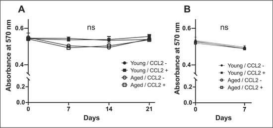
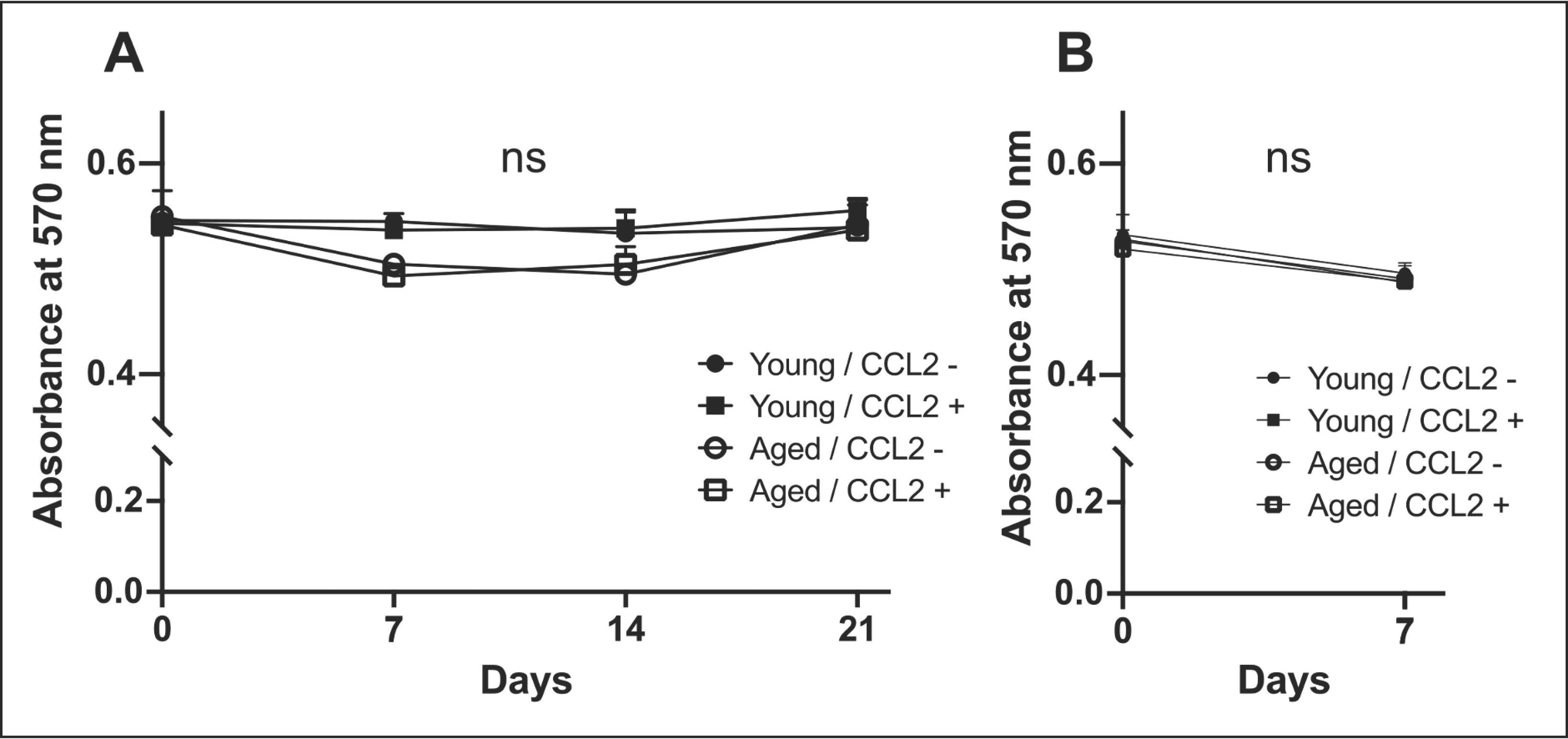
We performed the cell-viability assay using young and aged MSCs to examine whether CCL2 had any adverse effects. During the osteogenic (Figure 1A) and osteoclastogenic (Figure 1B) differentiation assays, there were no significant differences between CCL2-treated MSCs and untreated MSCs regarding cell viability.
Figure 1.
Cell-viability assay. Cell viabilities were assessed during the osteogenic (A) and osteoclastogenic (B) differentiation assays using the alamarBlue Cell-Viability Reagent. Both young and aged cells were cultured with or without recombinant murine CCL2 (10 ng/mL). Absorbance at 570 nm was measured. N = 4 independent mice per group, with one independent cell isolation per mouse. Cells from each mouse were divided into CCL2-treated and untreated groups. Each condition was assessed using three technical replicate wells. Different wells on separate plates were used for each time point. ns, not statistically significant.
3.2. CCL2 Enhanced Mineralization in MSCs–Macrophages Co-Culture in Both Young and Aged Cells
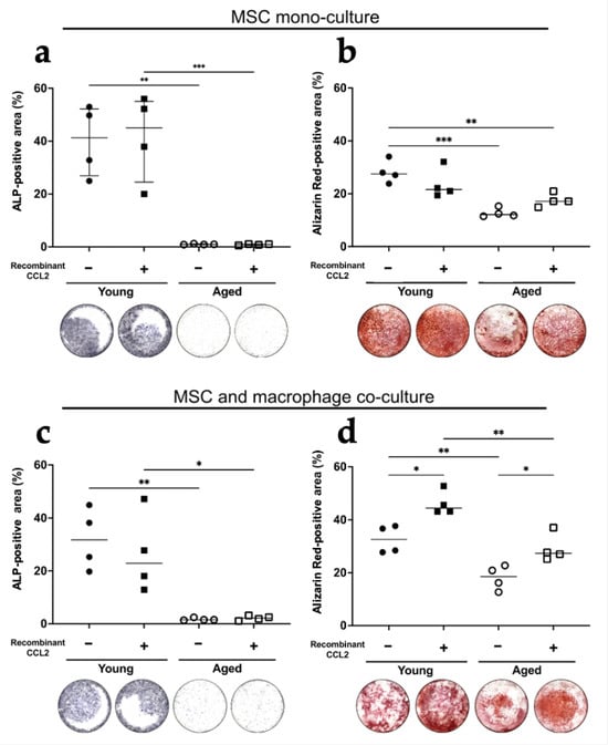
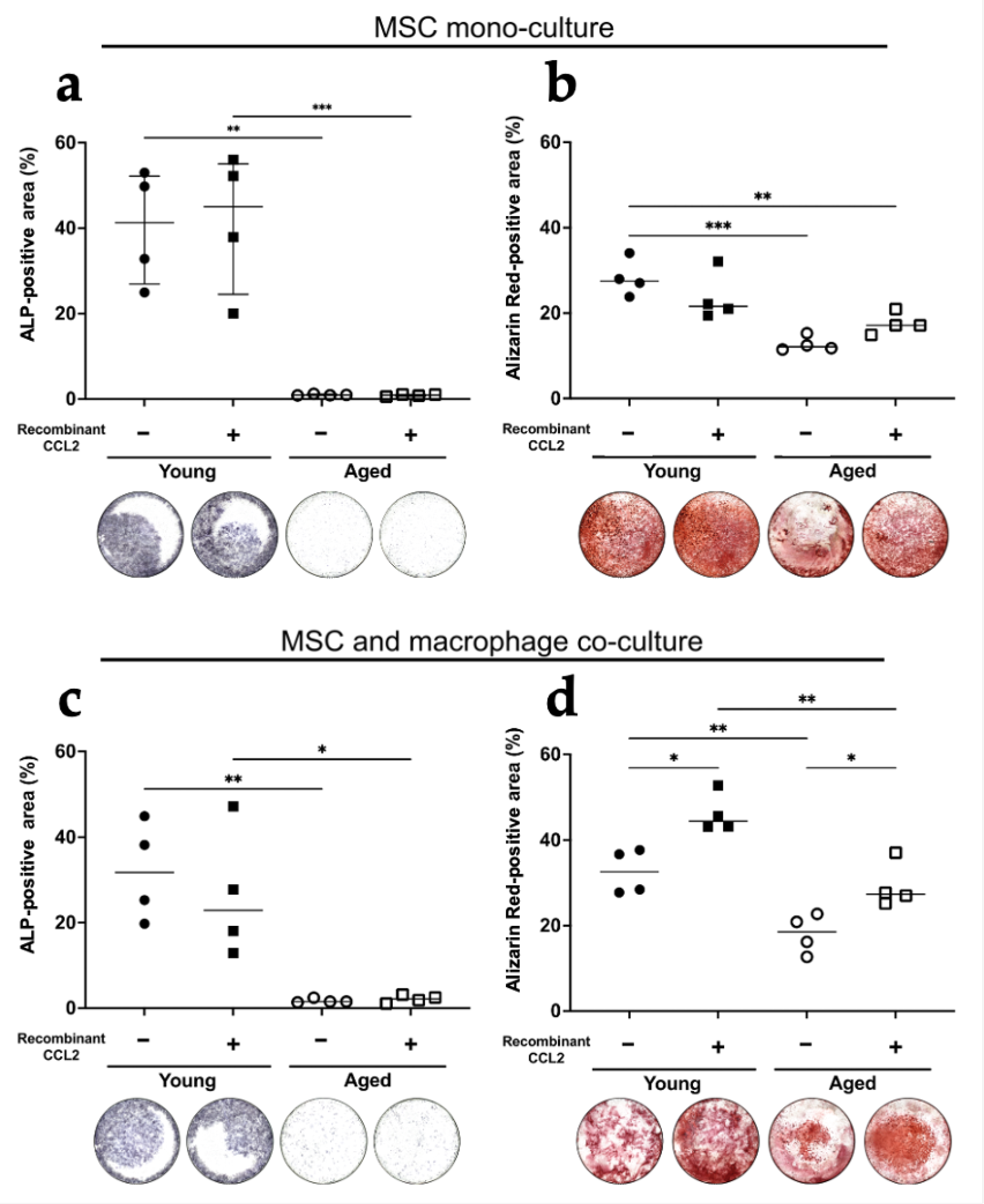
In the mono-cultures of MSCs, young murine cells showed a greater ALP-positive area than aged cells (p < 0.01). There was no significant difference between the recCCL2-treated group and the non-treated group (Figure 2a). These trends were similar for Alizarin-Red staining (Figure 2b). CCL2 did not exhibit a direct effect on osteogenesis in the mono-culture of MSCs. In the co-cultures of MSCs with macrophages, MSCs from young mice showed higher osteogenic ability than aged mice (p < 0.01). The group treated with recCCL2 showed a greater Alizarin-Red-positive area than the non-treated group both in young (p < 0.05) and aged (p < 0.05) cells, but there were no significant differences found in ALP staining (Figure 2c,d).
Figure 2.
Osteogenic differentiation assay. Quantitative analysis of (a) ALP and (b) Alizarin-Red-positive area proportion (%/well) and representative whole-well images of each group in MSCs mono-culture. Quantitative analysis of (c) ALP and (d) Alizarin-Red-positive area proportion (%/well) and representative whole-well images of each group in MSCs and macrophage co-culture. Purple staining colors indicates ALP-positive areas, while red staining indicates Alizarin Red-positive areas. N = 4 independent mice per group, with one independent cell isolation per mouse. Each condition was assessed using three technical replicate wells per mouse for each assay. * = p < 0.05, ** = p < 0.01, *** = p < 0.001.
3.3. CCL2 Increased M1 and Decreased M2 Proportion on Day 7 During the Osteogenic Differentiation Assay
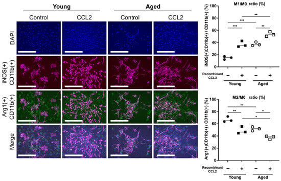
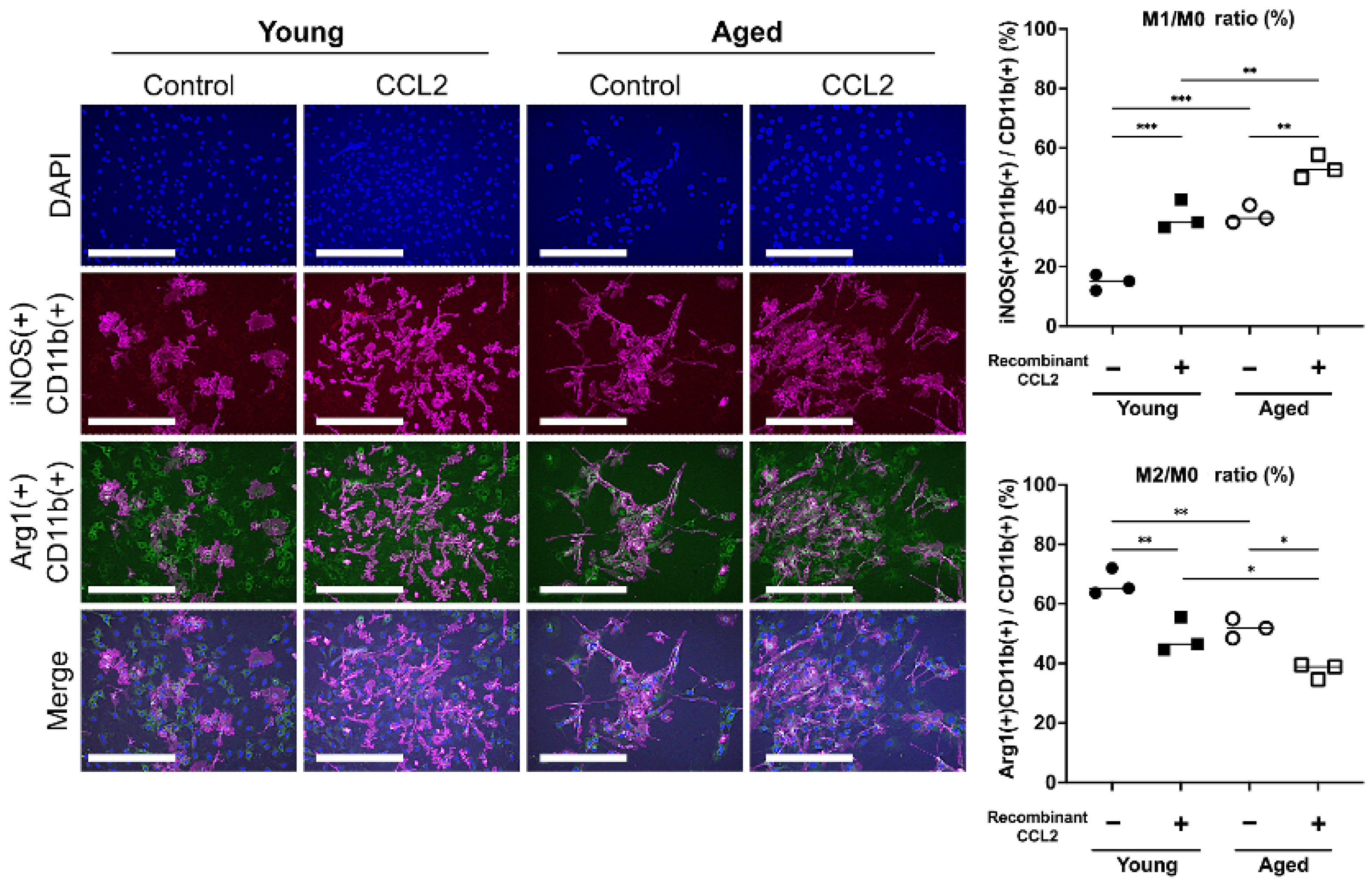
We performed the immunofluorescent staining to investigate the effect of CCL2 on macrophage phenotype during the osteogenic differentiation on day 7 (Figure 3). In the CCL2 non-treated group, aged cells showed a higher proportion of M1 (p < 0.001) and a lower proportion of M2 (p < 0.01) than young cells. These trends were the same in the CCL2-treated group (M1; p < 0.01, M2; p < 0.05). CCL2 increased the M1/M0 ratio (young; p < 0.001, aged; p < 0.01) and decreased the M2/M0 ratio (young; p < 0.01, aged; p < 0.05) both in young and aged cells.
Figure 3.
Immunofluorescence for macrophage phenotype on day 7 during the co-culture osteogenic differentiation assay. Representative images of immunofluorescence for macrophages are shown (scale bar = 100 μm). Quantitative assessments of the M1 positive (iNOS/CD11b double-positive) cells and the M2 positive (Arg1/CD11b double-positive) cells were performed. Blue indicates DAPI-stained nuclei, magenta indicates iNOS/CD11b staining, and green indivates Arg1/CD11b staining. N = 3 independent mice per group, with one independent cell isolation per mouse. Each condition was assessed using three technical replicate wells per mouse for this assay. * = p < 0.05, ** = p < 0.01, *** = p < 0.001.
3.4. CCL2 Increased the TRAP-Positive Cells and the Expression Level of C-fos and Dc-Stamp Both in Young and Aged Cells
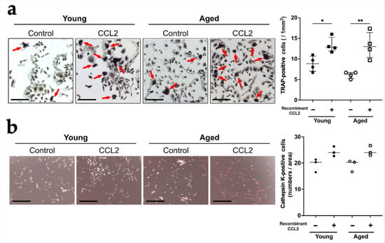
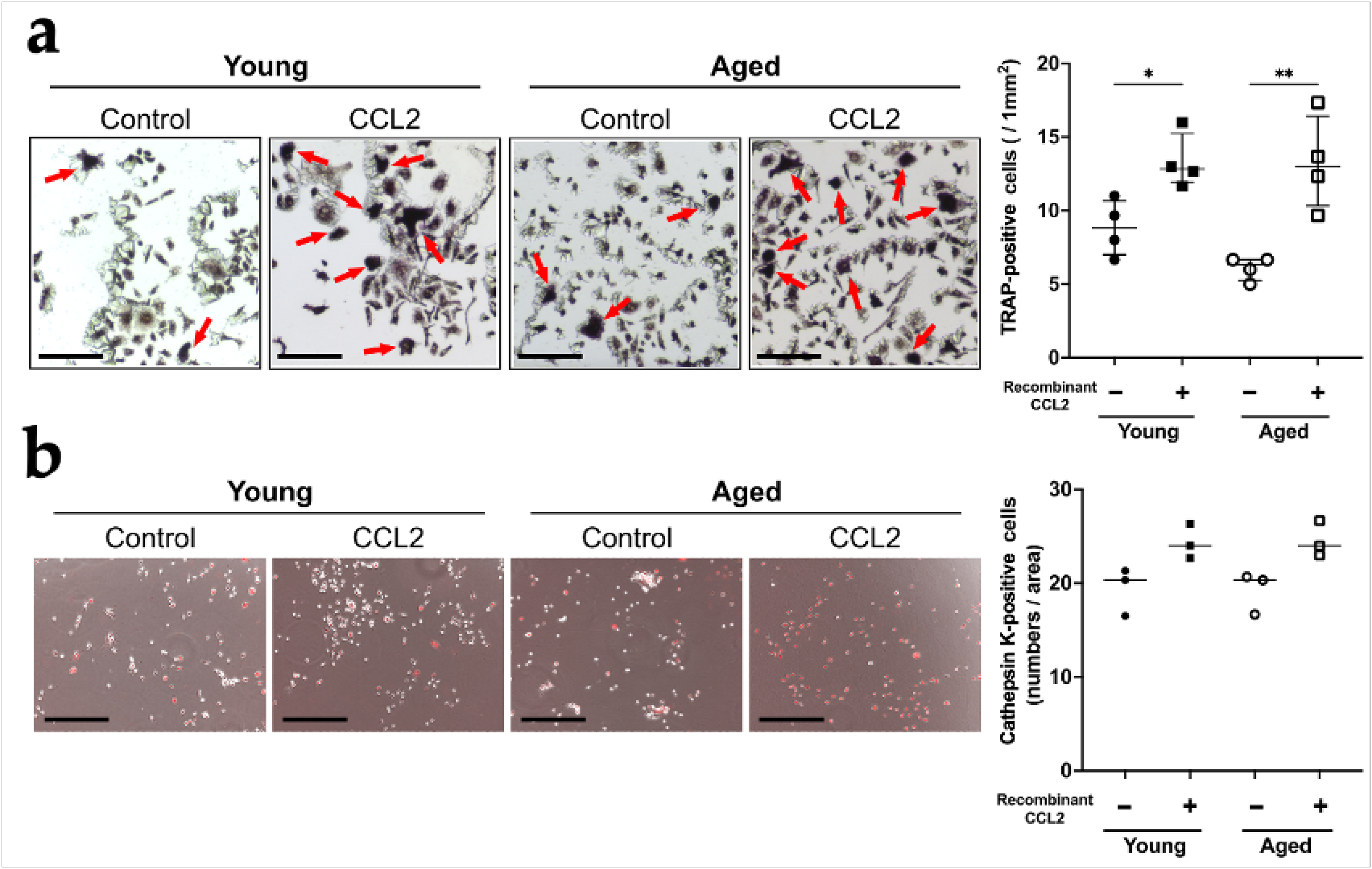
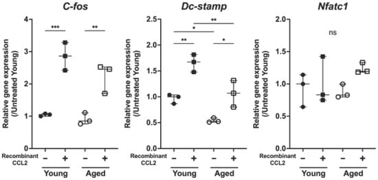
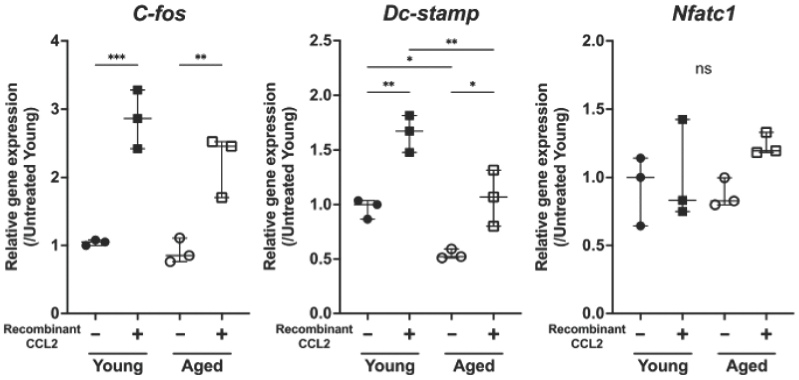
In the osteoclastogenic differentiation assay, the number of TRAP-positive cells was increased in the group treated with recCCL2 both in young (p < 0.05) and aged (p < 0.01) cells (Figure 4a). There was no significant difference in the number of Cathepsin K-positive cells (Figure 4b). To explore the mechanisms of CCL2 and osteoclastogenesis, we conducted RT-PCR using cells collected during the osteoclastogenic differentiation assay. We checked osteoclastogenesis-related gene expression of C-fos, Dc-stamp, and Nfatc1 (Figure 5). CCL2 treatment increased the expression levels of C-fos and Dc-stamp both in young (p < 0.001, p < 0.01) and aged (p < 0.01, p < 0.05) cells. Young cells showed a higher expression level of Dc-stamp than aged cells both under CCL2 non-treated (p < 0.05) and treated (p < 0.01) conditions.
Figure 4.
Osteoclastogenic differentiation assay. Quantitative analysis of (a) TRAP-positive cells (/1 mm2) (N = 4 independent mice per group) and (b) Cathepsin K-positive cells (/area) (N = 3 independent mice per group) and representative images of each group are shown (scale bar = 200 μm). In (a), red arrows indicate TRAP-positive multinucleated cells. In (b), red staining indicates Cathepsin K-positive cells. One independent cell isolation was performed per mouse. Each condition was assessed using three technical replicate wells per mouse. Three randomly selected microscopic fields were evaluated per well. * = p < 0.05, ** = p < 0.01.
Figure 5.
Gene expressions on day 7 during the osteoclastogenic differentiation assay. The results of RT-PCR on day 7 during the osteoclastogenic differentiation assay are shown. Values are shown as relative gene expression levels to the control group (untreated young mice group). N = 3 independent mice per group, with one independent cell isolation per mouse. Each sample was analyzed using three technical replicate RT-PCR reactions. * = p < 0.05, ** = p < 0.01, *** = p < 0.001. ns, not statistically significant.
4. Discussion
To maintain bone homeostasis, bone resorption and the breakdown of bone tissue must be balanced with bone formation, which would result in the development of new bone tissue [16]. With aging, the balance between removing old bone and forming new bone is interrupted, resulting in osteoporosis. Bone regeneration in aging is a complex process affected by lots of factors, including systemic and local cell signaling, osteogenic and osteoclastogenic cells, immune cells, and blood microenvironment [17]. Factors such as hormonal changes, oxidative-stress-induced DNA damage, and poor diet also lead to tissue abnormality and an imbalance in senescent bone homeostasis [18]. For example, the loss of estrogen after menopause causes bone loss mainly in women, which is further aggravated with aging [19,20]. Cells grown in high oxygen concentrations were induced a greater senescence, which means oxidative stress is an inducer of senescence [21]. At the cellular level, aging is related to an associated reduction in bone formation and an increase in marrow adipogenesis. Age-related functional decline in osteoblasts is a result of increased apoptosis [22], decreased proliferation, impaired osteoblast differentiation [23], increased osteoblast senescence [24], and dysfunctional osteoprogenitors [25]. Osteoprotegerin (OPG) is secreted by a variety of cells including osteoblasts, and OPG reduces bone loss by blocking the interaction between RANKL and RANK [26]. The secretion level of OPG is reduced due to the aging-related decrease of osteoblasts, thereby activating osteoclast resorption and promoting osteoporosis [18]. Therefore, with aging, bone mass significantly declines, and the components of bone become more fragile.
Chronic, low-grade inflammation occurs during the aging process, also referred to as “inflammaging”, and has been more widely implicated in the development of chronic diseases and aging [2]. Aging is associated with the accumulation of senescent cells in tissues and subsequent secretion of pro-inflammatory mediators via a senescence-associated secretory phenotype (SASP). C-C motif chemokine ligand 2 (CCL2), a member of the C-C chemokine superfamily, is a commonly secreted pro-inflammatory mediator in senescent cells, and CCL2 is highly involved in SASP [27]. CCL2 is one of the inflammatory chemokines, which crucially regulate leukocyte recruitment during inflammatory responses. CCL2 binds to the C-C motif chemokine receptor 2 (CCR2) to induce chemotactic activity and increase calcium influx [28,29,30]. Monocytes recruited by CCL2/CCR2 in a mouse osteoarthritis model led to increased inflammation and cartilage damage, relative to wild-type mice [31]. A community study of the relating serum CCL2 and knee radiographic osteoarthritis reported that higher CCL2 levels were associated with increased presence and progression of radiographic knee osteoarthritis [32]. In a rat model, increased serum levels of CCL2 were associated with lower bone-mineral density, and then validated using clinical patient samples, suggesting that CCL2 has therapeutic potential for predicting osteoporosis risk [33]. The association between CCL2/CCR2 and aging has been reported in a range of human disease fields. Aged vascular smooth-muscle cells showed increased production of CCL2 without any external stimulation [34]. Overexpression of CCL2 in mice led to accelerated aging compared to wild-type mice, dysfunction of metabolic pathways, and premature death [35]. The relationship between CCL2 and osteogenesis in terms of inflammation has been also reported. CCL2 expression in osteoblasts and mononuclear cells might be induced by lipopolysaccharide (LPS) under inflammatory conditions [36]. The expression of CCL2 in osteoblasts appears to mediate the paracrine recruitment of monocytes during bone remodeling, and CCL2 expression by osteoblasts is upregulated in response to inflammatory stimuli [37,38]. Based on this evidence, we hypothesized that CCL2 could influence bone remodeling-related cellular processes not only in young cells but also in aged cells.
Accumulating evidence has indicated that CCL2 is also an important regulator of osteoclastogenesis. Osteoclasts play principal roles in the absorption of old impaired bone tissues for subsequent osteogenesis, contributing to the initiation of bone regeneration [39,40]. CCL2 regulates the migration of osteoclast precursor cells and osteoclast formation and activity [41]. At the cellular level, CCL2 plays a pivotal role in the activation of bone resorption and the promotion of human osteoclast fusion [42,43]. CCL2 knockout led to reduced quantity and activity of osteoclasts, as well as reduced expression of Colony-stimulating factor-1 receptor (C-fms) and receptor activator of NFκB (RANKL) receptor. This was accompanied by the reduced expression of osteoclast-specific genes but no changes in bone formation genes, leading to increased bone mass [44,45]. On the other hand, in a hyper-occlusion model with CCL2−/− and CCR2−/− mice, enhanced osteoclastogenesis was observed, which indicated that CCL3 (MIP1-α) expression might compensate for deficient CCL2/CCR2 signaling during occlusal traumatism [46]. In the bone microenvironment, CCL2 expression is regulated by a variety of factors, such as RANKL [43], parathyroid hormone (PTH)/parathyroid hormone-related peptide (PTHrP) [47]. RANKL was shown to induce the expression of CCL2, leading to the stimulation of osteoclastogenesis [43]. PTH-induced osteoblastic expression of CCL2 mediated the recruitment of monocyte osteoclast precursors and NFκB ligand-stimulated osteoclastogenesis, leading to subsequent bone remodeling [48]. In CCL2 knockout mice, PTH was unable to increase trabecular bone mineral density and bone volume with a lack of increase in macrophage numbers, osteoclast surface, and osteoclast number compared with wild-type mice [49]. CCL2 alone cannot differentiate murine bone marrow macrophages into mature osteoclasts. Rather, CCL2 accompanied with RANKL/PTH highly increased the effect of RANKL/PTH on osteoclastogenesis [48].
In the present study, we demonstrated that CCL2 facilitated osteogenesis, particularly mineralization, in direct co-culture of MSCs with macrophages using young and aged female murine cells; CCL2 did not have this effect in the MSC mono-culture. This trend is compatible with previous studies using murine and rabbit male cells [50,51]. The strength of this study is that it used both young and aged murine cells and investigated the aging effect. The research about aging has been highly demanded, and its importance has been emphasized [52,53]. When performing the animal experiments, not only young but also aged subjects or aged models should be included. To reveal the aging difference would lead to solving the unmet problem of age-related human diseases. In this study, CCL2 increased the M1 and decreased M2 macrophage ratio in young and aged cells, which also enhances osteogenesis, particularly mineralization, in MSC–macrophage co-culture. Previous studies have shown that CCL2 regulates macrophage polarization through multiple signaling pathways. In particular, CCL2 has been reported to promote M1 macrophage polarization through the p38 MAPK and NF-κB signaling pathways [54,55]. These mechanisms may partly explain the CCL2-induced changes in macrophage phenotype observed in the present study. CCL2 also enhanced selected osteoclastogenic features in young and aged female cells. Osteoclasts play predominant roles in the resorption of old bone prior to subsequent osteogenesis, contributing to the initiation of bone regeneration [39]. CCL2 treatment increased the expression level of C-fos and Dc-stamp both in young and aged cells. Previous studies have shown that CCL2 promotes recruitment and fusion of osteoclast precursors and cooperates with RANKL-dependent osteoclast differentiation [41,43,56]. One possible explanation is that CCL2 may enhance recruitment and activation of monocyte-lineage osteoclast precursors and promote cell–cell fusion, consistent with the observed increases in C-fos, Dc-stamp, and TRAP-positive cells. The absence of significant increases in Nfatc1 expression and Cathepsin K-positive cells suggests that this effect may be more prominent during precursor activation and fusion than during terminal osteoclast maturation and functional activation. Furthermore, we found age-dependent differences in the expression of Dc-stamp, the master regulator of cell–cell fusion and osteoclastogenesis; young cells showed a higher expression level of Dc-stamp than aged cells regardless of CCL2 treatment. These results indicate that Dc-stamp may be a potential target for further investigation of age-related changes in bone regeneration.
The current study has limitations. First, this study describes only in vitro experiments; further, in vivo studies are desirable to translate the possibility of CCL2 into the clinical situation. Second, the causal relationship between CCL2-induced changes in macrophage phenotype and enhanced osteogenesis and the involvement of CCL2/CCR2 signaling were not directly examined. We did not perform experiments involving CCR2 inhibition or neutralization, or macrophage manipulation. Therefore, the investigation of the functional mechanisms of CCL2 in bone homeostasis was insufficient to reveal specific potential targets for bone regeneration. Further studies using macrophage manipulation and inhibition of CCL2/CCR2 signaling will be necessary to clarify the underlying mechanisms. Third, only age-matched MSC–macrophage co-culture were used, and cross-age co-cultures were not performed. We used only three representative genes for real-time RT-PCR experiments and did not evaluate the expression of osteogenic markers, such as RUNX2, SP7/Osterix, ALPL, COL1A1, and BGLAP/osteocalcin. In addition, functional bone resorption was not evaluated using assays such as the resorption-pit assay. Future studies using RNA sequencing and other novel methods will also clarify the functional signaling pathway and specific target genes related to CCL2 on bone regeneration in the context of aging.
5. Conclusions
In conclusion, we elucidated that CCL2 promotes osteogenesis, particularly mineralization, and enhances selected osteoclastogenic features in both young and aged cells. These findings suggest that CCL2 may have a potential role in bone remodeling in the context of aging.
Author Contributions
Conceptualization, Masakazu.Toya.; methodology, Masakazu.Toya.; software, Masakazu.Toya.; validation, R.H., T.I., I.S., M.M., M.L.L., and Y.S.E.; formal analysis, Masakazu.Toya. and R.H.; investigation, Masakazu.Toya., R.H., Q.G., and S.K.-H.C.; resources, S.B.G.; data curation, Masakazu.Toya. and R.H.; writing—original draft preparation, Masakazu.Toya.; writing—review and editing, R.H., M.L.L., Y.S.E., T.I., I.S., M.M., J.K., Masanori.Tsubosaka., Q.G., S.K.-H.C., and S.B.G.; visualization, Masakazu.Toya. and R.H.; supervision, S.K.-H.C.; project administration, S.B.G.; funding acquisition, S.B.G. All authors have read and agreed to the published version of the manuscript.
Funding
This research was partially funded by the National Institute of Arthritis and Musculoskeletal and Skin Diseases, Grant/Award Numbers: R01AR073145, R01AR063717.
Institutional Review Board Statement
The animal study protocol was approved by the Stanford University Laboratory Animal Care and Control Committee (APLAC reference number 9964, 24 October 2023).
Informed Consent Statement
Not applicable.
Data Availability Statement
The data presented in this study are available on request from the corresponding author. The data are not publicly available due to confidentiality concerns.
Acknowledgments
This research was supported by the Robert L. and Mary Ellenburg Chair in Surgery, Stanford University.
Conflicts of Interest
The authors declare no conflicts of interest.
References
- Williamson, S.; Landeiro, F.; McConnell, T.; Fulford-Smith, L.; Javaid, M.K.; Judge, A.; Leal, J. Costs of fragility hip fractures globally: A systematic review and meta-regression analysis. Osteoporos. Int. 2017, 28, 2791–2800. [Google Scholar] [CrossRef] [Scilit] [PubMed]
- Franceschi, C.; Garagnani, P.; Parini, P.; Giuliani, C.; Santoro, A. Inflammaging: A new immune-metabolic viewpoint for age-related diseases. Nat. Rev. Endocrinol. 2018, 14, 576–590. [Google Scholar] [CrossRef] [Scilit] [PubMed]
- Franceschi, C.; Campisi, J. Chronic inflammation (inflammaging) and its potential contribution to age-associated diseases. J. Gerontol. A Biol. Sci. Med. Sci. 2014, 69, S4–S9. [Google Scholar] [CrossRef] [Scilit] [PubMed]
- Maruyama, M.; Rhee, C.; Utsunomiya, T.; Zhang, N.; Ueno, M.; Yao, Z.; Goodman, S.B. Modulation of the Inflammatory Response and Bone Healing. Front. Endocrinol. 2020, 11, 386. [Google Scholar] [CrossRef] [Scilit] [PubMed]
- Pajarinen, J.; Lin, T.; Gibon, E.; Kohno, Y.; Maruyama, M.; Nathan, K.; Lu, L.; Yao, Z.; Goodman, S.B. Mesenchymal stem cell-macrophage crosstalk and bone healing. Biomaterials 2019, 196, 80–89. [Google Scholar] [CrossRef] [Scilit] [PubMed]
- Zhang, N.; Lo, C.-W.; Utsunomiya, T.; Maruyama, M.; Huang, E.; Rhee, C.; Gao, Q.; Yao, Z.; Goodman, S.B. PDGF-BB and IL-4 co-overexpression is a potential strategy to enhance mesenchymal stem cell-based bone regeneration. Stem Cell Res. Ther. 2021, 12, 40. [Google Scholar] [CrossRef] [Scilit] [PubMed]
- Nathan, K.; Lu, L.Y.; Lin, T.; Pajarinen, J.; Jämsen, E.; Huang, J.-F.; Romero-Lopez, M.; Maruyama, M.; Kohno, Y.; Yao, Z.; et al. Precise immunomodulation of the M1 to M2 macrophage transition enhances mesenchymal stem cell osteogenesis and differs by sex. Bone Jt. Res. 2019, 8, 481–488. [Google Scholar] [CrossRef] [Scilit] [PubMed]
- Lin, T.; Pajarinen, J.; Kohno, Y.; Huang, J.-F.; Maruyama, M.; Romero-Lopez, M.; Nathan, K.; Yao, Z.; Goodman, S.B. Trained murine mesenchymal stem cells have anti-inflammatory effect on macrophages, but defective regulation on T-cell proliferation. FASEB J. 2019, 33, 4203–4211. [Google Scholar] [CrossRef] [Scilit] [PubMed]
- Lin, T.-H.; Sato, T.; Barcay, K.R.; Waters, H.; Loi, F.; Zhang, R.; Pajarinen, J.; Egashira, K.; Yao, Z.; Goodman, S.B. NF-κB decoy oligodeoxynucleotide enhanced osteogenesis in mesenchymal stem cells exposed to polyethylene particle. Tissue Eng. Part A 2015, 21, 875–883. [Google Scholar] [CrossRef] [Scilit] [PubMed]
- Peister, A.; Mellad, J.A.; Larson, B.L.; Hall, B.M.; Gibson, L.F.; Prockop, D.J. Adult stem cells from bone marrow (MSCs) isolated from different strains of inbred mice vary in surface epitopes, rates of proliferation, and differentiation potential. Blood 2004, 103, 1662–1668. [Google Scholar] [CrossRef] [Scilit] [PubMed]
- Rampersad, S.N. Multiple applications of Alamar Blue as an indicator of metabolic function and cellular health in cell viability bioassays. Sensors 2012, 12, 12347–12360. [Google Scholar] [CrossRef] [Scilit] [PubMed]
- Al-Nasiry, S.; Geusens, N.; Hanssens, M.; Luyten, C.; Pijnenborg, R. The use of Alamar Blue assay for quantitative analysis of viability, migration and invasion of choriocarcinoma cells. Hum. Reprod. 2007, 22, 1304–1309. [Google Scholar] [CrossRef] [Scilit] [PubMed]
- Bankhead, P.; Loughrey, M.B.; Fernández, J.A.; Dombrowski, Y.; McArt, D.G.; Dunne, P.D.; McQuaid, S.; Gray, R.T.; Murray, L.J.; Coleman, H.G.; et al. QuPath: Open source software for digital pathology image analysis. Sci. Rep. 2017, 7, 16878. [Google Scholar] [CrossRef] [Scilit] [PubMed]
- Kushioka, J.; Toya, M.; Shen, H.; Hirata, H.; Zhang, N.; Huang, E.; Tsubosaka, M.; Gao, Q.; Teissier, V.; Li, X.; et al. Therapeutic effects of MSCs, genetically modified MSCs, and NFĸB-inhibitor on chronic inflammatory osteolysis in aged mice. J. Orthop. Res. 2022, 41, 1004–1013. [Google Scholar] [CrossRef] [Scilit] [PubMed]
- Lin, T.-H.; Pajarinen, J.; Sato, T.; Loi, F.; Fan, C.; Córdova, L.A.; Nabeshima, A.; Gibon, E.; Zhang, R.; Yao, Z.; et al. NF-κB decoy oligodeoxynucleotide mitigates wear particle-associated bone loss in the murine continuous infusion model. Acta Biomater. 2016, 41, 273–281. [Google Scholar] [CrossRef] [Scilit] [PubMed]
- Salhotra, A.; Shah, H.N.; Levi, B.; Longaker, M.T. Mechanisms of bone development and repair. Nat. Rev. Mol. Cell Biol. 2020, 21, 696–711. [Google Scholar] [CrossRef] [Scilit] [PubMed]
- Gruber, R.; Koch, H.; Doll, B.A.; Tegtmeier, F.; Einhorn, T.A.; Hollinger, J.O. Fracture healing in the elderly patient. Exp. Gerontol. 2006, 41, 1080–1093. [Google Scholar] [CrossRef] [Scilit] [PubMed]
- Chandra, A.; Rajawat, J. Skeletal Aging and Osteoporosis: Mechanisms and Therapeutics. Int. J. Mol. Sci. 2021, 22, 3553. [Google Scholar] [CrossRef] [Scilit] [PubMed]
- Manolagas, S.C. From estrogen-centric to aging and oxidative stress: A revised perspective of the pathogenesis of osteoporosis. Endocr. Rev. 2010, 31, 266–300. [Google Scholar] [CrossRef] [Scilit] [PubMed]
- Riggs, B.L.; Khosla, S.; Melton, L.J., 3rd. A unitary model for involutional osteoporosis: Estrogen deficiency causes both type I and type II osteoporosis in postmenopausal women and contributes to bone loss in aging men. J. Bone Miner. Res. 1998, 13, 763–773. [Google Scholar] [CrossRef] [Scilit] [PubMed]
- von Zglinicki, T.; Saretzki, G.; Döcke, W.; Lotze, C. Mild hyperoxia shortens telomeres and inhibits proliferation of fibroblasts: A model for senescence? Exp. Cell Res. 1995, 220, 186–193. [Google Scholar] [CrossRef] [Scilit] [PubMed]
- Jilka, R.L.; Almeida, M.; Ambrogini, E.; Han, L.; Roberson, P.K.; Weinstein, R.S.; Manolagas, S.C. Decreased oxidative stress and greater bone anabolism in the aged, when compared to the young, murine skeleton with parathyroid hormone administration. Aging Cell 2010, 9, 851–867. [Google Scholar] [CrossRef] [Scilit] [PubMed]
- Abdallah, B.M.; Haack-Sørensen, M.; Fink, T.; Kassem, M. Inhibition of osteoblast differentiation but not adipocyte differentiation of mesenchymal stem cells by sera obtained from aged females. Bone 2006, 39, 181–188. [Google Scholar] [CrossRef] [Scilit] [PubMed]
- Farr, J.N.; Fraser, D.G.; Wang, H.; Jaehn, K.; Ogrodnik, M.B.; Weivoda, M.M.; Drake, M.T.; Tchkonia, T.; LeBrasseur, N.K.; Kirkland, J.L.; et al. Identification of Senescent Cells in the Bone Microenvironment. J. Bone Miner. Res. 2016, 31, 1920–1929. [Google Scholar] [CrossRef] [Scilit] [PubMed]
- Kassem, M.; Marie, P.J. Senescence-associated intrinsic mechanisms of osteoblast dysfunctions. Aging Cell 2011, 10, 191–197. [Google Scholar] [CrossRef] [Scilit] [PubMed]
- Dutka, M.; Bobiński, R.; Wojakowski, W.; Francuz, T.; Pająk, C.; Zimmer, K. Osteoprotegerin and RANKL-RANK-OPG-TRAIL signalling axis in heart failure and other cardiovascular diseases. Heart Fail. Rev. 2022, 27, 1395–1411. [Google Scholar] [CrossRef] [Scilit] [PubMed]
- Pereira, B.I.; Devine, O.P.; Vukmanovic-Stejic, M.; Chambers, E.S.; Subramanian, P.; Patel, N.; Virasami, A.; Sebire, N.J.; Kinsler, V.; Valdovinos, A.; et al. Senescent cells evade immune clearance via HLA-E-mediated NK and CD8(+) T cell inhibition. Nat. Commun. 2019, 10, 2387. [Google Scholar] [CrossRef] [Scilit] [PubMed]
- Jarnagin, K.; Grunberger, D.; Mulkins, M.; Wong, B.; Hemmerich, S.; Paavola, C.; Bloom, A.; Bhakta, S.; Diehl, F.; Freedman, R.; et al. Identification of surface residues of the monocyte chemotactic protein 1 that affect signaling through the receptor CCR2. Biochemistry 1999, 38, 16167–16177. [Google Scholar] [CrossRef] [Scilit] [PubMed]
- Hemmerich, S.; Paavola, C.; Bloom, A.; Bhakta, S.; Freedman, R.; Grunberger, D.; Krstenansky, J.; Lee, S.; McCarley, D.; Mulkins, M.; et al. Identification of residues in the monocyte chemotactic protein-1 that contact the MCP-1 receptor, CCR2. Biochemistry 1999, 38, 13013–13025. [Google Scholar] [CrossRef] [Scilit] [PubMed]
- Paavola, C.D.; Hemmerich, S.; Grunberger, D.; Polsky, I.; Bloom, A.; Freedman, R.; Mulkins, M.; Bhakta, S.; McCarley, D.; Wiesent, L.; et al. Monomeric monocyte chemoattractant protein-1 (MCP-1) binds and activates the MCP-1 receptor CCR2B. J. Biol. Chem. 1998, 273, 33157–33165. [Google Scholar] [CrossRef] [Scilit] [PubMed]
- Raghu, H.; Lepus, C.M.; Wang, Q.; Wong, H.H.; Lingampalli, N.; Oliviero, F.; Punzi, L.; Giori, N.J.; Goodman, S.B.; Chu, C.R.; et al. CCL2/CCR2, but not CCL5/CCR5, mediates monocyte recruitment, inflammation and cartilage destruction in osteoarthritis. Ann. Rheum. Dis. 2017, 76, 914–922. [Google Scholar] [CrossRef] [Scilit] [PubMed]
- Longobardi, L.; Jordan, J.; Shi, X.; Renner, J.; Schwartz, T.; Nelson, A.; Barrow, D.; Kraus, V.; Spagnoli, A. Associations between the chemokine biomarker CCL2 and knee osteoarthritis outcomes: The Johnston County Osteoarthritis Project. Osteoarthr. Cartil. 2018, 26, 1257–1261. [Google Scholar] [CrossRef] [Scilit] [PubMed]
- Hu, Y.; Wang, L.; Zhao, Z.; Lu, W.; Fan, J.; Gao, B.; Luo, Z.; Jie, Q.; Shi, X.; Yang, L. Cytokines CCL2 and CXCL1 may be potential novel predictors of early bone loss. Mol. Med. Rep. 2020, 22, 4716–4724. [Google Scholar] [CrossRef] [Scilit] [PubMed]
- Song, Y.; Shen, H.; Schenten, D.; Shan, P.; Lee, P.J.; Goldstein, D.R. Aging enhances the basal production of IL-6 and CCL2 in vascular smooth muscle cells. Arterioscler. Thromb. Vasc. Biol. 2012, 32, 103–109. [Google Scholar] [CrossRef] [Scilit] [PubMed]
- Luciano-Mateo, F.; Cabré, N.; Baiges-Gaya, G.; Fernández-Arroyo, S.; Hernández-Aguilera, A.; Rodríguez-Tomàs, E.E.; Arenas, M.; Camps, J.; Menéndez, J.A.; Joven, J. Systemic overexpression of C-C motif chemokine ligand 2 promotes metabolic dysregulation and premature death in mice with accelerated aging. Aging 2020, 12, 20001–20023. [Google Scholar] [CrossRef] [Scilit] [PubMed]
- Jiang, Y.; Graves, D.T. Periodontal pathogens stimulate CC-chemokine production by mononuclear and bone-derived cells. J. Periodontol. 1999, 70, 1472–1478. [Google Scholar] [CrossRef] [Scilit] [PubMed]
- Graves, D.T.; Jiang, Y.; Valente, A.J. Regulated expression of MCP-1 by osteoblastic cells in vitro and in vivo. Histol. Histopathol. 1999, 14, 1347–1354. [Google Scholar] [CrossRef] [Scilit] [PubMed]
- Graves, D.T.; Jiang, Y.; Valente, A.J. The expression of monocyte chemoattractant protein-1 and other chemokines by osteoblasts. Front. Biosci. 1999, 4, D571–D580. [Google Scholar] [CrossRef] [Scilit] [PubMed]
- Charles, J.F.; Aliprantis, A.O. Osteoclasts: More than ’bone eaters. Trends Mol. Med. 2014, 20, 449–459. [Google Scholar] [CrossRef] [Scilit] [PubMed]
- Teitelbaum, S.L. Bone resorption by osteoclasts. Science 2000, 289, 1504–1508. [Google Scholar] [CrossRef] [Scilit] [PubMed]
- Siddiqui, J.A.; Partridge, N.C. CCL2/Monocyte Chemoattractant Protein 1 and Parathyroid Hormone Action on Bone. Front. Endocrinol. 2017, 8, 49. [Google Scholar] [CrossRef] [Scilit] [PubMed]
- Kim, M.S.; Day, C.J.; Selinger, C.I.; Magno, C.L.; Stephens, S.R.; Morrison, N.A. MCP-1-induced human osteoclast-like cells are tartrate-resistant acid phosphatase, NFATc1, and calcitonin receptor-positive but require receptor activator of NFkappaB ligand for bone resorption. J. Biol. Chem. 2006, 281, 1274–1285. [Google Scholar] [CrossRef] [Scilit] [PubMed]
- Kim, M.S.; Day, C.J.; Morrison, N.A. MCP-1 is induced by receptor activator of nuclear factor-{kappa}B ligand, promotes human osteoclast fusion, and rescues granulocyte macrophage colony-stimulating factor suppression of osteoclast formation. J. Biol. Chem. 2005, 280, 16163–16169. [Google Scholar] [CrossRef] [Scilit] [PubMed]
- Sul, O.; Ke, K.; Kim, W.; Kim, S.; Lee, S.; Kim, H.; Kim, S.; Suh, J.; Choi, H. Absence of MCP-1 leads to elevated bone mass via impaired actin ring formation. J. Cell. Physiol. 2012, 227, 1619–1627. [Google Scholar] [CrossRef] [Scilit] [PubMed]
- Miyamoto, K.; Ninomiya, K.; Sonoda, K.-H.; Miyauchi, Y.; Hoshi, H.; Iwasaki, R.; Miyamoto, H.; Yoshida, S.; Sato, Y.; Morioka, H.; et al. MCP-1 expressed by osteoclasts stimulates osteoclastogenesis in an autocrine/paracrine manner. Biochem. Biophys. Res. Commun. 2009, 383, 373–377. [Google Scholar] [CrossRef] [Scilit] [PubMed]
- Tsutsumi, T.; Kajiya, H.; Goto, K.T.; Takahashi, Y.; Okabe, K. Hyperocclusion up-regulates CCL3 expression in CCL2- and CCR2-deficient mice. J. Dent. Res. 2013, 92, 65–70. [Google Scholar] [CrossRef] [Scilit] [PubMed]
- Mulholland, B.S.; Forwood, M.R.; Morrison, N.A. Monocyte Chemoattractant Protein-1 (MCP-1/CCL2) Drives Activation of Bone Remodelling and Skeletal Metastasis. Curr. Osteoporos. Rep. 2019, 17, 538–547. [Google Scholar] [CrossRef] [Scilit] [PubMed]
- Li, X.; Qin, L.; Bergenstock, M.; Bevelock, L.M.; Novack, D.V.; Partridge, N.C. Parathyroid hormone stimulates osteoblastic expression of MCP-1 to recruit and increase the fusion of pre/osteoclasts. J. Biol. Chem. 2007, 282, 33098–33106. [Google Scholar] [CrossRef] [Scilit] [PubMed]
- Tamasi, J.A.; Vasilov, A.; Shimizu, E.; Benton, N.; Johnson, J.; Bitel, C.L.; Morrison, N.; Partridge, N.C. Monocyte chemoattractant protein-1 is a mediator of the anabolic action of parathyroid hormone on bone. J. Bone Miner. Res. 2013, 28, 1975–1986. [Google Scholar] [CrossRef] [Scilit] [PubMed]
- Toya, M.; Zhang, N.; Tsubosaka, M.; Kushioka, J.; Gao, Q.; Li, X.; Chow, S.K.-H.; Goodman, S.B. CCL2 promotes osteogenesis by facilitating macrophage migration during acute inflammation. Front. Cell Dev. Biol. 2023, 11, 1213641. [Google Scholar] [CrossRef] [Scilit] [PubMed]
- Shinohara, I.; Tsubosaka, M.; Toya, M.; Lee, M.L.; Kushioka, J.; Murayama, M.; Gao, Q.; Li, X.; Zhang, N.; Chow, S.K.-H.; et al. C-C Motif Chemokine Ligand 2 Enhances Macrophage Chemotaxis, Osteogenesis, and Angiogenesis during the Inflammatory Phase of Bone Regeneration. Biomolecules 2023, 13, 1665. [Google Scholar] [CrossRef] [Scilit] [PubMed]
- Mitchell, S.J.; Scheibye-Knudsen, M.; Longo, D.L.; de Cabo, R. Animal models of aging research: Implications for human aging and age-related diseases. Annu. Rev. Anim. Biosci. 2015, 3, 283–303. [Google Scholar] [CrossRef] [Scilit] [PubMed]
- Ackert-Bicknell, C.L.; Anderson, L.C.; Sheehan, S.; Hill, W.G.; Chang, B.; Churchill, G.A.; Chesler, E.J.; Korstanje, R.; Peters, L.L. Aging Research Using Mouse Models. Curr. Protoc. Mouse Biol. 2015, 5, 95–133. [Google Scholar] [CrossRef] [Scilit] [PubMed]
- Chen, L.; Pan, D.; Zhang, Y.; Zhang, E.; Ma, L. C-C Motif Chemokine 2 Regulates Macrophage Polarization and Contributes to Myocardial Infarction Healing. J. Interferon Cytokine Res. 2024, 44, 68–79. [Google Scholar] [CrossRef] [Scilit] [PubMed]
- Lu, L.Y.; Loi, F.; Nathan, K.; Lin, T.; Pajarinen, J.; Gibon, E.; Nabeshima, A.; Cordova, L.; Jämsen, E.; Yao, Z.; et al. Pro-Inflammatory M1 Macrophages Promote Osteogenesis by Mesenchymal Stem Cells via the COX-2-Prostaglandin E2 Pathway. J. Orthop. Res. 2017, 35, 2378–2385. [Google Scholar] [CrossRef] [Scilit] [PubMed]
- Khan, U.A.; Hashimi, S.M.; Bakr, M.M.; Forwood, M.R.; Morrison, N.A. CCL2 and CCR2 are essential for the formation of osteoclasts and foreign body giant cells. J. Cell. Biochem. 2016, 117, 382–389. [Google Scholar] [CrossRef] [Scilit] [PubMed]
Disclaimer/Publisher’s Note: The statements, opinions and data contained in all publications are solely those of the individual author(s) and contributor(s) and not of MDPI and/or the editor(s). MDPI and/or the editor(s) disclaim responsibility for any injury to people or property resulting from any ideas, methods, instructions or products referred to in the content. |
© 2026 by the authors. Licensee MDPI, Basel, Switzerland. This article is an open access article distributed under the terms and conditions of the Creative Commons Attribution (CC BY) license.