


Abstract
Background/Objectives: Glaucoma is a progressive optic neuropathy characterized by retinal ganglion cell loss and visual field deterioration despite adequate intraocular pressure control. Neuroprotective adjunct therapies have therefore been investigated, and citicoline has emerged as a potential candidate. We conducted a systematic review to summarize and critically appraise the evidence on the effects of citicoline, used as an adjunctive therapy, on visual function, functional vision, and quality of life in patients with glaucoma. Methods: A systematic literature search was performed in major electronic databases (PubMed, Scopus, and Web of Science) following PRISMA guidelines. Randomized and non-randomized clinical studies evaluating citicoline as adjunctive therapy in glaucoma were included. Outcomes of interest comprised visual function, electrophysiology, structural optical coherence tomography parameters, quality of life, and safety. Risk of bias was assessed using RoB 2 and ROBINS-I tools. Results: Fifteen studies were included in this review, ten randomized controlled trials and five non-randomized clinical trials. Included studies showed heterogeneous designs and follow-up durations. Several studies reported improvements in electrophysiological responses, including retinal and post-retinal functional markers, and relative stabilization of selected visual field indices, with some studies reporting preservation of retinal nerve fiber layer and ganglion cell complex thickness under stable intraocular pressure. However, these surrogate outcomes were not consistently accompanied by improvements in visual acuity or patient-reported quality-of-life measures. Methodological limitations and endpoint variability reduced comparability across studies. Across oral, topical, and intramuscular routes of administration, citicoline was generally reported to have a favorable safety and tolerability profile. Conclusions: Current evidence suggests that citicoline may exert neurobiological effects compatible with neuroprotection in glaucoma, but the clinical significance of these findings remains uncertain. Available data are insufficient to support routine clinical recommendation. Well-designed randomized trials with standardized, patient-relevant outcomes are required to determine whether these effects translate into meaningful visual benefit.
1. Introduction
Glaucomatous optic neuropathy (GON) comprises a heterogeneous group of chronic, progressive disorders characterized by structural and functional damage to the optic nerve. It remains one of the leading causes of irreversible blindness worldwide. Current projections estimate that more than 112 million individuals will be affected by glaucoma by 2040 [1,2]. The burden of glaucoma varies according to age and ethnicity, with a marked increase in prevalence after 60 years of age, higher prevalence and more aggressive disease progression among individuals of African descent, and increased risk of primary angle-closure glaucoma in Asian populations [1,3,4]. Although elevated intraocular pressure (IOP) remains the main modifiable risk factor, disease progression may occur despite apparently adequate IOP control, supporting the concept of glaucoma as a complex neurodegenerative disorder rather than a purely pressure-dependent disease [1,3,5].
At the cellular level, glaucoma is characterized by progressive loss of retinal ganglion cells (RGCs) and their axons, leading to thinning of the retinal nerve fiber layer (RNFL), optic nerve head damage, and visual field deterioration, with consequent limitations in visual performance and quality of life [6,7,8]. Diagnosis and longitudinal monitoring rely on integrating structural and functional assessments (Figure 1), including optical coherence tomography (OCT), standard automated perimetry, tonometry, and gonioscopy, which together support disease classification and the detection of progression [9,10,11]. Current treatment strategies primarily aim to reduce IOP through pharmacological, laser, or surgical approaches [4,12]. However, long-term topical therapy may be limited by ocular surface toxicity, periorbital adverse effects, treatment burden, and incomplete prevention of progression in some patients [3,7,12,13,14].
Figure 1.
Multimodal structural and functional assessment in a 70-year-old woman with open-angle glaucoma (OAG) under topical intraocular pressure (IOP)-lowering therapy. (Top left): Macular thickness map derived from the ILM–IPL/INL segmentation boundaries, corresponding functionally to the ganglion cell complex (GCC), obtained by SD-OCT Nidek RS-330®. (Top right): Standard Automated Perimetry (SAP) Octopus 301® perimeter (G1 program, dynamic strategy), demonstrating glaucomatous visual field defects. Bottom left: anterior chamber angle imaging by SD-OCT, including angle measurement parameters. (Image courtesy of Marta Araújo and Nádia Fernandes.).
Growing evidence that glaucoma involves the entire visual pathway, including the lateral geniculate nucleus and visual cortex, has provided the rationale for exploring neuroprotective strategies aimed at preserving neuronal integrity beyond IOP reduction [1,2,6,14]. Neuroprotective approaches may target mechanisms implicated in glaucomatous neurodegeneration, including glutamate-mediated excitotoxicity, mitochondrial dysfunction, oxidative stress, impaired axoplasmic transport, glial activation, and apoptotic pathways [2,8,15,16].
Citicoline (cytidine-5′-diphosphocholine, CDP-choline) has emerged as a promising neuroprotective adjunct in glaucoma (Figure 2). It is a naturally occurring endogenous compound and a precursor in the biosynthesis of acetylcholine and membrane phospholipids, including phosphatidylcholine and sphingomyelin, which are essential for neuronal membrane integrity and RGC survival [6,14]. Experimental and clinical evidence supports its neuroprotective effects in several neurological and ophthalmic conditions, including glaucoma, and clinical studies have increasingly investigated citicoline as an adjunct to conventional IOP-lowering therapy, reporting potential effects on structural and functional visual outcomes [5,12,15,16].
Figure 2.
Schematic representation of citicoline mechanisms in neuroprotection, neurorestoration, and neuroregeneration. Created by the authors with AI assistance and reviewed by the authors.
Despite this growing interest, the clinical evidence on citicoline in GON remains heterogeneous, particularly with respect to study design, route of administration, treatment duration, outcome measures, and methodological quality. These limitations restrict the ability to draw robust conclusions on its efficacy, effectiveness, and safety. Previous reviews have often focused on narrower efficacy endpoints, such as IOP, visual field indices, RNFL thickness, or selected electrophysiological parameters, with less emphasis on patient-centered outcomes, safety, formulation-related confounding, and the separate appraisal of randomized and non-randomized evidence. The present review therefore provides an updated and clinically oriented synthesis, integrating structural, functional, electrophysiological, quality-of-life, and safety outcomes while applying stricter intervention criteria and contemporary risk-of-bias assessment tools. Therefore, this systematic review was designed to assess the effects of citicoline as an adjunctive therapy to IOP-lowering treatment on visual function, functional vision, electrophysiological and structural parameters, quality of life, and safety in patients with open-angle glaucoma, across randomized and non-randomized clinical studies.
2. Materials and Methods
The present systematic review was developed in accordance with the Preferred Reporting Items for Systematic Reviews and Meta-Analysis (PRISMA 2020) guidelines [17]. The PRISMA checklist (Table S1) is included in the Supplementary Materials.
2.1. Protocol Registration
The protocol for this review was previously registered in the International Prospective Register of Systematic Reviews (PROSPERO; CRD42024501617).
2.2. Eligibility Criteria
To guide the literature search and eligibility criteria, the PICOS framework was used (Table 1).
Table 1.
PICOS framework.
Studies were eligible if they included patients with OAG receiving stable IOP-lowering medication and evaluated citicoline as an adjuvant treatment. Eligible interventions were defined as formulations containing citicoline as the sole active pharmacological agent. The presence of pharmaceutical excipients or delivery vehicles (e.g., hyaluronic acid, phospholipids, preservatives, or colorants) was not considered an exclusion criterion, once these components lack intrinsic neuroprotective activity. In contrast, formulations containing additional agents with potential neuroprotective or neuromodulatory effects (such as Ginkgo biloba, Bacopa monnieri, alpha-lipoic acid, or phosphatidylserine) were excluded to mitigate the risk of confounding, as their inclusion would hinder an accurate assessment of the specific effect attributable to citicoline. Consequently, 13 studies were excluded from this review based on these criteria. Eligible studies compared outcomes under citicoline supplementation with the same IOP-lowering medication alone (placebo, no additional treatment, or pre-treatment phase in before–after designs). Outcomes of interest included quantitative measures of visual function, structural retinal measures, electrophysiological parameters, quality-of-life measures, and safety outcomes. To provide a holistic assessment of efficacy and safety, the inclusion criteria encompassed both RCTs and NRCTs, including before–after studies. This strategy facilitates the appraisal of citicoline’s effectiveness in clinical scenarios where randomized evidence is currently sparse. Articles published in English, Portuguese, and Spanish were considered. Studies focusing on non-glaucomatous neuropathies and non-clinical evidence (e.g., reviews, abstracts, editorials, or animal studies) were also excluded.
2.3. Information Sources and Search Strategy
For a highly sensitive search strategy, the information sources were the biomedical electronic databases MEDLINE (via PubMed), SCOPUS, and Web of Science.
A comprehensive approach was employed, using descriptors related to the key terms, including citicoline and glaucoma, as well as their synonyms. These terms were combined using the Boolean operators “AND” and “OR” to identify and select eligible studies effectively. The search strategy, adapted for each biomedical electronic database, is available in the Supplemental Material (Table S2).
2.4. Selection Process
The articles obtained were exported to the Rayyan Systematic Reviews web application (Rayyan QCRI). Duplicates were excluded, and two independent reviewers analyzed the titles and abstracts to select eligible studies according to the inclusion and exclusion criteria. A third reviewer addressed discrepancies and lack of consensus between the two reviewers at these stages to make the final decision. The selection process was summarized using a PRISMA flow diagram.
2.5. Data Collection Process
After selecting the eligible studies, two independent reviewers extracted the data of interest using Excel (Microsoft Office 365) from the text, graphs, and tables of each article. A standardized extraction sheet was developed to collect key information, including study identification, population and intervention characteristics, and assessed outcomes. If there were any discrepancies between reviewers, a third reviewer was included to verify the data extracted.
2.6. Data Items
The registered data were:
- Population: Participants’ characteristics, such as total number, sex, and age;
- Intervention: Citicoline intervention characteristics, such as route of administration, pharmaceutical form, daily dose, frequency, and treatment duration/protocols;
- Comparison: Type of comparator, including IOP-lowering alone, placebo, no additional treatment, or baseline conditions in before–after studies);
- Primary outcomes: Quantitative measures of visual function (e.g., mean deviation (MD), pattern standard deviation (PSD), and visual field index (VFI)), structural retinal measures (optical coherence tomography (OCT), retinal nerve fiber layer (RNFL) and RGC layer thickness), electrophysiological parameters (pattern electroretinogram (PERG), visual evoked potentials (VEP)), quality of life, and safety outcomes (adverse events, need for treatment modification, and tolerability);
- Study Design: Study characteristics, such as study design category and design features;
- Other: study identification, such as authors, article title, duration, country, year of publication.
2.7. Risk of Bias and Quality Assessment
The included RCTs were independently assessed for risk of bias using the Cochrane Risk of Bias 2 (RoB 2) tool. For the crossover trial, the RoB 2 extension for crossover designs was applied, incorporating additional considerations regarding carry-over effects, adequacy of the washout period, and sequence randomization. RCTs were categorized as presenting “low risk of bias,” “some concerns,” or “high risk of bias,” in accordance with RoB 2 guidance [18]. NRCTs were evaluated using the Risk Of Bias In Non-randomized Studies of Interventions (ROBINS-I) tool and were classified as having “low,” “moderate,” “serious,” or “critical” risk of bias [19]. These instruments were selected to ensure a standardized evaluation of study quality, specifically addressing domain-specific biases such as randomization integrity in RCTs and confounding factors in NRCTs. Risk of bias assessments were conducted independently by two reviewers. Disagreements were resolved through discussion, and a third reviewer was consulted when consensus could not be reached. Risk of bias visualization was performed using the robvis (Risk-of-bias Visualization) tool [20].
2.8. Data Synthesis
Due to the anticipated heterogeneity in study designs, citicoline formulations, and outcome measures, a structured narrative synthesis was conducted following the Synthesis Without Meta-analysis (SWiM) guidelines [21]. Studies were grouped according to clinical outcomes: (1) functional parameters (e.g., visual field, electroretinogram); (2) structural assessments (e.g., retinal nerve fiber layer thickness); (3) quality of life; and (4) safety outcomes. Within each group, findings from RCTs and NRCTs were independently appraised to assess the consistency and direction of therapeutic effects. Key characteristics and results from each study were tabulated to facilitate cross-study comparison. Before deciding on a narrative synthesis, the feasibility of exploratory meta-analysis was considered for the most frequently reported outcomes, including visual field mean deviation, RNFL thickness, and electrophysiological parameters. However, quantitative pooling was not performed because only a small number of studies reported comparable data for each outcome, and these differed substantially in study design, comparator, administration route, dose, treatment duration, follow-up, and risk of bias. Outcome reporting was also inconsistent, including differences in visual field strategies, OCT-derived parameters, electrophysiological components, units of measurement, and reporting of absolute values versus change scores. In addition, some numerical data were estimated from graphical representations, further limiting the reliability of pooled estimates. Therefore, even exploratory pooled analyses were considered statistically unstable and potentially misleading, and a structured narrative synthesis was deemed the most appropriate approach.
3. Results
3.1. Study Selection
The initial database search yielded 313 records (Figure 3). After removing 127 duplicates, 186 unique citations underwent title and abstract screening. Of these, 149 records were excluded for failing to meet the predefined eligibility criteria. The remaining 37 reports were sought for full-text retrieval; however, eight articles could not be retrieved. A total of 29 full-text reports were assessed for eligibility; 14 were subsequently excluded for the following reasons: 13 studies involved combination therapies with other neuroprotective agents, and one was an observational study. Ultimately, 15 studies met all criteria and were included in this systematic review.
Figure 3.
PRISMA 2020 flow diagram illustrating the literature identification, screening, eligibility, and inclusion process for the systematic review. Adapted from Page et al. [17].
3.2. Study Design and Characteristics
The final selection comprised 15 studies, consisting of ten RCTs and five NRCTs, with notable differences between the two design categories. The key methodological features for each study are detailed below (Table 2). The included studies exhibited a heterogeneous landscape in terms of design features. Most RCTs used a parallel design, often with double-blind or assessor-blinded methodologies. Two recent multicenter RCTs used crossover designs to minimize intra-patient variability: one international double-blind placebo-controlled trial [22] and one Italian single-blind trial [23]. In contrast, NRCTs were predominantly single-arm, before-and-after or parallel open-label studies. The duration of the studies differed markedly, ranging from 1.5 months [24] to 10 years [25]. RCTs ranged from 3 months [26] to 3 years [27].
For both RCTs and NRCTs, a significant concentration of research was identified in Italy, which served as the primary site for most studies. Multicenter studies were observed in the RCT category, including Italian multicenter trials [23,27] and international European collaborations involving centers in Spain, the UK, Belgium, and Greece [22], and in the NRCT category, including Italian national collaborations [28].
3.3. Participant Characteristics
Across the included studies, sample sizes ranged from 11 to 146 participants (Table 2). Larger numbers of participants, ranging from 29 to 146, were typically observed in RCTs, whereas NRCTs involved smaller groups, ranging from 11 to 41. In studies reporting sex distribution, most showed no marked sex imbalance, although one included a higher proportion of male participants [23]. One NRCT reported mixed participation, with eight females and three males [24]. The mean age of participants reflects a predominantly elderly population. Typically, studies reported age as mean ± standard deviation and/or ranges, although a few did not provide any information [25,29]. RCTs reported mean ages ranging from 45.6 to 72.7 years [27,30]. NRCTs reported broad age ranges, spanning 25 to 75 years [31].
3.4. Comparator Characteristics
A wide variability in comparator strategies was observed across the included studies. Most of the RCTs used placebo-controlled, standard-treatment control, or crossover designs. In contrast, several NRCTs relied on before–after comparisons at different follow-up intervals, lacking a parallel control group. In most studies, participants continued standard IOP-lowering therapy, with citicoline evaluated as an adjunctive therapy.
3.5. Intervention Characteristics
The characteristics of citicoline interventions are summarized in Table 3.
A marked heterogeneity in intervention protocols was observed across studies, including route of administration, pharmaceutical form, dosage, frequency, and duration, with frequent use of cyclic treatment schedules in several studies.
Three main routes of administration and pharmaceutical forms were identified. Oral administration was the most common route, with solutions, tablets, or powders. Topical administration, such as citicoline 2% ophthalmic solution, was used in both RCTs [27,32] and in one NRCT [33]. Intramuscular administration was also utilized across both design categories [25,30,31,34]. Several studies did not provide any information regarding the pharmaceutical form [25,29,31,34].
Oral dosage ranged from 500 mg/day to 1600 mg/day, with 500 mg once daily being the most frequently adopted regimen [22,28,35,36]. Intramuscular administration was consistent across studies at 1000 mg/day during treatment periods. The standardized topical regimen across studies was 3 drops/day (approximately 3 mg/day of citicoline).
A distinctive feature observed across several studies was the use of intermittent cycles of treatment protocols, consisting of repeated treatment periods followed by washout intervals. These cycles were repeated for extended durations. In the longest NRCT, 15 days of intramuscular treatment were administered every 6 months, up to 10 years [25].
In one of the crossover designs, treatment duration was adjusted to control for potential carry-over effects [22]; in the other, only the 4-month citicoline-alone treatment period was considered for this review, because the comparator was a fixed combination containing citicoline and additional neuromodulatory components [23]. In contrast, some studies with oral, intramuscular, or topical administration used continuous treatment schedules rather than shorter predefined periods (10 days to 3 years).
Table 2.
Study and participant characteristics of the included studies.
Table 3.
Citicoline intervention characteristics of the included studies.
The interventions analyzed in this systematic review utilized a variety of commercial citicoline formulations, reflecting the evolution of pharmaceutical preparations over time. In RCTs, oral administration was predominantly performed using Cebrolux® (Tubilux Pharma, Roma, Italy) [34], Cebrolux 800® (Bausch & Lomb, Bridgewater, NJ, USA) [23,26], Neukron Ofta® (Omikron Italia, Roma, Italy) [35], and Neurotidine® (Omikron Italia, Italy) [22]. NRCTs reported citicoline administered orally as Neukron Ofta® (Omikron Italia, Italy) [28] or as locally compounded tablets by a hospital pharmacy using pharmaceutical-grade citicoline sourced from Kyowa, Japan [24]. Topical interventions in RCTs consistently used OMK1® (Omikron Italia, Italy), an ophthalmic solution containing high-molecular-weight hyaluronic acid and benzalkonium chloride as penetration enhancers [32]. Additionally, an NRCT study used OMK1-LF® (Omikron Italia, Italy), a preservative-free topical liposomal formulation [33]. One RCT reported intramuscular administration of Cebroton 1000® (Tubilux Pharma, Italy) [34], while one NRCT used Neuroton 1000® (Nuovo Consorzio Sanitario, Roma, Italy) [31]. One study did not report the specific commercial formulation used in the trial [25].
Adherence was systematically assessed in multiple studies using patient questionnaires, structured interviews [27,36], and, in oral trials, objective measures such as counting returned medication packaging [26]. High adherence rates were reported across all administration routes. Compliance with oral formulations was described as “appropriate” to “high” [28,35]. Oral citicoline was frequently noted as particularly suitable for elderly patients due to ease of administration compared to eye drops [29,35]. Topical administration achieved “good” to “very good” adherence, commonly defined as correct use during at least 80% of the study period [27,32,33]. In contrast, the intramuscular route requires healthcare assistance, which can be a barrier to compliance [34]. Most studies reported minimal participant exclusion due to non-compliance [32,36]. Authors also acknowledged that structured follow-up and close monitoring inherent to clinical trial settings contributed to the high adherence levels observed [27,28].
3.6. Clinical and Functional Effects of Citicoline
3.6.1. Intraocular Pressure and Structural OCT Outcomes
As summarized in Table 4, two included studies evaluated structural outcomes using SD-OCT. The inclusion of objective biomarkers, such as RNFL and GCC thickness, provides a more objective, anatomically grounded evaluation of potential neuroprotective effects, as structural changes in retinal layers are directly related to glaucomatous neurodegeneration. Within this context, Lanza et al. (2019) conducted a randomized prospective study evaluating the effect of oral citicoline over a 24-month follow-up period [35]. Essentially, IOP values remained stable and comparable between the treated and untreated groups throughout the study, thereby minimizing confounding pressure-related effects. Under these conditions, oral citicoline therapy was associated with a significantly slower rate of structural deterioration. Specifically, global RNFL thickness was significantly higher in the citicoline group compared with controls at 12 months (p < 0.007), 18 months (p < 0.0001), and 24 months (p < 0.0001). Sectorial analyses showed a similar pattern across quadrants.
Table 4.
Optical coherence tomography structural outcomes reported in the included studies.
Superior RNFL thickness differed significantly at 12 months (p < 0.0018), 18 months (p < 0.0001), and 24 months (p < 0.0001), while inferior RNFL thickness showed significant differences at 12 months (p < 0.003), 18 months (p < 0.0001), and 24 months (p < 0.0001). Notably, earlier divergence between groups was observed in the nasal (p < 0.016 at 6 months) and temporal sectors (p < 0.024 at 6 months), suggesting that sectorial OCT analysis may detect structural effects before global measures become significantly different. Macular parameters demonstrated a similar pattern throughout GCC thickness, with significant preservation in the citicoline group at 12 months (p < 0.003), 18 months (p < 0.0001), and 24 months (p < 0.0001). Intra-group comparisons indicated progressive thinning in the untreated cohort (generally p < 0.001 across time points). In contrast, structural changes in the citicoline group were smaller and less consistently significant, supporting a possible stabilizing effect [35]. Taken together, these findings suggest a possible structural effect under stable IOP conditions.
Complementing these findings, the study conducted by Rossetti et al. (2020) evaluated topical citicoline over three years [27]. As in the Lanza et al. (2019) study, IOP control was comparable between groups, allowing an assessment of structural outcomes without major pressure-related confounding. Using SD-OCT, the authors reported that mean RNFL loss over three years was significantly lower in the citicoline group (−1.86 μm) than in the placebo group (−2.99 μm; p = 0.02). Differences became statistically significant at later follow-up visits (at 30 and 36 months; p < 0.05). Sectorial analysis showed a significant protective effect in the inferior RNFL quadrant (p = 0.04), whereas the superior sector did not reach statistical significance (p = 0.1). Furthermore, changes in RNFL thickness were significantly associated with changes in 10–2 visual field mean deviation (p = 0.02), reinforcing the structural–functional relationship observed in treated patients [27].
The study by Ottobelli et al. (2013) evaluated the effects of oral citicoline supplementation in patients with POAG under controlled IOP conditions [28]. However, this investigation did not include a structural assessment using SD-OCT, and consequently, no data on RNFL or GCC thickness were reported. Similarly, the studies by Parisi et al. (2015) and Parisi et al. (2019) did not provide OCT-derived structural information, focusing exclusively on clinical and electrophysiological parameters [32,33]. As a result, their outcomes were limited to IOP-related assessments and did not address morphological changes in RNFL or GCC.
Overall, the available evidence suggests a possible structural benefit of citicoline in patients with glaucoma, particularly regarding the preservation of RNFL and GCC thickness under stable IOP conditions. However, this conclusion is based on only two randomized controlled trials reporting OCT-derived structural outcomes, while the remaining studies did not include morphological assessments. Although the findings were generally consistent, the limited number of studies, heterogeneous intervention protocols, and variability in follow-up duration preclude definitive conclusions regarding the magnitude and clinical significance of these structural effects.
3.6.2. Visual Acuity in Open-Angle Glaucoma
Across the studies included in the present systematic review, most participants were diagnosed with POAG or chronic OAG, generally under stable IOP control with topical hypotensive therapy. In contrast, the randomized controlled trial by Parisi et al. (1999) included 40 patients with open-angle glaucoma, all with previously documented IOP > 21 mmHg at diagnosis and subsequently controlled under beta-blocker monotherapy [30]. The randomized trial by Anton et al. (2022) included patients with chronic glaucoma, without evidence of angle-closure mechanisms [26]. Arrico et al. (2021) explicitly recruited 110 patients with stage IV POAG, whereas Rossi et al. (2025) included patients with POAG, well-controlled IOP, early visual field damage, and preserved visual acuity, and the multicenter crossover trial by Rossetti et al. (2023) enrolled patients with OAG and bilateral visual field damage [22,23,29].
In most studies, baseline best-corrected visual acuity (BCVA) was used primarily as an eligibility criterion rather than as a longitudinal outcome measure, generally requiring participants to have preserved central vision between 0.3 logMAR (≥20/40 Snellen) and 0.0 logMAR (20/20 Snellen). The populations included in the studies summarized in this review were similarly homogeneous, consisting predominantly of patients with POAG. BCVA was also primarily employed as an inclusion criterion, with required values typically ranging from normal or near-normal vision, between 0.0 and 0.1 logMAR (approximately 20/20 to 20/25 Snellen) or at least 0.3 logMAR (≥20/40 Snellen), or visual acuity > 0.7 decimal [23,30,33]. Consequently, the available evidence largely reflects outcomes in glaucoma patients with well-preserved baseline visual acuity.
Among studies evaluating BCVA or visual acuity as a clinical outcome, only one provided a detailed longitudinal assessment reporting improvement. Arrico et al. (2021) evaluated the long-term effect of oral citicoline therapy over three years in patients with POAG [29]. At baseline, all participants met the inclusion criterion of BCVA of at least 0.3 logMAR or better (≥20/40 Snellen), and at the end of follow-up, patients in the intervention group demonstrated a clinically significant improvement in visual acuity, with mean BCVA improving from approximately 0.1 logMAR (20/25 Snellen) at baseline to values better than 0.0 logMAR (better than 20/20 Snellen). In contrast, the control group showed a progressive decline in BCVA over the same period, trending toward worse than 0.1 logMAR (below 20/25 Snellen). These findings raise the possibility that prolonged citicoline therapy may influence central visual function in patients with POAG [29]. Rossi et al. (2025) also assessed visual acuity during follow-up, but no clinically relevant improvement attributable to the 4-month citicoline 800 mg treatment period was observed [23].
Overall, evidence regarding visual acuity remains limited. Most studies used BCVA primarily as an eligibility criterion rather than as a longitudinal outcome measure, and only one study reported improvement in BCVA following prolonged oral citicoline treatment [29].
3.6.3. Visual Field and Electrophysiological Functional Outcomes
The studies evaluated citicoline as an adjunctive intervention in patients with OAG using different administration routes and methodological approaches. Functional outcomes mainly included standard automated perimetry parameters, such as mean deviation (MD), pattern standard deviation (PSD), and visual field index (VFI) (Table 5). Electrophysiological outcomes included visual evoked potential (VEP) and pattern electroretinogram (PERG), which assess retinal and post-retinal bioelectrical activity related to RGC function and visual pathway conduction (Table 6). One study also evaluated retinocortical time and exploratory brain diffusion tensor imaging–magnetic resonance imaging (DTI-MRI) metrics of post-retinal visual pathways. Across the included studies, citicoline was administered either orally or topically, in combination with conventional IOP-lowering medication and compared with standard treatment alone or placebo.
Table 5.
Visual electrophysiology outcomes (VEP and PERG) reported in the included studies.
Table 6.
Perimetric visual field outcomes (Humphrey Perimetry) reported in the included studies.
Parisi et al. (1999) were among the first to investigate the electrophysiological effects of citicoline in glaucoma. In an RCT using intramuscular citicoline, the authors reported significant improvement in retinal and cortical bioelectrical activity. After 60 days of treatment, patients showed a significant increase in PERG P50–N95 amplitudes and a significant reduction in VEP P100 implicit times compared with baseline (p < 0.01). These improvements were significantly greater than those observed in the placebo group (p < 0.01). Following treatment interruption, electrophysiological parameters gradually returned toward baseline values, suggesting that the functional changes were treatment-associated and reversible [30]. In a later prospective controlled study conducted by Parisi et al. (2015), topical citicoline was evaluated in 24 eyes with OAG treated with citicoline eye drops for four months, while 23 control eyes received only standard beta-blocker therapy. After treatment, the citicoline group showed a significant increase in PERG P50–N95 amplitude (p < 0.01) and a significant shortening of VEP P100 implicit time (p < 0.01). These changes were not observed in untreated controls. Notably, a significant correlation was observed between increased PERG amplitude and shortened VEP latency (p < 0.01), consistent with an association between RGC function and neural conduction parameters. After a two-month washout period, electrophysiological parameters returned to baseline values, again supporting a reversible treatment-associated effect [32].
In another study conducted by Parisi et al. (2019), a new liposomal formulation of citicoline eye drops was investigated in a pilot functional study. After four months of treatment, patients showed a significant increase in PERG P50–N95 amplitude (p < 0.05) and a significant reduction in VEP P100 latency (p < 0.05). Moreover, individual improvements in VEP latency were significantly correlated with increases in PERG amplitude (p < 0.05). Although visual field parameters did not change significantly over this short follow-up period, the electrophysiological results suggested a possible improvement in retinal and post-retinal function following topical citicoline [33]. The prospective study conducted by Rejdak et al. (2003) evaluated whether similar effects could be achieved with oral administration of citicoline, in 21 glaucomatous eyes, for two-week courses of oral citicoline (1 g/day). The results showed a significant electrophysiological improvement in 62% of treated eyes. Mean VEP P100 latency decreased from 123.5 ± 3.9 ms to 111.9 ± 1.9 ms (p = 0.0008), while VEP amplitude increased from 6.56 ± 1.39 μV to 7.88 ± 1.16 μV (p = 0.04). These results indicated that orally administered citicoline may influence neural conduction along the visual pathway in a substantial proportion of glaucoma patients [24].
Rossi et al. (2025) further evaluated oral citicoline 800 mg in a multicenter, randomized, single-blind, crossover trial including patients with POAG and well-controlled IOP under prostaglandin analog monotherapy. For this review, only the 4-month citicoline-alone treatment period was considered. During this period, citicoline 800 mg was associated with improvements in several PERG parameters compared with baseline, including increased P50 and N95 amplitudes and shorter response latencies in different retinal sectors. Statistically significant changes were reported for inferior P50 amplitude, inferior N95 amplitude, central N95 latency, and inferior N95 latency. However, these electrophysiological changes were not accompanied by significant improvements in visual field parameters, visual acuity, or vision-related quality of life over the 4-month treatment period [23].
Parisi et al. (2025) evaluated oral citicoline solution 500 mg/day in a randomized, prospective, double-blind, placebo-controlled trial including patients with OAG treated with beta-blocker monotherapy. After 12 months, the citicoline group showed significant shortening of VEP P100 implicit time and increased VEP N75–P100 amplitude for both 60′ and 15′ stimuli, whereas no significant changes were observed in the placebo group. Significant improvements were also reported in PERG responses, including shorter P50 implicit times and higher P50–N95 amplitudes [36].
This study also introduced additional measures of post-retinal visual pathway function. Retinocortical time, calculated as the difference between VEP P100 and PERG P50 implicit times, was significantly shortened after citicoline treatment, suggesting a possible effect on post-retinal neural conduction. Shortening of VEP P100 implicit time was significantly correlated with changes in visual field mean deviation. DTI-MRI was also performed to assess post-retinal visual pathway metrics, including the lateral geniculate nucleus and optic radiations. Changes in these metrics were directionally associated with retinocortical time shortening, although the associations did not reach statistical significance [36].
Regarding visual field and related functional-structural outcomes, Rossetti et al. (2020) assessed the effects of citicoline eye drops on functional and structural glaucoma progression. After three years, the rate of deterioration in 24-2 visual field MD was −1.03 dB/year in the citicoline group, compared with −1.92 dB/year in the placebo group (p = 0.07). In the more central 10-2 visual field test, a significant difference was observed, with progression of −0.41 dB in treated patients versus −2.22 dB in controls (p = 0.02). RNFL thinning was also significantly reduced in the citicoline group (1.8 μm vs. 2.9 μm; p = 0.02), supporting a possible structure–function association compatible with a neuroprotective effect [27]. Ottobelli et al. (2013) reported favorable changes in visual field parameters during oral citicoline therapy. The rate of MD progression improved from −1.1 dB/year before treatment to −0.15 dB/year after two years of supplementation (p = 0.01). These changes occurred independently of intraocular pressure fluctuations, raising the possibility of an IOP-independent effect [28]. The study conducted by Arrico et al. (2021) that evaluated long-term oral citicoline therapy in advanced glaucoma, over 36 months, reported that mean MD improved significantly from −14 dB at baseline to −8.8 dB (p = 0.001), and PSD decreased from 13 dB to 11 dB (p = 0.001), without improvement in the control group, which showed continuous visual field deterioration [29]. Lanza et al. (2019) conducted a randomized, two-year study combining functional and structural assessments, demonstrating that, after 18 months, treated patients had significantly better MD values than controls (−7.25 vs. −8.64 dB; p = 0.039), and this difference increased at 24 months (p = 0.006). These findings were associated with significantly greater preservation of RNFL and GCC thickness in the citicoline group (p < 0.01), as shown by OCT analysis, with OCT detecting treatment effects earlier than perimetry [35]. In contrast to previous findings, Anton et al. (2022) investigated the short-term functional impact of citicoline alone and in combination with Docosahexaenoic Acid (DHA), with no significant changes observed in the citicoline group [26].
Overall, electrophysiological outcomes showed the most consistent pattern of treatment-associated changes, mainly involving PERG, VEP, and, in one recent study, retinocortical time. Visual field outcomes suggested a possible trend toward stabilization, particularly for mean deviation. However, findings were less consistent across studies and were not consistently accompanied by improvements in visual acuity or patient-reported outcomes. Exploratory DTI-MRI findings were reported in one study and may support post-retinal pathway involvement, although they should be interpreted cautiously. Overall, these results remain limited by methodological heterogeneity, differences in treatment duration, administration route, follow-up, and sample size.
3.6.4. Vision-Related Quality of Life
Two randomized crossover trials evaluated vision-related quality of life using the NEI VFQ-25 questionnaire [22,23]. Rossetti et al. (2023) investigated the effect of oral citicoline supplementation on quality of life in patients with chronic OAG in an international, randomized, double-masked, placebo-controlled, crossover clinical trial [22]. A total of 146 participants were enrolled and assigned to receive either citicoline (500 mg/day in a 10 mL oral solution) or placebo, with treatment periods of three months each and evaluations performed at baseline, 3, 6, and 9 months. The primary outcome was the change in the composite score of the 25-item National Eye Institute Visual Function Questionnaire (VFQ-25). The results showed a small but statistically significant improvement in the composite VFQ-25 score after six months of citicoline treatment compared with placebo (p = 0.0413), particularly in the placebo–citicoline sequence group.
Analysis of the VFQ-25 subscales revealed no statistically significant differences between the citicoline and placebo groups at most time points. The only subscale to show a significant improvement was “driving,” which changed significantly from baseline to nine months (p = 0.0334). When treatment effects were pooled across time points, citicoline provided a small but significant advantage over placebo in the composite VFQ-25 score (p = 0.0459). Importantly, no significant carry-over effect was detected in the crossover design (p = 0.9738), supporting the validity of the treatment comparisons [22]. At baseline, most participants exhibited high VFQ-25 scores, indicating relatively preserved vision-related quality of life. This ceiling effect may have limited the potential for measurable improvement in a substantial proportion of the sample.
Rossi et al. (2025) also assessed vision-related quality of life using the NEI VFQ-25 in a multicenter, randomized, single-blind, crossover trial comparing citicoline 800 mg with a fixed combination containing citicoline, homotaurine, vitamin B3, and pyrroloquinoline quinone. For this review, only the 4-month citicoline 800 mg treatment period was considered. During this period, no significant improvement in NEI VFQ-25 scores was observed with citicoline 800 mg alone. In contrast, significant improvements in the total mean score, general health, color vision, and peripheral vision scales were reported during supplementation with the fixed combination, suggesting that the observed quality-of-life effects in this trial cannot be attributed to citicoline monotherapy [23].
Quality of life was also assessed using the Short Form Health Survey (SF-36) general health questionnaire, with mental and physical component scores analyzed separately. No significant changes over time were observed in either treatment group for these summary measures (p ≥ 0.7656). Among all SF-36 subscales, “social functioning” was the only domain that showed a significant improvement from baseline in the citicoline group (p = 0.0251) [22].
Overall, evidence on patient-reported outcomes remains limited and inconsistent. Rossetti et al. (2023) [22] suggested a modest improvement in vision-related quality of life with oral citicoline 500 mg, whereas Rossi et al. (2025) [23] did not show improvement in NEI VFQ-25 scores during the citicoline 800 mg monotherapy period. Consequently, current evidence remains insufficient to determine whether electrophysiological or structural changes observed with citicoline translate into meaningful patient-perceived functional benefits.
3.6.5. Safety and Tolerability Profile
Across all included studies, citicoline was generally reported to be safe and well tolerated, regardless of route of administration. No serious adverse events or systemic treatment-related side effects were reported in any of the included studies [22,23,27,28,30,31,32,33,34,35,36]. In studies using oral citicoline, treatment was well-tolerated [22,23,28,29,34,35,36]. In topical administration trials, mild conjunctival hyperemia was observed, but investigators attributed this finding to the use of hypotensive medications rather than citicoline [27]. Ophthalmic safety assessments were monitored for corneal microabrasions, epithelial lesions, and alterations in tear film quality, and none were detected [33].
Citicoline treatment did not result in significant changes in IOP [23,26,27,32,33,36]. Most studies maintained participants on stable hypotensive regimens throughout the trial period. Several protocols explicitly excluded patients who required substantial adjustments to their topical IOP-lowering medication to minimize confounding effects. In one RCT, only 2.5% of participants were excluded for failing to maintain IOP below 18 mmHg [27]. Studies that monitored systemic parameters, including blood pressure, reported no significant variations associated with citicoline use [33]. Nevertheless, safety reporting was not standardized across studies, and longer-term, adequately powered trials are still required to better characterize uncommon adverse events.
3.7. Risk of Bias
The methodological quality of the included studies was evaluated using the Cochrane Risk of Bias 2 tool for RCTs and the ROBINS-I tool for NRCTs. Across the ten RCTs, a clear heterogeneity in methodological rigor was observed (Figure 4). Rossetti et al. (2020), Rossetti et al. (2023), and Parisi et al. (2025) were judged to be at low risk of bias across all domains, representing the most methodologically reliable evidence currently available on the use of citicoline in glaucoma [22,27,36]. In contrast, most of the remaining RCTs raised concerns, primarily regarding insufficient reporting of the randomization process, lack of allocation concealment, and limited masking. The Parisi et al. (1999) and Anton et al. (2022) studies were classified as high risk of bias due to deviations from intended interventions and selective reporting [26,30]. Selective reporting was common, largely due to the absence of trial registration and lack of prespecified protocols. Despite these limitations, the domains related to missing outcome data and measurement of outcomes were frequently judged as low risk.
Figure 4.
Risk of bias summary for included randomized controlled trials (RoB 2).
All five NRCTs were judged to be at serious to critical overall risk of bias (Figure 5). The most problematic domains were bias due to confounding and participant selection, as most studies employed before–after or uncontrolled designs. Lack of masking and potential learning effects in visual field testing further increased the risk of bias in outcome measurement. Only the classification of interventions was consistently judged as low risk, as exposure to citicoline was clearly defined.
Figure 5.
Risk of bias summary for included non-randomized clinical trials (ROBINS-I).
4. Discussion
Glaucoma is a progressive optic neuropathy and the second leading cause of moderate-to-severe irreversible visual impairment worldwide. This condition is characterized by the progressive loss of RGCs, resulting in gradual visual field deterioration. In addition to visual field defects, patients frequently experience other functional impairments, including reduced contrast sensitivity, difficulties with face recognition, and reading limitations, among others [3,7,8]. Citicoline has shown potential as a therapeutic adjunct in neurodegenerative disorders and various ophthalmic diseases, primarily due to its neuroprotective properties, including the reduction in glutamate-mediated excitotoxicity and oxidative stress [2,14].
Overall, no study included angle-closure glaucoma, secondary glaucoma, or other optic neuropathies. Therefore, the populations across the included studies can be considered relatively homogeneous, predominantly composed of patients with primary or chronic OAG receiving stable IOP-lowering therapy. This consistency strengthens the internal validity of the structural and functional comparisons while limiting generalizability to other glaucoma subtypes.
4.1. Structural Effects and IOP-Independent Mechanisms
Regarding structural outcomes, all studies evaluating changes in RNFL thickness suggested that adding citicoline to IOP-lowering therapy may help slow glaucomatous progression [7,27]. Higher mean RNFL thickness values were consistently observed in the citicoline groups. However, these findings should be interpreted with caution, given the relatively small sample sizes and limited follow-up duration across studies. Most authors acknowledged the need for larger, well-designed RCTs with longer follow-up to confirm the potential structural benefits of citicoline [7,15,27]. Among the studies assessing the GCL, two reported findings consistent with a possible protective effect of citicoline on disease progression [7,35]. Overall, higher mean RGC layer thickness was observed in the citicoline groups, suggesting a potential trend toward structural preservation.
In glaucoma, neuroprotection refers to therapeutic strategies aimed at preserving the structure and function of RGCs. These approaches aim to interfere with key pathogenic mechanisms involved in glaucomatous neurodegeneration, including inhibition of apoptotic pathways, modulation of glutamate-mediated excitotoxicity, reduction in oxidative stress, and support of mitochondrial function and cellular bioenergetics. Targeting these processes is considered essential to prevent progressive RGC loss and subsequent optic nerve damage. Citicoline has been proposed as a promising neuroprotective agent in glaucoma due to its multimodal actions, which include stabilization of neuronal membranes, enhancement of mitochondrial metabolism, and anti-apoptotic effects that may promote neuronal survival. This biological rationale supports investigating citicoline as a neuroprotective strategy in glaucoma management [37].
Regarding IOP, only one study reported a statistically significant reduction following citicoline treatment (p = 0.02) [28], whereas the remaining studies found no meaningful differences between groups at baseline or at follow-up. The relative stability of IOP across studies is compatible with the hypothesis that some reported changes may not be explained solely by additional IOP reduction. However, these data do not establish causality or definitively confirm a direct neuroprotective effect. Most trials intentionally excluded patients requiring significant hypotensive therapy modifications to minimize confounding, consistent with rigorous neuroprotection evaluation frameworks. The described pressure-independent effects align with both preclinical and clinical evidence. Animal models show that citicoline can preserve neural integrity and visual function without altering IOP, suggesting direct protection of RGCs and visual pathways [38]. Systematic reviews of neuroprotective strategies in glaucoma highlight that interventions targeting neuronal survival and function may modify disease progression, independently of IOP [39]. A recent comprehensive review of emerging neuroprotective agents emphasizes the role of citicoline in modulating mitochondrial stability, neurotransmitter balance, and excitotoxicity, mechanisms distinct from pressure reduction, contributing to improved electrophysiological and visual outcomes [40]. Taken together, these data reinforce the possibility of a neuroprotective profile, aligning with broader evidence that effective glaucoma management may require adjunctive strategies directly targeting neurodegeneration.
Collectively, these SD-OCT-based studies provide convergent structural evidence supporting a potential disease-modifying role of citicoline in glaucoma. Both oral and topical formulations were associated with reduced RNFL thinning compared with control groups under stable IOP conditions. The consistent inclusion of objective structural biomarkers strengthens the overall body of evidence and enhances interpretability within the framework of neuroprotection research. Nevertheless, heterogeneity in study design, treatment regimens (oral versus topical administration), outcome measures, and follow-up duration limits the comparability of findings and precludes definitive conclusions. Collectively, the available evidence suggests a possible structural benefit of citicoline as an adjunctive therapy in glaucoma. However, sample sizes were relatively modest and follow-up durations, although longer than in earlier electrophysiological studies, remain limited in the context of a chronic progressive disease. Therefore, while structural findings are promising, further large-scale, long-term trials incorporating standardized OCT protocols are warranted to confirm these observations.
4.2. Visual Field and Electrophysiological Functional Effects
Regarding functional outcomes, the most frequently assessed parameters included VF indices, such as mean deviation (MD), pattern standard deviation (PSD), and visual field index (VFI), as well as electrophysiological measures such as pattern electroretinography (PERG) and visual evoked potentials (VEP). Visual field performance was the most extensively evaluated outcome. Six studies reported statistically significant improvements in one or more visual field parameters (p < 0.05), whereas four did not observe significant intergroup differences (Table 6). These findings suggest possible functional effects associated with citicoline supplementation, although their consistency, durability, and clinical relevance remain uncertain [41]. An important methodological limitation across the included studies concerns the heterogeneity of the Humphrey Field Analyzer (HFA) protocols used to assess visual field outcomes. Although all trials employed SAP, different test programs were adopted, including the 24-2 [26,27,28,32,33], the 30-2 [29,35], and, in some cases, additional central 10-2 [27]. These programs differ in spatial sampling density and retinal area coverage, which may partially explain discrepancies in reported outcomes, particularly in studies where statistically significant effects were observed in 10-2 but not in 24-2 analyses [27]. Differences in baseline disease severity, follow-up duration, and outcome metrics, including MD, PSD, VFI, or progression slope [26,27,28,29,35], further limit direct inter-study comparability. The absence of standardized perimetric endpoints reduces the feasibility of pooled analyses and underscores the need for harmonized HFA protocols in future RCTs evaluating citicoline as adjunctive therapy in glaucoma.
Regarding electrophysiological outcomes, several studies reported improvements in VEP and PERG amplitudes and/or implicit times following citicoline supplementation [7,24,30,31,32,33,34]. Parisi et al. (2015), for example, observed significant improvements in PERG P50 amplitude and VEP P100 implicit time in the citicoline group (p < 0.01), findings that were consistent with corresponding changes in MD values [32].
More recently, Parisi et al. (2025) extended this electrophysiological framework by incorporating retinocortical time and exploratory DTI-MRI metrics in a randomized, prospective, double-blind, placebo-controlled trial of oral citicoline solution 500 mg/day. Retinocortical time, calculated as the difference between VEP P100 and PERG P50 implicit times, provides an electrophysiological index of post-retinal neural conduction and may help characterize functional integrity along the visual pathway beyond RGC activity alone. In this study, retinocortical time shortening was observed together with improved PERG and VEP responses, and VEP P100 implicit time shortening was significantly correlated with changes in visual field MD. Exploratory DTI-MRI metrics of the lateral geniculate nucleus and optic radiations were directionally associated with retinocortical time changes, although these associations did not reach statistical significance [36].
Across outcome domains, the apparently greater consistency of PERG and VEP findings should not be interpreted as stronger evidence of clinical efficacy. Electrophysiological parameters may be more sensitive to short-term modulation of RGC function and visual pathway conduction than visual field endpoints and structural OCT results, which usually require longer follow-up, standardized testing protocols, and adequate control of learning effects, baseline disease severity, and IOP stability. The inclusion of retinocortical time and DTI-MRI in Parisi et al. (2025) [36] broadens the mechanistic assessment of citicoline by extending the focus from retinal function to post-retinal visual pathway involvement. However, these findings remain mechanistic and exploratory and should not be interpreted as direct evidence of clinically meaningful visual benefit. Similarly, Rossi et al. (2025) reported PERG changes during the citicoline 800 mg treatment period without corresponding improvements in visual field parameters, visual acuity, or vision-related quality of life, reinforcing the need to distinguish electrophysiological responsiveness from patient-relevant clinical benefit [23]. The reversibility of PERG and VEP changes after treatment discontinuation in some studies supports a treatment-associated effect, but also raises the possibility of transient neuroenhancement rather than sustained neuroprotection. Therefore, discordance between electrophysiological, visual field and structural OCT outcomes likely reflects both biological differences between endpoints and methodological heterogeneity across studies.
The patient-centered implications of these findings, including visual acuity, functional vision, contrast sensitivity, and quality-of-life outcomes, are discussed separately below.
Long-term evidence on citicoline in glaucoma remains limited. The 10-year prospective study by Virno et al. (2000) reported an initial improvement in retinal sensitivity followed by long-term stabilization in treated patients, whereas untreated patients showed progressive visual field deterioration (p = 0.007) [25]. These findings suggest a possible effect on functional progression, although the magnitude of benefit should be interpreted cautiously because early non-randomized studies may overestimate treatment effects. Although later electrophysiological and multimodal studies provide additional mechanistic support, current data remain insufficient to confirm sustained long-term neuroprotection or disease-modifying benefit in routine clinical practice [37].
4.3. Patient-Centered Outcomes, Quality of Life, and Contrast Sensitivity
From a patient-centered perspective, the clinical significance of the reported findings remains uncertain. BCVA was generally recorded at baseline but not consistently reassessed during follow-up. Only one study provided a detailed longitudinal assessment and reported improved BCVA in the citicoline-treated group [29]. This represents an important limitation, as visual acuity remains a fundamental parameter in the evaluation of functional vision. However, high-contrast visual acuity alone may not adequately capture the visual difficulties experienced by patients with glaucoma in daily life.
Evidence on quality of life remains limited and inconsistent. Rossetti et al. (2023) reported a modest improvement in selected vision-related quality-of-life scores, assessed using the VFQ-25, among patients receiving oral citicoline supplementation; however, no significant differences were observed in overall quality of life as assessed by the SF-36 questionnaire [22]. The authors noted that relatively high baseline VFQ-25 composite scores may have limited the ability to detect meaningful changes and suggested that future studies should include patients at more advanced stages of disease, in whom the burden on daily functioning may be more pronounced [22]. In contrast, Rossi et al. (2025) did not report significant improvement in NEI VFQ-25 scores during the 4-month citicoline 800 mg monotherapy period [23]. In that study, quality-of-life improvements were observed only during supplementation with the fixed combination containing citicoline, homotaurine, vitamin B3, and pyrroloquinoline quinone, suggesting that these effects cannot be attributed to citicoline alone.
This discrepancy reinforces the need to distinguish neurobiological or electrophysiological responsiveness from clinically meaningful patient benefit. Although PERG, VEP, retinocortical time, OCT, and DTI-MRI metrics may provide objective evidence of retinal or post-retinal modulation, they do not directly measure how patients see, function, or experience disease burden in daily activities. In this context, Parisi et al. (2025) is relevant because it broadens the mechanistic evaluation of citicoline by integrating retinal, post-retinal, and neuroimaging biomarkers; however, these outcomes should be interpreted alongside functional vision and patient-reported measures to determine whether neurophysiological changes translate into meaningful clinical benefit [36].
Contrast sensitivity may help bridge this gap between surrogate neurobiological outcomes and patient-centered benefit (Figure 6). In the studies included in this review, no full-text article evaluated color vision or contrast sensitivity as dedicated functional outcomes, despite the recognized relevance of contrast sensitivity to visual quality in glaucoma [42]. Flaharty et al. (2024) reported that reduced contrast sensitivity is associated with difficulties in real-world activities such as reading, face recognition, mobility, stair negotiation, low-light navigation, and night driving, and may correlate more strongly with vision-related quality of life than conventional high-contrast visual acuity [43]. Similarly, Sverstad et al. (2025) highlighted central contrast sensitivity as a potentially sensitive functional endpoint in randomized controlled trials in glaucoma, particularly because it may detect impairment not fully reflected by best-corrected visual acuity or standard automated perimetry [44].
Figure 6.
The role of contrast sensitivity as a functional bridge between neurobiological effects and patient-centered outcomes in glaucoma. Created by the authors with AI assistance and reviewed by the authors.
Although combination formulations fall outside the primary scope of this review when they prevent attribution of effects specifically to citicoline, evidence from such studies may help inform future outcome selection. Marino et al. (2020) reported that a fixed combination containing citicoline, homotaurine, and vitamin E improved contrast sensitivity and vision-related quality of life without significant changes in IOP or standard automated perimetry [45]. These findings suggest that contrast sensitivity and patient-reported outcomes may be more responsive to functional changes than conventional clinical endpoints in some neuroprotection studies, although the contribution of citicoline alone cannot be isolated in combination formulations.
Overall, the available evidence suggests biological activity compatible with neuroprotection, but does not yet establish that citicoline produces clinically meaningful improvements in daily visual function or long-term patient-centered outcomes, given the recognized impact of glaucoma on daily activities such as driving, reading, and face recognition [46]. Future citicoline trials should therefore incorporate validated patient-reported outcomes and standardized functional vision measures, including contrast sensitivity, alongside electrophysiological and structural biomarkers. This integrated approach may help clarify whether citicoline provides meaningful benefits for patients’ daily visual function and vision-related quality of life.
4.4. Route of Administration, Adherence, and Feasibility
In ophthalmic clinical practice, citicoline-containing products have been increasingly used as adjunctive therapies in the management of glaucoma, particularly in patients with evidence of functional or structural progression despite adequate IOP control. Both topical and oral citicoline formulations have been investigated as coadjuvant interventions in glaucoma care, reflecting their growing integration into clinical practice as neuroprotective support alongside standard IOP-lowering treatment [12]. Citicoline eye drops have been evaluated as add-on therapy to standard hypotensive treatment, with the randomized placebo-controlled clinical trial by Rossetti et al. (2020) reporting findings compatible with functional and structural stabilization when combined with conventional IOP-lowering medications [26].
Adherence remains a critical determinant of long-term success in glaucoma management and often limits the real-world effectiveness of otherwise efficacious therapies. In the present review, citicoline was generally well accepted, with good compliance reported across studies using both self-reported and objective monitoring methods. The route of administration appeared to influence adherence behavior, although the available evidence does not allow a definitive conclusion that one formulation is clinically superior to another. Oral citicoline was the most frequently investigated route and appears easier to integrate into long-term treatment routines, particularly in elderly patients with reduced manual dexterity or already exposed to complex topical regimens. Topical citicoline has the advantage of ocular delivery and is supported by longer-term placebo-controlled evidence, including structural and functional stabilization over three years; however, the need for repeated daily instillation may remain a barrier in routine care, as real-world studies consistently demonstrate frequent administration errors, missed doses, and declining persistence with topical glaucoma therapy [47,48]. Intramuscular administration was associated with early electrophysiological improvement and long-term perimetric stabilization in older studies, but its requirement for healthcare assistance limits real-world acceptability. Therefore, oral and topical routes appear more feasible for chronic use, although efficacy superiority remains uncertain due to heterogeneity in study design, outcome measures, doses, follow-up, and risk of bias. The high adherence rates observed in clinical trials should also be interpreted cautiously, as structured follow-up and increased patient engagement are likely to have contributed to a trial effect; in real-world settings, up to half of patients may discontinue glaucoma treatment within the first year [49]. Thus, the practical benefit of citicoline may depend not only on biological efficacy, but also on simplified dosing strategies and targeted patient education to maintain long-term adherence outside controlled research settings.
4.5. Safety and Tolerability Profile
Our synthesis indicates that citicoline is consistently safe and well tolerated across different administration routes, with no serious adverse events or systemic treatment-related side effects reported in the included studies, even with long-term use. Local ocular tolerability was also favorable, and any mild conjunctival findings were attributed to concomitant hypotensive therapy rather than to citicoline itself [27]. Importantly, citicoline did not significantly influence IOP or systemic parameters, allowing its effects to be distinguished from those of IOP-lowering medication. Taken together, these findings support a favorable safety profile across diverse administration routes and follow-up durations. This observation is consistent with evidence from other clinical contexts. Systematic reviews in acute ischemic stroke and traumatic brain injury likewise report no increase in adverse events, mortality, or clinically relevant toxicity associated with citicoline administration [50,51]. The convergence of data from ophthalmological and neurological settings strengthens confidence in citicoline’s low risk. It supports its suitability for further evaluation as a long-term adjunctive strategy in glaucoma management.
4.6. Risk of Bias, Limitations, and Future Research Priorities
The risk-of-bias assessment revealed clear methodological differences between RCTs and NRCTs. Among the RCTs, Rossetti et al. (2020), Rossetti et al. (2023), and Parisi et al. (2025) showed an overall low risk of bias across RoB 2 domains, reflecting stronger methodological features such as masking, placebo control, randomization procedures, protocol registration, and appropriate statistical analysis [22,27,36]. The multicenter trials by Rossetti et al. (2020) and Rossetti et al. (2023), together with the recent double-blind placebo-controlled trial by Parisi et al. (2025), therefore represent the most methodologically reliable evidence currently available on citicoline in glaucoma. In contrast, the remaining RCTs showed greater methodological limitations, mainly related to deviations from intended interventions, incomplete masking, insufficient reporting of allocation concealment, lack of outcome prespecification, and selective reporting. Nevertheless, domains related to missing outcome data and outcome measurement were frequently judged as low risk, largely because many studies used objective and standardized assessments, such as automated perimetry, OCT, and electrophysiology.
Methodological limitations were substantially greater in NRCTs, most of which were classified as having serious or critical overall risk of bias according to ROBINS-I. The main concerns were confounding and participant selection inherent to before–after or single-arm designs, particularly when participants were selected based on disease progression. They were therefore susceptible to regression to the mean and time effects. Additional limitations included the absence of control groups, deviations from intended interventions, lack of masking, and selective outcome reporting. Overall, the evidence base shows a progressive improvement in methodological quality over time. While NRCTs provide supportive physiological and exploratory clinical data, conclusions regarding clinical effectiveness should rely primarily on randomized, adequately masked, placebo-controlled trials.
Beyond domain-level risk-of-bias judgments, the evidence base remains vulnerable to small-study effects and publication-related bias. Most studies enrolled limited samples, several were single-center or open-label, and non-randomized evidence was frequently relied on, including before–after designs with heterogeneous treatment protocols. In addition, repeated contributions from overlapping or closely related investigator groups may increase the influence of local clinical practices, outcome preferences, and analytic conventions, while reducing external reproducibility. Formal assessment of publication bias was not feasible because of the small number of studies per outcome and the absence of comparable effect estimates. Nevertheless, the predominance of positive functional or electrophysiological findings, limited trial registration, and concerns about selective reporting suggest that publication bias and selective positive reporting cannot be excluded. Therefore, the available evidence should be interpreted as suggestive rather than confirmatory, with greatest weight given to prospectively registered, adequately masked, placebo-controlled, multicenter randomized trials.
The absence of meta-analysis represents an additional limitation. However, given the small number of studies reporting comparable outcomes and the marked clinical and methodological heterogeneity across study designs, comparators, administration routes, doses, treatment durations, follow-up periods, and endpoint definitions, quantitative pooling would likely have produced imprecise and potentially misleading estimates.
A further limitation relates to the potential confounding effects arising from differences in the composition of commercially available citicoline formulations. Although eligibility criteria were designed to include only formulations in which citicoline was the sole active pharmacological agent, excipients and delivery vehicles such as hyaluronic acid, phospholipids, preservatives, or liposomal carriers may influence ocular surface tolerance, residence time, corneal penetration, and bioavailability. Therefore, although these components were not considered active neuroprotective co-interventions, formulation-related differences may have contributed to variability in treatment effects and complicated the attribution of observed outcomes solely to citicoline. Formulations containing additional components with potential neuromodulatory or antioxidant properties were excluded to minimize this risk of confounding, including B-complex vitamins when used as active ingredients, since thiamine, pyridoxine, and cobalamin have recognized roles in neuronal metabolism, neurotransmitter synthesis, and myelin integrity [52]. In contrast, B vitamins used exclusively as excipients or colorants, in non-therapeutic quantities, were not considered relevant co-interventions. Despite these efforts, residual confounding related to product composition and bioavailability cannot be completely excluded and should be considered when interpreting the findings of this review. This issue is further illustrated by Rossi et al. (2025), in which the fixed combination containing citicoline, homotaurine, vitamin B3, and pyrroloquinoline quinone showed greater effects than citicoline 800 mg alone for some outcomes, reinforcing the importance of distinguishing citicoline monotherapy from combination formulations in future trials [23].
Although a formal GRADE assessment was not prespecified in the review protocol and was not performed, the overall certainty of evidence should be considered limited across most outcome domains. Electrophysiological outcomes provide the most consistent evidence of treatment-associated changes. However, these remain surrogate mechanistic endpoints and are affected by small sample sizes, heterogeneous protocols, and variable risk of bias. Evidence for structural OCT outcomes and visual field progression is less certain because it derives from a limited number of studies with differences in follow-up duration, treatment route, and endpoint definitions. Evidence for visual acuity, functional vision, and quality-of-life outcomes remains very limited and should be considered exploratory. Safety findings were generally favorable across studies, but the available evidence is still insufficient to exclude uncommon or long-term adverse events. Therefore, current findings should be interpreted as low-certainty, hypothesis-generating evidence rather than definitive proof of clinically meaningful neuroprotection.
In summary, the available evidence suggests that citicoline may be associated with favorable changes in patients with OAG, particularly in electrophysiological parameters and selected visual field indices, with a possible trend toward structural preservation of RNFL and GCC when used as an adjunct to IOP-lowering therapy. These findings are compatible with a possible IOP-independent neurobiological effect, but they do not establish causality or confirm clinically meaningful neuroprotection. The current body of evidence remains limited by small sample sizes, short or heterogeneous follow-up periods, methodological variability, and inconsistent evaluation of clinically meaningful outcomes such as contrast sensitivity, visual acuity, functional vision, and quality of life.
Future research should prioritize adequately powered, prospectively registered, multicenter randomized controlled trials with standardized citicoline formulations, treatment regimens, comparators, follow-up protocols, and outcome definitions. Particular attention should be given to patients with progressive glaucoma despite well-controlled intraocular pressure, as these individuals may represent the population most likely to benefit from adjunctive neuroprotective therapies. Future trials should prioritize patient-relevant outcomes capable of bridging the gap between surrogate biomarkers and meaningful clinical benefit, including confirmed rates of visual field progression, particularly changes in central 10–2 and 24–2 mean deviation; time to clinically meaningful progression; preservation of reading ability; contrast sensitivity; mobility; driving-related visual function; treatment burden; adherence; and vision-related quality of life assessed with validated glaucoma-specific instruments. In addition, the recent study by Parisi et al. (2025) highlights the potential value of incorporating markers of post-retinal visual pathway function, such as retinocortical time and advanced neuroimaging metrics, into future trial designs [36]. Therefore, future studies should adopt a multimodal assessment framework integrating structural OCT outcomes, visual field indices, contrast sensitivity, electrophysiological biomarkers, retinocortical time, DTI-MRI when available, and patient-reported outcomes (Figure 7). Structural OCT and electrophysiological outcomes should remain important mechanistic and supportive endpoints. However, they should be interpreted alongside functional and patient-reported outcomes to clarify the clinical relevance of citicoline as an adjunctive strategy in glaucoma management.
Figure 7.
Proposed multimodal assessment framework for future clinical trials evaluating citicoline in glaucoma. Future studies should integrate structural OCT outcomes, visual field indices, contrast sensitivity, electrophysiological biomarkers, retinocortical time, advanced neuroimaging metrics, and patient-reported outcomes to better determine whether neurobiological effects translate into clinically meaningful visual benefit. Created by the authors with AI assistance and reviewed by the authors.
5. Conclusions
Current evidence suggests that citicoline may exert neurobiological effects compatible with neuroprotection as an adjunct to intraocular pressure-lowering therapy in open-angle glaucoma. Across randomized and non-randomized studies, treatment was associated with improvements in electrophysiological parameters and relative stabilization of selected visual field and structural OCT indices, supporting a possible mechanism beyond pressure reduction. However, the clinical relevance of these findings remains uncertain. The available literature is limited by methodological heterogeneity, small sample sizes, and inconsistent evaluation of patient-centered outcomes, and observed biological effects were not consistently accompanied by measurable improvements in quality of life. Citicoline showed a favorable safety profile and appears suitable for further long-term evaluation. Larger, well-designed randomized trials with standardized, patient-relevant outcomes are required to determine whether the observed neurobiological effects translate into meaningful visual benefit in clinical practice.
Supplementary Materials
The following supporting information can be downloaded at https://www.mdpi.com/article/10.3390/medicines13040031/s1. Table S1: PRISMA 2020 checklist; Table S2: Comprehensive search expression applied in the biomedical electronic databases MEDLINE (PubMed), Web of Science, and Scopus, as of 30 June 2026; Figure S1: Traffic light plot of individual risk of bias judgments for included randomized controlled trials (RoB 2); Figure S2: Traffic light plot of individual risk of bias judgments for included non-randomized clinical trials (ROBINS-I).
Author Contributions
Conceptualization, N.F.; methodology, P.M. and N.F.; software, D.L., L.L. and V.F.; validation, P.M. and N.F.; formal analysis, D.L., L.L. and V.F.; investigation, D.L., L.L., V.F., P.M. and N.F.; data curation, D.L., L.L., V.F. and P.M.; writing—original draft preparation, P.M.; writing—review and editing, P.M. and N.F.; supervision, N.F. All authors have read and agreed to the published version of the manuscript.
Funding
This research received no external funding.
Institutional Review Board Statement
Not applicable.
Informed Consent Statement
Not applicable.
Data Availability Statement
No additional data were generated beyond what is provided in the manuscript and Supplementary Material. Further inquiries can be directed to the corresponding author.
Acknowledgments
During the preparation of this manuscript, the authors used ChatGPT (OpenAI, GPT-5.5 Thinking; accessed in 2026) for language refinement, text structuring, and clarity improvement, and for the creation of schematic figures. The authors reviewed and edited all AI-assisted outputs. All scientific content, data extraction, data synthesis, interpretation, and conclusions were developed and validated by the authors, who take full responsibility for the content of this publication.
Conflicts of Interest
N.F. is affiliated with Clínica Topcare, a private clinic. This affiliation did not influence the design, conduct, analysis, interpretation, or writing of this systematic review. The authors declare no financial or commercial conflicts of interest related to citicoline, glaucoma therapies, or any products discussed in this manuscript.
References
- Kastner, A.; King, A.J. Advanced glaucoma at diagnosis: Current perspectives. Eye 2020, 34, 116–128. [Google Scholar] [CrossRef] [Scilit]
- Gandolfi, S.; Marchini, G.; Caporossi, A.; Scuderi, G.; Tomasso, L.; Brunoro, A. Cytidine 5′-diphosphocholine (Citicoline): Evidence for a neuroprotective role in glaucoma. Nutrients 2020, 12, 793. [Google Scholar] [CrossRef] [Scilit]
- Schuster, A.K.; Erb, C.; Hoffmann, E.M.; Dietlein, T.; Pfeiffer, N. The diagnosis and treatment of glaucoma. Dtsch. Arztebl. Int. 2020, 117, 225–234. [Google Scholar] [CrossRef] [Scilit]
- Steinmetz, J.D.; Bourne, R.R.; Briant, P.S.; Flaxman, S.R.; Taylor, H.R.; Jonas, J.B.; Abdoli, A.A.; Abrha, W.A.; Abualhasan, A.; Abu-Gharbieh, E.G.; et al. Causes of blindness and vision impairment in 2020 and trends over 30 years, and prevalence of avoidable blindness in relation to VISION 2020: The Right to Sight: An analysis for the Global Burden of Disease Study. Lancet Glob. Health 2021, 9, e144–e160. [Google Scholar] [CrossRef] [Scilit]
- Spaeth, G.L. European Glaucoma Society Terminology and Guidelines for Glaucoma. 5th Edition. BMJ 2021, 105, 1–169. [Google Scholar] [CrossRef] [Scilit]
- Parisi, V.; Oddone, F.; Ziccardi, L.; Roberti, G.; Coppola, G.; Manni, G. Citicoline and Retinal Ganglion Cells: Effects on Morphology and Function. Curr. Neuropharmacol. 2017, 16, 919–932. [Google Scholar] [CrossRef] [Scilit]
- Chițu, I.; Voinea, L.-M.; Istrate, S.; Vrapciu, A.; Ciuluvică, R.C.; Tudosescu, R. The neuroprotective role of citicoline treatment in glaucoma—6 months results of a prospective therapeutic trial. Rom. J. Ophthalmol. 2019, 63, 222–230. [Google Scholar] [CrossRef] [Scilit]
- Matteucci, A.; Varano, M.; Gaddini, L.; Mallozzi, C.; Villa, M.; Pricci, F.; Malchiodi-Albedi, F. Neuroprotective effects of citicoline in in vitro models of retinal neurodegeneration. Int. J. Mol. Sci. 2014, 15, 6286–6297. [Google Scholar] [CrossRef] [Scilit]
- Gigantelli, J.W.; Goldblum, K.; Johns, K.J. Glaucoma: Diagnosis and Management; NICE: London, UK, 2009. [Google Scholar]
- Carneiro, A. OCT, 1st ed.; Sociedade Portuguesa de Oftalmologia: Lisbon, Portugal, 2016; Available online: https://thea.pt/sites/default/files/documentos/oct_2016_small.pdf (accessed on 20 April 2026).
- Nolan, W.; Onakoya, A. Gonioscopy lenses. Community Eye Health J. 2021, 34, 40–42. Available online: https://pmc.ncbi.nlm.nih.gov/articles/PMC8862628/pdf/jceh_34_112_040.pdf (accessed on 20 April 2026). [CrossRef] [Scilit]
- Prinz, J.; Prokosch, V.; Liu, H.; Walter, P.; Fuest, M.; Migliorini, F. Efficacy of citicoline as a supplement in glaucoma patients: A systematic review. PLoS ONE 2023, 18, e0291836. [Google Scholar] [CrossRef] [Scilit]
- Pimenta, G.; Sousa, D.C.; Leal, I.; Marques-Neves, C.; Abegão Pinto, L. Prescription pattern of ocular hypotensive drugs in Portugal and its comparison with the European guidelines—PEM Study. Acta Ophthalmol. 2019, 97, e1030–e1031. [Google Scholar] [CrossRef] [Scilit]
- Secades, J.J.; Gareri, P. Citicoline: Pharmacological and clinical review, 2022 update. Rev. Neurol. 2022, 75, S1–S89. [Google Scholar] [CrossRef] [Scilit]
- Sahin, A.K.; Kapti, H.B.; Uzun, A. Effect of oral citicoline therapy on retinal nerve fiber layer and ganglion cell-inner plexiform layer in patients with primary open angle glaucoma. Int. J. Ophthalmol. 2022, 15, 483–488. [Google Scholar] [CrossRef] [Scilit]
- Chitu, I.; Tudosescu, R.; Leasu-Branet, C.; Voinea, L.-M. Citicoline—A neuroprotector with proven effects on glaucomatous disease. Rom. J. Ophthalmol. 2017, 61, 152–158. [Google Scholar] [CrossRef] [Scilit]
- Page, M.J.; McKenzie, J.E.; Bossuyt, P.M.; Boutron, I.; Hoffmann, T.C.; Mulrow, C.D.; Shamseer, L.; Tetzlaff, J.M.; Akl, E.A.; Brennan, S.E.; et al. The PRISMA 2020 statement: An updated guideline for reporting systematic reviews. BMJ 2021, 372, n71. [Google Scholar] [CrossRef] [Scilit]
- Sterne, J.A.C.; Savović, J.; Page, M.J.; Elbers, R.G.; Blencowe, N.S.; Boutron, I.; Cates, C.J.; Cheng, H.Y.; Corbett, M.S.; Eldridge, S.M.; et al. RoB 2: A revised tool for assessing risk of bias in randomised trials. BMJ 2019, 366, l4898. [Google Scholar] [CrossRef] [Scilit]
- Sterne, J.A.C.; Hernán, M.A.; Reeves, B.C.; Savović, J.; Berkman, N.D.; Viswanathan, M.; Henry, D.; Altman, D.G.; Ansari, M.T.; Boutron, I.; et al. ROBINS-I: A tool for assessing risk of bias in non-randomised studies of interventions. BMJ 2016, 355, i4919. [Google Scholar] [CrossRef] [Scilit]
- McGuinness, L.A.; Higgins, J.P.T. Risk-of-bias VISualization (robvis): An R package and Shiny web app for visualizing risk-of-bias assessments. Res. Synth. Methods 2021, 12, 55–61. [Google Scholar] [CrossRef] [Scilit]
- Campbell, M.; McKenzie, J.E.; Sowden, A.; Katikireddi, S.V.; Brennan, S.E.; Ellis, S.; Hartmann-Boyce, J.; Ryan, R.; Shepperd, S.; Thomas, J.; et al. Synthesis without meta-analysis (SWiM) in systematic reviews: Reporting guideline. BMJ 2020, 368, 16890. [Google Scholar] [CrossRef] [Scilit]
- Rossetti, L.; Goni, F.; Montesano, G.; Stalmans, I.; Topouzis, F.; Romano, D.; Galantin, E.; Delgado-Gonzales, N.; Giammaria, S.; Coco, G.; et al. The effect of citicoline oral solution on quality of life in patients with glaucoma: The results of an international, multicenter, randomized, placebo-controlled cross-over trial. Graefe’s Arch. Clin. Exp. Ophthalmol. 2023, 261, 1659–1668. [Google Scholar] [CrossRef] [Scilit]
- Rossi, G.C.M.; Rinaldi, M.; Matarazzo, F.; Strianese, D.; Campagna, G.; La Ragione, M.; Veneruso, P.E.; Scapagnini, G.; Costagliola, C. Randomized, Cross over, Multicenter, Single-Blind Study Comparing Citicoline 500 mg/Homotaurine 50 mg/Vitamin B3 54 mg/Pyrroloquinoline Quinone 5 mg (Neuprozin Mito®) and Citicoline 800 mg (Cebrolux®) on Pattern Electroretinogram (PERG) and Quality of Life in Patients with Primary Open-Angle Glaucoma with Well-Controlled Intraocular Pressure. J. Clin. Med. 2025, 14, 3774. [Google Scholar] [CrossRef] [Scilit]
- Rejdak, R.; Toczołowski, J.; Kurkowski, J.; Kamiński, M.L.; Rejdak, K.; Stelmasiak, Z.; Grieb, P. Oral citicoline treatment improves visual pathway function in glaucoma. Med. Sci. Monit. 2003, 9, PI24–PI28. [Google Scholar]
- Virno, M.; Pecori-Giraldi, J.; Liguori, A.; De Gregorio, F. The protective effect of citicoline on the progression of the perimetric defects in glaucomatous patients (perimetric study with a 10-year follow-up). Acta Ophthalmol. Scand. Suppl. 2000, 78, 56–57. [Google Scholar] [CrossRef] [Scilit]
- Anton, A.; Garcia, V.; Muñoz, M.; Gonzales, K.; Ayala, E.; Sanchez, E.d.M.; Morilla-Grasa, A. The Effect of Oral Citicoline and Docosahexaenoic Acid on the Visual Field of Patients with Glaucoma: A Randomized Trial. Life 2022, 12, 1481. [Google Scholar] [CrossRef] [Scilit]
- Rossetti, L.; Iester, M.; Tranchina, L.; Ottobelli, L.; Coco, G.; Calcatelli, E.; Ancona, C.; Cirafici, P.; Manni, G. Can treatment with citicoline eyedrops reduce progression in glaucoma? The results of a randomized placebo-controlled clinical trial. Eur. J. Gastroenterol. Hepatol. 2020, 29, 513–520. [Google Scholar] [CrossRef] [Scilit]
- Ottobelli, L.; Manni, G.L.; Centofanti, M.; Iester, M.; Allevena, F.; Rossetti, L. Citicoline Oral Solution in Glaucoma: Is There a Role in Slowing Disease Progression? Ophthalmologica 2013, 229, 219–226. [Google Scholar] [CrossRef] [Scilit]
- Arrico, L.; Compagno, S.; Pacella, F.; Bianchini, D.; Borrazzo, C.; Turchetti, P.; Malvasi, M.; Trovato Battagliola, E.; Pacella, E. Effects of oral citicoline in perimetric glaucoma defects. Senses Sci. 2021, 7, 1131–1144. [Google Scholar] [CrossRef]
- Parisi, V.; Manni, G.; Colacino, G.; Bucci, M.G. Cytidine-5′-Diphosphocholine (Citicoline) Improves Retinal and Cortical Responses in Patients with Glaucoma. Ophthalmology 1999, 106, 1126–1134. [Google Scholar] [CrossRef] [Scilit]
- Giraldi, J.P.; Virno, M.; Covelli, G.; Grechi, G.; De Gregorio, F. Therapeutic value of citicoline in the treatment of glaucoma (computerized and automated perimetric investigation). Int. Ophthalmol. 1989, 13, 109–112. [Google Scholar] [CrossRef] [Scilit]
- Parisi, V.; Centofanti, M.; Ziccardi, L.; Tanga, L.; Michelessi, M.; Roberti, G.; Manni, G. Treatment with citicoline eye drops enhances retinal function and neural conduction along the visual pathways in open angle glaucoma. Graefe’s Arch. Clin. Exp. Ophthalmol. 2015, 253, 1327–1340. [Google Scholar] [CrossRef] [Scilit]
- Parisi, V.; Oddone, F.; Roberti, G.; Tanga, L.; Carnevale, C.; Ziccardi, L.; Manni, G. Enhancement of Retinal Function and of Neural Conduction Along the Visual Pathway Induced by Treatment with Citicoline Eye Drops in Liposomal Formulation in Open Angle Glaucoma: A Pilot Electrofunctional Study. Adv. Ther. 2019, 36, 987–996. [Google Scholar] [CrossRef] [Scilit]
- Parisi, V.; Coppola, G.; Centofanti, M.; Oddone, F.; Angrisani, A.M.; Ziccardi, L.; Ricci, B.; Quaranta, L.; Manni, G. Evidence of the neuroprotective role of citicoline in glaucoma patients. Prog. Brain Res. 2008, 173, 541–554. [Google Scholar] [CrossRef] [Scilit]
- Lanza, M.; Carnevale, U.A.G.; Mele, L.; Sconocchia, M.B.; Bartollino, S.; Costagliola, C. Morphological and functional evaluation of oral citicoline therapy in chronic open-angle glaucoma patients: A pilot study with a 2-year follow-up. Front. Pharmacol. 2019, 10, 1117. [Google Scholar] [CrossRef] [Scilit]
- Parisi, V.; Ziccardi, L.; Tanga, L.; Barbano, L.; Tinelli, E.; Coppola, G.; Di Renzo, A.; Michelessi, M.; Roberti, G.; Carnevale, C.; et al. Citicoline Oral Solution Induces Functional Enhancement and Synaptic Plasticity in Patients with Open-Angle Glaucoma. J. Clin. Med. 2025, 15, 223. [Google Scholar] [CrossRef] [Scilit]
- Weinreb, R.N.; Aung, T.; Medeiros, F.A. The pathophysiology and treatment of glaucoma: A review. JAMA 2014, 311, 1901–1911. [Google Scholar] [CrossRef] [Scilit]
- van der Merwe, Y.; Murphy, M.C.; Sims, J.R.; Faiq, M.A.; Yang, X.-L.; Ho, L.C.; Conner, I.P.; Yu, Y.; Leung, C.K.; Wollstein, G.; et al. Citicoline Modulates Glaucomatous Neurodegeneration Through Intraocular Pressure-Independent Control. Neurotherapeutics 2021, 18, 1339–1359. [Google Scholar] [CrossRef] [Scilit]
- Wang, L.H.; Huang, C.H.; Lin, I.C. Advances in Neuroprotection in Glaucoma: Pharmacological Strategies and Emerging Technologies. Pharmaceuticals 2024, 17, 1261. [Google Scholar] [CrossRef] [Scilit]
- Martucci, A.; Cesareo, M.; Pinazo-Durán, M.D.; Aiello, F.; Pocobelli, G.; Mancino, R.; Nucci, C. Next-Gen Neuroprotection in Glaucoma: Synergistic Molecules for Targeted Therapy. J. Clin. Med. 2025, 14, 6145. [Google Scholar] [CrossRef] [Scilit]
- Oddone, F.; Rossetti, L.; Parravano, M.; Sbardella, D.; Coletta, M.; Ziccardi, L.; Roberti, G.; Carnevale, C.; Romano, D.; Manni, G.; et al. Citicoline in ophthalmological neurodegenerative disease: A comprehensive review. Pharmaceuticals 2021, 14, 281. [Google Scholar] [CrossRef] [Scilit]
- Sung, M.S.; Park, S.W. Spatial Contrast Sensitivity for the Diagnosis of Glaucoma. J. Korean Glaucoma Soc. 2015, 4, 14. [Google Scholar] [CrossRef] [Scilit]
- Flaharty, K.; Niziol, L.M.; Woodward, M.A.; Elam, A.; Bicket, A.; Killeen, O.J.; Zhang, J.; Johnson, L.; Kershaw, M.; John, D.A.; et al. Association of Contrast Sensitivity with Eye Disease and Vision-Related Quality of Life. Am. J. Ophthalmol. 2024, 261, 176–186. [Google Scholar] [CrossRef] [Scilit]
- Sverstad, A.; Møller, J.R.; Virgili, G.; Azuara-Blanco, A.; Freiberg, J.; Ahrensberg, S.; Kristianslund, O.; Petrovski, G.; Kolko, M. Central Contrast Sensitivity as an Outcome Measure in Randomized Controlled Trials in Glaucoma—A Systematic Review. Life 2025, 15, 1043. [Google Scholar] [CrossRef] [Scilit]
- Marino, P.F.; Rossi, G.C.M.; Campagna, G.; Capobianco, D.; Costagliola, C.; on behalf of QUALICOS Study Group. Effects of citicoline, homotaurine, and vitamin e on contrast sensitivity and visual-related quality of life in patients with primary open-angle glaucoma: A preliminary study. Molecules 2020, 25, 5614. [Google Scholar] [CrossRef] [Scilit]
- Gazzard, G.; Kolko, M.; Iester, M.; Crabb, D.P. A scoping review of quality of life questionnaires in glaucoma patients. J. Glaucoma 2021, 30, 732–743. [Google Scholar] [CrossRef] [Scilit]
- Olthoff, C.M.G.; Schouten, J.S.A.G.; Van De Borne, B.W.; Webers, C.A.B. Noncompliance with ocular hypotensive treatment in patients with glaucoma or ocular hypertension: An evidence-based review. Ophthalmology 2005, 112, 953–961.e7. [Google Scholar] [CrossRef] [Scilit]
- Tsai, J.C.; McClure, C.A.; Ramos, S.E.; Schlundt, D.G.; Pichert, J.W. Compliance Barriers in Glaucoma: A Systematic Classification. J. Glaucoma 2003, 12, 393–398. [Google Scholar] [CrossRef] [Scilit]
- Friedman, D.S.; Okeke, C.O.; Jampel, H.D.; Ying, G.-S.; Plyler, R.J.; Jiang, Y.; Quigley, H.A. Risk Factors for Poor Adherence to Eyedrops in Electronically Monitored Patients with Glaucoma. Ophthalmology 2009, 116, 1097–1105. [Google Scholar] [CrossRef] [Scilit]
- Martí-Carvajal, A.J.; Valli, C.; Martí-Amarista, C.E.; Solà, I.; Martí-Fàbregas, J.; Bonfill Cosp, X. Citicoline for treating people with acute ischemic stroke. Cochrane Database Syst. Rev. 2020, 8, CD013066. [Google Scholar] [CrossRef] [Scilit]
- Secades, J.J.; Trimmel, H.; Salazar, B.; González, J.A. Citicoline for the Management of Patients with Traumatic Brain Injury in the Acute Phase: A Systematic Review and Meta-Analysis. Life 2023, 13, 369. [Google Scholar] [CrossRef] [Scilit]
- Calderón-Ospina, C.A.; Nava-Mesa, M.O. B Vitamins in the nervous system: Current knowledge of the biochemical modes of action and synergies of thiamine, pyridoxine, and cobalamin. CNS Neurosci. Ther. 2020, 26, 5–13. [Google Scholar] [CrossRef] [Scilit]
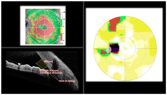
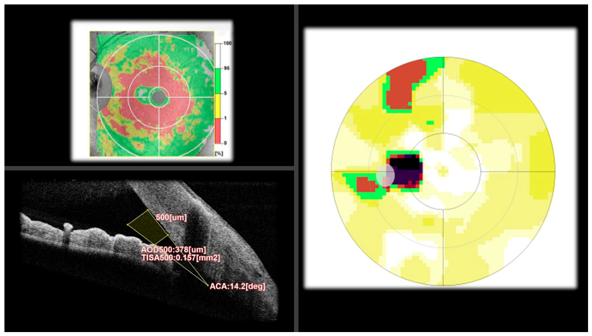
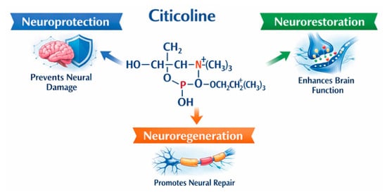
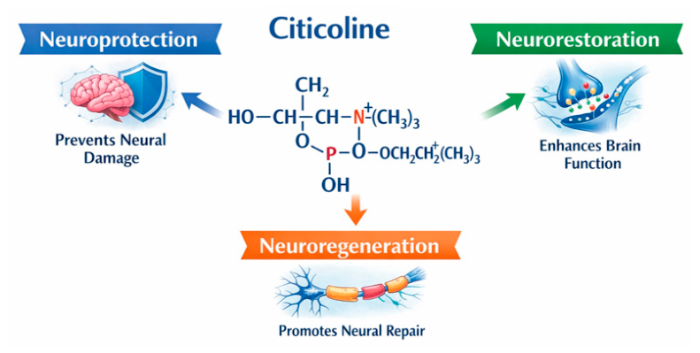
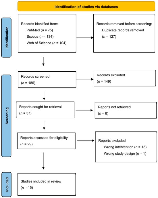
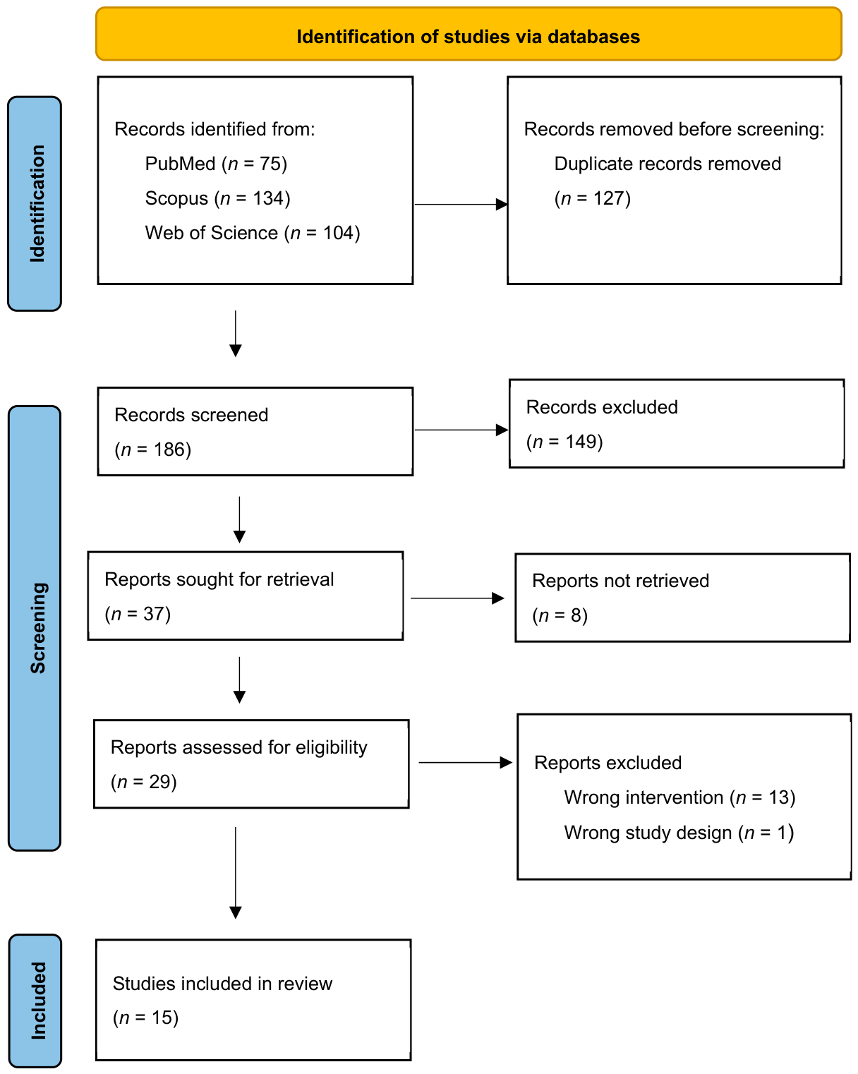
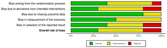
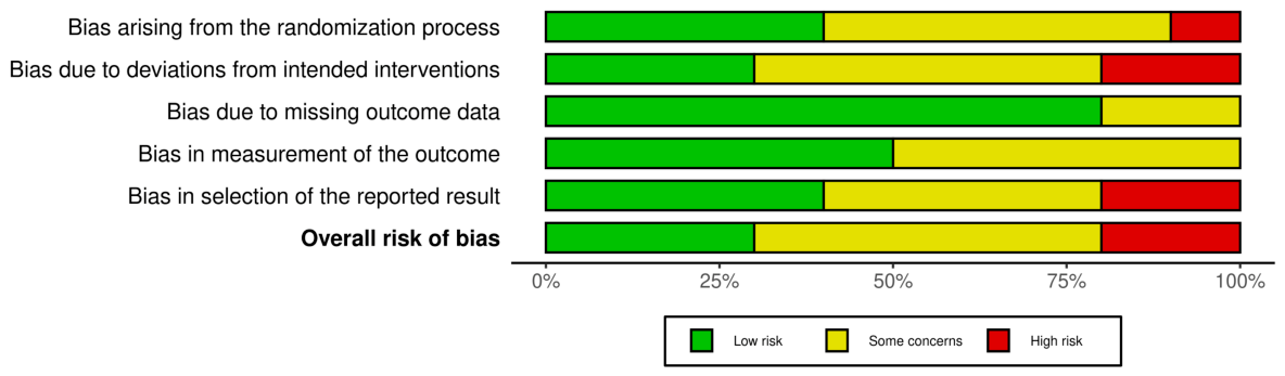
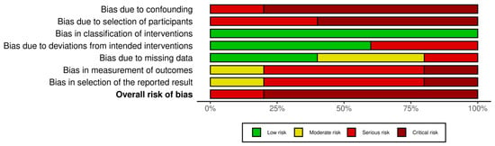
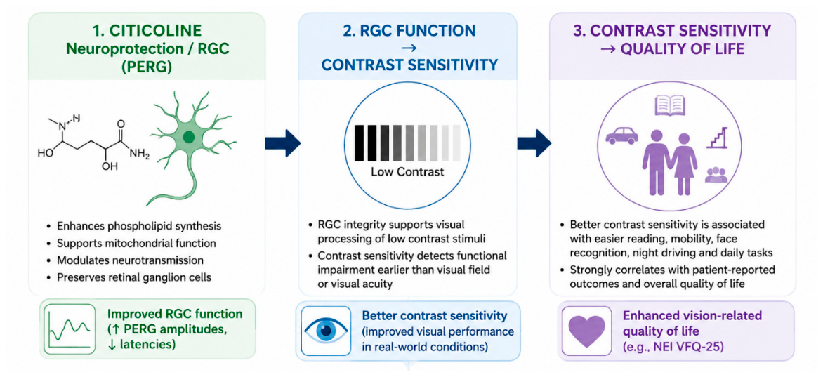
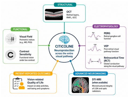
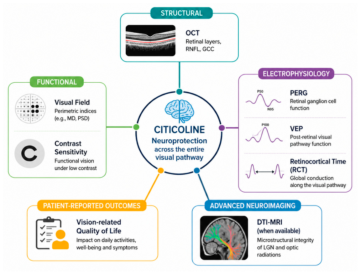

Disclaimer/Publisher’s Note: The statements, opinions and data contained in all publications are solely those of the individual author(s) and contributor(s) and not of MDPI and/or the editor(s). MDPI and/or the editor(s) disclaim responsibility for any injury to people or property resulting from any ideas, methods, instructions or products referred to in the content. |
© 2026 by the authors. Licensee MDPI, Basel, Switzerland. This article is an open access article distributed under the terms and conditions of the Creative Commons Attribution (CC BY) license.