


Abstract
6-PPD quinine (6-PPDQ) affects intestinal barrier function; however, its underlying mechanisms remain largely unknown. In the current study, we examined the role of reduction in phosphatidic acid synthesis in mediating the toxicity of 6-PPDQ in affecting intestinal barrier function. In Caenorhabditis elegans, 6-PPDQ exposure reduced the phosphatidic acid content, which was accompanied by the decreased expression of acl-5 and acl-6 encoding glycerol-3-phosphate acyltransferase. The RNAi of acl-5 and acl-6 lowered the phosphatidic acid content, enhanced intestinal permeability, and resulted in the increased accumulation of 6-PPDQ. Meanwhile, acl-5 and acl-6 RNAi caused susceptibility to 6-PPDQ toxicity by upregulating the expressions of insulin ligands and receptor genes and downregulating the expressions of daf-16 and its target genes. Moreover, the RNAi of acl-5 and acl-6 elevated the expression of let-363, and the RNAi of let-363 could reduce the expressions of insulin ligand genes and confer resistance to 6-PPDQ toxicity. The double RNAi of acl-5 and acl-6 caused more severe enhanced intestinal permeability and 6-PPDQ toxicity. Therefore, 6-PPDQ exposure potentially disrupts phosphatidic acid synthesis to affect intestinal barrier function by downregulating acl-5 and acl-6 expressions.
1. Introduction
Through different pathways, 6-PPD quinone (6-PPDQ) is generated from 6-PPD [1,2,3]. Besides acting as cause for coho salmon lethality [4], 6-PPDQ exists in different environments, especially in aquatic environments [5,6,7,8]. The range of environmentally relevant concentrations (ERCs) for 6-PPDQ is tens of μg/L or ng/L [4,9,10,11,12]. Exposure to 6-PPDQ causes multiple aspects of toxicity in organisms, including aquatic organisms [13,14,15,16,17,18,19,20]. Exposure to 6-PPDQ also results in organ injury in mammals, such as damage to the liver [21,22,23,24,25]. 6-PPDQ detection from human-related biological samples suggests a potential health risk to human beings [26,27,28,29,30]. After intraperitoneal injection, 6-PPDQ was found to be accumulated in multiple organs of mice [31,32].
Caenorhabditis elegans is useful to detect pollutant toxicity [32,33,34,35,36] and to determine its underlying molecular mechanisms [37,38,39,40]. This is largely due to the sensitivity to pollutant exposure in this animal model [41,42,43,44,45]. In nematodes, exposure to 6-PPDQ could cause several aspects of toxicity, including neurotoxicity and reproductive toxicity [46,47,48,49]. Some biochemical metabolisms (such as amino acid and vitamin D3 metabolisms) were disrupted by 6-PPDQ [50,51,52,53]. The organism’s lifespan was further decreased by 6-PPDQ, which was related to dysregulation of insulin/IGF-1 signaling [54], disruption in mitochondrial complexes’ function [55,56,57], and suppression in mitochondrial unfolded protein response (mt UPR) and mitophagy [58,59]. Moreover, 6-PPDQ resulted in disruption of C. elegans intestinal barrier function reflected by an enhancement in intestinal permeability, which was associated with the formation of susceptibility to 6-PPDQ toxicity [60]. However, the molecular basis for this observed 6-PPDQ intestinal toxicity is largely unclear.
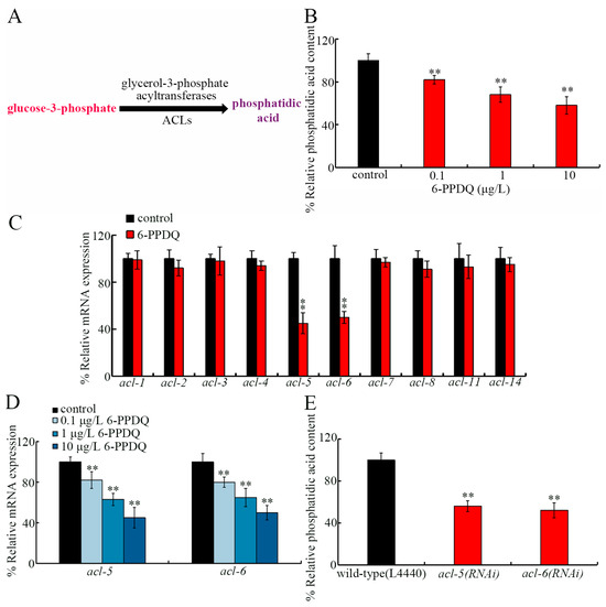
Phosphatidic acid, the simplest diacylglycerophospholipid, can exert diverse biological functions [61], particularly in regulating membrane assembly [62]. Growing evidence has indicated that phosphatidic acid synthesis is essential for maintaining cell membrane integrity [63,64]. We therefore hypothesized that 6-PPDQ might increase intestinal permeability by disrupting phosphatidic acid synthesis. In C. elegans, phosphatidic acid is synthesized from glycerol-3-phosphate through acylation reactions catalyzed by acyltransferases encoded by acl genes (Figure 1A) [65]. Among acl genes, acl-1–8, 11, and 14 can be expressed in the intestine (https://wormbase.org). In mammals, phosphatidic acid is known to bind to target of rapamycin (TOR) [66]. Inhibition in C. elegans LET-363/TOR has been shown to extend lifespan [67]. In this study, the effects of 6-PPDQ on phosphatidic acid content and expression of intestinal acl genes were first examined. Moreover, the role of candidate acl genes in modulating 6-PPDQ toxicity on intestinal barrier function and the underlying mechanism were determined. The findings highlighted the role of inhibition in ACL-5/6 in mediating 6-PPDQ damage to intestinal barrier function and causing susceptibility to 6-PPDQ toxicity by activating the mTOR–insulin signaling axis.
Figure 1.
Effect of 6-PPDQ exposure on synthesis of phosphatidic acid. (A) A diagram showing control of phosphatidic acid synthesis from glucose-3-phosphate by glucose-3-phosphate acyltransferases in nematodes. (B) Effect of 6-PPDQ exposure on phosphatidic acid content in wild-type N2 nematodes. N = 3. ** p < 0.01 vs. control. (C) Effect of 6-PPDQ (10 μg/L) on expression of intestinal acl genes in wild-type N2 nematodes. Thirty intact intestines were isolated for qRT-PCR analysis. N = 3. ** p < 0.01 vs. control. (D) Effect of 6-PPDQ exposure on expression of intestinal acl5 and acl-6 in wild-type N2 nematodes. Thirty intact intestines were isolated for qRT-PCR analysis. N = 3. ** p < 0.01 vs. control. (E) Effect of RNAi of acl-5 and acl-6 on phosphatidic acid content in 6-PPDQ-exposed wild-type N2 nematodes. N = 3. Exposure concentration of 6-PPDQ was 10 μg/L. ** p < 0.01 vs. wild-type(L4440). Data are presented as the mean ± standard deviation (SD).
2. Materials and Methods
2.1. Animal Maintenance
Animal strains (Table S1) were cultured according to standard C. elegans protocol [68] on an NGM plate fed with E. coli OP50. The used strains are from the Caenorhabditis Genetics Center (CGC). Gravid hermaphrodites were subjected to embryonic isolation using a lysis buffer in order to obtain L1-larvae [69].
2.2. Exposure
The 6-PPDQ (Toronto Research Chemicals, Toronto, ON, Canada) exposure concentrations were 0.1–10 μg/L, which correspond to those in actual water environments [4]. 6-PPDQ exposure was from L1-larave and lasted until the third day of adulthood (6.5 days) at 20 °C in darkness [70]. The 6-PPDQ was dissolved in dimethyl sulfoxide (DMSO), and DMSO solution diluted by K buffer in the same way as the 6-PPDQ solutions was used as the control solution. Exposure solutions of 6-PPDQ prepared by diluting the stock solution with K buffer (0.032 M KCl, 0.051 M NaCl) were refreshed daily. Nematodes were exposed to 6-PPDQ solutions with OP50 added as the food source. The exposure volume for each group was 1 mL in 12-well glass plates. Approximately 1000 nematodes were exposed to 6-PPDQ for each group. During the exposure, shaking at 150 rpm was performed for cultured nematodes. Nematodes were randomly assigned to groups for the assessment of different endpoints. During exposure, the 6-PPDQ working solutions were updated daily.
The concentrations of the 6-PPDQ exposure solutions were confirmed by HPLC-MS/MS (PerkinElmer, Waltham, MA, USA). The 6-PPDQ body accumulation was further examined by HPLC-MS/MS, and we provide the detailed experimental procedure in Text S1.
2.3. Phosphatidic Acid Content
A phosphatidic acid test kit (Shanghai Chutai Biotechnology Co., Shanghai, China) was used. Nematodes were weighed, homogenized, and centrifuged. Supernatant was measured for absorbance at 450 nm. Experiments were conducted in triplicate.
2.4. Endpoints
For the reactive oxygen species (ROS) assay, animals were treated using 1 μM CM-H2DCFDA for 2 h with shaking at 200 rpm [71]. Following incubation, ROS fluorescent signals were examined (excitation/emission wavelength: 488/510 nm). For the lipofuscin accumulation assay, fluorescent signals were analyzed under a DAPI filter [72]. Fifty animals were tested. Experiments were conducted in triplicate.
For locomotion assay, head thrashing was examined by observing movement direction along the X-axis and changes in the direction of the posterior (Y-axis) [71], and body bending was examined by tracking the mid-body bending direction [72]. Brood size refers to the total number of offspring for an individual nematode and was assessed until animals stopped egg-laying [73]. Fifty animals were tested. Experiments were conducted in triplicate.
2.5. Intestinal Permeability
Animals were stained using 5% erioglaucine disodium for 3 h [69]. Images were analyzed under bright field. Fifty animals were tested.
2.6. Gene Expression
Nematodes were lysed using pre-chilled TRIzol (Sangon Biotech. Co., Ltd., Shanghai, China). cDNA was prepared by M-MuLV reverse transcriptase (Sangon Biotech. Co., Ltd., Shanghai, China). Target gene expressions were assessed by qRT-PCR with tba-1 serving as reference gene [74]. The primers are available in Table S2.
2.7. RNA Interference (RNAi)
L1-larvae were treated on RNAi plates seeded by double-stranded RNA-expressing E. coli HT115. Their progeny were exposed to 6-PPDQ. Transgenic strains of VP303 and WM118 are tools for intestinal and muscle gene RNAi. Empty vector/L4440 served as control [74]. Figures S1 and S2 show the RNAi efficiency.
2.8. Data Analysis
One-way or two-way ANOVA (for multi-factor comparison) followed by the Tukey post hoc test was used to evaluate differences between different groups. A p-value of <0.01 (**) was deemed statistically significant.
3. Results
3.1. 6-PPDQ Inhibited Synthesis of Phosphatidic Acid
Phosphatidic acid content was decreased by 6-PPDQ (Figure 1B). Among intestinal acl genes, expressions of acl-1–4, acl-6, acl-8, acl-11, and acl-14 were not changed by 6-PPDQ; however, acl-5 and acl-6 expressions were reduced by 6-PPDQ (Figure 1C). After 6-PPDQ exposure, the decrease in acl-5 and acl-6 expressions were concentration dependent (Figure 1D). Moreover, phosphatidic acid content was inhibited by acl-5 and acl-6 RNAi (Figure 1E).
3.2. acl-5 and acl-6 RNAi Induced Susceptibility to 6-PPDQ Toxicity
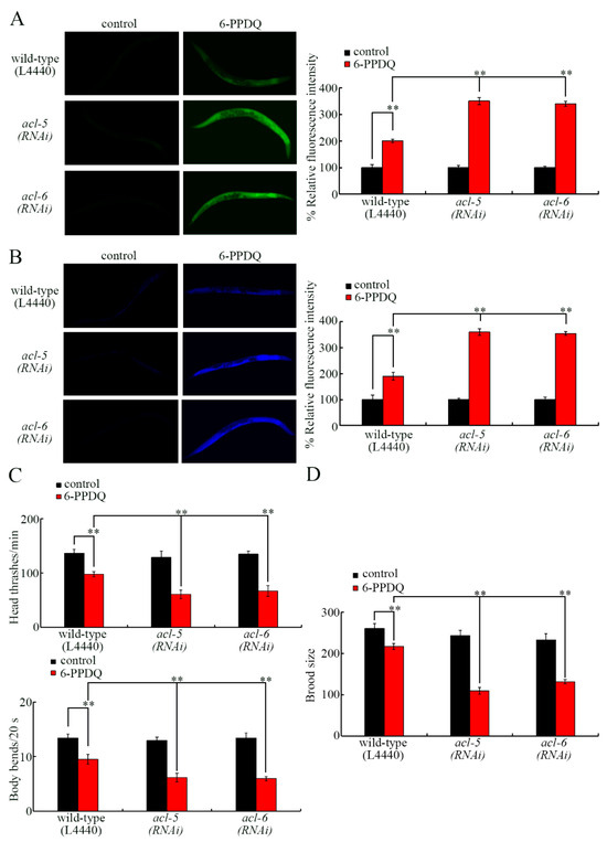
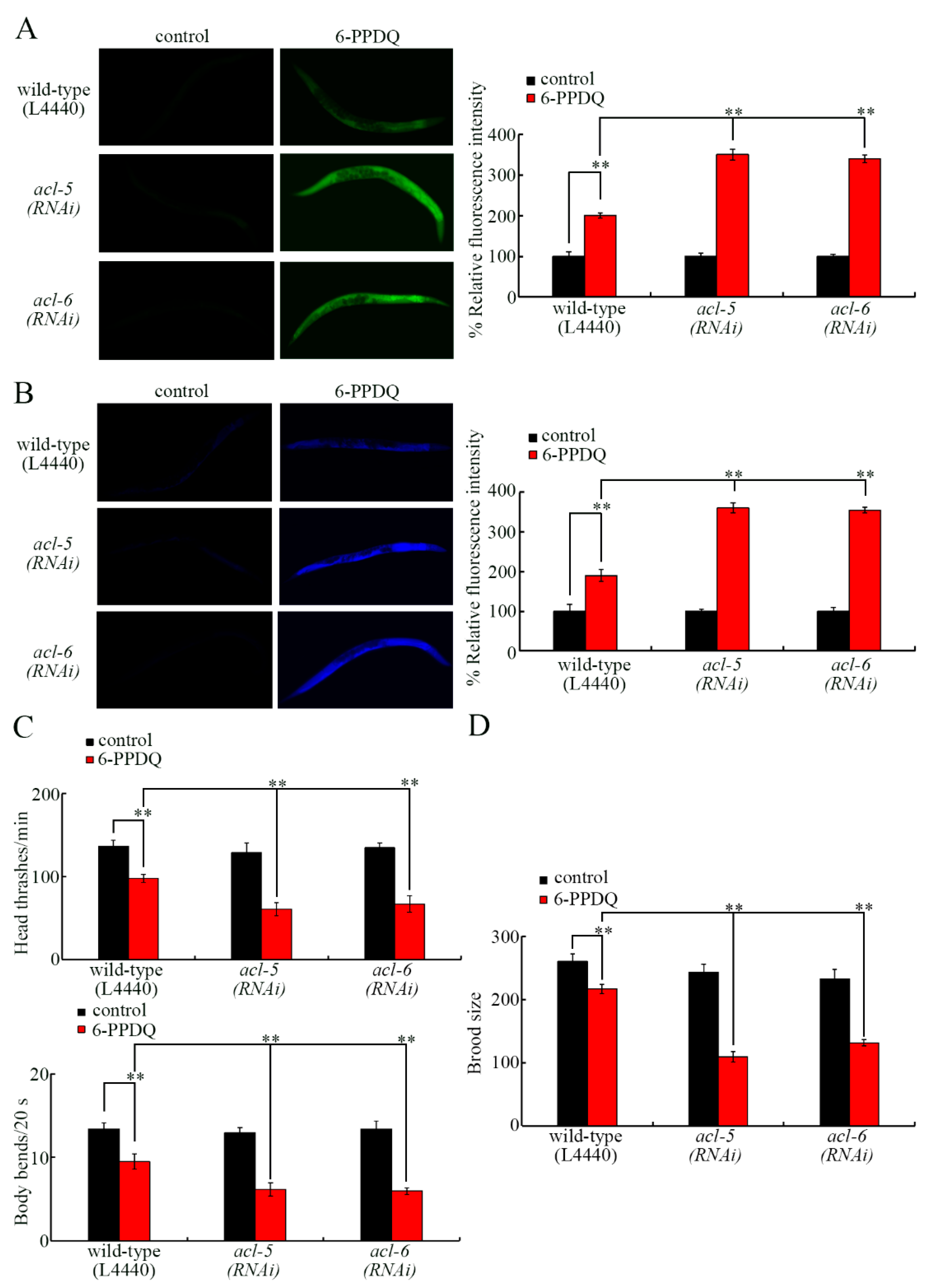
Using lipofuscin accumulation and ROS generation as intestinal-toxicity-related endpoints, the RNAi of acl-5 and acl-6 caused more severe 6-PPDQ intestinal toxicity in inducing lipofuscin accumulation and ROS generation compared with those in wild-type N2 nematodes (Figure 2A,B). Using locomotion as a neurotoxicity-related endpoint and brood size as a reproductive-toxicity-related endpoint, more severe 6-PPDQ neurotoxicity in inhibiting locomotion and reproductive toxicity in reducing brood size were caused by acl-5 and acl-6 RNAi compared with those in wild-type N2 nematodes (Figure 2C,D).
Figure 2.
Effect of RNAi of acl-5 and acl-6 on 6-PPDQ toxicity in causing ROS generation (A), inducing intestinal lipofuscin accumulation (B), decreasing locomotion (C), and reducing brood size (D) in wild-type N2 nematodes. N = 50. Exposure concentration of 6-PPDQ was 10 μg/L. ** p < 0.01. Data are presented as the mean ± standard deviation (SD).
3.3. Tissue-Specific Activity of ACL-5 and ACL-6 to Control 6-PPDQ Toxicity
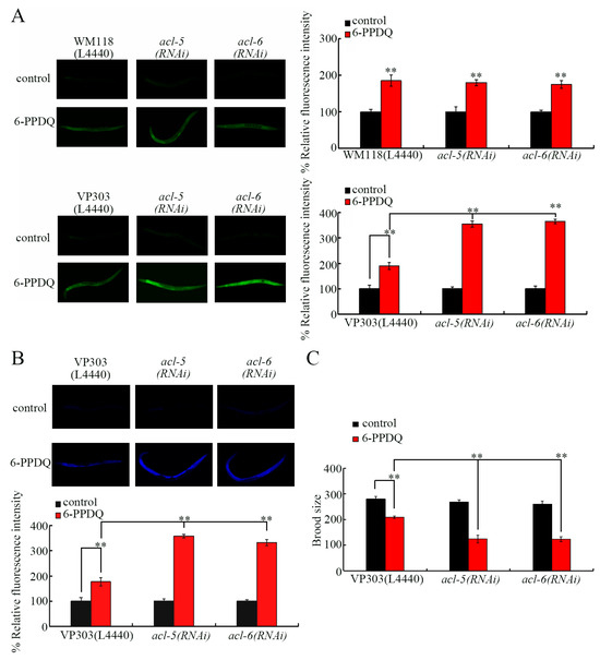
We next focused on ACL-5 and ACL-6 to examine their tissue-specific activity in controlling 6-PPDQ toxicity. C. elegans ACL-5 and ACL-6 are expressed in the muscle and intestine (https://wormbase.org). Nevertheless, using intestinal ROS generation as an endpoint, induction of intestinal ROS generation by 6-PPDQ was not changed by the muscle RNAi of acl-5, and acl-6 but could be increased by the intestinal RNAi of acl-5 and acl-6 (Figure 3A). Similarly, 6-PPDQ-caused lipofuscin accumulation was strengthened by intestinal acl-5 and acl-6 RNAi (Figure 3B). Additionally, 6-PPDQ-caused reproductive reduction was increased by intestinal acl-5 and acl-6 RNAi (Figure 3C).
Figure 3.
Tissue-specific activity of ACL-5 and ACL-6 in regulating 6-PPDQ toxicity. (A) Tissue-specific activity of ACL-5 and ACL-6 in regulating 6-PPDQ toxicity in causing intestinal ROS generation. N = 50. (B) Intestine-specific activity of ACL-5 and ACL-6 in regulating 6-PPDQ toxicity in causing intestinal lipofuscin accumulation. N = 50. (C) Intestine-specific activity of ACL-5 and ACL-6 in regulating 6-PPDQ toxicity in reducing brood size. Exposure concentration of 6-PPDQ was 10 μg/L. N = 50. ** p < 0.01. Data are presented as the mean ± standard deviation (SD).
3.4. ACL-5 and ACL-6 Modulated Intestinal Permeability
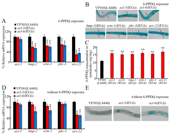
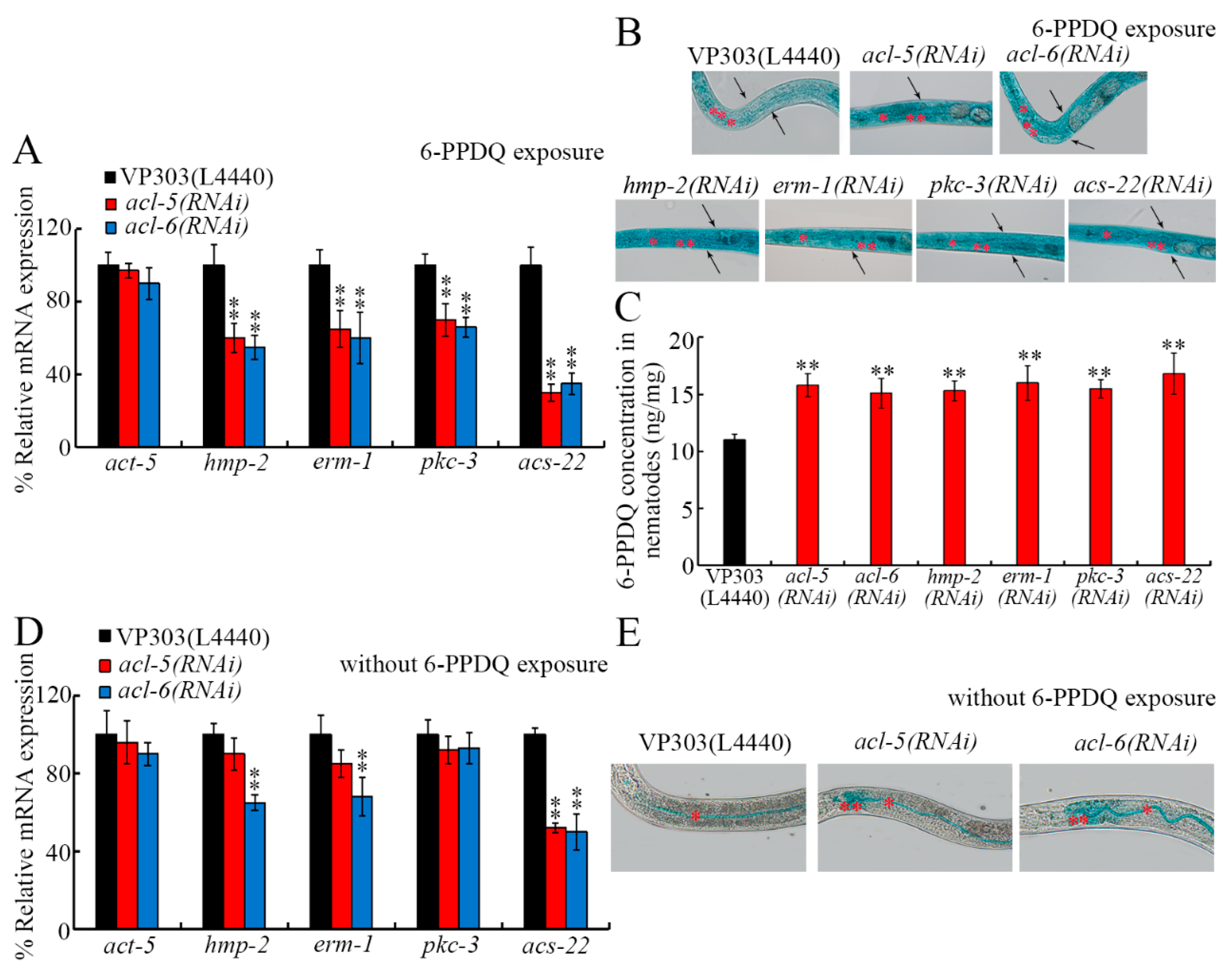
Some C. elegans proteins are shown to regulate intestinal permeability [72]. Considering the important association between phosphatidic acid metabolism and cell membrane integrity [63], we next examined the role of ACL-5 and ACL-6 in modulating the functional state of the intestinal barrier. In 6-PPDQ-exposed nematodes, although act-5 expression was not changed by acl-5 and acl-6 RNAi, expressions of pkc-3, erm-1, hmp-2, and acs-22 were decreased by acl-5 and acl-6 RNAi (Figure 4A). The intestinal permeability could be strengthened by 6-PPDQ [60]. The intestinal permeability of 6-PPDQ-exposed nematodes was enhanced not only by acl-5 and acl-6 RNAi but also by pkc-3, hmp-2, erm-1, and acs-22 RNAi (Figure 4B). Moreover, 6-PPDQ accumulation was increased by acl-5, acl-6, pkc-3, hmp-2, erm-1, and acs-22 RNAi (Figure 4C).
Figure 4.
Effect of intestinal RNAi of acl-5 and acl-6 on intestinal permeability under 6-PPDQ exposure or without 6-PPDQ exposure condition. (A) Effect of intestinal RNAi of acl-5 and acl-6 on expression of genes governing functional state of intestinal barrier in 6-PPDQ-exposed VP303 nematodes. Exposure concentration of 6-PPDQ was 10 μg/L. N = 3. ** p < 0.01 vs. VP303(L4440). (B) Effect of intestinal RNAi of acl-5, acl-6, hmp-2, erm-1, acs-22, and pkc-3 on intestinal permeability in 6-PPDQ-exposed VP303 nematodes. Exposure concentration of 6-PPDQ was 10 μg/L. Single asterisks and double asterisks indicate intestinal lumen and intestinal cells, respectively. Arrowheads indicate the body cavity. N = 50. (C) Effect of intestinal RNAi of acl-5, acl-6, hmp-2, erm-1, acs-22, and pkc-3 on 6-PPDQ accumulation in body of VP303 nematodes after exposure to 10 μg/L 6-PPDQ. N = 3. ** p < 0.01 vs. VP303(L4440). (D) Effect of intestinal RNAi of acl-5 and acl-6 on expression of genes governing functional state of intestinal barrier in VP303 nematodes under the condition without 6-PPDQ exposure N = 3. ** p < 0.01 vs. VP303(L4440). (E) Effect of intestinal RNAi of acl-5 and acl-6 on intestinal permeability in VP303 nematodes under the condition without 6-PPDQ exposure. Single asterisks and double asterisks indicate intestinal lumen and intestinal cells, respectively. N = 50. Data are presented as the mean ± standard deviation (SD).
Under normal conditions, although act-5, hmp-2, erm-1, and pkc-3 expressions were not affected by intestinal acl-5 RNAi, acs-22 expression was decreased by the intestinal RNAi of acl-5 (Figure 4D). Moreover, under normal conditions, expression of hmp-2, erm-1, and acs-22 could be decreased by acl-6 RNAi (Figure 4D). Under normal conditions, intestinal acl-5 and acl-6 RNAi could already even cause blue dye translocation to intestinal cells (Figure 4E).
3.5. Intestinal RNAi of acl-5 and acl-6 Affected Expression of Genes in Insulin Signaling Pathway
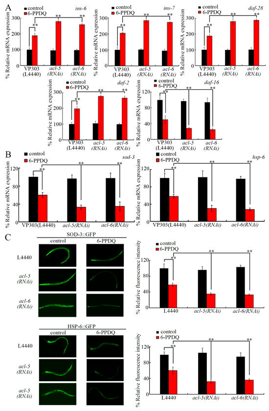
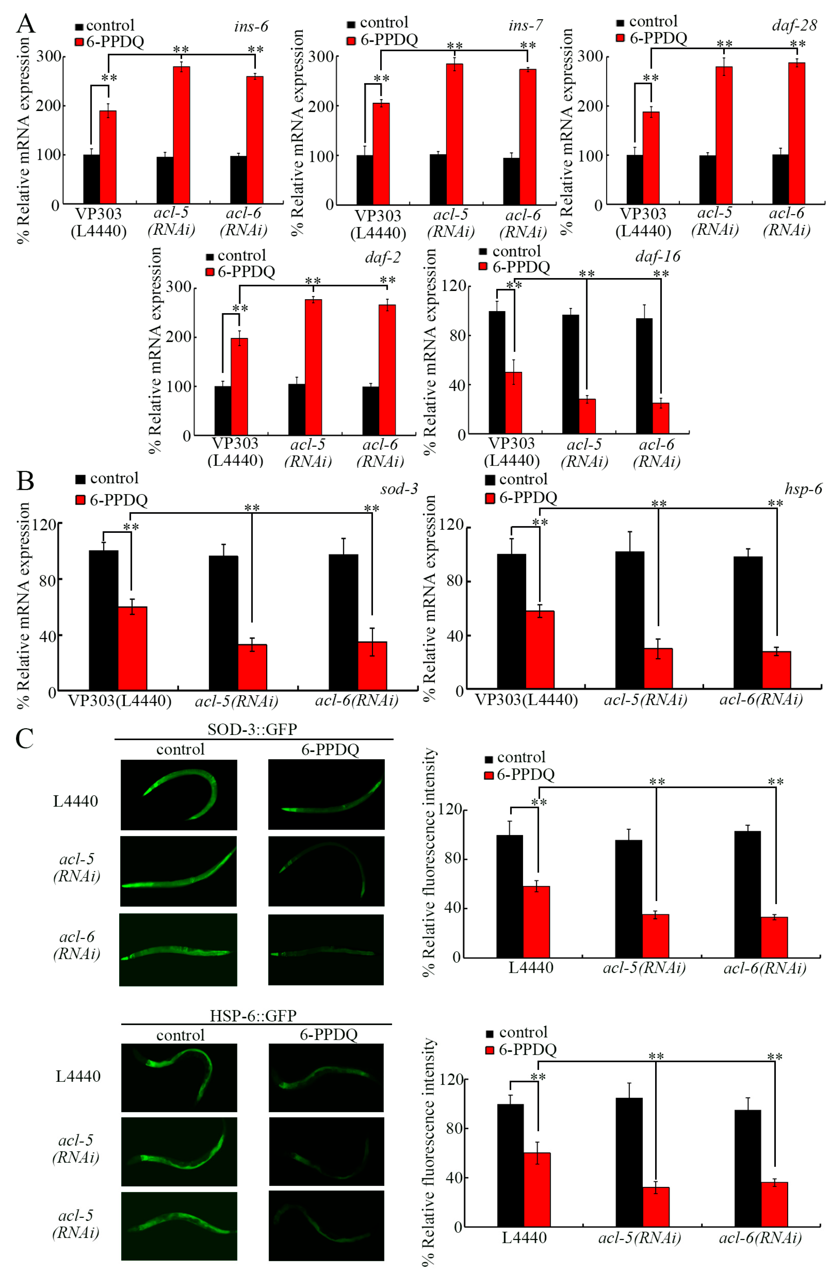
Insulin signaling functions in the C. elegans intestine to control pollutant toxicity [72]. Insulin ligands modulated the effect of 6-PPDQ toxicity on longevity by activating receptor DAF-2 and inhibiting DAF-16 and targets (SOD-3 and HSP-6) [75]. Among previously identified insulin ligand genes dysregulated by 6-PPDQ [75], the 6-PPDQ-caused increase in expression of ins-6, ins-7, and daf-28 was strengthened by intestinal acl-5 and acl-6 RNAi (Figure 5A). Additionally, the 6-PPDQ-caused increase in daf-2 expression and decrease in daf-16 expression were enhanced by intestinal acl-5 and acl-6 RNAi (Figure 5A). Moreover, the decrease in sod-3, hsp-6, SOD-3::GFP, and HSP-6::GFP expressions induced by 6-PPDQ was enhanced by intestinal acl-5 and acl-6 RNAi (Figure 5B,C).
Figure 5.
Effect of intestinal RNAi of acl-5 and acl-6 on expression of genes in insulin signaling pathways after 6-PPDQ exposure. (A) Effect of intestinal RNAi of acl-5 and acl-6 on expressions of ins-6, ins-7, daf-28, daf-2, and daf-16 in VP303 nematodes after 6-PPDQ exposure. N = 3. (B) Effect of intestinal RNAi of acl-5 and acl-6 on expressions of sod-3 and hsp-6 in VP303 nematodes after 6-PPDQ exposure. N = 3. (C) Effect of RNAi of acl-5 and acl-6 on expressions of SOD-3::GFP and HSP-6::GFP in CF1553 and SJ410 nematodes after 6-PPDQ exposure. N = 50. Exposure concentration of 6-PPDQ was 10 μg/L. ** p < 0.01. Data are presented as the mean ± standard deviation (SD).
3.6. Effect of Intestinal hmp-2, pkc-3, erm-1, acs-22, daf-28, ins-7, ins-6, daf-2, daf-16, sod-3, and hsp-6 RNAi on 6-PPDQ Toxicity
Using several endpoints, 6-PPDQ toxicity was increased by intestinal erm-1, hmp-2, pkc-3, acs-22, daf-16, sod-3, and hsp-6 RNAi (Figure S3A–C). Different from this, the 6-PPDQ-caused toxicity was inhibited by intestinal daf-28, ins-7, ins-6, and daf-2 RNAi (Figure S3A–C).
3.7. ACL-5 and ACL-6 Modulated Insulin Signals by Inhibiting LET-363
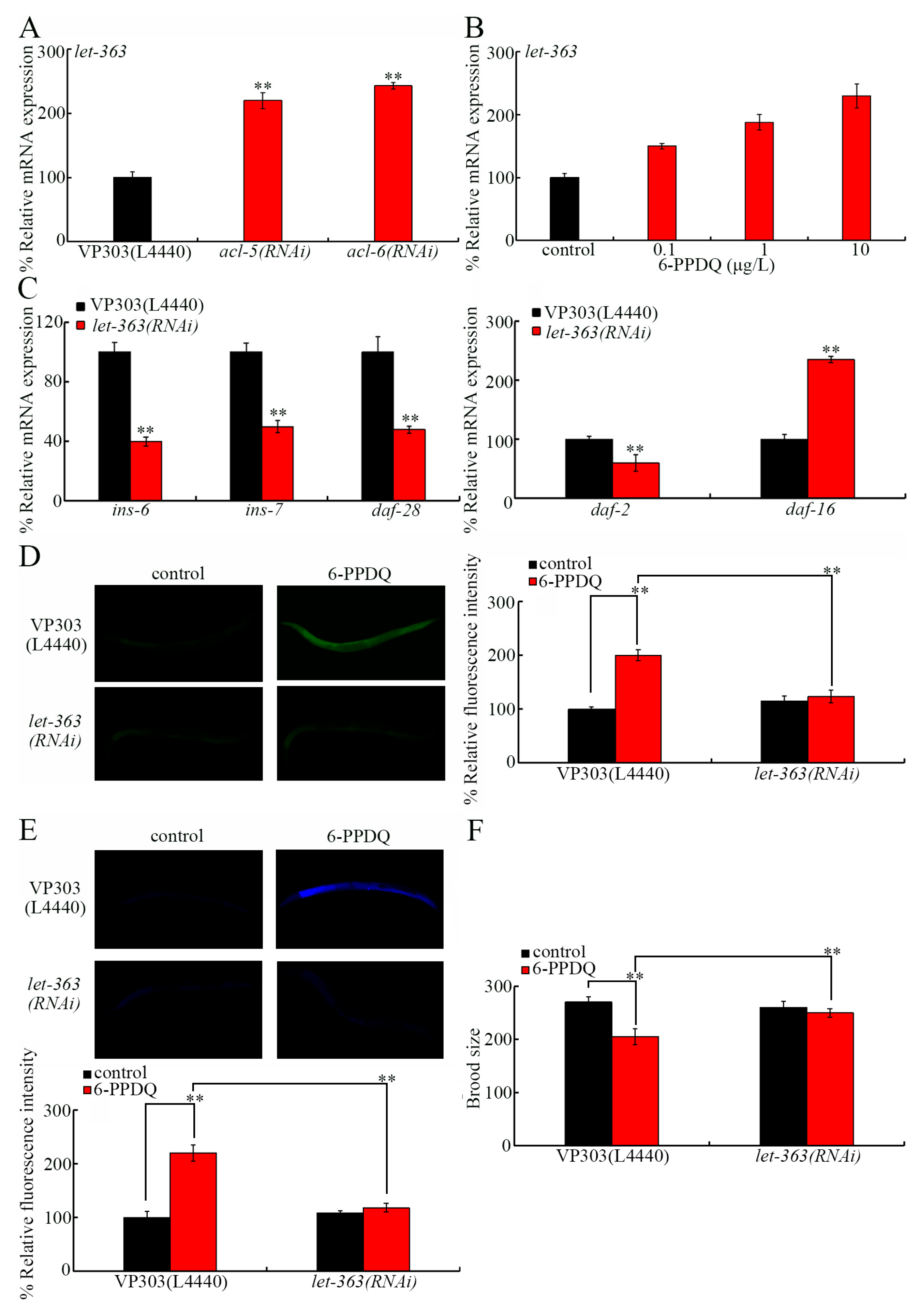
In mammals, phosphatidic acid can by sensed by and bind to TOR [66]. LET-363 is C. elegans TOR [76]. let-363 expression was increased by intestinal acl-5 and acl-6 RNAi (Figure 6A). 6-PPDQ increased intestinal let-363 expression (Figure 6B). The intestinal RNAi of let-363 decreased daf-28, ins-7, ins-6, and daf-2 expressions and increased daf-16 expression (Figure 6C). Besides this, after 6-PPDQ exposure, let-363 RNAi further increased sod-3 and hsp-6 expressions (Figure S4). 6-PPDQ-caused toxicity was inhibited by intestinal let-363 RNAi (Figure 6D–F).
Figure 6.
Role of LET-363 in controlling 6-PPDQ toxicity by activating insulin signals. (A) Effect of intestinal RNAi of acl-5 and acl-6 on expression of let-363 in 6-PPDQ-exposed VP303 nematodes. Exposure concentration of 6-PPDQ was 10 μg/L. N = 3. ** p < 0.01 vs. VP303(L44440). (B) Effect of 6-PPDQ exposure on intestinal let-363 expression in wild-type N2 nematodes. Thirty intact intestines were isolated for qRT-PCR analysis. N = 3. ** p < 0.01 vs. control. (C) Effect of intestinal RNAi of let-363 on expression of ins-6, ins-7, daf-28, daf-2, and daf-16 in 6-PPDQ-exposed VP303 nematodes. Exposure concentration of 6-PPDQ was 10 μg/L. N = 3. ** p < 0.01 vs. VP303(L4440). (D) Effect of intestinal RNAi of let-363 on 6-PPDQ toxicity in causing intestinal ROS generation in VP303 nematodes. Exposure concentration of 6-PPDQ was 10 μg/L. N = 50. ** p < 0.01. (E) Effect of intestinal RNAi of let-363 on 6-PPDQ toxicity in inducing intestinal lipofuscin accumulation in VP303 nematodes. Exposure concentration of 6-PPDQ was 10 μg/L. N = 50. ** p < 0.01. (F) Effect of intestinal RNAi of let-363 on 6-PPDQ toxicity in reducing brood size in VP303 nematodes. Exposure concentration of 6-PPDQ was 10 μg/L. N = 50. ** p < 0.01. Data are presented as the mean ± standard deviation (SD).
3.8. Effect of Double RNAi of acl-5 and acl-6 on Intestinal Permeability and 6-PPDQ Toxicity Induction
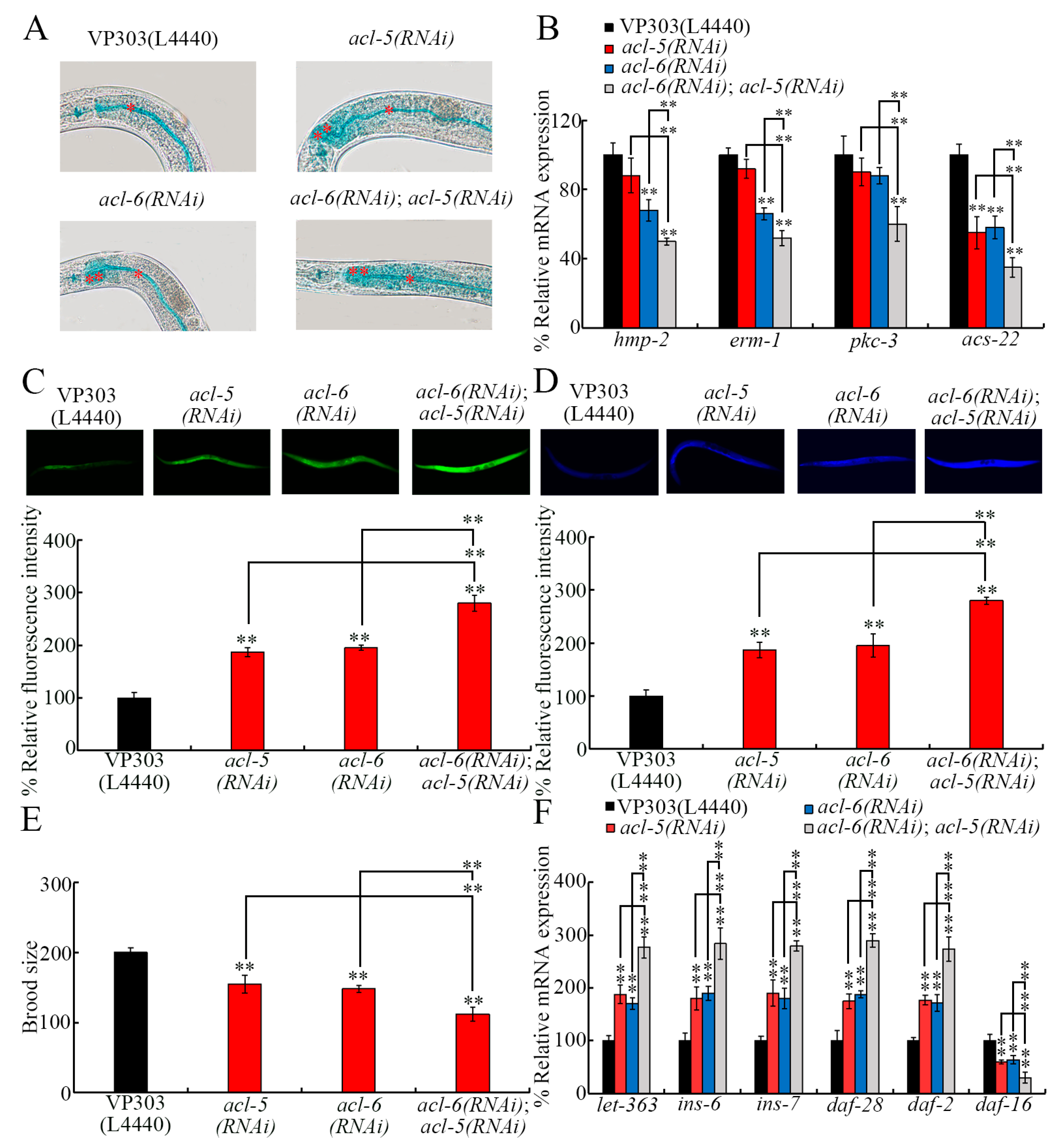
After double RNAi of acl-5 and acl-6, more severe intestinal permeability was detected in acl-6(RNAi);acl-5(RNAi) than in acl-5(RNAi) and acl-6(RNAi) (Figure 7A). Accompanying this, more severe decrease in hmp-2, erm-1, and acs-22 expressions were found in acl-6(RNAi);acl-5(RNAi) than in acl-5(RNAi) and/or acl-6(RNAi) (Figure 7B). Compared with no expressional change of pkc-3 in acl-5(RNAi) and/or acl-6(RNAi), pkc-3 expression was reduced in acl-6(RNAi);acl-5(RNAi) (Figure 7B).
Figure 7.
Effect of double RNAi of acl-5 and acl-6 on intestinal permeability and 6-PPDQ toxicity induction. (A) Effect of double RNAi of acl-5 and acl-6 on intestinal permeability in VP303 nematodes. Single asterisks and double asterisks indicate intestinal lumen and intestinal cells. N = 50. (B) Effect of double RNAi of acl-5 and acl-6 on expressions of hmp-2, erm-1, pkc-3, and acs-22 in VP303 nematodes. N = 3. ** p < 0.01 vs. VP303(L4440) (if not specially indicated). (C) Interaction between acl-5 and acl-6 in regulating 6-PPDQ toxicity in inducing intestinal ROS generation in VP303 nematodes. Exposure concentration of 6-PPDQ was 10 μg/L. N = 50. ** p < 0.01 vs. VP303(L4440) (if not specially indicated). (D) Interaction between acl-5 and acl-6 in regulating 6-PPDQ toxicity in inducing intestinal lipofuscin accumulation in VP303 nematodes. Exposure concentration of 6-PPDQ was 10 μg/L. N = 50. ** p < 0.01 vs. VP303(L4440) (if not specially indicated). (E) Interaction between acl-5 and acl-6 in regulating 6-PPDQ toxicity in reducing brood size in VP303 nematodes. Exposure concentration of 6-PPDQ was 10 μg/L. N = 50. ** p < 0.01 vs. VP303(L4440) (if not specially indicated). (F) Effect of double RNAi of acl-5 and acl-6 on expressions of let-363, ins-6, ins-7, daf-28, daf-2, and daf-16 in 6-PPDQ-exposed VP303 nematodes. Exposure concentration of 6-PPDQ was 10 μg/L. N = 3. ** p < 0.01 vs. VP303(L4440) (if not specially indicated). Data are presented as the mean ± standard deviation (SD).
Moreover, more severe 6-PPDQ toxicity was found in acl-6(RNAi);acl-5(RNAi) than in acl-5(RNAi) and/or acl-6(RNAi) (Figure 7C–E). Meanwhile, more severe increases in let-363, daf-28, ins-6, ins-7, and daf-2 expressions and decrease in daf-16 expression were also detected in acl-6(RNAi);acl-5(RNAi) than in acl-5(RNAi) and/or acl-6(RNAi) (Figure 7F).
4. Discussion
The intestine is the largest organ in nematodes. After 6-PPDQ exposure, C. elegans intestinal permeability could be enhanced [60], which suggests disrupted intestinal function. Considering the role of phosphatidic acid during cell membrane organization [62], we determined the effect of 6-PPDQ exposure on phosphatidic acid synthesis and its association with 6-PPDQ toxicity, especially the damage to intestinal function. We here observed a reduction in phosphatidic acid content, which was due to inhibition of its synthesis. Accompanying the reduction in phosphatidic acid content (Figure 1B), acl-5 and acl-6 expressions were decreased by 6-PPDQ (Figure 1C), and phosphatidic acid content could be reduced by acl-5 and acl-6 RNAi (Figure 1E). ACL-5 is the homolog of mammal endoplasmic reticulum (ER) GPAT4, and ACL-6 is the homolog of mammal mitochondrial GPAT1 and GPAT2 [77]. ACLs are predicted to catalyze the process generating phosphatidic acid, the precursor of membrane phospholipids [77]. Among intestinal acl genes encoding glycerol-3-phosphate acyltransferases, only the expressions of acl-5 and acl-6 were decreased by 6-PPDQ (Figure 1C). That is, the expression of not all intestinal acl genes had enough sensitivity to show a response to 6-PPDQ. Among these intestinal acl genes, our data suggested that expression of acl-5 and acl-6 may be highly sensitive after pollutant exposure. The reduction in phosphatidic acid content and decrease in acl-5 and acl-6 expression by 6-PPDQ suggested that 6-PPDQ at ERCs could reduce phosphatidic acid content by inhibiting certain glycerol-3-phosphate acyltransferases.
We further provide several lines of evidence to demonstrate that the decrease in acl-5 and acl-6 expressions mediated the induction of 6-PPDQ toxicity. That is, accompanied with the decrease in acl-5 and acl-6 expressions (Figure 1D), acl-5 and acl-6 RNAi caused susceptibility to 6-PPDQ toxicity. acl-5(RNAi) and acl-6(RNAi) showed susceptibility to 6-PPDQ toxicity at different aspects. Using intestinal endpoints, acl-5 and acl-6 RNAi induced susceptibility to 6-PPDQ intestinal toxicity (Figure 2A,B). Using locomotion as the endpoint, acl-5 and acl-6 RNAi caused susceptibility to 6-PPDQ neuronal toxicity (Figure 2C). Using brood size as the endpoint, acl-5 and acl-6 RNAi also induced susceptibility to 6-PPDQ reproductive toxicity (Figure 2D). Further examination of the effect of acl-5 and acl-6 overexpression is suggested in the future to further confirm these observations.
Moreover, ACL-5 and ACL-6 functioned in the intestine to control 6-PPDQ toxicity. The observations suggested that susceptibility of acl-5(RNAi) and acl-6(RNAi) to 6-PPDQ intestinal toxicity might be the direct effect of the RNAi of acl-5 and acl-6. In contrast, the detected susceptibility of acl-5(RNAi) and acl-6(RNAi) to 6-PPDQ neuronal toxicity and reproductive toxicity might be the indirect effect of the RNAi of acl-5 and acl-6.
For the mechanism of reduction in acl-5 and acl-6 expressions in mediating the 6-PPDQ toxicity on intestinal barrier function, we first found that more severe enhancement in intestinal permeability was caused by the RNAi of acl-5 and acl-6 after 6-PPDQ exposure (Figure 4B), which was accompanied by inhibited pkc-3, erm-1, hmp-2, and acs-22 expression (Figure 4A). The hmp-2 encodes beta-catenin [78]; erm-1 encodes ezrin–radixin–moesin [79]; pkc-3 encodes atypical protein kinase C [80]; and acs-22 encodes fatty acid acyl-CoA synthetase [81]. Mutation or RNAi of these genes causes increased intestinal permeability [82,83]. Meanwhile, we observed an increase in 6-PPDQ accumulation by the RNAi of acl-5, acl-6, pkc-3, erm-1, hmp-2, and acs-22 (Figure 4C). The intestinal RNAi of acl-5 and acl-6 could further cause enhanced intestinal permeability (Figure 4E). For the molecular basis of this enhanced intestinal permeability, intestinal acl-5 RNAi decreased acs-22 expression, and intestinal acl-6 RNAi decreased erm-1, hmp-2, and acs-22 expression (Figure 4D). Therefore, the observed more severe enhancement in intestinal permeability in 6-PPDQ-exposed acl-5/6(RNAi) was not only associated with ACL-5 and ACL-6 functions but also closely related to the role of PKC-3, ERM-1, HMP-2, and ACS-22. Nevertheless, besides the decrease in their expressions detected in acl-5(RNAi) and/or acl-6(RNAi) under normal conditions, pkc-3 expression was also decreased in 6-PPDQ-exposed acl-5(RNAi) and/or acl-6(RNAi) (Figure 4A). These may be because the intestinal RNAi of acl-5 and acl-6 induced susceptibility to 6-PPDQ damage (Figure 3), which further induced the decrease in pkc-3 expression. Furthermore, the intestinal RNAi of acl-5 and acl-6 and their downstream target genes induced susceptibility to 6-PPDQ damage (Figure 3 and Figure S3). After pollutant exposure, intestinal oxidative damage contributed to enhanced intestinal permeability [72]. Additionally, after 6-PPDQ exposure, these targeted genes’ expression was negatively correlated with intestinal oxidative stress [84]. That is, the induced oxidative stress might act together with the decrease in acl-5/6 and their target gene expressions to lead to the induction of enhanced intestinal permeability and 6-PPDQ accumulation.
For the effects of RNAi of acl-5 and acl-6 on the intestine, we further found that the intestinal RNAi of acl-5 and acl-6 induced susceptibility to 6-PPDQ toxicity by affecting the insulin signaling pathway. In the insulin signaling pathway, activation of insulin ligands and receptor DAF-2 and inhibition in DAF-16 and its targets mediated 6-PPDQ toxicity [75]. Moreover, the increased expressions of these ligands and receptor genes and the decrease in expressions of daf-16 and its two target genes were strengthened by acl-5/6 RNAi (Figure 5A–C). Meanwhile, 6-PPDQ-caused intestinal toxicity and reproductive toxicity were inhibited by the intestinal RNAi of these ligands and receptor genes and enhanced by the RNAi of daf-16 and its two target genes (Figure S3A–C). Therefore, the intestinal RNAi of acl-5 and acl-6 induced susceptibility to 6-PPDQ damage by activating insulin ligands and DAF-2 and inhibiting DAF-16 and its targets.
In cells, TOR acted as the sensor of phosphatidic acid [66]. LET-363 is the C. elegans TOR [85]. Moreover, we identified that the intestinal RNAi of acl-5 and acl-6 induced susceptibility to 6-PPDQ toxicity through the LET-363/TOR–insulin signaling axis. let-363 expression was activated by 6-PPDQ (Figure 6B), and its expression was accelerated by acl-5 and acl-6 RNAi (Figure 6A). Meanwhile, 6-PPDQ intestinal toxicity and reproductive toxicity were suppressed by let-363 RNAi (Figure 6D–F). More importantly, insulin ligand genes and daf-2 expressions were decreased by let-363 RNAi (Figure 6C), and daf-16 and target gene expressions were increased by let-363 RNAi (Figure 6C and Figure S4). Therefore, the 6-PPDQ-caused reduction in phosphatidic acid induced the increase in intestinal let-363 expression, which in turn activated certain intestinal insulin ligands and their receptors to mediate the induction of 6-PPDQ toxicity.
Furthermore, more severely enhanced intestinal permeability and decreased expression of related genes were found in acl-6(RNAi);acl-5(RNAi) than in acl-5(RNAi) and acl-6(RNAi) (Figure 7A,B). This provides an important mechanism to amplify 6-PPDQ damage to intestinal barrier function. The alteration in pkc-3, erm-1, hmp-2, and acs-22 expressions reflects the corresponding molecular basis for this amplification mechanism. Following this change, more severe 6-PPDQ toxicity was further observed in acl-6(RNAi);acl-5(RNAi) (Figure 7C–E). The more severe increase in expressions of let-363 and insulin ligand and receptor genes and decrease in daf-16 expression provide a molecular basis for the induction of this more severe 6-PPDQ toxicity to a certain degree.
5. Conclusions
In conclusion, phosphatidic acid content was reduced by 6-PPDQ. This reduction in phosphatidic acid content in 6-PPDQ-exposed nematodes was due to a decrease in the expressions of acl-5 and acl-6 governing phosphatidic acid synthesis. ACL-5 and ACL-6 functioned in the intestine to control 6-PPDQ toxicity. On the one hand, intestinal acl-5 and acl-6 RNAi disrupted intestinal barrier function reflected by enhanced intestinal permeability and a decrease in intestinal erm-1, hmp-2, and/or acs-22 expression. On the other hand, acl-5 and acl-6 RNAi caused susceptibility to 6-PPDQ by activating the intestinal TOR–insulin signaling axis. The double RNAi of acl-5 and acl-6 resulted in a more severe defect in intestinal permeability and 6-PPDQ toxicity. Our results provide an important molecular basis for 6-PPDQ in causing damage to intestinal barrier function. Additionally, the 6-PPDQ exposure risk in disrupting the phosphatidic acid metabolism of organisms is suggested.
Supplementary Materials
The following supporting information can be downloaded at https://www.mdpi.com/article/10.3390/toxics14030254/s1: Text S1: Extraction and instrumental analysis of 6-PPDQ. Figure S1: RNAi efficiency of acl-5 and acl-6. ** p < 0.01 vs. wild-type(L4440), WM118(L4440), or VP303(L4440). Figure S2: RNAi efficiency of hmp-2, erm-1, pkc-3, acs-22, ins-6, ins-7, daf-28, daf-2, daf-16, sod-3, hsp-6, and let-363. ** p < 0.01 vs. VP303(L4440). Figure S3: Effect of intestinal RNAi of hmp-2, erm-1, pkc-3, acs-22, ins-6, ins-7, daf-28, daf-2, daf-16, sod-3, and hsp-6 on 6-PPDQ toxicity in causing intestinal ROS generation (A), in inducing intestinal lipofuscin accumulation (B), and in reducing brood size (C). Exposure concentration of 6-PPDQ was 10 μg/L. ** p < 0.01. Figure S4: Effect of RNAi of let-363 on expression of SOD-3 and HSP-6 in 6-PPDQ-exposed nematodes. (A) Effect of RNAi of let-363 on expression of sod-3 and hsp-6 in 6-PPDQ-exposed nematodes. (B) Effect of RNAi of let-363 on expression of SOD-3::GFP in 6-PPDQ-exposed nematodes. (C) Effect of RNAi of let-363 on expression of HSP-6::GFP in 6-PPDQ-exposed nematodes. Exposure concentration of 6-PPDQ was 10 μg/L. ** p < 0.01. Table S1: Information for C. elegans strains. Table S2: Primer information for qRT-PCR.
Author Contributions
Investigation and writing of the draft, J.W.; supervision and review of the draft, Q.B. and D.W. All authors have read and agreed to the published version of the manuscript.
Funding
This research received no external funding.
Institutional Review Board Statement
Not applicable.
Informed Consent Statement
Not applicable.
Data Availability Statement
The original data presented in this study are included in the article/Supplementary Materials; further inquiries can be directed to the corresponding author.
Conflicts of Interest
The authors declare no conflict of interest.
Correction Statement
This article has been republished with a minor correction to the Institutional Review Board Statement and Informed Consent Statement. This change does not affect the scientific content of the article.
References
- Li, Y.; Zeng, J.; Liang, Y.; Zhao, Y.; Zhang, S.; Chen, Z.; Zhang, J.; Shen, X.; Wang, J.; Zhang, Y.; et al. A Review of N-(1,3-dimethylbutyl)-N’-phenyl-p-phenylenediamine (6PPD) and its derivative 6PPD-quinone in the environment. Toxics 2024, 12, 394. [Google Scholar] [CrossRef] [Scilit] [PubMed]
- Ihenetu, S.C.; Xu, Q.; Khan, Z.H.; Kazmi, S.S.U.H.; Ding, J.; Sun, Q.; Li, G. Environmental fate of tire-rubber related pollutants 6PPD and 6PPD-Q: A review. Environ. Res. 2024, 258, 119492. [Google Scholar] [CrossRef] [Scilit] [PubMed]
- Sivalingam, S.; Gayathri, J.; Boopathy, G.; Choi, D.; Sangeetha Selvan, K. Comprehensive review on environmental pollution caused by 6PPD-quinone and remediation strategies. RSC Adv. 2026, 16, 1943–1955. [Google Scholar] [CrossRef] [Scilit]
- Tian, Z.; Zhao, H.; Peter, K.T.; Gonzalez, M.; Wetzel, J.; Wu, C.; Hu, X.; Prat, J.; Mudrock, E.; Hettinger, R.; et al. A Ubiquitous tire rubber–derived chemical induces acute mortality in coho salmon. Science 2021, 371, 185–189. [Google Scholar] [CrossRef] [Scilit]
- Jiang, Y.; Wang, C.; Ma, L.; Gao, T.; Wāng, Y. Environmental profiles, hazard identification, and toxicological hallmarks of emerging tire rubber-related contaminants 6PPD and 6PPD-quinone. Environ. Int. 2024, 187, 108677. [Google Scholar] [CrossRef] [Scilit]
- Zhang, Y.; Yan, L.; Wang, L.; Zhang, H.; Chen, J.; Geng, N. A nation-wide study for the occurrence of PPD antioxidants and 6PPD-quinone in road dusts of China. Sci. Total Environ. 2024, 922, 171393. [Google Scholar] [CrossRef] [Scilit]
- Hiki, K.; Yamamoto, H. Concentration and leachability of N-(1,3-dimethylbutyl)-N’-phenyl-p-phenylenediamine (6PPD) and its quinone transformation product (6PPD-Q) in road dust collected in Tokyo, Japan. Environ. Pollut. 2022, 302, 119082. [Google Scholar] [CrossRef] [Scilit]
- Yan, X.; Xiao, J.; Kiki, C.; Zhang, Y.; Manzi, H.P.; Zhao, G.; Wang, S.; Sun, Q. Unraveling the fate of 6PPD-Q in aquatic environment: Insights into formation, dissipation, and transformation under natural conditions. Environ. Int. 2024, 191, 109004. [Google Scholar] [CrossRef] [Scilit]
- Chen, X.; He, T.; Yang, X.; Gan, Y.; Qing, X.; Wang, J.; Huang, Y. Analysis, environmental occurrence, fate and potential toxicity of tire wear compounds 6PPD and 6PPD-quinone. J. Hazard. Mater. 2023, 452, 131245. [Google Scholar] [CrossRef] [Scilit]
- Lane, R.F.; Smalling, K.L.; Bradley, P.M.; Greer, J.B.; Gordon, S.E.; Hansen, J.D.; Kolpin, D.W.; Spanjer, A.R.; Masoner, J.R. Tire-derived contaminants 6PPD and 6PPD-Q: Analysis, sample handling, and reconnaissance of United States stream exposures. Chemosphere 2024, 363, 142830. [Google Scholar] [CrossRef] [Scilit] [PubMed]
- Xu, S.; Wang, Q.; Lao, J.Y.; Cao, Y.; Hong, P.; Chen, C.; Lam, E.Y.; Fang, J.K.; Lee, S.; Leung, K.M.Y. Typical tire additives in river water: Leaching, Transformation, and environmental risk assessment. Environ. Sci. Technol. 2024, 58, 18940–18949. [Google Scholar] [CrossRef] [Scilit]
- Cao, G.; Wang, W.; Zhang, J.; Wu, P.; Qiao, H.; Li, H.; Huang, G.; Yang, Z.; Cai, Z. Occurrence and fate of substituted p-phenylenediamine-derived quinones in Hong Kong wastewater treatment plants. Environ. Sci. Technol. 2023, 57, 15635–15643. [Google Scholar] [CrossRef] [Scilit] [PubMed]
- Yi, J.; Ruan, J.; Yu, H.; Wu, B.; Zhao, J.; Wang, H.; Chen, R.; Yang, Q.; Chen, J.; Sun, D. Environmental fate, toxicity, and mitigation of 6PPD and 6PPD-quinone: Current understanding and future directions. Environ. Pollut. 2025, 30, 126352. [Google Scholar] [CrossRef] [Scilit]
- Zhang, S.Y.; Gan, X.; Shen, B.; Jiang, J.; Shen, H.; Lei, Y.; Liang, Q.; Bai, C.; Huang, C.; Wu, W.; et al. 6PPD and its metabolite 6PPDQ induce different developmental toxicities and phenotypes in embryonic zebrafish. J. Hazard. Mater. 2023, 455, 131601. [Google Scholar] [CrossRef] [Scilit]
- Prosser, R.S.; Salole, J.; Hang, S. Toxicity of 6PPD-quinone to four freshwater invertebrate species. Environ. Pollut. 2023, 337, 122512. [Google Scholar] [CrossRef] [Scilit]
- Botelho, M.T.; Militão, G.G.; Brinkmann, M.; Umbuzeiro, G.A. Toxicity and mutagenicity studies of 6PPD-quinone in a marine invertebrate species and bacteria. Environ. Mol. Mutagen. 2023, 64, 335–341. [Google Scholar] [CrossRef] [Scilit]
- Varshney, S.; O’Connor, O.L.; Gora, A.H.; Rehman, S.; Kiron, V.; Siriyappagouder, P.; Dahle, D.; Kögel, T.; Ørnsrud, R.; Olsvik, P.A. Mixture toxicity of 6PPD-quinone and polystyrene nanoplastics in zebrafish. Environ. Pollut. 2024, 348, 123835. [Google Scholar] [CrossRef] [Scilit] [PubMed]
- Jiao, F.; Zhao, Y.; Yue, Q.; Wang, Q.; Li, Z.; Lin, W.; Han, L.; Wei, L. Chronic toxicity mechanisms of 6PPD and 6PPD-quinone in zebrafish. Environ. Sci. Ecotechnol. 2025, 25, 100567. [Google Scholar] [CrossRef] [Scilit]
- Di, S.; Liu, Z.; Zhao, H.; Li, Y.; Qi, P.; Wang, Z.; Xu, H.; Jin, Y.; Wang, X. Chiral perspective evaluations: Enantioselective hydrolysis of 6PPD and 6PPD-quinone in water and enantioselective toxicity to Gobiocypris rarus and Oncorhynchus mykiss. Environ. Int. 2022, 166, 107374. [Google Scholar] [CrossRef] [Scilit]
- Liu, Z.; Feng, Y.; Sun, W.; Wang, B.; Shi, C.; Ran, R.; Zhang, Y.; Lu, L.; Zhang, H. Environmental concentrations of 6PPD and 6PPD-quinone induce hepatic lipid metabolism disorders in male black-spotted frogs. J. Hazard. Mater. 2024, 480, 136400. [Google Scholar] [CrossRef] [Scilit] [PubMed]
- Fang, L.; Fang, C.; Di, S.; Yu, Y.; Wang, C.; Wang, X.; Jin, Y. Oral exposure to tire rubber-derived contaminant 6PPD and 6PPD-quinone induce hepatotoxicity in mice. Sci. Total Environ. 2023, 869, 161836. [Google Scholar] [CrossRef] [Scilit] [PubMed]
- Yao, K.; Kang, Q.; Liu, W.; Chen, D.; Wang, L.; Li, S. Chronic exposure to tire rubber-derived contaminant 6PPD-quinone impairs sperm quality and induces the damage of reproductive capacity in male mice. J. Hazard. Mater. 2024, 470, 134165. [Google Scholar] [CrossRef] [Scilit] [PubMed]
- Yu, H.; Zhang, W.; Wang, D.; Shi, B.; Zhu, Y.; Hu, W.; He, J.; Hong, J.; Xu, X.; Zheng, X.; et al. Exposure to 6PPD-Q induces dysfunctions of ovarian granulosa cells: Its potential role in PCOS. J. Hazard. Mater. 2025, 486, 137037. [Google Scholar] [CrossRef] [Scilit]
- Yang, M.; Jing, Q.; Zhang, W.; Wang, Q.; Zhong, L.; Huang, Y.; Qin, Y. From exposure models to multi-organ toxicity of 6PPD-quinone in mice: A mini-review. Ecotoxicol. Environ. Saf. 2026, 310, 119812. [Google Scholar] [CrossRef] [Scilit]
- Fang, L.; Xu, J.; Fang, C.; Jin, Y. Oral exposure to tire rubber-derived contaminant 6PPD and 6PPD-quinone induces intestinal toxicity in mice. Toxicology 2025, 518, 154285. [Google Scholar] [CrossRef] [Scilit]
- Shi, R.; Zhang, Z.; Zeb, A.; Fu, X.; Shi, X.; Liu, J.; Wang, J.; Wang, Q.; Chen, C.; Sun, W.; et al. Environmental occurrence, fate, human exposure, and human health risks of p-phenylenediamines and their quinones. Sci. Total Environ. 2024, 957, 177742. [Google Scholar] [CrossRef] [Scilit]
- Deng, M.; Ji, X.; Peng, B.; Fang, M. In vitro and in vivo biotransformation profiling of 6PPD-quinone toward their detection in human urine. Environ. Sci. Technol. 2024, 58, 9113–9124. [Google Scholar] [CrossRef] [Scilit]
- Chen, H.; Jin, H.; Ren, F.; Guo, R.; Zhu, J.; Huang, K. Enantioselectivity in human urinary excretion of N-(1,3-dimethylbutyl)-N’-phenyl-1,4-benzenediamine (6PPD) and 6PPD-quinone. Environ. Pollut. 2025, 378, 126489. [Google Scholar] [CrossRef] [Scilit] [PubMed]
- Liu, C.; Zhao, X.; Guo, L.; Yu, Q.; Zhang, W.; Peng, Z.; Gao, Y.; Gong, X.; Li, P.; Jiao, H.; et al. Emerging N-(1,3-dimethylbutyl)-N’-phenyl-p-phenylenediamine (6PPD) and 6PPD quinone in paired human plasma and urine from Tianjin, China: Preliminary assessment with demographic factors. J. Hazard. Mater. 2024, 476, 134818. [Google Scholar] [CrossRef] [Scilit]
- Song, S.; Gao, Y.; Feng, S.; Cheng, Z.; Huang, H.; Xue, J.; Zhang, T.; Sun, H. Widespread occurrence of two typical N, N’-substituted p-phenylenediamines and their quinones in humans: Association with oxidative stress and liver damage. J. Hazard. Mater. 2024, 468, 133835. [Google Scholar] [CrossRef] [Scilit]
- He, W.-M.; Chao, J.; Gu, A.-H.; Wang, D.-Y. Evaluation of 6-PPD quinone toxicity on lung of male BALB/c mice by quantitative proteomics. Sci. Total Environ. 2024, 922, 171220. [Google Scholar] [CrossRef] [Scilit] [PubMed]
- He, W.-M.; Gu, A.-H.; Wang, D.-Y. Four-week repeated exposure to tire-derived 6-PPD quinone causes multiple organ injury in male BALB/c mice. Sci. Total Environ. 2023, 894, 164842. [Google Scholar] [CrossRef] [Scilit]
- Zhou, Y.; Wang, C.; Nie, Y.; Wu, L.; Xu, A. 2,4,6-trinitrotoluene causes mitochondrial toxicity in Caenorhabditis elegans by affecting electron transport. Environ. Res. 2024, 252, 118820. [Google Scholar] [CrossRef] [Scilit]
- Wei, J.; Zhang, Y.; Shi, W.; Lu, L.; Zhou, Q.; Pu, Y.; Yin, L. Copper exposure induces neurotoxicity through ferroptosis in C. elegans. Chem. Biol. Interact. 2025, 407, 111369. [Google Scholar] [CrossRef] [Scilit]
- Shang, Y.; Chen, K.; Ni, H.; Zhu, X.; Yuan, X.; Wang, Y.; Liu, X.; Cui, Z.; Niu, Y.; Shi, Y.; et al. Environmentally relevant concentrations of perfluorobutane sulfonate impair locomotion behaviors and healthspan by downregulating mitophagy in C. elegans. J. Hazard. Mater. 2024, 480, 135938. [Google Scholar] [CrossRef] [Scilit]
- Luo, Y.; Xu, H.; Peng, P.; Lu, X.; Zhou, Q.; Gao, Z.; Tang, C.; Yin, H.; Cai, Y.; Mahai, G.; et al. Polyhalogenated carbazole impairs dopaminergic neurons through dysregulation of liquid-liquid phase separation in Caenorhabditis elegans. Adv. Sci. 2025, 12, e02486. [Google Scholar] [CrossRef] [Scilit] [PubMed]
- Nicolai, M.M.; Pirritano, M.; Gasparoni, G.; Aschner, M.; Simon, M.; Bornhorst, J. Manganese-induced toxicity in C. elegans: What can we learn from the transcriptome? Int. J. Mol. Sci. 2022, 23, 10748. [Google Scholar] [CrossRef] [Scilit] [PubMed]
- Yu, Y.; Hua, X.; Chen, H.; Yang, Y.; Dang, Y.; Xiang, M. Tetrachlorobisphenol A mediates reproductive toxicity in Caenorhabditis elegans via DNA damage-induced apoptosis. Chemosphere 2022, 300, 134588. [Google Scholar] [CrossRef] [Scilit]
- Shi, C.; Wang, C.; Zeng, L.; Peng, Y.; Li, Y.; Hao, H.; Zheng, Y.; Chen, C.; Chen, H.; Zhang, J.; et al. Triphenyl phosphate induced reproductive toxicity through the JNK signaling pathway in Caenorhabditis elegans. J. Hazard. Mater. 2023, 446, 130643. [Google Scholar] [CrossRef] [Scilit]
- Cao, Z.; Wang, M.; Zhou, T.; Xu, A.; Du, H. Whole-genome sequencing reveals germ cell mutagenicity of alpha-endosulfan in Caenorhabditis elegans. Environ. Sci. Technol. 2022, 56, 16024–16032. [Google Scholar] [CrossRef] [Scilit]
- Zhu, J.; Miao, G.; Jiang, H.; Su, H.; Wang, Y.; Chen, L.; Zhang, J.; Wang, Y. Polystyrene nanoplastics at predicted environmental concentrations enhance the toxicity of copper on Caenorhabditis elegans. Ecotoxicol. Environ. Saf. 2024, 282, 116749. [Google Scholar] [CrossRef] [Scilit] [PubMed]
- Li, H.; Li, H.; Zuo, N.; Lang, D.; Du, W.; Zhang, P.; Pan, B. Can the concentration of environmentally persistent free radicals describe its toxicity to Caenorhabditis elegans? Evidence provided by neurotoxicity and oxidative stress. J. Hazard. Mater. 2024, 469, 133823. [Google Scholar] [CrossRef] [Scilit] [PubMed]
- Liu, H.; Wu, Y.; Wang, Z. Long-term exposure to polystyrene nanoparticles at environmentally relevant concentration causes suppression in heme homeostasis signal associated with transgenerational toxicity induction in Caenorhabditis elegans. J. Hazard. Mater. 2023, 459, 132124. [Google Scholar] [CrossRef] [Scilit] [PubMed]
- Shu, C.; Hu, D.; Cao, S.; Hou, B.; Gu, S.; Fu, L.; Zhang, F.; Wang, D. Exposure to 77PD quinone inhibits longevity and healthspan via affecting mitochondrial signals in Caenorhabditis elegans. Ecotoxicol. Environ. Saf. 2026, 310, 119767. [Google Scholar] [CrossRef] [Scilit]
- Nagar, Y.; Thakur, R.S.; Parveen, T.; Patel, D.K.; Ram, K.R.; Satish, A. Toxicity assessment of parabens in Caenorhabditis elegans. Chemosphere 2020, 246, 125730. [Google Scholar] [CrossRef] [Scilit]
- Liu, Z.-Y.; Bian, Q.; Wang, D.-Y. 6-PPD quinone induces response of nuclear hormone receptors in the germline associated with formation of reproductive toxicity in Caenorhabditis elegans. J. Hazard. Mater. 2025, 495, 138815. [Google Scholar] [CrossRef] [Scilit]
- Wang, Y.-X.; Wu, J.-W.; Wang, D.-Y. 6-PPD quinone causes lipid accumulation across multiple generations differentially affected by metabolic sensors and components of COMPASS complex in Caenorhabditis elegans. Environ. Pollut. 2025, 366, 125539. [Google Scholar] [CrossRef] [Scilit]
- Wang, W.; Hu, G.-C.; Li, Y.-H.; Wang, D.-Y. 6-PPD quinone inhibits ammonia excretion to cause multiple aspects of toxicity in Caenorhabditis elegans by activating dual oxidase complex-SKN-1 axis. Environ. Pollut. 2026, 390, 127528. [Google Scholar] [CrossRef] [Scilit]
- Wan, X.; Liang, G.-Y.; Wang, D.-Y. 6-PPD quinone at environmentally relevant concentrations disrupts citric acid cycle in Caenorhabditis elegans: Role of reduction in acetyl CoA and pyruvate contents. Environ. Chem. Ecotoxicol. 2025, 7, 1119–1129. [Google Scholar] [CrossRef] [Scilit]
- Wang, W.; Li, Y.-H.; Wang, D.-Y. Long-term exposure to 6-PPD quinone inhibits glutamate synthesis and glutamate receptor function associated with its toxicity induction in Caenorhabditis elegans. Toxics 2025, 13, 434. [Google Scholar] [CrossRef] [Scilit]
- Wang, W.; Li, Y.-H.; Wang, D.-Y. Exposure to 6-PPD quinone disrupts adsorption and catabolism of leucine to cause mitochondrial dysfunction in Caenorhabditis elegans. Toxics 2025, 13, 544. [Google Scholar] [CrossRef] [Scilit]
- Wang, W.; Li, Y.-H.; Wang, D.-Y. Simultaneously PYCR-1 and ALH-6 inhibition exacerbates 6-PPD quinone toxicity via disrupting proline and glutamate metabolisms and activating insulin signals in Caenorhabditis elegans. J. Environ. Sci. 2026, in press. [Google Scholar] [CrossRef] [Scilit]
- Wu, J.-W.; Li, L.-E.; Hu, D.-Y.; Liu, R.; Bian, Q.; Wang, D.-Y. Environmentally relevant concentrations of 6-PPDQ disrupts vitamin D3 adsorption and receptor function in Caenorhabditis elegans. Environ. Sci. Process. Impacts 2025, 27, 2798–2808. [Google Scholar] [CrossRef] [Scilit]
- Wang, Y.-X.; Hu, G.-C.; Wang, D.-Y. 6-PPD quinone reduces lifespan by activating a feedback loop between cholesterol transformation related signal and insulin signaling in C. elegans. J. Environ. Sci. 2025, in press. [Google Scholar] [CrossRef] [Scilit]
- Hua, X.; Liang, G.-Y.; Chao, J.; Wang, D.-Y. Exposure to 6-PPD quinone causes damage on mitochondrial complex I/II associated with lifespan reduction in Caenorhabditis elegans. J. Hazard. Mater. 2024, 472, 134598. [Google Scholar] [CrossRef] [Scilit]
- Hua, X.; Wang, D.-Y. 6-PPD quinone causes alteration in ubiquinone-mediated complex III associated with toxicity on mitochondrial function and longevity in Caenorhabditis elegans. J. Environ. Chem. Engineer. 2025, 13, 116571. [Google Scholar] [CrossRef] [Scilit]
- Hua, X.; Wang, D.-Y. 6-PPD quinone at environmentally relevant concentrations activates feedback response of electron transport chain to mediate damage on mitochondrial function and longevity in Caenorhabditis elegans. Environ. Chem. Ecotoxicol. 2025, 7, 2356–2365. [Google Scholar] [CrossRef] [Scilit]
- Hua, X.; Wang, D.-Y. 6-PPD quinone at environmentally relevant concentrations induced damage on longevity in C. elegans: Mechanistic insight from inhibition in mitochondrial UPR response. Sci. Total Environ. 2024, 954, 176275. [Google Scholar] [CrossRef] [Scilit]
- Hua, X.; Wang, D.-Y. Environmentally relevant concentration of 6-PPD quinone inhibits two types of mitophagy to cause mitochondrial dysfunction and lifespan reduction in Caenorhabditis elegans. Environ. Sci. Process. Impacts 2025, 27, 1928–1940. [Google Scholar] [CrossRef] [Scilit]
- Hua, X.; Feng, X.; Liang, G.-Y.; Chao, J.; Wang, D.-Y. Long-term exposure to tire-derived 6-PPD quinone causes intestinal toxicity by affecting functional state of intestinal barrier in Caenorhabditis elegans. Sci. Total Environ. 2023, 861, 160591. [Google Scholar] [CrossRef] [Scilit]
- Kooijman, E.E.; Burger, K.N. Biophysics and function of phosphatidic acid: A molecular perspective. Biochim. Biophys. Acta 2009, 1791, 881–888. [Google Scholar] [CrossRef] [Scilit]
- Zhukovsky, M.A.; Filograna, A.; Luini, A.; Corda, D.; Valente, C. Phosphatidic acid in membrane rearrangements. FEBS Lett. 2019, 593, 2428–2451. [Google Scholar] [CrossRef] [Scilit]
- Pang, D.; Liao, S.; Wang, W.; Mu, L.; Li, E.; Shen, W.; Liu, F.; Zou, Y. Destruction of the cell membrane and inhibition of cell phosphatidic acid biosynthesis in Staphylococcus aureus: An explanation for the antibacterial mechanism of morusin. Food Funct. 2019, 10, 6438–6446. [Google Scholar] [CrossRef] [Scilit]
- Zhang, Q.; Liu, Q.; Xue, H.; Bi, Y.; Li, X.; Xu, X.; Liu, Z.; Prusky, D. ROS mediated by TrPLD3 of Trichothecium roseum participated cell membrane integrity of apple fruit by influencing phosphatidic acid metabolism. Food Microbiol. 2024, 120, 104484. [Google Scholar] [CrossRef] [Scilit] [PubMed]
- Watts, J.L.; Ristow, M. Lipid and carbohydrate metabolism in Caenorhabditis elegans. Genetics 2017, 207, 413–446. [Google Scholar] [CrossRef] [Scilit]
- Foster, D.A. Phosphatidic acid and lipid-sensing by mTOR. Trends Endocrinol. Metab. 2013, 24, 272–278. [Google Scholar] [CrossRef] [Scilit] [PubMed]
- Smith, H.J.; Lanjuin, A.; Sharma, A.; Prabhakar, A.; Nowak, E.; Stine, P.G.; Sehgal, R.; Stojanovski, K.; Towbin, B.D.; Mair, W.B. Neuronal mTORC1 inhibition promotes longevity without suppressing anabolic growth and reproduction in C. elegans. PLoS Genet. 2023, 19, e1010938. [Google Scholar] [CrossRef] [Scilit]
- Brenner, S. The genetics of Caenorhabditis elegans. Genetics 1974, 77, 71–94. [Google Scholar] [CrossRef] [Scilit] [PubMed]
- Wang, Y.-X.; Wang, D.-Y. Effect of disruption in intestinal barrier function during transgenerational process on nanoplastic toxicity induction in Caenorhabditis elegans. Environ. Sci. Nano 2025, 12, 2741–2749. [Google Scholar] [CrossRef] [Scilit]
- Wang, Y.-X.; Hu, G.-C.; Wang, D.-Y. Increased S-adenosyl methionine strengthens the suppression in mitochondrial unfolded protein response induced by 6-PPD quinone at environmentally relevant concentrations in Caenorhabditis elegans. Environ. Pollut. 2025, 386, 127231. [Google Scholar] [CrossRef] [Scilit]
- Hu, D.-Y.; Wang, Y.-X.; Hu, G.-C.; Liu, R.; Wang, D.-Y. 6-PPD quinone inhibited retinoic acid synthesis mediates toxicity through feedback loop between ALH-3/DHS-19-SEX-1 axis and intestinal signals in Caenorhabditis elegans. J. Environ. Expo. Assess. 2025, 4, 40. [Google Scholar] [CrossRef] [Scilit]
- Wang, D.-Y. Target Organ Toxicology in Caenorhabditis elegans; Springer Nature Singapore Pte Ltd.: Singapore, 2019. [Google Scholar]
- Wu, J.-W.; Shao, Y.-T.; Hua, X.; Li, Y.-H.; Wang, D.-Y. Photo-aged polylactic acid microplastics causes severe transgenerational decline in reproductive capacity in C. elegans: Insight into activation of DNA damage checkpoints affected by multiple germline histone methyltransferases. Environ. Pollut. 2025, 382, 126697. [Google Scholar] [CrossRef] [Scilit]
- Shu, C.; Wang, W.; Cao, S.; Hou, B.; Gu, S.; Fu, L.; Zhang, F.; Wang, D. 6-PPD quinone induces lifespan reduction by causing immunosuppression via DAF-16/PMK-1 signaling in Caenorhabditis elegans. Environ. Chem. Ecotoxicol. 2026, 8, 1176–1184. [Google Scholar] [CrossRef] [Scilit]
- Hua, X.; Wang, D.-Y. Exposure to 6-PPD quinone at environmentally relevant concentrations inhibits both lifespan and healthspan in C. elegans. Environ. Sci. Technol. 2023, 57, 19295–19303. [Google Scholar] [CrossRef] [Scilit] [PubMed]
- Sheaffer, K.L.; Updike, D.L.; Mango, S.E. The Target of Rapamycin pathway antagonizes pha-4/FoxA to control development and aging. Curr. Biol. 2008, 18, 1355–1364. [Google Scholar] [CrossRef] [Scilit]
- Ohba, Y.; Sakuragi, T.; Kage-Nakadai, E.; Tomioka, N.H.; Kono, N.; Imae, R.; Inoue, A.; Aoki, J.; Ishihara, N.; Inoue, T.; et al. Mitochondria-type GPAT is required for mitochondrial fusion. EMBO J. 2013, 32, 1265–1279. [Google Scholar] [CrossRef] [Scilit] [PubMed]
- Natarajan, L.; Witwer, N.E.; Eisenmann, D.M. The divergent Caenorhabditis elegans beta-catenin proteins BAR-1, WRM-1 and HMP-2 make distinct protein interactions but retain functional redundancy in vivo. Genetics 2001, 159, 159–172. [Google Scholar] [CrossRef] [Scilit]
- Van Fürden, D.; Johnson, K.; Segbert, C.; Bossinger, O. The C. elegans ezrin-radixin-moesin protein ERM-1 is necessary for apical junction remodelling and tubulogenesis in the intestine. Dev. Biol. 2004, 272, 262–276. [Google Scholar] [CrossRef] [Scilit]
- Bossinger, O.; Klebes, A.; Segbert, C.; Theres, C.; Knust, E. Zonula adherens formation in Caenorhabditis elegans requires dlg-1, the homologue of the Drosophila gene discs large. Dev. Biol. 2001, 230, 29–42. [Google Scholar] [CrossRef] [Scilit] [PubMed]
- Kage-Nakadai, E.; Kobuna, H.; Kimura, M.; Gengyo-Ando, K.; Inoue, T.; Arai, H.; Mitani, S. Two very long chain fatty acid acyl-CoA synthetase genes, acs-20 and acs-22, have roles in the cuticle surface barrier in Caenorhabditis elegans. PLoS ONE 2010, 5, e8857. [Google Scholar] [CrossRef] [Scilit] [PubMed]
- Liu, H.-L.; Guo, D.-Q.; Kong, Y.; Rui, Q.; Wang, D.-Y. Damage on functional state of intestinal barrier by microgravity stress in nematode Caenorhabditis elegans. Ecotoxicol. Environ. Saf. 2019, 183, 109554. [Google Scholar] [CrossRef] [Scilit] [PubMed]
- Qu, M.; Xu, K.-N.; Li, Y.-H.; Wong, G.; Wang, D.-Y. Using acs-22 mutant Caenorhabditis elegans to detect the toxicity of nanopolystyrene particles. Sci. Total Environ. 2018, 643, 119–126. [Google Scholar] [CrossRef] [Scilit] [PubMed]
- Wang, Y.-X.; Liang, G.-Y.; Chao, J.; Wang, D.-Y. Comparison of intestinal toxicity in enhancing intestinal permeability and in causing ROS production of six PPD quinones in Caenorhabditis elegans. Sci. Total Environ. 2024, 927, 172306. [Google Scholar] [CrossRef] [Scilit]
- Meissner, B.; Boll, M.; Daniel, H.; Baumeister, R. Deletion of the intestinal peptide transporter affects insulin and TOR signaling in Caenorhabditis elegans. J. Biol. Chem. 2004, 279, 36739–36745. [Google Scholar] [CrossRef] [Scilit] [PubMed]




Disclaimer/Publisher’s Note: The statements, opinions and data contained in all publications are solely those of the individual author(s) and contributor(s) and not of MDPI and/or the editor(s). MDPI and/or the editor(s) disclaim responsibility for any injury to people or property resulting from any ideas, methods, instructions or products referred to in the content. |
© 2026 by the authors. Licensee MDPI, Basel, Switzerland. This article is an open access article distributed under the terms and conditions of the Creative Commons Attribution (CC BY) license.