


Abstract
Bisphenol A (BPA) is one of the most extensively studied endocrine-disrupting chemicals, but its increasing replacement by alternative bisphenols has raised concerns about the safety of these substitutes. In the present study, we compared the effects of BPA, bisphenol F (BPF), bisphenol S (BPS), bisphenol Z (BPZ), bisphenol AP (BPAP), and bisphenol A diglycidyl ether (BADGE) in Daphnia magna, focusing on lipid storage, feeding activity, photomotor behaviour, and molecular biomarkers related to lipid metabolism and endocrine regulation. Feeding, behavioural, and gene transcription effects were assessed fo-llowing short term exposures of adolescent females for 1 day and lipid storage effects after exposures during an entire instar (3–4 days). The tested compounds produced distinct biological responses. BPA increased lipid storage accumulation, whereas exposure to BPF, BPS, BPZ, BPAP, and BADGE generally resulted in reduced lipid storage reserves. Several compounds also decreased feeding activity, suggesting that altered energy intake may contribute to the observed changes in lipid storage. Behavioural analyses revealed signi-ficant effects on photomotor responses, particularly after exposure to BPA, BPF, BPS, and BADGE. At the molecular level, bisphenol exposure modified the expression of genes involved in lipid metabolism, lipid transport, and endocrine-related pathways. BPA induced the expression of genes associated with lipid synthesis and storage, while compounds causing lipid storage depletion more frequently affected genes involved in lipid mobilisation and fatty acid metabolism. Marked effects were also observed for endocrine biomarkers. BADGE increased the expression of Neverland, a key gene in ecdysteroid biosynthesis, while reducing the expression of the juvenoid-responsive gene hb2 and vitellogenin. In contrast, BPF significantly increased hb2 transcription. Correlation analyses further supported links between metabolic, behavioural, and endocrine responses. Overall, the results show that bisphenol analogues differ substantially in their mode of action and biological effects. While BPA displayed clear obesogenic properties, most replacement bisphenols reduced lipid storage and altered endocrine-regulated pathways. These findings indicate that BPA substitutes cannot be assumed to represent safer alternatives and highlight the need for a broader evaluation of their ecological impacts in aquatic organisms.
1. Introduction
Bisphenols are extensively used in the manufacture of polycarbonate plastics, epoxy resins, thermal paper, food-contact materials, and numerous consumer products [1]. Among them, bisphenol A (BPA) has received particular attention because of its widespread environmental occurrence and its ability to interfere with endocrine-regulated processes in vertebrates and invertebrates [2]. Growing evidence linking BPA exposure to reproductive, developmental, metabolic, and neurological effects has led to increasing regulatory restrictions and the progressive replacement of BPA with structurally related analogues, including bisphenol S (BPS), bisphenol F (BPF), bisphenol Z (BPZ), bisphenol AP (BPAP), and bisphenol A diglycidyl ether (BADGE) [3,4]. However, the safety of these alternatives remains uncertain, as many share physicochemical properties and biological activities similar to those of BPA.
The extensive production and use of bisphenols have resulted in their continuous release into aquatic ecosystems through industrial effluents, wastewater treatment plant discharges, and urban runoff [1]. Consequently, aquatic organisms are chronically exposed to complex mixtures of parent bisphenols and their substitutes. Recent studies have demonstrated that several BPA alternatives display endocrine-disrupting properties comparable to or even greater than those of BPA, challenging the assumption that replacement compounds represent safer substitutes [3,4]. While most available studies have focused on reproductive and developmental toxicity [5], less attention has been given to their potential effects on metabolic regulation and cognition [6,7,8].
Beyond their classical endocrine activity, bisphenols can interfere with lipid synthesis, storage, and mobilisation, leading to profound alterations in energy homeostasis [7,9,10]. In vertebrates, BPA has been widely described as an obesogenic compound capable of promoting adipogenesis and lipid storage accumulation [8]. Similar effects have been reported in other aquatic invertebrates, suggesting that disruption of energy metabolism may represent a common response to bisphenol exposure across diverse taxa [11,12]. Nevertheless, the capacity of BPA substitutes to disrupt lipid metabolism remains poorly understood, particularly in freshwater crustaceans. Because energy reserves are essential for growth, reproduction, and survival, alterations in lipid homeostasis may ultimately affect population fitness and ecosystem functioning, as has been reported in the crustacean Daphnia magna [13].
Behavioural responses are among the most sensitive indicators of contaminant exposure, often occurring at concentrations below those affecting survival or reproduction [14,15,16]. Changes in locomotion, phototaxis, and stimulus responsiveness are particularly relevant from an ecological perspective because they can directly affect predator avoidance, resource acquisition and habitat selection [14]. These responses may result from disruption of neurotransmission and neuroendocrine signalling pathways, which are increasingly recognised targets of endocrine-disrupting chemicals [14,16,17]. Given the close interplay between energy metabolism, endocrine regulation, and neural function, the behavioural effects observed in the present study may be linked to the metabolic disturbances induced by bisphenol exposure. Nevertheless, the mechanistic relationships between bisphenol-induced metabolic alterations and behavioural responses remain poorly understood.
The freshwater cladoceran D. magna is a well-established model organism in ecotoxicology due to its ecological relevance, sensitivity to contaminants, and availability of extensive genomic resources [18]. Moreover, D. magna possesses highly conserved endocrine pathways, including ecdysteroid and juvenoid signalling systems, which regulate development, moulting, reproduction, and energy allocation [19,20,21]. Molecular bio-markers associated with these pathways, such as Neverland (nvd), a key enzyme in ecdysteroid biosynthesis, and hb2, a juvenoid-responsive hemoglobin gene, provide valuable tools for investigating endocrine disruption mechanisms [20,22,23]. Likewise, genes involved in lipid metabolism, lipid transport, and neurotransmitter synthesis can help link organism-level responses with underlying molecular processes [24,25].
Although BPA substitutes and BPA-derived compounds such as BPS, BPF, BPZ, BPAP, and BADGE are increasingly detected in aquatic environments and have been reported to affect development, reproduction, oxidative status, and endocrine function, considerably less is known about their effects on ecologically relevant behavioural traits, feeding performance, lipid homeostasis, and endocrine-regulated molecular pathways in aquatic invertebrates. Existing studies have predominantly focused on survival, growth, reproductive performance, and biochemical responses, leaving important gaps regarding the mechanistic relationships between endocrine disruption, metabolism, and behaviour in freshwater crustaceans [26,27,28].
Therefore, the aim of the present study was to assess and compare the effects of BPA, BPF, BPS, BPZ, BPAP, and BADGE on D. magna combining lipid storage accumulation, feeding activity, photomotor behaviour, and gene expression analyses. We hypothesised that bisphenols would differentially affect energy metabolism and behaviour and that these effects would be associated with alterations in genes involved in lipid regulation, neurotransmission and endocrine signalling. By integrating apical and mechanistic endpoints, this study provides new insights into the metabolic and endocrine-disrupting potential of both BPA and its increasingly used substitutes.
2. Materials and Methods
2.1. Chemicals
Bisphenol A (BPA, CAS No. 80-05-7), bisphenol F (BPF, CAS No. 620-92-8), bisphenol S (BPS, CAS No. 80-09-1), bisphenol Z (BPZ, CAS No. 843-55-0), bisphenol AP (BPAP, CAS No. 1571-75-1), and bisphenol A diglycidyl ether (BADGE-Cl, CAS No. 1675-54-3) were purchased from Sigma-Aldrich (St. Louis, MO, USA) at analytical grade purity (>98%). Methanol and water used for chemical preparation and analysis were of HPLC grade and obtained from Merck (Darmstadt, Germany).
2.2. Experimental Animals
A single D. magna clone F, extensively characterised in previous studies [29] was used for dopaminergic drug exposure assays. Bulk cultures of 10 animals/L were maintained in hard reconstituted water [30] as previously described [13], hereafter named ASTM hard water. Cultures were fed every other day with Chlorella vulgaris Beijerinck 5 × 10⁵ cells/mL, corresponding to 1.8 μg C/mL [13]. The culture medium was changed every other day, and neonates were removed within 24 h. Photoperiod was set to 14 h light: 10 h dark cycle and temperature at 20 ± 1 °C.
2.3. Experimental Design and Exposures
2.3.1. Storage Lipid Quantification by Nile Red Staining
Groups of five neonates were cultured in 150 mL of ASTM hard water and fed C. vulgaris at a concentration of 5 × 10⁵ cells mL−1 until the end of the third juvenile instar (approximately 4–8 h before the third moult; 5–6 d post-hatching at 24 ± 1 °C). At this stage, juveniles were individually exposed to bisphenols in separate experiments in 50 mL. For BPA, BPF, BPS, BPZ, and BPAP, three concentrations (5, 15, and 50 μM) were tested, whereas five concentrations were evaluated for BADGE (0.5, 1.5, 5, 15, and 50 μM), together with a solvent control. Exposure concentrations were selected based on preliminary experiments conducted with BPA [12]. Additional lower concentrations were included for BADGE because of its higher toxicity, which produced approximately 10% and 50% mortality at 15 and 50 μM, respectively. No mortality was observed for the remaining bisphenols at the tested concentrations. Acetone was used as carrier solvent at a final concentration of 20 μL L−1 in all treatments, including controls. The experiment was designed to evaluate changes in neutral lipid storage using the Nile Red assay following previous procedures [31]. Exposures covered the entire adolescent period, and females were sampled immediately after the fourth moult and following egg laying into the brood pouch (3–4 days exposure). Test solutions were renewed every other day using freshly prepared media. Ten replicates were conducted per treatment. Water samples (5 mL) were taken from freshly prepared test solutions (N = 5) (time 0), at 24 h and after renewal (48 h) to quantify that the actual concentrations reached the nominal concentrations. Detailed procedures for chemical analyses, Nile Red staining, and fluorescence quantification are provided in Appendix A.
2.3.2. Feeding, Behaviour, and Gene Transcription Experiments
Newly released neonates were cultured in groups of 50 individuals in 1.5 L ASTM hard water and fed C. vulgaris at the same density described above for 5 d. Subsequently, feeding, behaviour, and gene transcription studies were conducted in three set of experiments.
Feeding
In the first experiment, groups of five juveniles were exposed in 100 mL of media for 24 h to the selected bisphenol concentrations and solvent control in the presence of food (5 × 10⁵ cells mL−1 C. vulgaris). Five replicates were performed per treatment. Feeding activity was expressed as a percentage of the corresponding control following previous procedures [12]. Three replicate test vessels filled with the same culture medium but with no animals were used as blank replicates to measure that algal densities did not increase during feeding assays. The mean initial algal cell concentration of the experimental vessels was measured at the start and end of tests. Cell density was estimated from absorbance measurements at λ = 650 nm in a dual-beam spectrophotometer (Uvikon 941, Kontron AG, Zurich, Switzerland) using standard calibration curves based on at least 20 data points, with an r2 > 0.98. Feeding results are reported as % relative to controls.
Behaviour
A second set of experiments evaluated behavioural responses to selected concentrations chosen around the lowest observed effect levels identified in either the feeding or Nile Red assays. Twenty juveniles per treatment were individually exposed in 40 mL ASTM hard water without food for 24 h. Following exposure, photomotor behaviour was assessed using DanioVision observation chambers (Noldus Information Technology, Wageningen, The Netherlands) coupled with EthoVision XT 14 software. The response magnitude was quantified as photomotor responses measured as the distance moved after each light stimulus/regimen including basal locomotor and visual motor activities (VMR) following 5 min of exposure to dark and light conditions, respectively [32], and maximum (Max) photomotor response, and habituation to 30 repeated light stimuli of 1 s [33]. Habituation was then defined as the progressive reduction in the photomotor response across successive light stimuli. Finally, phototaxis was measured as the percentage of animals occupying the dark half of each well after 10 min of light exposure. Wells of a 24-well plate were divided into illuminated and dark compartments using custom acrylic inserts that were opaque to white light but transparent to infrared light, permitting behavioural recording while maintaining a stable light gradient [34]. Further methodology methods are in Appendix A.
Gene Transcription Analyses
A third set of experiments was conducted to evaluate transcriptional responses using Integrated Fluidic Circuits (IFCs), the Biomark X9™ System (Standard BioTools Inc., Boston, MA, USA), for high-throughput quantitative real-time PCR (qPCR). Up to 24 target genes and one reference gene (G3PDH) were analysed (Table A1; Appendix A). Gene selection was based on previous studies [24,25,35]. The gene panel included fourteen genes involved in lipid metabolism, four genes associated with dopaminergic and serotonergic pathways, one gene related to energy metabolism (aconitase, ACON), two genes involved in ecdysteroid signalling (E75 and Neverland), one gene associated with juvenoid signalling (hb2), and one reproduction-related gene (vtg). Exposure concentrations were selected around the lowest effect levels identified in the feeding and Nile Red experiments. Groups of five juveniles were exposed in 150 mL ASTM hard water without food for 24 h. Five biological replicates were conducted per treatment. Following exposure, the five organisms from each replicate were pooled into a microcentrifuge tube, preserved in 0.5 mL RNAlatter™, and stored at −80 °C until RNA extraction. Primer sequences were obtained from published studies and genomic resources available through the NCBI and WfleaBase databases and are listed in Table A1; Appendix A. Detailed protocols for RNA extraction, cDNA synthesis, qPCR conditions, amplification efficiencies, and amplicon sizes are provided in Appendix A.
2.4. Chemical Analyses
Nominal concentrations of bisphenol A (BPA), bisphenol F (BPF), bisphenol S (BPS), bisphenol Z (BPZ), bisphenol AP (BPAP), and bisphenol A diglycidyl ether (BADGE) were verified by high-performance liquid chromatography coupled to tandem mass spectrometry (HPLC-MS/MS). Water samples (10 mL, N = 5) of each tested bisphenol at 5, 15, and 50 µM were collected immediately after preparation of the exposure solutions and after 24 and 48 h. Further analytical method details are in Appendix A.
2.5. Data Analysis
Nile Red fluorescence, feeding activity, gene expression levels (expressed as fold change relative to controls), and behavioural endpoints were analysed separately for each experiment using one-way analysis of variance (ANOVA). When significant treatment effects were detected, differences from the solvent control were identified using Dunnett’s post hoc test. Percentage data were arcsine square-root transformed prior to analysis, while behavioural endpoints were log-transformed when necessary to meet ANOVA assumptions of data normality and variance homoscedasticity. Pearson correlations were conducted using log or arcsine square-root transformed data when required to satisfy normality assumptions. Statistical analyses were performed using IBM SPSS Statistics (Version 32, IBM Corp., Armonk, NY, USA). Statistical significance was established at p < 0.05. Statistical significance was considered at p < 0.05. For gene transcription, false-discovery-rate correction was not applied because the analysis focused on a priori selected candidate genes associated with lipid metabolism, neurotransmission, and endocrine regulation rather than an exploratory transcriptomic dataset. Nevertheless, some statistically significant responses, particularly those with p values near the significance threshold, should be considered indicative rather than definitive evidence of treatment effect.
3. Results
3.1. Chemical Analyses
Measured concentrations remained generally within 80–120% of nominal values throughout the exposure period, confirming adequate exposure conditions nominal (Table A2, Appendix A). BADGE showed the greatest decline, reaching approximately 70% of nominal concentrations after 48 h. Nevertheless, the man time-weighted concentration for all tested compounds were within 20% of nominal values, which is consider acceptable in ecotoxicological testing. Thus, nominal values are reported instead of actual ones. Furthermore, test media were renewed every other day with freshly prepared solutions, minimising exposure loss.
3.2. Storage Lipids
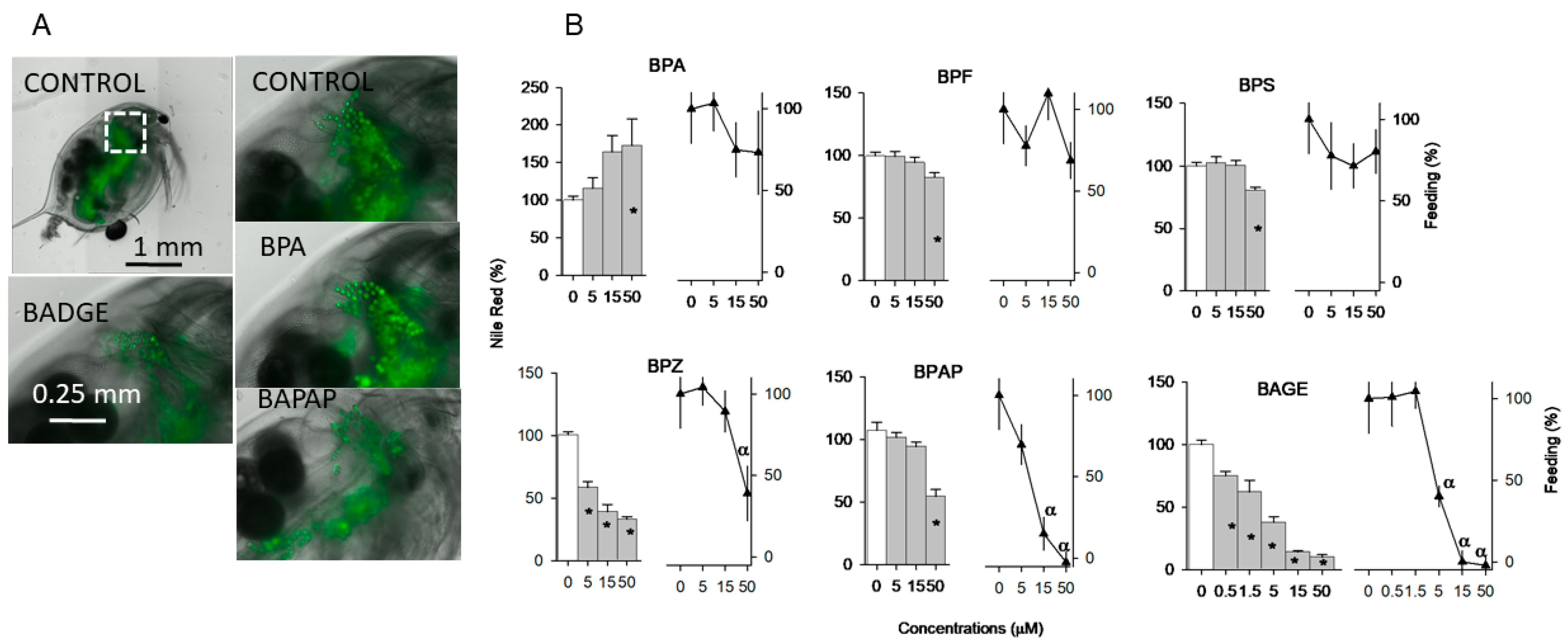
Five of the six tested bisphenols (BPF, BPS, BPZ, BPAP, BADGE) reduced lipid droplet fluorescence in a concentration-related manner, whereas the positive control BPA increased them (Figure 1; Stats in Table A3, Appendix A). Note, however, that BPZ, BPAP, and BADGE also significantly inhibited feeding.
Figure 1.
Accumulation of storage lipids in lipids droplets measured by the Nile Red assay of D. magna individuals exposed to the studied bisphenols. Selected fluorescent images of lipid droplets across treatments (A) and relative fluorescence values relative to controls ((B); %, Mean SE, N = 10). Feeding inhibition responses relative to controls are also depicted as triangles in B. * or α indicate significant treatment differences in % lipid droplet fluorescence or feeding, respectively, against controls following ANOVAs and Dunnett’s tests. Dashed square in the first image of graph A indicates the amplificated area to visualize the lipid droplets in the rest of images.
3.3. Behaviour
Four out of the six tested bisphenols altered at least one of the five studied photomotor responses: BPA, BPF, and BPS significantly impaired (p < 0.05) Max responses mostly at 15 µM. At 50 µM, BPA also significantly impaired (p < 0.05) basal activity and increased VMR. At 0.5 µM, BADGE increased significantly (p < 0.05) Max, and at 1.5 µM impaired basal activity (Table 1; Stats Table A4, Appendix A).
Table 1.
Behavioural results across the studied bisphenols and experiments. Bold values mean significant differences from control values following ANOVA and different from controls. Sample size differences (N) resulted from occasional failure of automated video-tracking and exclusion of incorrect recordings.
3.4. Molecular Responses
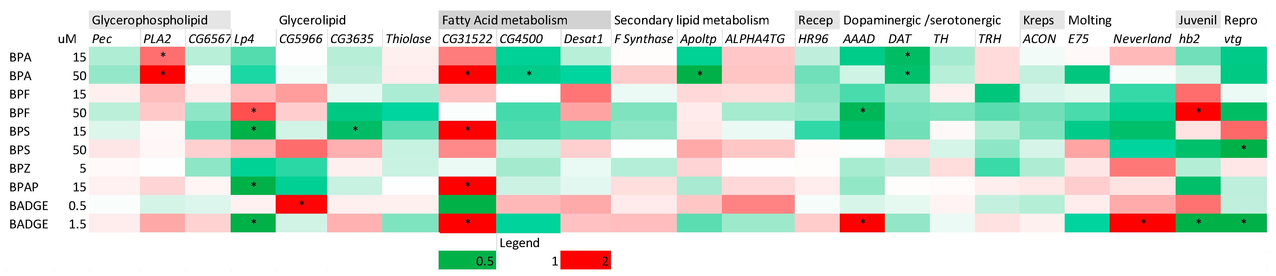
The transcription of several lipid metabolic genes were de-regulated by the selected BPs (Figure 2; Stats in Table A5, Appendix A). BPA that enhanced storage lipid accumulation upregulated the phospholipase gene A (PLA2), that is involved in lipid remodelling, and the fatty acid elongase CG31522. It also downregulated the fatty acid ligase and lipid transporter CG4500 and Adopt.y. All bisphenol substitutes except BPZ reduced lipid storage and induced either up- or downregulation of genes involved in lipid metabolism and endocrine regulation Lp4 (TG lipase), and upregulated CG31522. BPS also downregulated the lipase CG3635 gene and BADGE upregulate the TG lipase CG5966 one. BPA downregulated the dopamine transporter gen DAT and BAGE upregulated the DOPA decarboxylase gene, AAAD and the moulting gene enzyme Neverland. Conversely BADGE also down-regulated the juvenoid-responsive hemoglobin gene hb2 and the reproduction-related gene vtg. BPF upregulated hb2 and downregulated vtg. Interestingly, although not significant, BPA and BPF downregulated vtg.
Figure 2.
Heatmap of gene transcription patterns (fold changes) across the studied BPs. * Significant differences from controls (p < 0.05) following ANOVA and Dunnett’s or Student t tests of Table A5, Appendix A. Boxplots of gene transcriptions are in Figure A1, Appendix A. On the first row, main gene metabolic pathways are depicted. Legend indicates relative fold change colour thresholds.
3.5. Bivariate Correlations Between Apical and Gene Responses
Correlation analyses between apical and molecular responses revealed significant positive associations of storage lipid accumulation with phospholipase A, the lipid transporter gene Apoltp, and negative ones with the phosphocholine cytidyltransferase Pec, and the nuclear receptor HR96 (Figure 3). Non-significant (p < 0.05) but high (>0.5) positive associations also included the lipid-acetyl-CoA-acyltransferase Thiolase and that of the lipid-galactosyltransferase ALPHA4TG. Negative high but non-significant associations included three genes involved in fatty acid metabolism (CG3635, CG4500, and Desat1). Storage lipid accumulation was also negatively correlated with DAT, likely due the contribution of BPA.
Figure 3.
Heatmap of Pearson correlations between apical and gene responses across the tested BPs. * Significant correlations p < 0.05. Lipid, Feed, Max, Hab, and Phototax are, respectively, storage lipids measured as Nile red fluorescence, feeding, habituation, and phototaxis. Legend indicates Pearson correlated values thresholds for colours.
From the tested behavioural traits, maximal responses to light (Max) was positively and significantly related with AAAD but also showed high positive correlations with the rest of the studied neurological related genes, and VMR had the opposite pattern. Max was also significantly and positively related with HR96 and VMR was negatively associated with it. Many behavioural traits were associated with the moulting metabolism gene Neverland as follows: Max showed a positive significant correlation whereas habituation and phototaxis showed a negative one.
4. Discussion
The present study demonstrates that structurally related bisphenols differ markedly in their capacity to disrupt energy metabolism, behaviour, and endocrine-regulated pathways in D. magna. Although most compounds altered storage lipid reserves, the direction and magnitude of responses varied considerably, suggesting different mechanisms of action despite their common bisphenolic structure. Despite the fact that BADGE concentrations declined during exposure, measured concentrations remained within an acceptable range for most treatments and renewal of test solutions likely minimised any influence on biological responses. Importantly, several effects occurred at sublethal concentrations and were associated with transcriptional alterations consistent with disturbances in lipid metabolism, neurophysiology, and hormonal regulation. Overall, our findings indicate that replacement bisphenols cannot be considered environmentally benign alternatives to BPA.
The storage lipid accumulation assay revealed two contrasting modes of action. BPA induced responses consistent with obesogenic-like effects, including increased storage lipid accumulation and regulation of lipid-metabolism genes [24,36], whereas BPF, BPS, BPZ, BPAP, and BADGE decreased storage lipid reserves. BPA results agree with previous findings [11,12,24]. However, interpretation of the storage lipid decrease must consider the concomitant inhibition of feeding observed for several compounds. Reduced food intake is expected to decrease the availability of carbon and energy for triacylglycerol synthesis and storage, thereby contributing indirectly to lower Nile Red fluorescence. Such indirect effects have previously been reported in D. magna exposed to metabolic disruptors and highlight the importance of simultaneously assessing feeding activity when evaluating obesogenic responses. Nevertheless, the lack of a strict correspondence between feeding inhibition and lipid depletion at specific concentrations of BPS (50 µM), BPZ (5 and 15 µM), and BADGE (0.5 and 1.5 µM) suggests that these compounds may also interact directly with lipid metabolic pathways, independently of their effects on food intake. For BADGE, however, interpretation of the Nile Red results should be made with caution at high concentrations since substantial mortality was observed at 15 and 50 µM. Consequently, the severe feeding inhibition recorded at these concentrations likely contributed to the reduction in Nile Red fluorescence, reflecting a depletion of lipid stores rather than a direct effect on lipid metabolism alone.
Previous studies indicate that bisphenol analogues can interfere with lipid homeostasis, although the direction of the response varies among compounds, tissues, and species. BPF has frequently been associated with hepatic lipid accumulation and steatosis-like phenotypes in humans [37], whereas BPS has been reported to either stimulate lipid deposition or reduce lipid levels depending on the biological context in zebrafish [38]. In contrast, BADGE has been described as a PPARγ antagonist capable of suppressing adipogenic processes and reducing lipid storage in mice [39]. These observations support the view that structural modifications among bisphenol analogues can differentially affect lipid synthesis, β-oxidation, and lipid droplet dynamics, potentially explaining the compound-specific patterns of Nile Red responses observed in the present study.
The transcriptional results partly support this interpretation. BPA, the only compound increasing lipid accumulation, induced genes associated with lipid remodelling and fatty acid synthesis, including PLA2 and CG31522, while repressing genes involved in lipid transport and fatty acid activation. Of particular interest is the strong induction of CG31522, a putative fatty-acid elongase-like gene that was also regulated by several bisphenols reducing lipid levels. A previous study identified this gene as responsive to compounds altering lipid homeostasis in Daphnia [24], suggesting that it may re-present a sensitive marker of xenobiotic-induced perturbations of fatty acid metabolism. In contrast, compounds reducing lipid storage frequently regulated lipases (Lp4, CG3635, CG5966) and genes involved in fatty acid metabolism, indicating attempts to compensate for altered energetic status. The significant correlations between lipid accumulation and genes such as PLA2, Apoltp, Pec, and HR96 further support a link between the measured apical response and underlying metabolic regulation. Indeed, the nuclear receptor HR96 is considered to be a master lipid regulator in Daphnia [40,41]. Pec and PLA2 belongs to the Kennedy and Lands cycle of glycerophospholipid remodelling, that is part of the lipid droplet metabolic pathways described in D. magna [24]. In arthropods the lipid transporter protein gene Apoltp transfers diacylglycerols, sterols, and other lipids between lipoprotein particles and target tissues [42].
Although behavioural effects were generally less pronounced than energy metabolic alterations, BPA, BPF, BPS, and BADGE modified one or more photomotor responses. The consistent reduction in maximum locomotor activity by BPA, BPF, and BPS suggests a common mode of action affecting neuronal responsiveness to light stimulation. Interestingly, BPA at the highest concentration simultaneously reduced basal locomotion while increasing VMR, indicating a more complex alteration of sensorimotor integration rather than generalised hypoactivity. Behavioural endpoints are increasingly recognised as highly sensitive measures of contaminant exposure in cladocerans, often responding before toxicity becomes apparent [33]. The relatively selective nature of the observed effects suggests that bisphenols may target specific neuronal pathways involved in stimulus processing rather than causing broad neurological impairment. Supporting the previous argument in larval zebrafish, it was reported that BPA exposure altered pre-pulse inhibition of the C-start reflex, which is equivalent to the D. magna Max parameter, indicating defects in sensory information processing, while general locomotion was unaffected [43]. Developmental BPA exposure significantly altered the optomotor response (OMR), a visually regulated behaviour [44], demonstrating that BPA can modify responsiveness to light-driven visual cues and sensory processing pathways. Furthermore, although not significant, most of the tested BPs decreased D. magna basal locomotion activity, which is in line with the reported defects of BPF on zebrafish spontaneous locomotion and startle responses, which were associated with motor neuron degeneration, myelination defects, and altered neurotransmitter profiles [45].
Molecular responses provide additional evidence supporting neurophysiological effects. BPA reduced expression of the dopamine transporter gene (DAT), whereas BADGE increased expression of AAAD, a key enzyme involved in dopamine and serotonin biosynthesis. Since catecholaminergic signalling participates in the regulation of locomotion and photobehaviour in Daphnia [32,33], disturbances in these pathways could explain, at least partly, the observed alterations in photomotor responses. This interpretation is reinforced by the correlation analysis, where maximum swimming activity (Max) was positively associated with AAAD expression while VMR showed opposite relationships with several neurotransmission-related genes. Although transcriptional changes do not necessarily translate into altered neurotransmitter concentrations, their consistency with behavioural responses suggests that neuroendocrine pathways represent relevant targets of bisphenol exposure.
Particularly interesting were the alterations observed in endocrine-related biomarkers. BADGE significantly increased the expression of Neverland, a key enzyme catalysing the first step of ecdysteroid biosynthesis [46]. In arthropods, Neverland is required for the conversion of cholesterol into 7-dehydrocholesterol and therefore regulates the production of moulting hormones [23,46,47]. Increased expression of this gene may indicate activation of the ecdysteroid pathway or compensatory responses aimed at maintaining hormonal homeostasis. The significant correlations between Neverland expression and behavioural endpoints, including positive associations with maximum swimming activity and phototaxis and negative associations with habituation, suggest a potential link between endocrine regulation and nervous system function. Such interactions are plausible given the known role of ecdysteroids in crustacean development and neuronal differentiation [48]. The ecdysis process itself is a hormonally controlled behavioural programme driven by neuropeptides and ecdysteroids. Changes in ecdysteroid signalling alter expression of neuropeptides involved in motor patterns associated with moulting [49].
The transcription of hb2, a juvenoid-responsive haemoglobin gene [22] varied across the tested bisphenols. Hb2 is frequently used as a biomarker of methyl farnesoate or juvenile hormone-like signalling in Daphnia [22]. BPF significantly up-regulated hb2 whereas BADGE down-regulated it, suggesting that different bisphenols may exert opposite effects on juvenoid-regulated pathways. Because juvenoid hormones regulate not only development and sex determination but also energy allocation and reproduction [36,50,51], disruption of hb2 expression could indicate broader endocrine consequences extending beyond oxygen transport. The concurrent down-regulation of vtg by BPF and BADGE further supports potential endocrine-disrupting activity affecting reproductive physiology. Although vitellogenin responses were modest, combined alterations of hb2, vtg, and Neverland point toward interference with both juvenoid and ecdysteroid signalling systems.
The concentrations used in the present study were selected to elicit sublethal responses affecting lipid metabolism, behaviour, and endocrine-related pathways and hence to study the mechanisms of action. Although the selected concentrations tested are generally above those reported in most surface waters, bisphenol analogues such as BPS, BPF, and BPAF are widely detected in aquatic environments. Reported concentrations range from the low ng L−1 level to the low µg L−1 level in wastewater-impacted systems, while sediments may act as important reservoirs and secondary sources of exposure [27].
5. Conclusions
Taken together, the results indicate that bisphenols affect multiple physiological systems in D. magna, with lipid metabolism appearing as the most sensitive target. Behavioural and molecular responses reveal that these effects are not restricted to energy homeostasis but also involve neurophysiological and endocrine-regulated processes. BPA primarily displayed obesogenic characteristics, whereas most replacement bisphenols reduced lipid reserves and altered feeding behaviour. The transcriptional responses of Neverland, hb2, and vtg additionally suggest that disruption of hormonal signalling may contribute to the observed phenotypes. These findings reinforce growing evidence that BPA substitutes may possess toxicological profiles comparable to, or distinct from, the parent compound and emphasise the need for integrated assessments combining apical and mechanistic endpoints when evaluating the ecological risks of emerging bisphenols.
Author Contributions
Conceptualisation, C.B.; methodology, C.B., A.J.-L., M.G.-M., L.S. and L.N.-M.; formal analysis, C.B., A.J.-L. and M.G.-M.; investigation, C.B., A.J.-L. and M.G.-M.; resources, C.B.; data curation, C.B.; writing—original draft preparation, C.B.; writing—review and editing, C.B.; visualisation, C.B.; supervision, C.B. and L.N.-M.; project administration, C.B.; funding acquisition, C.B. All authors have read and agreed to the published version of the manuscript.
Funding
This work was supported by the grants PID2023-151815OB-I00 funded by Spanish Ministry of Science and Innovation (MCIN), Spain and by the European Partnership for the Assessment of Risks from Chemicals (PARC), Grant agreement ID: 101057014 funded by Horizon Europe.
Institutional Review Board Statement
Not applicable.
Informed Consent Statement
Not applicable.
Data Availability Statement
The original contributions presented in this study are included in the article/Appendix A. Further inquiries can be directed to the corresponding authors.
Conflicts of Interest
The authors declare no conflicts of interest.
Appendix A
Appendix A.1. Chemical Analyses
Water samples were filtered through 0.22 μm PTFE syringe filters prior to analysis. Analyses were performed using an Acquity HPLC system coupled to a Triple Quadrupole Detector (Waters, Milford, MA, USA). Chromatographic separation was achieved using an Acquity UPLC BEH C18 column (100 mm × 2.1 mm, 1.7 μm particle size) fitted with a VanGuard BEH C18 pre-column (5 mm × 2.1 mm, 1.7 μm particle size) (Waters, Milford, MA, USA) operated at 40 °C and a flow rate of 0.4 mL min−1. The mobile phase consisted of water containing 2 mM ammonium fluoride (A) and methanol (B). Elution was performed using a gradient from 40% to 95% B over 8 min, held at 95% B for 3 min, and returned to the initial conditions for column equilibration. The injection volume was 5 μL.
The mass spectrometer was equipped with an electrospray ionization (ESI) source operating in positive and negative ionization modes. BPA, BPF, BPS, BPZ, and BPAP were monitored in ESI− mode, whereas BADGE was monitored in ESI+ mode. Data acquisition was performed in multiple reaction monitoring (MRM) mode. Compound identification was based on retention time and the corresponding MRM transitions optimized using analytical standards. Quantification was performed by external calibration using solvent standards prepared in methanol–water (50:50, v/v). Calibration curves were linear over the concentration range studied (R2 > 0.99). Measured concentrations were within 80–120% of nominal concentrations; therefore, nominal concentrations were used throughout the study.
Appendix A.2. Nile Red Determination
The Nile Red stock solution was prepared in acetone and stored protected from light following previous procedures [31]. Just before use, the working solution was prepared by diluting the stock solution to 1.5 μM in ASTM. Live individuals were then exposed to Nile Red working solution in the dark for 1 h at 20 °C. After incubation, animals were placed in 100 mL ASTM for 1 min to allow clearance of Nile Red residuals. Following clearance, animals were placed individually in 1.5 mL centrifuge tubes, the remaining water was removed, and samples were sonicated in 300 μL of isopropanol. The homogenised extract was then centrifuged at 10,000× g. We used 200 μL of supernatant to measure Nile Red fluorescence using an excitation/emission wavelength of 530/590 nm and a microplate fluorescence reader (Synergy 2, BioTek, Winooski, VT, USA). Each treatment had one animal per sample (10 replicates in total). For each quantification and treatment, 10 blanks (animals not exposed to Nile Red) were used to account for background levels of fluorescence. For fluorescence visualization of lipid droplets in selected treatments in whole animals, animals were exposed in ASTM hard water to Nile Red also for 2 h and then images were obtained with the aid of a microscopy EVOS 70000 (Evos B.V., Amsterdam, The Netherlands) using GFP.
Appendix A.3. Behaviour
Following exposure, organisms were distributed individually to 24 well plate filled with 1 mL of exposure media. Treatments were randomised across plates and 24 replicates per treatment were conducted. Photomotor responses included basal activity under darkness, enhanced photomotor responses upon light stimuli (Max and visual motor responses-VMR), habituation to repetitive stimuli, and negative phototaxis using previous developed assays were conducted in the DanioVision observation chambers (Noldus, Wageningen, The Netherlands) and the software EthoVision® XT 14 was used for video recording, data acquisition, and analyses [32,33,34].
Appendix A.4. Gene Transcription
Extraction and purification of the mRNA content in pools of 5 D. magna were performed using the on-column RNeasy Mini Kit and the RNase-Free DNase Set (QIAGEN, Venlo, The Netherlands) in tandem, following the manufacturer’s instructions. Obtained RNA was quantified and its quality was checked in a NanoDrop D-1000 Spectrophotometer (NanoDrop Technologies, Wilmington, DE, USA). Samples presenting a ratio 230/260–260/280 between 1.9 and 2.1 were selected. RNA integrity was checked using an Agilent 2100 Bioanalyzer (Agilent Technologies, Santa Clara, CA, USA). Only the samples showing RIN values above 9 were further used for qPCR analysis. The reverse transcriptase (RT) step was performed using the Transcriptor First Strand cDNA Synthesis Kit (Roche, Basel, Switzerland) following the manufacturer’s protocol, using 1 µg of RNA and oligo (dT) as the primer. Primers for the studied genes were designed with Primer Quest (IDT Technologies, Coralville, IA, USA) and are listed in the Appendix A section, Table A1. Primer efficiency was determined by the serial dilution [52], being between 95 and 100% for all tested genes. qPCR amplified fragments were purified and concentrated by DNA clean and Concentrator™ 5 (Zymo Research, Irvine, CA, USA) and their sequence was confirmed by capillary sequencing, amplifying fragments in both directions using both forward and reverse primers.
Gene transcription analysis was performed by Quantitative Real-Time Reverse Transcriptase Polymerase Chain Reaction (qRT-PCR) using a high-throughput qPCR equipment, the Biomark X9™ System (Standard BioTools Inc.). In this study, we used a 24.192 Dynamic Array™ IFC (Standard BioTools Inc.) to analyze 65 samples and 24 genes, whose primers are in Table A1. Before IFC analysis, cDNA samples underwent pre-amplification to increase their quantity and ensure targeted gene detection. Preamp Master Mix (Standard BioTools Inc.) was used following the manufacturer’s protocol, and reactions were run in a thermocycler (Mastercycler® X50, Eppendorf AG, Hamburg, Germany) with a programme of 95 °C for 2 min, 12 cycles of 95 °C for 15 s, and 60 °C for 4 min, followed by a hold at 4 °C. Samples were then cleaned with Exonuclease I (Standard BioTools Inc.), processed at 37 °C for 30 min, 80 °C for 15 min, and held at 4 °C, after which were 10-fold diluted. Non-template and RT-minus controls were included to check for primer dimers or DNA contamination. The IFC was then loaded with pre-amplified cDNA, target genes, and necessary reagents, and analyzed using the Biomark X9™ System according to the manufacturer’s instruction. Quantitative PCR was performed using a hot-start activation step at 95 °C for 60 s, followed by 30 amplification cycles consisting of denaturation at 96 °C for 5 s and annealing/extension at 60 °C for 20 s. Fluorescence data were acquired during each annealing/extension step. Amplicon specificity was verified by melting curve analysis from 60 °C to 95 °C, with a temperature increment of 1 °C every 3 s. Relative abundance values of all genes were calculated from the second derivative of their respective amplification curve. Cp values of target genes were compared to the corresponding reference gene (G3PDH), whose CPs varied less than 0.5 cycles across all samples. Results are depicted as fold changes relative to controls ΔCT.
Figure A1.
Boxplot of the transcription patterns of the tested genes across BPs. Different colours differentiate different treatments.
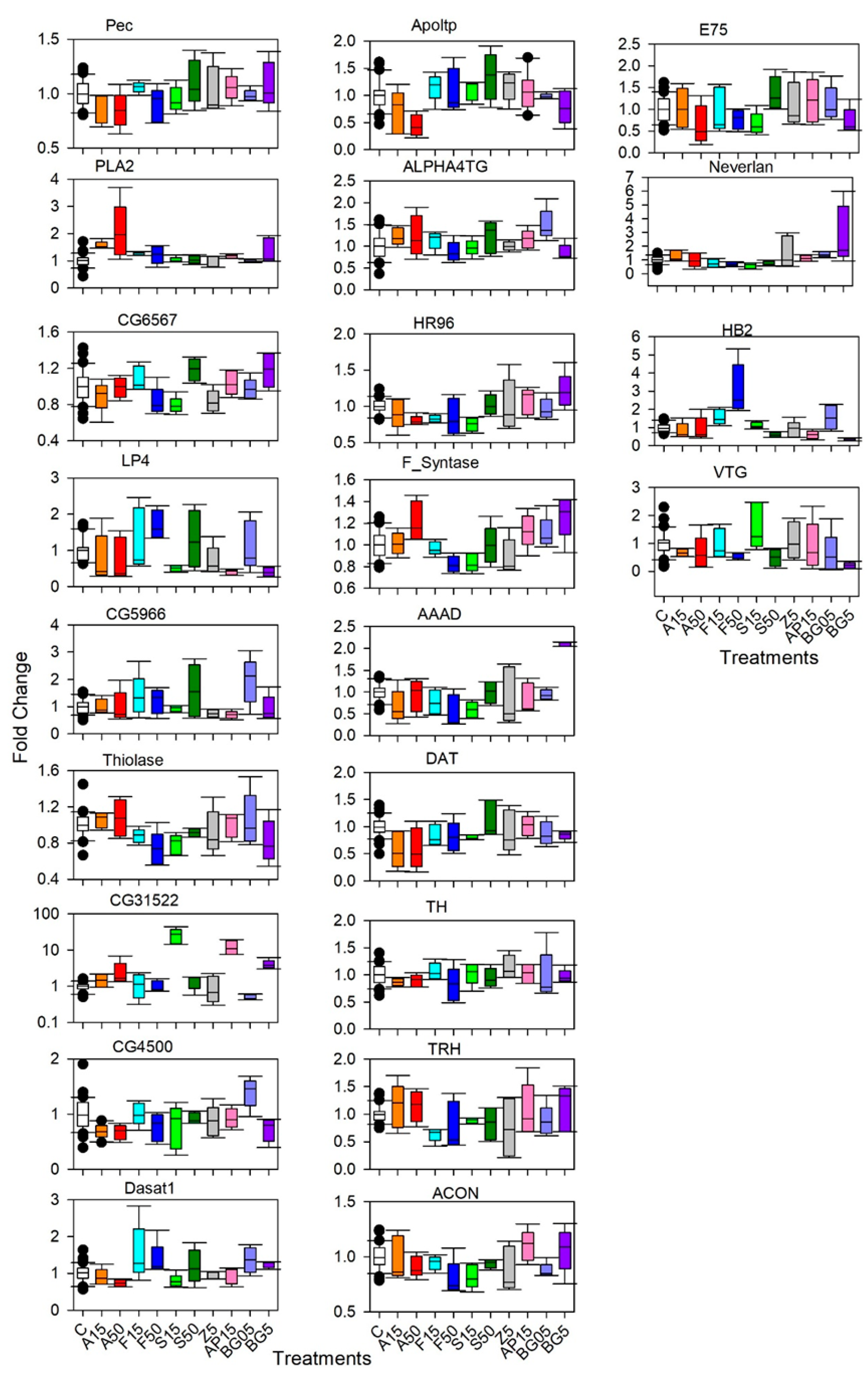
Table A1.
Studied genes: Gene names, accession numbers, primer sequences, efficiency, amplicon sizes, and gene descriptions.
Table A2.
Measured concentrations (µM) of freshly prepared (t0) and old test solutions at 24 band 48 h (t24, t48, respectively). Mean and standard deviation. N = 5.
Table A3.
One way ANOVA results testing for concentrations effects of the studies bisphenols on relative Nile Red fluorescence and feeding responses. Feeding responses of all BPs were tested in a single experiment whereas for Nile Red one experiment was performed for each BPs. df, F, and p are, respectively, degrees of freedom, Fisher’s coefficient, and probability level.
Table A4.
One way ANOVA results testing for concentrations effects of the studied bisphenols on the studied photomotor responses across the three experiments performed. df, F, and p are, respectively, degrees of freedom, Fisher’s coefficient, and probability level.
Table A5.
Statistical results for one way ANOVA or Student t test comparing gene transcription responses of BPs exposed organisms with those of controls. Three different experiments were performed. In the Table legend, the tested treatments in each experiment are depicted as the compound abbreviation and the tested concentration in µM. df, F, and p are, respectively, degrees of freedom, Fisher’s coefficient, and probability level.
References
- Hahladakis, J.N.; Iacovidou, E.; Gerassimidou, S. An Overview of the Occurrence, Fate, and Human Risks of the Bisphenol-A Present in Plastic Materials, Components, and Products. Integr. Environ. Assess. Manag. 2023, 19, 45–62. [Google Scholar] [CrossRef] [Scilit] [PubMed]
- Oehlmann, J.; Schulte-Oehlmann, U.; Kloas, W.; Jagnytsch, O.; Lutz, I.; Kusk, K.O.; Wollenberger, L.; Santos, E.M.; Paull, G.C.; Van Look, K.J.W.; et al. A Critical Analysis of the Biological Impacts of Plasticizers on Wildlife. Philos. Trans. R. Soc. B Biol. Sci. 2009, 364, 2047–2062. [Google Scholar] [CrossRef] [Scilit] [PubMed]
- Pelch, K.; Wignall, J.A.; Goldstone, A.E.; Ross, P.K.; Blain, R.B.; Shapiro, A.J.; Holmgren, S.D.; Hsieh, J.-H.; Svoboda, D.; Auerbach, S.S.; et al. A Scoping Review of the Health and Toxicological Activity of Bisphenol A (BPA) Structural Analogues and Functional Alternatives. Toxicology 2019, 424, 152235. [Google Scholar] [CrossRef] [Scilit] [PubMed]
- Rochester, J.R.; Bolden, A.L. Bisphenol S and F: A Systematic Review and Comparison of the Hormonal Activity of Bisphenol A Substitutes. Environ. Health Perspect. 2015, 123, 643–650. [Google Scholar] [CrossRef] [Scilit] [PubMed]
- Maffini, M.V.; Rubin, B.S.; Sonnenschein, C.; Soto, A.M. Endocrine Disruptors and Reproductive Health: The Case of Bisphenol-A. Mol. Cell. Endocrinol. 2006, 254–255, 179–186. [Google Scholar] [CrossRef] [Scilit] [PubMed]
- Welch, C.; Mulligan, K. Does Bisphenol A Confer Risk of Neurodevelopmental Disorders? What We Have Learned from Developmental Neurotoxicity Studies in Animal Models. Int. J. Mol. Sci. 2022, 23, 2894. [Google Scholar] [CrossRef] [Scilit] [PubMed]
- Legeay, S.; Faure, S. Is Bisphenol A an Environmental Obesogen? Fundam. Clin. Pharmacol. 2017, 31, 594–609. [Google Scholar] [CrossRef] [Scilit] [PubMed]
- Viberg, H.; Fredriksson, A.; Buratovic, S.; Eriksson, P. Dose-Dependent Behavioral Disturbances after a Single Neonatal Bisphenol A Dose. Toxicology 2011, 290, 187–194. [Google Scholar] [CrossRef] [Scilit] [PubMed]
- Molina-Molina, J.-M.; Amaya, E.; Grimaldi, M.; Sáenz, J.-M.; Real, M.; Fernández, M.F.; Balaguer, P.; Olea, N. In Vitro Study on the Agonistic and Antagonistic Activities of Bisphenol-S and Other Bisphenol-A Congeners and Derivatives via Nuclear Receptors. Toxicol. Appl. Pharmacol. 2013, 272, 127–136. [Google Scholar] [CrossRef] [Scilit] [PubMed]
- Heindel, J.J.; Newbold, R.; Schug, T.T. Endocrine Disruptors and Obesity. Nat. Rev. Endocrinol. 2015, 11, 653–661. [Google Scholar] [CrossRef] [Scilit] [PubMed]
- Fuertes, I.; Jordão, R.; Casas, F.; Barata, C. Allocation of Glycerolipids and Glycerophospholipids from Adults to Eggs in Daphnia magna: Perturbations by Compounds That Enhance Lipid Droplet Accumulation. Environ. Pollut. 2018, 242, 1702–1710. [Google Scholar] [CrossRef] [Scilit] [PubMed]
- Jordão, R.; Garreta, E.; Campos, B.; Lemos, M.F.L.; Soares, A.M.V.M.; Tauler, R.; Barata, C. Compounds Altering Fat Storage in Daphnia magna. Sci. Total Environ. 2016, 545–546, 127–136. [Google Scholar] [CrossRef] [Scilit] [PubMed]
- Barata, C.; Baird, D.J. Determining the Ecotoxicological Mode of Action of Toxicants from Measurements on Individuals: Results from Short Duration Chronic Tests with Daphnia magna Straus. Aquat. Toxicol. 2000, 48, 195–209. [Google Scholar] [CrossRef] [Scilit] [PubMed]
- Faria, M.; Barata, C.; Raldúa, D. Behavioral Impairment in Aquatic Organisms Exposed to Neurotoxic Pollutants. Toxics 2022, 10, 243. [Google Scholar] [CrossRef] [Scilit] [PubMed]
- Raldua, D.; Campos, B.; Barata, C.; Piña, B.; García-Reyero, N.; Babin, P.J. Deciphering Emerging Toxicological Effects of Pharmaceuticals on Aquatic Organisms by Using Daphnia magna and Danio rerio as Model Organisms. In Comprehensive Analytical Chemistry; Elsevier B.V.: Amsterdam, The Netherlands, 2013; Volume 62, pp. 611–647. [Google Scholar] [CrossRef] [Scilit]
- Legradi, J.B.; Di Paolo, C.; Kraak, M.H.S.; van der Geest, H.G.; Schymanski, E.L.; Williams, A.J.; Dingemans, M.M.L.; Massei, R.; Brack, W.; Cousin, X.; et al. An Ecotoxicological View on Neurotoxicity Assessment. Environ. Sci. Eur. 2018, 30, 46. [Google Scholar] [CrossRef] [Scilit] [PubMed]
- Raldúa, D.; Piña, B. In Vivo Zebrafish Assays for Analyzing Drug Toxicity. Expert Opin. Drug Metab. Toxicol. 2014, 10, 685–697. [Google Scholar] [CrossRef] [Scilit] [PubMed]
- Orsini, L.; Gilbert, D.; Podicheti, R.; Jansen, M.; Brown, J.B.; Solari, O.S.; Spanier, K.I.; Colbourne, J.K.; Rusch, D.B.; Decaestecker, E.; et al. Daphnia magna Transcriptome by RNA-Seq across 12 Environmental Stressors. Sci. Data 2016, 3, 160030. [Google Scholar] [CrossRef] [Scilit] [PubMed]
- LeBlanc, G.A. Crustacean Endocrine Toxicology: A Review. Ecotoxicology 2007, 16, 61–81. [Google Scholar] [CrossRef] [Scilit] [PubMed]
- Toyota, K.; Miyakawa, H.; Hiruta, C.; Furuta, K.; Ogino, Y.; Shinoda, T.; Tatarazako, N.; Miyagawa, S.; Shaw, J.R.; Iguchi, T. Methyl Farnesoate Synthesis Is Necessary for the Environmental Sex Determination in the Water Flea Daphnia Pulex. J. Insect Physiol. 2015, 80, 22–30. [Google Scholar] [CrossRef] [Scilit] [PubMed]
- Miyakawa, H.; Toyota, K.; Hirakawa, I.; Ogino, Y.; Miyagawa, S.; Oda, S.; Tatarazako, N.; Miura, T.; Colbourne, J.K.; Iguchi, T. A Mutation in the Receptor Methoprene-Tolerant Alters Juvenile Hormone Response in Insects and Crustaceans. Nat. Commun. 2013, 4, 1856. [Google Scholar] [CrossRef] [Scilit] [PubMed]
- Gorr, T.A.; Rider, C.V.; Wang, H.Y.; Olmstead, A.W.; LeBlanc, G.A. A Candidate Juvenoid Hormone Receptor Cis-Element in the Daphnia magna Hb2 Hemoglobin Gene Promoter. Mol. Cell. Endocrinol. 2006, 247, 91–102. [Google Scholar] [CrossRef] [Scilit] [PubMed]
- Sumiya, E.; Ogino, Y.; Toyota, K.; Miyakawa, H.; Miyagawa, S.; Iguchi, T. Neverland Regulates Embryonic Moltings through the Regulation of Ecdysteroid Synthesis in the Water Flea Daphnia magna, and May Thus Act as a Target for Chemical Disruption of Molting. J. Appl. Toxicol. 2016, 36, 1476–1485. [Google Scholar] [CrossRef] [Scilit] [PubMed]
- Fuertes, I.; Jordão, R.; Piña, B.; Barata, C. Time-Dependent Transcriptomic Responses of Daphnia magna Exposed to Metabolic Disruptors That Enhanced Storage Lipid Accumulation. Environ. Pollut. 2019, 249, 99–108. [Google Scholar] [CrossRef] [Scilit] [PubMed]
- Carrillo, M.P.; Moro, H.; Piña, B.; Vila-Costa, M.; Barata, C. Post-Embryonic Consequences of Embryonic Exposure: Mechanisms of Developmental Toxicity Induced by Selective Serotonin Re-Uptake Inhibitors and Tributyltin in Daphnia magna. Environ. Pollut. 2026, 399, 128203. [Google Scholar] [CrossRef] [Scilit] [PubMed]
- Fabrello, J.; Matozzo, V. Bisphenol Analogs in Aquatic Environments and Their Effects on Marine Species—A Review. J. Mar. Sci. Eng. 2022, 10, 1271. [Google Scholar] [CrossRef] [Scilit]
- Liu, J.; Zhang, L.; Lu, G.; Jiang, R.; Yan, Z.; Li, Y. Occurrence, Toxicity and Ecological Risk of Bisphenol A Analogues in Aquatic Environment—A Review. Ecotoxicol. Environ. Saf. 2021, 208, 111481. [Google Scholar] [CrossRef] [Scilit] [PubMed]
- Qian, L.; Chen, C.; Guo, L.; Deng, J.; Zhang, X.; Zheng, J.; Wang, G.; Zhang, X. Developmental and Reproductive Impacts of Four Bisphenols in Daphnia magna. Int. J. Mol. Sci. 2022, 23, 14561. [Google Scholar] [CrossRef] [Scilit] [PubMed]
- Baird, D.J.; Barata, C. Genetic Variation in the Response of Daphnia to Toxic Substances: Implications for Risk Assessment. In Genetics And Ecotoxicology; Forbes, V.E., Ed.; Taylor & Francis: New York, NY, USA, 1998; pp. 207–221. [Google Scholar] [CrossRef] [Scilit]
- ASTM (Ed.) Standard Guide for Conducting Renewal Life-Cycle Toxicity Tests with Daphnia magna. In Annual Book of ASTM Standards, E 1193-94; American Society for Testing and Materials: Philadelphia, PA, USA, 1994; pp. 512–518. [Google Scholar]
- Jordão, R.; Casas, J.; Fabrias, G.; Campos, B.; Piña, B.; Lemos, M.F.L.; Soares, A.M.V.M.; Tauler, R.; Barata, C. Obesogens beyond Vertebrates: Lipid Perturbation by Tributyltin in the Crustacean Daphnia magna. Environ. Health Perspect. 2015, 123, 813–819. [Google Scholar] [CrossRef] [Scilit] [PubMed]
- Bellot, M.; Faria, M.; Gómez-Canela, C.; Raldúa, D.; Barata, C. Pharmacological Modulation of Behaviour, Serotonin and Dopamine Levels in Daphnia magna Exposed to the Monoamine Oxidase Inhibitor Deprenyl. Toxics 2021, 9, 187. [Google Scholar] [CrossRef] [Scilit] [PubMed]
- Bedrossiantz, J.; Martínez-Jerónimo, F.; Bellot, M.; Raldua, D.; Gómez-Canela, C.; Barata, C. A High-Throughput Assay for Screening Environmental Pollutants and Drugs Impairing Predator Avoidance in Daphnia magna. Sci. Total Environ. 2020, 740, 140045. [Google Scholar] [CrossRef] [Scilit] [PubMed]
- Bellot, M.; Gómez-Canela, C.; Barata, C. Phototactic Behaviour and Neurotransmitter Profiles in Two Daphnia magna Clones: Vertical and Horizontal Responses to Fish Kairomones and Psychotropic Drugs. Sci. Total Environ. 2022, 830, 154684. [Google Scholar] [CrossRef] [Scilit] [PubMed]
- Rivetti, C.; López-Perea, J.J.; Laguna, C.; Piña, B.; Mateo, R.; Eljarrat, E.; Barceló, D.; Barata, C. Integrated Environmental Risk Assessment of Chemical Pollution in a Mediterranean Floodplain by Combining Chemical and Biological Methods. Sci. Total Environ. 2017, 583, 248–256. [Google Scholar] [CrossRef] [Scilit] [PubMed]
- Jordão, R.; Campos, B.; Piña, B.; Tauler, R.; Soares, A.M.V.M.; Barata, C. Mechanisms of Action of Compounds That Enhance Storage Lipid Accumulation in Daphnia magna. Environ. Sci. Technol. 2016, 50, 13565–13573. [Google Scholar] [CrossRef] [Scilit] [PubMed]
- Wang, J.; Yu, P.; Xie, X.; Wu, L.; Zhou, M.; Huan, F.; Jiang, L.; Gao, R. Bisphenol F Induces Nonalcoholic Fatty Liver Disease-like Changes: Involvement of Lysosome Disorder in Lipid Droplet Deposition. Environ. Pollut. 2021, 271, 116304. [Google Scholar] [CrossRef] [Scilit] [PubMed]
- Qin, J.-Y.; Ru, S.; Wang, W.; Hao, L.; Wei, S.; Zhang, J.; Xiong, J.-Q.; Wang, J.; Zhang, X. Unraveling the Mechanism of Long-Term Bisphenol S Exposure Disrupted Ovarian Lipids Metabolism, Oocytes Maturation, and Offspring Development of Zebrafish. Chemosphere 2021, 277, 130304. [Google Scholar] [CrossRef] [Scilit] [PubMed]
- Wang, Y.; Pan, Z.; Chen, F. Inhibition of PPARγ by Bisphenol A Diglycidyl Ether Ameliorates Dexamethasone-Induced Osteoporosis in a Mouse Model. J. Int. Med. Res. 2019, 47, 6268–6277. [Google Scholar] [CrossRef] [Scilit] [PubMed]
- Sengupta, N.; Gerard, P.D.; Baldwin, W.S. Perturbations in Polar Lipids, Starvation Survival and Reproduction Following Exposure to Unsaturated Fatty Acids or Environmental Toxicants in Daphnia magna. Chemosphere 2016, 144, 2302–2311. [Google Scholar] [CrossRef] [Scilit] [PubMed]
- Karimullina, E.; Li, Y.; Ginjupalli, G.K.; Baldwin, W.S. Daphnia HR96 Is a Promiscuous Xenobiotic and Endobiotic Nuclear Receptor. Aquat. Toxicol. 2012, 116–117, 69–78. [Google Scholar] [CrossRef] [Scilit] [PubMed]
- Palm, W.; Sampaio, J.L.; Brankatschk, M.; Carvalho, M.; Mahmoud, A.; Shevchenko, A.; Eaton, S. Lipoproteins in Drosophila Melanogaster—Assembly, Function, and Influence on Tissue Lipid Composition. PLoS Genet. 2012, 8, e1002828. [Google Scholar] [CrossRef] [Scilit] [PubMed]
- Scaramella, C.; Alzagatiti, J.B.; Creighton, C.; Mankatala, S.; Licea, F.; Winter, G.M.; Emtage, J.; Wisnieski, J.R.; Salazar, L.; Hussain, A.; et al. Bisphenol A Exposure Induces Sensory Processing Deficits in Larval Zebrafish during Neurodevelopment. eNeuro 2022, 9, ENEURO.0020-22.2022. [Google Scholar] [CrossRef] [Scilit] [PubMed]
- Crowley-Perry, M.; Barberio, A.J.; Zeino, J.; Winston, E.R.; Connaughton, V.P. Zebrafish Optomotor Response and Morphology Are Altered by Transient, Developmental Exposure to Bisphenol-A. J. Dev. Biol. 2021, 9, 14. [Google Scholar] [CrossRef] [Scilit] [PubMed]
- Kim, Y.; Kim, S.S.; Park, B.H.; Hwang, K.-S.; Bae, M.A.; Cho, S.-H.; Kim, S.; Park, H.-C. Mechanism of Bisphenol F Affecting Motor System and Motor Activity in Zebrafish. Toxics 2023, 11, 477. [Google Scholar] [CrossRef] [Scilit] [PubMed]
- Yoshiyama-Yanagawa, T.; Enya, S.; Shimada-Niwa, Y.; Yaguchi, S.; Haramoto, Y.; Matsuya, T.; Shiomi, K.; Sasakura, Y.; Takahashi, S.; Asashima, M.; et al. The Conserved Rieske Oxygenase DAF-36/Neverland Is a Novel Cholesterol-Metabolizing Enzyme. J. Biol. Chem. 2011, 286, 25756–25762. [Google Scholar] [CrossRef] [Scilit] [PubMed]
- Yoshiyama, T.; Namiki, T.; Mita, K.; Kataoka, H.; Niwa, R. Neverland Is an Evolutionally Conserved Rieske-Domain Protein That Is Essential for Ecdysone Synthesis and Insect Growth. Development 2006, 133, 2565–2574. [Google Scholar] [CrossRef] [Scilit] [PubMed]
- Kamiyama, T.; Niwa, R. Transcriptional Regulators of Ecdysteroid Biosynthetic Enzymes and Their Roles in Insect Development. Front. Physiol. 2022, 13, 823418. [Google Scholar] [CrossRef] [Scilit] [PubMed]
- Oliphant, A.; Alexander, J.L.; Swain, M.T.; Webster, S.G.; Wilcockson, D.C. Transcriptomic Analysis of Crustacean Neuropeptide Signaling during the Moult Cycle in the Green Shore Crab, Carcinus Maenas. BMC Genom. 2018, 19, 711. [Google Scholar] [CrossRef] [Scilit] [PubMed]
- Borst, D.W.; Laufer, H.; Landau, M.; Chang, E.S.; Hertz, W.A.; Baker, F.C.; Schooley, D.A. Methyl Farnesoate and Its Role in Crustacean Reproduction and Development. Insect Biochem. 1987, 17, 1123–1127. [Google Scholar] [CrossRef] [Scilit]
- Olmstead, A.W.; Leblanc, G.A. Juvenoid Hormone Methyl Farnesoate Is a Sex Determinant in the Crustacean Daphnia magna. J. Exp. Zool. 2002, 293, 736–739. [Google Scholar] [CrossRef] [Scilit] [PubMed]
- Tichopad, A.; Dilger, M.; Schwarz, G.; Pfaffl, M.W. Standardized Determination of Real-Time PCR Efficiency from a Single Reaction Set-Up. Nucleic Acids Res. 2003, 31, e122. [Google Scholar] [CrossRef] [Scilit] [PubMed]



Disclaimer/Publisher’s Note: The statements, opinions and data contained in all publications are solely those of the individual author(s) and contributor(s) and not of MDPI and/or the editor(s). MDPI and/or the editor(s) disclaim responsibility for any injury to people or property resulting from any ideas, methods, instructions or products referred to in the content. |
© 2026 by the authors. Licensee MDPI, Basel, Switzerland. This article is an open access article distributed under the terms and conditions of the Creative Commons Attribution (CC BY) license.