


Abstract
Micro- and nanoplastics can coexist with lead (Pb) in environmental media, raising the possibility of maternal co-exposure during pregnancy. Polystyrene nanoplastics (PS-NPs), owing to their high specific surface area, can adsorb Pb2+, potentially altering its bioavailability and tissue distribution through a proposed Trojan horse effect. This narrative review integrates evidence from placental, reproductive, and nonplacental models to explore the potential effects of PS-NPs and Pb co-exposure on placental trophoblast cells. Based on the available evidence, we propose a trophoblast-centered, testable framework in which PS-NPs and Pb, whether transiently associated or arriving through separate pathways, may converge at the maternal–fetal interface, promote oxidative stress, disrupt endoplasmic reticulum homeostasis, and shift unfolded protein response signaling toward apoptosis. By linking potential co-exposure pathways with trophoblast stress responses, this framework provides experimentally tractable directions for reproductive toxicology. Direct evidence demonstrating that Pb remains associated with PS-NPs during maternal circulation or within the placenta is limited; thus, the proposed framework should be regarded as a testable hypothesis rather than an established mechanism. Future studies should prioritize environmentally relevant exposure levels, factorial co-exposure designs, particle-specific tracking, and human-relevant trophoblast or placental models to define mixture interactions and establish causal relationships within the proposed pathway.
1. Introduction
The placenta is a critical transient organ located at the maternal–fetal interface. It carries out several key physiological functions essential for fetal development, including nutrient transport, gas exchange and metabolic waste removal. Trophoblast cells constitute major cellular components of the placenta and mediate many of these functions [1]. Trophoblast proliferation, differentiation, fusion, and invasion are tightly regulated and are essential for normal placental development. Conversely, disturbances in trophoblast cell proliferation, differentiation, or invasion can compromise placentation and contribute to adverse pregnancy outcomes, including preeclampsia, recurrent miscarriage, and fetal growth restriction [2]. In recent years, the reproductive toxicity induced by environmental contaminants has gained growing research attention. Epidemiological and toxicological evidence indicates that environmental pollutants, including polycyclic aromatic hydrocarbons and heavy metals, can reach the placenta through the maternal circulation and adversely affect trophoblast and placental function [3].
Under environmental weathering, including solar radiation, thermal oxidation, mechanical stress and biological processes, larger plastic fragments can break down into microplastics (MPs), generally defined as plastic particles ranging from 1 μm to 5 mm [4]. MPs may undergo further degradation and fragmentation to form nanoplastics (NPs), commonly defined as particles smaller than 1 μm [5]. Compared with MPs, NPs are smaller and may have a greater potential for biological-barrier penetration, cellular internalization, and transport within biological systems [6]. These properties may confer greater biological reactivity, thereby elevating the potential risks associated with exposure. Common polymer types in environmental micro- and nanoplastics (MNPs) include polyethylene, polystyrene, and polyvinyl chloride. Among them, polystyrene (PS) is one of the major polymer resins produced worldwide [7], and one of the most commonly reported polymer types in human biomonitoring studies [8]. Therefore, polystyrene nanoplastics (PS-NPs) are widely used as experimental model particles in nanoplastic toxicology. Experimental studies indicate that PS-NPs can reach placental tissue and, under certain exposure conditions, disrupt placental homeostasis and fetal development through oxidative stress, ferroptosis, and other cellular responses [9,10].
Lead (Pb), a persistent heavy metal with well-established neurotoxic and reproductive effects, is also widely distributed in the environment. Pb may cross the placenta by interfering with transport pathways used by essential divalent cations, such as calcium ion (Ca2+). Placental Pb accumulation has been associated with impaired placental function and adverse developmental outcomes in offspring [11].
Under real-world exposure conditions, environmental pollutants rarely exist in a single form, but are more encountered as complex mixtures. Owing to their high specific surface area and surface chemistry, NPs can adsorb metal ions under certain environmental conditions [12]. Such interactions have led to the hypothesis of a potential “Trojan horse” effect, whereby particle association may alter the environmental fate and bioavailability of Pb [13]. However, adsorption observed under environmental or laboratory conditions does not establish that Pb remains associated with PS-NPs in biological fluids or that the two contaminants are transported together to or across the placenta. Although the individual toxic effects of PS-NPs and Pb have been widely investigated, their combined effects have mainly been examined in nonplacental models, including the liver and intestine [14,15]. Direct evidence of combined exposure in placental trophoblast models remains unavailable, and its potential effects at the maternal–fetal interface are therefore unresolved.
Against this background, this review proposes a trophoblast-centered framework to explore the potential effects of combined PS-NPs and Pb exposure at the maternal–fetal interface. Two non-exclusive exposure scenarios are considered: Pb may remain transiently associated with PS-NPs during maternal circulation, or the two contaminants may reach the placenta separately and converge at the trophoblast surface or following cellular uptake. Their combined effects may potentially increase oxidative stress, disturb endoplasmic reticulum (ER) homeostasis, and shift the unfolded protein response (UPR) toward apoptosis signaling. The novelty of this framework does not lie in these stress pathways, but in integrating potential exposure routes with downstream trophoblast injury while distinguishing direct placental evidence from cross-model mechanistic inference. Both the proposed transport routes and the intracellular sequence remain hypotheses that require validation in factorial co-exposure studies.
2. Literature Search Strategy
For this narrative review, structured, non-exhaustive searches were conducted in PubMed and Web of Science. No restriction on publication year was applied, and the final search was performed on 15 October 2026. The principal search terms covered three concept groups: polystyrene nanoplastics (“polystyrene nanoplastics”, “polystyrene nanoparticles”, “PS-NPs”, or “PSNPs”), lead (“lead”, “Pb”, or “lead acetate”), and placental and trophoblast terms (“placenta”, “placental”, “trophoblast”, or “trophoblast cells”). Synonyms within each group were combined using “OR”, and the concept groups were combined using “AND”. Supplementary searches paired terms for PS-NPs and/or Pb with “co-exposure”, “combined exposure”, “maternal exposure”, “pregnancy”, “oxidative stress”, “endoplasmic reticulum stress”, “apoptosis”, and “reproductive toxicity”. Searches addressing human exposure and epidemiological relevance additionally included “microplastics”, “nanoplastics”, “biomonitoring”, and “pregnancy outcomes”. Publications were selected according to their relevance to particle–Pb interactions, maternal exposure and pregnancy outcomes, placental transport, trophoblast injury, or mechanisms relevant to the proposed framework. Original studies using placental or trophoblast models were prioritized, while nonplacental co-exposure studies and studies of either contaminant alone were included as complementary evidence.
3. Environmental Co-Exposure and the Trojan Horse Hypothesis
3.1. Contaminant Mixtures in Environmental Media
In real-world environments, contaminants rarely occur in complete isolation and may instead occur as complex mixtures, a phenomenon commonly termed the “cocktail” effect [16]. MNPs and Pb may co-occur in water, soil, or air. Their presence in the same matrix, however, does not show that Pb is bound to plastic particle surfaces. Field evidence that Pb remains stably associated with environmental plastic particles is still limited.
Laboratory studies have shown that NPs can adsorb metal ions, including Pb2+ [17,18]. Adsorption–desorption behavior is influenced by both solution chemistry and particle properties, including pH, ionic strength, particle size, polymer type, surface charge, surface functionalization, aging, and medium composition. Smaller particles generally provide a larger specific surface area and have shown greater Pb adsorption in the tested microplastic systems [19,20], whereas aggregation can alter the availability of surface binding sites [21].
Aging may roughen particle surfaces and introduce oxygen-containing functional groups, but its net effect on Pb retention varies with the aging process and exposure matrix [19,21,22]. Dissolved organic matter and coexisting ions may enhance or inhibit adsorption through Pb complexation, competition for surface binding sites, or bridging interactions [20,22]. Importantly, these findings do not establish that Pb remains associated with intact NPs in natural waters or biological fluids. They indicate that interactions with plastic particles may alter Pb transport or bioavailability under certain conditions. Results obtained with pristine particles in simplified aqueous media may therefore not represent the behavior of aged particles in environmental or biological matrices.
3.2. Micro- and Nanoplastic-Mediated Pb Transport and Potential Toxicological Interactions
The process by which MNPs serve as carriers to promote the entry of heavy metals into organisms is termed the “Trojan horse” effect [23]. The central premise of this effect is that particle carriers can alter the biodistribution and bioavailability of heavy metals. Uptake of free Pb2+ by organisms may be limited by competition for ion channels, precipitation reactions and complexation reactions. Pb2+ associated with the surfaces of MNPs may be internalized together with the particles through endocytic pathways, potentially altering cellular uptake. However, this possibility has not been established in physiological fluids or placental models.
In a mouse study, co-exposure to PS-NPs and Pb was associated with an increased hepatic Pb burden; however, this finding does not establish transport of intact particle-associated Pb [14]. In vitro, Pb2+ enrichment on PS-NPs enhanced cytotoxicity in 293T and LO2 cells and was associated with oxidative, mitochondrial, and membrane injury [24]. Studies in neuronal models showed that PS-NPs increased cellular or tissue Pb accumulation, and one study implicated clathrin-mediated endocytosis in this effect [25,26]. These findings support the plausibility of a Trojan horse-like uptake process in nonplacental models, but they do not establish persistence of particle-associated Pb in maternal blood, placental transfer, or trophoblast cell uptake.
Beyond potential carrier-mediated delivery, co-exposure to MNPs and Pb may generate combined toxic effects. Intracellular exposure to both contaminants may increase cellular injury because MNPs can induce oxidative stress, membrane disturbance, and mitochondrial dysfunction, whereas Pb can inhibit enzymatic activity, disrupt antioxidant defense systems, and impair deoxyribonucleic acid (DNA) repair. Studies involving co-exposure to silica NPs and Pb have reported additive or synergistic interactions under specific experimental conditions [27]. Similarly, hepatic and intestinal studies of PS-NPs and Pb have reported enhanced or cell-type-specific combined effects [14,15]. Nevertheless, enhanced toxicity following co-exposure should not automatically be interpreted as synergism. Formal synergistic interactions have not been consistently demonstrated across models or endpoints. The nature and magnitude of the interaction may depend on particle physicochemical properties, exposure concentration, mixture ratio, exposure duration, and cell type. Accordingly, the effects of co-exposure to MNPs and Pb should be described as additive, synergistic, or antagonistic only when these interactions have been evaluated using an appropriate factorial design. Such designs should include each contaminant alone and in combination across multiple concentrations and mixture ratios. Observed mixture response should then be compared with predictions from a prespecified additivity model; agreement with the prediction indicates additivity, whereas significant deviations toward greater or lower toxicity support synergism or antagonism, respectively.
3.3. Human Exposure Pathways and Potential Placental Risks
Humans may be co-exposed to MNPs and Pb through ingestion and inhalation, although the extent to which Pb remains associated with MNPs during uptake is still not understood. Dietary ingestion represents a principal potential exposure route, but its relative contribution to total human exposure remains uncertain [6]. Contaminated drinking water, seafood such as shellfish [28], and crops grown in contaminated environments may account for the simultaneous intake of plastic particles and Pb. Nevertheless, the detection of both contaminants within the same food or environmental matrix does not necessarily prove that Pb is directly bound to the surfaces of MNPs.
Inhalation represents another plausible exposure route. Airborne microplastics and metal-containing particulate matter may coexist in environments such as subway air [29]. After inhalation, sufficiently small particles, or chemicals carried or released by them, may cross the alveolar–capillary barrier and enter the systemic circulation [6]. Co-occurrence in air does not mean that Pb remains associated with plastic particles during uptake or circulation. In airway fluids and blood, protein-corona formation and changes in pH or ionic composition may alter PS-NP aggregation and Pb adsorption [19,22,30,31]. Most blood Pb partitions into erythrocytes, and much of the plasma fraction is protein-bound [32]. If a fraction of Pb were to remain associated with PS-NPs in the maternal circulation, the two could, in principle, reach the placenta together. Alternatively, PS-NPs and bioavailable Pb species may reach the placenta through separate pathways, and their effects could converge on shared cellular stress responses in trophoblast cells. Neither particle-associated delivery nor convergence of their effects has been demonstrated directly in placental co-exposure models.
3.4. Human Biomonitoring and Epidemiological Evidence
Plastic material has been detected in several pregnancy-associated human samples. In a study of 12 term pregnancies, MPs were identified in maternal blood, placenta, and umbilical venous blood [33]. A separate analysis of 62 placentas reported plastic polymer concentrations ranging from 6.5 to 685 μg/g [34]. MPs were also detected in placentas from pregnancies affected by intrauterine growth restriction [35].
Maternal Pb exposure is supported by a larger epidemiological evidence base. In the UK Avon Longitudinal Study of Parents and Children (ALSPAC) cohort (n = 4285), maternal whole-blood Pb concentrations averaged 3.67 μg/dL; higher levels were associated with preterm delivery and lower birth weight [36]. A subsequent systematic review concluded that sufficient epidemiological evidence links prenatal Pb exposure with preterm birth [37]. The broader reproductive effects of Pb and other heavy metals have also been reviewed recently [38]. These two bodies of human evidence remain separate. No identified pregnancy cohort has jointly quantified PS-NP and Pb exposure, and no validated biomarker currently provides a size-resolved estimate of maternal PS-NP exposure. Experimental co-exposure studies also have limited relevance to pregnancy. A nonpregnant mouse liver study used 0.1 mg/kg PS-NPs administered by gavage together with 1 g/L Pb in drinking water for four weeks [14], with a nominal Pb concentration far exceeding the WHO provisional drinking-water guideline value of 0.01 mg/L [39]. A zebrafish study used 200 μg/L PS-NPs and 50 μg/L Pb [15]. Differences in species, exposure route, and matrix prevent direct extrapolation from these experimental data to maternal internal or placental doses in humans. These findings therefore support hazard identification and mechanistic plausibility rather than quantitative risk estimates for typical human exposure.
Future pregnancy cohorts could combine maternal whole-blood Pb measurements with contamination-controlled, polymer-specific analyses of maternal blood, placental tissue, and cord blood [33,34,40,41]. Whole-blood Pb is an established exposure biomarker, whereas measurements of MNPs in biological samples remain candidate exposure metrics. Pyrolysis–gas chromatography–mass spectrometry (Py-GC/MS) can estimate polymer mass, while Raman- or Fourier-transform infrared spectroscopy-based methods can characterize detectable particles and polymer types. Further analytical standardization is required, particularly for NPs. Even with these analytical methods, concurrent detection of MNPs and Pb in the same biological samples would indicate co-exposure but would not establish particle-associated Pb transport or validate the proposed co-exposure mechanism.
4. Potential Toxicokinetic Pathways to the Placenta
Defining maternal absorption, systemic distribution, and accumulation is critical for evaluating combined toxicity. Toxicokinetic evidence requires careful interpretation because intravenous administration bypasses absorption barriers, while oral and inhalational exposure more closely represents common environmental routes.
4.1. Maternal Biodistribution and Placental Accumulation of PS-NPs
After oral exposure, a small fraction of PS-NPs may cross the intestinal barrier and enter the systemic circulation, while intravenously administered particles enter the bloodstream directly [42]. After entering the circulatory system, PS-NPs are not uniformly distributed throughout the body, but tend to accumulate in organs rich in reticuloendothelial system components such as liver and spleen. Although most of these particles are trapped by the mononuclear phagocyte system and sequestered in liver and spleen tissues, some PS-NPs escape rapid clearance and reach the placenta. A study in pregnant mice documented that, following intravenous administration of PS-NPs, particle-associated signals could be detected within placental blood vessels and maternal blood sinuses [43]. However, intravenous administration bypasses gastrointestinal and respiratory absorption barriers and delivers a defined particle dose directly into the maternal circulation. Such studies are informative for post-absorption distribution and placental localization but cannot estimate environmental absorption or human placental dose and should be interpreted separately from studies using oral or inhalational exposure [44]. Reported placental accumulation and fetal transfer vary with species, gestational stage, exposure route, particle properties, dose, and detection method. Differences among studies may arise from limitations of the analytical methods used, for example sample contamination, insufficient specificity for particle identification, and difficulties in distinguishing intact particles from free fluorescent markers and particle degradation products. Therefore, such factors should be considered when comparing studies.
4.2. Placental Transport of Pb and the Role of the “Trojan Horse” Effect
After entering the maternal circulation, most blood Pb partitions into erythrocytes, whereas only a small fraction remains in plasma, much of which is protein-bound [32]. Bioavailable Pb in plasma can cross the placenta [11], and its transport may involve calcium-permeable channels or divalent metal transporters. If some Pb remains associated with the surfaces of PS-NPs during maternal circulation, its placental uptake could differ from that of dissolved Pb. Human placental explants have been shown to internalize polystyrene microplastics (PS-MPs) [45], demonstrating the capacity for placental particle uptake but not establishing the co-internalization of particle-associated Pb. If co-internalization occurs, progressive acidification within endolysosomal compartments, from approximately pH 6.5 to 4.5 [46], could protonate surface binding sites and increase competition by H+, thereby shifting the adsorption–desorption equilibrium toward Pb2+ release relative to extracellular conditions [21,47]. However, neither intracellular Pb release nor its kinetics has been demonstrated in trophoblast cells, and the effect may vary with particle surface chemistry, remodeling of the biomolecular corona, ionic composition, and Pb speciation.
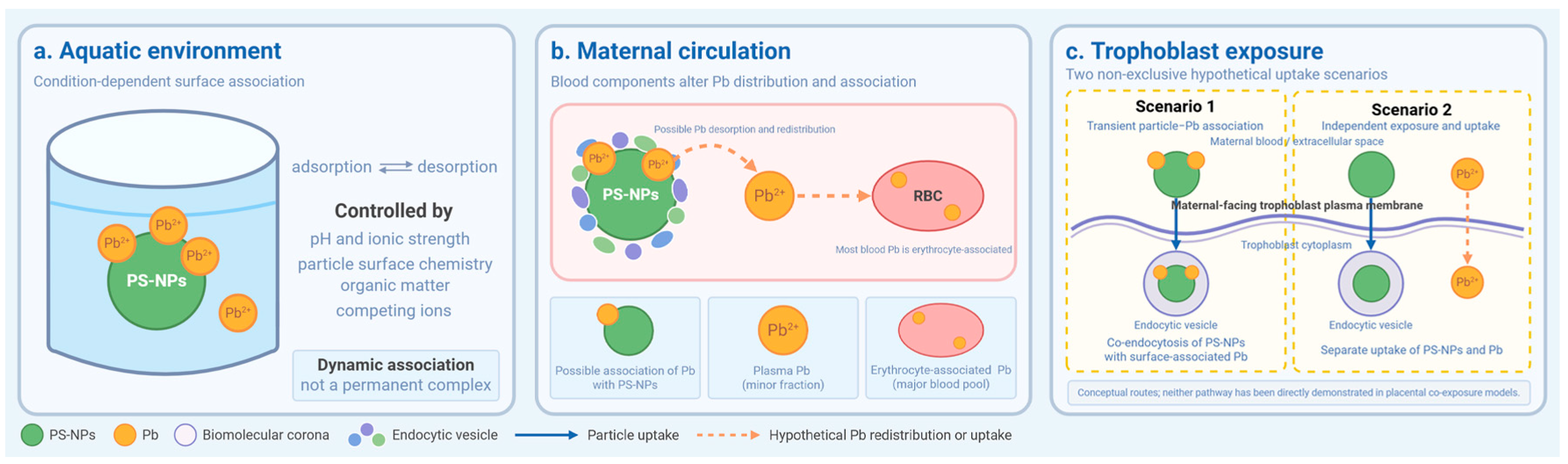
Alternatively, PS-NPs and Pb may enter trophoblast cells through separate pathways. Figure 1 summarizes these two non-exclusive scenarios; neither scenario has been directly demonstrated during co-exposure at the maternal–fetal interface, and the relative contributions of particle-associated and bioavailable Pb remain unknown.
Figure 1.
Reversible interactions between PS-NPs and Pb, and possible routes of trophoblast cell exposure. (a) In aquatic environments, Pb2+ adsorption onto and desorption from PS-NPs depend on pH, ionic strength, particle surface chemistry, organic matter, and competing ions. Particle-Pb association is dynamic rather than a permanent complex. (b) In maternal circulation, biomolecular corona formation and blood components may alter Pb association with PS-NPs. Most blood Pb is associated with erythrocytes, whereas a smaller fraction resides in plasma. Whether surface-associated Pb persists or redistributes between blood compartments remains unknown. (c) Two non-exclusive hypothetical scenarios are illustrated: co-endocytosis of PS-NPs with transiently associated Pb (Scenario 1), and independent uptake of PS-NPs and Pb (Scenario 2). RBC, red blood cell.
4.3. Potential Fetal Distribution
Under some exposure conditions, NPs and bioavailable Pb may cross the placental barrier and enter the fetal compartment. In an animal study, NPs were detected in several fetal organs, including the liver, heart, and brain, following maternal administration [48], although reported translocation varies with particle properties and experimental design.
Therefore, the placenta may represent an early target organ of toxicity rather than the sole target organ. Even when fetal particle burden is low, placental injury may impair nutrients and oxygen transport and thereby indirectly affect fetal development. Direct fetal exposure may also occur if PS-NPs, Pb, or transiently particle-associated Pb cross the barrier. Because the fate of particle-associated Pb remains poorly understood, future studies should combine particle-specific imaging with quantitative Pb analysis and simultaneously examine maternal blood, distinct placental compartments, and fetal tissues.
5. The Placenta as a Potential Target of Combined Toxicity
5.1. Main Functions of the Placenta
The placenta continues to grow and remodel throughout pregnancy to support fetal development. Its functions include nutrient transport, gas exchange, metabolic waste removal, hormone secretion, and immune regulation. The disruption of placental architecture is associated with preeclampsia, fetal growth restriction, and recurrent miscarriage [49].
5.2. The Murine Placental Labyrinth as a Model-Specific Experimental Compartment
In the murine placenta, the labyrinth is the principal site of maternal–fetal exchange and therefore constitutes a relevant experimental compartment for investigating the potential placental effects of co-exposure to PS-NPs and Pb. The interhemal barrier includes a single discontinuous layer of mononuclear sinusoidal trophoblast giant cells lining the maternal blood sinuses, two continuous syncytiotrophoblast layers (SynT-I and SynT-II), and fetal capillary endothelium on the fetal side [50]. Together, these layers regulate the exchange of gases, nutrients, and other substances between the maternal and fetal circulations, including transporter-dependent nutrient transfer [51].
Labyrinth trophoblast cells are located at the maternal–fetal exchange interface and have intensive transport and metabolic activity. These features may increase their sensitivity to disturbances in redox balance and protein homeostasis. However, direct evidence of PS-NPs and Pb co-localization, co-internalization, or combined trophoblast toxicity is currently lacking. Key structural differences between the murine placental labyrinth and the human terminal chorionic villus are summarized in Table 1.
Table 1.
Major structural differences between the murine placental labyrinth and the human terminal chorionic villus.
Although mouse and human placentas are both hemochorial, their exchange barriers are not equivalent. The murine labyrinth contains discontinuous sinusoidal trophoblast giant cells and two syncytiotrophoblast layers, whereas the term human villus contains a single continuous syncytiotrophoblast layer with sparse cytotrophoblasts. These architectural differences may affect particle contact, uptake, and transfer. Findings from the murine labyrinth therefore cannot be directly extrapolated to human placental uptake or barrier crossing.
5.3. Susceptibility to Environmental Contaminants
Normal placental development and homeostasis rely on the coordinated regulation of trophoblast differentiation, fusion, survival, endocrine function, and vascular remodeling. Syncytin proteins participate in cytotrophoblast fusion and syncytiotrophoblast formation [59], while hypoxia-inducible factors and vascular endothelial growth factor regulate cellular responses to oxygen and placental angiogenesis [60]. In humans, interactions between maternal immune cells and extravillous trophoblast cells, including signaling mediated by killer-cell immunoglobulin-like receptors and human leukocyte antigen C, contribute to trophoblast cell invasion and vascular remodeling [61]. These human-specific processes provide important biological context but cannot be directly mapped onto individual trophoblast subtypes within the murine placenta.
This homeostatic balance is highly susceptible to disruption by exogenous pollutants. Experimental studies have shown that PS-NPs can accumulate in placental tissue, including trophoblast cells, and impair trophoblast viability or function [62,63,64]. Exposure to PS-NPs has also been associated with placental oxidative stress, ferroptosis-related signaling, and compromised barrier integrity [65]. Pb can accumulate in placental tissue and cross the placental barrier [66], while maternal Pb exposure has also been associated with an increased risk of preterm birth [37]. In pregnant mice, co-exposure to PS-NPs and Cadmium (Cd) increased placental Cd accumulation and disrupted the uterine–placental micro-environment, with mitochondrial reactive oxygen species (mtROS)–modulate voltage-dependent anion channel 1 (VDAC1)-related mitophagy implicated [67]. Although this study examined Cd rather than Pb, its findings suggest that PS-NPs may alter placental metal accumulation and toxicity. Taken together with studies examining PS-NPs and Pb separately, the available evidence indicates that either contaminant may reach or adversely affect the placenta. However, their combined effects on trophoblast cells remain unknown, including whether co-exposure produces additive, synergistic, antagonistic, or other interactions. Table 2 summarizes evidence relevant to the proposed framework and outlines the key limitations of the underlying studies.
Table 2.
Grouped experimental evidence of the proposed placental trophoblast framework for PS-NPs and Pb exposure.
Evidence category: “Placental/trophoblast” denotes studies of either contaminant or plastic particles in placental or trophoblast models; “Nonplacental” denotes co-exposure studies of PS particles and Pb conducted in nonplacental models; and “Related exposure/stress models” denotes studies involving non-identical but mechanistically relevant exposures or experimental systems. These studies do not constitute direct evidence of PS-NP/Pb co-exposure, nor do they establish cross-species equivalence. None of the studies listed directly examined co-exposure to PS-NPs and Pb in placental trophoblast cells. hCG, human chorionic gonadotropin; IRE1α, inositol-requiring enzyme 1 alpha; NF-κB, nuclear factor kappa B; PS-COOH, carboxylated polystyrene; XBP1s, spliced X-box-binding protein 1.
6. Potential Mechanisms of Co-Exposure
Direct mechanistic evidence from placental trophoblast cells co-exposed to PS-NPs and Pb is currently unavailable. The existing evidence is derived primarily from two sources: placental or trophoblast studies examining either contaminant alone, which support individual components of the proposed pathway, and combined-exposure studies conducted in nonplacental models. In the latter, co-exposure to PS particles and Pb has been associated with redox disturbance and cellular or tissue injury in mouse liver, zebrafish intestine, earthworm coelomocytes, and mouse ovary [14,15,74,82]. In the mouse liver model, co-exposure to PS-NPs and Pb increased hepatic Pb accumulation, oxidative damage, inflammation, and caspase-9/3 activation [14]. The ovarian study identified PKR-like endoplasmic reticulum kinase (PERK)/eukaryotic initiation factor 2α (eIF2α)-associated ER stress and apoptosis following co-exposure to PS microplastics and Pb [82]. These findings provide a rationale for testing an oxidative stress–ER stress–apoptosis sequence in placental trophoblast cells but do not demonstrate this sequence at the maternal–fetal interface. Canonical UPR studies provide additional mechanistic context but should not be interpreted as direct evidence for the proposed co-exposure pathway.
6.1. Oxidative Stress as a Candidate Early Event
Oxidative stress arises when reactive oxygen species (ROS) production exceeds cellular antioxidant capacity. However, an increase in ROS alone does not establish oxidative injury or determine its position within a toxic pathway. Evidence is more informative when ROS accumulation is accompanied by lipid or protein oxidation, impaired antioxidant defenses, or functional injury [83,84,85].
Direct trophoblast studies support the susceptibility of these cells to PS-NP-induced redox disturbance. In HTR-8/SVneo cells, PS-NPs increased intracellular ROS and inflammatory mediator production while reducing cell viability, cell-cycle progression, migration, and invasion [63]. Carboxylated PS particles also caused concentration-dependent DNA damage and impaired migration and invasion [68]. In primary human term trophoblast cells, 20-nm PS-NPs rapidly increased ROS and induced greater cytotoxicity than 100-nm particles [72]. At relatively high concentrations, PS-NPs also increased ROS and reduced mitochondrial membrane potential in Swan.71 cells [64]. In JEG-3 placental cells, smaller and amino-modified PS-NPs generally produced greater cytotoxicity, ROS accumulation, DNA damage, inflammatory responses, cell-cycle arrest, and apoptosis than larger, carboxylated, or unmodified particles [70]. These findings show that PS-NPs can increase ROS and impair human trophoblast function, although they do not establish ROS generation as the initiating event. Pb exposure has likewise been associated with mitochondrial alterations in human trophoblast cell models. Pb reduced mitochondrial DNA copy number in JEG-3, BeWo, and HTR-8/SVneo cells and, under hypoxic conditions, decreased mitochondrial membrane potential in BeWo cells [73]. These cell-line- and condition-dependent findings demonstrate mitochondrial susceptibility to Pb exposure but do not directly establish ROS generation or identify oxidative stress as the primary upstream event.
Excessive ROS can disturb ER redox homeostasis and protein folding, providing a biologically plausible link between oxidative stress to UPR activation [86]. Importantly, this connection has been examined directly in trophoblast models. In trophoblast cells and early placental explants, hydrogen peroxide (H2O2)-induced oxidative stress activated IRE1α/XBP1s signaling and impaired trophoblast function; N-acetyl-L-cysteine attenuated these effects [75]. This study demonstrates that oxidative stress can engage UPR signaling in trophoblast cells, although it did not involve PS-NPs or Pb. Oxidative stress should therefore be regarded as a candidate early event in the proposed co-exposure pathway, pending direct temporal and intervention studies in placental trophoblast co-exposure models.
6.2. ER Stress as a Potential Amplifier
ER stress develops when protein-folding demand exceeds ER capacity, leading to activation of the UPR. The three principal UPR branches—PERK, inositol-requiring enzyme 1 alpha (IRE1α), and activating transcription factor 6 (ATF6)—initially reduce protein-folding pressure and promote cellular adaptation. If ER homeostasis is not restored, sustained PERK/eIF2α/ATF4/C/EBP homologous protein (CHOP) and IRE1α/c-Jun N-terminal kinase (JNK) signaling may shift the response toward apoptosis [87,88,89]. This canonical UPR framework provides mechanistic context but does not demonstrate that co-exposure activates the same sequence in placental trophoblast cells.
Evidence linking plastic-particle exposure to placental ER perturbation has increased. In HTR-8/SVneo cells, PS-NP exposure caused ER dilation and vesiculation, accompanied by loss of membrane-associated ribosomes and other ultrastructural signs of cellular stress [69]. In pregnant mice, PS-MPs induced placental ER stress and apoptosis together with activation of glucose-regulated protein (GRP78)/IRE1α/JNK signaling [78]. Exposure to MNPs also activated PERK/eIF2α/activating transcription factor 4 (ATF4) signaling in mouse placentas and human trophoblast cells, and PERK inhibition partially rescued impaired trophoblast syncytialization [76]. Single-cell transcriptomic analysis further revealed increased expression of ER-stress-related genes in placental trophoblast populations following MNP exposure [79]. Calcium dyshomeostasis may represent an additional mechanism that intersects with ER responses. In mouse placenta and HTR-8/SVneo cells, PS-NPs disrupted Ca2+ homeostasis through signal transducing adaptor molecule 2-associated changes in lysosomal inositol 1,4,5-trisphosphate receptor type 3 (IP3R3) degradation and TRPV6-mediated Ca2+ influx [77], but this study did not establish ER-stress-dependent Ca2+ release or caspase-12 activation. Together, these studies identify ER perturbation as one component of the cellular responses to plastic-particle exposure in placental models, although the evidence ranges from ultrastructural and transcriptomic associations to pathway-level intervention.
Evidence for Pb-associated placental ER perturbation is derived mainly from animal studies. In lead-exposed rats, placental trophoblast degeneration, rough-ER distension, loss of membrane-associated ribosomes, and increased NF-κB expression were reported; in human placentas, NF-κB expression correlated with maternal blood Pb, although structural abnormalities were not detected [80]. A subsequent rat study reported ER swelling, expansion, and vacuolization together with increased caspase-12 mRNA expression [81]. These findings associate Pb exposure with placental ER injury but do not establish ER stress as the mediator of Pb-induced trophoblast injury.
6.3. Apoptosis as a Potential Outcome of Unresolved ER Stress
Placental and trophoblast studies indicate that severe or unresolved ER stress can culminate in apoptosis. Severe ER stress in human placental tissue and JEG-3/JAR cells has been associated with translational repression, impaired proliferation, and CHOP-related apoptotic signaling. In primary human villous trophoblast cells, palmitate activated the UPR and induced CHOP- and caspase-dependent apoptosis [90]. Sustained CHOP signaling can alter the balance of B-cell lymphoma 2 (BCL-2) family proteins and promote downstream caspase activation. In rodents, ER Ca2+ dysregulation may additionally engage calpain and caspase-12; this pathway should be interpreted as species-dependent because functional caspase-12 is absent in most humans [91,92].
Evidence from the individual contaminants further supports apoptosis as a relevant outcome. At relatively high concentrations, PS-NPs increased ROS and apoptosis, reduced mitochondrial membrane potential and BCL-2 expression, and increased cleaved caspase-2/3 in Swan.71 cells; BCL-2 overexpression attenuated apoptosis [64]. In HTR-8/SVneo cells, high concentrations of 50- and 200-nm PS-NPs were associated with cell-cycle alterations, reduced mitochondrial membrane potential, and increased apoptosis, whereas 2-μm PS-MPs produced no comparable apoptotic response [71]. Under hypoxic conditions, Pb reduced mitochondrial membrane potential and increased apoptosis in BeWo cells, although responses varied among trophoblast cell lines [73]. In rat placenta, gestational Pb exposure caused ER swelling, expansion, and vacuolization, accompanied by increased caspase-12 mRNA expression and TUNEL-positive apoptosis [81]. Thus, PS-NPs and Pb can independently engage apoptosis-related processes in trophoblast or placental models, although the upstream pathways are not identical.
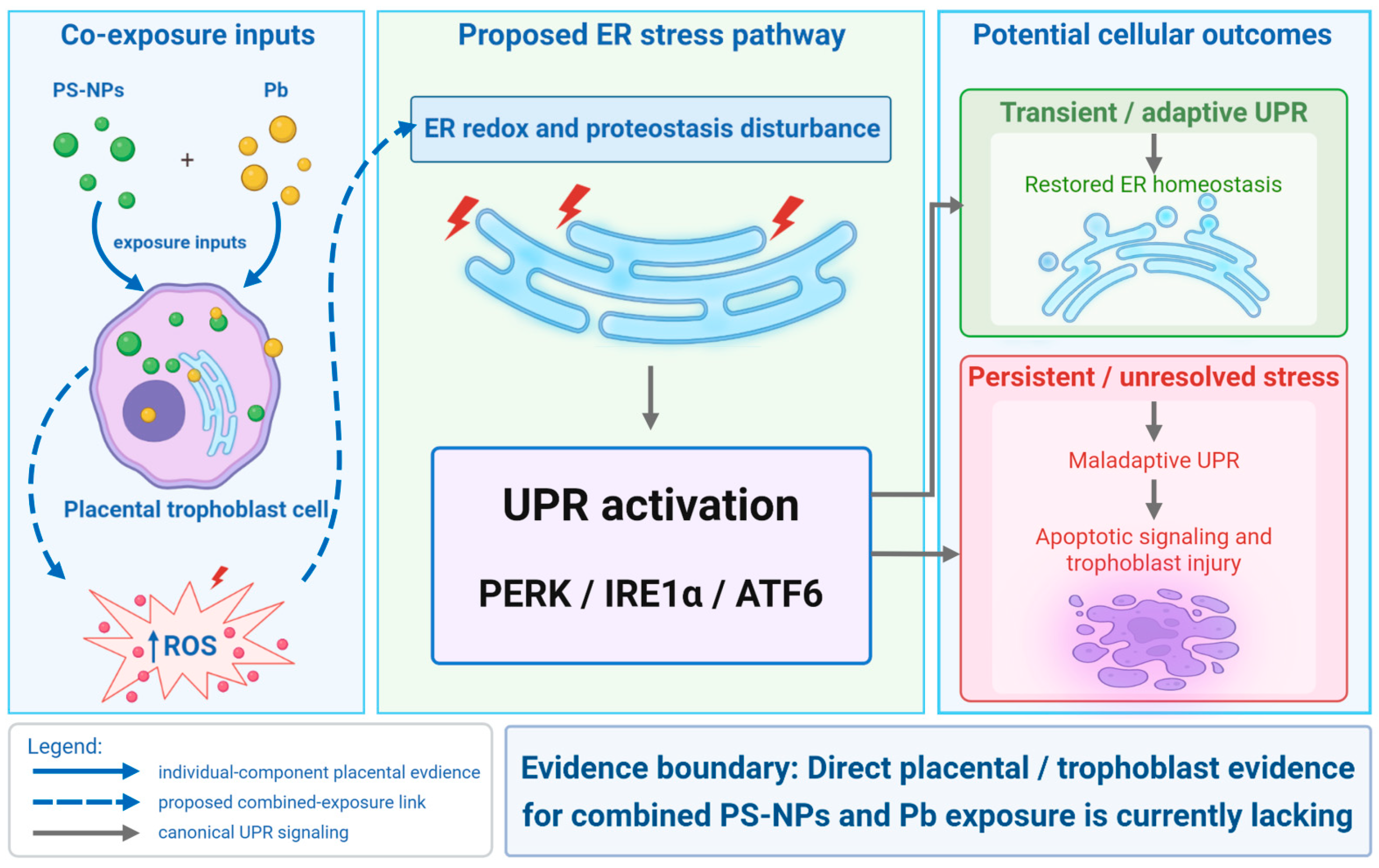
On this basis, we hypothesize that co-exposure to PS-NPs and Pb may intensify redox imbalance, prolong ER stress, and increase the likelihood of trophoblast apoptosis. This sequence, summarized in Figure 2, remains a testable framework rather than an established co-exposure mechanism. Apoptosis is treated as the principal proposed outcome, whereas calcium dyshomeostasis, altered autophagy, inflammation, ferroptosis, and mitochondrial dysfunction may interact with or modify this response. Direct validation will require factorial co-exposure studies with temporal profiling and pathway-specific inhibition or rescue.
Figure 2.
Proposed ER stress pathway for placental trophoblast responses to co-exposure to PS-NPs and Pb. Individual-contaminant studies provide placental or trophoblast evidence for oxidative and related cellular stress responses, but whether PS-NPs and Pb jointly disrupt ER redox balance and proteostasis remains hypothetical. ER disturbance may activate the PERK, IRE1α, and ATF6 branches of the UPR. Transient or adaptive UPR signaling may restore ER homeostasis, whereas persistent stress may favor maladaptive UPR signaling, apoptotic signaling, and trophoblast injury. Solid blue arrows indicate placental or trophoblast evidence from individual-contaminant studies; dashed blue arrows indicate proposed co-exposure links; and gray arrows indicate canonical UPR relationships. The complete sequence has not been directly demonstrated in placental trophoblast co-exposure models.
7. Strengths and Limitations
A key strength of this review is its integration of evidence from environmental toxicology, placental biology, and cellular stress research while explicitly distinguishing direct evidence from cross-model inference. However, no identified study has directly examined combined exposure to PS-NPs and Pb in placental trophoblast cells. The proposed framework therefore relies partly on placental studies of the individual contaminants and on co-exposure evidence from nonplacental models. Translation of these findings to human pregnancy is further constrained by species differences in placental structure and by variation in particle properties, exposure routes, doses, experimental models, and analytical methods. As a structured narrative review, this work is intended to identify mechanistic gaps, and generate testable hypotheses, rather than to establish causality or provide quantitative estimates of human pregnancy risk.
8. Clinical Implications and Priorities for Future Research
Current evidence does not establish a causal link between maternal co-exposure to PS-NPs and Pb and specific adverse pregnancy outcomes, nor does it justify clinical recommendations. Future research should prioritize the generation of evidence linking exposure, placental effects, and pregnancy outcomes. An initial priority is to improve joint exposure assessment by pairing repeated maternal whole-blood Pb measurements with contamination-controlled, polymer-specific analyses of maternal blood, placental tissue, and cord blood. Whole-blood Pb is an established exposure biomarker, whereas plastic-related measurements should currently be regarded as candidate exposure metrics rather than validated biomarkers. Factorial co-exposure studies spanning multiple doses and mixture ratios should then be conducted using primary human trophoblast cells, trophoblast organoids, and placental explants. Primary cells and organoids are suitable for mechanistic testing, whereas explants retain tissue-level organization. Ex vivo placental perfusion and placenta-on-a-chip systems could further be used to evaluate placental retention, barrier function, and maternal–fetal transfer under human-relevant conditions. Observed mixture responses should be compared with predictions from a prespecified additivity model to distinguish additive, synergistic, and antagonistic interactions. Time-course and pathway-intervention studies are also needed to test the proposed oxidative stress–ER stress–apoptosis sequence. Ultimately, prospective pregnancy cohorts linking reliable exposure measurements with placental or neonatal endpoints will be required to determine clinical relevance.
9. Conclusions
PS-NPs and Pb may coexist in environmental media, but no study has directly examined their combined effects in placental trophoblast cells. Pb adsorption to plastic surfaces has been demonstrated under laboratory conditions; however, whether this association persists in maternal blood or alters placental delivery remains unknown. Placental and trophoblast studies show that each contaminant can disrupt cellular homeostasis, whereas nonplacental co-exposure models support, but do not establish, the plausibility of a sequence involving oxidative stress, unresolved ER stress, and apoptosis.
Accordingly, the trophoblast-centered framework proposed here should be regarded as a testable model rather than an established mechanism or an estimate of human pregnancy risk. Validation will require characterization of particle–Pb interactions in biological media, factorial co-exposure designs across relevant doses and mixture ratios, particle-specific tracking, and time-resolved intervention studies in complementary human placental models. Prospective pregnancy cohorts should combine established Pb biomonitoring with contamination-controlled, polymer measurements in paired maternal blood, placental tissue, and cord-blood samples. Such evidence is needed to determine whether PS-NPs and Pb interact at the maternal–fetal interface and whether the proposed pathway has clinical relevance.
Author Contributions
Conceptualization, L.L.; writing—original draft, L.L., Y.Z., F.Z., X.D. and Z.Y.; writing—review and editing, L.L., Y.Z., F.Z., X.D., Z.Y. and G.W.; visualization, G.W. All authors have read and agreed to the published version of the manuscript.
Funding
This work was supported by the PhD Research Startup Fund of Mudanjiang Medical University (2021-MYBSKY036), Mudanjiang Medical University Science Foundation Torch Program (2022-MYHJ-005), Heilongjiang Provincial Natural Science Foundation (PL2025H224), and the Mudanjiang City Key Research and Development Plan Project (HA25CR091).
Institutional Review Board Statement
Not applicable.
Informed Consent Statement
Not applicable.
Data Availability Statement
No new data were created or analyzed in this study. Data sharing is not applicable to this article.
Acknowledgments
During the preparation of this manuscript, the authors used ChatGPT (OpenAI, GPT–5.6) to assist with English-language editing, sentence restructuring, the organization of selected passages, and the preliminary conceptual development of the Graphical Abstract and Figure 1 and Figure 2. No AI-generated graphical elements were incorporated into the final figures. The final artwork was independently redrawn by the authors using BioRender. The authors reviewed and revised all AI-assisted output and take full responsibility for the content of the manuscript.
Conflicts of Interest
The authors declare no conflicts of interest.
References
- Turco, M.Y.; Moffett, A. Development of the human placenta. Development 2019, 146, dev163428. [Google Scholar] [CrossRef] [Scilit]
- Aye, I.L.M.H.; Tong, S.; Charnock-Jones, D.S.; Smith, G.C.S. The human placenta and its role in reproductive outcomes revisited. Physiol. Rev. 2025, 105, 2305–2376. [Google Scholar] [CrossRef] [Scilit]
- Bloom, M.S.; Varde, M.; Newman, R.B. Environmental toxicants and placental function. Best Pract. Res. Clin. Obstet. Gynaecol. 2022, 85, 105–120. [Google Scholar] [CrossRef] [Scilit]
- Lakhiar, I.A.; Yan, H.; Zhang, J.; Wang, G.; Deng, S.; Bao, R.; Zhang, C.; Syed, T.N.; Wang, B.; Zhou, R.; et al. Plastic Pollution in Agriculture as a Threat to Food Security, the Ecosystem, and the Environment: An Overview. Agronomy 2024, 14, 548. [Google Scholar] [CrossRef] [Scilit]
- Gigault, J.; Halle, A.T.; Baudrimont, M.; Pascal, P.Y.; Gauffre, F.; Phi, T.L.; El Hadri, H.; Grassl, B.; Reynaud, S. Current opinion: What is a nanoplastic? Environ. Pollut. 2018, 235, 1030–1034. [Google Scholar] [CrossRef] [Scilit]
- Pan, Y.; Janjua, T.I.; Thomas, K.V.; Shepherd, C.E.; Rauert, C. A critical review of micro- and nanoplastic permeation in the human body. Microplast. Nanoplast. 2026, 6, 21. [Google Scholar] [CrossRef] [Scilit]
- Geyer, R.; Jambeck, J.R.; Law, K.L. Production, use, and fate of all plastics ever made. Sci. Adv. 2017, 3, e1700782. [Google Scholar] [CrossRef] [Scilit]
- Xu, H.; Yu, Z.; Xie, Y. Quantitative human biomonitoring of micro- and nanoplastics: Exposure profiles, mechanistic insights, and health implications. J. Hazard. Mater. 2026, 502, 141054. [Google Scholar] [CrossRef] [Scilit]
- He, Y.; Li, Q.; Sun, Q.; Li, H.; Yu, T.; Chen, M.; Zhang, G.; Zhang, B.; Wang, W.; Ju, S. Gut microbiota contributes to polystyrene nanoplastics-induced fetal growth restriction by disturbing placental nicotinamide metabolism. J. Nanobiotechnol. 2025, 23, 561. [Google Scholar] [CrossRef] [Scilit]
- Chen, Z.; Zheng, M.; Wan, T.; Li, J.; Yuan, X.; Qin, L.; Zhang, L.; Hou, T.; Liu, C.; Li, R. Gestational exposure to nanoplastics disrupts fetal development by promoting the placental aging via ferroptosis of syncytiotrophoblast. Environ. Int. 2025, 197, 109361. [Google Scholar] [CrossRef] [Scilit]
- Goyer, R.A. Transplacental transport of lead. Environ. Health Perspect. 1990, 89, 101–105. [Google Scholar] [CrossRef]
- Li, X.; Hu, S.; Yu, Z.; He, F.; Zhao, X.; Liu, R. New Evidence for the Mechanisms of Nanoplastics Amplifying Cadmium Cytotoxicity: Trojan Horse Effect, Inflammatory Response, and Calcium Imbalance. Environ. Sci. Technol. 2025, 59, 9471–9485. [Google Scholar] [CrossRef] [Scilit]
- Hildebrandt, L.; Nack, F.L.; Zimmermann, T.; Pröfrock, D. Microplastics as a Trojan horse for trace metals. J. Hazard. Mater. Lett. 2021, 2, 100035. [Google Scholar] [CrossRef] [Scilit]
- Yuan, H.; Wen, S.; Zhao, Y.; Hu, L.; Xu, H. Polystyrene nanoplastics exacerbated Pb-induced liver toxicity in mice. Toxicol. Res. 2023, 12, 446–456. [Google Scholar] [CrossRef] [Scilit]
- Yu, J.; Chen, L.; Gu, W.; Liu, S.; Wu, B. Heterogeneity effects of nanoplastics and lead on zebrafish intestinal cells identified by single-cell sequencing. Chemosphere 2022, 289, 133133. [Google Scholar] [CrossRef] [Scilit]
- Cedergreen, N. Quantifying synergy: A systematic review of mixture toxicity studies within environmental toxicology. PLoS ONE 2014, 9, e96580. [Google Scholar] [CrossRef] [Scilit]
- Pokhrel, A.; Islam, M.S.; Mitra, S. Generation of Eroded Nanoplastics from Real World Wastes and Their Capacity for Heavy Metal Adsorption. ACS ES&T Water 2025, 5, 2291–2299. [Google Scholar] [CrossRef] [Scilit]
- Li, T.; Cao, X.; Cui, X.; Zhao, R.; Chen, H.; Xue, W.; Cui, Z.; Tan, X.; Ni, S. Competitive adsorption of lead and cadmium onto nanoplastics with different charges: Two-dimensional correlation spectroscopy study. Environ. Sci. Pollut. Res. 2023, 30, 72884–72899. [Google Scholar] [CrossRef] [Scilit]
- Muthuraja, R.; Ou, B.; Thangavelu, M.; Narhayanan, T.N.; Chittamart, N.; Janjaroen, D. Effects of particle size and aging on heavy metal adsorption by polypropylene and polystyrene microplastics under varying environmental conditions. Chemosphere 2024, 369, 143843. [Google Scholar] [CrossRef] [Scilit]
- Nguyen, T.-B.; Ho, T.-B.-C.; Huang, C.P.; Chen, C.-W.; Chen, W.-H.; Hsieh, S.; Hsieh, S.-L.; Dong, C.-D. Adsorption of lead(II) onto PE microplastics as a function of particle size: Influencing factors and adsorption mechanism. Chemosphere 2022, 304, 135276. [Google Scholar] [CrossRef] [Scilit]
- Blancho, F.; Davranche, M.; Léon, A.; Marsac, R.; Reynauld, S.; Grassl, B.; Gigault, J. Mechanistic description of lead sorption onto nanoplastics. Environ. Sci. Nano 2024, 11, 1671–1681. [Google Scholar] [CrossRef] [Scilit]
- Liu, S.; Huang, J.; Zhang, W.; Shi, L.; Yi, K.; Zhang, C.; Pang, H.; Li, J.; Li, S. Investigation of the adsorption behavior of Pb(II) onto natural-aged microplastics as affected by salt ions. J. Hazard. Mater. 2022, 431, 128643. [Google Scholar] [CrossRef] [Scilit]
- Zhao, W.G.; Tian, Y.M.; Zhao, P.; Zhao, L.A.; Jin, C. Research Progress on Trojan-horse Effect of Microplastics and Heavy Metals in Freshwater Environment. Huan Jing Ke Xue 2023, 44, 1244–1257. [Google Scholar] [CrossRef] [Scilit]
- Li, Y.; Li, Y.; Li, J.; Song, Z.; Zhang, C.; Guan, B. Toxicity of polystyrene nanoplastics to human embryonic kidney cells and human normal liver cells: Effect of particle size and Pb2+ enrichment. Chemosphere 2023, 328, 138545. [Google Scholar] [CrossRef] [Scilit]
- Li, Q.; Zhang, Y.; Hu, Y.; Liu, Z.; Cui, S.; Wei, D.; Yue, L.; Du, Q.; Liu, T.; Huang, D.; et al. The mitochondrial fission-mitophagy axis drives neuronal S-phase arrest and hippocampal damage in co-exposure to polystyrene nanoplastics and lead. Toxicol. Lett. 2026, 420, 111896. [Google Scholar] [CrossRef] [Scilit]
- Yue, L.; Li, Q.; Liu, T.; Du, Q.; Hu, Y.; Wei, D.; Huang, D.; Qi, Y. Polystyrene nanoplastics enhance lead-induced ferroptosis in HT22 cells via clathrin-mediated endocytosis. Toxicology 2026, 527, 154591. [Google Scholar] [CrossRef] [Scilit]
- Lu, C.F.; Yuan, X.Y.; Li, L.Z.; Zhou, W.; Zhao, J.; Wang, Y.M.; Peng, S.Q. Combined exposure to nano-silica and lead induced potentiation of oxidative stress and DNA damage in human lung epithelial cells. Ecotoxicol. Environ. Saf. 2015, 122, 537–544. [Google Scholar] [CrossRef] [Scilit]
- Tanaviyutpakdee, P.; Karnpanit, W. Exposure Assessment of Heavy Metals and Microplastic-like Particles from Consumption of Bivalves. Foods 2023, 12, 3018. [Google Scholar] [CrossRef] [Scilit]
- Roy, D.; Murmu, M.; Mandal, S.; Banerjee, P.; Lee, M.; Park, J. Microplastics bound to fine particulate matter in subway air: A pathway for toxic metal transport to enhanced carcinogenicity in human lungs. J. Hazard. Mater. 2026, 501, 140721. [Google Scholar] [CrossRef] [Scilit]
- Li, X.; He, E.; Jiang, K.; Peijnenburg, W.J.G.M.; Qiu, H. The crucial role of a protein corona in determining the aggregation kinetics and colloidal stability of polystyrene nanoplastics. Water Res. 2021, 190, 116742. [Google Scholar] [CrossRef] [Scilit]
- Sadiq, A.; Litti, L.; Bogialli, S.; Arrigoni, G.; Pastore, P.; Abdolahpur Monikh, F. The influence of nanoplastics’ surface charge on the formation of protein corona and the subsequent sorption of Cd2+ and Pb2+ ions. Colloids Surf. C Environ. Asp. 2026, 4, 100088. [Google Scholar] [CrossRef] [Scilit]
- Ong, C.N.; Lee, W.R. Distribution of lead-203 in human peripheral blood in vitro. Occup. Environ. Med. 1980, 37, 78–84. [Google Scholar] [CrossRef] [Scilit]
- Sun, H.; Su, X.; Mao, J.; Liu, Y.; Li, G.; Du, Q. Microplastics in maternal blood, fetal appendages, and umbilical vein blood. Ecotoxicol. Environ. Saf. 2024, 287, 117300. [Google Scholar] [CrossRef] [Scilit]
- Garcia, M.A.; Liu, R.; Nihart, A.; El Hayek, E.; Castillo, E.; Barrozo, E.R.; Suter, M.A.; Bleske, B.; Scott, J.; Forsythe, K.; et al. Quantitation and identification of microplastics accumulation in human placental specimens using pyrolysis gas chromatography mass spectrometry. Toxicol. Sci. 2024, 199, 81–88. [Google Scholar] [CrossRef] [Scilit]
- Amereh, F.; Amjadi, N.; Mohseni-Bandpei, A.; Isazadeh, S.; Mehrabi, Y.; Eslami, A.; Naeiji, Z.; Rafiee, M. Placental plastics in young women from general population correlate with reduced foetal growth in IUGR pregnancies. Environ. Pollut. 2022, 314, 120174. [Google Scholar] [CrossRef] [Scilit]
- Taylor, C.M.; Golding, J.; Emond, A.M. Adverse effects of maternal lead levels on birth outcomes in the ALSPAC study: A prospective birth cohort study. BJOG 2015, 122, 322–328. [Google Scholar] [CrossRef] [Scilit]
- Eaves, L.A.; Lodge, E.K.; Rohin, W.R.; Roell, K.R.; Manuck, T.A.; Fry, R.C. Prenatal metal(loid) exposure and preterm birth: A systematic review of the epidemiologic evidence. J. Expo. Sci. Environ. Epidemiol. 2025, 35, 696–708. [Google Scholar] [CrossRef] [Scilit]
- Marconi, G.; Di Resta, C.; Naclerio, A.; Banfi, G.; Tomaiuolo, R. Heavy metals and human reproductive toxicity: Mechanisms, pregnancy outcomes, and mitigation strategies. Reprod. Toxicol. 2026, 139, 109104. [Google Scholar] [CrossRef] [Scilit]
- World Health Organization. Guidelines for Drinking-Water Quality: Fourth Edition Incorporating the First and Second Addenda; World Health Organization: Geneva, Switzerland, 2022. [Google Scholar]
- Barbosa, F.; Tanus-Santos, J.E.; Gerlach, R.F.; Parsons, P.J. A Critical Review of Biomarkers Used for Monitoring Human Exposure to Lead: Advantages, Limitations, and Future Needs. Environ. Health Perspect. 2005, 113, 1669–1674. [Google Scholar] [CrossRef] [Scilit]
- Durkin, A.M.; Zou, R.; Boucher, J.M.; Boyles, M.S.P.; van Boxel, J.; Bustamante, M.; Christopher, E.A.; Dadvand, P.; Dusza, H.M.; van Duursen, M.; et al. Investigating Exposure and Hazards of Micro- and Nanoplastics During Pregnancy and Early Life (AURORA Project): Protocol for an Interdisciplinary Study. JMIR Res. Protoc. 2024, 13, e63176. [Google Scholar] [CrossRef] [Scilit]
- Zhang, H.J.; Li, S.; Wang, X.L.; Zhang, K.D.; Fang, H.T.; Wu, X.; Huang, Z.; Jiang, W.; Yang, L.; Tan, Q.G.; et al. Size-dependent translocation of polystyrene nanoplastics across biological barriers in mammals. Nat. Commun. 2025, 17, 1116. [Google Scholar] [CrossRef] [Scilit]
- Kenesei, K.; Murali, K.; Czeh, A.; Piella, J.; Puntes, V.; Madarasz, E. Enhanced detection with spectral imaging fluorescence microscopy reveals tissue- and cell-type-specific compartmentalization of surface-modified polystyrene nanoparticles. J. Nanobiotechnol. 2016, 14, 55. [Google Scholar] [CrossRef] [Scilit]
- Bongaerts, E.; Nawrot, T.S.; Van Pee, T.; Ameloot, M.; Bové, H. Translocation of (ultra)fine particles and nanoparticles across the placenta; a systematic review on the evidence of in vitro, ex vivo, and in vivo studies. Part. Fibre Toxicol. 2020, 17, 56. [Google Scholar] [CrossRef] [Scilit]
- Marques, A.L.; Weingrill, R.; Ospina-Prieto, S.; Pires, K.; Cavalcante, I.; Sousa, A.K.; Cavalcante, I.C.; Oliveira, L.; Souza, S.; Fonseca, E.J.; et al. Polystyrene microplastics internalization by term placental chorionic villi explants. Placenta 2026, 173, 102–110. [Google Scholar] [CrossRef] [Scilit]
- Hu, Y.-B.; Dammer, E.B.; Ren, R.-J.; Wang, G. The endosomal-lysosomal system: From acidification and cargo sorting to neurodegeneration. Transl. Neurodegener. 2015, 4, 18. [Google Scholar] [CrossRef] [Scilit]
- Lu, X.; Zeng, F.; Wei, S.; Gao, R.; Abdurahman, A.; Wang, H.; Liang, W. Effects of humic acid on Pb2+ adsorption onto polystyrene microplastics from spectroscopic analysis and site energy distribution analysis. Sci. Rep. 2022, 12, 8932. [Google Scholar] [CrossRef] [Scilit]
- Fournier, S.B.; D’Errico, J.N.; Adler, D.S.; Kollontzi, S.; Goedken, M.J.; Fabris, L.; Yurkow, E.J.; Stapleton, P.A. Nanopolystyrene translocation and fetal deposition after acute lung exposure during late-stage pregnancy. Part. Fibre Toxicol. 2020, 17, 55. [Google Scholar] [CrossRef] [Scilit]
- Shao, X.; Yu, W.; Yang, Y.; Wang, F.; Yu, X.; Wu, H.; Ma, Y.; Cao, B.; Wang, Y.L. The mystery of the life tree: The placentas. Biol. Reprod. 2022, 107, 301–316. [Google Scholar] [CrossRef] [Scilit]
- Elmore, S.A.; Cochran, R.Z.; Bolon, B.; Lubeck, B.; Mahler, B.; Sabio, D.; Ward, J.M. Histology Atlas of the Developing Mouse Placenta. Toxicol. Pathol. 2022, 50, 60–117, Correction in Toxicol. Pathol. 2023, 53, 153. https://doi.org/10.1177/01926233231175051. [Google Scholar] [CrossRef] [Scilit]
- Watson, E.D.; Cross, J.C. Development of structures and transport functions in the mouse placenta. Physiology 2005, 20, 180–193. [Google Scholar] [CrossRef] [Scilit]
- Georgiades, P.; Ferguson-Smith, A.C.; Burton, G.J. Comparative Developmental Anatomy of the Murine and Human Definitive Placentae. Placenta 2002, 23, 3–19. [Google Scholar] [CrossRef] [Scilit]
- Woods, L.; Perez-Garcia, V.; Hemberger, M. Regulation of Placental Development and Its Impact on Fetal Growth—New Insights From Mouse Models. Front. Endocrinol. 2018, 9, 570. [Google Scholar] [CrossRef] [Scilit]
- Carter, A.M. Animal Models of Human Placentation—A Review. Placenta 2007, 28, S41–S47. [Google Scholar] [CrossRef] [Scilit]
- Burton, G.J.; Fowden, A.L. The placenta: A multifaceted, transient organ. Philos. Trans. R. Soc. B Biol. 2015, 370, 20140066. [Google Scholar] [CrossRef] [Scilit]
- Mori, M.; Ishikawa, G.; Luo, S.-S.; Mishima, T.; Goto, T.; Robinson, J.M.; Matsubara, S.; Takeshita, T.; Kataoka, H.; Takizawa, T. The Cytotrophoblast Layer of Human Chorionic Villi Becomes Thinner but Maintains Its Structural Integrity During Gestation1. Biol. Reprod. 2007, 76, 164–172. [Google Scholar] [CrossRef] [Scilit]
- Shibata, S.; Kobayashi, E.H.; Kobayashi, N.; Oike, A.; Okae, H.; Arima, T. Unique features and emerging in vitro models of human placental development. Reprod. Med. Biol. 2020, 19, 301–313. [Google Scholar] [CrossRef] [Scilit]
- Blundell, C.; Tess, E.R.; Schanzer, A.S.R.; Coutifaris, C.; Su, E.J.; Parry, S.; Huh, D. A microphysiological model of the human placental barrier. Lab Chip 2016, 16, 3065–3073. [Google Scholar] [CrossRef] [Scilit]
- Wang, Q.; Shi, Y.; Bian, Q.; Zhang, N.; Wang, M.; Wang, J.; Li, X.; Lai, L.; Zhao, Z.; Yu, H. Molecular mechanisms of syncytin-1 in tumors and placental development related diseases. Discov. Oncol. 2023, 14, 104. [Google Scholar] [CrossRef] [Scilit]
- Ietta, F.; Wu, Y.; Winter, J.; Xu, J.; Wang, J.; Post, M.; Caniggia, I. Dynamic HIF1A regulation during human placental development. Biol. Reprod. 2006, 75, 112–121. [Google Scholar] [CrossRef] [Scilit]
- Papuchova, H.; Meissner, T.B.; Li, Q.; Strominger, J.L.; Tilburgs, T. The Dual Role of HLA-C in Tolerance and Immunity at the Maternal-Fetal Interface. Front. Immunol. 2019, 10, 2730. [Google Scholar] [CrossRef] [Scilit]
- Wang, B.; Xie, X.; Fan, N.; Deng, Q.; Shi, N.; Long, D.; Huang, W.; Zhu, S.; Chen, Z.; Cheng, X.; et al. Polystyrene Nanoparticles Disrupt Oxidative Phosphorylation and Impair Placental Development in Mice. Toxics 2026, 14, 158. [Google Scholar] [CrossRef] [Scilit]
- Hu, J.; Zhu, Y.; Zhang, J.; Xu, Y.; Wu, J.; Zeng, W.; Lin, Y.; Liu, X. The potential toxicity of polystyrene nanoplastics to human trophoblasts in vitro. Environ. Pollut. 2022, 311, 119924. [Google Scholar] [CrossRef] [Scilit]
- Wan, S.; Wang, X.; Chen, W.; Wang, M.; Zhao, J.; Xu, Z.; Wang, R.; Mi, C.; Zheng, Z.; Zhang, H. Exposure to high dose of polystyrene nanoplastics causes trophoblast cell apoptosis and induces miscarriage. Part. Fibre Toxicol. 2024, 21, 13. [Google Scholar] [CrossRef] [Scilit]
- He, Z.; Cai, J.; Liu, R.; Yan, W.; Yang, G.; Yu, R.; Xin, L.; Wan, Z. Polystyrene nanoparticles induced adverse pregnancy outcomes via the activation of placental ferroptosis and gut microbiota dysfunction. Reprod. Toxicol. 2026, 139, 109113. [Google Scholar] [CrossRef] [Scilit]
- Orct, T.; Sekovanic, A.; Kljakovic-Gaspic, Z. A Three-Decade Overview of Cadmium and Lead in Placentas of Postpartum Women: A Review of Evidence from Croatia (1990s–2019). Life 2026, 16, 343. [Google Scholar] [CrossRef] [Scilit]
- Zhang, R.; Nie, P.; Fu, F. Polystyrene nanoplastics exacerbated the toxicity of cadmium: From the perspective of the placenta-uterus micro-environment. Free Radic. Biol. Med. 2026, 254, 690–700. [Google Scholar] [CrossRef] [Scilit]
- Nacka-Aleksić, M.; Vilotić, A.; Pirković, A.; Živanović, M.; Ljujić, B.; Jovanović Krivokuća, M. Nano-scale dangers: Unravelling the impact of nanoplastics on human trophoblast invasion. Chem. Biol. Interact. 2025, 405, 111317. [Google Scholar] [CrossRef] [Scilit]
- Ragusa, A.; Cristiano, L.; Di Vinci, P.; Familiari, G.; Nottola, S.A.; Macchiarelli, G.; Svelato, A.; De Luca, C.; Rinaldo, D.; Neri, I.; et al. Artificial plasticenta: How polystyrene nanoplastics affect in-vitro cultured human trophoblast cells. Front. Cell Dev. Biol. 2025, 13, 1539600. [Google Scholar] [CrossRef] [Scilit]
- Shen, F.; Li, D.; Guo, J.; Chen, J. Mechanistic toxicity assessment of differently sized and charged polystyrene nanoparticles based on human placental cells. Water Res. 2022, 223, 118960. [Google Scholar] [CrossRef] [Scilit]
- Wan, D.; Liu, Y.; Chang, Q.; Liu, Z.; Wang, Q.; Niu, R.; Gao, B.; Guan, Q.; Xia, Y. Micro/Nanoplastic Exposure on Placental Health and Adverse Pregnancy Risks: Novel Assessment System Based upon Targeted Risk Assessment Environmental Chemicals Strategy. Toxics 2024, 12, 553. [Google Scholar] [CrossRef] [Scilit]
- Poinsignon, L.; Lefrère, B.; Ben Azzouz, A.; Chissey, A.; Colombel, J.; Djelidi, R.; Ferecatu, I.; Fournier, T.; Beaudeux, J.-L.; Lespes, G.; et al. Exposure of the human placental primary cells to nanoplastics induces cytotoxic effects, an inflammatory response and endocrine disruption. J. Hazard. Mater. 2025, 490, 137713. [Google Scholar] [CrossRef] [Scilit]
- Dai, Y.; Xu, X.; Huo, X.; Schuitemaker, J.H.N.; Faas, M.M. Differential effect of lead and cadmium on mitochondrial function and NLRP3 inflammasome activation in human trophoblast. J. Physiol. 2026, 604, 4225–4246. [Google Scholar] [CrossRef] [Scilit]
- Guo, S.; Shi, H.; Qi, Y.; Tian, G.; Wang, T.; He, F.; Li, X.; Liu, R. Environmental relevant concentrations of polystyrene nanoplastics and lead co-exposure triggered cellular cytotoxicity responses and underlying mechanisms in Eisenia fetida. Sci. Total Environ. 2023, 905, 167264. [Google Scholar] [CrossRef] [Scilit]
- Mukherjee, I.; Dhar, R.; Singh, S.; Sharma, J.B.; Nag, T.C.; Mridha, A.R.; Jaiswal, P.; Biswas, S.; Karmakar, S. Oxidative stress-induced impairment of trophoblast function causes preeclampsia through the unfolded protein response pathway. Sci. Rep. 2021, 11, 18415. [Google Scholar] [CrossRef] [Scilit]
- Cheng, Y.; Li, Y.; Zhang, Y.; Liu, H.; Yang, B.; Zhu, J.; Kuang, H. Gestational exposure to micro- and nanoplastics leads to poor pregnancy outcomes by impairing placental trophoblast syncytialization. Environ. Pollut. 2025, 381, 126520. [Google Scholar] [CrossRef] [Scilit]
- Chen, Z.; Yin, X.; Geng, Y.-Q.; Gao, R.; Zhang, Y.; Ma, Y.; Mu, X.; Chen, X.; Li, F.; He, J. Subchronic Exposure to Polystyrene Nanoplastics Disrupts Placental Development and Calcium Homeostasis: Insights from In Vivo and In Vitro Models. ACS Nano 2025, 19, 13825–13841. [Google Scholar] [CrossRef] [Scilit]
- Bai, J.; Wang, Y.; Deng, S.; Yang, Y.; Chen, S.; Wu, Z. Microplastics caused embryonic growth retardation and placental dysfunction in pregnant mice by activating GRP78/IRE1alpha/JNK axis induced apoptosis and endoplasmic reticulum stress. Part. Fibre Toxicol. 2024, 21, 36. [Google Scholar] [CrossRef] [Scilit]
- Zhang, T.; Tian, H.; Teng, Y.; Duan, Z.; Dai, H.; Cheng, D.; Han, J.; Liu, M. Micro- and nanoplastic (MNPs) exposure at single-cell resolution impaired placental function and cellular dynamics. Ecotoxicol. Environ. Saf. 2025, 306, 119332. [Google Scholar] [CrossRef] [Scilit]
- Wang, Y.-y.; Sui, K.-x.; Li, H.; Ma, H.-y. The effects of lead exposure on placental NF-κB expression and the consequences for gestation. Reprod. Toxicol. 2009, 27, 190–195. [Google Scholar] [CrossRef] [Scilit]
- Wang, Y.; Hu, H.; Li, H.; Ma, H.; Xu, F.; Qu, B. Effects of lead exposure on placental cellular apoptosis and endoplasmic reticulum stress in rats. Chin. Med. J. 2014, 127, 1744–1748. [Google Scholar] [CrossRef] [Scilit]
- Feng, Y.; Yuan, H.; Wang, W.; Xu, Y.; Zhang, J.; Xu, H.; Fu, F. Co-exposure to polystyrene microplastics and lead aggravated ovarian toxicity in female mice via the PERK/eIF2α signaling pathway. Ecotoxicol. Environ. Saf. 2022, 243, 113966, Correction in Ecotoxicol. Environ. Saf. 2026, 1, 120257. https://doi.org/10.1016/j.ecoenv.2026.120257. [Google Scholar] [CrossRef] [Scilit]
- Jomova, K.; Alomar, S.Y.; Alwasel, S.H.; Nepovimova, E.; Kuca, K.; Valko, M. Several lines of antioxidant defense against oxidative stress: Antioxidant enzymes, nanomaterials with multiple enzyme-mimicking activities, and low-molecular-weight antioxidants. Arch. Toxicol. 2024, 98, 1323–1367. [Google Scholar] [CrossRef] [Scilit]
- Sies, H.; Jones, D.P. Reactive oxygen species (ROS) as pleiotropic physiological signalling agents. Nat. Rev. Mol. Cell Biol. 2020, 21, 363–383. [Google Scholar] [CrossRef] [Scilit]
- Jena, A.B.; Samal, R.R.; Bhol, N.K.; Duttaroy, A.K. Cellular Red-Ox system in health and disease: The latest update. Biomed. Pharmacother. 2023, 162, 114606. [Google Scholar] [CrossRef] [Scilit]
- Ong, G.; Logue, S.E. Unfolding the Interactions between Endoplasmic Reticulum Stress and Oxidative Stress. Antioxidants 2023, 12, 981. [Google Scholar] [CrossRef] [Scilit]
- Ron, D.; Walter, P. Signal integration in the endoplasmic reticulum unfolded protein response. Nat. Rev. Mol. Cell Biol. 2007, 8, 519–529. [Google Scholar] [CrossRef] [Scilit]
- Hetz, C.; Chevet, E.; Oakes, S.A. Proteostasis control by the unfolded protein response. Nat. Cell Biol. 2015, 17, 829–838, Correction in Nat. Cell Biol. 2015, 17, 1088. https://doi.org/10.1038/ncb3184. [Google Scholar] [CrossRef] [Scilit]
- Karna, K.K.; Shin, Y.S.; Choi, B.R.; Kim, H.K.; Park, J.K. The Role of Endoplasmic Reticulum Stress Response in Male Reproductive Physiology and Pathology: A Review. World J. Mens. Health 2020, 38, 484–494. [Google Scholar] [CrossRef] [Scilit]
- Colvin, B.N.; Longtine, M.S.; Chen, B.; Costa, M.L.; Nelson, D.M. Oleate attenuates palmitate-induced endoplasmic reticulum stress and apoptosis in placental trophoblasts. Reproduction 2017, 153, 369–380. [Google Scholar] [CrossRef] [Scilit]
- Momoi, T. Caspases involved in ER stress-mediated cell death. J. Chem. Neuroanat. 2004, 28, 101–105. [Google Scholar] [CrossRef] [Scilit]
- Fischer, H.; Koenig, U.; Eckhart, L.; Tschachler, E. Human caspase 12 has acquired deleterious mutations. Biochem. Biophys. Res. Commun. 2002, 293, 722–726. [Google Scholar] [CrossRef] [Scilit]

Disclaimer/Publisher’s Note: The statements, opinions and data contained in all publications are solely those of the individual author(s) and contributor(s) and not of MDPI and/or the editor(s). MDPI and/or the editor(s) disclaim responsibility for any injury to people or property resulting from any ideas, methods, instructions or products referred to in the content. |
© 2026 by the authors. Licensee MDPI, Basel, Switzerland. This article is an open access article distributed under the terms and conditions of the Creative Commons Attribution (CC BY) license.