


Abstract
Despite their crucial biological role as metabolites, reactive oxygen and reactive nitrogen species (ROS and RNS) can have a negative effect on organisms when their cellular contents overwhelm the normal equilibrium provided by antioxidant defenses. Important biomolecules, such as lipids, proteins, and nucleic acids (i.e., DNA), can be damaged by their oxidative effects, resulting in malfunction or a shorter lifespan of cells and, eventually, of the whole organism. Oxidative stress can be defined as the consequence of an imbalance of pro-oxidants and antioxidants due to external stress sources (e.g., exposure to xenobiotics, UV radiation, or thermic stress). It can be evaluated by monitoring specific biomarkers to determine the state of health of breathing organisms. Assessments of ROS, RNS, specific degenerative oxidative reaction products, and antioxidant system efficiency (antioxidant enzyme activities and antioxidant compound contents) have been extensively performed for this purpose. A wide variety of analytical methods for measuring these biomarkers exist in the literature; most of these methods involve indirect determination via spectrophotometric and spectrofluorometric techniques. This review reports a collection of studies from the last decade regarding contaminant-induced oxidative stress in insects, with a brief description of the analytical methods utilized.
1. Introduction
With more than one million known species, insects represent the earth’s most prosperous group of animals. Insects have been popular as model organisms for more than 100 years for studying the themes of biology, such as aging and senescence [1,2], or diseases, such as dementia [3]. Over the past two decades, insects have emerged as bioindicators of environmental status [4,5,6,7,8,9]. As bioindicators, bees, mosquitoes, beetles, dragonflies, and moths can be used to measure the effects of environmental contaminants, such as heavy metals and pesticides, on ecosystems [4,9,10,11], and their protection is essential to ensure their continued use as models for the preservation of other living organisms, including humans. The use of insects as model organisms in oxidative stress studies is essentially linked to their ability to show rapid changes in morphological and physiological parameters, such as lifespan, brood size, and growth rate, and to the ease of management, which allows for the evaluation of exposure to chemical contaminants both in laboratory conditions and in real environmental conditions [8,12].
The toxic effects of contaminants on organisms can often be mediated by oxidative stress mechanisms, which may lead to sublethal outcomes [12,13,14,15,16,17,18,19,20,21]. Oxidative stress is generally defined as a detrimental condition resulting from an imbalance in the cellular oxidant/antioxidant system, in favor of oxidant species [22,23]. Within this framework, three main types of biomarkers are commonly used for assessment: (i) reactive species, either total or specific (RS); (ii) products of oxidative damage to biomolecules, such as lipids, proteins, or DNA; and (iii) components of the antioxidant defense system, including enzyme activities and low-molecular-weight antioxidants [14]. This classification clarifies the conceptual distinction between oxidative stress—representing the imbalance itself—and oxidative damage, which refers to the measurable biological consequences of that imbalance on cellular components. Reactive oxygen species (ROS) and reactive nitrogen species (RNS) are the primary classes of compounds (oxidant species) involved in oxidative stress. Even though they are generally referred to as free radicals because most of them are radicals (e.g., hydroxyl, ·OH; superoxide, O2·−; or nitrogen oxide, NO), they comprise a variety of nonradical RS that are able to produce an oxidative effect on the organism, such as ozone, singlet oxygen, H2O2, or nitrous acid [24]. Reactive oxygen species and RNS can be of both exogenous and endogenous origin: they can be generated by high-energy irradiation (UV light, X-rays, or gamma-rays); be products of metal-catalyzed reactions; be present as pollutants in the atmosphere; be produced by neutrophils and macrophages during inflammation; and be byproducts of mitochondria-catalyzed electron transport reactions and other mechanisms [25]. Because of their biological role in healthy cells and tissues as specific regulatory molecules [26], ROS and RNS can be referred to as metabolites [27].
In healthy cells and tissues, free radicals, or, more generally, RS, are controlled by an antioxidant system that reacts to their excess content or reactivity via the upregulation of antioxidants and related enzymes [26]. Therefore, an efficient antioxidant system is essential for maintaining the physiological level of RS and preventing damage to biological molecules. The antioxidant defenses present in animal species, such as insects, are based on similar mechanisms that can be categorized into three main groups for their typology of action [28]: the first group contains all the mechanisms that act to prevent RS reactions with biomolecules, such as superoxide dismutase (SOD), catalase (CAT), and selenium-dependent glutathione peroxidase (GPx-I); the second comprises those mechanisms for terminating RS chain reactions free radical scavengers, mainly low-molecular-weight antioxidants, such as glutathione (GSH), vitamins C and E, and carotenoids, which act in this way; and finally, the third group includes all the mechanisms responsible for the elimination of RS effects, for example, by repairing damaged biomolecules. Enzymes such as selenium-independent glutathione peroxidase (GPx-II) [29], glutathione S-transferase (GST) [30], and 8-oxoguanine glycosylase (OGG1) [31] are dedicated to this work.
External sources of stress (such as exposure to xenobiotics and irradiation) can induce RS overproduction in cells (or an efficient reduction in antioxidant defenses) when oxidant reactions overwhelm the defense system efficiency so that enzymes and radical scavengers are not able to detoxify the cell, oxidative stress conditions are reached, and damage to important biomolecules such as lipids, proteins, and DNA occurs [32,33]. The biomolecules most susceptible to oxidative damage are lipids: peroxidation of polyunsaturated fatty acids promoted by ·OH can lead to the production of two typical aldehydes (recognized as markers of lipid oxidative decay), malondialdehyde and 4-hydroxynonenale, with the latter able to react with proteins, impairing their functions [33]. Reactive species can also react directly with proteins: side-chain oxidation and backbone fragmentation can lead to carbonyl function formation, with consequent activity reduction [34]. Since most RSs are produced during cellular respiration, mitochondrial DNA is highly susceptible to oxidation; 8-oxoguanine (8-oxoG), with the formation of DNA strand breaks, is one of the most representative products of oxidative damage to DNA [31].
The determination of oxidative stress levels can be achieved by measuring the content of specific biomarkers, generally through indirect colorimetric or fluorometric assays, in which the formation or depletion of a specific reactant or an enzyme substrate is measured. These chemical assays can be susceptible to interference from the organic matrix under analysis; therefore, a specific pretreatment of the samples is often necessary.
This review briefly reports a collection of analytical methods, biomarker analyses, and sample treatments for assessing oxidative stress in insects induced by environmental contaminants, as previously discussed in Messi [35]. Articles published in the last decade were selected, including some highly relevant ones published more than 10 years ago, resulting from a keyword search for ‘oxidative stress’, ‘insect’, and ‘environmental contaminants’ (on Scopus or PubMed).
2. Temporal Distribution and Contextual Issues of Papers
Articles with at least one biomarker of oxidative stress were considered. Figure 1 visually categorizes the main biomarkers discussed in this review into three broad groups: RS, antioxidant defense systems, and markers of oxidative damage. This classification helps readers to better understand the different types of molecules involved in oxidative stress processes and provides a clear framework for interpreting the studies included in this review.
Figure 1.
Biomarkers of oxidative stress are divided by typology: reactive species, the antioxidant defense system, and oxidative damage. 8-OHdG: 8-hydroxy-2′-deoxyguanosine; α-toc: alpha-tocopherol; CAT: catalase; GPx: glutathione peroxidase; GR: glutathione reductase; GSH: reduced glutathione; GST: glutathione-S-transferase; LPO: lipid peroxidation; MTLPs: metallothionein-like proteins; PC: protein carbonyl; PT: protein thiol; SOD: superoxide dismutase; tSH: total thiols.
The selection criteria limited the analysis to 57 articles (Figure 2), encompassing a total of 205 assays of oxidative stress (Figure 3), which were conducted on 27 different insect species belonging to 9 different orders (Figure 4). Further details on the methods considered—including insect species, sample type, exposure conditions, effects evaluated, analytical methods, and sample reading wavelengths—are provided in Table A1, Table A2, Table A3, Table A4, Table A5, Table A6, Table A7, Table A8, Table A9, Table A10, Table A11, Table A12, Table A13, Table A14 and Table A15.
Figure 2.
Number of articles by year.
Figure 3.
Occurrence of oxidative stress assays. Reactive species (blue), antioxidative enzyme activities (green), low-molecular-weight antioxidants (light green), and oxidative damage assays (orange).
Figure 4.
Occurrence of involved insects grouped by order; representative common names are reported in brackets.
Figure 2 shows the total number of articles by year. The year with the highest number of publications was 2023, with 14 articles (23%), and the year with the lowest number of publications was 2018, with zero articles. The distribution of papers over the years appeared well balanced, although with some fluctuations. Many studies have demonstrated that studying oxidative stress in insects is relevant and evolving.
Figure 3 shows the percentages of articles that used oxidative stress assays. Most articles used CAT and lipid damage assays (65 and 63% of articles, respectively), whereas 56 and 54% of the articles used SOD and GST assays. Catalase and SOD are antioxidant enzymes that are commonly studied together because they work simultaneously. In particular, SOD is a catalyst for the dismutation of O2·− into H2O2, and CAT can eliminate H2O2, catalyzing the transfer of electrons [11]. As described in the following sections, CAT and SOD are very straightforward and practical tests that involve the evaluation of oxidative stress via UV-vis spectrophotometric analysis of the decrease in the absorbance signal of H2O2. Lipid damage may be associated with depletion of the antioxidant activity of SOD, CAT, and GST [36]. Methods using these latter assays will also be described in the following sections.
Species of interest in the reviewed articles are reported by order in Figure 4 as follows: Periplaneta americana [37], Reticulitermes speratus [38]), Coleoptera (Anaceana globulus [39], Leptinotarsa decemlineata [40], Tenebrio molitor [41], Trachyderma hispida [16]), Diptera (Aedes aegypti [42], Chironomus kiiensis [43], Chironomus riparius [20,44,45,46], Drosophila melanogaster [15,19,21,47,48,49,50,51,52], Hermetia illucens [53]), Hemiptera (Oncopeltus fasciatus [54,55]), Hymenoptera (Apis cerana [56], Apis mellifera [10,11,17,57,58,59,60,61,62,63], Atta sexdens [64]), Lepidoptera (Bombyx mori [65,66,67], Galleria mellonella [68,69,70,71,72,73], Ostrinia nubilalis [74], Spodoptera exigua [75,76], Spodoptera litura [77]), Orthoptera (Acheta domesticus [78], Aiolopus thalassinus [79,80], Locusta migratoria [13]), and Trichoptera (Hydropsyche pellucidula [81], Stenopsyche marmorata [82]). In addition, Diptera: Chironomidae; Odonata: Gomphus and Lestes [83]; and Orthoptera: Acridoidea [84] have also been studied.
As shown in Figure 4, the three most studied insects are flies (e.g., D. melanogaster), moths (e.g., G. mellonella), and bees (e.g., A. mellifera). This choice is likely because D. melanogaster and G. mellonella are economically important insects that are easy to breed, have numerous offspring, have a short development cycle, and have a known genome, whereas A. mellifera is a very valuable insect that needs to be protected. In addition, D. melanogaster exhibits behavioral and biochemical similarities with vertebrates. In particular, it shares a consolidated genetic homology with humans and, therefore, allows us to easily test alternatives that counteract oxidative stress-related disorders due to exposure to toxic substances in humans [18,47].
3. Analytical Methods
3.1. Sample Treatment
Typically, pooled samples or individual insects are washed with purified water, dried, weighed, and stored at −80 °C before analysis. The homogenization of samples is conducted on ice-chilled buffer [usually phosphate-buffered saline (PBS) or Tris-HCl at a specific pH, with EDTA, inhibitors of metabolic pathways, or other additives when required by the method or by necessity], water, a glass-Teflon grinder [42,48], a homogenizer [13,40], or a glass stick [85]. The homogenates are usually centrifuged at a low speed to separate the undissolved materials, and the supernatant is then collected and treated, depending on the method protocol. The nuclear, mitochondrial, and cytosolic fractions were collected via differential centrifugation. Hosamani and Muralidhara [48] reported that the nuclear fraction was obtained from the pellet after centrifugation at 7800× g for 10 min; then, the postnuclear supernatant collected was divided into mitochondrial and cytosolic fractions by centrifugation at 10,000× g for 10 min (the mitochondrial fraction as the pellet and the cytosolic fraction as the postmitochondrial supernatant). Different spinning conditions have also been reported: Tetreau et al. [42], according to Riaz et al. [86], separated the cytosolic fraction by ultracentrifugation at 100,000× g for 1 h at 4 °C; a nuclear fraction, especially that used in the comet assay, was reported to be collected by centrifuging at 2500× g for 5 min at 4 °C [16]. Further treatment steps can follow, and since they are linked to specific assay protocols, they will not be reported here for brevity (more information can be found in the methods references; a complete guide on insect hemolymph collection can be found in Łoś and Strachecka [87]).
3.2. Reactive Species Assays
3.2.1. Nitrogen Oxides
The determination of nitrogen oxides (NOs) content in locusts [13] and fruit flies [15,47,49] was indirectly performed via colorimetric determination of nitrite (NO2−), and nitrate (NO3−) content via the Griess reaction (Table A1). The determination of NOs can be achieved by measuring the absorbance at 492 [88,89], 548 [90], or 550 nm [47,91] of the diazo-dye product of the two-step Griess reaction of dinitrogen trioxide (N2O3), generated by the acid-catalyzed formation of nitrous acid from NO2− or by autoxidation of NO, with sulfanilamide and N-(1-napthyl)ethylenediamine (Griess reagent). When extracellular fluid quantification of NO2− and NO3− is needed, NO2− reductase enzymes can be efficiently applied to reduce any NO3− present to NO2− before determination [88]. Grace insect medium, which is a modification of Wyatt’s medium, can support the maintenance of insect cells [90]. The samples were incubated at room temperature with Griess reagent at a 1:1 ratio [47,49] for 20 min [47,49]. The NO content is reported in NO2− equivalents (nmol/L), referred to as the NO2− calibration curve [47,49], and is finally normalized to grams of protein [13], with the advantage of easy comparison. Alternatively, NO content is expressed with respect to the weight of the tissue analyzed [15].
In fruit flies exposed to Cd(II) [47] in combination with Fe(II) + rotenone [49], an increased level of NO was detected. However, when exposed to rotenone at only 50 µM, an increase in NO content was observed when the fly’s whole body was analyzed [15], whereas no significant change was observed in the fly’s head [49]. An amelioration of NO levels, attributed to the antioxidant properties, was observed when Cd(II) and rotenone were administered to flies with the flavonoid hesperidin or Syagrus coronata fixed oil, respectively [15,49], reverting their values to the control levels.
3.2.2. Reactive Oxygen Species
Reactive oxygen species (Table A2) were monitored in silkworms, termites, fruit flies, and beet armyworms via fluorescence emission of 2,7-dichlorofluorescein (DCF) resulting from 2,7-dichlorofluorescein diacetate (DCF-DA) oxidation [21,48,50,65,66,75]. In contrast, Peng et al. [41] reported the use of dihydroethidium (DHE) at an excitation wavelength (Ex) of 488–535 nm and an emission wavelength (Em) of 610 nm to determine the ROS content in mealworm larvae. The DCF method consists of adding DCF-DA to the sample and incubating it in the dark at room temperature [48] or 37 °C [50,65,75] for a specific interval from 15 min [48] to 1 h [92], depending on the author. After this period, the quantification of ROS is determined by comparison of fluorescence units (at Ex = 485–500 nm and Em = 525–530 nm) and expressed in relative fluorescence units with respect to a blank [50,65] or in pmol of DCF formed per minute [48]; standardization to protein weight was sometimes also adopted [48,65].
The exposure of female silkworm individuals to graphene oxide nanoparticles (GONPs) increased ROS levels in ovary tissues [66]. An increase in the concentration of ROS was also observed in fruit flies exposed to Eugenia uniflora leaf essence oil (used in popular medicine) [50] or the pesticide paraquat [48], as well as in fruit fly [21] and beet armyworm cell cultures [75] exposed to Sb(III) and camptothecin (alkaloid isolated from Camptotheca acuminita) [93], respectively. The midguts, Malpighian tubules, and fat body tissues of silkworm larvae fed polystyrene nanoparticles did not significantly differ from those of the control larvae [65].
3.2.3. Hydrogen Peroxide
The methods for determining H2O2 in insects (honeybees, silkworms, fruit flies, and black soldier flies) are shown in Table A3. The H2O2 content in honeybees exposed to environmental pollution [10] or fed Se(IV) or Se(VI) [57] was determined with commercial kits. H2O2-specific oxidation of Amplex Red to resorufin (Abs: 570 nm; Ex/Em = 535/587 nm) was employed. Fang et al. [66] reported the same working wavelength, which used a commercial kit for the assay without providing enough information to retrieve the method.
Fruit flies exposed to Cd(II) [47], paraquat [48], or Fe(II) and rotenone [49] were analyzed according to Wolff’s method [94]. The colorimetric assay is based on the FOX 1 reagent, where the H2O2-specific oxidation of ferrous ions in xylenol orange (ferrous ammonium sulfate, sorbitol, sulfuric acid, and xylenol orange) is followed at 560 nm.
Finally, Abdelfattah and Renault [53] evaluated the H2O2 concentration in black soldier fly larvae fed different treated feeds (Cd, Fe, Pb, or catechol) via triiodide spectrophotometric detection [95]. The method is based on oxidation by H2O2 in an acidic medium of potassium iodide to form triiodide, which shows two intense absorption peaks at 285 and 350 nm. However, the reported working wavelength for triiodide determination is 240 nm.
A calibration curve of H2O2 was employed for quantification. The results are reported in various units: µmol/mL [47], µmol/L [49], nmol/mg of protein [48,66], nmol/mg of sample [10], ppm [53], or nmol/individual [57].
According to the authors, the H2O2 content significantly increased, with respect to the control, in honeybees exposed to Se(IV) or Se(VI) [57]; in silkworm ovarian tissues exposed to GOMPs [66]; in fruit flies exposed to Fe(II) and rotenone together, but not individually [49]; in those exposed to Cd(II) [47]; and in those exposed to paraquat [48]. An increase in H2O2, attributed to its antioxidant properties, was observed when Cd(II) was administered to S. coronata fixed oil-treated flies [47], which returned H2O2 to control levels.
3.2.4. Superoxide
Specific determination of O2−∙ was performed on fruit flies [48] and mosquito larvae [42]. Two different analytical methods were used (Table A4). One is based on the specific oxidation of dihydroethidium (DHE) by superoxide anions into fluorescent 2-hydroxyethidium [96], with an optimal Ex/Em of 490/590 nm reported [48]. The other is based on the reduction of nitroblue tetrazolium (NBT) chloride by O2−∙ via a one-electron transfer reaction to yield monoformazan (NBT+) [96]. In accordance with Reynaud et al. [97], with some modifications, high specificity and precise quantification of superoxide concentration were reported by Tetreau et al. [42]. Briefly, the author added NBT directly to the sample (mosquito larvae) and incubated it for 2 h in darkness at room temperature. The nonreacted NBT was washed twice in ethanol, and the formazan (NBT+) formed was air-dried and then dissolved in a KOH/DMSO solution. A working wavelength of 630 nm was set to measure NBT+ absorbance. The results are reported for superoxide generated in mitochondria as “fluorescence units/min/mg of protein” [48] or directly as the optical density, OD630 [42].
In fruit flies exposed to paraquat (PQ) for 24 h, the superoxide content increased significantly for a PQ dose higher than 20 mM [48] compared with that of the control. To simulate low to high (0.5–50 µg/L) polycyclic aromatic hydrocarbon (PAH)-contaminated water exposure [42], mosquito larvae were treated with fluoranthene and/or benzo[a]pyrene (BaP) for 24 h and then irradiated with or without UV-A rays (365 nm) for 1 h (UV index = 1.14). The results indicated that superoxide formation was dependent on dose and treatment. An increase in PAH concentration led to a decrease in superoxide formation, especially when BaP was utilized. Irradiation treatment was observed to be responsible for a significant increase in superoxide content, which was 80% greater than that of the corresponding control. Considering the low degree of irradiation to which the larvae were exposed, this result highlights the importance of working in a controlled environment (e.g., in darkness) for the risk of alterations or artifacts.
3.3. Enzyme Activity Assays
3.3.1. Catalase Activity
Oxidative stress conditions were evaluated by measuring CAT activity in insects of various typologies, mainly model organisms, exposed to various contaminants (Table A5). The activity of the CAT enzyme, which is responsible for the regulation of cellular peroxides, has been evaluated in Orthoptera, which are exposed to nanoparticles [13,78], insecticides [84], and different types of severe environmental pollution [79,80]; in termites, beetles, and bugs, which are exposed to ferrous ions [54], pesticides [37,40], and environmental pollution or irradiation [16,38,39]; in bees, which are exposed to heavy metals [56,58] and pesticides [59,60]; in Lepidoptera, which are exposed to nanoparticles [65,66,67,68,69], heavy metals [70], pesticides and plant growth regulating compounds [71,72,73,77]; and in flies, which are exposed to azo dyes [19], essential oils [50], pesticides [20,44,48,49], antibiotics [45], metals [47,49], and environmental pollution [46,82,83]. The choice of the proper insect is driven by the aim of the specific study; for example, terrestrial beetles were taken into consideration in soil-related studies [16], and aquatic insects were studied for water contaminants [39].
Commonly, the method utilized for measuring the enzyme activity of CAT consists of monitoring the consumption of H2O2 by a sample at 240 nm in potassium or sodium phosphate buffer (pH 7.0) [11,73,98,99,100,101,102,103,104,105,106,107,108,109] or Tris-HCl (pH 7.5) and EDTA [39,110] for a few minutes at 22, 25, or 37 °C. Rainio et al. [40], according to Deisseroth and Dounce [111] and Fossati et al. [112], reported the quantification of H2O2 using a different chromogenic system by the reaction of 3,5-dichloro-2-hydroxybenzenesulfonic acid (DCHBS) with 4-aminophenazone (ampyrone) and H2O2, catalyzed by horseradish peroxidase enzyme (HRP), with the formation of a chromophore product having a strong absorbance peak at 520 nm. Similarly, to shift the working wavelength from the UV to the visible range (405 nm), residual H2O2 was quantified by measuring the yellowish complex obtained by the addition of ammonium molybdate (a commercial kit used by Li et al. [56]). Like the previous method, this method consists of the indirect quantification of H2O2 decomposed at specific intervals by stopping enzymatic activity and measuring the depletion of the initial H2O2 via a secondary reaction.
Assessment of CAT activity via gel zymography was performed by Manna et al. [84] on field insects of Acridoidea (order Orthoptera), according to Zerbetto et al. [113]. Optical density comparison of zymogram bands allows the relative quantification of protease enzyme activities. ImageJ software was used for this purpose. The results were generally expressed as µmol H2O2 decomposed (or decreased in absorbance)/min/mg protein. The H2O2 concentration was determined from the measured absorbance using the Beer–Lambert law. Different values of the molar extinction coefficient at 240 nm have been reported in the literature (35.0 [37], 39.4 [51], 44.1 mM−1 cm−1 [48]), or alternatively by comparison with a standardized CAT solution [82].
Generally, the results obtained have led to a significant dependency on the dose or exposure time of the contaminants [45,60,69,73,77] in relation to the specific insect explored, e.g., gender-dependent [51], age and social role [59], and tissue analyzed [69,78]. A significant correlation between CAT activity and bioaccumulated metals (Fe, Mn, and Zn) was found in honeybees exposed to different types of environmental pollution [11].
3.3.2. Superoxide Dismutase Activity
Oxidative stress conditions were evaluated in various insects, and the activity of the enzyme responsible for the dismutation of superoxide anions was monitored (Table A6). Both CAT and SOD have indeed been investigated in relation to H2O2 and superoxide anions, respectively, since they are considered the first antioxidant cellular response to these ROS.
According to the reviewed studies, SOD activity can be indirectly determined by monitoring a secondary chromogenic redox reaction, the mechanism of which is related to superoxide anions. Then, a competition mechanism is set, and the activity of the SOD enzyme can be evaluated by comparing the inhibition of the autoxidation rate of various well-known systems with respect to a reference (a control free of the SOD competition reaction). The systems used in the reviewed articles include pyrogallol [64,77,81,114,115], epinephrine [21,53,79,80,116], quercetin [48,50,51,117,118], and BXT-01050 (a tetracyclic catechol) [16,119] autoxidation. An alternative proposed mechanism relies on coupling the superoxide anion scavenging reaction (involving SOD) with a chromogenic redox reaction promoted by superoxide anion, which is produced in situ by the xanthine/xanthine oxidase system. For this purpose, NBT [19,120], p-iodonitrotetrazolium (INT) [59,121], tetrazolium salt (WST) [82,83], and cytochrome-C [44,45,65,68,69,70,122] oxidation can be monitored. This second methodology is preferably used in commercial kits [13,40,41,56,60,66,71,72,73,82] since ad hoc systems are easily made and branded by companies. An alternative to NBT that leads to water-insoluble mono- or diformazan, which needs to be solubilized in DMSO before the absorbance at 560 nm is read [19,120], is the water-soluble tetrazolium salt “WST-1” [82,83,123]. This salt overcomes some drawbacks of the use of NBT, mainly by leading to the formation of a water-soluble form of formazan at 450 nm and avoiding direct interactions with xanthine oxidase [123]. All these methods share a similar principle; essentially, they differ in the specific working conditions (e.g., pH and temperature) that depend on the optimal conditions for the compounds employed. In contrast, as previously reported for CAT determination, according to Weydert and Cullen [124], gel zymography techniques can be applied for the determination of SOD activity [84]; the support of image analyzer software and long analysis time must be considered.
As usual for enzymatic activity measurements, the results were reported in enzymatic units, defined as the concentration of SOD able to reduce the rate of reaction of the competitive system by 50%, which was monitored spectrophotometrically for a short interval at a specific wavelength (see Table A6) depending on the chromophore product formed.
Like CAT activity, SOD activity changes in a contaminant dose- and exposure time-dependent manner [60,69,71,72,77]. La Porta et al. [11] reported a significantly moderate to strong correlation between SOD activity levels and some bioaccumulated metals (Cr and Mn) in honeybees from different polluted environments.
3.3.3. Glutathione S-Transferase Activity
Glutathione S-transferase catalyzes the conjugation of GSH with organic compounds through their electrophilic centers, initiating the detoxification process by neutralizing their alkylating potential and increasing their solubility in water; it is also responsible for GSH-mediated peroxide reduction [30]. The determination of GST activity was performed in A. aegypti [42], A. mellifera [11,59,60,61,62], A. cerana [56], B. mori [65], C. kiiensis [43], C. riparius [20,44,46], D. melanogaster [47,48,49,50,51,52], G. mellonella [68,69,70,71,72,73], H. pellucidula [81], L. decemlineata [40], L. migratoria [13], O. fasciatus [54], S. litura [77], and T. hispida [16] (Table A7), followed by a spectrophotometer at 340 nm to determine the formation of S-(2,4-dinitrophenyl)glutathione, the product of the conjugation of GSH with 1-chloro-2,4-dinitrobenzene (CDNB) [86,98,105,125,126,127,128,129,130,131,132]. The authors differ from each other by a few modifications in the protocol applications; generally, a sample aliquot was added to a reaction mixture made of GSH and CDNB in PBS at pH 6.5 [11,48,77], 6.9 [42], 7.0 [47,49,50,52], or 7.4 [61,62,65] in the presence or absence of EDTA and other additives, e.g., protease inhibitors (phenylmethylsulfonyl fluoride, PMSF), or dithiothreitol (to prevent GSH oxidation) [43]. The reactions were conducted at a constant temperature (ranging from 25 to 30 °C), and the absorbance was observed after a few minutes when the samples stabilized (from 1 to 8 min).
Glutathione S-transferase activity is usually reported as nmol of conjugated CDNB/min/mg protein through a calibration curve or by applying the Beer–Lambert equation with ε340 = 9.6 mM−1 cm−1 [54,73] or ε340 = 5.3 mM−1 cm−1 [46].
Similar to what has been previously reported for other enzyme activities (CAT and SOD), dependency on dose [52,59,60,61,71,72], time of exposure [61], age and social role [59], sex [51], and analyzed tissue [61] has been observed for GST activity changes. In addition, low-intensity UV-A irradiation (UV index = 1.17) significantly increased GST activity in mosquito larvae [42]. Therefore, all these are external factors that should be taken into consideration when comparing enzyme activities.
3.3.4. Glutathione Reductase Activity
Assessment of glutathione reductase (GR) activity (Table A8) was performed to evaluate the regenerative capacity of glutathione in its reduced form (GSH), which is responsible for the functionality of GSH-related enzymes (such as GST and GPx) (Figure 5).
Figure 5.
The biochemical environment of glutathione.
The traditional determination of GSH content is conducted via reaction with Ellman’s reagent (DTNB) [15,49,133], although this method involves thiol interference; therefore, the total glutathione content can be determined by monitoring the depletion of DTNB when an excess of GR and NADPH is present. Glutathione reductase activity can be determined by monitoring the formation of NADP+ when there is an excess of GSSG. When GPx activity is requested, the formation of NADP+ can be monitored while consuming a known quantity of organic hydroperoxide, with an excess of GR and NADPH; H2O2 can also be used as a substrate, but care must be taken to reduce competitive CAT activity (e.g., by adding sodium azide). Spectrophotometric determination of GR activity was performed by following the formation of GSH from a known quantity of the GR substrate oxidized glutathione (GSSG). Two approaches were followed: the first [126,134] consists of monitoring the formation of GSH by following the oxidation of NADPH to NADP+ associated with the process [11,44], where a decrease in absorbance is monitored at 340 nm, ε340 = 6.22 mM−1 cm−1 [126]; the second [40] is based on quantifying the GSH formed at various intervals by reaction with DTNB, of which the reduced form yields an increase in absorbance at 412 nm. Glutathione reductase activity was expressed in nmol of NADP+ produced/min/mg protein [44].
Glutathione reductase activity changes in samples of potato beetles exposed to glyphosate [40] and harlequin flies exposed to spinosad and indoxacarb [44] did not differ significantly from the reference. Instead, a moderately significant negative correlation was observed between GR activity levels and bioaccumulated Ni and Fe concentrations in honeybees; no significant relationship was reported with the other metals investigated [11].
3.3.5. Glutathione Peroxidase Activity
The reviewed studies evaluated the cellular capacity of reducing hydroperoxide variations by comparing GPx activity between treated samples and reference samples in A. domesticus [78], A. globulus [39], A. mellifera [11,59], C. riparius [44], D. melanogaster [19], G. mellonella [68,69,70,72], H. pellucidula [81], L. decemlineata [40], and L. migratoria [13]. GPx exists in two main forms. The first, GPx-II, is able to catalyze the reduction of organic hydroperoxides to the corresponding alcohol and water; the other, GPx-I, which is selenium dependent, is responsible for the reduction of H2O2 [29,30,135] (Figure 5). In the reviewed articles (Table A9), the methods adopted for assessing GPx activity are based on the same mechanism described by Lawrence and Burk [135] and Paglia and Valentine [136], with the following modifications [137,138,139]: the assay consists of monitoring the oxidation of NADPH to NADP+ generated from the recycling reaction of GSSG to GSH, catalyzed by GR, and associated with the action of GPx on a hydroperoxide substrate. The most commonly employed substrates are cumene hydroperoxide [70,78] and H2O2 [11,40]. To prevent interference caused by CAT activity on the shared substrate [140], sodium azide (NaN3) was used when H2O2 was used.
Generally, the reactive mixture was prepared in EDTA and PBS, and the reaction was monitored at 340 nm. An extinction coefficient of 6.22 mM−1 cm−1 [72,126,141] was used. Glutathione peroxidase activity was determined by Paleolog et al. [59] through the purpurogallin test [100]. As described previously [87,105], a mixture of pyrogallol and H2O2 in PBS buffer at pH 6.8 was incubated for 5 min at 25 °C after the sample was added. Consequently, the reaction is stopped with H2SO4, and the formation of purpurogallin, resulting from the oxidation of pyrogallol, facilitated by peroxidase and H2O2, is determined with a spectrophotometer at 420 nm.
The quantification of GPx activity is expressed in terms of nmol of NADP+ produced/min/mg protein [44,68,69,72,81], units/mg protein [13,19,39,59,70], or µmol Cumene-OOH/min/mg protein [78].
3.4. Low-Molecular-Weight Antioxidant Molecule Assays
3.4.1. Glutathione
Oxidative stress conditions can be evaluated through the glutathione content for its antioxidant and antitoxic behavior as a cofactor of GPx and GST [142,143]. This tripeptide (consisting of glycine, cysteine, and glutamic acid) plays a key role in the antioxidant defense by acting as a reductant against potentially toxic H2O2 and other peroxides, such as lipid hydroperoxides. Because of its nucleophilic behavior, it is also responsible for xenobiotic detoxification through a mechanism that is promoted by conjugation through the cysteine thiol group to the electrophilic centers (Figure 5). The levels of glutathione used for assessing changes in beetles or bugs exposed to heavy metal-polluted water [39] or pesticides [37,40] and ants [64] or flies exposed to colorant additives [19], pesticides [20,48], or Sb [21] are reported in Table A10. The principle of the assay is generally based on the reaction of GSH with Ellman’s reagent, 5,5′-dithiobis-(2-nitrobenzoic acid) (DTNB), to form glutathione disulfide (GSSG) and 2-nitro-5-mercaptobenzoic acid, the latter exhibiting an intense absorbance peak at 412 nm [133,144,145,146]. However, the application of this method produces a response for the total reduced thiols; to avoid interference from other thiols, a specific, sensitive, rapid, and reliable procedure for the total glutathione assay has been adopted [20,64]. The procedure follows the depletion of DTNB by the action of GSH, which is continuously recycled via the GR/NADPH system [147,148]. To reduce the contribution of protein thiols, trichloroacetic acid (TCA) [37] or sulfosalicylic acid (SSA) [40] was added to separate the proteins via precipitation. The estimation of the ratio of reduced to oxidized glutathione (GSH:GSSG), which is used as an index for oxidative stress assessment, was subsequently performed by Rainio et al. [40] via a commercial kit (K005-F1, Arbor). In this method, GSH is determined via a fluorescent probe (ThioStarTM, Ex/Em = 405/510 nm) as a substitute for DTNB in Ellman’s procedure; subsequently, the GSSG assay is conducted via the activation of the GR-NADPH recycling system. Total glutathione is then calculated by adding the contributions of GSH and GSSG [149].
Hosamani and Muralidhara [48] reported a different fluorometric procedure than previously described; the method consists of measuring the formation of a fluorescent complex, o-phthalaldehyde (OPT), with both reduced and oxidized glutathione at Ex/Em = 345/425 nm [150]. The addition of formaldehyde suppresses the interference of endogenous histidine-containing compounds. A calibration curve with GSH standards was used to quantify GSH via each method described.
3.4.2. Total Reduced Thiols
The levels of total reduced thiols (nonenzymatic antioxidants), as shown in Table A11, were investigated in fruit flies exposed to Cd(II) [47] or rotenone pesticide [15,49]; in each study, the assessment of total reduced thiols (nonenzymatic antioxidants) was performed via Ellman’s reagent method [133,151]. The samples were prepared in phosphate buffer (pH 7.4), and the DTNB reagent was added and incubated at room temperature for 30 min [15,47,49]. The formation of a yellowish product (2-nitro-5-mercaptobenzoic acid) was then read at 412 nm [47,49,133]. A working wavelength of 405 nm was also reported [15].
The results were reported, referring to a GSH calibration curve, in µmol of GSH/mg of protein [15,47,49], or µmol of GSH/g of tissue [15]. They showed a significant decrease in RSH content in fruit flies exposed to rotenone, but only when the whole body was analyzed [15], or in heads when rotenone was administered together with FeSO4 [49].
3.4.3. α-Tocopherol
Alpha-tocopherol (vitamin E) is a significant nonenzymatic antioxidant with the ability to counteract the oxidation of polyunsaturated fatty acids of biological membranes [152]; its change in content, relative to the control, has been studied in honeybees exposed to metals [17] or to various combinations of pesticides and metals [63] (Table A12). The assessment was conducted using the liquid chromatography technique, according to a method described by Helmer et al. [153], through hexane/acetone extraction and reversed-phase column separation (in a methanol/water gradient), with analytical detection at 292 nm [17,63].
Honeybees exposed for 10 days to environmentally relevant levels of Al(II), Cd(II), Pb(II), Fe(II), atrazine, and glyphosate revealed a concentration dependency of α-tocopherol content versus Cd and Pb [17]; nonsignificant variations were observed in the other contaminants investigated [17,63].
3.5. Oxidative Damage Assays
3.5.1. Lipid Damage
Lipid peroxidation has been studied in A. thalassinus [79,80], A. globulus [39], A. cerana [56], A. mellifera [17,60,63], B. mori [65,66], C. riparius [20,44,45], Chironomidae, Gomphus, and Lestes [83], D. melanogaster [15,19,21,47,48,49,50,51], G. mellonella [72,73], H. pellucidula [81], L. decemlineata [40], L. migratoria [13], O. fasciatus [54,55], O. nubilalis [74], P. americana [37], R. speratus [38], S. exigua [75,76], S. litura [77], S. marmorata [82], T. molitor [41], and T. hispida [16] (Table A13). Oxidative stress can be assessed by measuring the levels of thiobarbituric acid reactive substances (TBARSs) as byproducts of lipid peroxidation (LPO), particularly malondialdehyde (MDA) [13,15,16,17,19,20,37,39,44,47,49,50,54,63,66,75,76,77,153,154,155,156,157,158,159,160,161,162]. Since the assay is not MDA-specific and a wide range of compounds (not related to LPO) also react with TBA [163,164], this method estimates TBARSs. Malondialdehyde or its precursors, such as 1,1,3,3-tetramethoxy-propane [155], are generally used as external standards to quantify TBARS content. Otherwise, a coefficient of molar extinction, ε535 = 1.56 × 10⁵ M−1 cm−1, is also used [37,72,73].
Another approach used for determining LPO consists of analyzing the lipid hydroperoxide (LHP) content obtained through the ferrous oxidation–xylenol orange assay, FOX-II [51,79,80,165]. The method consists of the oxidation (in acidic conditions at room temperature) of ferrous ions by hydroperoxides; the newly formed ferric acid can be bound with xylenol orange to produce a chromophore complex with strong absorbance between 540 and 600 nm [51,79,80,165,166]. The FOX-II assay is recognized as highly specific to hydroperoxides in general. Therefore, lipid isolation should be performed before analysis. Lipid hydroperoxide content is commonly expressed in cumene hydroperoxide (CHP) equivalents/g wet tissue [165]. Generally, the determination of LPO is carried out via a spectrophotometer at 530 nm [72,73], 535 nm [20,37,44,49,59,65,66,155,156,157,159,161,162], or 532 nm [13,19,38,41,47,48,50,55,74,75,76,77,154,155,158,160,167], or a fluorimeter at Ex/Em = 532/553 nm [17,63,153], 520/535 nm [81,168], or 530/560 nm [16,82,155,169]. The reaction was conducted in acidic media at 45 °C for 1 h [83], 90 °C for 45 min [49], 30 min [39], 1 h [47,50], 100 °C for 15 min [75,77,80], 20 min [37], 30 min [38,39], or 1 h [15,17,65,76].
Lipid peroxidation increased significantly with respect to the control in honeybees [63], tobacco worms [77], wax moths [72], and flies [20,44,45]. Hosamani and Muralidhara [48] reported a significant increase in fruit flies exposed to paraquat in the mitochondrial fraction, but a nonsignificant change in the cytosolic fraction. Exposure to heavy metals resulted in a significant increase in A. mellifera when exposed to Al [17], whereas LPO levels did not change significantly for similar exposures to Cd or Pb. In contrast, the exposure of A. cerana, as well as European corn borers [74] and fruit flies [47], to Cd significantly increased [56]. Exposure to TiO2 or Al2O3 nanoparticles did not affect the LPO in O. fasciatus [55].
3.5.2. Protein Damage
An evaluation of the ROS-induced modifications of proteins in A. thalassinus [79,80], A. mellifera [10,17,57,62], D. melanogaster [47,49,51], H. illucens [53], R. speratus [38], and S. exigua [75] was performed in terms of carbonyl groups and thiol groups (Table A14). In particular, the formation of carbonyl compounds is the most general and widely used marker of severe protein oxidation [170,171,172]. The methods described in this review were based on reactions, under strongly acidic conditions, of carbonyl groups with 2,4-dinitrophenylhydrazine (DNPH) to form 2,4-dinitrophenylhydrazone (DNP), which has an absorbance at 366, 370, or 375 nm. The authors made minor modifications to this method; the following approaches differ in terms of the sample homogenization buffer utilized, such as PBS with or without additives (Triton X-100, CaCl2) [53,79] or Tris-HCl [10,38], as well as the solvent employed for rinsing the pellet-forming proteins from unreacted DNPH (ice cold acetone or ethanol/ethyl acetate, 1:1 mixture) [47,53,75]. The protein carbonyl content was quantified via an extinction coefficient of ε370 = 22 mM−1 cm−1 [51] or ε375 = 6.36 mM−1 cm−1 [75]. The total protein content is typically reported as the weight of total protein.
As described by Rovenko et al. [51], the assay of protein thiols was performed using Ellman’s method for determining sulfhydryl groups in tissues. The content was estimated by comparing total and low-molecular-weight thiols and analyzing the protein-containing and deproteinized fractions of the supernatants. The amount of protein in the supernatants was assayed after centrifugation with bovine serum albumin as the standard [173].
Research has revealed significant increases in the carbonyl group content in proteins compared with the control in fruit flies exposed to Cd(II) [47] or to a mixture of Fe(II) and rotenone, but not to Fe(II) or rotenone administered alone [49]. Honeybees exposed to heavy metals or metalloids presented increased protein damage when they were fed Cd(II) [17] or Se(IV)/(VI) [57], whereas Al(II) and Pb(II) did not affect proteins. Interestingly, a controlled sucrose/yeast diet, which produces a deficit of carbohydrates in D. melanogaster larvae, resulted in a decrease in protein carbonyls and protein thiols [51]. These findings provide valuable insights into the effects of ROS-induced protein modification in different organisms.
3.5.3. DNA Damage
Studies on the effects of oxidative stress on insects, such as A. domesticus [78], A. thalassinus [79], B. mori [66], C. riparius [44], G. mellonella [72], R. speratus [38], S. litura [77], and T. hispida [16], involving DNA damage are shown in Table A15. The most commonly used approach is the comet assay, an alkaline gel electrophoresis-based method that can be used to measure DNA damage in individual eukaryotic cells [78,174,175,176,177]. This technique permits the evaluation of DNA strand break levels in samples with the assistance of image-analyzer software, examining the shape of single-cell nuclei unwound DNA spots forced to migrate. Parameters such as tail intensity, tail length, and tail moment (product of tail DNA% by tail length), percentage of severed cells (number of cells showing DNA damage/total number of cells), and olive moment (product of tail DNA% and distance of head and tail centroids) were employed in the statistical analysis; see Gyori et al. [178] for a more detailed description of the parameters.
The alkaline precipitation assay [179,180] was performed by Monteiro et al. [44]. Cell lysis was conducted via the addition of sodium dodecyl sulfate detergent (SDS) to NaOH, Tris, and EDTA. SDS-associated nucleoproteins and genomic DNA precipitate were obtained with the addition of KCl, and the separation of damaged DNA, which was collected from the supernatant, was achieved by centrifuging the mixture (8000× g, for 4 min). Hoechst dye was added to quantify strain breaks, and fluorescence emission was measured versus a whole mixture without a sample (a blank) at Ex/Em = 360/450 nm.
A different approach was used to determine the 8-hydroxy-2′-deoxyguanosine (8-OHdG) content of DNA. The concentration of 8-OHdG in the extracted insect DNA was determined via commercial kits (a competitive enzyme-linked immunosorbent assay utilizing a monoclonal antibody) [38,66,77].
An increase in DNA damage was observed in Lepidoptera exposed to agrochemicals [72,77] and to graphene oxide NPs [66], in house crickets exposed to diamond NPs [78], and in desert beetles exposed to textile industry soil (heavy metal contamination) [16]. A comparison of termites, queens, and workers exposed to UV-B revealed a significant increase in workers only [38]. A nonsignificant change in DNA strain breaks was observed in harlequin flies exposed to spinosad and indoxacarb [44].
4. Conclusions
Exposure to environmental pollutants can lead to numerous adverse effects, including oxidative stress phenomena. Using insects and particular biomarkers is useful for evaluating oxidative stress induced by environmental pollutants. The studies collected in this review indicate that the fundamental chemistry underlying the analytical methods has remained consistent since the last century. However, advancements in instrumentation and methodological protocols have enabled reductions in sample volumes and the use of safer reagents, aligning with modern green chemistry practices.
Nevertheless, sample preparation must be performed with caution to ensure sample stability and reduce the possibility of oxidative damage to tissues/cells/biomolecules during collection. Therefore, standardized sampling procedures and analytical methods for evaluating oxidative stress are urgently needed to determine the extent of this problem and fully understand its effects on all living organisms, including humans.
Author Contributions
Conceptualization, M.M. and M.L.A.; resources, M.M. and M.L.A.; writing—original draft preparation, M.M. and R.G.; writing—review and editing, M.L.A.; visualization, M.M. and R.G.; supervision, M.L.A.; funding acquisition, M.M. and M.L.A. All authors have read and agreed to the published version of the manuscript.
Funding
This research was funded by the project 2023, RM123188F73F6255 (Principal Investigator M.L. Astolfi), financed by Sapienza University of Rome; and the Ph.D. project of Marcello Messi on green topics, PON Research and Innovation 2014–2020 project, funded by FSE REACT-EU.
Institutional Review Board Statement
Not applicable.
Informed Consent Statement
Not applicable.
Data Availability Statement
No new data were created or analyzed in this study. Data sharing is not applicable to this article.
Acknowledgments
This article is based on a small part of the PhD thesis by Marcello Messi, titled “Probiotics and medicinal plants as “green” tools to protect bees and hive products from chemicals,” completed at Sapienza University of Rome, Rome, Italy, in 2025.
Conflicts of Interest
The authors declare no conflicts of interest.
Abbreviations
The following abbreviations are used in this manuscript:
| 8-OHdG | 8-hydroxy-2′-deoxyguanosine |
| 8-oxoG | 8-oxoguanine |
| BaP | benzo[a]pyrene |
| CAT | catalase |
| CDNB | 1-chloro-2,4-dinitrobenzene |
| CHP | cumene hydroperoxide |
| DCF-DA | 2,7-dichlorofluorescein diacetate |
| DCHBS | 3,5-dichloro-2-hydroxybenzenesulfonic acid |
| DHE | dihydroethidium |
| DNP | 2,4-dinitrophenylhydrazone |
| DNPH | 2,4-dinitrophenylhydrazine |
| DTNB | 5,5′-dithiobis-(2-nitrobenzoic acid) |
| GONPs | graphene oxide nanoparticles |
| GPx | glutathione peroxidase |
| GPx-I | selenium-dependent glutathione peroxidase |
| GPx-II | selenium-independent glutathione peroxidase |
| GR | glutathione reductase |
| GSH | glutathione |
| GSSG | glutathione disulfide |
| GST | glutathione S-transferase |
| HRP | horseradish peroxidase enzyme |
| INT | p-iodonitrotetrazolium |
| LHP | lipid hydroperoxide |
| LPO | lipid peroxidation |
| MDA | malondialdehyde |
| MTLPs | metallothionein-like proteins |
| NBT | nitroblue tetrazolium |
| NBT+ | formazan |
| OGG1 | 8-oxoguanine glycosylase |
| OPT | o-phthalaldehyde |
| PAH | polycyclic aromatic hydrocarbon |
| PC | protein carbonyls |
| PMSF | phenylmethylsulfonyl fluoride |
| PQ | paraquat |
| PT | protein thiols |
| RNS | reactive nitrogen species |
| ROS | reactive oxygen species |
| RS | reactive species |
| SOD | superoxide dismutase |
| SSA | sulfosalicylic acid |
| TBARS | thiobarbituric acid reactive substances |
| TCA | trichloroacetic acid |
| Tsh | total thiols |
| WST | tetrazolium salt |
| α-toc | alpha-tocopherol |
Appendix A
Table A1.
Determination of nitrogen oxides (NOs) via the Griess colorimetric reaction test [88,89].
Table A2.
Determination of reactive oxygen species (ROS).
Table A3.
Determination of hydrogen peroxide.
Table A4.
Determination of superoxide.
Table A5.
Determination of catalase (CAT) activity.
Table A6.
Determination of superoxide dismutase (SOD) activity.
Table A7.
Determination of glutathione s-transferase (GST) activity.
Table A8.
Determination of glutathione reductase (GR).
Table A9.
Determination of glutathione peroxidase (GPx) activity.
Table A10.
Determination of glutathione (GSH) content.
Table A11.
Determination of total reduced thiols.
Table A12.
Determination of alpha-tocopherol.
Table A13.
Determination of lipid damage.
Table A14.
Determination of protein damage.
Table A15.
Determination of DNA damage.
References
- Grotewiel, M.S.; Martin, I.; Bhandari, P.; Cook-Wiens, E. Functional Senescence in Drosophila melanogaster. Ageing Res. Rev. 2005, 4, 372–397. [Google Scholar] [CrossRef] [Scilit] [PubMed]
- Song, J.; Jiang, G.; Zhang, J.; Guo, J.; Li, Z.; Hao, K.; Liu, L.; Cheng, Z.; Tong, X.; Dai, F. Metformin Prolongs Lifespan through Remodeling the Energy Distribution Strategy in Silkworm, Bombyx mori. Aging 2019, 11, 240–248. [Google Scholar] [CrossRef] [Scilit] [PubMed]
- Goodman, L.D.; Bellen, H.J. Recent Insights into the Role of Glia and Oxidative Stress in Alzheimer’s Disease Gained from Drosophila. Curr. Opin. Neurobiol. 2022, 72, 32–38. [Google Scholar] [CrossRef] [Scilit] [PubMed]
- Astolfi, M.L.; Conti, M.E.; Messi, M.; Marconi, E. Probiotics as a Promising Prophylactic Tool to Reduce Levels of Toxic or Potentially Toxic Elements in Bees. Chemosphere 2022, 308, 136261. [Google Scholar] [CrossRef] [Scilit]
- Astolfi, M.L.; Conti, M.E.; Ristorini, M.; Frezzini, M.A.; Papi, M.; Massimi, L.; Canepari, S. An Analytical Method for the Biomonitoring of Mercury in Bees and Beehive Products by Cold Vapor Atomic Fluorescence Spectrometry. Molecules 2021, 26, 4878. [Google Scholar] [CrossRef] [Scilit]
- Conti, M.E.; Astolfi, M.L.; Finoia, M.G.; Massimi, L.; Canepari, S. Biomonitoring of Element Contamination in Bees and Beehive Products in the Rome Province (Italy). Environ. Sci. Pollut. Res. 2022, 29, 36057–36074. [Google Scholar] [CrossRef] [Scilit]
- Conti, M.E.; Astolfi, M.L.; Mele, G.; Ristorini, M.; Vitiello, G.; Massimi, L.; Canepari, S.; Finoia, M.G. Performance of Bees and Beehive Products as Indicators of Elemental Tracers of Atmospheric Pollution in Sites of the Rome Province (Italy). Ecol. Indic. 2022, 140, 109061. [Google Scholar] [CrossRef] [Scilit]
- Chowdhury, S.; Dubey, V.K.; Choudhury, S.; Das, A.; Jeengar, D.; Sujatha, B.; Kumar, A.; Kumar, N.; Semwal, A.; Kumar, V. Insects as Bioindicator: A Hidden Gem for Environmental Monitoring. Front. Environ. Sci. 2023, 11, 1146052. [Google Scholar] [CrossRef] [Scilit]
- Parikh, G.; Rawtani, D.; Khatri, N. Insects as an Indicator for Environmental Pollution. Environ. Claims J. 2021, 33, 161–181. [Google Scholar] [CrossRef] [Scilit]
- Giampaoli, O.; Messi, M.; Merlet, T.; Sciubba, F.; Canepari, S.; Spagnoli, M.; Astolfi, M.L. Landfill Fire Impact on Bee Health: Beneficial Effect of Dietary Supplementation with Medicinal Plants and Probiotics in Reducing Oxidative Stress and Metal Accumulation. Environ. Sci. Pollut. Res. 2023, 32, 10331–10347. [Google Scholar] [CrossRef] [Scilit]
- La Porta, G.; Magara, G.; Goretti, E.; Caldaroni, B.; Dörr, A.J.M.; Selvaggi, R.; Pallottini, M.; Gardi, T.; Cenci-Goga, B.T.; Cappelletti, D.; et al. Applying Artificial Neural Networks to Oxidative Stress Biomarkers in Forager Honey Bees (Apis mellifera) for Ecological Assessment. Toxics 2023, 11, 661. [Google Scholar] [CrossRef] [Scilit]
- Chaitanya, R.K.; Shashank, K.; Sridevi, P. Oxidative Stress in Invertebrate Systems. In Free Radicals and Diseases; InTech: London, UK, 2016. [Google Scholar]
- Arafat, E.A.; El-Sayed, D.S.; Hussein, H.K.; Flaven-Pouchon, J.; Moussian, B.; El-Samad, L.M.; El Wakil, A.; Hassan, M.A. Entomotherapeutic Role of Periplaneta americana Extract in Alleviating Aluminum Oxide Nanoparticles-Induced Testicular Oxidative Impairment in Migratory Locusts (Locusta migratoria) as an Ecotoxicological Model. Antioxidants 2023, 12, 653. [Google Scholar] [CrossRef] [Scilit]
- Demirci-Çekiç, S.; Özkan, G.; Avan, A.N.; Uzunboy, S.; Çapanoğlu, E.; Apak, R. Biomarkers of Oxidative Stress and Antioxidant Defense. J. Pharm. Biomed. Anal. 2022, 209, 114477. [Google Scholar] [CrossRef] [Scilit]
- dos Santos Nunes, R.G.; de Amorim, L.C.; Bezerra, I.C.; da Silva, A.J.; dos Santos, C.A.L.; Gubert, P.; de Menezes, I.R.A.; Duarte, A.E.; Barros, L.M.; da Silveira Andrade-da-Costa, B.L.; et al. Syagrus Coronata Fixed Oil Prevents Rotenone-Induced Movement Disorders and Oxidative Stress in Drosophila melanogaster. J. Toxicol. Environ. Health A 2024, 87, 497–515. [Google Scholar] [CrossRef] [Scilit]
- El-Gendy, A.H.; Augustyniak, M.; Toto, N.A.; Al Farraj, S.; El-Samad, L.M. Oxidative Stress Parameters, DNA Damage and Expression of HSP70 and MT in Midgut of Trachyderma hispida (Forskål, 1775) (Coleoptera: Tenebrionidae) from a Textile Industry Area. Environ. Pollut. 2020, 267, 115661. [Google Scholar] [CrossRef] [Scilit]
- Gauthier, M.; Aras, P.; Jumarie, C.; Boily, M. Low Dietary Levels of Al, Pb and Cd May Affect the Non-Enzymatic Antioxidant Capacity in Caged Honey Bees (Apis mellifera). Chemosphere 2016, 144, 848–854. [Google Scholar] [CrossRef] [Scilit]
- Gomes, K.K.; Dos Santos, A.B.; Dos Anjos, J.S.; Leandro, L.P.; Mariano, M.T.; Pinheiro, F.L.; Farina, M.; Franco, J.L.; Posser, T. Increased Iron Levels and Oxidative Stress Mediate Age-Related Impairments in Male and Female Drosophila melanogaster. Oxid. Med. Cell. Longev. 2023, 2023, 7222462. [Google Scholar] [CrossRef] [Scilit] [PubMed]
- Karaman, M. Biochemical and Molecular Assessment of Oxidative Stress in Fruit Fly Exposed to Azo Dye Brilliant Black PN. Mol. Biol. Rep. 2024, 51, 150. [Google Scholar] [CrossRef] [Scilit] [PubMed]
- Rodrigues, A.C.M.; Gravato, C.; Quintaneiro, C.; Barata, C.; Soares, A.M.V.M.; Pestana, J.L.T. Sub-Lethal Toxicity of Environmentally Relevant Concentrations of Esfenvalerate to Chironomus riparius. Environ. Pollut. 2015, 207, 273–279. [Google Scholar] [CrossRef] [Scilit] [PubMed]
- Wang, X.; Zhou, P.; Zhang, Z.; Huang, Q.; Chen, X.; Ji, L.; Cheng, X.; Shi, Y.; Yu, S.; Tang, J.; et al. A Drosophila Model of Gestational Antimony Exposure Uncovers Growth and Developmental Disorders Caused by Disrupting Oxidative Stress Homeostasis. Free Radic. Biol. Med. 2023, 208, 418–429. [Google Scholar] [CrossRef] [Scilit] [PubMed]
- Dai, D.-F.; Chiao, Y.A.; Marcinek, D.J.; Szeto, H.H.; Rabinovitch, P.S. Mitochondrial Oxidative Stress in Aging and Healthspan. Longev. Healthspan 2014, 3, 6. [Google Scholar] [CrossRef] [Scilit]
- Kramer, B.H.; Nehring, V.; Buttstedt, A.; Heinze, J.; Korb, J.; Libbrecht, R.; Meusemann, K.; Paxton, R.J.; Séguret, A.; Schaub, F.; et al. Oxidative Stress and Senescence in Social Insects: A Significant but Inconsistent Link? Philos. Trans. R. Soc. B Biol. Sci. 2021, 376, 20190732. [Google Scholar] [CrossRef] [Scilit]
- Evans, P.; Halliwell, B. Free Radicals and Hearing: Cause, Consequence, and Criteria. Ann. N. Y. Acad. Sci. 1999, 884, 19–40. [Google Scholar] [CrossRef] [Scilit]
- Valko, M.; Rhodes, C.J.; Moncol, J.; Izakovic, M.; Mazur, M. Free Radicals, Metals and Antioxidants in Oxidative Stress-Induced Cancer. Chem. Biol. Interact. 2006, 160, 1–40. [Google Scholar] [CrossRef] [Scilit] [PubMed]
- Lushchak, V.I. Free Radicals, Reactive Oxygen Species, Oxidative Stress and Its Classification. Chem. Biol. Interact. 2014, 224, 164–175. [Google Scholar] [CrossRef] [Scilit] [PubMed]
- Munro, D.; Treberg, J.R. A Radical Shift in Perspective: Mitochondria as Regulators of Reactive Oxygen Species. J. Exp. Biol. 2017, 220, 1170–1180. [Google Scholar] [CrossRef] [Scilit]
- Deska, M. Activity of Antioxidant Enzymes under Induced Oxidative Stress. J. Ecol. Eng. 2020, 21, 42–51. [Google Scholar] [CrossRef] [Scilit]
- Katerji, M.; Filippova, M.; Duerksen-Hughes, P. Approaches and Methods to Measure Oxidative Stress in Clinical Samples: Research Applications in the Cancer Field. Oxidative Med. Cell. Longev. 2019, 2019, 1279250. [Google Scholar] [CrossRef] [Scilit] [PubMed]
- Vašková, J.; Kočan, L.; Vaško, L.; Perjési, P. Glutathione-Related Enzymes and Proteins: A Review. Molecules 2023, 28, 1447. [Google Scholar] [CrossRef] [Scilit]
- Van Houten, B.; Santa-Gonzalez, G.A.; Camargo, M. DNA Repair after Oxidative Stress: Current Challenges. Curr. Opin. Toxicol. 2018, 7, 9–16. [Google Scholar] [CrossRef] [Scilit]
- Koch, R.E.; Hill, G.E. An Assessment of Techniques to Manipulate Oxidative Stress in Animals. Funct. Ecol. 2017, 31, 9–21. [Google Scholar] [CrossRef] [Scilit]
- Pisoschi, A.M.; Pop, A. The Role of Antioxidants in the Chemistry of Oxidative Stress: A Review. Eur. J. Med. Chem. 2015, 97, 55–74. [Google Scholar] [CrossRef] [Scilit]
- Headlam, H.A.; Davies, M.J. Markers of Protein Oxidation: Different Oxidants Give Rise to Variable Yields of Bound and Released Carbonyl Products. Free Radic. Biol. Med. 2004, 36, 1175–1184. [Google Scholar] [CrossRef] [Scilit] [PubMed]
- Messi, M. Probiotics and Medicinal Plants as “Green” Tools to Protect Bees and Hive Products from Chemicals. Ph.D. Thesis, Sapienza University of Rome, Rome, Italy, 2025. Available online: https://hdl.handle.net/11573/1738987 (accessed on 14 August 2025).
- Anet, A.; Olakkaran, S.; Kizhakke Purayil, A.; Hunasanahally Puttaswamygowda, G. Bisphenol A Induced Oxidative Stress Mediated Genotoxicity in Drosophila melanogaster. J. Hazard. Mater. 2019, 370, 42–53. [Google Scholar] [CrossRef] [Scilit]
- Jankowska, M.; Augustyn, B.; Maliszewska, J.; Przeździecka, B.; Kubiak, D.; Chełchowska, O.; Kaczorek, J.; Knop, D.; Krajnik, K.; Kletkiewicz, H.; et al. Sublethal Biochemical, Behavioral, and Physiological Toxicity of Extremely Low Dose of Bendiocarb Insecticide in Periplaneta americana (Blattodea: Blattidae). Environ. Sci. Pollut. Res. 2023, 30, 47742–47754. [Google Scholar] [CrossRef] [Scilit] [PubMed]
- Tasaki, E.; Kobayashi, K.; Matsuura, K.; Iuchi, Y. An Efficient Antioxidant System in a Long-Lived Termite Queen. PLoS ONE 2017, 12, e0167412. [Google Scholar] [CrossRef] [Scilit] [PubMed]
- Shonouda, M.L.; El-Samad, L.M.; Mokhamer, E.; Toto, N. Use of Oxidative Stress and Genotoxic Biomarkers of Aquatic Beetles Anaceana globulus (Coleoptera: Hydrophilidae) as Biomonitors of Water Pollution. J. Entomol. 2016, 13, 122–131. [Google Scholar] [CrossRef] [Scilit]
- Rainio, M.J.; Margus, A.; Virtanen, V.; Lindström, L.; Salminen, J.-P.; Saikkonen, K.; Helander, M. Glyphosate-Based Herbicide Has Soil-Mediated Effects on Potato Glycoalkaloids and Oxidative Status of a Potato Pest. Chemosphere 2020, 258, 127254. [Google Scholar] [CrossRef] [Scilit]
- Peng, B.-Y.; Sun, Y.; Zhang, X.; Sun, J.; Xu, Y.; Xiao, S.; Chen, J.; Zhou, X.; Zhang, Y. Unveiling the Residual Plastics and Produced Toxicity during Biodegradation of Polyethylene (PE), Polystyrene (PS), and Polyvinyl Chloride (PVC) Microplastics by Mealworms (Larvae of Tenebrio molitor). J. Hazard. Mater. 2023, 452, 131326. [Google Scholar] [CrossRef] [Scilit]
- Tetreau, G.; Chandor-Proust, A.; Faucon, F.; Stalinski, R.; Akhouayri, I.; Prud’homme, S.M.; Régent-Kloeckner, M.; Raveton, M.; Reynaud, S. UV Light and Urban Pollution: Bad Cocktail for Mosquitoes? Aquat. Toxicol. 2014, 146, 52–60. [Google Scholar] [CrossRef] [Scilit]
- Sun, L.; Wang, J.; Li, X.; Cao, C. Effects of Phenol on Glutathione S-Transferase Expression and Enzyme Activity in Chironomus kiiensis Larvae. Ecotoxicology 2019, 28, 754–762. [Google Scholar] [CrossRef] [Scilit] [PubMed]
- Monteiro, H.R.; Pestana, J.L.T.; Novais, S.C.; Soares, A.M.V.M.; Lemos, M.F.L. Toxicity of the Insecticides Spinosad and Indoxacarb to the Non-Target Aquatic Midge Chironomus riparius. Sci. Total Environ. 2019, 666, 1283–1291. [Google Scholar] [CrossRef] [Scilit]
- Xie, Z.; Tang, J.; Wu, X.; Li, X.; Hua, R. Bioconcentration, Metabolism and the Effects of Tetracycline on Multiple Biomarkers in Chironomus riparius Larvae. Sci. Total Environ. 2019, 649, 1590–1598. [Google Scholar] [CrossRef] [Scilit]
- Caballero-Carretero, P.; Carrasco-Navarro, V.; Kukkonen, J.V.K.; Martínez-Guitarte, J.L. Gene Expression Analysis of Chironomus riparius in Response to Acute Exposure to Tire Rubber Microparticles and Leachates. Environ. Pollut. 2024, 342, 123111. [Google Scholar] [CrossRef] [Scilit]
- Asejeje, F.O.; Ogunro, O.B.; Asejeje, G.I.; Adewumi, O.S.; Abolaji, A.O. An Assessment of the Ameliorative Role of Hesperidin in Drosophila melanogaster Model of Cadmium Chloride-Induced Toxicity. Comp. Biochem. Physiol. Part C Toxicol. Pharmacol. 2023, 263, 109500. [Google Scholar] [CrossRef] [Scilit]
- Hosamani, R. Muralidhara Acute Exposure of Drosophila melanogaster to Paraquat Causes Oxidative Stress and Mitochondrial Dysfunction. Arch. Insect Biochem. Physiol. 2013, 83, 25–40. [Google Scholar] [CrossRef] [Scilit]
- Adedara, A.O.; Otenaike, T.A.; Olabiyi, A.A.; Adedara, I.A.; Abolaji, A.O. Neurotoxic and Behavioral Deficit in Drosophila melanogaster Co-Exposed to Rotenone and Iron. Metab. Brain Dis. 2023, 38, 349–360. [Google Scholar] [CrossRef] [Scilit]
- Da Cunha, F.A.B.; Wallau, G.L.; Pinho, A.I.; Nunes, M.E.M.; Leite, N.F.; Tintino, S.R.; Da Costa, G.M.; Athayde, M.L.; Boligon, A.A.; Coutinho, H.D.M.; et al. Eugenia uniflora Leaves Essential Oil Induces Toxicity in Drosophila melanogaster: Involvement of Oxidative Stress Mechanisms. Toxicol. Res. 2015, 4, 634–644. [Google Scholar] [CrossRef] [Scilit]
- Rovenko, B.M.; Kubrak, O.I.; Gospodaryov, D.V.; Perkhulyn, N.V.; Yurkevych, I.S.; Sanz, A.; Lushchak, O.V.; Lushchak, V.I. High Sucrose Consumption Promotes Obesity Whereas Its Low Consumption Induces Oxidative Stress in Drosophila melanogaster. J. Insect Physiol. 2015, 79, 42–54. [Google Scholar] [CrossRef] [Scilit]
- Halmenschelager, P.T.; da Rocha, J.B.T. Biochemical CuSO4 Toxicity in Drosophila melanogaster Depends on Sex and Developmental Stage of Exposure. Biol. Trace Elem. Res. 2019, 189, 574–585. [Google Scholar] [CrossRef] [Scilit] [PubMed]
- Alaaeldin Abdelfattah, E.; Renault, D. Does the Presence of Heavy Metal and Catechol Contaminants in Organic Waste Challenge the Physiological Performance of the Bioconverter Hermetia illucens? J. Insect Physiol. 2023, 144, 104469. [Google Scholar] [CrossRef] [Scilit]
- Ferrero, A.; Torreblanca, A.; Garcerá, M.D. Assessment of the Effects of Orally Administered Ferrous Sulfate on Oncopeltus fasciatus (Heteroptera: Lygaeidae). Environ. Sci. Pollut. Res. 2017, 24, 8551–8561. [Google Scholar] [CrossRef] [Scilit] [PubMed]
- López-Muñoz, D.; Ochoa-Zapater, M.A.; Torreblanca, A.; Garcerá, M.D. Evaluation of the Effects of Titanium Dioxide and Aluminum Oxide Nanoparticles through Tarsal Contact Exposure in the Model Insect Oncopeltus fasciatus. Sci. Total Environ. 2019, 666, 759–765. [Google Scholar] [CrossRef] [Scilit]
- Li, Z.; Guo, D.; Wang, C.; Chi, X.; Liu, Z.; Wang, Y.; Wang, H.; Guo, X.; Wang, N.; Xu, B.; et al. Toxic Effects of the Heavy Metal Cd on Apis cerana Cerana (Hymenoptera: Apidae): Oxidative Stress, Immune Disorders and Disturbance of Gut Microbiota. Sci. Total Environ. 2024, 912, 169318. [Google Scholar] [CrossRef] [Scilit]
- Alburaki, M.; Smith, K.D.; Adamczyk, J.; Karim, S. Interplay between Selenium, Selenoprotein Genes, and Oxidative Stress in Honey Bee Apis mellifera L. J. Insect Physiol. 2019, 117, 103891. [Google Scholar] [CrossRef] [Scilit]
- Schmarsow, R.; de la Paz Moliné, M.; Damiani, N.; Domínguez, E.; Medici, S.K.; Churio, M.S.; Gende, L.B. Toxicity and Sublethal Effects of Lead (Pb) Intake on Honey Bees (Apis mellifera). Chemosphere 2023, 344, 140345. [Google Scholar] [CrossRef] [Scilit]
- Paleolog, J.; Wilde, J.; Miszczak, A.; Gancarz, M.; Strachecka, A. Antioxidation Defenses of Apis Mellifera Queens and Workers Respond to Imidacloprid in Different Age-Dependent Ways: Old Queens Are Resistant, Foragers Are Not. Animals 2021, 11, 1246. [Google Scholar] [CrossRef] [Scilit] [PubMed]
- Wang, D.; Lv, L.; Gao, Z.; Zhu, Y.-C.; Weng, H.; Yang, G.; Wang, Y. Joint Toxic Effects of Thiamethoxam and Flusilazole on the Adult Worker Honey Bees (Apis mellifera L.). Environ. Pollut. 2023, 317, 120806. [Google Scholar] [CrossRef] [Scilit] [PubMed]
- Almasri, H.; Tavares, D.A.; Pioz, M.; Sené, D.; Tchamitchian, S.; Cousin, M.; Brunet, J.-L.; Belzunces, L.P. Mixtures of an Insecticide, a Fungicide and a Herbicide Induce High Toxicities and Systemic Physiological Disturbances in Winter Apis mellifera Honey Bees. Ecotoxicol. Environ. Saf. 2020, 203, 111013. [Google Scholar] [CrossRef] [Scilit]
- Badiou-Bénéteau, A.; Benneveau, A.; Géret, F.; Delatte, H.; Becker, N.; Brunet, J.L.; Reynaud, B.; Belzunces, L.P. Honeybee Biomarkers as Promising Tools to Monitor Environmental Quality. Environ. Int. 2013, 60, 31–41. [Google Scholar] [CrossRef] [Scilit]
- Jumarie, C.; Aras, P.; Boily, M. Mixtures of Herbicides and Metals Affect the Redox System of Honey Bees. Chemosphere 2017, 168, 163–170. [Google Scholar] [CrossRef] [Scilit]
- Poiani, S.B.; Dobeš, P.; Kunc, M.; Pereira, M.C.; Bueno, O.C.; Hyršl, P. The Influence of Selected Insecticides on the Oxidative Response of Atta Sexdens (Myrmicinae, Attini) Workers. Neotrop. Entomol. 2023, 52, 1088–1099. [Google Scholar] [CrossRef] [Scilit]
- Parenti, C.C.; Binelli, A.; Caccia, S.; Della Torre, C.; Magni, S.; Pirovano, G.; Casartelli, M. Ingestion and Effects of Polystyrene Nanoparticles in the Silkworm Bombyx mori. Chemosphere 2020, 257, 127203. [Google Scholar] [CrossRef] [Scilit] [PubMed]
- Fang, Y.; Lu, Z.; Li, M.; Qu, J.; Ye, W.; Li, F.; Wei, J.; Sun, H.; Li, B. An Assessment of the Reproductive Toxicity of GONPs Exposure to Bombyx mori. Ecotoxicol. Environ. Saf. 2021, 210, 111888. [Google Scholar] [CrossRef] [Scilit] [PubMed]
- Belal, R.; Gad, A. Zinc Oxide Nanoparticles Induce Oxidative Stress, Genotoxicity, and Apoptosis in the Hemocytes of Bombyx mori Larvae. Sci. Rep. 2023, 13, 3520. [Google Scholar] [CrossRef] [Scilit]
- Sezer Tuncsoy, B.; Tuncsoy, M.; Gomes, T.; Sousa, V.; Teixeira, M.R.; Bebianno, M.J.; Ozalp, P. Effects of Copper Oxide Nanoparticles on Tissue Accumulation and Antioxidant Enzymes of Galleria mellonella L. Bull. Environ. Contam. Toxicol. 2019, 102, 341–346. [Google Scholar] [CrossRef] [Scilit]
- Tuncsoy, B.; Tuncsoy, M. Particle Size Effect of Micro and Nano Aluminium Oxides on Antioxidant Defence System of Model Organism Galleria mellonella. Bull. Environ. Contam. Toxicol. 2023, 110, 75. [Google Scholar] [CrossRef] [Scilit] [PubMed]
- Mese, Y.; Tuncsoy, B.; Ozalp, P. Effects of Cu, Zn and Their Mixtures on Bioaccumulation and Antioxidant Enzyme Activities in Galleria mellonella L. (Lepidoptera: Pyralidae). Ecotoxicology 2022, 31, 649–656. [Google Scholar] [CrossRef] [Scilit] [PubMed]
- Altuntaş, H. Determination of Gibberellic Acid (GA3)-Induced Oxidative Stress in a Model Organism Galleria mellonella L. (Lepidoptera: Pyralidae). Environ. Entomol. 2015, 44, 100–105. [Google Scholar] [CrossRef] [Scilit]
- Altuntaş, H.; Duman, E.; Kılıç, G. Juglone Induced Oxidative and Genotoxic Stress in the Model Insect Galleria mellonella L. (Pyralidae: Lepidoptera). Int. J. Trop. Insect Sci 2020, 40, 611–619. [Google Scholar] [CrossRef] [Scilit]
- Dere, B.; Altuntaş, H.; Nurullahoğlu, Z.U. Insecticidal and Oxidative Effects of Azadirachtin on the Model Organism Galleria mellonella L. (Lepidoptera: Pyralidae). Arch. Insect Biochem. Physiol. 2015, 89, 138–152. [Google Scholar] [CrossRef] [Scilit]
- Vukašinović, E.L.; Čelić, T.V.; Kojić, D.; Franeta, F.; Milić, S.; Ninkov, J.; Blagojević, D.; Purać, J. The Effect of Long Term Exposure to Cadmium on Ostrinia Nubilalis Growth, Development, Survival Rate and Oxidative Status. Chemosphere 2020, 243, 125375. [Google Scholar] [CrossRef] [Scilit]
- Ren, X.; Zhang, L.; Zhang, Y.; Mao, L.; Jiang, H. Oxidative Stress Induced by Camptothecin and Hydroxyl-Camptothecin in IOZCAS-Spex-II Cells of Spodoptera exigua Hübner. Comp. Biochem. Physiol. Part C Toxicol. Pharmacol. 2019, 216, 52–59. [Google Scholar] [CrossRef] [Scilit]
- Pompka, A.; Szulińska, E.; Kafel, A. Starvation and Cadmium Affect Energy Reserves and Oxidative Stress in Individuals of Spodoptera exigua. Ecotoxicology 2022, 31, 1346–1355. [Google Scholar] [CrossRef] [Scilit]
- Jameel, M.; Alam, M.F.; Younus, H.; Jamal, K.; Siddique, H.R. Hazardous Sub-Cellular Effects of Fipronil Directly Influence the Organismal Parameters of Spodoptera litura. Ecotoxicol. Environ. Saf. 2019, 172, 216–224. [Google Scholar] [CrossRef] [Scilit] [PubMed]
- Karpeta-Kaczmarek, J.; Dziewięcka, M.; Augustyniak, M.; Rost-Roszkowska, M.; Pawlyta, M. Oxidative Stress and Genotoxic Effects of Diamond Nanoparticles. Environ. Res. 2016, 148, 264–272. [Google Scholar] [CrossRef] [Scilit]
- Yousef, H.A.; Abdelfattah, E.A.; Augustyniak, M. Antioxidant Enzyme Activity in Responses to Environmentally Induced Oxidative Stress in the 5th Instar Nymphs of Aiolopus thalassinus (Orthoptera: Acrididae). Environ. Sci. Pollut. Res. 2019, 26, 3823–3833. [Google Scholar] [CrossRef] [Scilit] [PubMed]
- Yousef, H.A.; Abdelfattah, E.A.; Augustyniak, M. Evaluation of Oxidative Stress Biomarkers in Aiolopus thalassinus (Orthoptera: Acrididae) Collected from Areas Polluted by the Fertilizer Industry. Ecotoxicology 2017, 26, 340–350. [Google Scholar] [CrossRef] [Scilit] [PubMed]
- Piccardo, M.; Bertoli, M.; Pastorino, P.; Barceló, D.; Provenza, F.; Lesa, D.; Anselmi, S.; Elia, A.; Prearo, M.; Pizzul, E.; et al. Lethal and Sublethal Responses of Hydropsyche pellucidula (Insecta, Trichoptera) to Commercial Polypropylene Microplastics after Different Preconditioning Treatments. Toxics 2021, 9, 256. [Google Scholar] [CrossRef] [Scilit]
- Suzuki, J.; Nakano, D.; Imamura, M.; Yamamoto, R.; Fujita, M. Assessing a Polluted River Environment by Oxidative Stress Biomarker Responses in Caddisfly Larvae. Sci. Total Environ. 2019, 696, 134005. [Google Scholar] [CrossRef] [Scilit]
- Patang, F.; Soegianto, A. Oxidative Responses of Macro-Invertebrates in Relation to Environmental Variables in Rivers of East Kalimantan, Indonesia. Chem. Ecol. 2020, 36, 855–867. [Google Scholar] [CrossRef] [Scilit]
- Manna, B.; Dutta, S.M.; Dalapati, S.; Maiti, S. Oxidative Stress-Induced Toxicity and DNA Stability in Some Agri-Field Based Livestock/Insect by Widely Used Pesticides. Comb. Chem. High Throughput Screen 2020, 23, 972–980. [Google Scholar] [CrossRef] [Scilit] [PubMed]
- Milivojević, T.; Glavan, G.; Božič, J.; Sepčić, K.; Mesarič, T.; Drobne, D. Neurotoxic Potential of Ingested ZnO Nanomaterials on Bees. Chemosphere 2015, 120, 547–554. [Google Scholar] [CrossRef] [Scilit] [PubMed]
- Riaz, M.A.; Poupardin, R.; Reynaud, S.; Strode, C.; Ranson, H.; David, J.P. Impact of Glyphosate and Benzo[a]Pyrene on the Tolerance of Mosquito Larvae to Chemical Insecticides. Role of Detoxification Genes in Response to Xenobiotics. Aquat. Toxicol. 2009, 93, 61–69. [Google Scholar] [CrossRef] [Scilit]
- Łoś, A.; Strachecka, A. Fast and Cost-Effective Biochemical Spectrophotometric Analysis of Solution of Insect “Blood” and Body Surface Elution. Sensors 2018, 18, 1494. [Google Scholar] [CrossRef] [Scilit]
- Bryan, N.S.; Grisham, M.B. Methods to Detect Nitric Oxide and Its Metabolites in Biological Samples. Free Radic. Biol. Med. 2007, 43, 645–657. [Google Scholar] [CrossRef] [Scilit]
- Grisham, M.B.; Granger, D.N.; Lefer, D.J. Modulation of Leukocyte–Endothelial Interactions by Reactive Metabolites of Oxygen and Nitrogen: Relevance to Ischemic Heart Disease. Free Radic. Biol. Med. 1998, 25, 404–433. [Google Scholar] [CrossRef] [Scilit]
- Ajjuri, R.R.; O’Donnell, J.M. Novel Whole-Tissue Quantitative Assay of Nitric Oxide Levels in Drosophila Neuroinflammatory Response. J. Vis. Exp. 2013, 82, 50892. [Google Scholar] [CrossRef] [Scilit]
- Green, L.C.; Wagner, D.A.; Glogowski, J.; Skipper, P.L.; Wishnok, J.S.; Tannenbaum, S.R. Analysis of Nitrate, Nitrite, and [15N]Nitrate in Biological Fluids. Anal. Biochem. 1982, 126, 131–138. [Google Scholar] [CrossRef] [Scilit]
- Pérez-Severiano, F.; Santamaría, A.; Pedraza-Chaverri, J.; Medina-Campos, O.N.; Ríos, C.; Segovia, J. Increased Formation of Reactive Oxygen Species, but No Changes in Glutathione Peroxidase Activity, in Striata of Mice Transgenic for the Huntington’s Disease Mutation. Neurochem. Res. 2004, 29, 729–733. [Google Scholar] [CrossRef] [Scilit]
- Danta, C.C.; Sahu, A.N. Naturally Occurring Anticancer Drugs. In Medicinal Chemistry of Chemotherapeutic Agents; Acharya, P.C., Kurosu, M., Eds.; Elsevier: Amsterdam, The Netherlands, 2023; pp. 539–588. [Google Scholar]
- Wolff, S.P. Ferrous Ion Oxidation in Presence of Ferric Ion Indicator Xylenol Orange for Measurement of Hydroperoxides. In Methods in Enzymology; Academic Press: San Diego, CA, USA, 1994; Volume 233, pp. 182–189. ISSN 0076-6879. [Google Scholar]
- Junglee, S.; Urban, L.; Sallanon, H.; Lopez-Lauri, F. Optimized Assay for Hydrogen Peroxide Determination in Plant Tissue Using Potassium Iodide. Am. J. Analyt. Chem. 2014, 5, 730–736. [Google Scholar] [CrossRef]
- Tarpey, M.M.; Fridovich, I. Methods of Detection of Vascular Reactive Species. Circ. Res. 2001, 89, 224–236. [Google Scholar] [CrossRef] [Scilit] [PubMed]
- Reynaud, S.; Duchiron, C.; Deschaux, P. 3-Methylcholanthrene Increases Phorbol 12-Myristate 13-Acetate-Induced Respiratory Burst Activity and Intracellular Calcium Levels in Common Carp (Cyprinus carpio L.) Macrophages. Toxicol. Appl. Pharmacol. 2001, 175, 1–9. [Google Scholar] [CrossRef] [Scilit]
- Parenti, C.C.; Ghilardi, A.; Della Torre, C.; Magni, S.; Del Giacco, L.; Binelli, A. Evaluation of the Infiltration of Polystyrene Nanobeads in Zebrafish Embryo Tissues after Short-Term Exposure and the Related Biochemical and Behavioural Effects. Environ. Pollut. 2019, 254, 112947. [Google Scholar] [CrossRef] [Scilit]
- Aebi, H. Catalase in Vitro. In Methods in Enzymology; Academic Press: San Diego, CA, USA, 1984; Volume 105, pp. 121–126. ISBN 0076-6879. [Google Scholar]
- Chance, B.; Maehly, A.C. Assay of Catalases and Peroxidases. In Methods in Enzymology; Academic Press: San Diego, CA, USA, 1955; pp. 764–775. [Google Scholar]
- Claiborne, A. Catalase Activity. In Handbook of Methods for Oxygen Radical Research; CRC Press: Boca Raton, FL, USA, 1985; pp. 283–284. [Google Scholar]
- Cohen, G.; Dembiec, D.; Marcus, J. Measurement of Catalase Activity in Tissue Extracts. Anal. Biochem. 1970, 34, 30–38. [Google Scholar] [CrossRef] [Scilit]
- Greenwald, R.A. Handbook Methods for Oxygen Radical Research, 1st ed.; CRC Press: Boca Raton, FL, USA, 2018; ISBN 9781351072922. [Google Scholar]
- Lück, H. Catalase. In Methods of Enzymatic Analysis; Elsevier: Amsterdam, The Netherlands, 1965; pp. 885–894. [Google Scholar]
- Strachecka, A.; Olszewski, K.; Paleolog, J. Varroa Treatment with Bromfenvinphos Markedly Suppresses Honeybee Biochemical Defence Levels. Entomol. Exp. Appl. 2016, 160, 57–71. [Google Scholar] [CrossRef] [Scilit]
- Dubovskii, I.M.; Grizanova, E.V.; Chertkova, E.A.; Slepneva, I.A.; Komarov, D.A.; Vorontsova, Y.L.; Glupov, V. V Generation of Reactive Oxygen Species and Activity of Antioxidants in Hemolymph of the Moth Larvae Galleria mellonella (L.) (Lepidoptera: Piralidae) at Development of the Process of Encapsulation. J. Evol. Biochem. Physiol. 2010, 46, 35–43. [Google Scholar] [CrossRef] [Scilit]
- Orta-Zavalza, E.; Briones-Martin-del-Campo, M.; Castano, I.; Penas, A.D. Las Catalase Activity Assay in Candida Glabrata. Bio-Protocol 2014, 4, e1072. [Google Scholar] [CrossRef] [Scilit]
- Prokić, M.; Borković-Mitić, S.; Krizmanić, I.; Gavrić, J.; Despotović, S.; Gavrilović, B.; Radovanović, T.; Pavlović, S.; Saičić, Z. Comparative Study of Oxidative Stress Parameters and Acetylcholinesterase Activity in the Liver of Pelophylax Esculentus Complex Frogs. Saudi J. Biol. Sci. 2017, 24, 51–58. [Google Scholar] [CrossRef] [Scilit]
- Della Torre, C.; Parolini, M.; Del Giacco, L.; Ghilardi, A.; Ascagni, M.; Santo, N.; Maggioni, D.; Magni, S.; Madaschi, L.; Prosperi, L.; et al. Adsorption of B(α)P on Carbon Nanopowder Affects Accumulation and Toxicity in Zebrafish (Danio Rerio) Embryos. Environ. Sci. Nano 2017, 4, 1132–1146. [Google Scholar] [CrossRef] [Scilit]
- Iuchi, Y.; Okada, F.; Onuma, K.; Onoda, T.; Asao, H.; Kobayashi, M.; Fujii, J. Elevated Oxidative Stress in Erythrocytes Due to a SOD1 Deficiency Causes Anaemia and Triggers Autoantibody Production. Biochem. J. 2007, 402, 219–227. [Google Scholar] [CrossRef] [Scilit]
- Deisseroth, A.; Dounce, A.L. Catalase: Physical and Chemical Properties, Mechanism of Catalysis, and Physiological Role. Physiol. Rev. 1970, 50, 319–375. [Google Scholar] [CrossRef] [Scilit]
- Fossati, P.; Prencipe, L.; Berti, G. Use of 3,5-Dichloro-2-Hydroxybenzenesulfonic Acid/4-Aminophenazone Chromogenic System in Direct Enzymic Assay of Uric Acid in Serum and Urine. Clin. Chem. 1980, 26, 227–231. [Google Scholar] [CrossRef] [Scilit]
- Zerbetto, E.; Vergani, L.; Dabbeni-Sala, F. Quantification of Muscle Mitochondrial Oxidative Phosphorylation Enzymes via Histochemical Staining of Blue Native Polyacrylamide Gels. Electrophoresis 1997, 18, 2059–2064. [Google Scholar] [CrossRef] [Scilit]
- Gao, R.; Yuan, Z.; Zhao, Z.; Gao, X. Mechanism of Pyrogallol Autoxidation and Determination of Superoxide Dismutase Enzyme Activity. Bioelectrochem. Bioenerg. 1998, 45, 41–45. [Google Scholar] [CrossRef] [Scilit]
- Marklund, S.; Marklund, G. Involvement of the Superoxide Anion Radical in the Autoxidation of Pyrogallol and a Convenient Assay for Superoxide Dismutase. Eur. J. Biochem. 1974, 47, 469–474. [Google Scholar] [CrossRef] [Scilit]
- Misra, H.P.; Fridovich, I. The Role of Superoxide Anion in the Autoxidation of Epinephrine and a Simple Assay for Superoxide Dismutase. J. Biol. Chem. 1972, 247, 3170–3175. [Google Scholar] [CrossRef] [Scilit] [PubMed]
- Kostyuk, V.A.; Potapovich, A.I. Superoxide-Driven Oxidation of Quercetin and a Simple Sensitive Assay for Determination of Superoxide Dismutase. Biochem. Int. 1989, 19, 1117–1124. [Google Scholar]
- Lozinsky, O.V.; Lushchak, O.V.; Kryshchuk, N.I.; Shchypanska, N.Y.; Riabkina, A.H.; Skarbek, S.V.; Maksymiv, I.V.; Storey, J.M.; Storey, K.B.; Lushchak, V.I. S-Nitrosoglutathione-Induced Toxicity in Drosophila melanogaster: Delayed Pupation and Induced Mild Oxidative/Nitrosative Stress in Eclosed Flies. Comp. Biochem. Physiol. A Mol. Integr. Physiol. 2013, 164, 162–170. [Google Scholar] [CrossRef] [Scilit]
- Nebot, C.; Moutet, M.; Huet, P.; Xu, J.Z.; Yadan, J.C.; Chaudiere, J. Spectrophotometric Assay of Superoxide Dismutase Activity Based on the Activated Autoxidation of a Tetracyclic Catechol. Anal. Biochem. 1993, 214, 442–451. [Google Scholar] [CrossRef] [Scilit] [PubMed]
- Sun, Y.; Oberley, L.W.; Li, Y. A Simple Method for Clinical Assay of Superoxide Dismutase. Clin. Chem. 1988, 34, 497–500. [Google Scholar] [CrossRef] [Scilit] [PubMed]
- Podczasy, J.J.; Wei, R. Reduction of Iodonitrotetrazolium Violet by Superoxide Radicals. Biochem. Biophys. Res. Commun. 1988, 150, 1294–1301. [Google Scholar] [CrossRef] [Scilit] [PubMed]
- McCord, J.M.; Fridovich, I. Superoxide Dismutase. J. Biol. Chem. 1969, 244, 6049–6055. [Google Scholar] [CrossRef] [Scilit]
- Ukeda, H.; Kawana, D.; Maeda, S.; Sawamura, M. Spectrophotometric Assay for Superoxide Dismutase Based on the Reduction of Highly Water-Soluble Tetrazolium Salts by Xanthine-Xanthine Oxidase. Biosci. Biotechnol. Biochem. 1999, 63, 485–488. [Google Scholar] [CrossRef] [Scilit]
- Weydert, C.J.; Cullen, J.J. Measurement of Superoxide Dismutase, Catalase and Glutathione Peroxidase in Cultured Cells and Tissue. Nat. Protoc. 2010, 5, 51–66. [Google Scholar] [CrossRef] [Scilit]
- Habig, W.H.; Pabst, M.J.; Jakoby, W.B. Glutathione S-Transferases. J. Biol. Chem. 1974, 249, 7130–7139. [Google Scholar] [CrossRef] [Scilit] [PubMed]
- Elia, A.C.; Dörr, A.J.M.; Mastrangelo, C.; Prearo, M.; Abete, M.C. Glutathione and Antioxidant Enzymes in the Hepatopancreas of Crayfish Procambarus Clarkii (Girard, 1852) of Lake Trasimeno (Italy). Bull. Français Pêche Piscic. 2006, 380–381, 1351–1361. [Google Scholar] [CrossRef] [Scilit]
- Carvalho, S.M.; Belzunces, L.P.; Carvalho, G.A.; Brunet, J.L.; Badiou-Beneteau, A. Enzymatic Biomarkers as Tools to Assess Environmental Quality: A Case Study of Exposure of the Honeybee Apis mellifera to Insecticides. Environ. Toxicol. Chem. 2013, 32, 2117–2124. [Google Scholar] [CrossRef] [Scilit]
- Booth, J.; Boyland, E.; Sims, P. An Enzyme from Rat Liver Catalysing Conjugations with Glutathione. Biochem. J. 1961, 79, 516–524. [Google Scholar] [CrossRef] [Scilit]
- Mannervik, B.; Guthenberg, C. Glutathione Transferase (Human Placenta). In Methods in Enzymology; Academic Press: San Diego, CA, USA, 1981; Volume 77, pp. 231–235. [Google Scholar]
- Habig, W.H.; Jakoby, W.B. Assays for Differentiation of Glutathione S-Transferases. In Methods in Enzymology; Academic Press: San Diego, CA, USA, 1981; Volume 77, pp. 398–405. ISSN 0076-6879. [Google Scholar]
- Guthenberg, C.; Ålin, P.; Mannervik, B. Glutathione Transferase from Rat Testis. In Methods in Enzymology; Academic Press: San Diego, CA, USA, 1985; Volume 113, pp. 507–510. ISSN 0076-6879. [Google Scholar]
- Carmagnol, F.; Sinet, P.-M.; Rapin, J.; Jerome, H. Glutathione-S-Transferase of Human Red Blood Cells; Assay, Values in Normal Subjects and in Two Pathological Circumstances: Hyperbilirubinemia and Impaired Renal Function. Clin. Chim. Acta 1981, 117, 209–217. [Google Scholar] [CrossRef] [Scilit]
- Ellman, G.L. Tissue Sulfhydryl Groups. Arch. Biochem. Biophys. 1959, 82, 70–77. [Google Scholar] [CrossRef] [Scilit]
- Cribb, A.E.; Leeder, J.S.; Spielberg, S.P. Use of a Microplate Reader in an Assay of Glutathione Reductase Using 5,5′-Dithiobis(2-Nitrobenzoic Acid). Anal. Biochem. 1989, 183, 195–196. [Google Scholar] [CrossRef] [Scilit]
- Lawrence, R.A.; Burk, R.F. Glutathione Peroxidase Activity in Selenium-Deficient Rat Liver. Biochem. Biophys. Res. Commun. 1976, 71, 952–958. [Google Scholar] [CrossRef] [Scilit]
- Paglia, D.E.; Valentine, W.N. Studies on the Quantitative and Qualitative Characterization of Erythrocyte Glutathione Peroxidase. J. Lab. Clin. Med. 1967, 70, 158–169. [Google Scholar]
- Mohandas, J.; Marshall, J.J.; Duggin, G.G.; Horvath, J.S.; Tiller, D.J. Differential Distribution of Glutathione and Glutathione-Related Enzymes in Rabbit Kidney. Biochem. Pharmacol. 1984, 33, 1801–1807. [Google Scholar] [CrossRef] [Scilit]
- Wendel, A. Glutathione Peroxidase. In Methods in Enzymology; Elsevier: Amsterdam, The Netherlands, 1981; Volume 77, pp. 325–333. ISSN 0076-6879. [Google Scholar]
- Flohé, L.; Günzler, W.A. Assays of Glutathione Peroxidase. In Methods in Enzymology; Academic Press: San Diego, CA, USA, 1984; Volume 105, pp. 114–120. ISSN 0076-6879. [Google Scholar]
- Rainio, M.J.; Margus, A.; Lehmann, P.; Helander, M.; Lindström, L. Effects of a Glyphosate-Based Herbicide on Survival and Oxidative Status of a Non-Target Herbivore, the Colorado Potato Beetle (Leptinotarsa decemlineata). Comp. Biochem. Physiol. Part—C Toxicol. Pharmacol. 2019, 215, 47–55. [Google Scholar] [CrossRef] [Scilit]
- Badary, O.A.; Abdel-Maksoud, S.; Ahmed, W.A.; Owieda, G.H. Naringenin Attenuates Cisplatin Nephrotoxicity in Rats. Life Sci. 2005, 76, 2125–2135. [Google Scholar] [CrossRef] [Scilit]
- Drisko, J.A. Chelation Therapy. In Integrative Medicine; Rakel, D., Ed.; Elsevier: Amsterdam, The Netherlands, 2018; pp. 1004–1015.e3. [Google Scholar]
- Gad, S.C. Glutathione. In Encyclopedia of Toxicology, 4th ed.; Wexler, P., Ed.; Academic Press: Oxford, UK, 2024; pp. 1001–1002. ISBN 978-0-323-85434-4. [Google Scholar]
- Beutler, E.; Duron, O.; Kelly, B.M. Improved Method for the Determination of Blood Glutathione. J. Lab. Clin. Med. 1963, 61, 882–888. [Google Scholar] [PubMed]
- Sedlak, J.; Lindsay, R.H. Estimation of Total, Protein-Bound, and Nonprotein Sulfhydryl Groups in Tissue with Ellman’s Reagent. Anal. Biochem. 1968, 25, 192–205. [Google Scholar] [CrossRef] [Scilit] [PubMed]
- Baker, M.A.; Cerniglia, G.J.; Zaman, A. Microtiter Plate Assay for the Measurement of Glutathione and Glutathione Disulfide in Large Numbers of Biological Samples. Anal. Biochem. 1990, 190, 360–365. [Google Scholar] [CrossRef] [Scilit] [PubMed]
- Anderson, M.E. Determination of Glutathione and Glutathione Disulfide in Biological Samples. In Methods in Enzymology; Academic Press: San Diego, CA, USA, 1985; Volume 113, pp. 548–555. ISSN 0076-6879. [Google Scholar]
- Tietze, F. Enzymic Method for Quantitative Determination of Nanogram Amounts of Total and Oxidized Glutathione: Applications to Mammalian Blood and Other Tissues. Anal. Biochem. 1969, 27, 502–522. [Google Scholar] [CrossRef] [Scilit]
- Rainio, M.J.; Eeva, T.; Lilley, T.; Stauffer, J.; Ruuskanen, S. Effects of Early-Life Lead Exposure on Oxidative Status and Phagocytosis Activity in Great Tits (Parus major). Comp. Biochem. Physiol. Part—C Toxicol. Pharmacol. 2015, 167, 24–34. [Google Scholar] [CrossRef] [Scilit]
- Mokrasch, L.C.; Teschke, E.J. Glutathione Content of Cultured Cells and Rodent Brain Regions: A Specific Fluorometric Assay. Anal. Biochem. 1984, 140, 506–509. [Google Scholar] [CrossRef] [Scilit]
- Jollow, D.J.; Mitchell, J.R.; Zampaglione, N.; Gillette, J.R. Bromobenzene-Induced Liver Necrosis. Protective Role of Glutathione and Evidence for 3,4-Bromobenzene Oxide as the Hepatotoxic Metabolite. Pharmacology 2008, 11, 151–169. [Google Scholar] [CrossRef] [Scilit]
- Coskun, M.; Kayis, T.; Gulsu, E.; ALP, E. Effects of Selenium and Vitamin E on Enzymatic, Biochemical, and Immunological Biomarkers in Galleria mellonella L. Sci. Rep. 2020, 10, 9953. [Google Scholar] [CrossRef] [Scilit]
- Helmer, S.H.; Kerbaol, A.; Aras, P.; Jumarie, C.; Boily, M. Effects of Realistic Doses of Atrazine, Metolachlor, and Glyphosate on Lipid Peroxidation and Diet-Derived Antioxidants in Caged Honey Bees (Apis mellifera). Environ. Sci. Pollut. Res. 2015, 22, 8010–8021. [Google Scholar] [CrossRef] [Scilit] [PubMed]
- Tsikas, D. Assessment of Lipid Peroxidation by Measuring Malondialdehyde (MDA) and Relatives in Biological Samples: Analytical and Biological Challenges. Anal. Biochem. 2017, 524, 13–30. [Google Scholar] [CrossRef] [Scilit] [PubMed]
- Ohkawa, H.; Ohishi, N.; Yagi, K. Reaction of Linoleic Acid Hydroperoxide with Thiobarbituric Acid. J. Lipid Res. 1978, 19, 1053–1057. [Google Scholar] [CrossRef] [Scilit]
- Bird, R.P.; Draper, H.H. Comparative Studies on Different Methods of Malonaldehyde Determination. In Methods in Enzymology; Academic Press: San Diego, CA, USA, 1984; Volume 105, pp. 299–305. ISSN 0076-6879. [Google Scholar]
- Draper, H.H.; Squires, E.J.; Mahmoodi, H.; Wu, J.; Agarwal, S.; Hadley, M. A Comparative Evaluation of Thiobarbituric Acid Methods for the Determination of Malondialdehyde in Biological Materials. Free Radic. Biol. Med. 1993, 15, 353–363. [Google Scholar] [CrossRef] [Scilit]
- Puntel, R.L.; Nogueira, C.W.; Rocha, J.B.T. Krebs Cycle Intermediates Modulate Thiobarbituric Acid Reactive Species (TBARS) Production in Rat Brain In Vitro. Neurochem. Res. 2005, 30, 225–235. [Google Scholar] [CrossRef] [Scilit]
- Cheeseman, K.H.; Slater, T.F. An Introduction to Free Radical Biochemistry. Br. Med. Bull. 1993, 49, 481–493. [Google Scholar] [CrossRef] [Scilit]
- Bar-Or, D.; Rael, L.T.; Lau, E.P.; Rao, N.K.R.; Thomas, G.W.; Winkler, J.V.; Yukl, R.L.; Kingston, R.G.; Curtis, C.G. An Analog of the Human Albumin N-Terminus (Asp-Ala-His-Lys) Prevents Formation of Copper-Induced Reactive Oxygen Species. Biochem. Biophys. Res. Commun. 2001, 284, 856–862. [Google Scholar] [CrossRef] [Scilit] [PubMed]
- Vuori, K.; Lehtonen, K.; Kanerva, M.; Peltonen, H.; Nikinmaa, M.; Berezina, N.; Boikova, E. Oxidative Stress Biomarkers in the Copepod Limnocalanus Macrurus from the Northern Baltic Sea: Effects of Hydrographic Factors and Chemical Contamination. Mar. Ecol. Prog. Ser. 2015, 538, 131–144. [Google Scholar] [CrossRef] [Scilit]
- Buege, J.A.; Aust, S.D. Microsomal Lipid Peroxidation. In Methods in Enzymology; Fleischer, S., Packer, L., Eds.; Academic Press: San Diego, CA, USA, 1978; Volume 52, pp. 302–310. ISSN 0076-6879. [Google Scholar]
- Kosugi, H.; Kojima, T.; Kikugawa, K. Thiobarbituric Acid-Reactive Substances from Peroxidized Lipids. Lipids 1989, 24, 873–881. [Google Scholar] [CrossRef] [Scilit]
- Guillén-Sans, R.; Guzmán-Chozas, M. The Thiobarbituric Acid (TBA) Reaction in Foods: A Review. Crit. Rev. Food Sci. Nutr. 1998, 38, 315–350. [Google Scholar] [CrossRef] [Scilit] [PubMed]
- Hermes-Lima, M.; Willmore, W.G.; Storey, K.B. Quantification of Lipid Peroxidation in Tissue Extracts Based on Fe(III)Xylenol Orange Complex Formation. Free Radic. Biol. Med. 1995, 19, 271–280. [Google Scholar] [CrossRef] [Scilit] [PubMed]
- Bou, R.; Codony, R.; Tres, A.; Decker, E.A.; Guardiola, F. Determination of Hydroperoxides in Foods and Biological Samples by the Ferrous Oxidation-Xylenol Orange Method: A Review of the Factors That Influence the Method’s Performance. Anal. Biochem. 2008, 377, 1–15. [Google Scholar] [CrossRef] [Scilit]
- Cervera, A.; Maymo, A.C.; Martinez-Pardo, R.; Garcera, M.D. Antioxidant Enzymes in Oncopeltus fasciatus (Heteroptera: Lygaeidae) Exposed to Cadmium. Environ. Entomol. 2003, 32, 705–710. [Google Scholar] [CrossRef] [Scilit]
- Uchiyama, M.; Mihara, M. Determination of Malonaldehyde Precursor in Tissues by Thiobarbituric Acid Test. Anal. Biochem. 1978, 86, 271–278. [Google Scholar] [CrossRef] [Scilit]
- Suzuki, J.; Imamura, M.; Nakano, D.; Yamamoto, R.; Fujita, M. Effects of Water Turbidity and Different Temperatures on Oxidative Stress in Caddisfly (Stenopsyche marmorata) Larvae. Sci. Total Environ. 2018, 630, 1078–1085. [Google Scholar] [CrossRef] [Scilit]
- Levine, R.L.; Williams, J.A.; Stadtman, E.P.; Shacter, E. Carbonyl Assays for Determination of Oxidatively Modified Proteins. In Methods in Enzymology; Academic Press: San Diego, CA, USA, 1994; Volume 233, pp. 346–357. ISSN 0076-6879. [Google Scholar]
- Levine, R.L.; Garland, D.; Oliver, C.N.; Amici, A.; Climent, I.; Lenz, A.-G.; Ahn, B.-W.; Shaltiel, S.; Stadtman, E.R. Determination of Carbonyl Content in Oxidatively Modified Proteins. In Methods in Enzymology; Academic Press: San Diego, CA, USA, 1990; Volume 186, pp. 464–478. ISSN 0076-6879. [Google Scholar]
- Dalle-Donne, I.; Rossi, R.; Colombo, R.; Giustarini, D.; Milzani, A. Biomarkers of Oxidative Damage in Human Disease. Clin. Chem. 2006, 52, 601–623. [Google Scholar] [CrossRef] [Scilit]
- Bradford, M.M. A Rapid and Sensitive Method for the Quantitation of Microgram Quantities of Protein Utilizing the Principle of Protein-Dye Binding. Anal. Biochem. 1976, 72, 248–254. [Google Scholar] [CrossRef]
- Singh, N.P.; McCoy, M.T.; Tice, R.R.; Schneider, E.L. A Simple Technique for Quantitation of Low Levels of DNA Damage in Individual Cells. Exp. Cell. Res. 1988, 175, 184–191. [Google Scholar] [CrossRef] [Scilit]
- Duman, E.; Altuntaş, H. Azadirachtin’in Galleria mellonella lepidoptera: Pyralidae üzerindeki genotoksisitesi. Biol. Divers Conserv. 2018, 11, 24–30. [Google Scholar]
- Abdelfattah, E.A.; Augustyniak, M.; Yousef, H.A. Biomonitoring of Genotoxicity of Industrial Fertilizer Pollutants in Aiolopus thalassinus (Orthoptera: Acrididae) Using Alkaline Comet Assay. Chemosphere 2017, 182, 762–770. [Google Scholar] [CrossRef] [Scilit]
- Bilbao, C.; Ferreiro, J.A.; Comendador, M.A.; Sierra, L.M. Influence of Mus201 and Mus308 Mutations of Drosophila melanogaster on the Genotoxicity of Model Chemicals in Somatic Cells in Vivo Measured with the Comet Assay. Mutat. Res. 2002, 503, 11–19. [Google Scholar] [CrossRef] [Scilit] [PubMed]
- Gyori, B.M.; Venkatachalam, G.; Thiagarajan, P.S.; Hsu, D.; Clement, M.-V. OpenComet: An Automated Tool for Comet Assay Image Analysis. Redox Biol. 2014, 2, 457–465. [Google Scholar] [CrossRef] [Scilit] [PubMed]
- Olive, P.L. DNA Precipitation Assay: A Rapid and Simple Method for Detecting DNA Damage in Mammalian Cells. Environ. Mol. Mutagen. 1988, 11, 487–495. [Google Scholar] [CrossRef] [Scilit] [PubMed]
- de Lafontaine, Y.; Gagné, F.; Blaise, C.; Costan, G.; Gagnon, P.; Chan, H.M. Biomarkers in Zebra Mussels (Dreissena polymorpha) for the Assessment and Monitoring of Water Quality of the St Lawrence River (Canada). Aquat. Toxicol. 2000, 50, 51–71. [Google Scholar] [CrossRef] [Scilit]
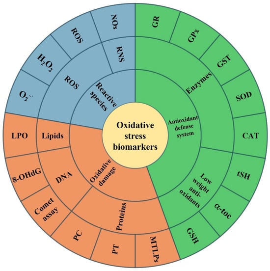
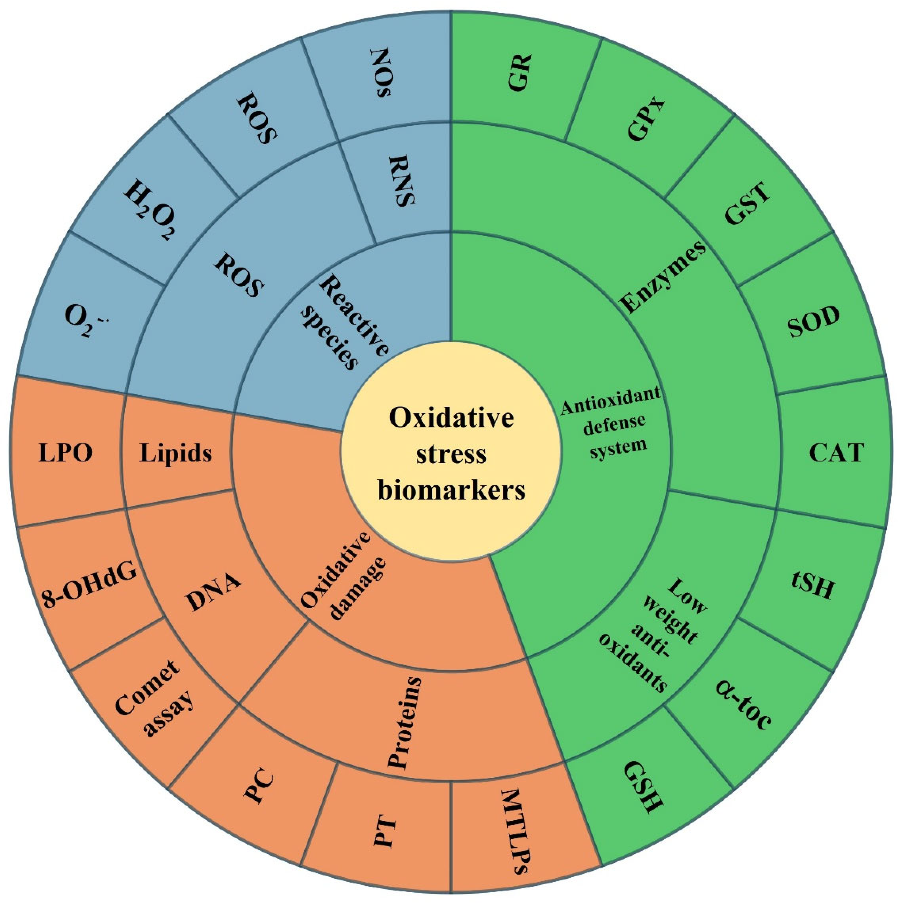
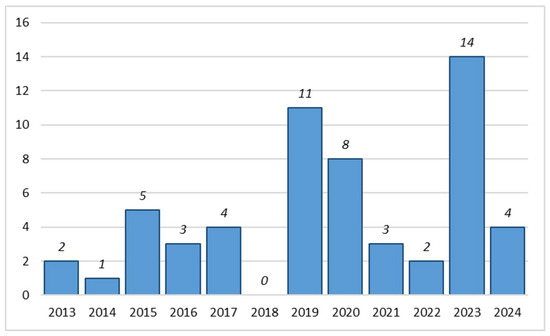
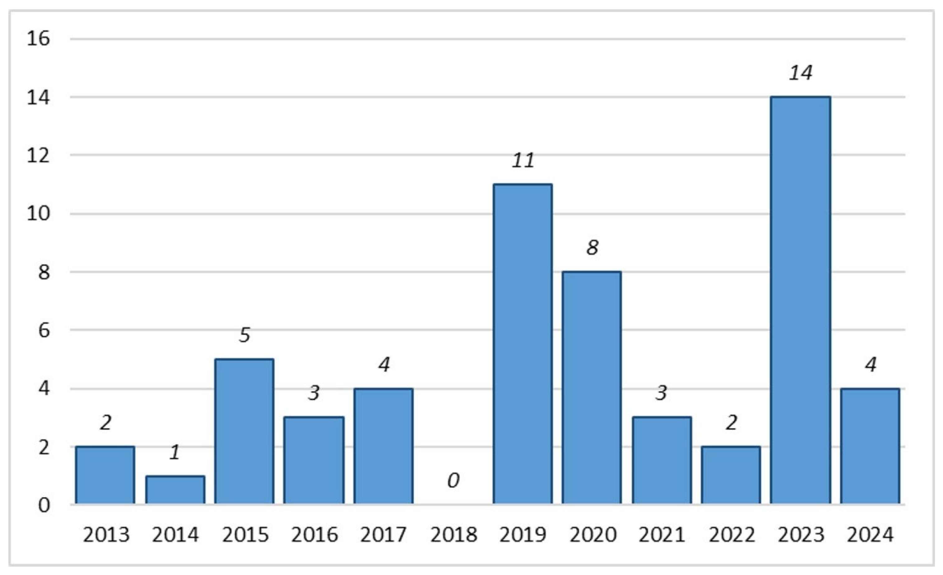
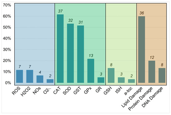
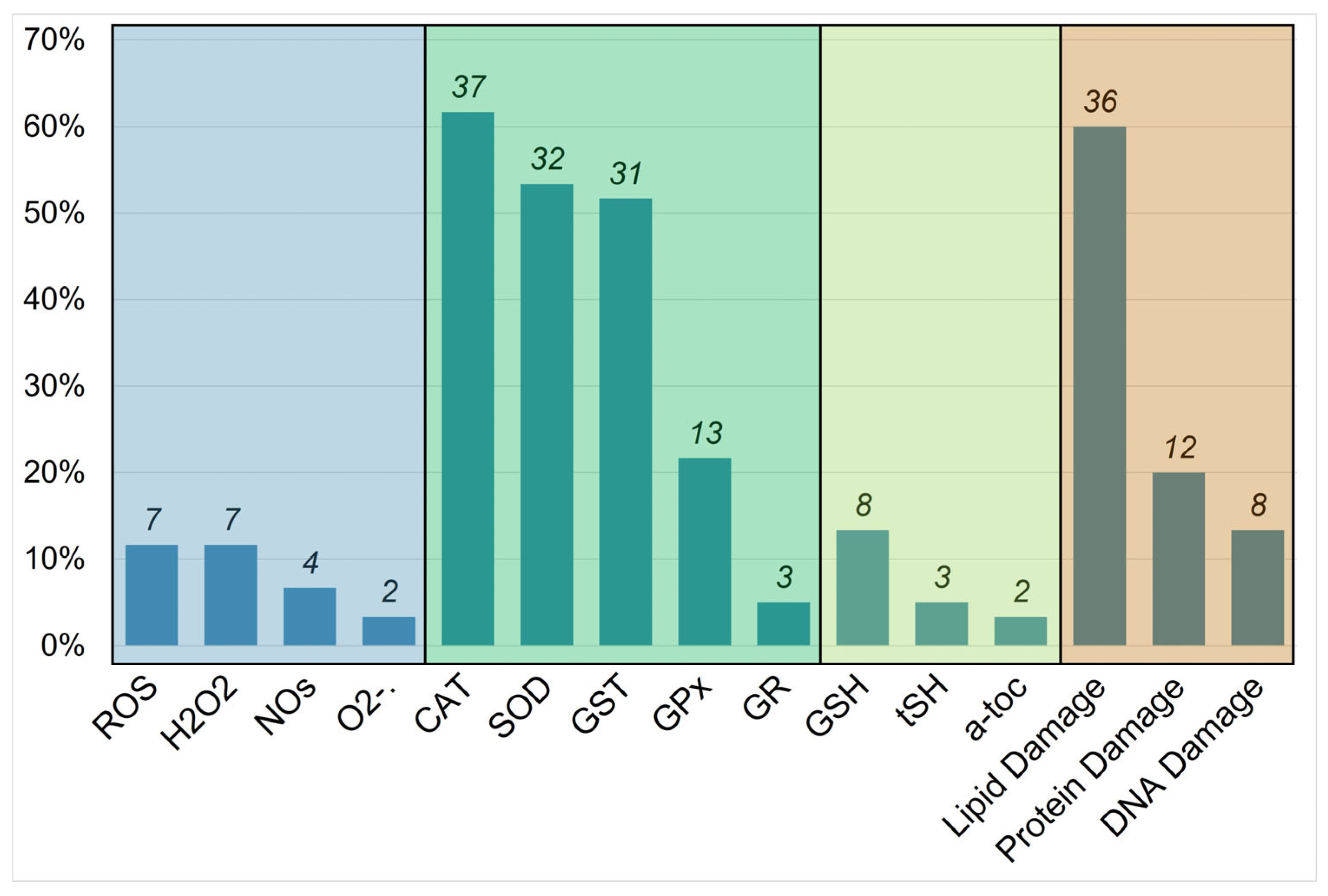
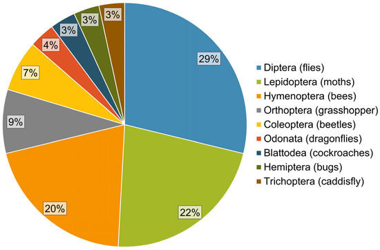
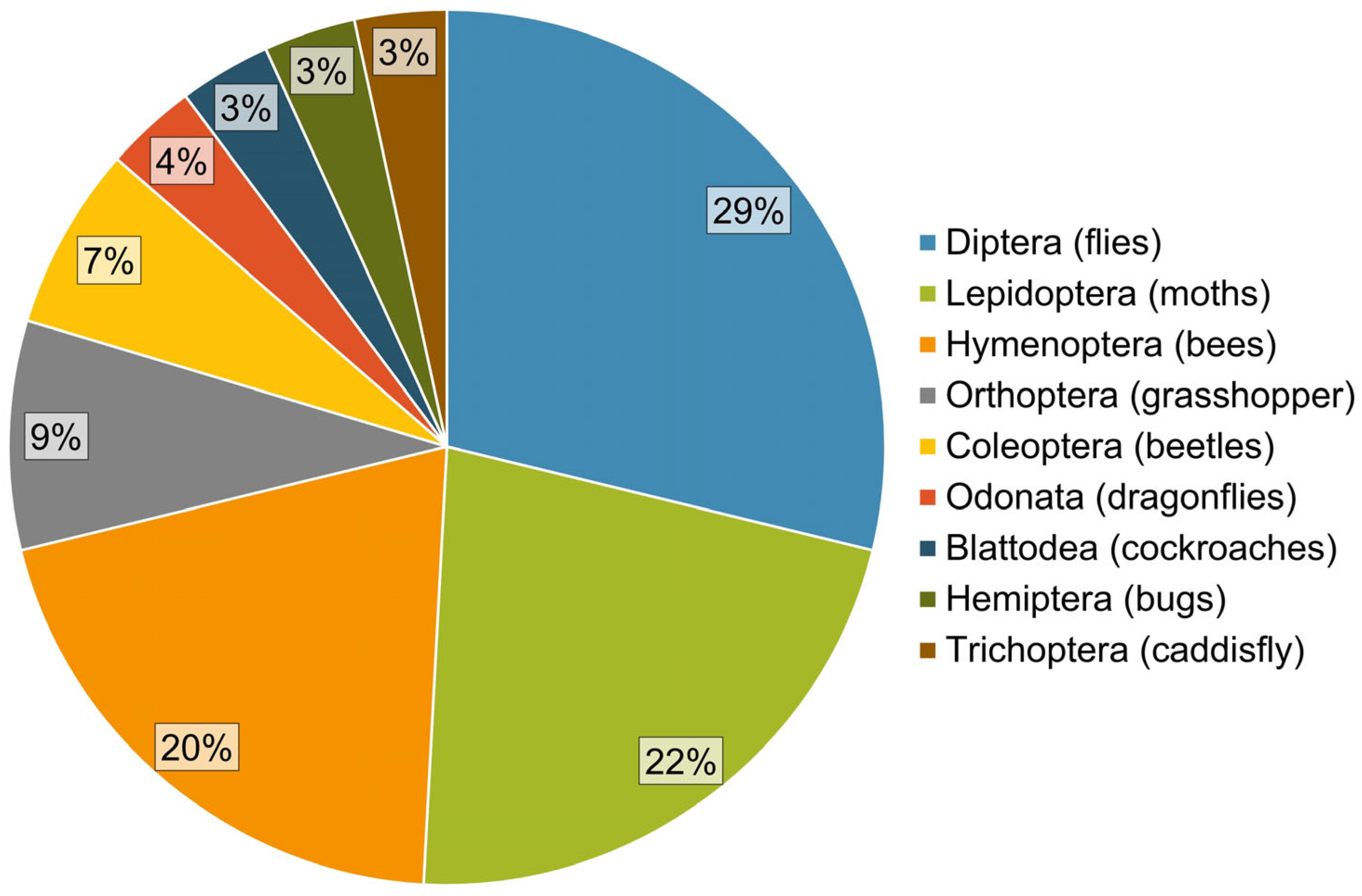
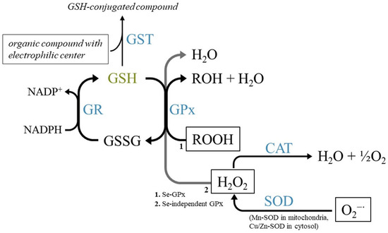
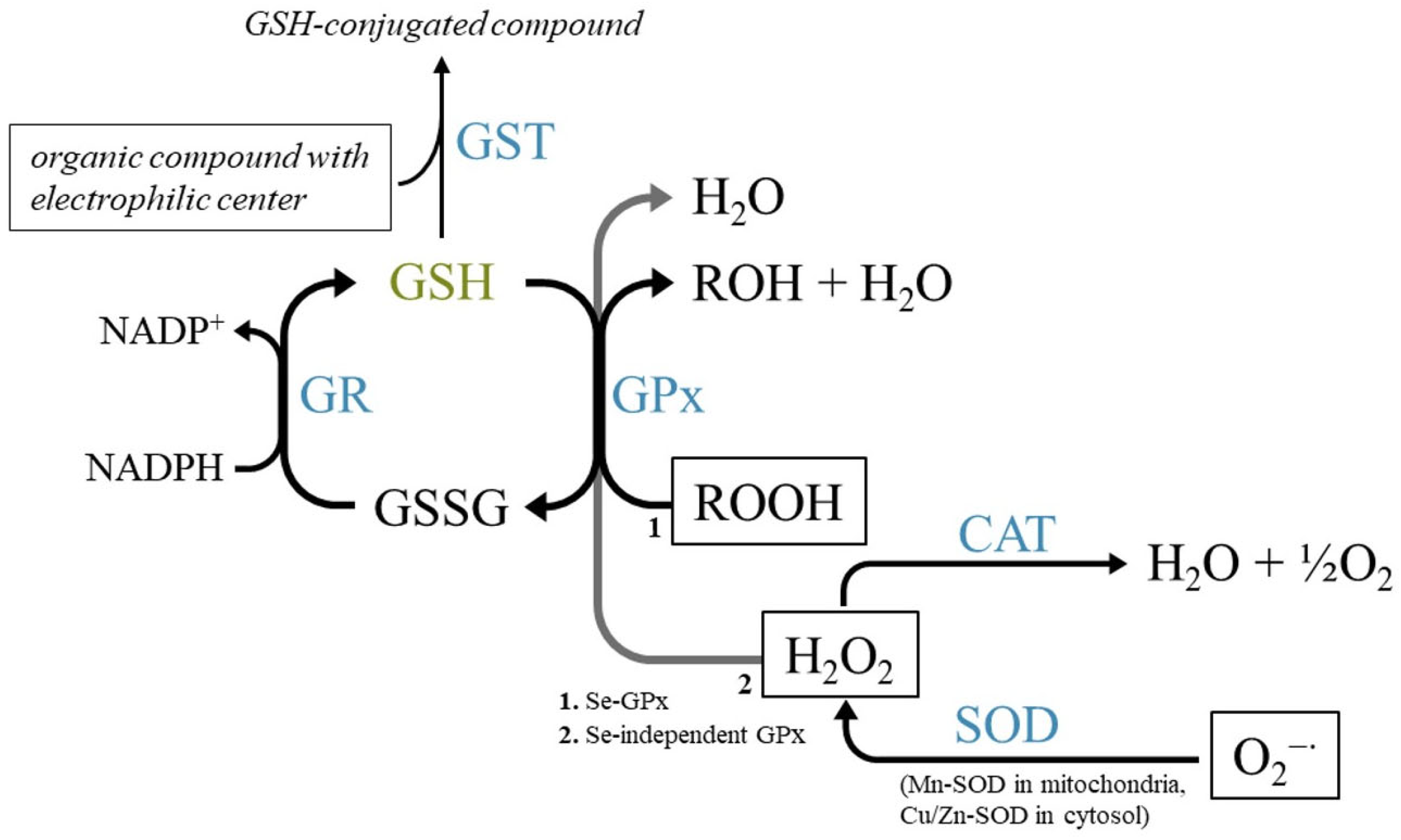
Disclaimer/Publisher’s Note: The statements, opinions and data contained in all publications are solely those of the individual author(s) and contributor(s) and not of MDPI and/or the editor(s). MDPI and/or the editor(s) disclaim responsibility for any injury to people or property resulting from any ideas, methods, instructions or products referred to in the content. |
© 2025 by the authors. Licensee MDPI, Basel, Switzerland. This article is an open access article distributed under the terms and conditions of the Creative Commons Attribution (CC BY) license (https://creativecommons.org/licenses/by/4.0/).