


Abstract
Background: Artificial intelligence (AI) is increasingly being integrated into dental implantology, particularly in treatment planning, a critical phase for implant success. Traditionally dependent on clinician expertise, planning can now be supported by AI-assisted systems that aim to improve diagnostic accuracy, precision, and efficiency. Objective: To synthesise recent evidence on the use of AI in dental implant planning, particularly its ability to analyse cone beam computed tomography (CBCT) imaging to identify edentulous regions and assess bone dimensions compared with conventional planning methods. Methods: A systematic search was conducted across PubMed, Scopus, Google Scholar, and the Cochrane Library, with additional manual searches from October 2024 to July 2025. Eligibility was defined using the Population, Intervention, Comparison, Outcome (PICO) framework, focusing on adults undergoing implant procedures planned using AI-assisted CBCT imaging and deep learning (DL) models, particularly U-Net architectures, for CBCT segmentation. Results: Ten studies were included, AI systems demonstrated high accuracy (92–99.7%) in detecting teeth and edentulous regions, with precision and recall frequently exceeding 90%. AI-assisted planning also showed improved efficiency, and, in one study, higher implant success rates compared with traditional planning (92% vs. 78%). However, variability in study design, inconsistent reporting, and limited ethical oversight were noted. Conclusions: AI, particularly DL models applied to CBCT imaging, shows strong potential to enhance diagnostic precision and efficiency in dental implant planning. Nevertheless, the field requires standardised evaluation metrics, larger datasets, and well-designed clinical trials before widespread clinical implementation.
1. Introduction
Dental implants have become a cornerstone of modern restorative dentistry, providing a fixed, reliable, and long-lasting solution for tooth replacement. Unlike conventional prosthetics, implants preserve jawbone structure, maintain adjacent tooth integrity, and significantly improve oral function and aesthetics, resulting in enhanced patient quality of life [1]. Their success has been well-documented across both partially dentate and fully edentulous patients, with studies reporting high survival rates and favourable long-term outcomes [2]. However, these outcomes are not guaranteed and depend heavily on precise, well-executed treatment planning [3,4]. Effective implant planning involves careful evaluation of anatomical structures, occlusion, and patient-specific factors such as bone volume, density, and overall oral health. It plays a critical role in ensuring accurate implant positioning, minimizing complications, and improving both surgical and prosthetic outcomes [5]. Radiographic assessment, particularly through the use of cone beam computed tomography (CBCT), has become an essential diagnostic tool in this process, offering detailed three-dimensional imaging of bone morphology, nerve pathways, and sinus cavities [6,7]. When combined with computer-aided design and manufacturing (CAD/CAM), clinicians are able to fabricate surgical guides that improve precision and predictability in implant placement [8,9]. More recently, the incorporation of AI into clinical workflows has begun to significantly transform the methodology and precision of dental implant treatment planning. AI, particularly through deep learning (DL) techniques such as convolutional neural networks (CNNs), enables automated interpretation of CBCT scans, identifying edentulous regions, assessing bone dimensions, and aiding in virtual implant placement with a high degree of accuracy [10,11]. These systems can enhance clinical efficiency, reduce variability caused by human interpretation, and support better surgical outcomes.
While digital tools like CBCT and intraoral scanners have already improved the technical workflow, AI introduces a new level of diagnostic precision and decision-making support [12]. This is especially relevant given the inherent subjectivity and potential for human error in clinical judgement. AI systems, when properly trained, can support clinicians by providing consistent, data-driven analyses, thus reducing risks associated with anatomical misinterpretation or planning errors.
Despite its promising capabilities, the application of AI in dental implantology remains relatively novel. The existing literature often focuses on diagnostic applications, with fewer studies evaluating its role in treatment planning itself. A recent study by Alqutaibi et al. [13] examined AI’s ability to detect missing teeth and assess bone dimensions from CBCT scans, suggesting strong potential for improving planning accuracy. However, the evidence base is still emerging, and more comprehensive evaluations are needed to understand AI’s full impact in clinical settings.
This review aims to fill that gap by critically examining current research on AI-assisted implant planning, particularly in the context of CBCT image analysis. By evaluating the effectiveness of AI technologies in identifying implant sites, measuring bone, and supporting treatment workflows, this study seeks to clarify AI’s role in enhancing implant success, surgical precision, and overall patient outcomes.
2. Materials and Methods
2.1. Review Protocol and Reporting Standards
This systematic review was conducted in accordance with Preferred Reporting Items for Systematic Reviews and Meta-Analyses (PRISMA) guidelines [14].
2.2. Eligibility Criteria
The eligibility criteria for study selection were based on the PICO (Population, Intervention, Comparison, Outcome) framework [15], ensuring a structured and focused review process Table 1.
Table 1.
(Population, Intervention, Comparison, Outcome) framework.
To ensure the review’s clinical relevance, systematic reviews, case reports, expert opinions, in vitro studies, finite element analysis studies, and abstract-only publications were excluded. These study types were excluded due to a lack of clinical outcome data or low levels of evidence [17].
2.3. Search Strategy
A comprehensive literature search was conducted in October 2024, with a final search performed in July 2025 (from 2014–2025) to capture the most recent publications. The search covered five major electronic databases: PubMed/MEDLINE, Scopus, Google Scholar, and the Cochrane Library.
To maximise the sensitivity and relevance of the search, a combination of keywords, Medical Subject Headings (MeSH) terms, and Boolean operators was used. The following terms were combined:
(“Dental implant” OR “dental implant success” OR “dental implant outcome” OR “dental implant aesthetics” OR “dental implant precision” OR “dental implant planning”) AND (“Artificial intelligence” OR “AI” OR “machine learning” OR “deep learning”).
Additional manual searches were carried out via: The British Dental Journal (BDJ), Reference lists of relevant studies, and grey literature databases.
Search results were imported into Mendeley reference manager, which was used to organise studies and remove duplicates. The remaining records were screened in two stages: (1) titles and abstracts were reviewed against the inclusion criteria; (2) full texts were examined to confirm alignment with the study objectives and eligibility criteria.
2.4. Data Extraction and Study Characteristics
Key information from eligible studies was extracted and summarised in a structured Study Characteristics Table (Table 1). The data included:
- •
- Authors and year of publication
- •
- Study design
- •
- Country and institution of origin
- •
- Type and size of dataset
- •
- Data collection timeframe
- •
- Area of interest (e.g., detection of edentulous areas, bone dimension analysis)
- •
- Stated objectives of each study
This standardised format facilitated consistent comparison and synthesis of the included literature. Data extraction was initially carried out and the following study characteristics and outcome measures were extracted to allow comparison between AI-assisted and conventional implant planning methods: study design, sample size, AI model or algorithm used, CBCT imaging parameters, accuracy of edentulous region detection, bone dimension assessment, diagnostic precision, sensitivity and specificity where reported, and reported clinical outcomes such as implant placement accuracy or success rate. These characteristics were summarised and compared across studies, as presented in Table 2.
2.5. Quality Assessment
The methodological quality of all included studies was assessed using the Critical Appraisal Skills Programme (CASP) checklists, which provide structured appraisal tools for a variety of study designs, including randomised controlled trials (RCTs), observational studies, and cohort studies.
The CASP tool guided evaluation in key areas, such as:
- Clarity of research aims
- Appropriateness of study design
- Transparency of data collection methods
- Consideration of ethical issues
- Rigour of data analysis
Each study was reviewed over the course and a Quality Assessment Table (Supplementary Materials Table S1) summarises the results, with “Can’t tell” used when full-text papers lacked sufficient detail.
Although variability was noted in areas such as sample size justification and ethical reporting, all studies were considered to meet the minimum quality threshold for inclusion. Full CASP checklists for each study are available in Supplementary Materials Table S2.
2.6. Data Synthesis
A narrative approach was employed for data synthesis in this systematic review. The data synthesis process was conducted. Quantitative findings from the studies included were summarized descriptively, with key statistical outcomes highlighted where available. Integration of the data was guided by a convergent synthesis design, in which findings were initially analysed separately and subsequently brought together to inform the overall conclusions. The synthesis and interpretation of the results were cross-checked with any discrepancies resolved through discussion.
3. Results
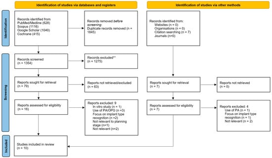
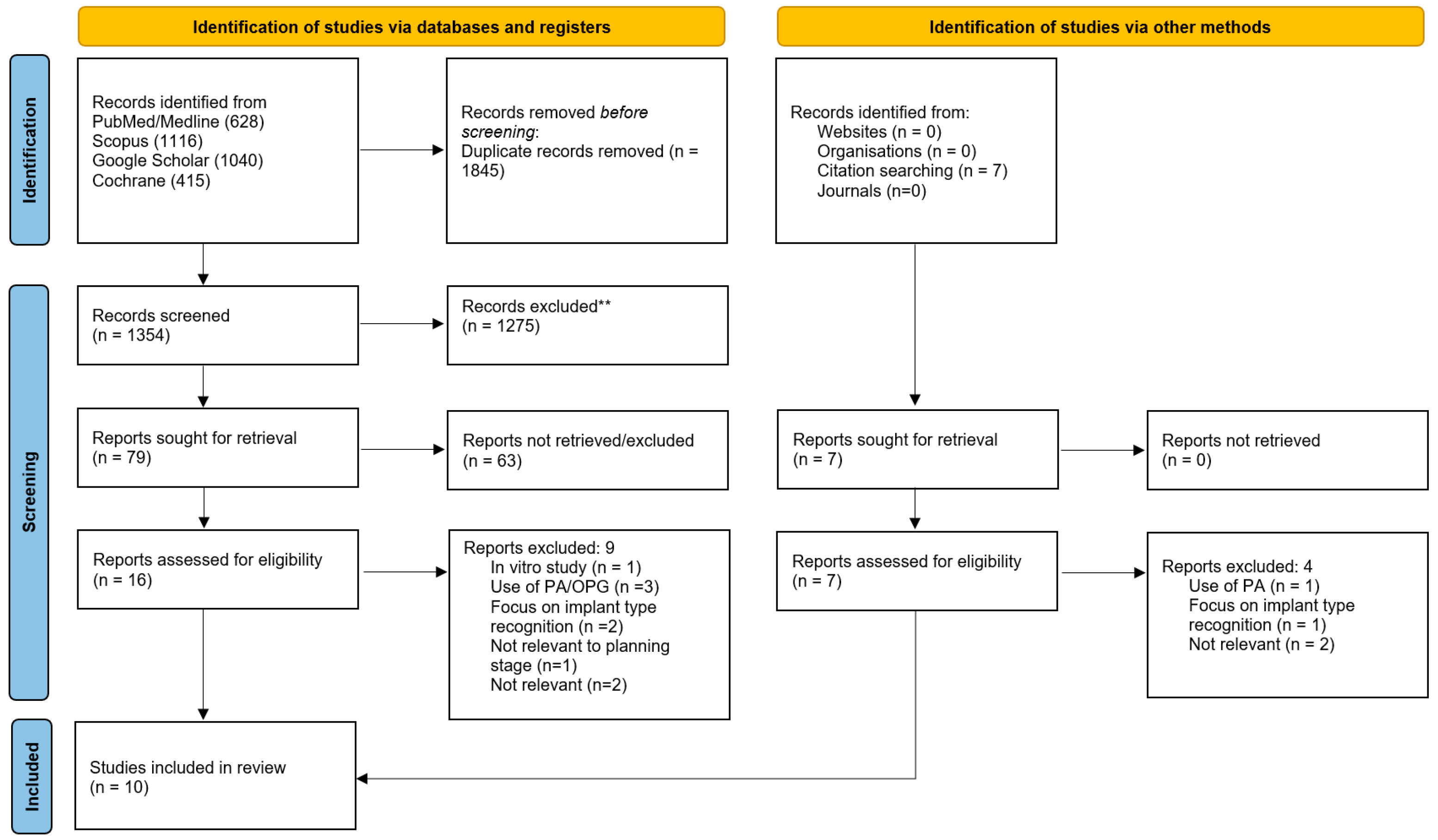
Following a comprehensive electronic search across five major databases, a total of 3199 articles were initially identified. After the removal of 1845 duplicates, 1354 records remained for title and abstract screening. These were assessed against the predefined inclusion and exclusion criteria, leading to the exclusion of 1275 articles that did not meet the eligibility requirements. A further screening of the remaining 79 studies was cconducted, and 16 articles were deemed potentially relevant and selected for full-text review. Of these, nine studies were excluded due to not meeting the inclusion criteria. One study was excluded because it was conducted in vitro and did not involve real patient data [18]. Several others were excluded for relying on two-dimensional imaging methods, such as panoramic or periapical radiographs, rather than CBCT, which was required for inclusion [18,19,20,21]. Additionally, two studies focused primarily on the identification or classification of dental implant systems using AI, rather than on planning potential implant workflows, which fell outside the scope of this review [22,23]. One study was excluded due to its focus on the restoration phase of a dental implant [24]. Finally, two studies were excluded for lacking relevance to AI entirely, focusing instead on robotics and digital dentistry applications [25,26].
Furthermore, manual citation searching identified seven further studies for potential inclusion. After full-text assessment, four of these were excluded for reasons consistent with those applied during the database screening. One study was excluded due to the use of 2D imaging [26]. Another was excluded as it focused on the detection of an implant system using AI [27], and the remaining two studies were excluded because they were unrelated to the review’s scope, with one addressing digital workflows and the other investigating peri-implant bone loss [28,29]. Following both the electronic and manual search processes, a total of ten studies met the eligibility criteria and were included in this review.
Figure 1 is a PRISMA flowchart representing the study selection and screening process. This systematic review was conducted in accordance with the Preferred Reporting Items for Systematic Reviews and Meta-Analyses (PRISMA) 2020 guidelines. The PRISMA flow diagram describing the study selection process is presented in Figure 1, and the completed PRISMA checklist is provided as Supplementary Materials Table S1. The 10 studies’ characteristics included in this review are presented in chronological order in Table 2, and the AI technology used and outcomes of the study are presented in the Section 3 in Table 4.
Figure 1.
PRISMA flowchart representing the study selection and screening process.
Table 2.
Study characteristics table.
Table 2, shows a total of ten studies were included in this systematic review, comprising a mix of retrospective studies, observational studies and a randomised controlled trial. The studies were conducted across diverse geographic regions, including Turkey, Belgium, Saudi Arabia, India, the United Arab Emirates, and potentially others, reflecting a growing global interest in the application of AI in dental implantology. All included studies used CBCT scans as their primary imaging modality, with dataset sizes ranging from 10 to 800 scans. The time frames of data collection varied, with some studies having no mention of it, and others reporting periods as recent as 2024; highlighting the up-to-date nature of the review.
The majority of studies focused on the identification and segmentation of missing teeth regions and available bone using CBCT imaging; however, the cross-sectional study by Satapathy et al. [36] evaluated the difference between AI-generated treatment plans and traditionally generated plans. The randomized control trial by Rajan et al. [35] also compared the outcomes of AI-assisted planning with human-generated planning.
Notably, there appears to be more concentration of research on AI’s capabilities in detecting missing teeth and anatomical landmarks, with fewer studies specifically investigating the overall efficiency and clinical impact of AI-driven implant planning processes. This review offers an up-to-date summary of recent findings and points out both advancements and gaps in the current literature.
A range of several different parameters were used to measure the outcomes of the results, and these varied between studies. They commonly included accuracy, precision, recall, F1 score and segmentation overlap metrics such as Dice Similarity Coefficient (DSC) and Jaccard index. These metrics provide a comprehensive insight into the effectiveness, reliability and applicability of the AI models used. See Table 3.
Table 3.
Metrics table.
Table 4 below presents a summary of the key outcomes from the studies included in this systematic review, focusing on the performance and clinical effectiveness of various AI technologies used in dental implant planning with CBCT imaging. The table outlines the type of AI software used (DL, CNN), the exact software used (if mentioned) and if additional models were applied (U-net). Whether the systems were overseen or validated by human specialists is also included. Each study’s outcomes are reported in terms of accuracy, precision, recall, success rates, segmentation performance, or comparative clinical metrics. Together, these data illustrate the growing role and effectiveness of AI in enhancing diagnostic accuracy, improving treatment planning efficiency, and supporting clinical decision-making in implant dentistry.
Table 4.
Outcomes of included studies.
The studies summarized in Table 5 highlight the strong effectiveness of AI—particularly deep learning (DL) techniques—in dental imaging and treatment planning. With the exception of one study, Rajan et al. [35], all investigations employed convolutional neural networks (CNNs) as a central component of their methodological frameworks.
Table 5.
Quality assessment table.
Out of the ten studies reviewed, eight achieved a CASP score of 70% or higher, indicating strong methodological quality. Six studies [31,32,34,37,38,39] scored 90%, demonstrating rigorous research practices. Conversely, the single RCT by Rajan et al. (2024) [35] scored lower due to vague reporting, and two other studies, Satapathy et al. (2024); Bodhe et al. (2022) [33,36], also lacked clarity, particularly in software disclosure and sample details.
4. Discussion
This systematic review aimed to evaluate the role of artificial intelligence (AI), particularly deep learning models applied to CBCT imaging, in dental implant treatment planning. The main findings indicate that AI systems demonstrate high diagnostic accuracy in detecting anatomical structures and edentulous regions, while also significantly improving planning efficiency. The studies included reported performance comparable to, or in some cases exceeding, that of clinicians, particularly in segmentation tasks and treatment planning workflows.
High levels of diagnostic accuracy reported across studies highlight the strength of AI in anatomical detection, particularly for tooth and edentulous region identification. The consistently high performance observed suggests that deep learning models are well-suited to CBCT image analysis, where pattern recognition and segmentation are critical. In addition to accuracy, substantial improvements in time efficiency indicate that AI has the potential to streamline clinical workflows and reduce clinician workload. Gerhardt et al. [31] reported 99.7% accuracy in tooth detection and significant time efficiency (1.5 s vs. 98 s for human planning). Similarly, Bayrakdar et al. [30] found high detection rates for the mandibular canal (72.2%) and missing teeth (95.3%).
Several studies highlighted how expert input and specific model architectures improved outcomes. Al-Sarem et al. [32] showed that adding U-Net to various CNNs significantly boosted performance, with DenseNet169 achieving 94% F1-scores. Al-Asali et al. [37] found expert annotations improved the Dice Similarity Coefficient (DSC) from 0.81 to 0.93. Elgarba et al. [38] demonstrated statistically faster AI-generated plans (198 vs. 435 s), with 95% requiring no major corrections, closely matching human performance (96%).
However, these findings should be interpreted cautiously. The variability in reported accuracy across anatomical structures, such as lower performance in sinus or canal detection, suggests that AI reliability may still depend on anatomical complexity and image quality. Furthermore, the reliance on expert annotation in several studies indicates that optimal AI performance often requires human input, reinforcing the role of AI as a supportive rather than autonomous tool.
There were concerns around inconsistent reporting, limited expert involvement, and ethical oversight. Only four studies explicitly referenced adherence to the Declaration of Helsinki, and two lacked any mention of ethical approval. Some studies also showed data discrepancies or lacked transparency in AI tool specifications, reducing reproducibility. Despite these issues, the overall findings indicate that AI can match or exceed human performance in dental implant planning, though further methodological consistency and ethical compliance are needed for clinical integration. This systematic review synthesised evidence from ten studies exploring the integration of AI particularly convolutional neural networks (CNNs) and deep learning (DL) models into CBCT-based dental implant planning. The collective findings strongly support AI’s emerging role as a powerful tool in diagnostic imaging and treatment planning, with most studies demonstrating high accuracy, efficiency, and clinical relevance of AI-driven segmentation and analysis.
A key finding across the reviewed literature is the efficacy of CNNs and DL models in accurately identifying complex dental anatomical structures such as edentulous areas, adjacent teeth, mandibular canals, and maxillary sinuses Nogueira-Reis et al. Several studies such as Gerhardt et al. and Elgarba et al. [31,38] showed that these systems not only maintained clinical precision but also significantly improved time efficiency, reducing planning time from minutes to mere seconds. This aligns with the broader literature in medicine, where AI-supported diagnostics have also shown enhanced accuracy and reduced workload [40,41,42].
The study by Al-Asali et al. [37] underscored the importance of expert-guided annotations in improving AI model performance, a trend mirrored in other domains of healthcare, where hybrid models, combining AI with clinician input, outperform either in isolation. This is consistent with findings by Raciti et al. [43] and Gulshan et al. [44] in oncology and ophthalmology respectively, where AI has shown diagnostic capabilities equal to or greater than those of clinicians, particularly in image interpretation tasks.
Several reviewed studies highlighted variability in AI performance between the maxilla and mandible. In general, higher accuracy was achieved in mandibular segmentation (DSC: 0.94) compared to maxillary structures (DSC: 0.907) [45]. This likely reflects anatomical complexity: the mandible is denser, better defined, and less variable than the maxilla, which has more intricate features like the nasal floor and sinus cavities [30,33]. The mandibular canal, despite being within the more consistently segmented mandible, remains a challenge for AI due to its small diameter and close proximity to surrounding structures [45]. Nonetheless, AI’s ability to identify such critical landmarks is paramount in minimising surgical risks such as nerve injury, sinus perforation, or root damage [31,32]. Beyond anatomical segmentation, AI has also shown potential in prosthetic design and biomechanical planning. Although not included in this review due to eligibility criteria, studies such as Cho et al. [24] demonstrate how DL models can replicate tooth morphology and design implant-supported crowns with near-human precision. Similarly, Alotaibi et al. [39] showed promising, though still developing, accuracy in AI-predicted implant length and diameter, indicating the potential of AI in supporting but not yet replacing prosthodontic expertise.
Implant placement accuracy is not purely dependent on anatomy; biomechanical factors like angulation, bone quality, occlusion, and aesthetics also play a crucial role in long-term success. Poor planning can lead to peri-implantitis, prosthetic complications, and compromised function [46,47]. AI tools, when integrated into surgical-restorative workflows, offer a means to optimise all these factors systematically.
The Relu® Creator, used in two of the reviewed studies [31,38], exemplifies the practical implementation of AI in dentistry. With FDA approval and cloud-based accessibility, it demonstrates the real-world feasibility of integrating AI into dental workflows. However, like many AI systems, its performance is still highly dependent on input data quality, and human validation remains essential, with only 35% of AI-generated plans requiring no modifications [38].
Technologically, the review also highlights the distinction between 2D U-net and 3D U-net architectures. While 2D U-nets are faster and require less data, they lack the volumetric context necessary for detailed CBCT analysis, making 3D U-nets the preferred choice for high-accuracy segmentation in implant planning [48,49]. Meanwhile, CNNs like VGG, YOLO, and ResNet remain superior for classification and detection, particularly in 2D images like OPGs or bitewings.
Compared to fields like radiology or oncology, AI in dentistry is still underdeveloped, a trend attributed largely to limited, non-standardised datasets and restricted data access due to the decentralised nature of dental practice. In contrast, medical datasets are larger, standardised, and often integrated across national health systems [50,51]. Moreover, current AI systems are not autonomous and should be viewed as decision-support tools, not replacements for clinical judgment. As shown in the reviewed studies, hybrid human-AI collaboration consistently yielded better outcomes than AI or humans alone [37,42].
The responsible use of AI in dentistry also raises ethical and regulatory considerations. Transparency is key: patients should be informed when AI tools are involved in their care, and consent must be obtained [52]. There is also a critical need to avoid bias by ensuring AI models are trained on diverse, representative datasets [53]. Furthermore, the successful integration of AI into dental practice depends heavily on clinician education. Dental professionals must not only learn how to operate AI tools but also understand their limitations, interpret outputs, and know when to override suggestions based on clinical context [54,55]. Without this, the risk of over-reliance or misapplication increases.
5. Limitations
In this study there was a relatively small number of included studies, many of which were based on single-centre and limited CBCT datasets, which restricts the generalisability of results and highlights the need for larger, multi-centre investigations. In addition, substantial heterogeneity across studies in terms of AI models, dataset characteristics, annotation methods, and outcome measures limits direct comparability and weakens the overall strength of evidence. Methodological transparency was also inconsistent, with incomplete reporting of data sources, validation processes, and model development in several studies, raising concerns regarding reproducibility and potential bias. Furthermore, variability inherent to CBCT imaging, including differences in resolution, artefacts, and annotation standards, may have influenced AI performance and contributed to inconsistencies in reported outcomes. The absence of standardised performance measures ranging from accuracy and sensitivity to Dice Similarity Coefficient (DSC) and F1-score limits comparability and reduces the strength of collective conclusions. Importantly, most studies focused on technical accuracy rather than clinically meaningful endpoints, and there remains a lack of robust, long-term, real-world validation and regulatory integration. As such, while AI demonstrates promising potential in implant planning, its translation into routine clinical practice remains limited, emphasising the need for standardised evaluation frameworks, improved reporting quality, and clinically driven research. Future research should prioritise the development of large, standardised, and well-annotated CBCT datasets, alongside multi-centre clinical validation studies using unified evaluation metrics.
6. Conclusions
AI shows strong potential to enhance CBCT-based dental implant planning by improving diagnostic accuracy, anatomical landmark identification, and workflow efficiency, while reducing operator variability. However, current evidence is limited by small, non-diverse datasets, methodological heterogeneity, and a lack of robust external and prospective clinical validation, restricting generalisability and routine clinical application.
Future research should focus on large-scale, multicentre studies using standardised evaluation frameworks and rigorous validation protocols. Long-term prospective data on safety, reliability, and real-world performance are essential before widespread clinical adoption can be justified.
Supplementary Materials
The following supporting information can be downloaded at: https://www.mdpi.com/article/10.3390/dj14050248/s1, Table S1: PRISMA 2020 Checklist; Table S2: CASP Checklist.
Author Contributions
Conceptualisation, G.Z.; online software, G.Z., A.S., R.S.K., and A.S.; validation, R.S.K., G.Z., C.U. and S.W.; formal analysis, G.Z.; resources, C.U. and S.W.; writing—original draft preparation, G.Z. and R.S.K.; writing—review and editing, A.S., S.W. and C.U.; visualisation, G.Z. and R.S.K.; supervision, R.S.K., G.Z. and A.S.; project administration, S.W., C.U. and G.Z.; funding acquisition, G.Z., A.S., S.W. and R.S.K. All authors have read and agreed to the published version of the manuscript.
Funding
This research received no external funding.
Institutional Review Board Statement
Not applicable.
Informed Consent Statement
Not applicable.
Data Availability Statement
No new data were created or analyzed in this study.
Acknowledgments
ICE dental Hospital.
Conflicts of Interest
The authors declare no conflict of interest.
References
- Reissmann, D.R.; Dard, M.; Lamprecht, R.; Struppek, J.; Heydecke, G. Oral health-related quality of life in subjects with implant-supported prostheses: A systematic review. J. Dent. 2017, 65, 22–40. [Google Scholar] [CrossRef] [Scilit]
- Buser, D.; Mericske-Stern, R.; Bernard, J.P.; Behneke, A.; Behneke, N.; Hirt, H.P.; Belser, U.C.; Lang, N.P. Long-term evaluation of non-submerged ITI implants. Part 1: 8-year life table analysis of a prospective multi-center study with 2359 implants. Clin. Oral Implants Res. 1997, 8, 161–172. [Google Scholar] [CrossRef] [Scilit] [PubMed]
- Buser, D.; Chappuis, V.; Belser, U.C.; Chen, S. Implant placement post extraction in esthetic single tooth sites: When immediate, when early, when late? Periodontol. 2000 2017, 73, 84–102. [Google Scholar] [CrossRef] [Scilit] [PubMed]
- Jivraj, S.; Chee, W.; Corrado, P. Treatment planning of the edentulous maxilla. Br. Dent. J. 2006, 201, 261–279. [Google Scholar] [CrossRef] [Scilit] [PubMed]
- Pjetursson, B.E.; Lang, N.P. Prosthetic treatment planning on the basis of scientific evidence. J. Oral Rehabil. 2008, 35, 72–79. [Google Scholar] [CrossRef] [Scilit]
- Worthington, P.; Rubenstein, J.; Hatcher, D.C. The role of cone-beam computed tomography in the planning and placement of implants. J. Am. Dent. Assoc. 2010, 141, 19S–24S. [Google Scholar] [CrossRef] [Scilit]
- Correa, L.R.; Spin-Neto, R.; Stavropoulos, A.; Schropp, L.; da Silveira, H.E.D.; Wenzel, A. Planning of dental implant size with digital panoramic radiographs, CBCT-generated panoramic images, and CBCT cross-sectional images. Clin. Oral Implants Res. 2014, 25, 690–695. [Google Scholar] [CrossRef] [Scilit]
- Van Assche, N.; Vercruyssen, M.; Coucke, W.; Teughels, W.; Jacobs, R.; Quirynen, M. Accuracy of computer-aided implant placement. Clin. Oral Implants Res. 2012, 23, 112–123. [Google Scholar] [CrossRef] [Scilit]
- Gargallo-Albiol, J.; Barootchi, S.; Marques-Guasch, J. Fully Guided Versus Half-Guided and Freehand Implant Placement: Systematic Review and Meta-analysis. Int. J. Oral Maxillofac. Implants 2020, 35, 1159. [Google Scholar] [CrossRef] [Scilit]
- Zakirov, A.; Ezhov, M.; Gusarev, M.; Alexandrovsky, V.; Shumilov, E. Dental pathology detection in 3D cone-beam CT. arXiv 2018. [Google Scholar] [CrossRef] [Scilit]
- Du, G.; Cao, X.; Liang, J.; Chen, X.; Zhan, Y. Medical image segmentation based on U-net: A review. J. Imaging Sci. Technol. 2020, 64, 1. [Google Scholar] [CrossRef] [Scilit]
- Dioguardi, M.; Spirito, F.; Quarta, C.; Sovereto, D.; Basile, E.; Ballini, A.; Caloro, G.A.; Troiano, G.; Muzio, L.L.; Mastrangelo, F. Guided dental implant surgery: Systematic review. J. Clin. Med. 2023, 12, 1490. [Google Scholar] [CrossRef] [Scilit]
- Alqutaibi, A.Y.; Algabri, R.; Ibrahim, W.I.; Elawady, D. Dental implant planning using artificial intelligence: A systematic review and meta-analysis. J. Prosthet. Dent. 2024, 134, 1619–1629. [Google Scholar] [CrossRef] [Scilit]
- Page, M.J.; McKenzie, J.E.; Bossuyt, P.M.; Boutron, I.; Hoffmann, T.C.; Mulrow, C.D.; Shamseer, L.; Tetzlaff, J.M.; Akl, E.A.; Brennan, S.E.; et al. The PRISMA 2020 statement: An updated guideline for reporting systematic reviews. BMJ 2021, 372, n71. [Google Scholar] [CrossRef] [Scilit] [PubMed]
- Richardson, W.S.; Wilson, M.C.; Nishikawa, J.; Hayward, R.S. The well-built clinical question: A key to evidence-based decisions. ACP J. Club 1995, 123, A12–A13. [Google Scholar] [PubMed]
- Bohner, L.; Hanisch, M.; Kleinheinz, J.; Jung, S. Dental implants in growing patients: A systematic review. Br. J. Oral Maxillofac. Surg. 2019, 57, 397–406. [Google Scholar] [CrossRef] [Scilit]
- Petrisor, B.; Bhandari, M. The hierarchy of evidence: Levels and grades of recommendation. Indian J. Orthop. 2007, 41, 11–15. [Google Scholar] [CrossRef] [Scilit] [PubMed]
- Liu, Y.; Chen, Z.C.; Chu, C.H.; Deng, F.L. Transfer learning via artificial intelligence for guiding implant placement in the posterior mandible: An in vitro study. Res. Sq. 2021, preprint. [Google Scholar] [CrossRef] [Scilit]
- Liu, C.-H.; Lin, C.-J.; Hu, Y.-H.; You, Z.-H. Predicting the failure of dental implants using supervised learning techniques. Appl. Sci. 2018, 8, 698. [Google Scholar] [CrossRef] [Scilit]
- Sukegawa, S.; Yoshii, K.; Hara, T.; Matsuyama, T.; Yamashita, K.; Nakano, K.; Takabatake, K.; Kawai, H.; Nagatsuka, H.; Furuki, Y. Multi-task deep learning model for classification of dental implant brand and treatment stage using dental panoramic radiograph images. Biomolecules 2021, 11, 815. [Google Scholar] [CrossRef] [Scilit]
- Park, W.; Huh, J.K.; Lee, J.H. Automated deep learning for classification of dental implant radiographs using a large multi-center dataset. Sci. Rep. 2023, 13, 4862. [Google Scholar] [CrossRef] [Scilit] [PubMed]
- Takahashi, T.; Nozaki, K.; Gonda, T.; Mameno, T.; Wada, M.; Ikebe, K. Identification of dental implants using deep learning—Pilot study. Int. J. Implant Dent. 2020, 6, 53. [Google Scholar] [CrossRef] [Scilit]
- Lee, J.H.; Kim, Y.T.; Lee, J.B. Identification of dental implant systems from low-quality and distorted dental radiographs using AI trained on a large multi-center dataset. Sci. Rep. 2024, 14, 12606. [Google Scholar] [CrossRef] [Scilit]
- Cho, J.H.; Çakmak, G.; Choi, J.; Lee, D.; Yoon, H.I.; Yilmaz, B.; Schimmel, M. Deep learning-designed implant-supported posterior crowns: Assessing time efficiency, tooth morphology, emergence profile, occlusion, and proximal contacts. J. Dent. 2024, 147, 105142. [Google Scholar] [CrossRef] [Scilit] [PubMed]
- Shi, J.; Liu, B.; Wu, X.; Liu, M.; Zhang, Q.; Lai, H.; Tonetti, M.S. Improved positional accuracy of dental implant placement using a haptic and machine-vision-controlled collaborative surgery robot: A pilot randomized controlled trial. J. Clin. Periodontol. 2024, 51, 24–32. [Google Scholar] [CrossRef] [Scilit]
- Fasih, P.; Yari, A.; Kamali Hakim, L.; Kashe, N.N. Artificial intelligence in detecting and segmenting vertical misfit of prosthesis in radiographic images of dental implants: A cross-sectional analysis. Clin. Oral Implants Res. 2025, 36, 578–588. [Google Scholar] [CrossRef] [Scilit]
- da Mata Santos, R.P.; Vieira Oliveira Prado, H.E.; Aranha Neto, I.S.; Alves de Oliveira, G.A.; Vespasiano Silva, A.I.; Gonçalves Zenóbio, E.; Ricardo Manzi, F. Automated identification of dental implants using artificial intelligence. Int. J. Oral Maxillofac. Implants 2021, 36, 918. [Google Scholar] [CrossRef] [Scilit] [PubMed]
- Guo, D.; Mühlemann, S.; Pan, S.; Zhou, Y.; Jung, R.E. A double-blind randomized within-subject study to evaluate clinical applicability of four digital workflows for the fabrication of posterior single implant crown. Clin. Oral Implants Res. 2023, 34, 1319–1329. [Google Scholar] [CrossRef] [Scilit]
- Zhang, H.; Shan, J.; Zhang, P.; Chen, X.; Jiang, H. Trabeculae microstructure parameters serve as effective predictors for marginal bone loss of dental implant in the mandible. Sci. Rep. 2020, 10, 18437. [Google Scholar] [CrossRef] [Scilit]
- Bayraktar, Y.; Ayan, E. Diagnosis of interproximal caries lesions with deep convolutional neural network in digital bitewing radiographs. Clin. Oral Investig. 2021, 26, 623–632. [Google Scholar] [CrossRef] [Scilit]
- do Nascimento Gerhardt, M.; Fontenele, R.C.; Leite, A.F.; Lahoud, P.; Van Gerven, A.; Willems, H.; Smolders, A.; Beznik, T.; Jacobs, R. Automated detection and labelling of teeth and small edentulous regions on cone-beam computed tomography using convolutional neural networks. J. Dent. 2022, 122, 104139. [Google Scholar] [CrossRef] [Scilit]
- Al-Sarem, M.; Al-Asali, M.; Alqutaibi, A.Y.; Saeed, F. Enhanced Tooth Region Detection Using Pretrained Deep Learning Models. Int. J. Environ. Res. Public Health 2022, 19, 15414. [Google Scholar] [CrossRef] [Scilit]
- Bodhe, R.; Sivakumar, S.; Raghuwanshi, A. Design and Development of Deep Learning Approach for Dental Implant Planning. In 2022 International Conference on Green Energy, Computing and Sustainable Technology (GECOST); IEEE: New York, NY, USA, 2022; pp. 269–274. [Google Scholar]
- Moufti, M.A.; Trabulsi, N.; Ghousheh, M.; Fattal, T.; Ashira, A.; Danishvar, S. Developing an artificial intelligence solution to autosegment the edentulous mandibular bone for implant planning. Eur. J. Dent. 2023, 17, 1330–1337. [Google Scholar] [CrossRef] [Scilit]
- Rajan, R.S.S.; Kumar, H.S.K.; Sekhar, A.; Nadakkavukaran, D.; Feroz, S.M.A.; Gangadharappa, P. Evaluating the Role of AI in Predicting the Success of Dental Implants Based on Preoperative CBCT Images: A Randomized Controlled Trial. J. Pharm. Bioallied Sci. 2024, 16, S886–S888. [Google Scholar] [CrossRef] [Scilit] [PubMed] [PubMed Central]
- Satapathy, S.K.; Aishwarya Kunam Rashme Rashme Pooja Priyadarshini Sudarsanam Gupta, A.; Kiran, S. AI-Assisted Treatment Planning for Dental Implant Placement: Clinical vs AI-Generated Plans. J. Pharm. Bioallied Sci. 2024, 16, S942–S944. [Google Scholar] [CrossRef] [Scilit]
- Al-Asali, M.; Alqutaibi, A.Y.; Al-Sarem, M.; Saeed, F. Deep learning-based approach for 3D bone segmentation and prediction of missing tooth region for dental implant planning. Sci. Rep. 2024, 14, 13888. [Google Scholar] [CrossRef] [Scilit] [PubMed]
- Elgarba, B.M.; Rocharles Cavalcante Fontenele Mangano, F.; Jacobs, R. Novel AI-based automated virtual implant placement: Artificial versus Human Intelligence. J. Dent. 2024, 147, 105146. [Google Scholar] [CrossRef] [Scilit] [PubMed]
- Alotaibi, S.; Alsomali, M.; Alghamdi, S.; Alfadda, S.; Alturaiki, I.; Al-Ekrish, A.; Altwaijry, N. Automatic placement of simulated dental implants within CBCT images in optimum positions: A deep learning model. Med. Biol. Eng. Comput. 2025, 63, 2325–2339. [Google Scholar] [CrossRef] [Scilit]
- Nogueira-Reis, F.; Morgan, N.; Suryani, I.R.; Tabchoury, C.P.M.; Jacobs, R. Full virtual patient generated by artificial intelligence-driven integrated segmentation of craniomaxillofacial structures from CBCT images. J. Dent. 2023, 141, 104829. [Google Scholar] [CrossRef] [Scilit]
- Chen, M.; Wang, Y.; Wang, Q.; Shi, J.; Wang, H.; Ye, Z.; Xue, P.; Qiao, Y. Impact of human and artificial intelligence collaboration on workload reduction in medical image interpretation. NPJ Digit. Med. 2024, 7, 349. [Google Scholar] [CrossRef] [Scilit]
- Patel, B.N.; Rosenberg, L.; Willcox, G.; Baltaxe, D.; Lyons, M.; Irvin, J.; Rajpurkar, P.; Amrhein, T.; Gupta, R.; Halabi, S.; et al. Human-machine partnership with artificial intelligence for chest radiograph diagnosis. NPJ Digit. Med. 2019, 2, 111. [Google Scholar] [CrossRef] [Scilit] [PubMed]
- Raciti, P.; Sue, J.; Ceballos, R.; Godrich, R.; Kunz, J.D.; Kapur, S.; Reuter, V.; Grady, L.; Kanan, C.; Klimstra, D.S.; et al. Novel artificial intelligence system increases the detection of prostate cancer in whole slide images of core needle biopsies. Mod. Pathol. 2020, 33, 2058–2066. [Google Scholar] [CrossRef] [Scilit] [PubMed]
- Gulshan, V.; Peng, L.; Coram, M.; Stumpe, M.C.; Wu, D.; Narayanaswamy, A.; Venugopalan, S.; Widner, K.; Madams, T.; Cuadros, J.; et al. Development and validation of a deep learning algorithm for detection of diabetic retinopathy in retinal fundus photographs. JAMA 2016, 316, 2402–2410. [Google Scholar] [CrossRef] [Scilit] [PubMed]
- Alahmari, M.; Alahmari, M.; Almuaddi, A.; Abdelmagyd, H.; Rao, K.; Hamdoon, Z.; Alsaegh, M.; Chaitanya, N.C.S.K.; Shetty, S. Accuracy of artificial intelligence-based segmentation in maxillofacial structures: A systematic review. BMC Oral Health 2025, 25, 350, Erratum in BMC Oral Health 2025, 25, 418. https://doi.org/10.1186/s12903-025-05833-6. [Google Scholar] [CrossRef] [Scilit] [PubMed] [PubMed Central]
- Katafuchi, M.; Weinstein, B.F.; Leroux, B.G.; Chen, Y.W.; Daubert, D.M. Restoration contour is a risk indicator for peri-implantitis: A cross-sectional radiographic analysis. J. Clin. Periodontol. 2018, 45, 225–232. [Google Scholar]
- Canullo, L.; Peñarrocha, M.; Monje, A.; Catena, A.; Peñarrocha, D. Association Between Clinical and Microbiologic Cluster Profiles and Peri-implantitis. Int. J. Oral Maxillofac. Implants 2017, 32, 1054–1064. [Google Scholar]
- Zheng, Q.; Gao, Y.; Zhou, M.; Li, H.; Lin, J.; Zhang, W.; Chen, X. Semi or fully automatic tooth segmentation in CBCT images: A review. PeerJ Comput. Sci. 2024, 10, e1994. [Google Scholar] [CrossRef] [Scilit]
- Ronneberger, O.; Fischer, P.; Brox, T. U-Net: Convolutional networks for biomedical image segmentation. In Medical Image Computing and Computer-Assisted Intervention—MICCAI 2015; Springer: Cham, Switzerland, 2015; pp. 234–241. [Google Scholar] [CrossRef] [Scilit]
- FDA. Artificial Intelligence and Machine Learning in Software as a Medical Device. U.S. Food and Drug Administration. Available online: https://www.fda.gov/medical-devices/software-medical-device-samd/artificial-intelligence-and-machine-learning-software-medical-device (accessed on 25 December 2015).
- NICE. HeartFlow FFRCT for Estimating Fractional Flow Reserve from Coronary CT Angiography (MTG32); National Institute for Health and Care Excellence: Manchester, UK, 2017. [Google Scholar]
- Char, D.S.; Shah, N.H.; Magnus, D. Implementing AI in Healthcare: Ethical Challenges. N. Engl. J. Med. 2018, 378, 981–983. [Google Scholar] [CrossRef] [Scilit]
- Schwendicke, F.; Samek, W.; Krois, J. Artificial Intelligence in Dentistry: Chances and Challenges. J. Dent. Res. 2020, 99, 769–774. [Google Scholar] [CrossRef] [Scilit]
- Topol, E. High-Performance Medicine: The Convergence of Human and Artificial Intelligence. Nat. Med. 2019, 25, 44–56. [Google Scholar] [CrossRef] [Scilit]
- World Medical Association. World Medical Association Declaration of Helsinki: Ethical principles for medical research involving human subjects. JAMA 2013, 310, 2191–2194. [Google Scholar] [CrossRef] [Scilit] [PubMed]

Disclaimer/Publisher’s Note: The statements, opinions and data contained in all publications are solely those of the individual author(s) and contributor(s) and not of MDPI and/or the editor(s). MDPI and/or the editor(s) disclaim responsibility for any injury to people or property resulting from any ideas, methods, instructions or products referred to in the content. |
© 2026 by the authors. Licensee MDPI, Basel, Switzerland. This article is an open access article distributed under the terms and conditions of the Creative Commons Attribution (CC BY) license.