Standardization and Validation of Digital Volumetric Measurement Methods for Alveolar Cleft Defects Using 3D Imaging

 , , , , ,
Abstract
1. Introduction
2. Materials and Methods
2.1. Data and Model Preparation
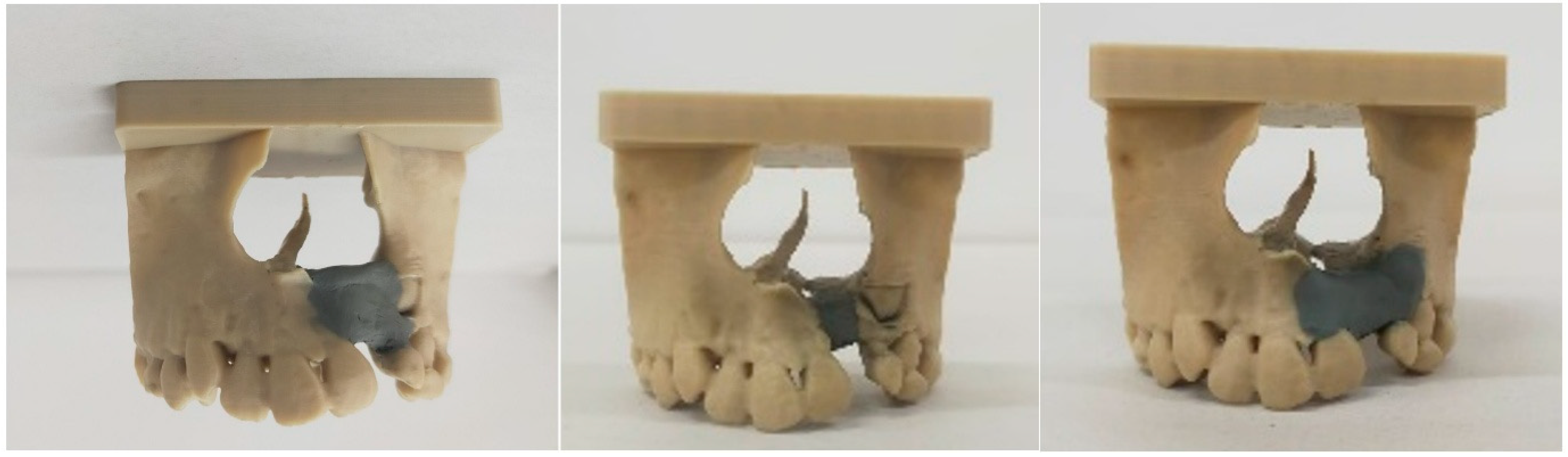
2.2. Quantitative Evaluation of Specialists’ Plans
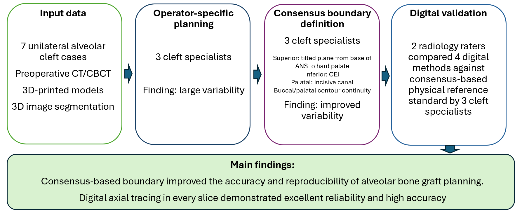
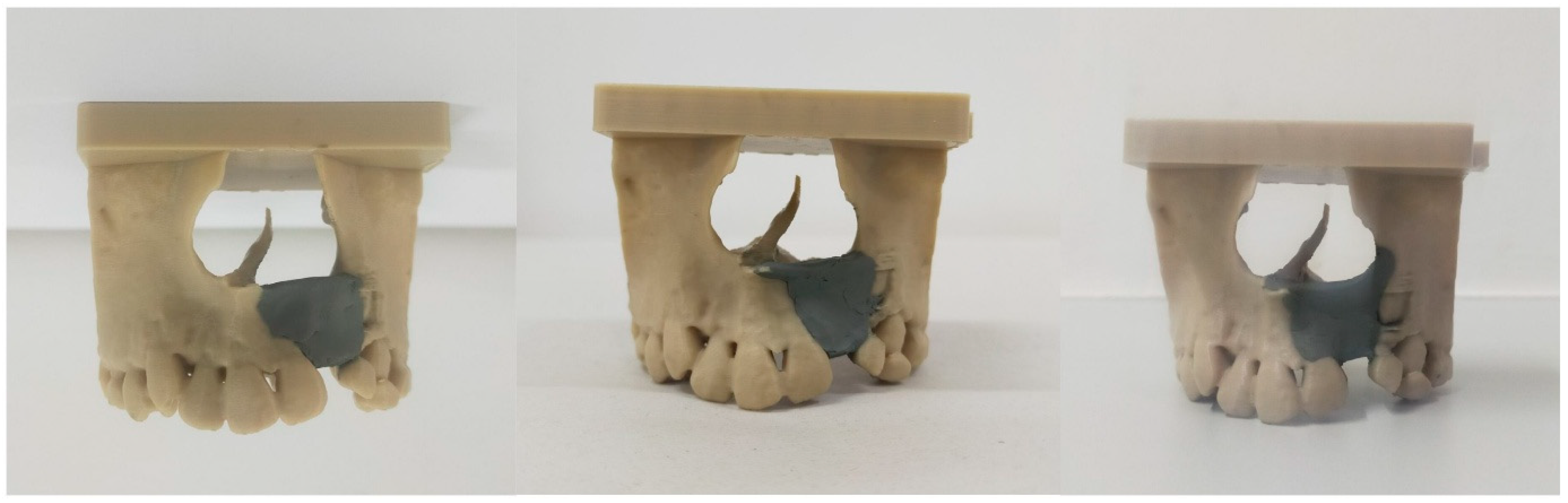
2.3. Development of a Consensus-Derived Boundary Definition for Idealized Alveolar Bone Graft Filling
- •
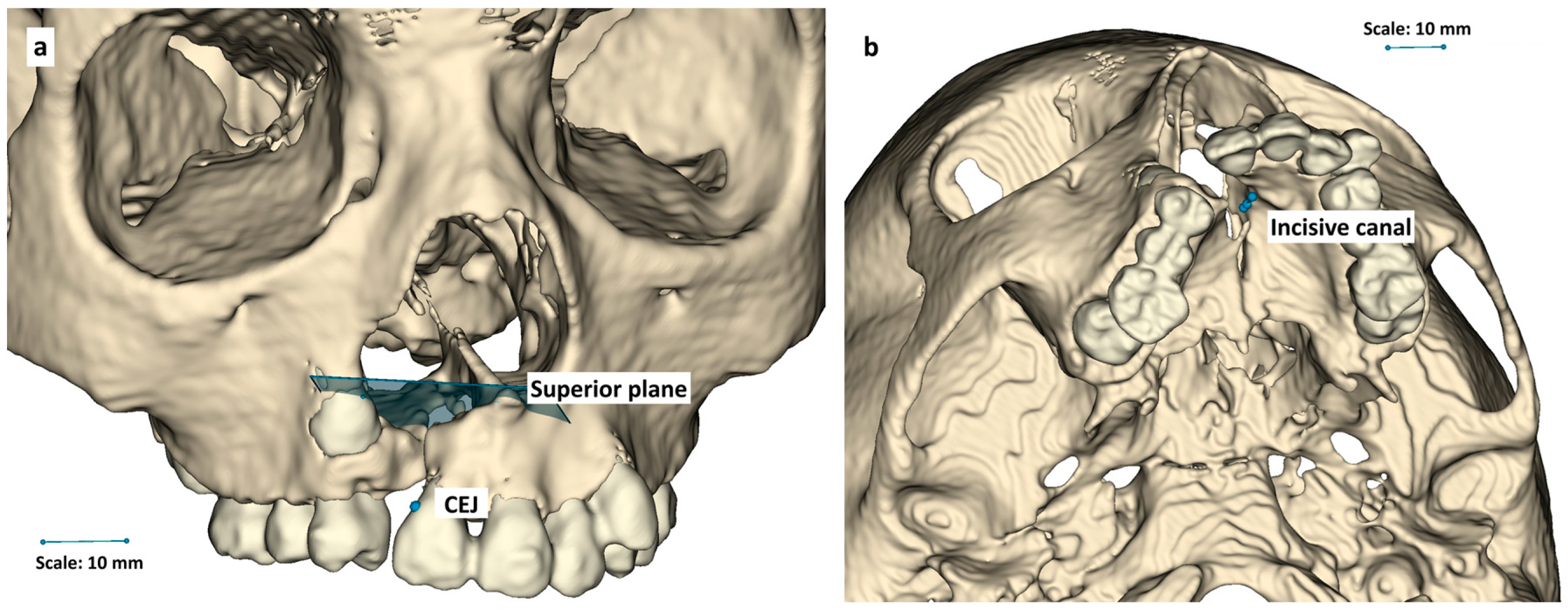
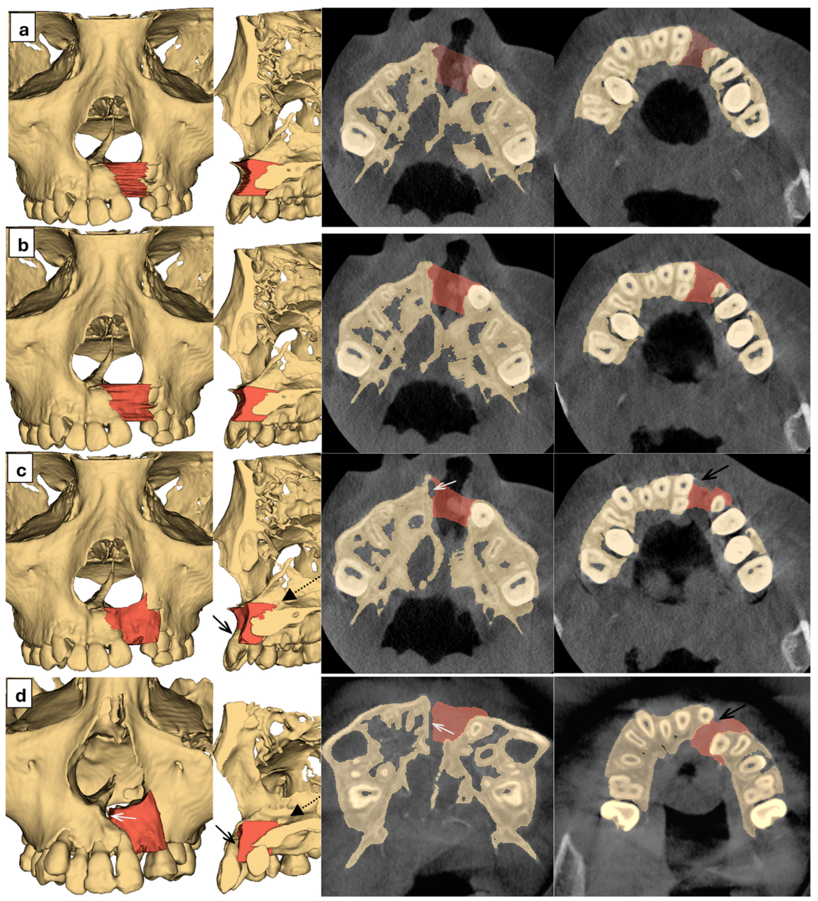
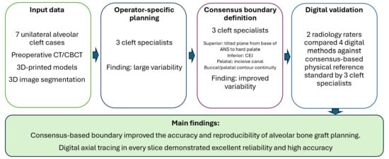
- Upper boundary: A tilted plane connecting the base of the anterior nasal spine (ANS) to the upper surface of the hard palate on the cleft side.
- •
- Lower boundary: The cemento-enamel junctions (CEJs).
- •
- Lateral boundary: Contiguous with adjacent hard tissues.
- •
- Buccal contour: Symmetrical with the contralateral side, creating smooth continuity between segments.
- •
- Palatal contour of the alveolar bone: Symmetrical with the contralateral side, creating smooth continuity between segments.
- •
- In cases involving the palate, the superior palatal boundary line passes through the anterior edge of the incisive canal.
2.4. Quantitative and Qualitative Validation of Various Digital Measurement Methods Against 3D-Printed Models
- •
- Method A: Manual tracing in every axial slice.
- •
- Method B: Manual tracing in every fifth slice (1.25 mm interval), followed by automatic interpolation.
- •
- Method C: Mirroring patient’s skull, followed by landmark-based superimposition.
- •
- Method D: Mirroring patient’s skull, followed by mesh-based superimposition.
3. Results
3.1. Exploration of Operator-Specified Plan
3.2. Consensus-Defined Alveolar Defect Volume
3.3. Quantitative and Qualitative Validation of Various Digital Measurement Methods Against 3D-Printed Models
4. Discussion
4.1. Boundary of Alveolar Defect Filling
4.2. Digital Measurement Methods
4.3. Implications and Future Work
4.4. Strengths and Limitations
5. Conclusions
Author Contributions
Funding
Institutional Review Board Statement
Informed Consent Statement
Data Availability Statement
Acknowledgments
Conflicts of Interest
References
- Guo, J.; Li, C.; Zhang, Q.; Wu, G.; Deacon, S.A.; Chen, J.; Hu, H.; Zou, S.; Ye, Q. Secondary Bone Grafting for Alveolar Cleft in Children with Cleft Lip or Cleft Lip and Palate. Cochrane Database Syst. Rev. 2011, 6, CD008050. [Google Scholar] [CrossRef]
- Kibe, T.; Maeda-Iino, A.; Takahashi, T.; Kamakura, S.; Suzuki, O.; Nakamura, N. A Follow-up Study on the Clinical Outcomes of Alveolar Reconstruction Using Octacalcium Phosphate Granules and Atelocollagen Complex. J. Oral. Maxillofac. Surg. 2021, 79, 2462–2471. [Google Scholar] [CrossRef]
- Yu, X.; Guo, R.; Li, W. Comparison of 2- and 3-Dimensional Radiologic Evaluation of Secondary Alveolar Bone Grafting of Clefts: A Systematic Review. Oral Surg. Oral Med. Oral Pathol. Oral Radiol. 2020, 130, 455–463. [Google Scholar] [CrossRef]
- Wang, X.; Tian, X.; Zeng, Y.; He, H.; Yuan, W. Advancing 3D Volumetric Analysis of Unilateral Alveolar Clefts: A Study Comparing Methods for Improved Clinical Precision. Clin. Oral Investig. 2025, 29, 373. [Google Scholar] [CrossRef]
- Saraswati, I.; Priaminiarti, M.; Nakamura, N.; Kiswanjaya, B.; Ariawan, D.; Suhartanto, H. Volumetric Measurement of Alveolar Clefts for Bone Graft Planning: A Systematic Review. Open Dent. J. 2025, 19, e18742106389355. [Google Scholar] [CrossRef]
- Padwa, B.L.; Tio, P.; Garkhail, P.; Nuzzi, L.C. Predictors of Outcomes in 900 Alveolar Bone Grafts. Plast. Reconstr. Surg. 2023, 154, 605–614. [Google Scholar] [CrossRef]
- López-Cedrún, J.L.; Gonzalez-Landa, G.; Figueroa, A. Isolated Keratinized Gingiva Incision in Alveolar Cleft Bone Grafts Improves Qualitative Outcomes: A Single Surgeon’s 23 Year Experience. J. Cranio-Maxillofac. Surg. 2014, 42, 1692–1697. [Google Scholar] [CrossRef]
- Stulberg, J.J.; Huang, R.; Kreutzer, L.; Ban, K.; Champagne, B.J.; Steele, S.R.; Johnson, J.K.; Holl, J.L.; Greenberg, C.C.; Bilimoria, K.Y. Association Between Surgeon Technical Skills and Patient Outcomes. JAMA Surg. 2020, 155, 960. [Google Scholar] [CrossRef]
- Guidry, C.A.; Newhook, T.E.; Turrentine, F.E.; Sohn, M.-W.; Sawyer, R.G.; Jones, R.S. Observations on Surgeons’ Case Selection, Morbidity, and Mortality Following Board Certification. Ann. Surg. 2016, 263, 487–492. [Google Scholar] [CrossRef]
- Seifeldin, S.A. Is Alveolar Cleft Reconstruction Still Controversial? (Review of Literature). Saudi Dent. J. 2016, 28, 3–11. [Google Scholar] [CrossRef]
- Parmar, S.; Datarkar, A.; Valvi, B.; Deshpande, A. Evaluation of Maxillary Alveolar Ridge Formation and Ridge Continuity after Secondary Alveolar Bone Grafting Using Cancellous and Cortico-Cancellous Bone Graft in Unilateral Cleft Alveolus Using Cone Beam Computed Tomographic Scan—A Randomized Controlled Trial. Oral Maxillofac. Surg. 2023, 28, 315–321. [Google Scholar] [CrossRef]
- Suomalainen, A.; Åberg, T.; Rautio, J.; Hurmerinta, K. Cone Beam Computed Tomography in the Assessment of Alveolar Bone Grafting in Children with Unilateral Cleft Lip and Palate. Eur. J. Orthod. 2014, 36, 603–611. [Google Scholar] [CrossRef]
- Choi, H.S.; Choi, H.G.; Kim, S.H.; Park, H.J.; Shin, D.H.; Jo, D.I.; Kim, C.K.; Uhm, K.I. Influence of the Alveolar Cleft Type on Preoperative Estimation Using 3d Ct Assessment for Alveolar Cleft. Arch. Plast. Surg. 2012, 39, 477–482. [Google Scholar] [CrossRef]
- Kesztyűs, A.; Würsching, T.; Nemes, B.; Pálvölgyi, L.; Nagy, K. Evaluation of 3D Visualization, Planning and Printing Techniques in Alveolar Cleft Repair, and Their Effect on Patients’ Burden. J. Stomatol. Oral Maxillofac. Surg. 2022, 123, 484–489. [Google Scholar] [CrossRef]
- Chou, P.-Y.; Denadai, R.; Hallac, R.R.; Dumrongwongsiri, S.; Hsieh, W.-C.; Pai, B.C.J.; Lo, L.-J. Comparative Volume Analysis of Alveolar Defects by 3D Simulation. J. Clin. Med. 2019, 8, 1401. [Google Scholar] [CrossRef]
- Kadhum, A.H.; Al-Zubaidi, S.; Abdulkareem, S.S. Effect of the Infill Patterns on the Mechanical and Surface Characteristics of 3D Printing of PLA, PLA+ and PETG Materials. ChemEngineering 2023, 7, 46. [Google Scholar] [CrossRef]
- Muhammad, A.R.; Sakura, R.R.; Dwilaksana, D.; Sumarji; Trifiananto, M. Layer Height, Temperature Nozzle, Infill Geometry and Printing Speed Effect on Accuracy 3D Printing PETG. REM Rekayasa Energi Manufaktur J. 2022, 7, 81–88. [Google Scholar] [CrossRef]
- Saraswati, I.; Priaminiarti, M.; Kiswanjaya, B. Defect Volume in Alveolar Clefts: Systematic Review and Workflows Using Slicer. In Current Research and Trends in Dental and Medical Technology; CRC Press: London, UK, 2025; pp. 607–616. [Google Scholar]
- Jotikasthira, P.; Chaiworawitkul, M.; Khwanngern, K.; Powcharoen, W. Correlation Between Presurgical Alveolar Cleft Volume Measured by Simulation Software Using CBCT and Actual Bone Volume Used for Grafting. Cleft Palate Craniofacial J. 2023, 62, 10556656231220502. [Google Scholar] [CrossRef]
- Shirzadeh, A.; Rahpeyma, A.; Khajehahmadi, S. A Prospective Study of Chin Bone Graft Harvesting for Unilateral Maxillary Alveolar Cleft During Mixed Dentition. J. Oral Maxillofac. Surg. Off. J. Am. Assoc. Oral Maxillofac. Surg. 2018, 76, 180–188. [Google Scholar] [CrossRef] [PubMed]
- de Rezende Barbosa, G.L.; Wood, J.S.; Pimenta, L.A.; Maria de Almeida, S.; Tyndall, D.A. Comparison of Different Methods to Assess Alveolar Cleft Defects in Cone Beam CT Images. Dentomaxillofacial Radiol. 2016, 45, 20150332. [Google Scholar] [CrossRef]
- Pfeifer, T.M.; Grayson, B.H.; Cutting, C.B. Nasoalveolar Molding and Gingivoperiosteoplasty Versus Alveolar Bone Graft: An Outcome Analysis of Costs in the Treatment of Unilateral Cleft Alveolus. Cleft Palate. Craniofac. J. 2002, 39, 26–29. [Google Scholar] [CrossRef] [PubMed]
- Potemra, H.M.K.; De Leon, F.S.; Liu, C.; Pfaff, M.J.; Bradley, J.P.; Wilson, L.F.; Lee, J.C. Abstract 110: Association Between Repeat Alveolar Bone Grafting and Anxiety in Teenagers with Cleft Lip and Palate. Plast. Reconstr. Surg. Glob. Open 2019, 7, 77. [Google Scholar] [CrossRef]
- Padwa, B.L.; Tio, P.; Garkhail, P.; Nuzzi, L.C. Cone Beam Computed Tomographic Analysis Demonstrates a 94% Radiographic Success Rate in 783 Alveolar Bone Grafts. J. Oral Maxillofac. Surg. 2022, 80, 633–640. [Google Scholar] [CrossRef]
- Zubovic, E.; Skolnick, G.B.; Said, A.M.; Nissen, R.J.; Snyder-Warwick, A.K.; Patel, K.B. Mind the Gap: Alveolar Bone Graft Revision in the Era of Computed Tomography. Cleft Palate-Craniofacial J. Off. Publ. Am. Cleft Palate-Craniofacial Assoc. 2022, 59, 246–253. [Google Scholar] [CrossRef]
- Stoop, C.C.; Janssen, N.G.; Ten Harkel, T.C.; Rosenberg, A.J.W.P. A Novel and Practical Protocol for Three-Dimensional Assessment of Alveolar Cleft Grafting Procedures. Cleft Palate-Craniofacial J. Off. Publ. Am. Cleft Palate-Craniofacial Assoc. 2023, 60, 601–607. [Google Scholar] [CrossRef]
- Kochhar, A.S.; Sidhu, M.S.; Prabhakar, M.; Bhasin, R.; Kochhar, G.K.; Dadlani, H.; Spagnuolo, G.; Mehta, V.V. Intra- And Interobserver Reliability of Bone Volume Estimation Using OsiriX Software in Patients with Cleft Lip and Palate Using Cone Beam Computed Tomography. Dent. J. 2021, 9, 14. [Google Scholar] [CrossRef]
- Liang, X.; Jacobs, R.; Hassan, B.; Li, L.; Pauwels, R.; Corpas, L.; Souza, P.C.; Martens, W.; Shahbazian, M.; Alonso, A.; et al. A Comparative Evaluation of Cone Beam Computed Tomography (CBCT) and Multi-Slice CT (MSCT): Part I. On Subjective Image Quality. Eur. J. Radiol. 2010, 75, 265–269. [Google Scholar] [CrossRef]
- Liang, X.; Lambrichts, I.; Sun, Y.; Denis, K.; Hassan, B.; Li, L.; Pauwels, R.; Jacobs, R. A Comparative Evaluation of Cone Beam Computed Tomography (CBCT) and Multi-Slice CT (MSCT). Part II: On 3D Model Accuracy. Eur. J. Radiol. 2010, 75, 270–274. [Google Scholar] [CrossRef]
- Andriola, F.D.O.; Haas Junior, O.L.; Guijarro-Martínez, R.; Hernández-Alfaro, F.; Oliveira, R.B.D.; Pagnoncelli, R.M.; Swennen, G.R. Computed Tomography Imaging Superimposition Protocols to Assess Outcomes in Orthognathic Surgery: A Systematic Review with Comprehensive Recommendations. Dentomaxillofacial Radiol. 2022, 51, 20210340. [Google Scholar] [CrossRef] [PubMed]
- Wang, X.; Pastewait, M.; Wu, T.-H.; Lian, C.; Tejera, B.; Lee, Y.-T.; Lin, F.-C.; Wang, L.; Shen, D.; Li, S.; et al. 3D Morphometric Quantification of Maxillae and Defects for Patients with Unilateral Cleft Palate via Deep Learning-Based CBCT Image Auto-Segmentation. Orthod. Craniofac. Res. 2021, 24, 108–116. [Google Scholar] [CrossRef] [PubMed]
- Diaz, A.L.; Kantar, R.; Podolsky, D.J.; Flores, R.L. Simulation in Cleft Care: Evolution, Evidence, and Training the Future Surgeon. Plast. Reconstr. Surg. 2025, 156, 56S–71S. [Google Scholar] [CrossRef] [PubMed]
- Alqutaibi, A.Y.; Hamadallah, H.H.; Oqbi, H.F.; Almuzaini, S.A.; Borzangy, S. Current Applications and Future Perspective of Virtual Reality in Dental Education and Practice in Saudi Arabia: A Scoping Review. Saudi Dent. J. 2024, 36, 1406–1416. [Google Scholar] [CrossRef] [PubMed]
- Ahn, G.; Lee, J.-S.; Yun, W.-S.; Shim, J.-H.; Lee, U.-L. Cleft Alveolus Reconstruction Using a Three-Dimensional Printed Bioresorbable Scaffold With Human Bone Marrow Cells. J. Craniofac. Surg. 2018, 29, 1880–1883. [Google Scholar] [CrossRef] [PubMed]



| Measurement | Reference Standard | Method A | Method B | Method C | Method D | ||||
|---|---|---|---|---|---|---|---|---|---|
| Rater 1 | Rater 2 | Rater 1 | Rater 2 | Rater 1 | Rater 2 | Rater 1 | Rater 2 | ||
| Mean defect volume | 1.280 ± 0.241 | 1.254 ± 0.385 | 1.281 ± 0.379 | 1.271 ± 0.325 | 1.274 ± 0.307 | 1.287 ± 0.292 | 0.803 ± 0.378 | 1.167 ± 0.436 | 0.989 ± 0.333 |
| Intra-rater ICC | Not applicable | 0.991 A | 0.971 A | 0.883 B | 0.951 A | 0.691 C | 0.919 A | 0.924 A | 0.977 A |
| Inter-rater ICC | Not applicable | 0.965 A | 0.921 A | 0.017 D | 0.323 D | ||||
Disclaimer/Publisher’s Note: The statements, opinions and data contained in all publications are solely those of the individual author(s) and contributor(s) and not of MDPI and/or the editor(s). MDPI and/or the editor(s) disclaim responsibility for any injury to people or property resulting from any ideas, methods, instructions or products referred to in the content. |
© 2026 by the authors. Licensee MDPI, Basel, Switzerland. This article is an open access article distributed under the terms and conditions of the Creative Commons Attribution (CC BY) license.
Share and Cite
Saraswati, I.; Priaminiarti, M.; Ariawan, D.; Sumardi, S.; Kiswanjaya, B.; Putra, B.T.; Bachtiar-Iskandar, H.H.; Nakamura, N.; Hak, M.S.; Suhartanto, H.; et al. Standardization and Validation of Digital Volumetric Measurement Methods for Alveolar Cleft Defects Using 3D Imaging. Dent. J. 2026, 14, 247. https://doi.org/10.3390/dj14050247
Saraswati I, Priaminiarti M, Ariawan D, Sumardi S, Kiswanjaya B, Putra BT, Bachtiar-Iskandar HH, Nakamura N, Hak MS, Suhartanto H, et al. Standardization and Validation of Digital Volumetric Measurement Methods for Alveolar Cleft Defects Using 3D Imaging. Dentistry Journal. 2026; 14(5):247. https://doi.org/10.3390/dj14050247
Chicago/Turabian StyleSaraswati, Inka, Menik Priaminiarti, Dwi Ariawan, Sariesendy Sumardi, Bramma Kiswanjaya, Bayu Trinanda Putra, Hanna H. Bachtiar-Iskandar, Norifumi Nakamura, Muhammad Syafrudin Hak, Heru Suhartanto, and et al. 2026. "Standardization and Validation of Digital Volumetric Measurement Methods for Alveolar Cleft Defects Using 3D Imaging" Dentistry Journal 14, no. 5: 247. https://doi.org/10.3390/dj14050247
APA StyleSaraswati, I., Priaminiarti, M., Ariawan, D., Sumardi, S., Kiswanjaya, B., Putra, B. T., Bachtiar-Iskandar, H. H., Nakamura, N., Hak, M. S., Suhartanto, H., & Mitsuyasu, T. (2026). Standardization and Validation of Digital Volumetric Measurement Methods for Alveolar Cleft Defects Using 3D Imaging. Dentistry Journal, 14(5), 247. https://doi.org/10.3390/dj14050247