


Abstract
Background/Objectives: It is essential to test the accuracy of artificial intelligence-assisted tools that detect dental pathologies from radiographs. This study aimed to evaluate the test characteristics of an artificial intelligence-assisted convolutional neural network-based prototype used for automated radiographic detection. Methods: A total of 300 panoramic and 100 intraoral periapical radiographs were collected between January 2020 and 2024 and then analyzed by two trained, independent specialist evaluators. The diagnostic consensus, “ground truth”, was labeled as follows: BL: bone loss; C: caries; F: filling; I: implants; IT: impacted teeth; P: prosthesis; PC: post-core; PR: periapical radiolucency; RF: root fillings; and RR: retained roots. The radiographs were uploaded to the prototype, and the results were compared. Sensitivity, specificity, positive predictive value, and negative predictive value were calculated using Stata version 15.0 (StataCorp). Results: Overall, most of the outcomes demonstrated sensitivity greater than 82%, with values ranging from 66.41% (65.47,67.36) for BL to 100% (100.00,100.00) for I. For all outcomes, specificity was greater than 93%, with values ranging from 93.61% (93.12,94.10) for BL to 100% for I. The overall values for all the test characteristics for the periapical radiographs were above 85%. The key errors identified in the qualitative analysis were errors in tooth identification, failure to detect recurrent caries under fillings and crowns, impacted canines, and inaccurate identification of extensive fillings as crowns. Conclusions: The prototype demonstrated high sensitivity and specificity in identifying dental pathologies. Accuracy in identifying bone loss, teeth that have migrated, including impacted canines, secondary caries, and differentiating extensive fillings from crowns requires further improvement.
1. Introduction
The interpretation of dental radiographs is a critical element of clinical assessment in dentistry; however, the accuracy of these interpretations can differ greatly among clinicians, especially between those with limited experience and seasoned practitioners. Research has shown that undergraduate dental students often face difficulties in consistently analyzing radiographic images, underscoring the need for structured and tailored training programs to build strong diagnostic competence [1]. Such variability reinforces the importance of implementing decision-support systems that can help standardize radiographic evaluations and minimize practitioner-dependent inconsistencies.
Artificial intelligence (AI)—the ability of computer systems to perform tasks that typically require human cognitive functions, such as recognizing patterns, interpreting visual information, and making classifications—has become increasingly influential in medical imaging. Advanced AI models, particularly those based on deep learning and convolutional neural networks (CNNs), are capable of processing extensive imaging datasets with notable speed and accuracy [2]. In dentistry, these technologies are being used to streamline diagnostic workflows, lower associated costs, and assist clinicians with the initial interpretation of radiographic images [3,4]. Compared with earlier rule-based approaches, modern deep learning techniques automatically extract meaningful features from raw data, enhancing the reliability of image interpretation and object detection.
Panoramic radiographs are among the most commonly employed imaging tools in everyday dental practice due to the fact that they offer wide anatomical coverage, relatively low radiation exposure, and economic advantages [5,6,7,8,9]. These radiographs provide a broad view of maxillofacial structures and aid with the identification of conditions such as dental caries, periodontal bone defects, impacted teeth, and other abnormalities. Improvements in digital imaging technology have further increased the suitability of panoramic images for automated computational analysis [10,11].
Initial efforts to automate tasks such as tooth detection and numbering utilized manual pixel-based segmentation or traditional machine learning techniques, including support vector machines, feedforward networks, and sequence alignment methods [12,13]. These approaches, however, lacked the sophistication needed to distinguish subtle radiographic differences. Advances in CNN architectures and the incorporation of more refined image augmentation methods have significantly enhanced the accuracy and adaptability of AI models in dental imaging [14], contributing to the growth of interest in applying AI to panoramic radiograph interpretation.
A recent overview of systematic reviews investigating the applications of artificial intelligence in the analysis of dental panoramic radiographs has identified a significant body of research [15] examining the use of AI for detecting and categorizing a variety of dental and maxillofacial findings, with encouraging results reported in areas such as caries detection, assessment of periodontal bone loss, age estimation, and the identification of mandibular lesions, dental implants, and other structural anomalies. Despite these advancements, most previous studies focused on only one type of condition or a limited number of radiographic features. At present, a comprehensive evaluation of an AI-based system capable of identifying a wide range of clinically meaningful dental pathologies from panoramic images, including bone loss, caries, restorations, implants, impacted teeth, prostheses, posts and cores, periapical radiolucency, root fillings, and retained roots has not yet been performed.
This gap is particularly important because clinicians typically interpret multiple concurrent findings rather than isolated abnormalities. An AI system capable of detecting numerous conditions simultaneously could help standardize the diagnostic process, reduce missed findings, and serve as a training tool for students and less experienced practitioners. Although the field is expanding rapidly, few investigations have provided such an all-encompassing analysis or assessed the diagnostic performance of AI across a wide spectrum of dental pathologies.
To address this need, the present study examines the diagnostic performance of a prototype AI-based tool designed to automatically detect a comprehensive range of dental findings on panoramic radiographs. This study helps to advance the existing literature by (1) analyzing a broader set of radiographic conditions than previously explored, (2) testing the system using real clinical data, and (3) offering insights that may support future improvements and the integration of AI-driven diagnostic technologies in dental practice.
2. Materials and Methods
2.1. Study Design
A retrospective cross-sectional study design was employed. Ethical approval for this study was obtained from the Research Ethics Committee, Ajman University (D-F-H-28-Aug, 26 August 2024).
2.2. Sample Selection
The sample size was determined by assuming an expected average prevalence (P) of 0.8 and a precision level (d) of 0.05, as previously described by Bonfanti-Gris. [16]. To account for the potential coexistence of the target pathologies in a single radiograph and to enhance the statistical power of this study, a total of 300 radiographs were analyzed. Eight hundred panoramic radiographs, taken using the same machine and with the same exposure parameters, were collected between January 2020 and January 2024 from the College of Dentistry at Ajman University. Panoramic radiographs were obtained using a digital panoramic unit (Carestream CS 8100SC 3D, Carestream Dental, [USA]), operating within 73 kVp and 10 mA, with an exposure cycle of 11 s. Automatic exposure control was used, and the parameters were adjusted according to patient size. Intraoral periapical radiographs were taken using an intraoral X-ray unit operating at 65 kVp and 10 mA with an exposure time ranging from 0.40 to 0.60 s for adult patients depending on the tooth region.
2.3. Study Protocol
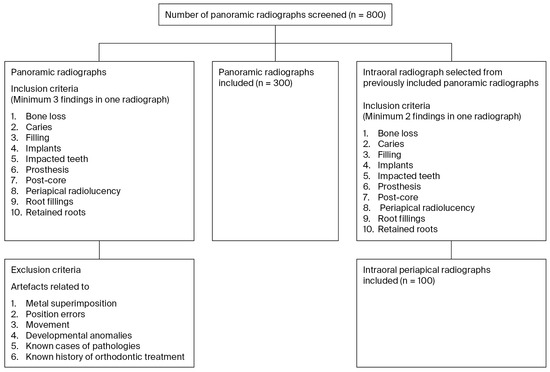
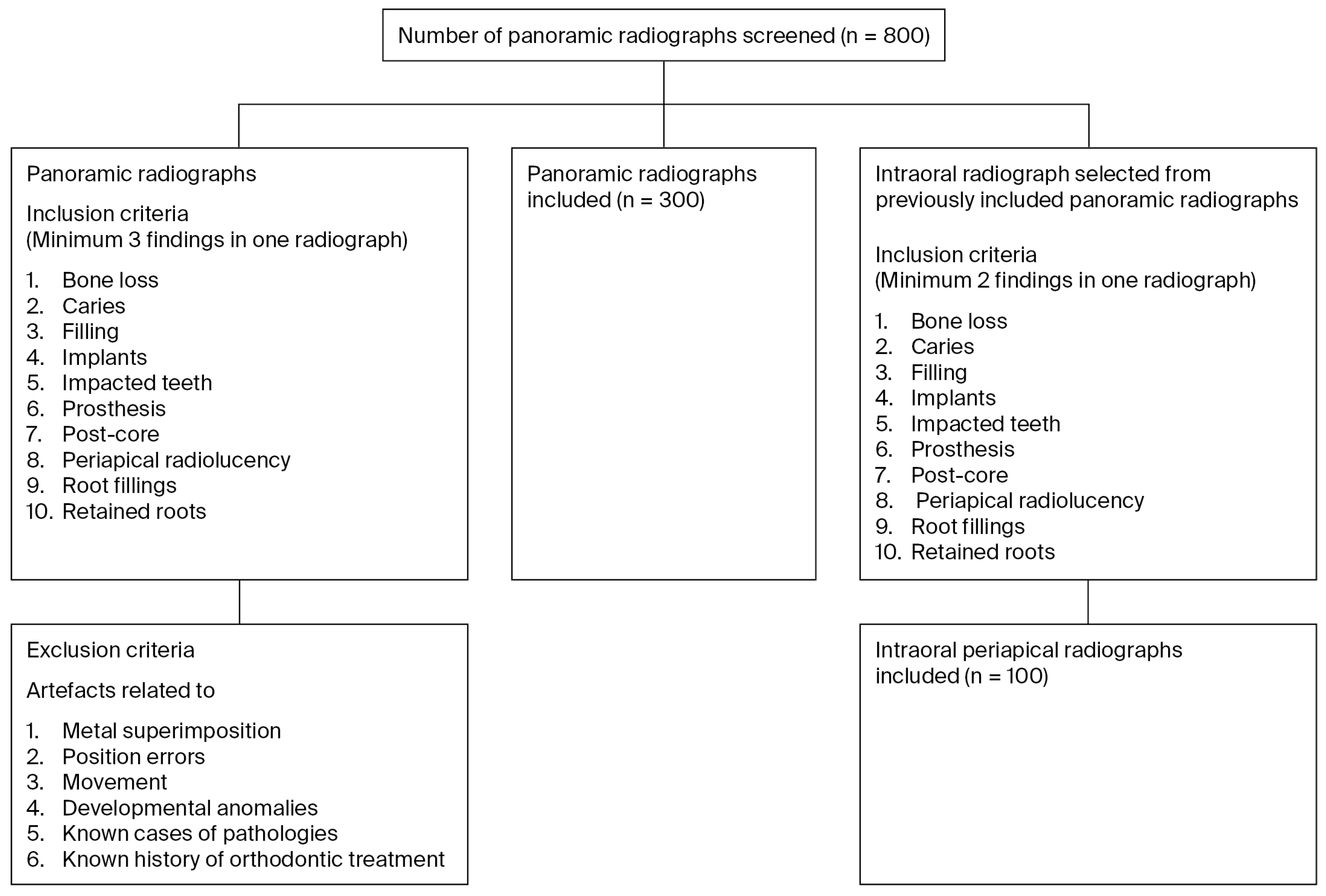
Initially, 800 panoramic radiographs were screened by a research assistant, and of these, 300 were included to ensure the presence of at least three identification parameters in one radiograph. The identification parameters are listed as follows: BL: bone loss; C: caries; F: filling; I: implants; IT: impacted teeth; P: prosthesis; PC: post-core; PR: periapical radiolucency; RF: root fillings; and RR: retained roots. Radiographs with artifacts related to metal superimposition, position errors, movement, developmental anomalies, known cases of pathologies, and known history of orthodontic treatment were excluded from this study. A corresponding intraoral periapical radiograph was selected from 100 randomly selected panoramic radiographs. It was ensured that each of the selected intraoral periapical radiographs had at least two of the included parameters. Two independent specialist evaluators with comparable clinical experience of more than 8 years (one specialist with postgraduate training and one with experience in the endodontic and pedodontic department) were trained in how to perform the radiographic identification by undergoing a calibration process. The flowchart for the selection process is depicted in Figure 1.
Figure 1.
Flowchart for the selection of radiographs.
During the calibration process, twenty radiographs were carefully selected to ensure the presence of all the diagnostic criteria of interest. Each specialist entered the findings as per the coding described as follows: BL: bone loss; C: caries; F: filling; I: implants; IT: impacted teeth; P: prosthesis; PC: post-core; PR: periapical radiolucency; RF: root fillings; and RR: retained roots. After the entries were made in separate sheets, the research assistant screened them for any discrepancies. In the event of any discrepancy, a meeting was called for the specialist evaluators to discuss the disagreement and reach a diagnostic consensus. The diagnostic consensus was specified as the “ground truth”. If no agreement was reached between the two leading operators, a third reviewer evaluated the radiograph to achieve consensus among the evaluators. The radiograph was excluded if consensus could not be reached. Subsequently, using the methodology employed for the calibration process, a comprehensive analysis of all the selected radiographs was performed, as outlined previously. The existing dental structures were labeled with Federation Dentaire International (FDI) nomenclature according to the following categories: BL: bone loss; C: caries; F: filling; I: implants; IT: impacted teeth; P: prosthesis; PC: post-core; PR: periapical radiolucency; RF: root fillings; and RR: retained roots. To assess bone levels, each mesial and distal surface of a tooth was marked as having bone loss of less than 33% (bone less absent) and greater than 33% (bone loss present) via visual estimation. The selected periapical radiographs (100) were also assessed in a similar way. Subsequently, the research assistant uploaded the images to the prototype. The specialist evaluators were blinded to the AI output. No modifications were made to the quality of the radiograph to ensure consistency between the identification environment for the specialists and the prototype. The detection threshold of the program was adjusted to 0%, which corresponds to the percentage of trustworthiness with which the software indicates the presence of any dental structure or treatment. Thus, the prototype was able to identify the structures regardless of the reliability percentage obtained.
2.4. Statistical Analysis
The diagnostic sensitivity, specificity, positive predictive value (PPV), negative predictive value (NPV), receiver operating characteristic curve (ROC), and area under the ROC of the tested dataset were calculated using Stata version 15.0 (StataCorp) for statistical analysis [17]. Descriptive analysis was also performed. For the panoramic radiographs, sensitivity, specificity, PPV, and NPV results were generated for specific sections, including overall, maxillary anterior, maxillary posterior, mandibular anterior, and mandibular posterior regions, for all the following outcomes: BL, C, F, I, IT, P, PC, PR, RF, and RR. ChatGPT version 5.0 was used to generate the figure displaying the overall test characteristics for all outcomes. For the intraoral periapical radiographs, the overall sensitivity, specificity, PPV, and NPV for the identification of any outcome present were calculated. Outcome-specific analyses were not performed due to the insufficient number of cases for each individual outcome.
3. Results
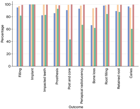
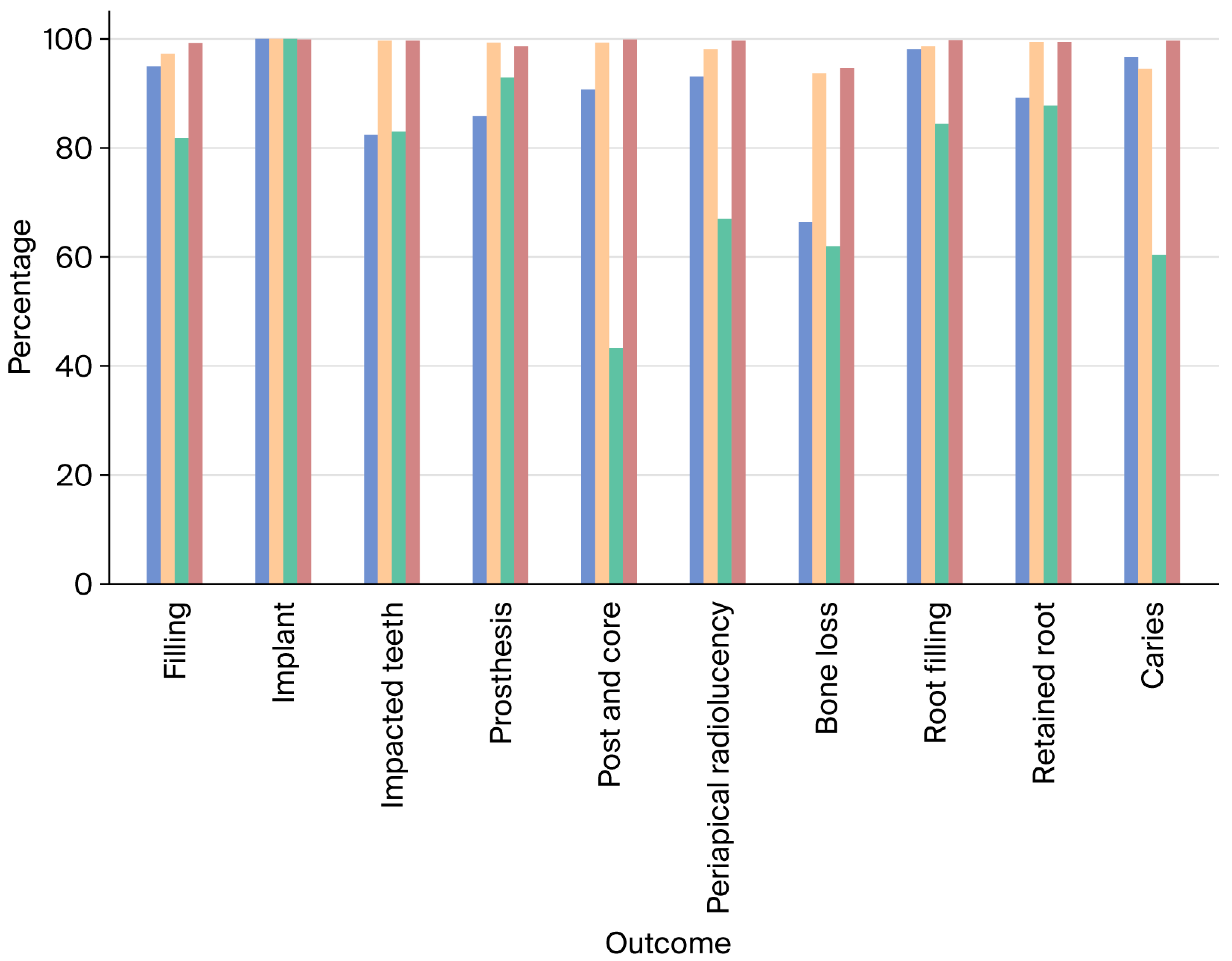
Overall, most of the outcomes demonstrated sensitivity greater than 82%, with values ranging from 66.41% for BL to 100% for I. For all outcomes, specificity was greater than 93%, with values ranging from 93.61% for BL to 100% for I. The overall values for all the test characteristics for the periapical radiographs were above 85%. The values for all the test characteristics across all the outcomes are provided in Figure 2.
Figure 2.
Test Characteristics (Overall) (In order: Sensitivity, Specificity, Positive Predictive Value, Negative Predictive Value). The confidence intervals for each value represented in the figure is provided in Table 1.
For the maxillary anterior region, most of the outcomes demonstrated sensitivity greater than 90%, with values ranging from 66.67% for IT to 100% for I. For all outcomes, specificity was greater than 93%, with values ranging from 93.84% for C to 100% for I. For the maxillary posterior region, most of the outcomes demonstrated high sensitivity (>82%). The sensitivity values ranged from 68.65% for BL to 100% for I. For all outcomes, specificity was high (>95%). Furthermore, the specificity values ranged from 95.09% for BL to 100% for I. The values for the test characteristics across all the outcomes are provided in Table 1.
Table 1.
Test characteristics.
For the mandibular anterior region, most of the outcomes demonstrated high sensitivity (>83%). The sensitivity values ranged from 83.33% for PR to 100% for I. For all outcomes, specificity was high (>88%). Furthermore, the specificity values ranged from 88.26% for BL to 100% for I. For the mandibular posterior region, most of the outcomes demonstrated high sensitivity (>80%). The sensitivity values ranged from 53.16% for BL to 100% for I. For all outcomes, specificity was high (>93%). Moreover, the specificity values ranged from 93.45% for C to 100% for I.
The ROCs for all the outcomes across all the regions are provided in Appendix A.
The qualitative analysis of the radiographs revealed various challenges, as summarized in Table 2.
Table 2.
Qualitative assessment of challenges.
For the periapical radiographs, the prototype demonstrated the following values for the test characteristics [Sensitivity = 85.26% (81.44,88.56), Specificity = 98.79% (97.52,99.51), PPV = 98.02% (95.95,99.04), NPV = 90.51% (88.30,92.33).
4. Discussion
The prototype software demonstrated high sensitivity and specificity in detecting dental pathologies [18].
Previous studies have shown high sensitivity in detecting dental caries from radiographs, with reported sensitivity values ranging from 72.26% to 94.15% [19,20,21,22,23,24,25,26,27,28,29,30,31,32,33,34,35,36].
The identification of endodontic features, such as root canal fillings and post-cores, has demonstrated reasonable accuracy. However, as highlighted in recent research, evaluating the quality of root canal fillings still requires further training [37].
The prototype effectively identified the presence or absence of dental implants on radiographs. Nonetheless, additional training is needed to ensure that classification of implant types is accurate, success is predicted, and implant designs are optimized, consistent with the findings from prior studies that employed AI in implant dentistry [38,39,40,41,42,43,44].
Compared with previous investigations assessing periapical radiolucency, the prototype demonstrated similar sensitivity and improved specificity [45,46,47,48,49,50,51,52,53,54,55,56,57,58,59,60,61,62,63,64,65,66]. Notably, variations were observed among studies in terms of projection geometry, exposure parameters, film contrast, and film speed. Furthermore, differences in the radiographic data across studies must be acknowledged. These factors are crucial when interpreting pooled accuracy results from studies that use AI-based tools for detecting dental pathologies.
Sensitivity for detecting bone loss was generally lower than that for other outcomes. Prior deep learning-based studies have reported sensitivities ranging from 75% to 94%, with sample sizes varying from 100 to 2276 panoramic radiographs [66,67,68,69,70]. As per the revised periodontal disease classification, the criteria employed to determine radiographic bone loss are divided into three categories: <15%, 15–33%, and >33%. In the current study, the criteria were revised to include only two categories (<33% and >33%) to determine whether the prototype can dichotomously detect critical bone loss. Exact measurements were not made on the radiographs using digital measurement tools, as clinicians do not conventionally use accurate caliper-based measurements on each proximal root surface to determine the percentage of bone loss. Subjectivity may have also played a role when determining bone loss measurements. Additional refinement and training of the prototype are required to detect bone loss. However, radiographic bone loss on its own does not determine the clinical prognosis for the teeth, and so this must be supplemented with clinical measurements.
Sensitivity for identifying impacted teeth was lower compared with that for other outcomes. This may be attributed to the challenge of determining vertical impactions and also impactions with minimal mesial or distal tilt. However, a sensitivity greater than 80%, even in a diagnostically challenging clinical situation, may be considered acceptable. The identification of impacted teeth exclusively through radiographic evaluation may also be challenging for clinicians. Ideally, a combination of clinical and radiographic examinations must be performed [71]. Sensitivity for detecting impacted teeth was significantly lower in the maxillary anterior region, primarily due to the failure to detect impacted maxillary canines. The incidence of canine impactions is reported to be higher in the maxillary region than in the mandibular region, with a higher incidence reported in females [72]. The training of the prototype may have been primarily focused on detecting impacted teeth in the posterior region. Further training is required to include case types in the anterior region of the jaws, specifically canines and premolars.
The prototype found it challenging to differentiate extensive mesio-occluso-distal amalgam fillings from crowns. Extensive radio-opacity in the coronal region presents a challenge when it comes to distinguishing between the delineated margins of a crown and the less sharp margins of a filling. Further challenges in identifying cervical margins may be incorporated using cervical burnout. Training the prototype with additional images of an appropriate resolution may help to improve its accuracy.
For the periapical radiographs, an outcome-based evaluation was not performed owing to the limited sample size achieved for each outcome. The overall test characteristics were calculated based on the number of findings accurately identified when compared with the ground truth. The prototype was determined to have a high accuracy based on the test characteristics, with values above 85%. Periapical radiographs are more accurate for the definitive identification of caries, bone loss, and similar findings that require higher-resolution images [73]. Therefore, the prototype may help the clinician when cross-checking their findings and may even indicate conclusions that might be missed due to an intense working schedule.
For the current study, two specialists and a reviewer were employed to determine the diagnostic consensus, the “ground truth”. A similar approach has been used previously to establish the baseline truth in radiographic identification from dental radiographs [74,75]. Future validation of the prototype may involve incorporating additional reviewers for agreement, particularly in the domains of bone loss detection and tooth impaction. It is recommended that the reference standards for validating AI-assisted radiographic detection follow the established guidelines, and they can be approached in a stage-wise manner [76,77].
PPV and NPV are functions of sensitivity, specificity, and prevalence. In general, as disease prevalence increases, so does the PPV. The low prevalence of individual outcomes, compared with the total data points, which amount to more than 8000 individual teeth, may be the reason for the lower PPVs for some outcomes in the current study. In low-prevalence populations, the number of true positives will be small compared with the number of false positives, even if the test is highly accurate at distinguishing between those who do and do not have the disease. As a result, the PPV drops despite the high sensitivity and specificity.
It has been reported previously that AI tools show high diagnostic accuracy in identifying a variety of intraoral pathologies from two-dimensional radiographs [78]. Testing the diagnostic accuracy of each newly introduced tool is imperative to safeguard the enhancement of the efficiency of these tools as a support system for clinicians. Reductions in false positives and negatives pave the way for enhanced accuracy. It has been found that AI-assisted radiographic identification can improve the diagnostic accuracy of clinicians examining panoramic radiographs, resulting in a decrease in false positives [79]. Furthermore, AI tools are capable of improving the efficiency of the diagnostic process by increasing speed and highlighting potential findings [80].
The limitations of the tool used in this study may be attributed to selection bias with the radiographs used for the training of the model, which may limit its generalizability. However, this may only be substantiated by further testing in diverse clinical practices internationally. The prototype shows promise for applications in supplementing clinicians with limited experience and reducing errors. Furthermore, standardized use of the prototype across a practice with clinicians with varying levels of experience may result in taking a standardized approach to documentation, thus minimizing errors with medico-legal implications.
5. Conclusions
The dental prototype used in this study demonstrated high sensitivity and specificity in identifying dental pathologies. However, further improvements are required to enhance the accuracy of the tool when identifying teeth that have migrated, including impacted canines, secondary caries, and differentiating extensive fillings from crowns.
Funding
This research was funded by EM2AI Pte. Ltd., UEN No. 201838370K, 2 Clementi Loop, #04-01, Logis Hub @ Clementi, Singapore 129809 (RESDEN-014). The article processing charge for the publication of this article was funded by Ajman University.
Institutional Review Board Statement
This study was conducted in accordance with the Declaration of Helsinki and approved by the Institutional Review Board of AJMAN UNIVERSITY (D-F-H-28-Aug, 26 August 2024).
Informed Consent Statement
Not applicable.
Data Availability Statement
The original contributions presented in this study are included in the article. Further inquiries can be directed to the corresponding author.
Acknowledgments
During the preparation of this manuscript and study, the author used ChatGPT version 5.0 for generating Figure 2, and refinement for English language proficiency. The author have reviewed the output for accuracy and terminology and takes full responsibility for the content of this publication. The results of this study were presented as an oral presentation at the FDI World Dental Congress, 2025, titled “Accuracy of An AI-Assisted Dental Prototype in Radiographic Detection”.
Conflicts of Interest
This research was funded by EM2AI Pte. Ltd., UEN No. 201838370K, 2 Clementi Loop, #04-01, Logis Hub @ Clementi, Singapore 129809. The company was not involved in any aspect of the research, including study design, selection of radiographs, data entry, analysis of findings, and interpretation of the results. This research was conducted at Ajman University, UAE.
Appendix A
Figure A1.
Receiver Operating Characteristic curve for filling in the maxillary posterior region.
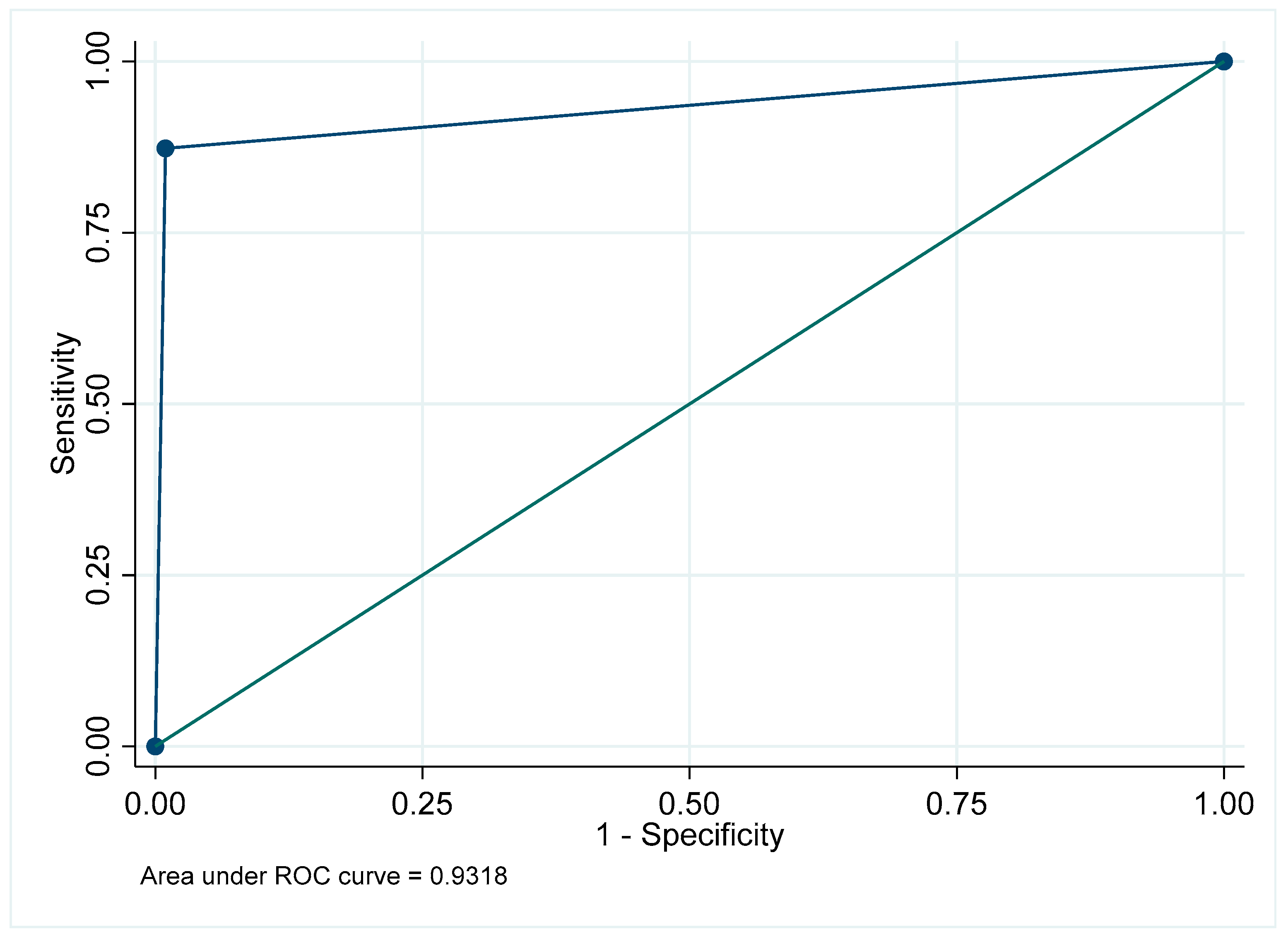
Figure A2.
Receiver Operating Characteristic curve for filling in the mandibular posterior region.
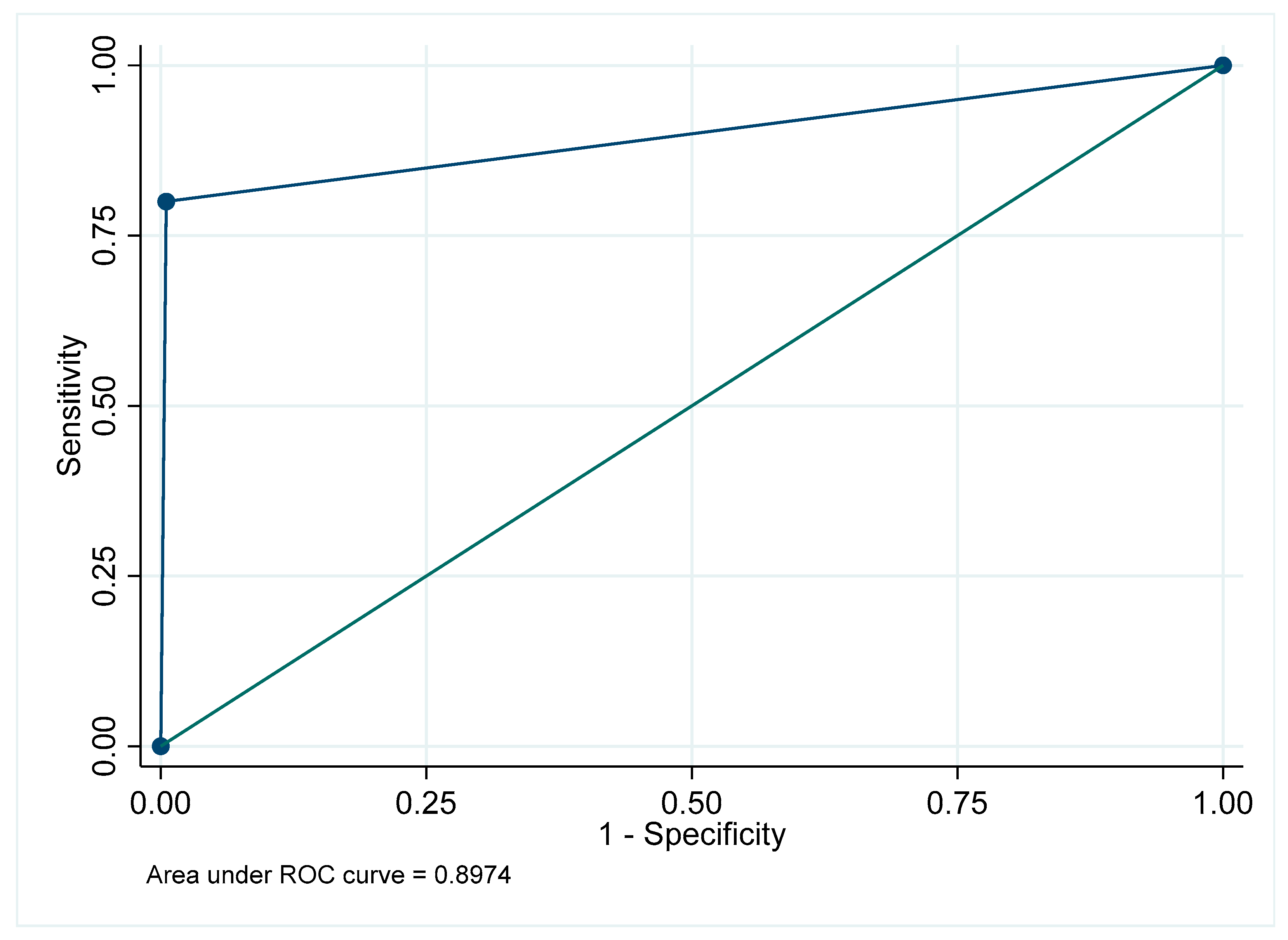
Figure A3.
Receiver Operating Characteristic curve for filling in the maxillary anterior region.
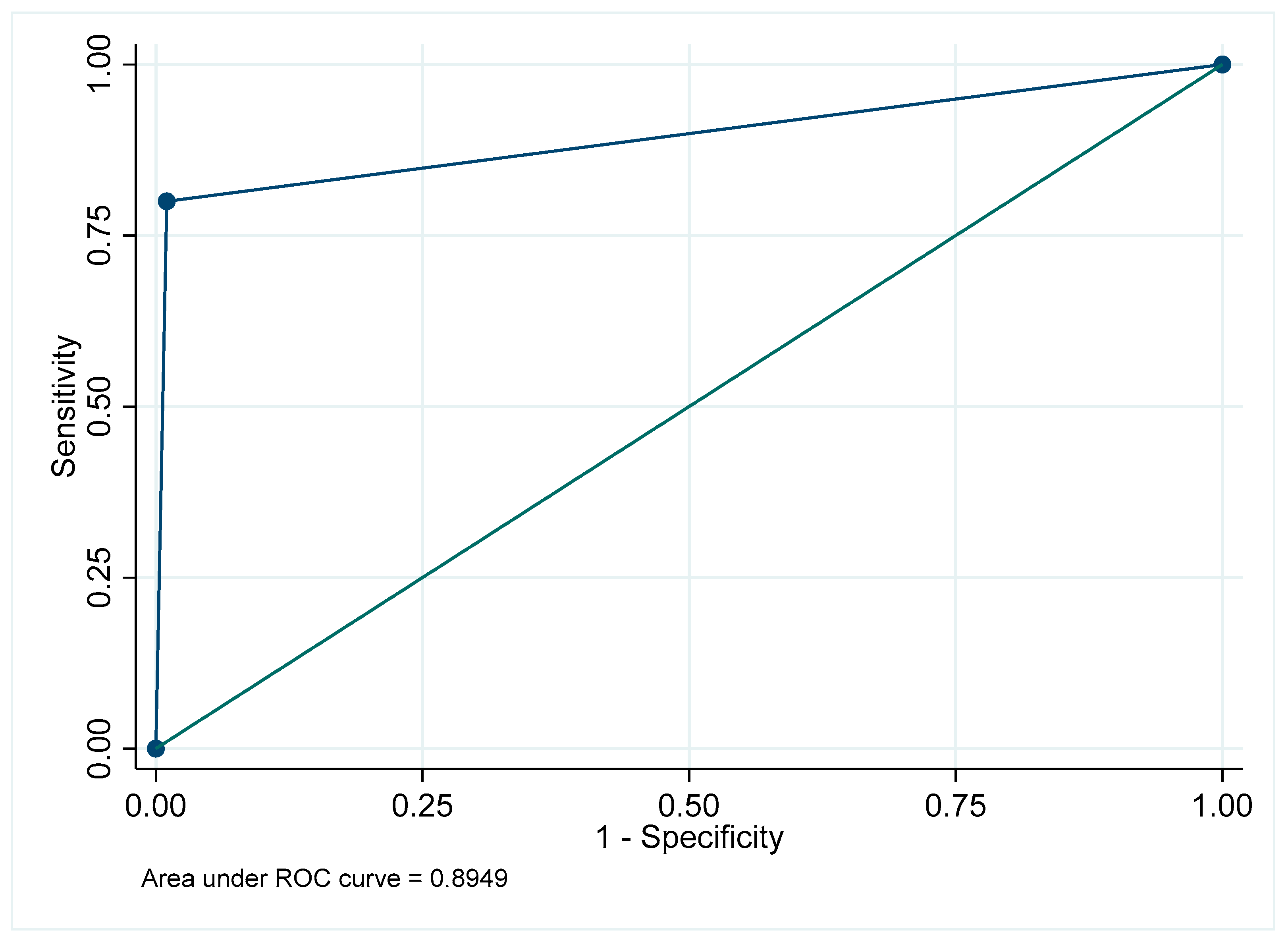
Figure A4.
Receiver Operating Characteristic curve for filling in the mandibular anterior region.
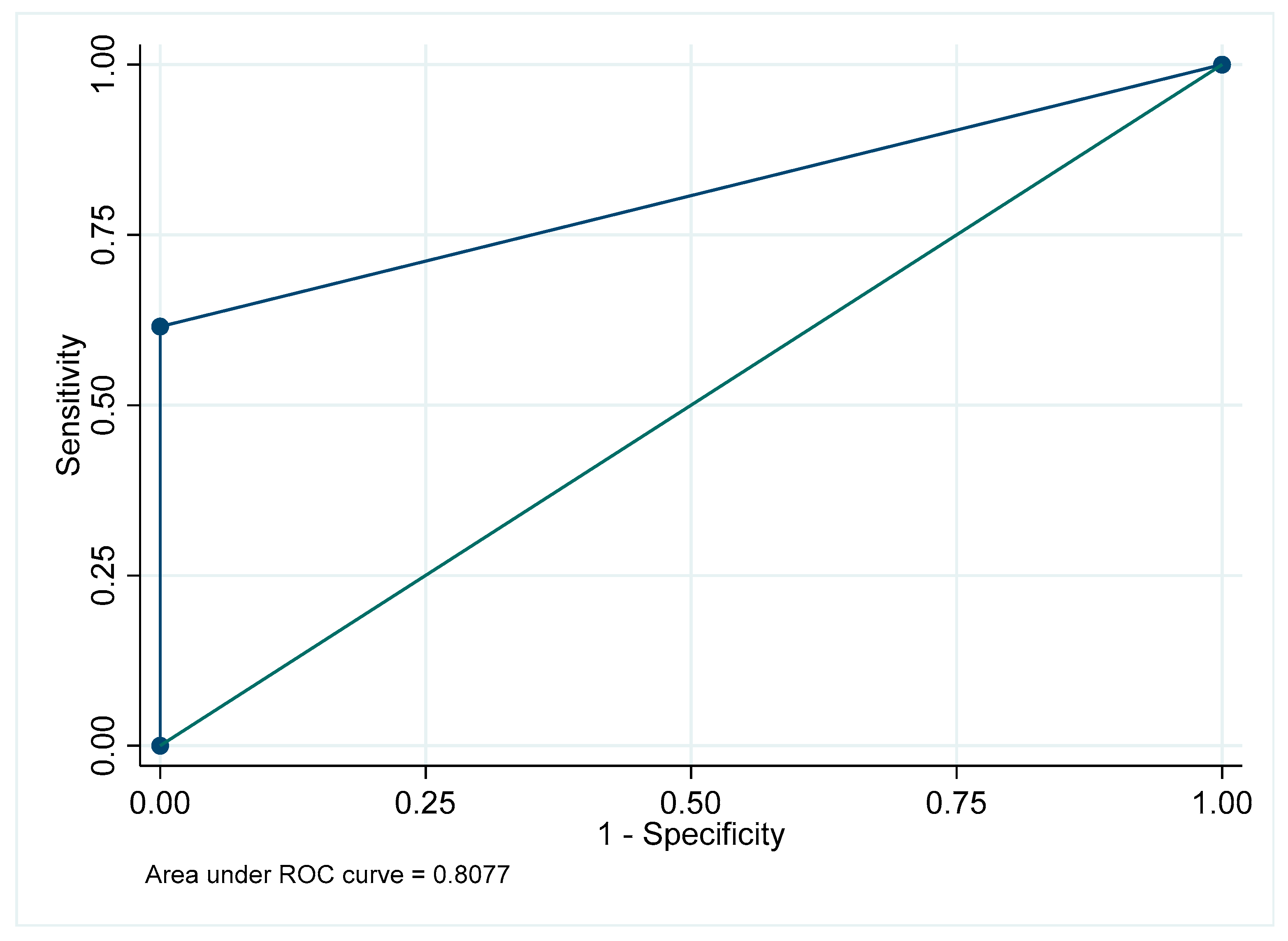
Figure A5.
Receiver Operating Characteristic curve for filling: Overall.
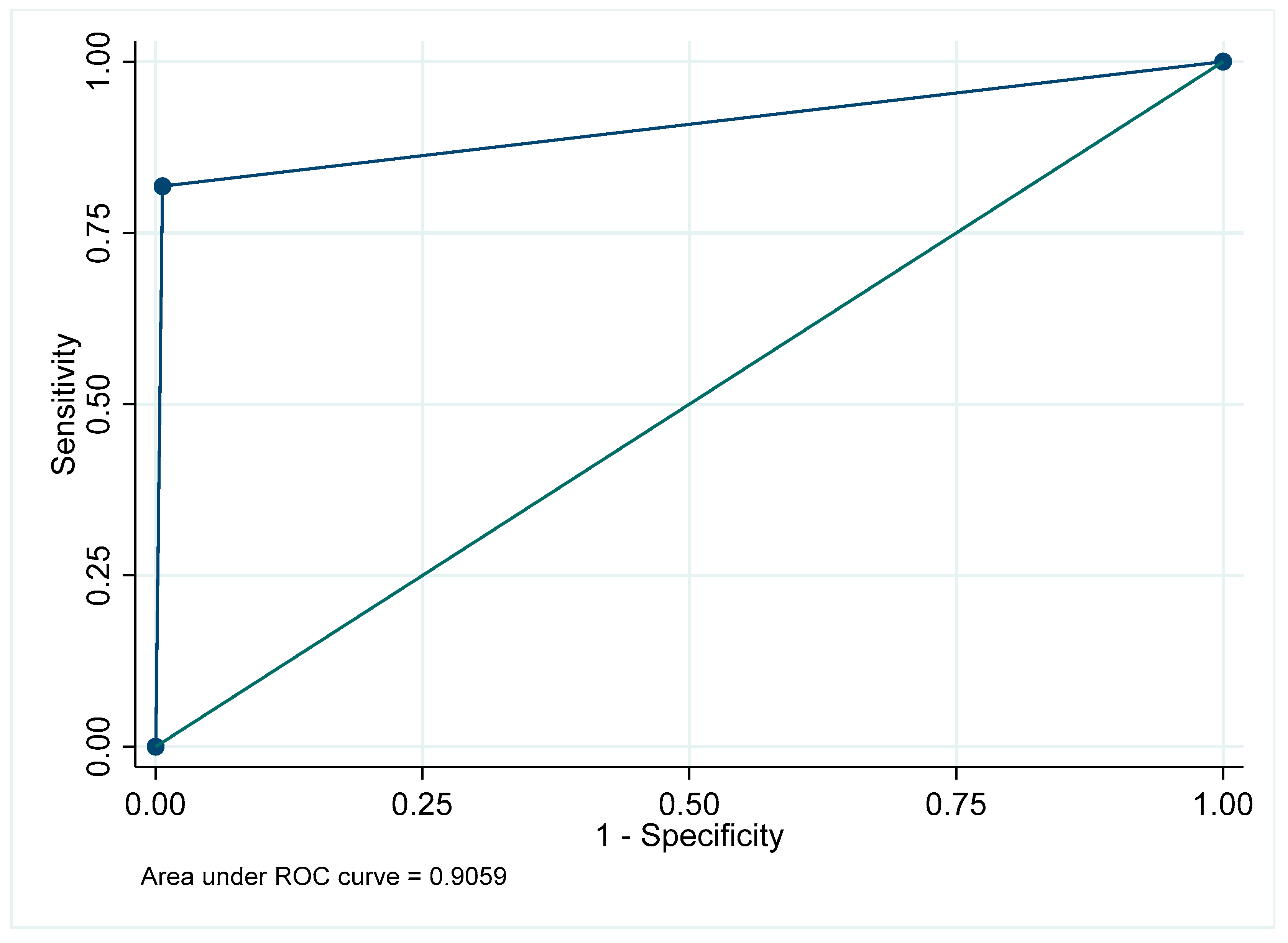
Figure A6.
Receiver Operating Characteristic curve for implants in the maxillary posterior region.
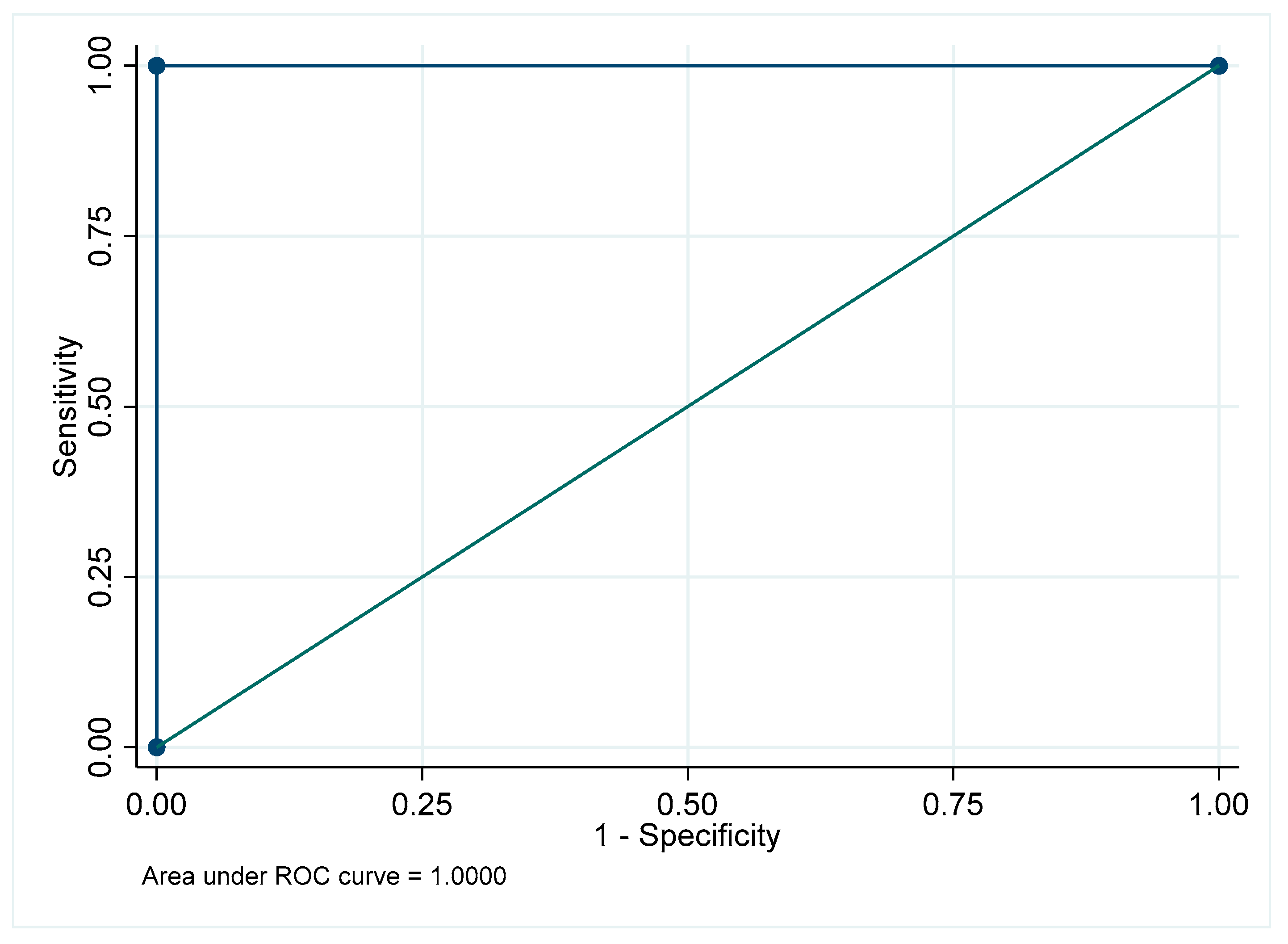
Figure A7.
Receiver Operating Characteristic curve for implants in the mandibular posterior region.
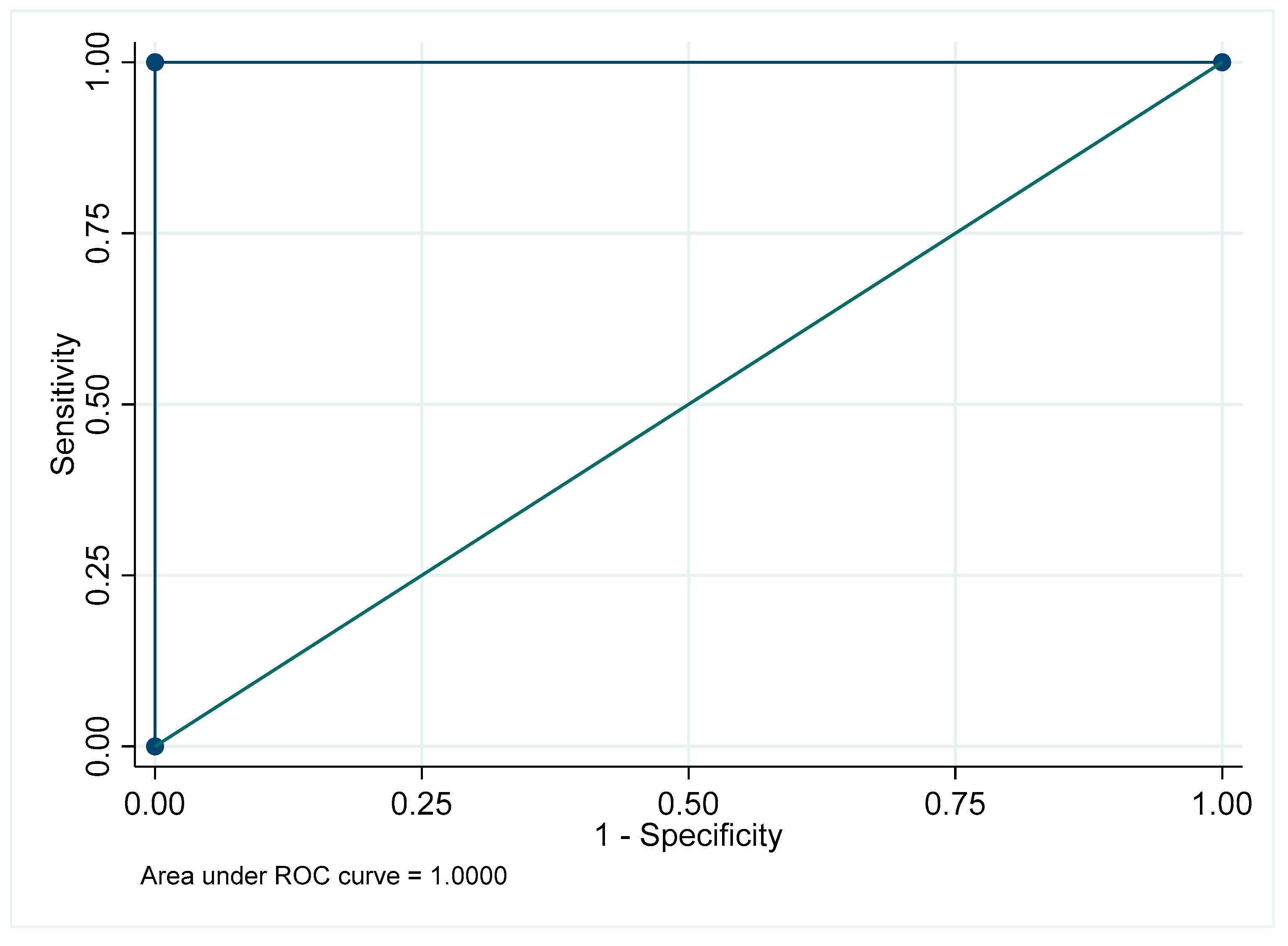
Figure A8.
Receiver Operating Characteristic curve for implants in the maxillary anterior region.
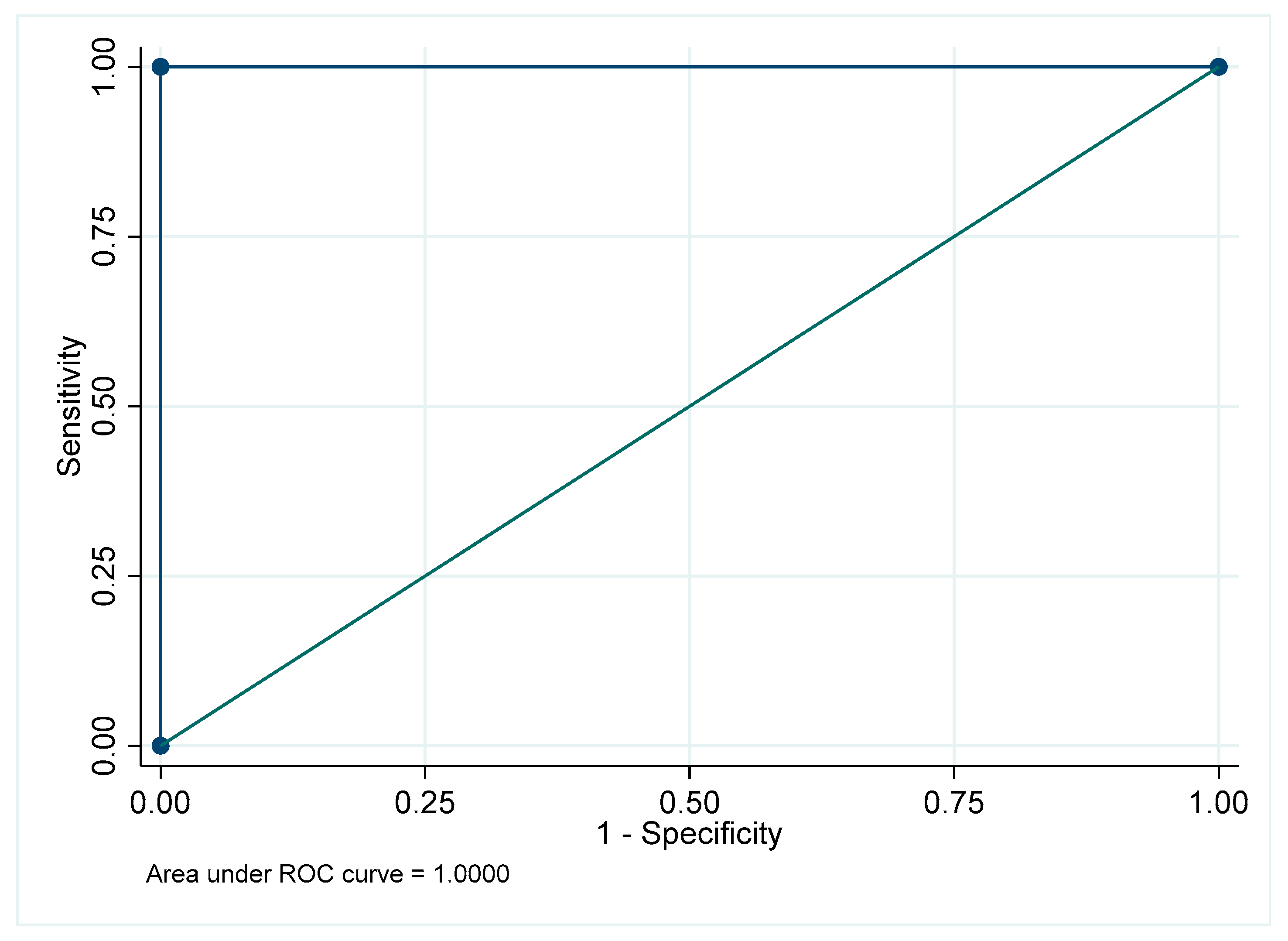
Figure A9.
Receiver Operating Characteristic curve for implants: Overall.
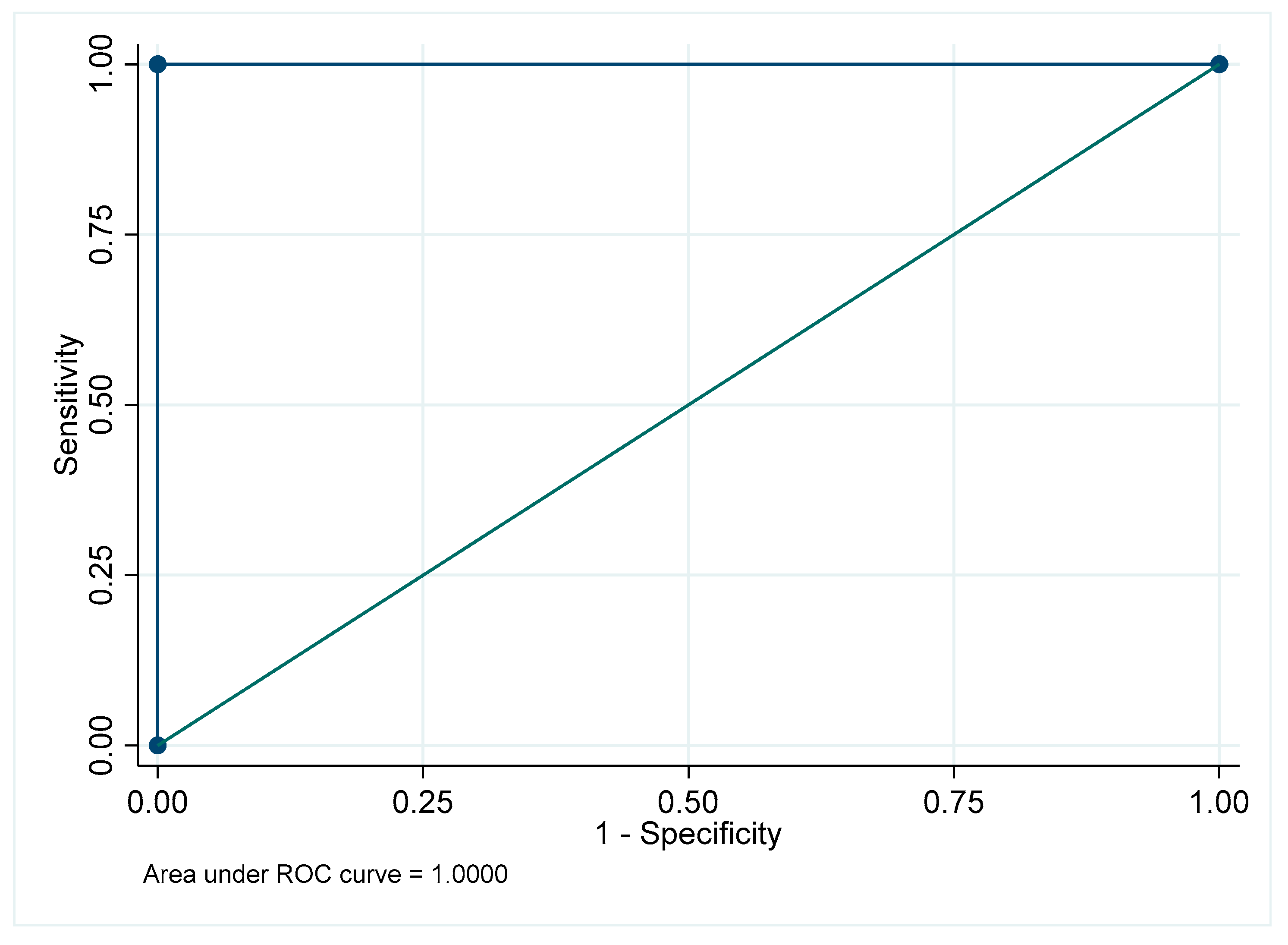
Figure A10.
Receiver Operating Characteristic curve for impacted teeth in the maxillary posterior region.
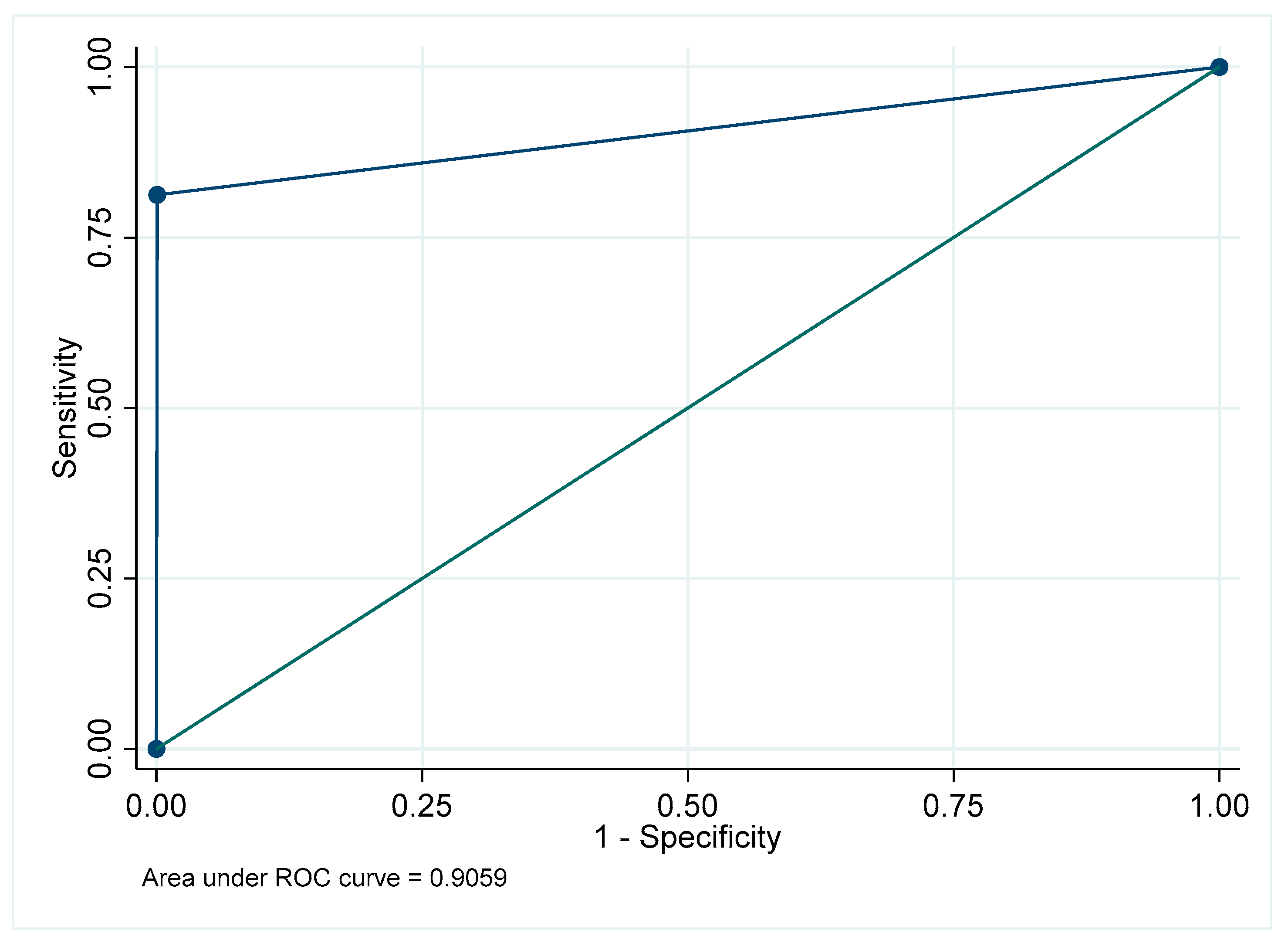
Figure A11.
Receiver Operating Characteristic curve for impacted teeth in the mandibular posterior region.
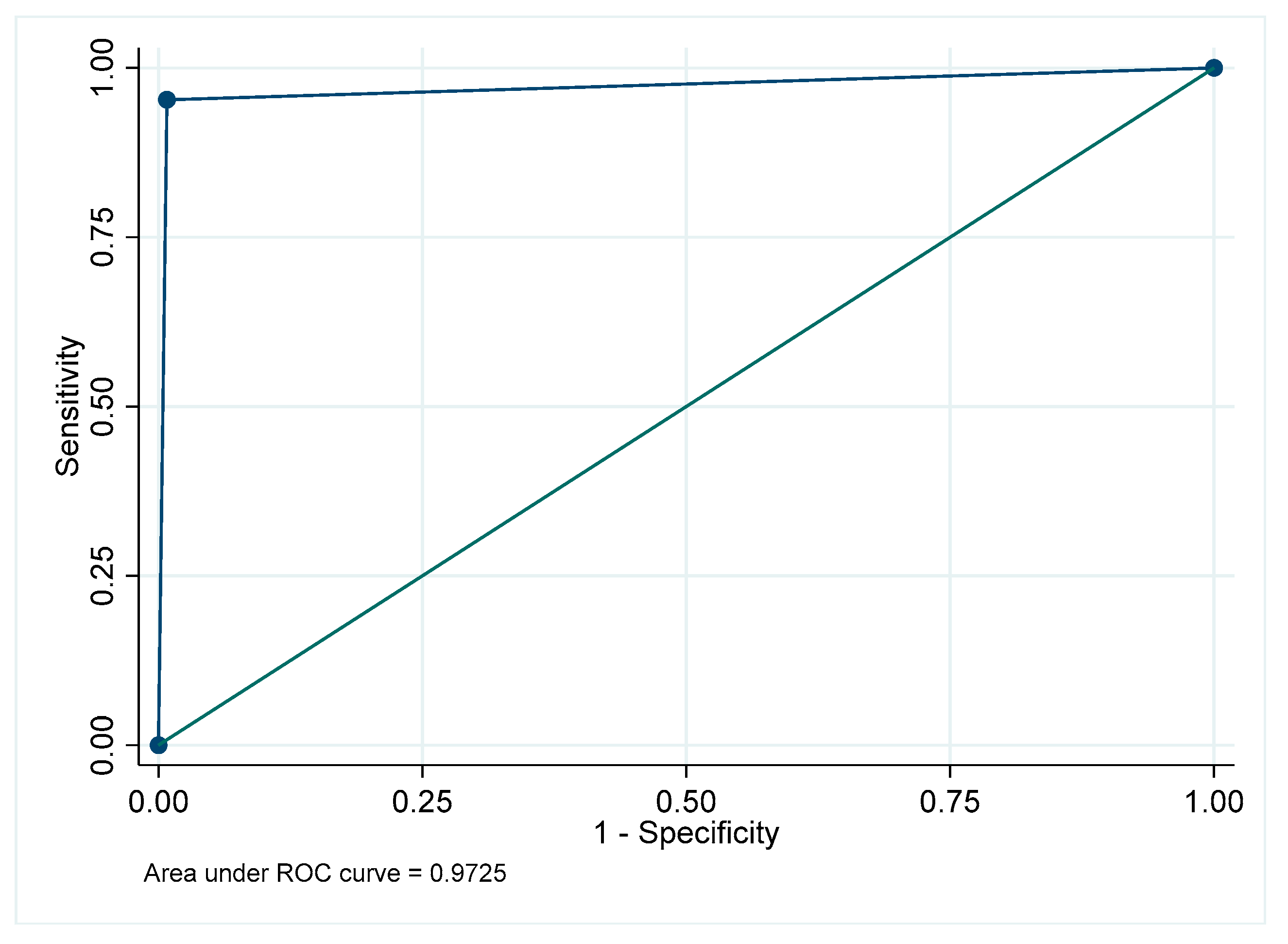
Figure A12.
Receiver Operating Characteristic curve for impacted teeth in the maxillary anterior region.
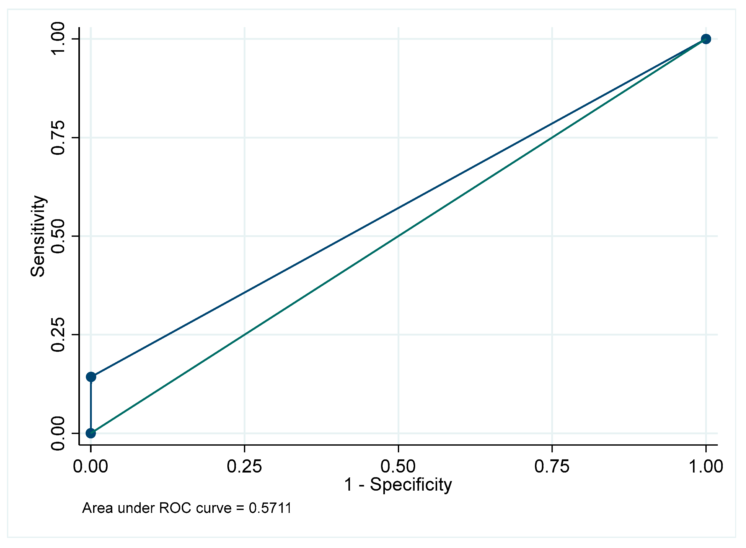
Figure A13.
Receiver Operating Characteristic curve for impacted teeth: Overall.
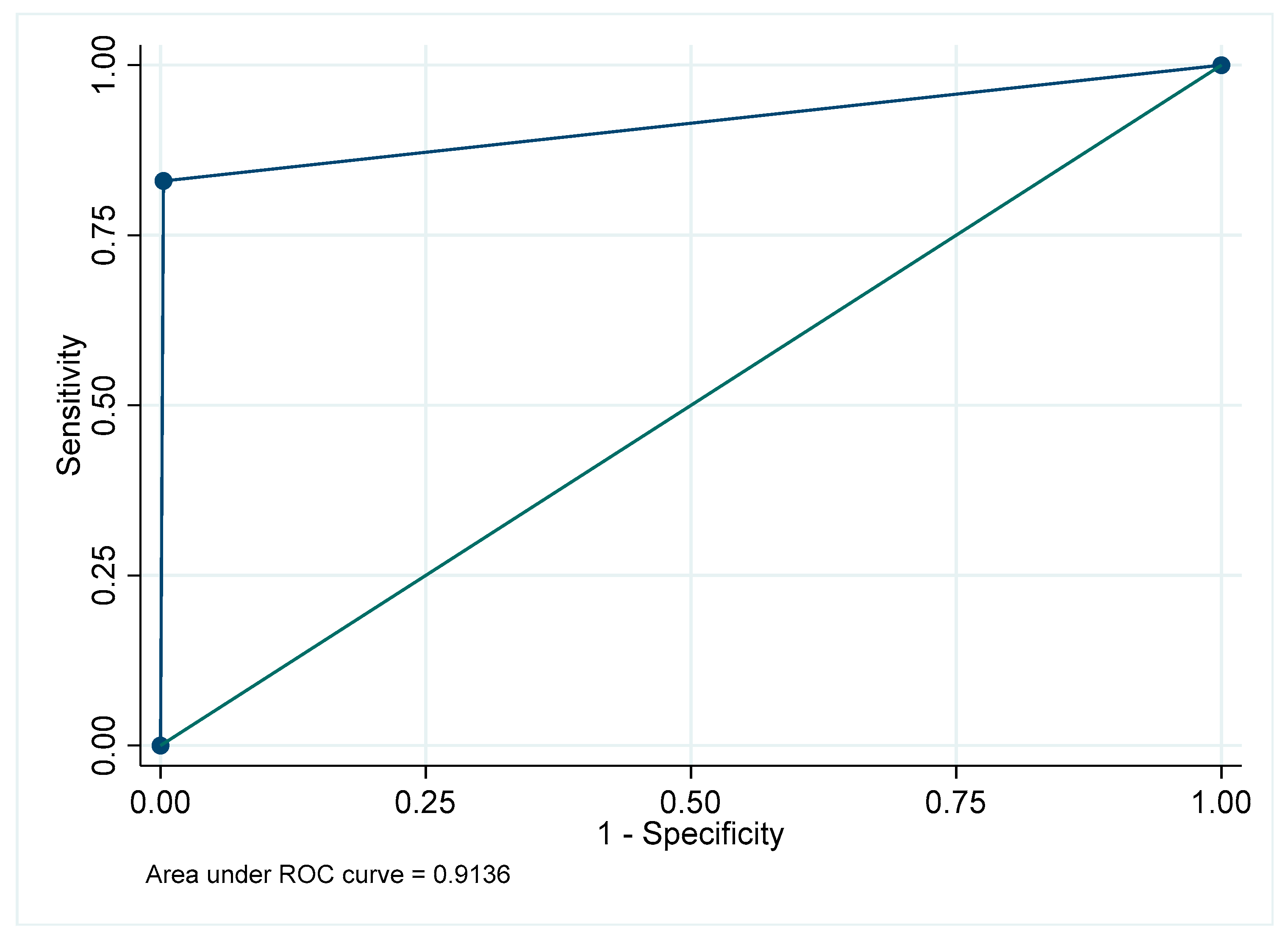
Figure A14.
Receiver Operating Characteristic curve for prosthesis in the maxillary posterior region.
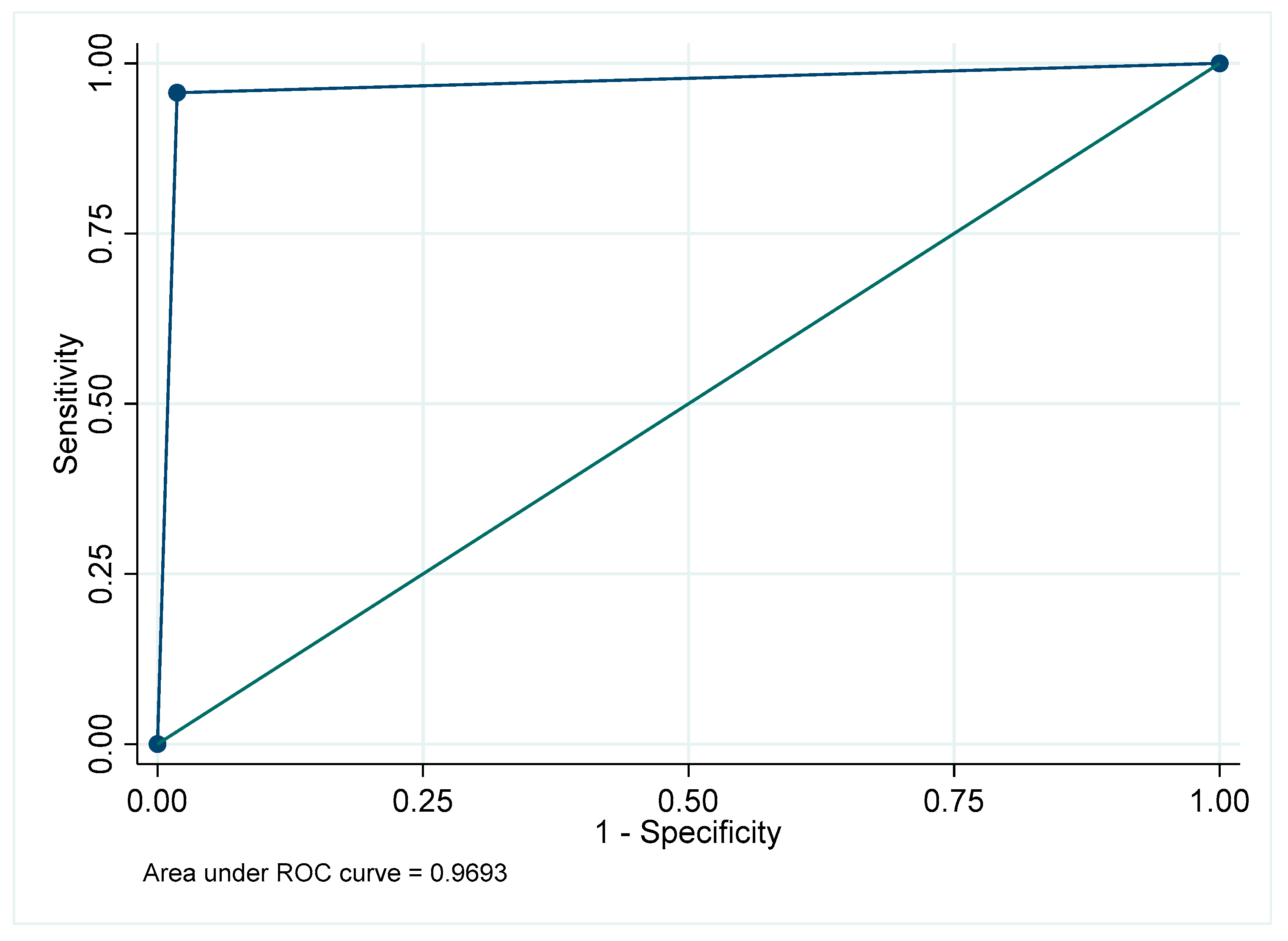
Figure A15.
Receiver Operating Characteristic curve for prosthesis in the mandibular posterior region.
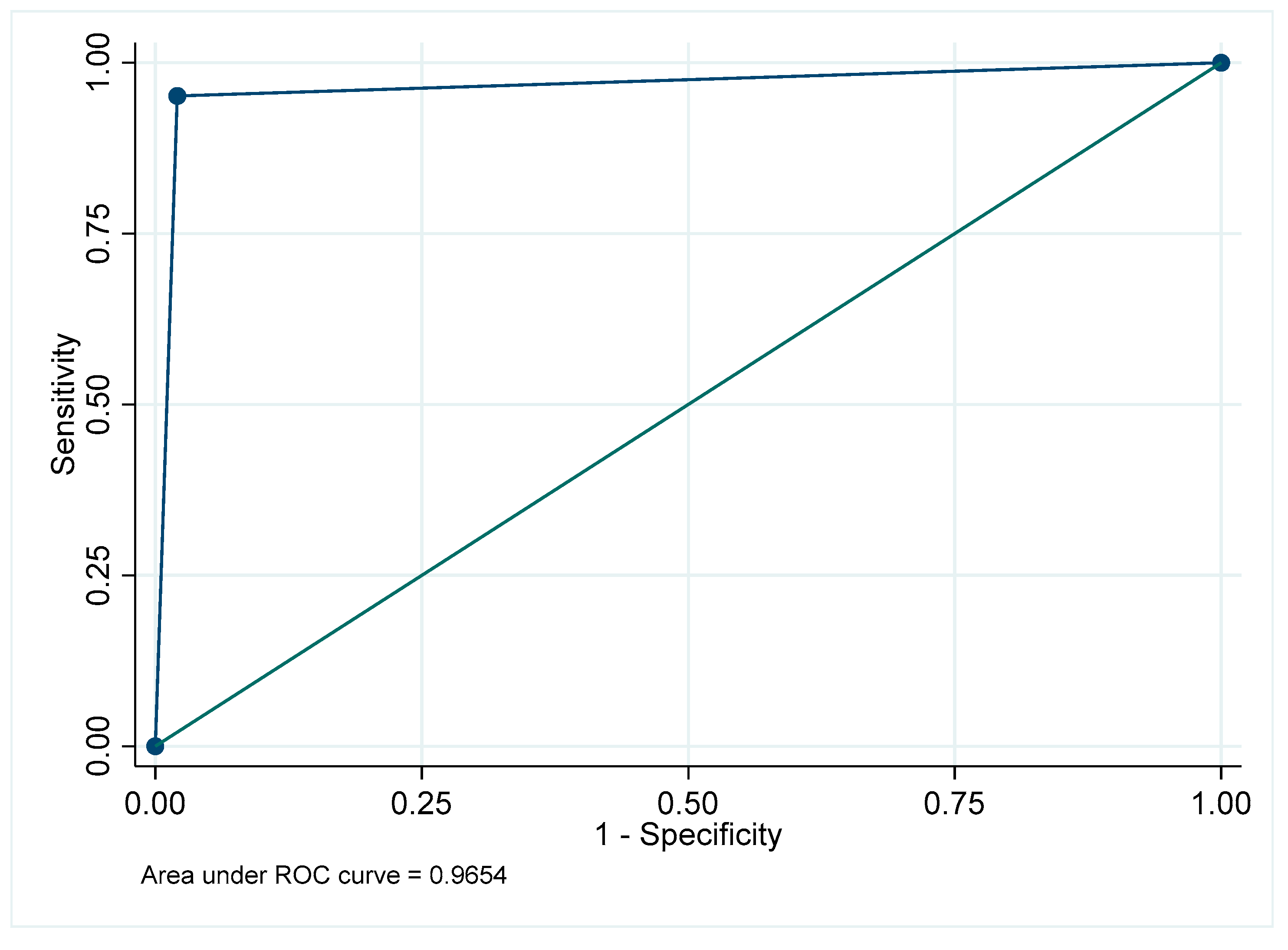
Figure A16.
Receiver Operating Characteristic curve for prosthesis in the maxillary anterior region.
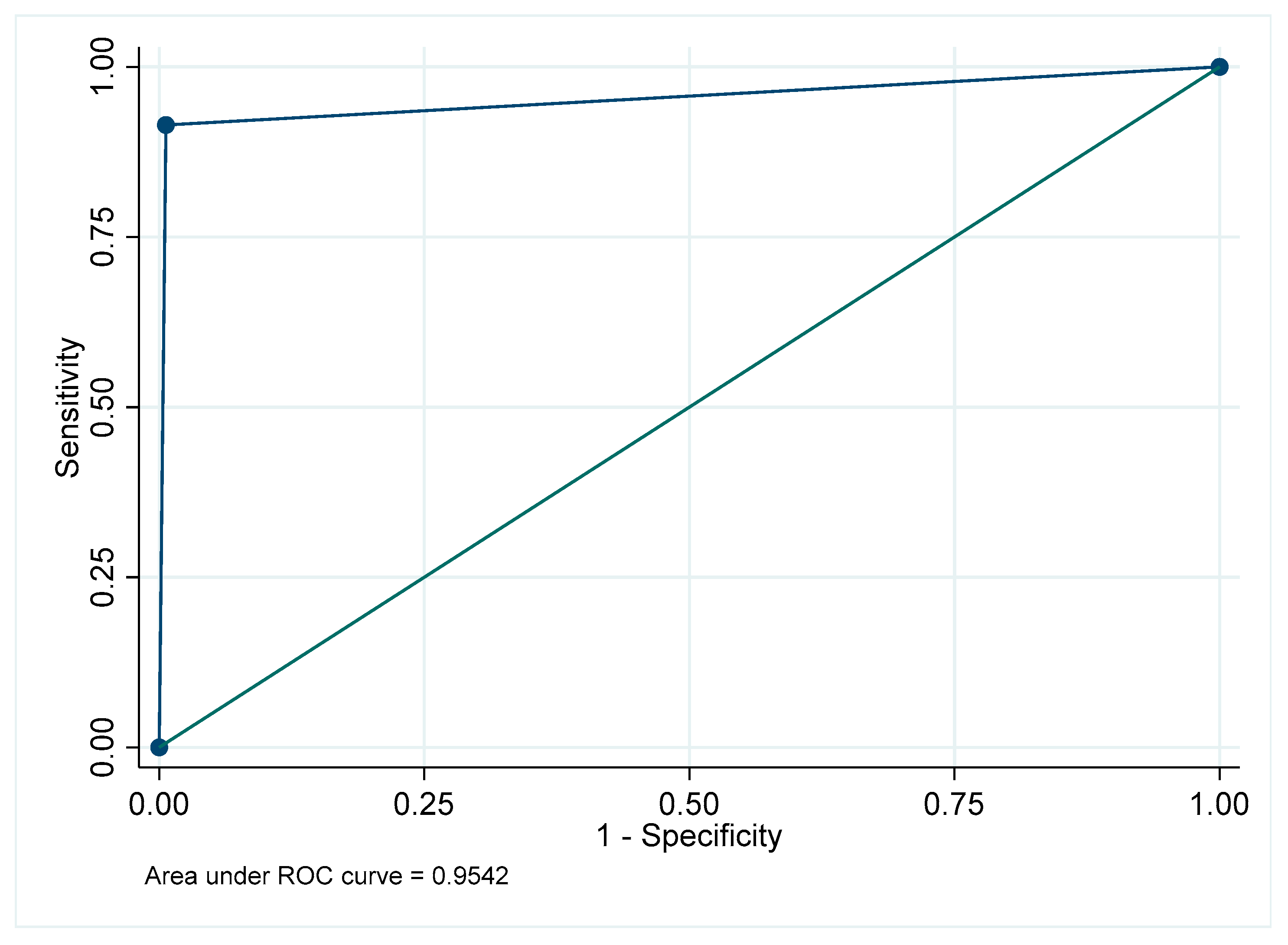
Figure A17.
Receiver Operating Characteristic curve for prosthesis in the mandibular anterior region.
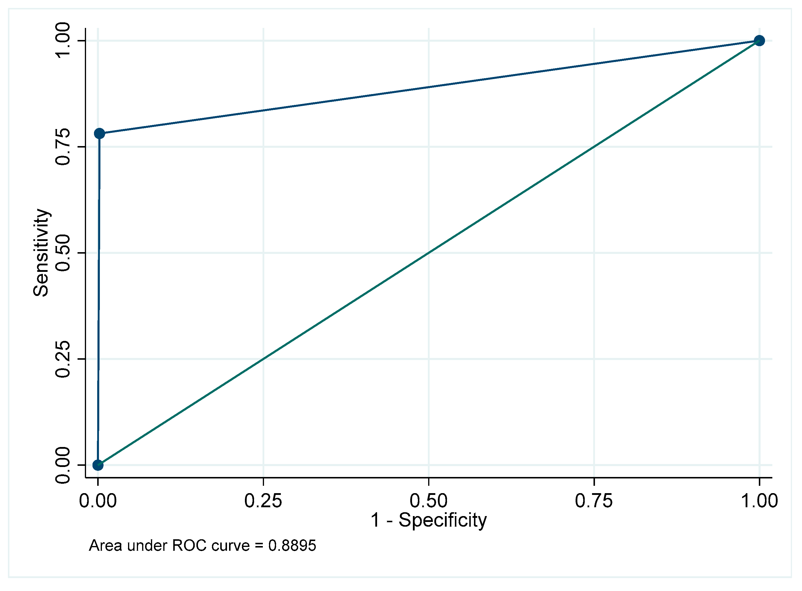
Figure A18.
Receiver Operating Characteristic curve for prosthesis: Overall.
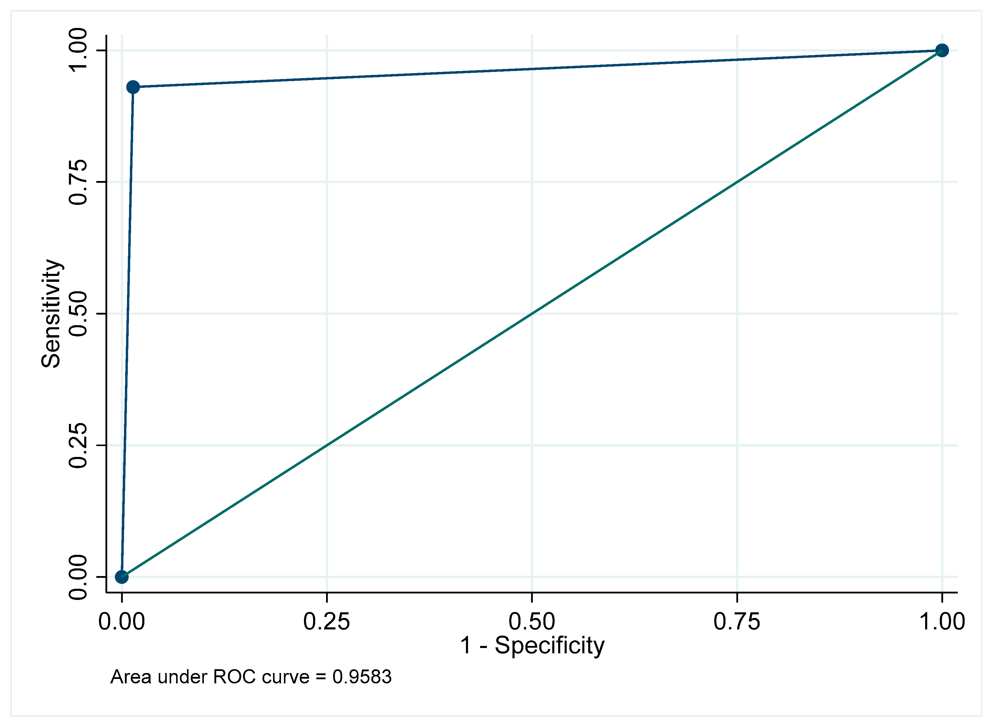
Figure A19.
Receiver Operating Characteristic curve for post and core in the maxillary posterior region.
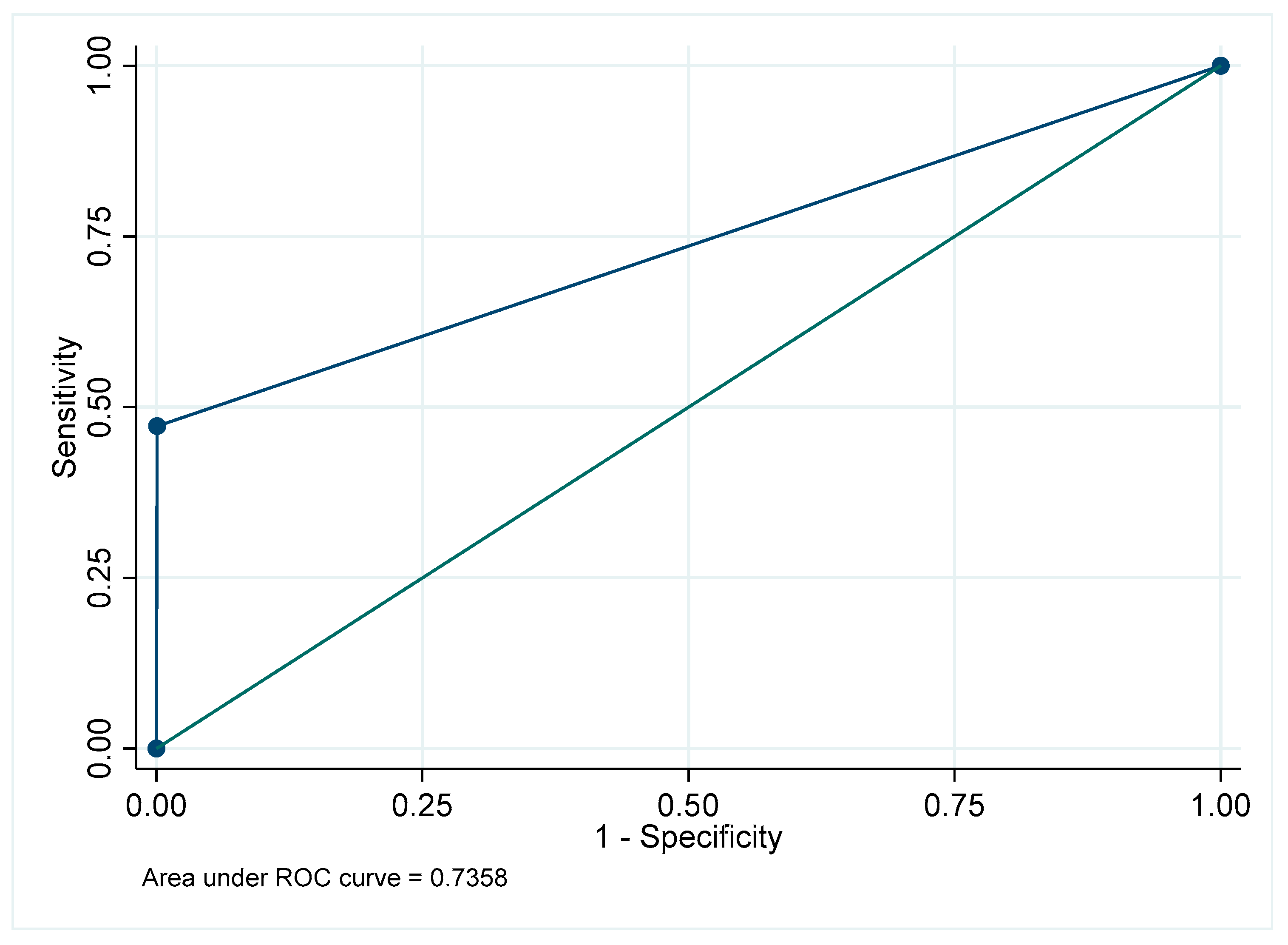
Figure A20.
Receiver Operating Characteristic curve for post and core in the mandibular posterior region.
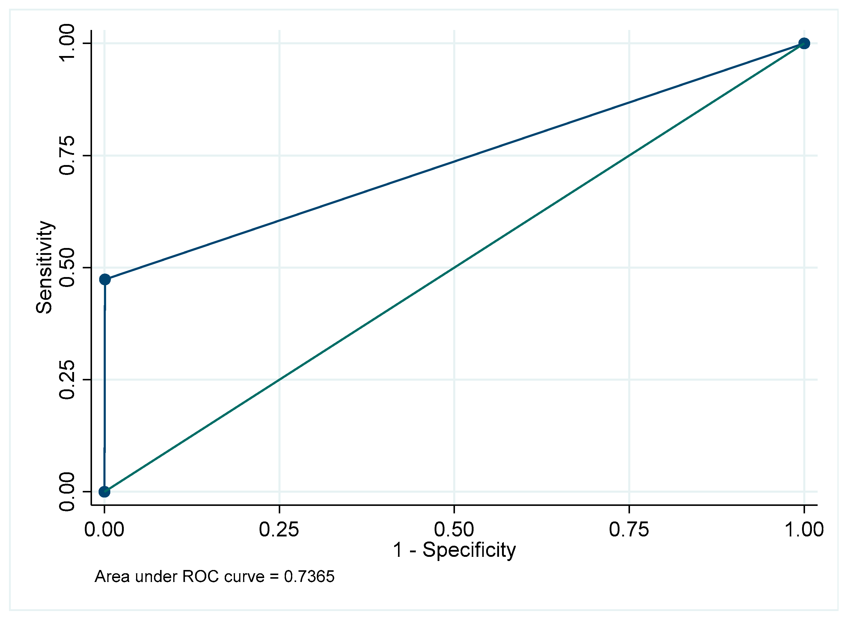
Figure A21.
Receiver Operating Characteristic curve for post and core in the maxillary anterior region.
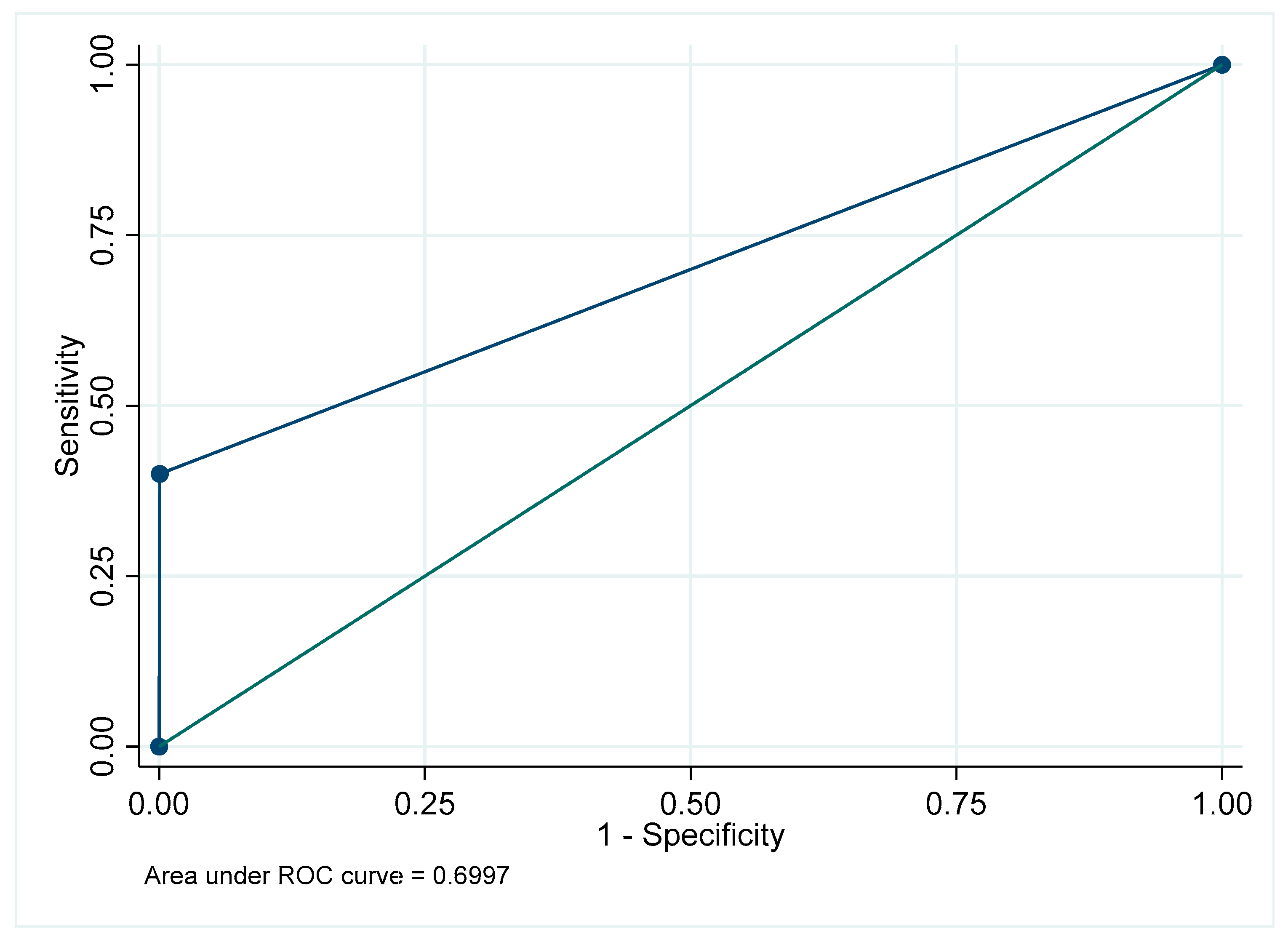
Figure A22.
Receiver Operating Characteristic curve for post and core in the mandibular anterior region.
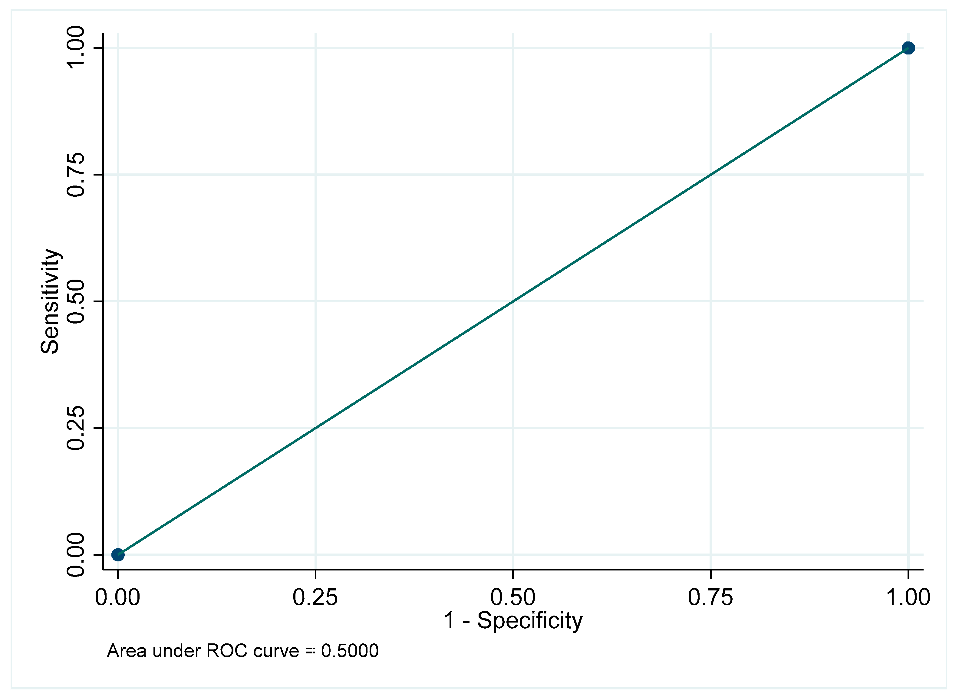
Figure A23.
Receiver Operating Characteristic curve for post and core: Overall.
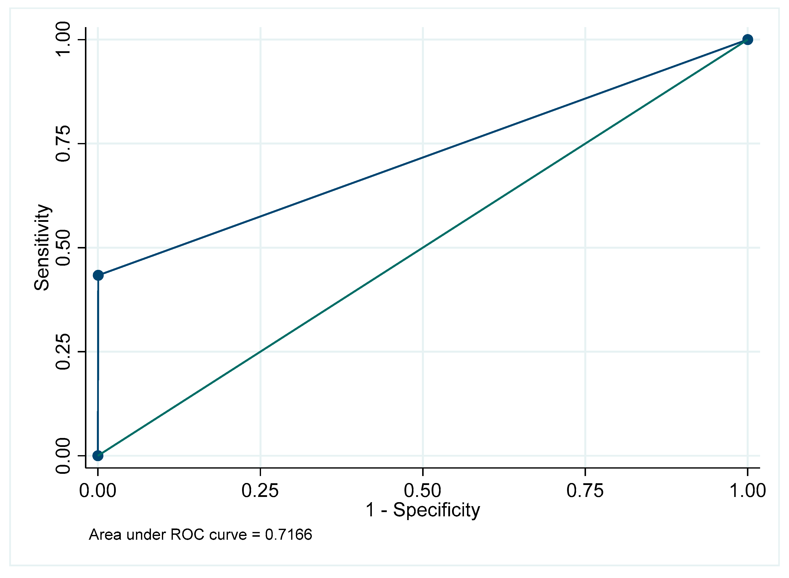
Figure A24.
Receiver Operating Characteristic curve for periapical radiolucency in the maxillary posterior region.
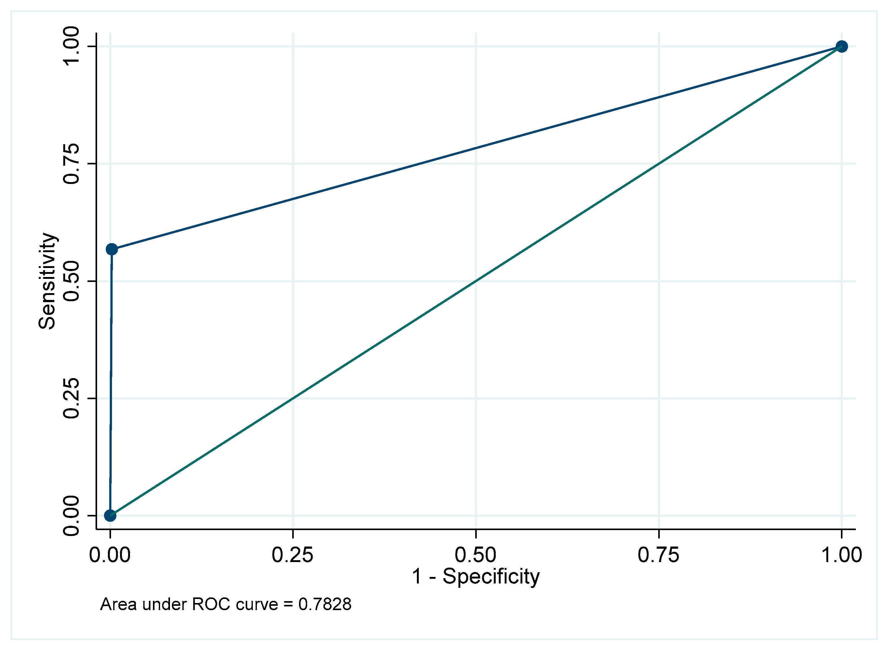
Figure A25.
Receiver Operating Characteristic curve for periapical radiolucency in the mandibular posterior region.
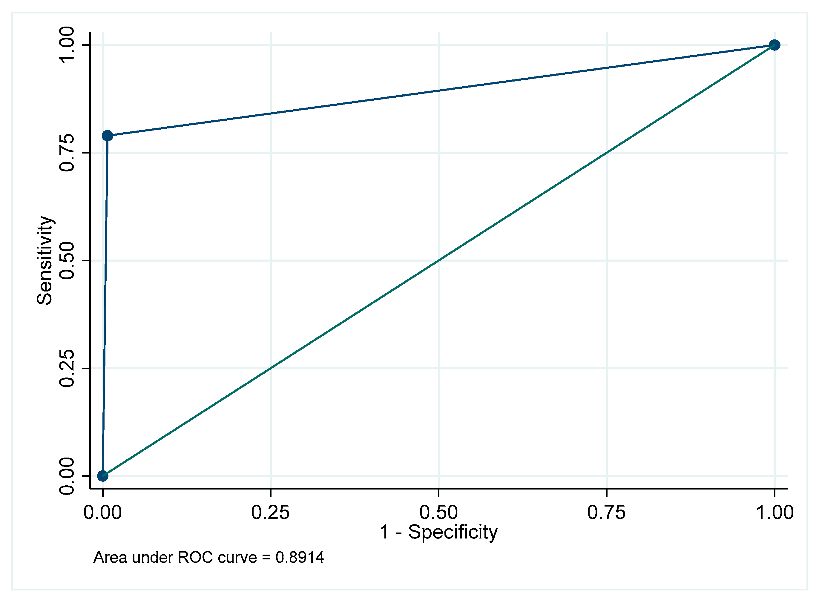
Figure A26.
Receiver Operating Characteristic curve for periapical radiolucency in the maxillary anterior region.
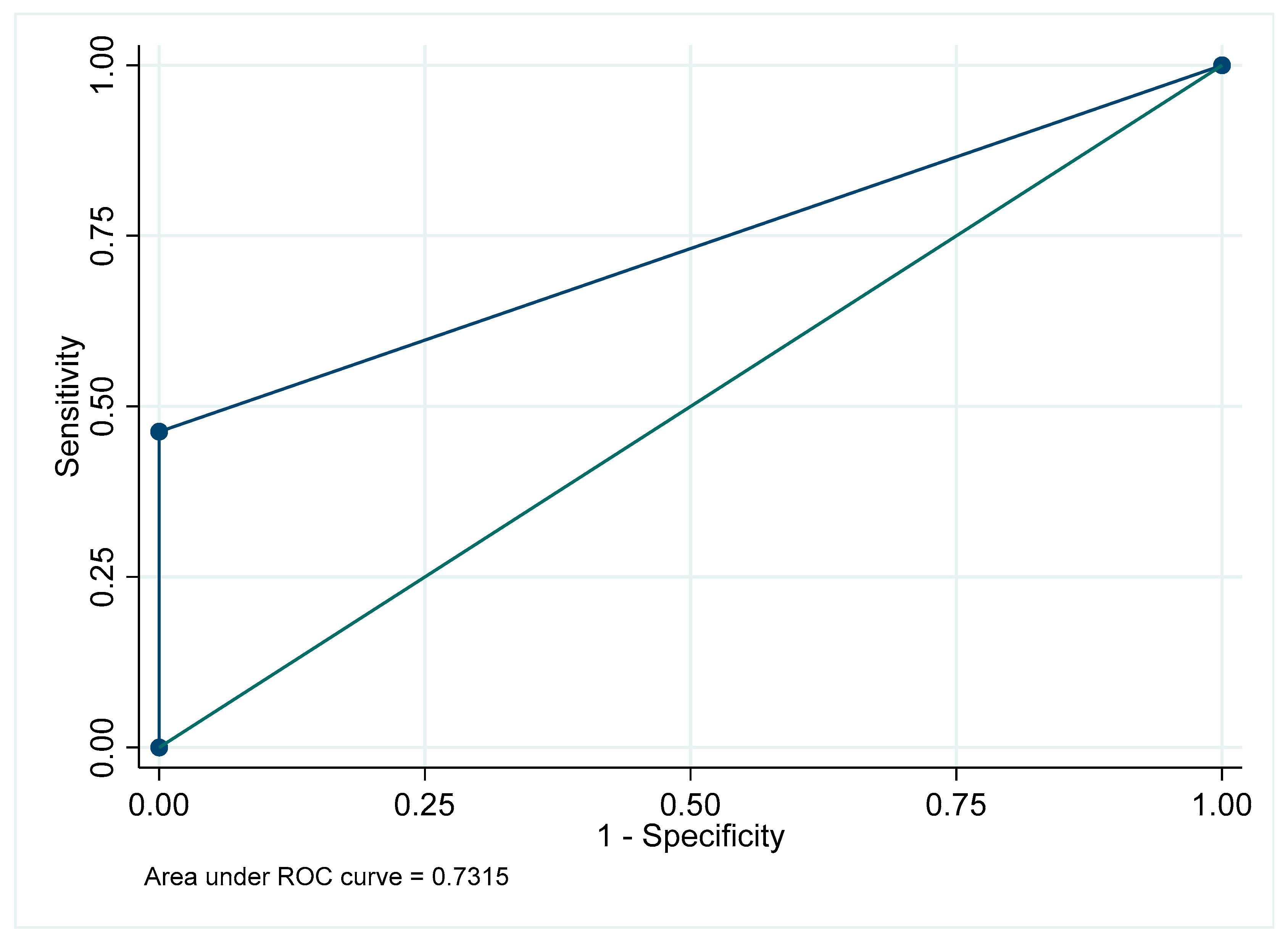
Figure A27.
Receiver Operating Characteristic curve for periapical radiolucency in the mandibular anterior region.
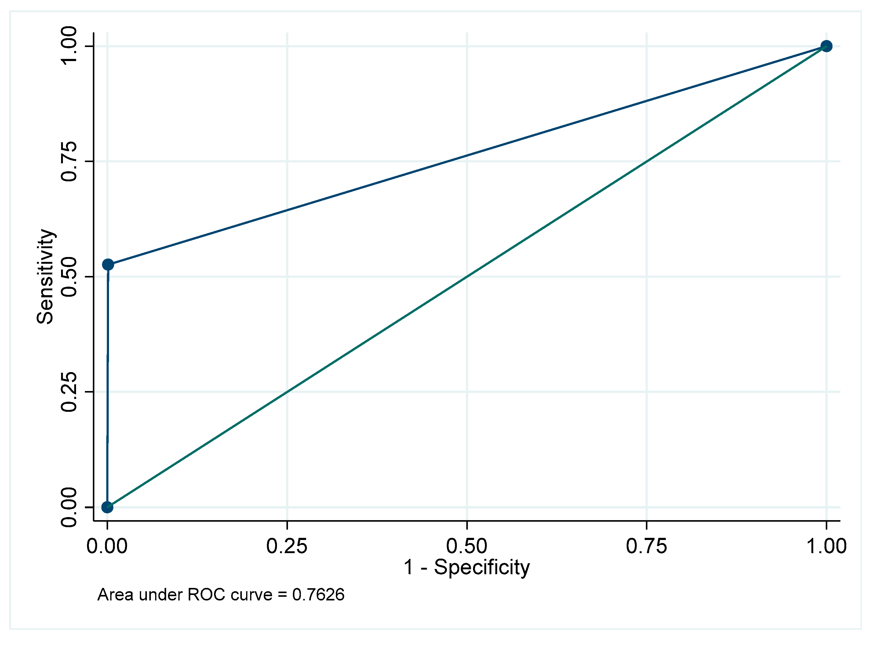
Figure A28.
Receiver Operating Characteristic curve for periapical radiolucency: Overall.
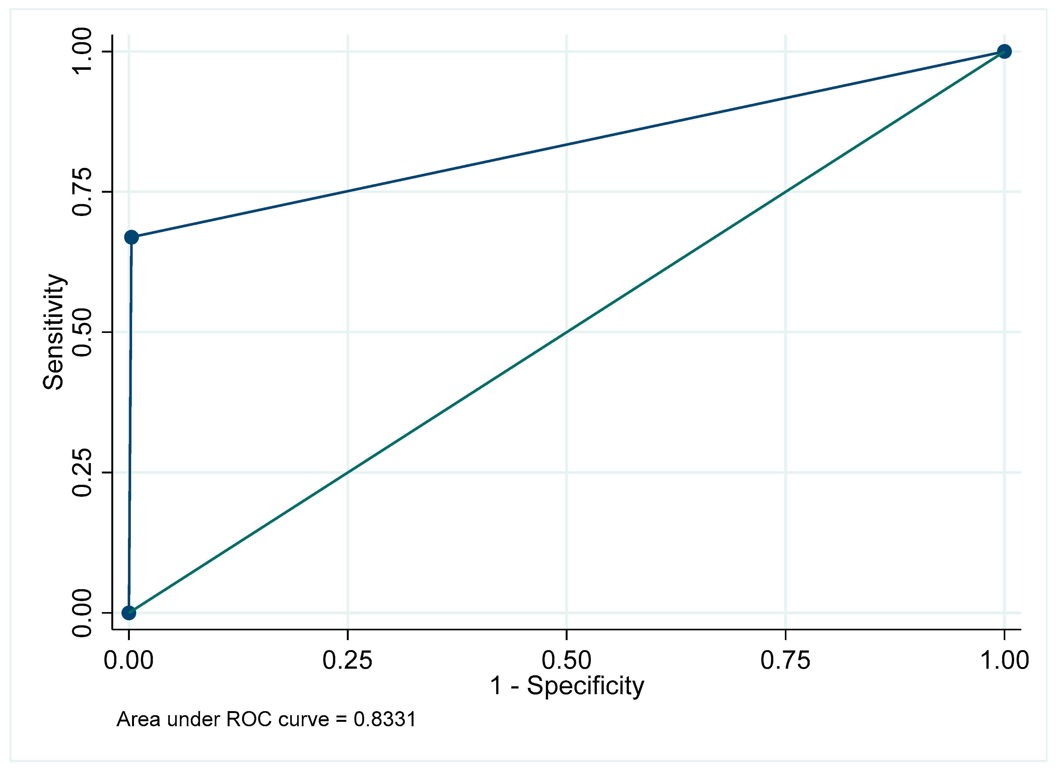
Figure A29.
Receiver Operating Characteristic curve for bone loss in the maxillary posterior region.
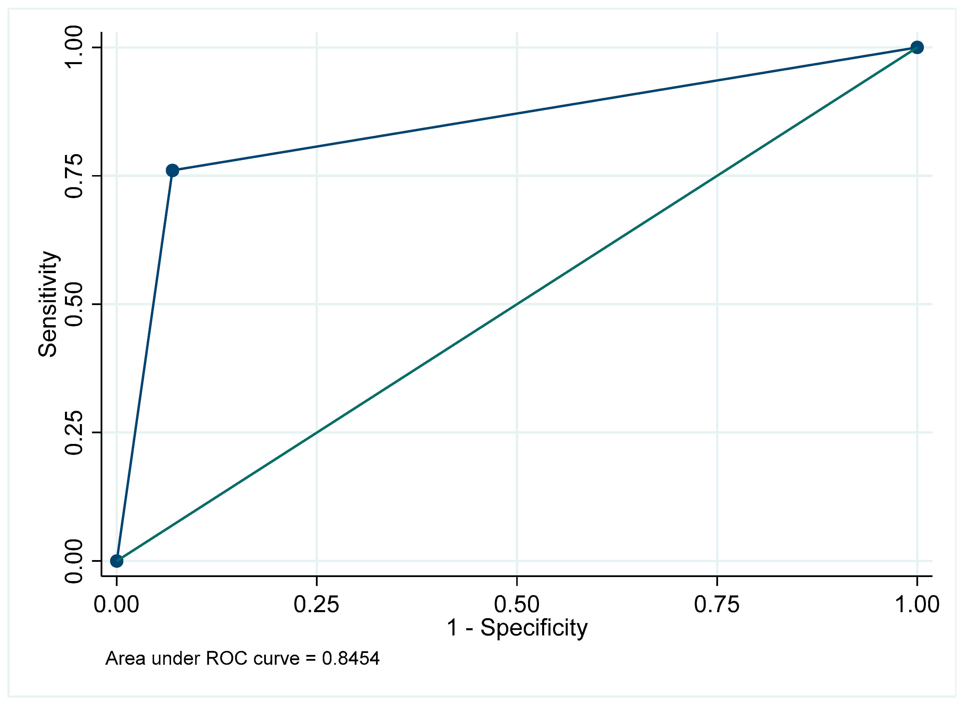
Figure A30.
Receiver Operating Characteristic curve for bone loss in the mandibular posterior region.
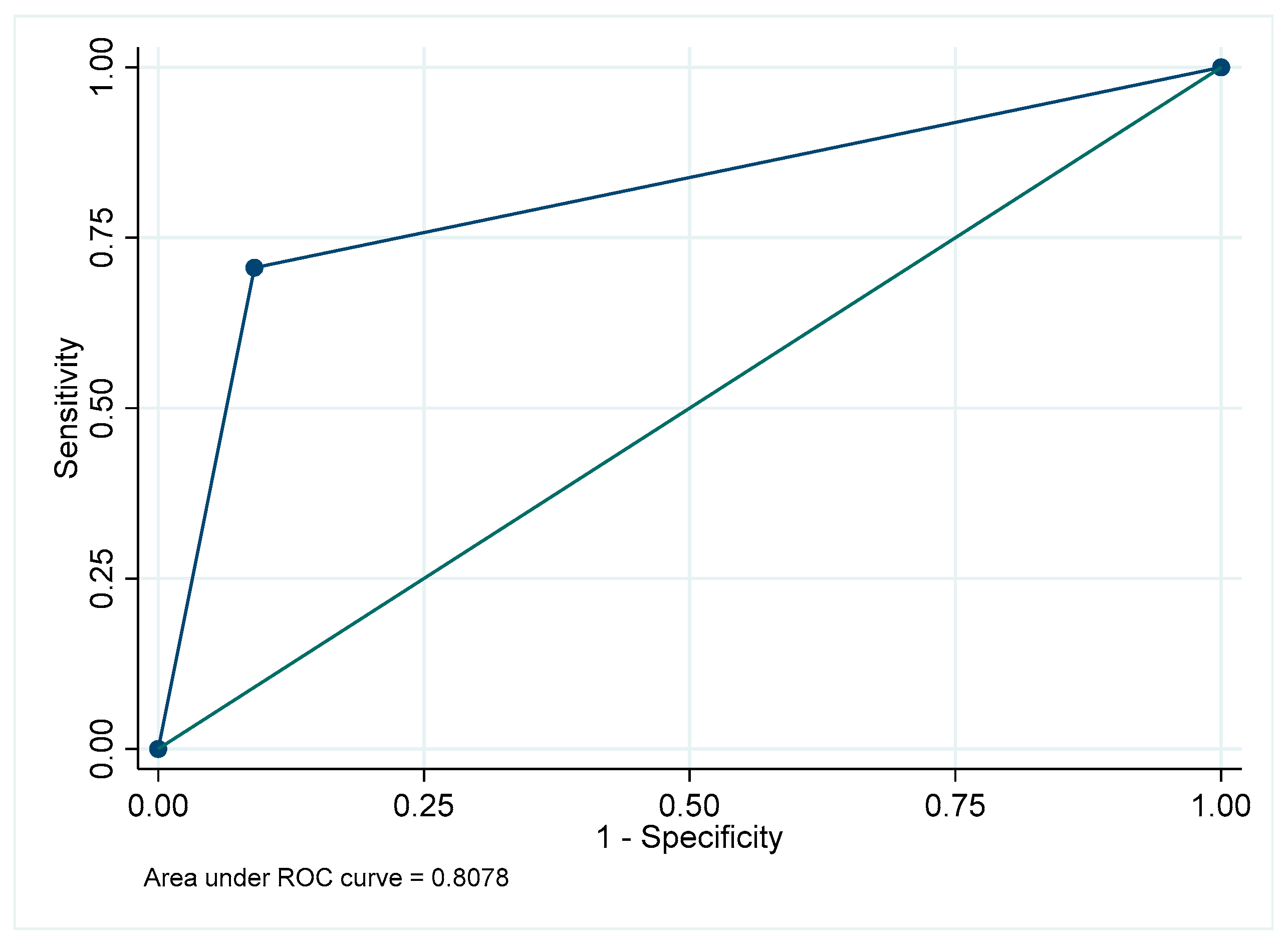
Figure A31.
Receiver Operating Characteristic curve for bone loss in the maxillary anterior region.
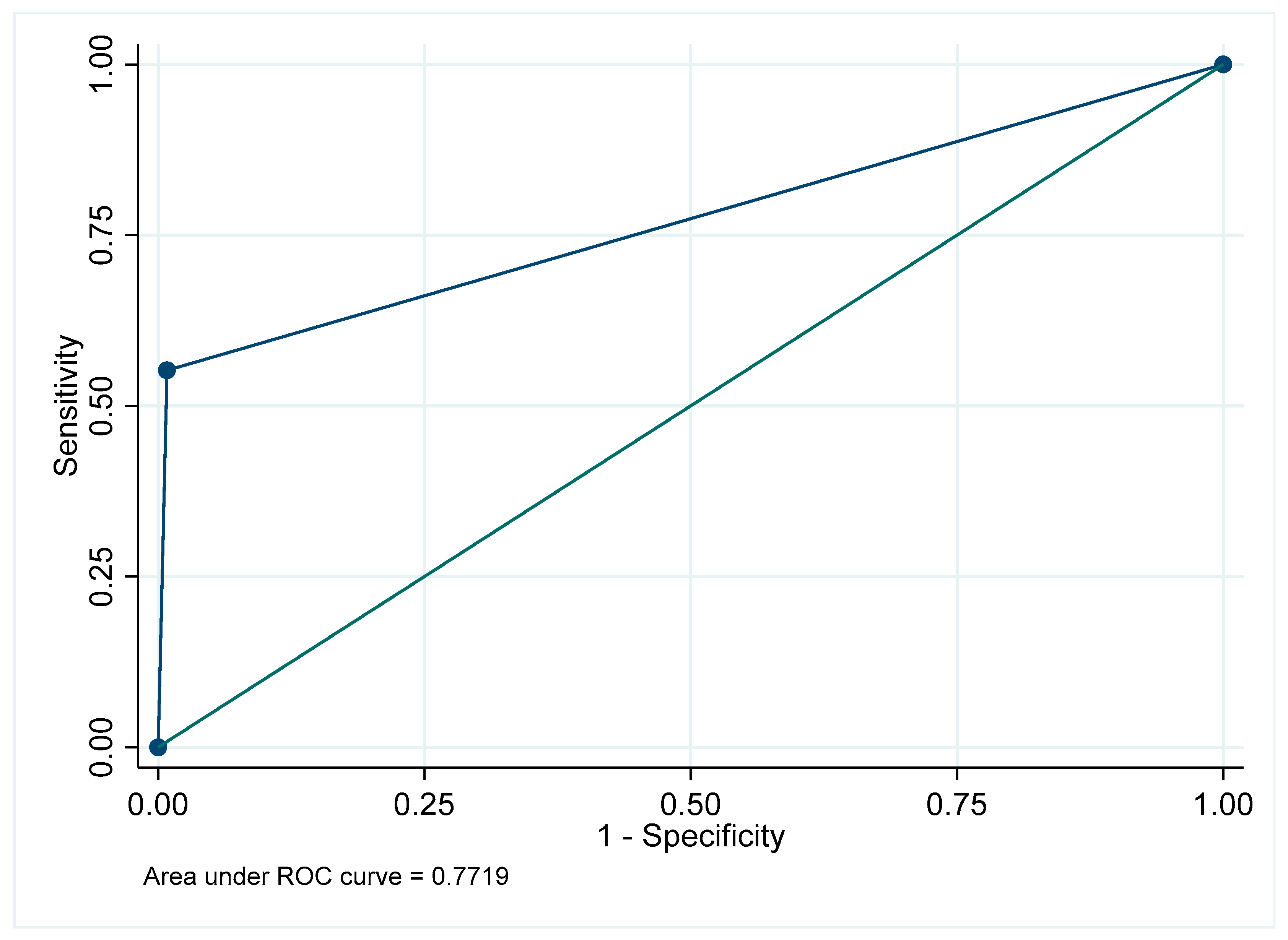
Figure A32.
Receiver Operating Characteristic curve for bone loss in the mandibular anterior region.
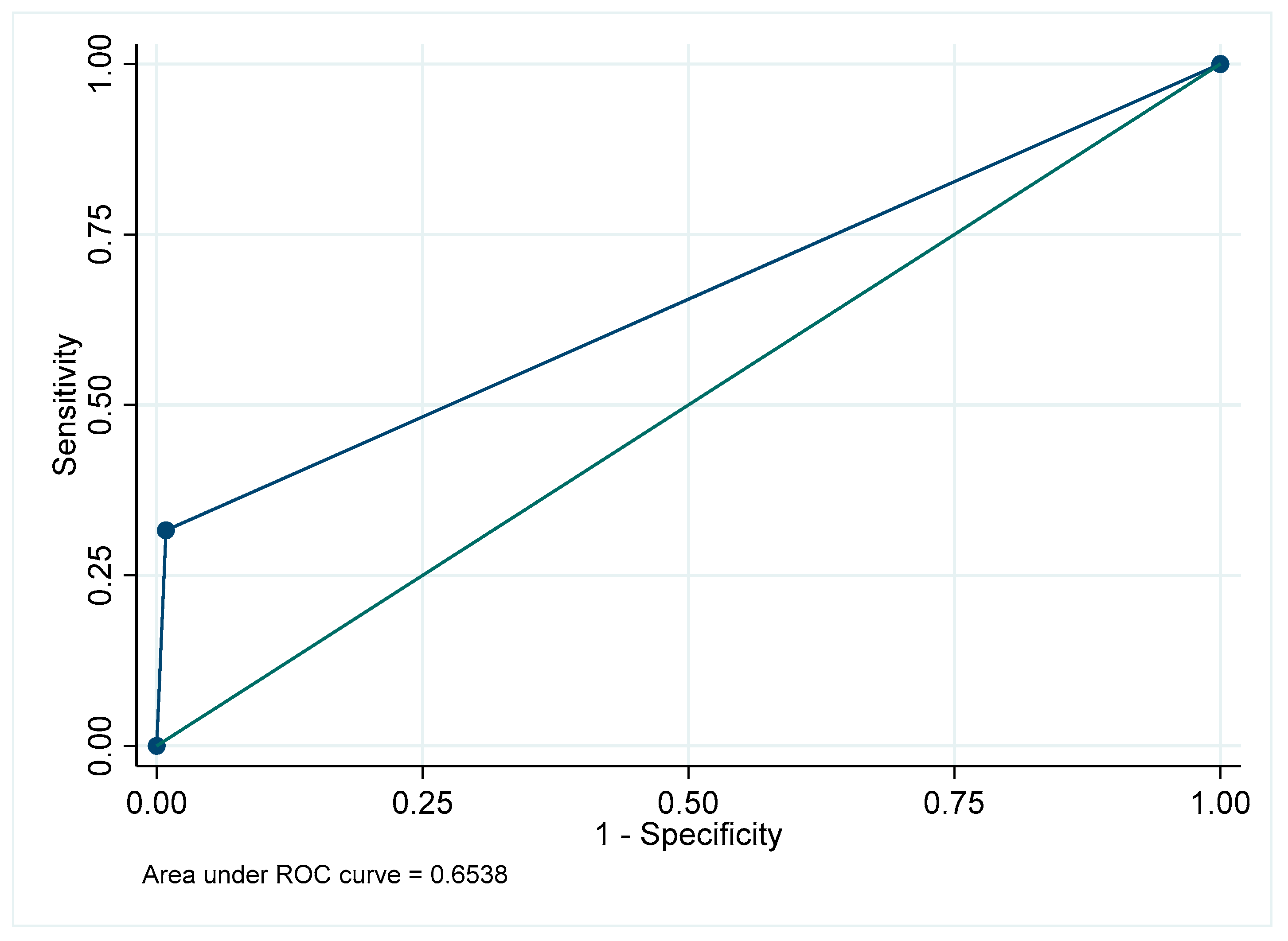
Figure A33.
Receiver Operating Characteristic curve for bone loss: Overall.
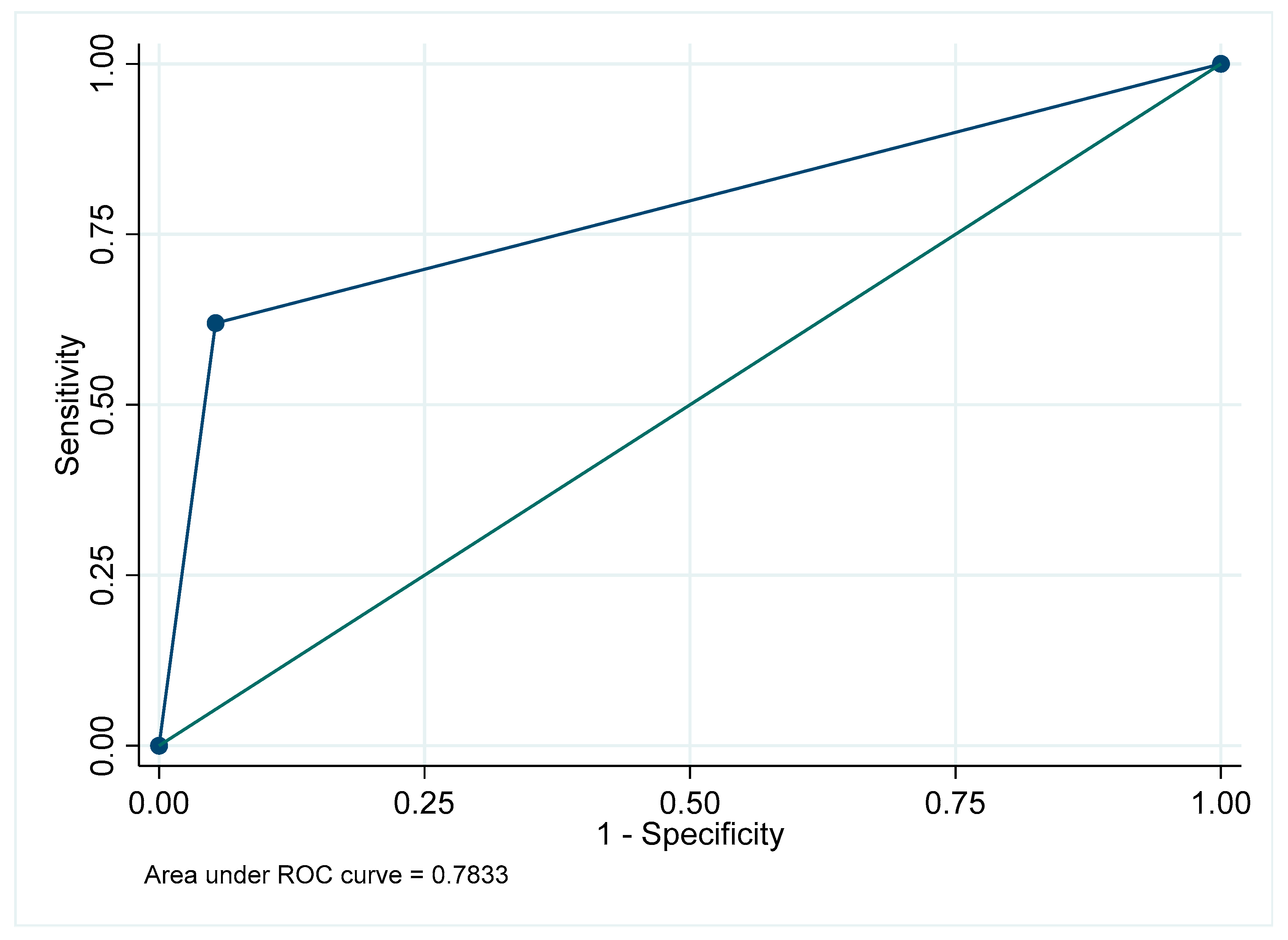
Figure A34.
Receiver Operating Characteristic curve for root fillings in the maxillary posterior region.
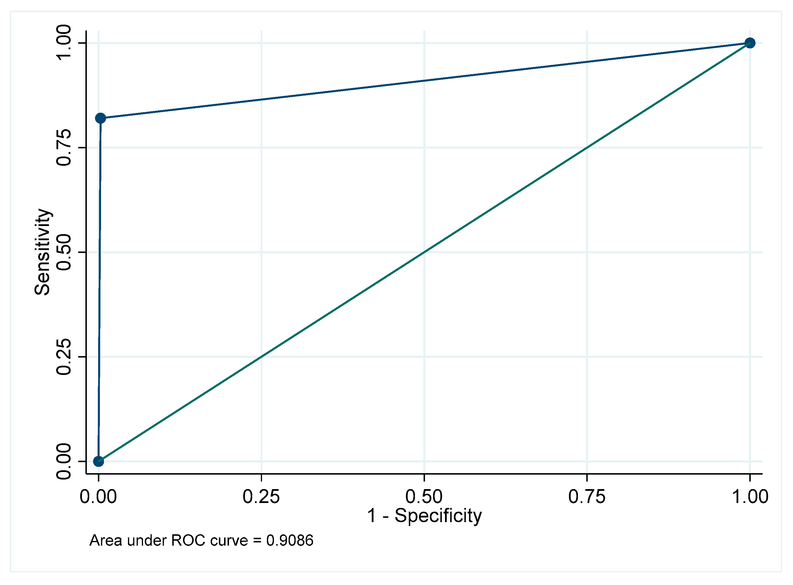
Figure A35.
Receiver Operating Characteristic curve for root fillings in the mandibular posterior region.
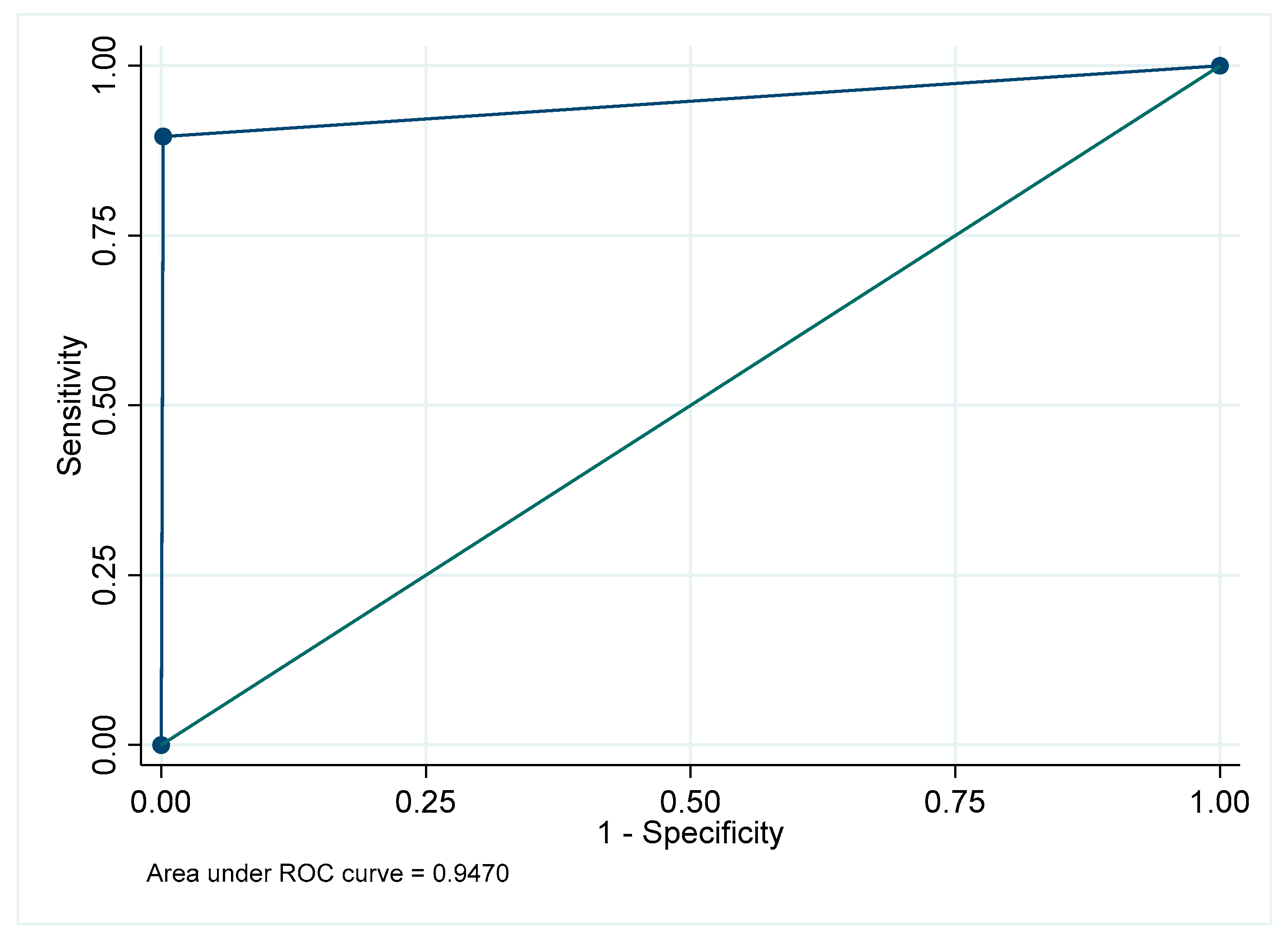
Figure A36.
Receiver Operating Characteristic curve for root fillings in the maxillary anterior region.
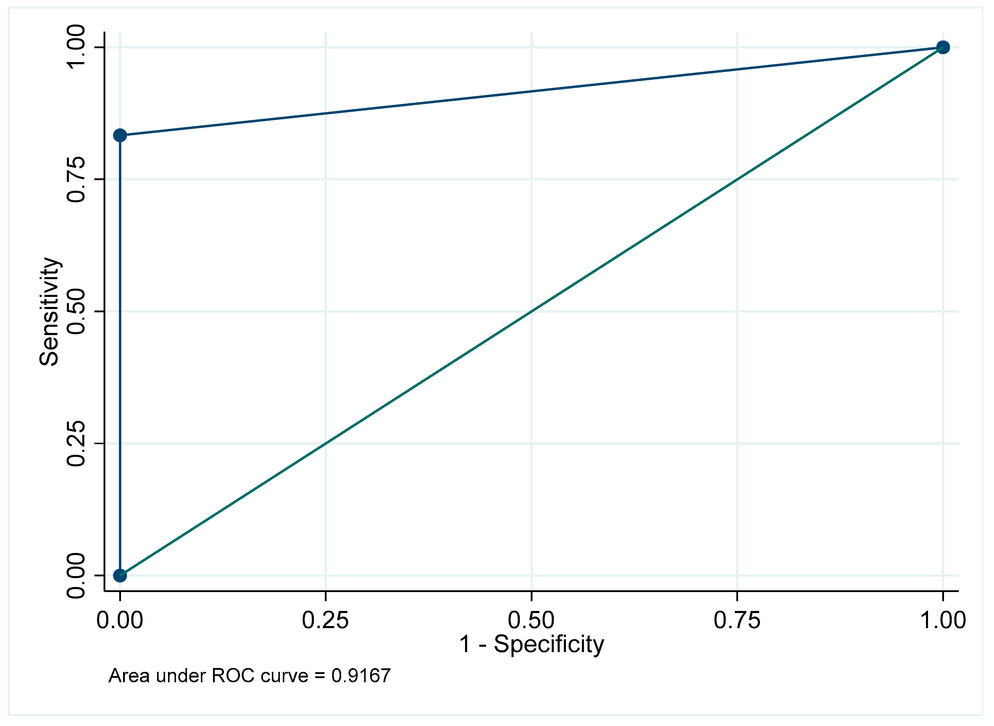
Figure A37.
Receiver Operating Characteristic curve for root fillings in the mandibular anterior region.
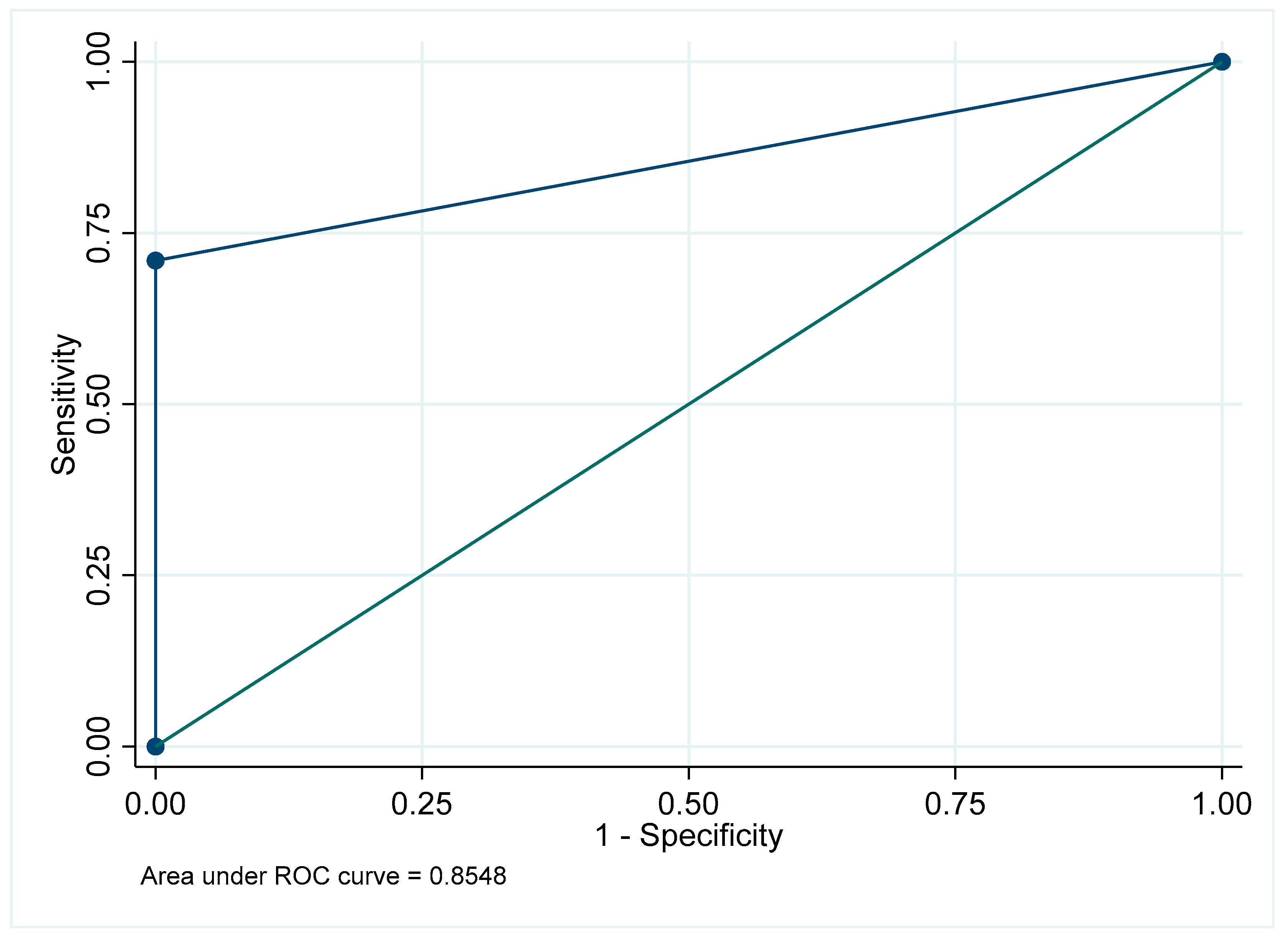
Figure A38.
Receiver Operating Characteristic curve for root fillings: Overall.
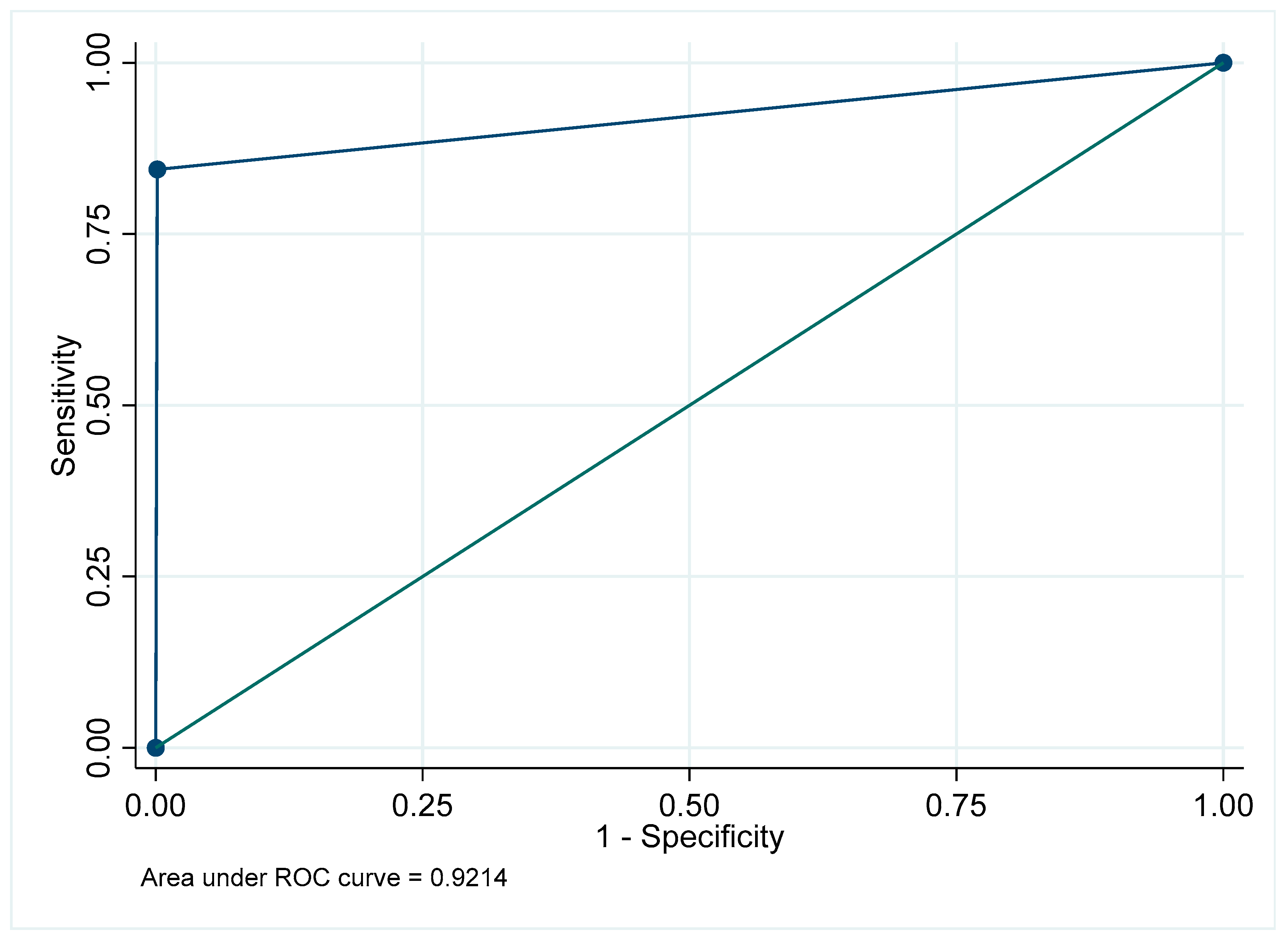
Figure A39.
Receiver Operating Characteristic curve for retained root in the maxillary posterior region.
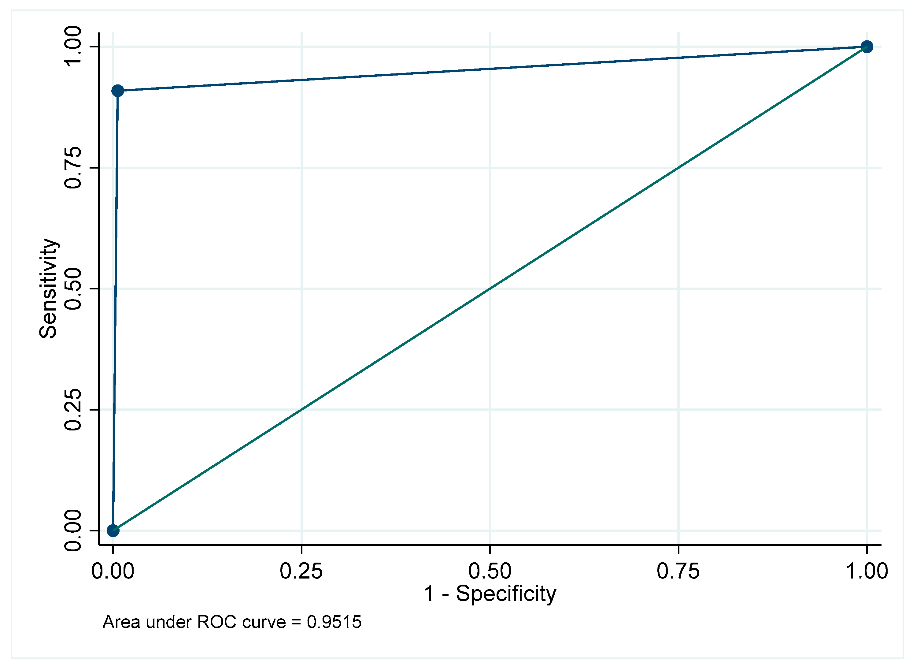
Figure A40.
Receiver Operating Characteristic curve for retained root in the mandibular posterior region.
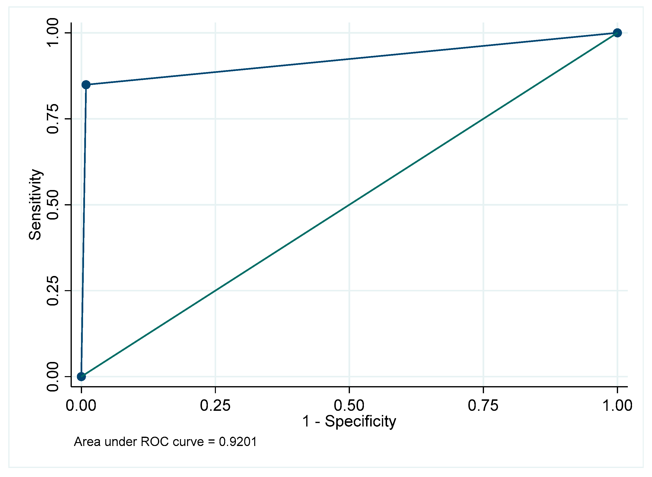
Figure A41.
Receiver Operating Characteristic curve for retained root in the maxillary anterior region.
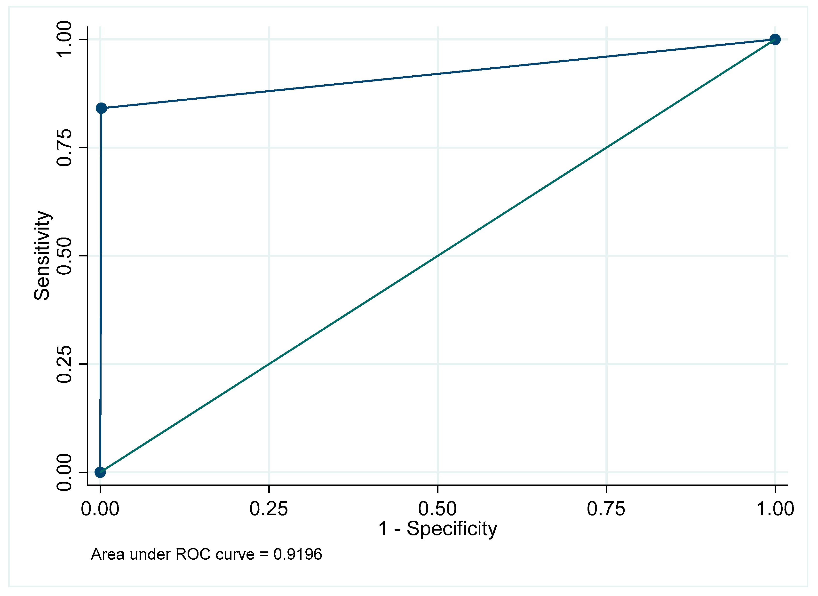
Figure A42.
Receiver Operating Characteristic curve for retained root in the mandibular anterior region.
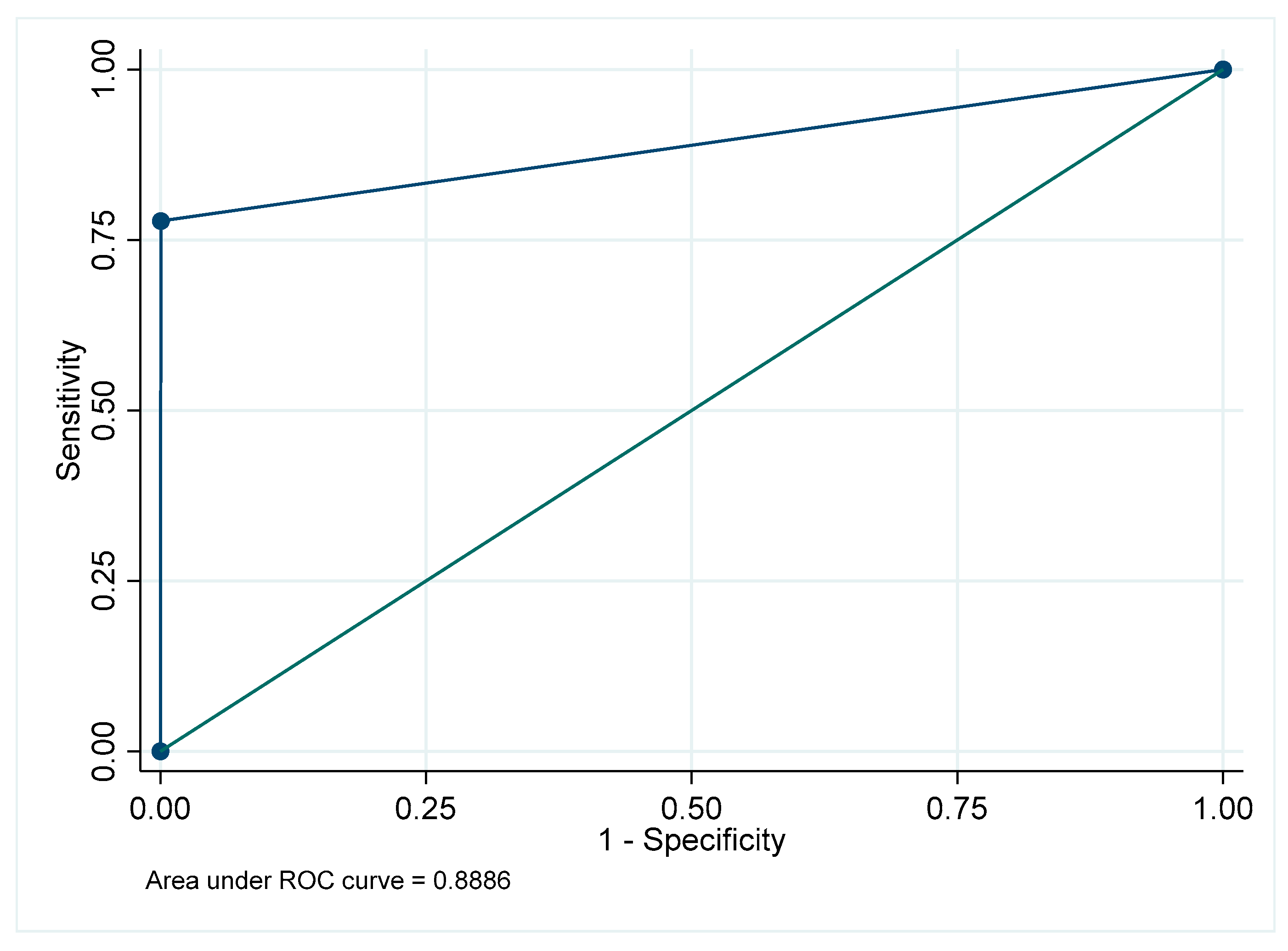
Figure A43.
Receiver Operating Characteristic curve for retained root: Overall.
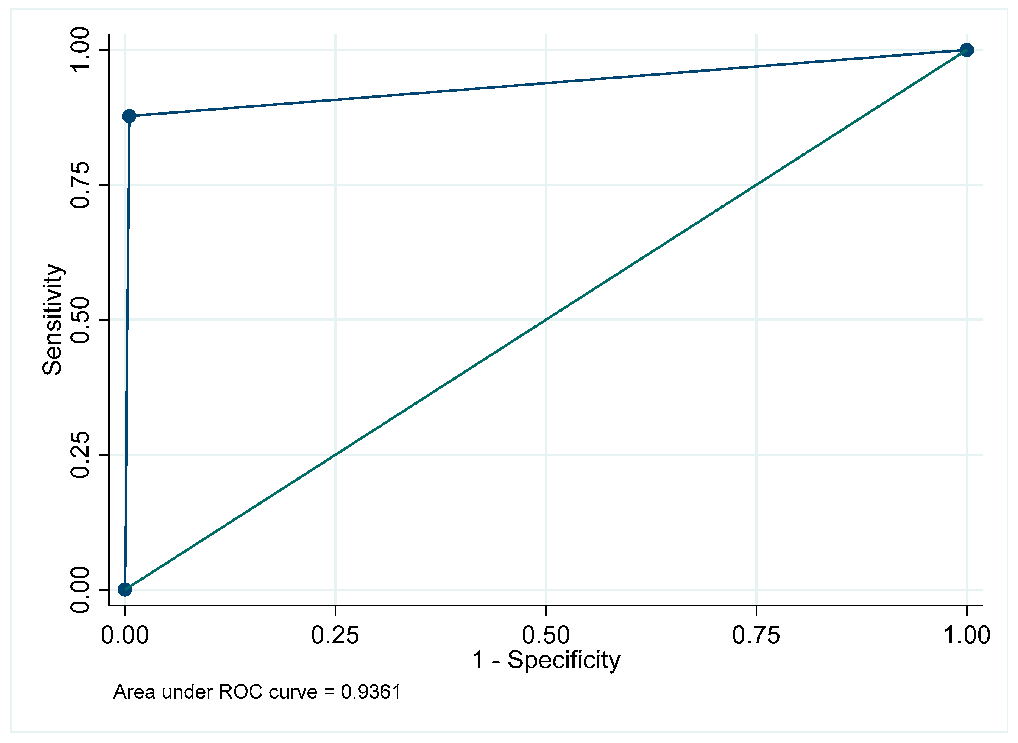
Figure A44.
Receiver Operating Characteristic curve for caries in the maxillary posterior region.
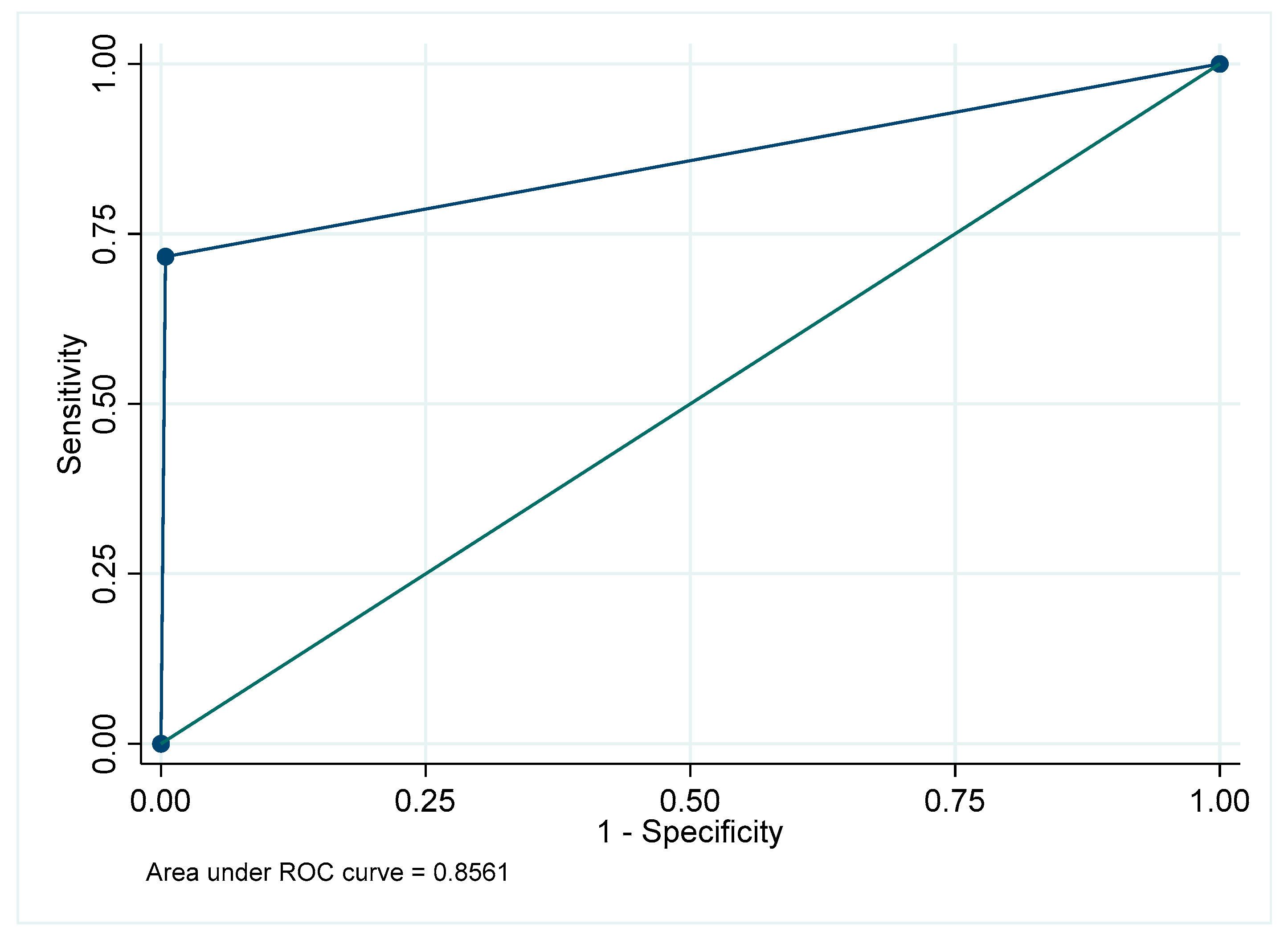
Figure A45.
Receiver Operating Characteristic curve for caries in the mandibular posterior region.
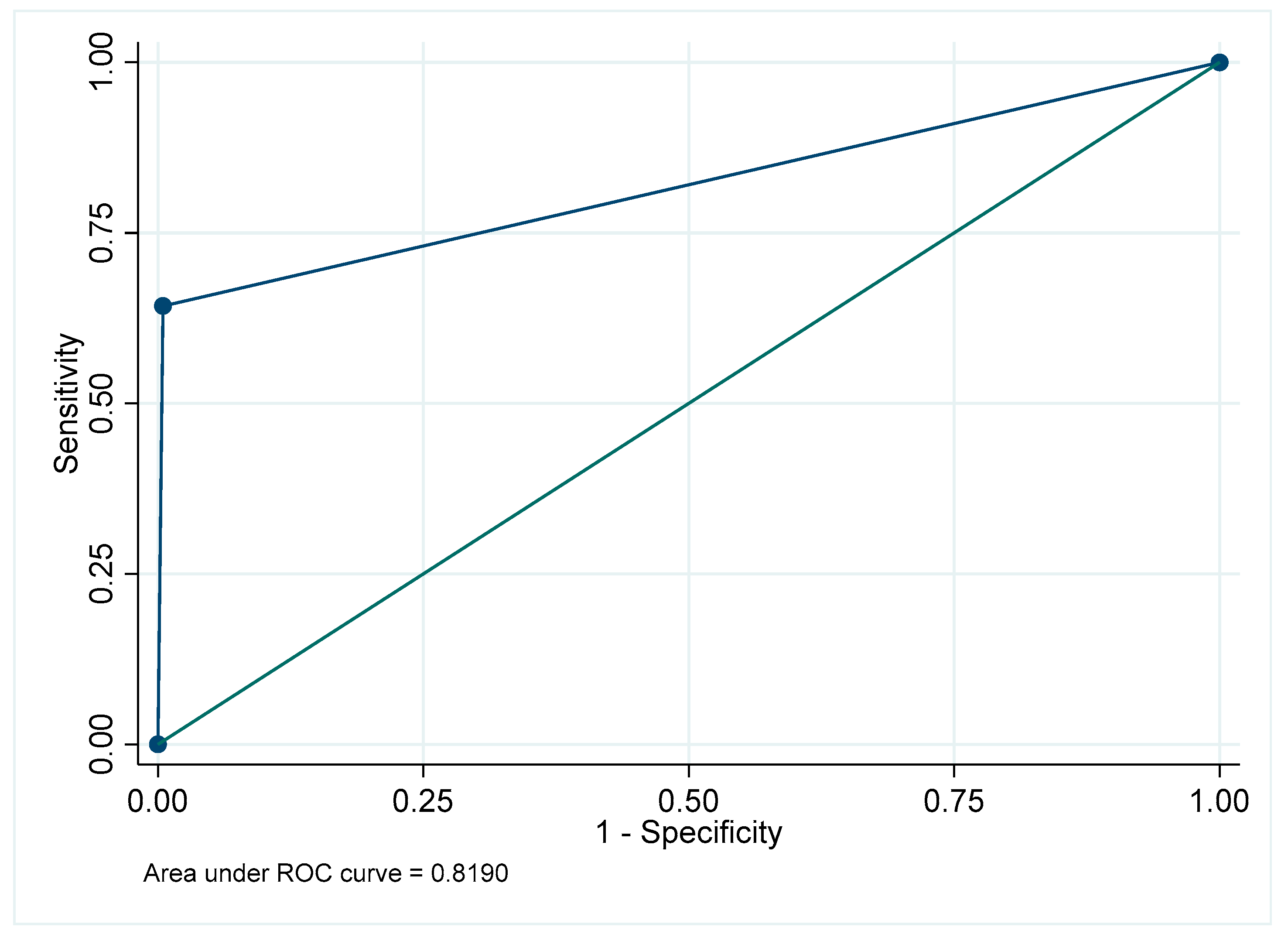
Figure A46.
Receiver Operating Characteristic curve for caries in the maxillary anterior region.
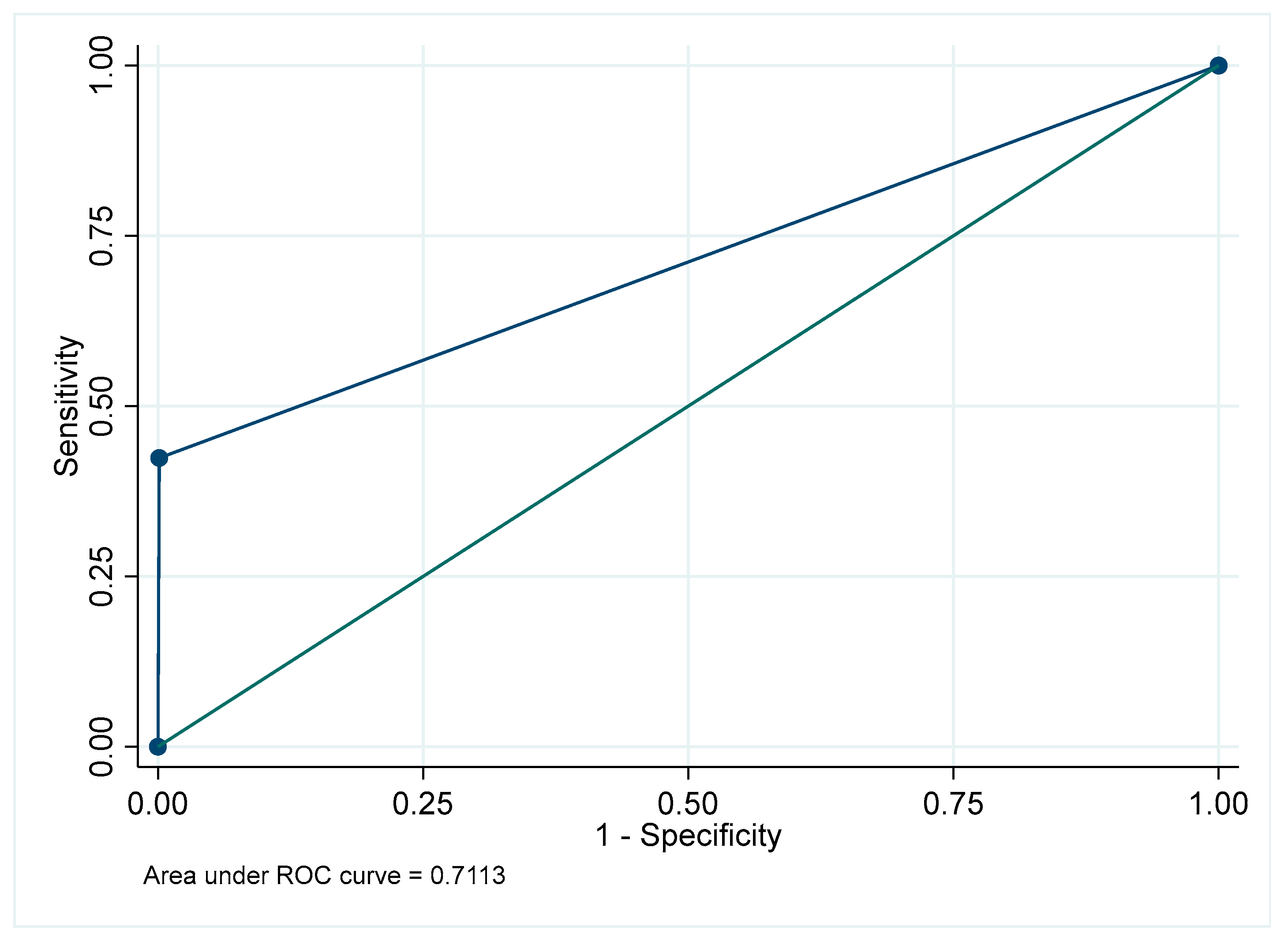
Figure A47.
Receiver Operating Characteristic curve for caries in the mandibular anterior region.
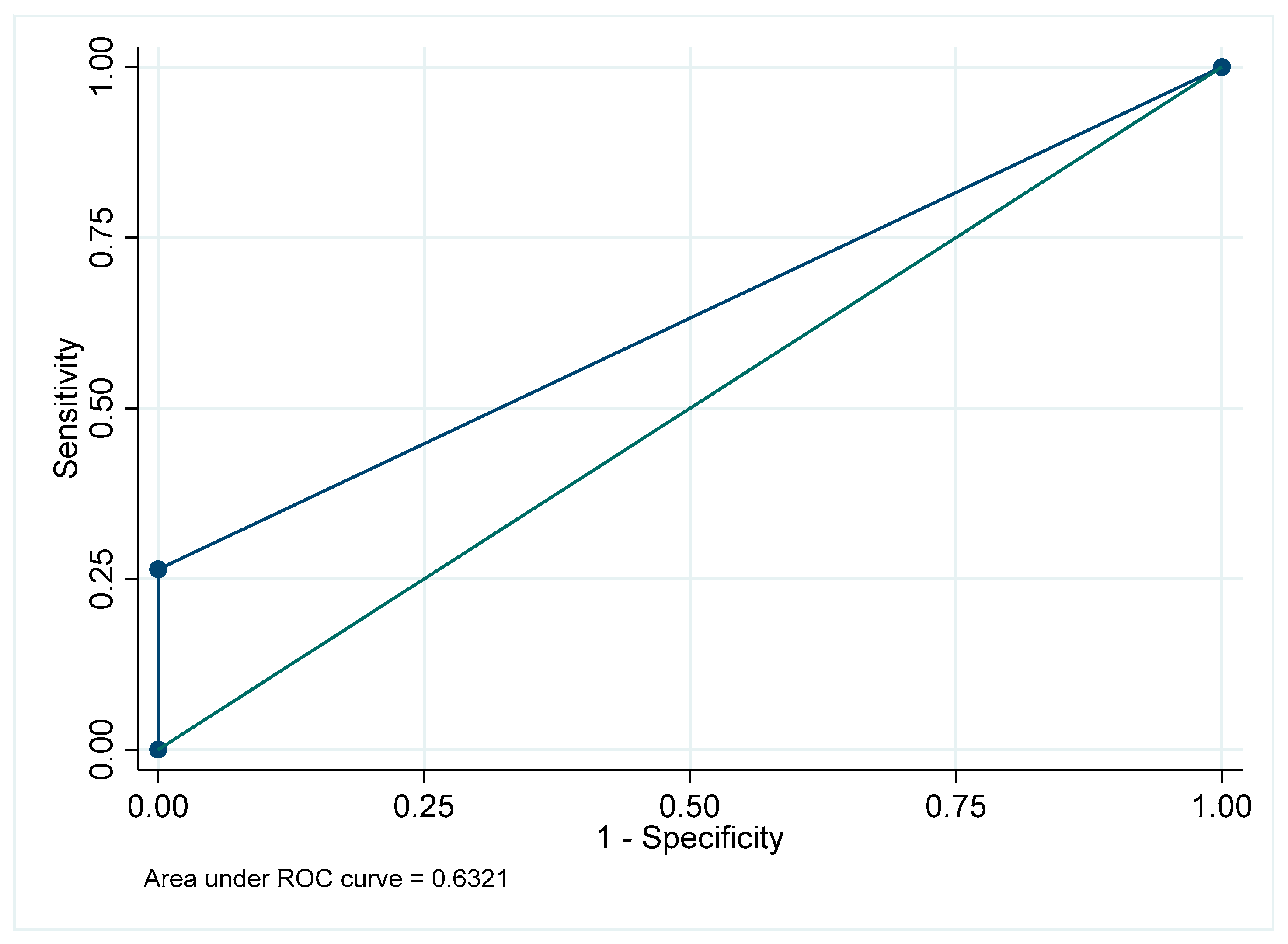
Figure A48.
Receiver Operating Characteristic curve for caries: Overall.
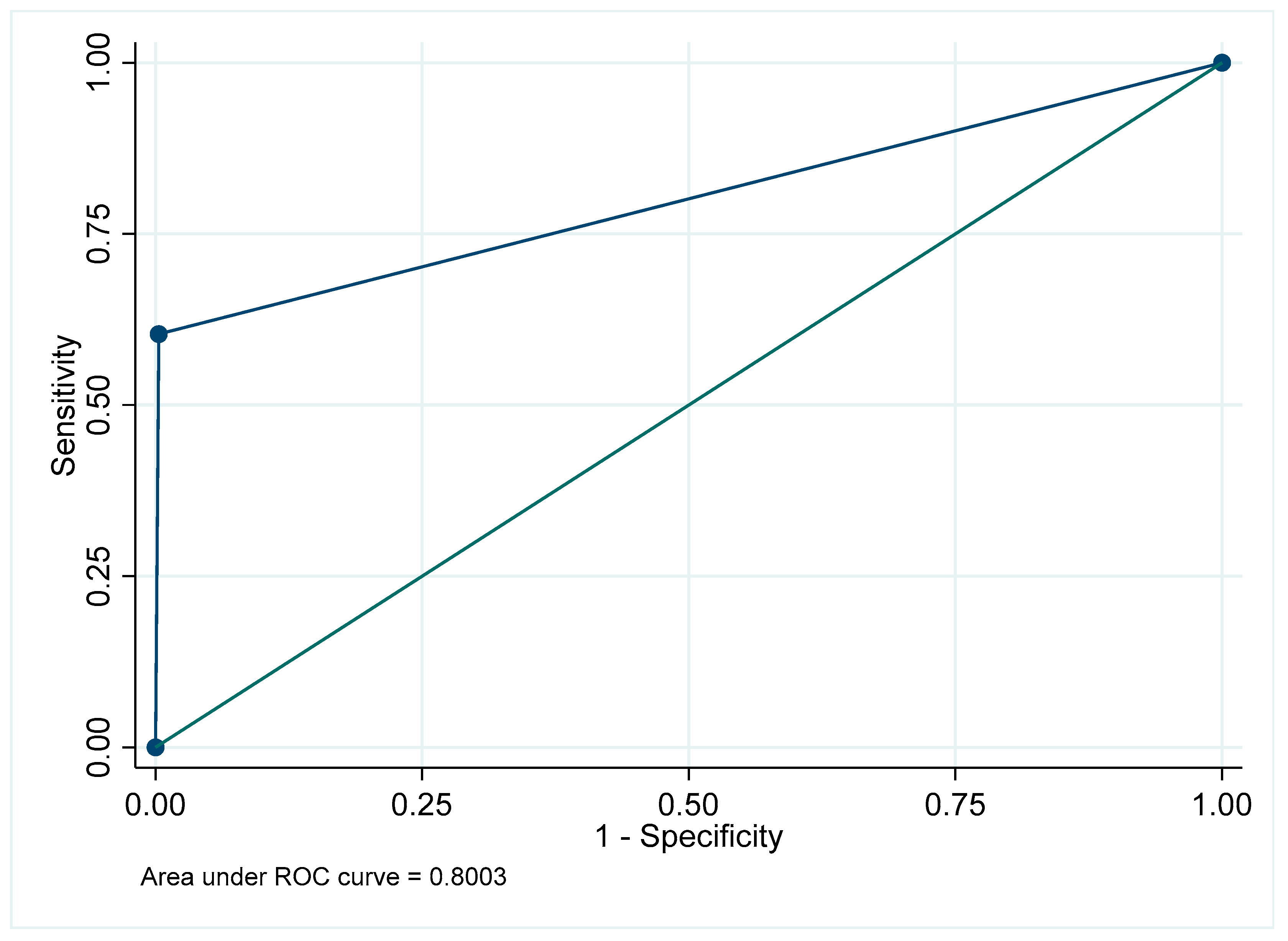
References
- Muthu, M.S.; Cho, V.Y.; FelsyPremila, G.; Nirmal, L.; Sujitha, P.; Bhavyaa, R.; King, N.M.; Anthonappa, R.P. Visual interpretation of clinical images among dental students using eye-tracking technology. J. Dent. Educ. 2024, 88, 606–613. [Google Scholar]
- Khanagar, S.B.; Al-Ehaideb, A.; Maganur, P.C.; Vishwanathaiah, S.; Patil, S.; Baeshen, H.A.; Sarode, S.C.; Bhandi, S. Developments, applications, and performance of artificial intelligence in dentistry: A systematic review. J. Dent. Sci. 2021, 16, 508–522. [Google Scholar] [CrossRef] [Scilit] [PubMed]
- Doi, K. Computer-aided diagnosis in medical imaging: Historical review, current status and future potential. Comput. Med. Imaging Graph. 2007, 31, 198–211. [Google Scholar] [CrossRef] [Scilit]
- Tuzoff, D.V.; Tuzova, L.N.; Bornstein, M.M.; Krasnov, A.S.; Kharchenko, M.A.; Nikolenko, S.I.; Sveshnikov, M.M.; Bednenko, G.B. Tooth detection and numbering in panoramic radiographs using convolutional neural networks. Dentomaxillofac. Radiol. 2019, 48, 20180051. [Google Scholar] [CrossRef] [Scilit]
- American Dental Association Council on Scientific Affairs. The use of dental radiographs: Update and recommendations. J. Am. Dent. Assoc. 2006, 137, 1304–1312. [Google Scholar] [CrossRef] [Scilit]
- Nardi, C.; Calistri, L.; Pradella, S.; Desideri, I.; Lorini, C.; Colagrande, S. Accuracy of orthopantomography for apical periodontitis without endodontic treatment. J. Endod. 2017, 43, 1640–1646. [Google Scholar] [CrossRef] [Scilit]
- Sun, W.; Xia, K.; Tang, L.; Liu, C.; Zou, L.; Liu, J. Accuracy of panoramic radiography in diagnosing maxillary sinus–root relationship: A systematic review and meta-analysis. Angle Orthod. 2018, 88, 819–829. [Google Scholar] [CrossRef] [Scilit]
- DeAngelis, A.F.; Barrowman, R.A.; Harrod, R.; Nastri, A.L. Maxillofacial emergencies: Maxillofacial trauma. Emerg. Med. Australas. 2014, 26, 530–537. [Google Scholar] [CrossRef] [Scilit]
- Chayra, G.A.; Meador, L.R.; Laskin, D.M. Comparison of panoramic and standard radiographs for the diagnosis of mandibular fractures. J. Oral Maxillofac. Surg. 1986, 44, 677. [Google Scholar] [CrossRef] [Scilit] [PubMed]
- Yeom, H.G.; Kim, J.E.; Huh, K.H.; Yi, W.J.; Heo, M.S.; Lee, S.S.; Choi, S.C.; Lee, S.J. Development of a panoramic resolution phantom for comprehensive evaluation of the horizontal and vertical resolution of panoramic radiography. Sci. Rep. 2020, 10, 16529. [Google Scholar] [CrossRef] [Scilit] [PubMed]
- Brüllmann, D.; Schulze, R.K. Spatial resolution in CBCT machines for dental/maxillofacial applications: What do we know today? Dentomaxillofac. Radiol. 2015, 44, 20140204. [Google Scholar] [CrossRef] [Scilit]
- Lin, P.L.; Lai, Y.H.; Huang, P.W. An effective classification and numbering system for dental bitewing radiographs using the tooth region and contour information. Pattern Recognit. 2010, 43, 1380–1392. [Google Scholar] [CrossRef] [Scilit]
- Miki, Y.; Muramatsu, C.; Hayashi, T.; Zhou, X.; Hara, T.; Katsumata, A.; Fujita, H. Classification of teeth in cone-beam CT using deep convolutional neural network. Comput. Biol. Med. 2017, 80, 24–29. [Google Scholar] [CrossRef] [Scilit]
- Ma, J.; Hu, C.; Zhou, P.; Jin, F.; Wang, X.; Huang, H. Review of Image Augmentation Used in Deep Learning-Based Material Microscopic Image Segmentation. Appl. Sci. 2023, 13, 6478. [Google Scholar] [CrossRef] [Scilit]
- Turosz, N.; Chęcińska, K.; Chęciński, M.; Brzozowska, A.; Nowak, Z.; Sikora, M. Applications of artificial intelligence in the analysis of dental panoramic radiographs: An overview of systematic reviews. Dentomaxillofac. Radiol. 2023, 52, 20230284. [Google Scholar] [CrossRef] [Scilit]
- Bonfanti-Gris, M.; Garcia-Cañas, A.; Alonso-Calvo, R.; Salido Rodriguez-Manzaneque, M.P.; Pradies Ramiro, G. Evaluation of an artificial intelligence web-based software to detect and classify dental structures and treatments in panoramic radiographs. J. Dent. 2022, 126, 104301. [Google Scholar] [CrossRef] [Scilit] [PubMed]
- Chaimani, A.; Higgins, J.P.T.; Mavridis, D.; Spyridonos, P.; Salanti, G. Graphical tools for network meta-analysis in STATA. PLoS ONE 2013, 8, e76654. [Google Scholar] [CrossRef] [Scilit]
- Kelly, H.; Bull, A.; Russo, P.; McBryde, E.S. Estimating sensitivity and specificity from positive predictive value, negative predictive value and prevalence: Application to surveillance systems for hospital-acquired infections. J. Hosp. Infect. 2008, 69, 164–168. [Google Scholar] [CrossRef] [Scilit] [PubMed]
- Chen, I.D.S.; Yang, C.M.; Chen, M.J.; Chen, M.C.; Weng, R.M.; Yeh, C.H. Deep learning-based recognition of periodontitis and dental caries in dental X-ray images. Bioengineering 2023, 10, 911. [Google Scholar] [CrossRef] [Scilit] [PubMed]
- Zhu, H.; Cao, Z.; Lian, L.; Ye, G.; Gao, H.; Wu, J. CariesNet: A deep learning approach for segmentation of multi-stage caries lesion from oral panoramic X-ray image. Neural Comput. Appl. 2023, 35, 16051–16059. [Google Scholar] [CrossRef] [Scilit]
- Bayraktar, Y.; Ayan, E. Diagnosis of interproximal caries lesions with deep convolutional neural network in digital bitewing radiographs. Clin. Oral Investig. 2022, 26, 623–632. [Google Scholar] [CrossRef] [Scilit]
- Mao, Y.C.; Chen, T.Y.; Chou, H.S.; Lin, S.Y.; Liu, S.Y.; Chen, Y.A.; Liu, Y.L.; Chen, C.A.; Huang, Y.C.; Chen, S.L.; et al. Caries and Restoration Detection Using Bitewing Film Based on Transfer Learning with CNNs. Sensors 2021, 21, 4613. [Google Scholar] [CrossRef] [Scilit]
- De Araujo Faria, V.; Azimbagirad, M.; Viani Arruda, G.; Fernandes Pavoni, J.; Cezar Felipe, J.; Dos Santos, E.M.C.M.F.; Murta Junior, L.O. Prediction of Radiation-Related Dental Caries Through PyRadiomics Features and Artificial Neural Network on Panoramic Radiography. J. Digit. Imaging 2021, 34, 1237–1248. [Google Scholar] [CrossRef] [Scilit]
- Hur, S.H.; Lee, E.Y.; Kim, M.K.; Kim, S.; Kang, J.Y.; Lim, J.S. Machine learning to predict distal caries associated with impacted third molars. Sci. Rep. 2021, 11, 15447. [Google Scholar] [CrossRef] [Scilit]
- Lee, S.; Oh, S.I.; Jo, J.; Kang, S.; Shin, Y.; Park, J.W. Deep learning for early dental caries detection in bitewing radiographs. Sci. Rep. 2021, 11, 16807. [Google Scholar] [CrossRef] [Scilit] [PubMed]
- Vinayahalingam, S.; Kempers, S.; Limon, L.; Deibel, D.; Maal, T.; Hanisch, M. Classification of caries in third molars on panoramic radiographs using deep learning. Sci. Rep. 2021, 11, 12609. [Google Scholar] [CrossRef] [Scilit]
- Mertens, S.; Krois, J.; Cantu, A.G.; Arsiwala, L.T.; Schwendicke, F. Artificial intelligence for caries detection: Randomized trial. J. Dent. 2021, 115, 103849. [Google Scholar] [CrossRef] [Scilit] [PubMed]
- Moran, M.; Faria, M.; Giraldi, G.; Bastos, L.; Oliveira, L.; Conci, A. Classification of Approximal Caries in Bitewing Radiographs Using Convolutional Neural Networks. Sensors 2021, 21, 5192. [Google Scholar] [CrossRef] [Scilit]
- Lian, L.; Zhu, T.; Zhu, F.; Zhu, H. Deep learning for caries detection and classification. Diagnostics 2021, 11, 1672. [Google Scholar] [CrossRef] [Scilit] [PubMed]
- Zheng, L.; Wang, H.; Mei, L.; Chen, Q.; Zhang, Y.; Zhang, H. Artificial intelligence in digital cariology: A new tool for the diagnosis of deep caries and pulpitis using convolutional neural networks. Ann. Transl. Med. 2021, 9, 763. [Google Scholar] [CrossRef] [Scilit]
- Bayrakdar, I.S.; Orhan, K.; Akarsu, S.; Çelik, Ö.; Atasoy, S.; Pekince, A.; Yasa, Y.; Bilgir, E.; Sağlam, H.; Aslan, A.F.; et al. Deep-learning approach for caries detection and segmentation on dental bitewing radiographs. Oral Radiol. 2021, 37, 237–248. [Google Scholar] [CrossRef] [Scilit]
- Devlin, H.; Williams, T.; Graham, J.; Ashley, M. The ADEPT study: A comparative study of dentists’ ability to detect enamel-only proximal caries in bitewing radiographs with and without the use of AssistDent artificial intelligence software. Br. Dent. J. 2021, 231, 481–485. [Google Scholar] [CrossRef] [Scilit]
- Szabó, V.; Orhan, K.; Dobó-Nagy, C.; Veres, D.S.; Manulis, D.; Ezhov, M.; Sanders, A.; Szabó, B.T. Deep Learning-Based Periapical Lesion Detection on Panoramic Radiographs. Diagnostics 2025, 15, 510. [Google Scholar] [CrossRef] [Scilit]
- Geetha, V.; Aprameya, K.S.; Hinduja, D.M. Dental caries diagnosis in digital radiographs using back-propagation neural network. Health Inf. Sci. Syst. 2020, 8, 8. [Google Scholar] [CrossRef] [Scilit]
- Cantu, A.G.; Gehrung, S.; Krois, J.; Chaurasia, A.; Rossi, J.G.; Gaudin, R.; Elhennawy, K.; Schwendicke, F. Detecting caries lesions of different radiographic extension on bitewings using deep learning. J. Dent. 2020, 100, 103425. [Google Scholar] [CrossRef] [Scilit]
- Lee, J.H.; Kim, D.H.; Jeong, S.N.; Choi, S.H. Detection and diagnosis of dental caries using CNN. J. Dent. 2018, 77, 106–111. [Google Scholar] [CrossRef] [Scilit] [PubMed]
- Jin, L.; Du, B.; Xu, Z.; Bai, H.; Ding, P.; Zhang, Z.; Pan, Y.; Lin, Y.; Li, Z.; Rausch-Fan, X.; et al. Deep-learning network for automated evaluation of root-canal filling radiographic quality. Eur. J. Med. Res. 2025, 30, 297. [Google Scholar] [CrossRef] [Scilit] [PubMed]
- Hadj Saïd, M.; Le Roux, M.K.; Catherine, J.H.; Lan, R. Development of an Artificial Intelligence Model to Identify a dental implant from a radiograph. Int. J. Oral Maxillofac. Implant. 2020, 36, 1077–1082. [Google Scholar] [CrossRef] [Scilit]
- Lee, J.H.; Jeong, S.N. Efficacy of deep convolutional neural network algorithm for the Identification and classification of dental implant systems using panoramic and periapical radiographs: A Pilot Study. Medicine 2020, 99, e20787. [Google Scholar] [CrossRef] [Scilit]
- Sukegawa, S.; Yoshii, K.; Hara, T.; Yamashita, K.; Nakano, K.; Yamamoto, N. Deep neural networks for dental implant classification. Biomolecules 2020, 10, 984. [Google Scholar] [CrossRef] [Scilit] [PubMed]
- Kim, J.E.; Nam, N.E.; Shim, J.S.; Jung, Y.H.; Cho, B.H.; Hwang, J.J. Transfer learning via deep neural networks for implant fixture classification using periapical radiographs. J. Clin. Med. 2020, 9, 1117. [Google Scholar] [CrossRef] [Scilit]
- Lee, J.H.; Kim, Y.T.; Lee, J.B.; Jeong, S.N. A Performance comparison between automated deep learning and dental professionals in classification of dental implant systems in dental imaging: A Multi-Centre Study. Diagnostics 2020, 10, 910. [Google Scholar] [CrossRef] [Scilit] [PubMed]
- Takahashi, T.; Nozaki, K.; Gonda, T.; Mameno, T.; Wada, M.; Ikebe, K. Identification of dental implants using deep learningPilot study. Int. J. Implant Dent. 2020, 6, 53. [Google Scholar] [CrossRef] [Scilit] [PubMed]
- Caton, J.G.; Armitage, G.; Berglundh, T.; Chapple, I.L.C.; Jepsen, S.; Kornman, K.S. A new classification scheme for periodontal and peri-implant diseases and conditions: Introduction and key changes from the 1999 classification. J. Clin. Periodontol. 2018, 45, S1–S8. [Google Scholar] [CrossRef] [Scilit]
- Ari, T.; Sağlam, H.; Öksüzoğlu, H.; Kazan, O.; Bayrakdar, İ.Ş.; Duman, S.B.; Çelik, Ö.; Jagtap, R.; Futyma-Gąbka, K.; Różyło-Kalinowska, I.; et al. Automatic Feature Segmentation in Dental Periapical Radiographs. Diagnostics 2022, 12, 3081. [Google Scholar] [CrossRef] [Scilit] [PubMed]
- Bayrakdar, I.S.; Orhan, K.; Çelik, Ö.; Bilgir, E.; Sağlam, H.; Kaplan, F.A.; Görür, S.A.; Odabaş, A.; Aslan, A.F.; Różyło-Kalinowska, I. A U-Net Approach to Apical Lesion Segmentation on Panoramic Radiographs. Biomed. Res. Int. 2022, 2022, 7035367. [Google Scholar] [CrossRef] [Scilit]
- Calazans, M.A.A.; Ferreira, F.A.B.S.; Alcoforado, M.L.M.G.; Santos, A.D.; Pontual, A.D.A.; Madeiro, F. Automatic Classification System for Periapical Lesions in Cone-Beam Computed Tomography. Sensors 2022, 22, 6481. [Google Scholar] [CrossRef] [Scilit]
- Caputo, B.; Gigante, G.E. Analysis of periapical lesion using statistical textural features. Stud. Health Technol. Inform. 2000, 77, 1231–1234. [Google Scholar]
- Chen, H.; Li, H.; Zhao, Y.; Zhao, J.; Wang, Y. Dental disease detection on periapical radiographs based on deep convolutional neural networks. Int. J. Comput. Assist. Radiol. Surg. 2021, 16, 649–661. [Google Scholar] [CrossRef] [Scilit]
- Chuo, Y.; Lin, W.M.; Chen, T.Y.; Chan, M.L.; Chang, Y.S.; Lin, Y.R.; Lin, Y.J.; Shao, Y.H.; Chen, C.A.; Chen, S.L.; et al. A High-Accuracy Detection System: Based on Transfer Learning for Apical Lesions on Periapical Radiograph. Bioengineering 2022, 9, 777. [Google Scholar] [CrossRef] [Scilit]
- Ekert, T.; Krois, J.; Meinhold, L.; Elhennawy, K.; Emara, R.; Golla, T.; Schwendicke, F. Deep Learning for the Radiographic Detection of Apical Lesions. J. Endod. 2019, 45, 917–922.e5. [Google Scholar] [CrossRef] [Scilit]
- Endres, M.G.; Hillen, F.; Salloumis, M.; Sedaghat, A.R.; Niehues, S.M.; Quatela, O.; Hanken, H.; Smeets, R.; Beck-Broichsitter, B.; Rendenbach, C.; et al. Development of a Deep Learning Algorithm for Periapical Disease Detection in Dental Radiographs. Diagnostics 2020, 10, 430. [Google Scholar] [CrossRef] [Scilit]
- Ezhov, M.; Gusarev, M.; Golitsyna, M.; Yates, J.M.; Kushnerev, E.; Tamimi, D.; Aksoy, S.; Shumilov, E.; Sanders, A.; Orhan, K. Clinically applicable artificial intelligence system for dental diagnosis with CBCT. Sci. Rep. 2021, 11, 15006. [Google Scholar] [CrossRef] [Scilit]
- Fatima, A.; Shafi, I.; Afzal, H.; Mahmood, K.; Díez, I.T.; Lipari, V.; Ballester, J.B.; Ashraf, I. Deep Learning-Based Multiclass Instance Segmentation for Dental Lesion Detection. Healthcare 2023, 11, 347. [Google Scholar] [CrossRef] [Scilit]
- Hamdan, M.H.; Tuzova, L.; Mol, A.; Tawil, P.Z.; Tuzoff, D.; Tyndall, D.A. The effect of a deep-learning tool on dentists’ performances in detecting apical radiolucencies on periapical radiographs. Dentomaxillofac. Radiol. 2022, 51, 20220122. [Google Scholar] [CrossRef] [Scilit]
- Kim, C.; Jeong, H.; Park, W.; Kim, D. Tooth-Related Disease Detection System Based on Panoramic Images and Optimization Through Automation: Development Study. JMIR Med. Inform. 2022, 10, e38640. [Google Scholar] [CrossRef] [Scilit]
- Kirnbauer, B.; Hadzic, A.; Jakse, N.; Bischof, H.; Stern, D. Automatic Detection of Periapical Osteolytic Lesions on Cone-beam Computed Tomography Using Deep Convolutional Neuronal Networks. J. Endod. 2022, 48, 1434–1440. [Google Scholar] [CrossRef] [Scilit] [PubMed]
- Krois, J.; Garcia Cantu, A.; Chaurasia, A.; Patil, R.; Chaudhari, P.K.; Gaudin, R.; Gehrung, S.; Schwendicke, F. Generalizability of deep learning models for dental image analysis. Sci. Rep. 2021, 11, 6102. [Google Scholar] [CrossRef] [Scilit]
- Li, C.W.; Lin, S.Y.; Chou, H.S.; Chen, T.Y.; Chen, Y.A.; Liu, S.Y.; Liu, Y.L.; Chen, C.A.; Huang, Y.C.; Chen, S.L.; et al. Detection of Dental Apical Lesions Using CNNs on Periapical Radiograph. Sensors 2021, 21, 7049. [Google Scholar] [CrossRef] [Scilit] [PubMed]
- Moidu, N.P.; Sharma, S.; Chawla, A.; Kumar, V.; Logani, A. Deep learning for categorization of endodontic lesion based on radiographic periapical index scoring system. Clin. Oral Investig. 2022, 26, 651–658. [Google Scholar] [CrossRef] [Scilit] [PubMed]
- Ngoc, V.T.N.; Viet, D.H.; Anh, L.K.; Minh, D.Q.; Nghia, L.L.; Loan, H.K.; Tuan, T.M.; Ngan, T.T.; Nguyen, T.T. Periapical Lesion Diagnosis Support System Based on X-ray Images Using Machine Learning Technique. World J. Dent. 2021, 12, 189–193. [Google Scholar] [CrossRef] [Scilit]
- Setzer, F.C.; Shi, K.J.; Zhang, Z.; Yan, H.; Yoon, H.; Mupparapu, M.; Li, J. Artificial Intelligence for the Computer-aided Detection of Periapical Lesions in Cone-beam Computed Tomographic Images. J. Endod. 2020, 46, 987–993. [Google Scholar] [CrossRef] [Scilit]
- Shafi, I.; Sajad, M.; Fatima, A.; Aray, D.G.; Lipari, V.; Diez, I.T.; Ashraf, I. Teeth Lesion Detection Using Deep Learning and the Internet of Things Post-COVID-19. Sensors 2023, 23, 6837. [Google Scholar] [CrossRef] [Scilit] [PubMed]
- Song, I.S.; Shin, H.K.; Kang, J.H.; Kim, J.E.; Huh, K.H.; Yi, W.J.; Lee, S.S.; Heo, M.S. Deep learning-based apical lesion segmentation from panoramic radiographs. Imaging Sci. Dent. 2022, 52, 351–357. [Google Scholar] [CrossRef] [Scilit] [PubMed]
- Ver Berne, J.; Saadi, S.B.; Politis, C.; Jacobs, R. A deep learning approach for radiological detection and classification of radicular cysts and periapical granulomas. J. Dent. 2023, 135, 104581. [Google Scholar] [CrossRef] [Scilit]
- Yilmaz, E.; Kayikcioglu, T.; Kayipmaz, S. Computer-aided diagnosis of periapical cyst and keratocystic odontogenic tumor on cone beam computed tomography. Comput. Methods Programs Biomed. 2017, 146, 91–100. [Google Scholar] [CrossRef] [Scilit]
- Kim, J.; Lee, H.S.; Song, I.S.; Jung, K.H. DeNTNet: Deep Neural Transfer Network for the detection of periodontal bone loss using panoramic dental radiographs. Sci. Rep. 2019, 9, 17615. [Google Scholar] [CrossRef] [Scilit]
- Chang, H.J.; Lee, S.J.; Yong, T.H.; Shin, N.Y.; Jang, B.G.; Kim, J.E.; Huh, K.H.; Lee, S.S.; Heo, M.S.; Choi, S.C.; et al. Deep Learning Hybrid Method to Automatically Diagnose Periodontal Bone Loss and Stage Periodontitis. Sci. Rep. 2020, 10, 7531. [Google Scholar] [CrossRef] [Scilit] [PubMed]
- Krois, J.; Ekert, T.; Meinhold, L.; Golla, T.; Kharbot, B.; Wittemeier, A.; Dörfer, C.; Schwendicke, F. Deep Learning for the Radiographic Detection of Periodontal Bone Loss. Sci. Rep. 2019, 9, 8495. [Google Scholar] [CrossRef] [Scilit]
- Thanathornwong, B.; Suebnukarn, S. Automatic detection of periodontal compromised teeth in digital panoramic radiographs using faster regional convolutional neural networks. Imaging Sci. Dent. 2020, 50, 169–174. [Google Scholar] [CrossRef] [Scilit]
- Zadrożny, Ł.; Regulski, P.; Brus-Sawczuk, K.; Czajkowska, M.; Parkanyi, L.; Ganz, S.; Mijiritsky, E. Artificial Intelligence Application in Assessment of Panoramic Radiographs. Diagnostics 2022, 12, 224. [Google Scholar] [CrossRef] [Scilit] [PubMed]
- Kumar, S.; Mehrotra, P.; Bhagchandani, J.; Singh, A.; Garg, A.; Kumar, S.; Sharma, A.; Yadav, H. Localization of impacted canines. J. Clin. Diagn. Res. 2015, 9, ZE11–ZE14. [Google Scholar] [CrossRef] [Scilit] [PubMed]
- Agastra, E.; Saettone, M.; Parrini, S.; Cugliari, G.; Deregibus, A.; Castroflorio, T. Impacted Permanent Mandibular Canines: Epidemiological Evaluation. J. Clin. Med. 2023, 12, 5375. [Google Scholar] [CrossRef] [Scilit]
- Zanini, L.G.K.; Rubira-Bullen, I.R.F.; Nunes, F.L.D.S. A Systematic Review on Caries Detection, Classification, and Segmentation from X-Ray Images: Methods, Datasets, Evaluation, and Open Opportunities. J. Imaging Inform. Med. 2024, 37, 1824–1845. [Google Scholar] [CrossRef] [Scilit] [PubMed]
- Issa, J.; Jaber, M.; Rifai, I.; Mozdziak, P.; Kempisty, B.; Dyszkiewicz-Konwińska, M. Diagnostic Test Accuracy of Artificial Intelligence in Detecting Periapical Periodontitis on Two-Dimensional Radiographs: A Retrospective Study and Literature Review. Medicina 2023, 59, 768. [Google Scholar] [CrossRef] [Scilit]
- Duggan, G.E.; Reicher, J.J.; Liu, Y.; Tse, D.; Shetty, S. Improving reference standards for validation of AI-based radiography. Br. J. Radiol. 2021, 94, 20210435. [Google Scholar] [CrossRef] [Scilit]
- Huhtanen, J.T.; Nyman, M.; Blanco Sequeiros, R.; Koskinen, S.K.; Pudas, T.K.; Kajander, S.; Niemi, P.; Aronen, H.J.; Hirvonen, J. Comparative accuracy of two commercial AI algorithms for musculoskeletal trauma detection in emergency radiographs. Eur. J. Radiol. 2024, 178, 111593. [Google Scholar] [CrossRef] [Scilit]
- Bhat, S.; Birajdar, G.K.; Patil, M.D. A comprehensive survey of deep learning algorithms and applications in dental radiograph analysis. Healthc. Anal. 2023, 4, 100282. [Google Scholar] [CrossRef] [Scilit]
- Pul, U.; Tichy, A.; Pitchika, V.; Schwendicke, F. Impact of artificial intelligence assistance on diagnosing periapical radiolucencies: A randomized controlled trial. J. Dent. 2025, 160, 105868. [Google Scholar] [CrossRef] [Scilit]
- Kwiatek, J.; Leśna, M.; Piskórz, W.; Kaczewiak, J. Comparison of the Diagnostic Accuracy of an AI-Based System for Dental Caries Detection and Clinical Evaluation Conducted by Dentists. J. Clin. Med. 2025, 14, 1566. [Google Scholar] [CrossRef] [Scilit]

Disclaimer/Publisher’s Note: The statements, opinions and data contained in all publications are solely those of the individual author(s) and contributor(s) and not of MDPI and/or the editor(s). MDPI and/or the editor(s) disclaim responsibility for any injury to people or property resulting from any ideas, methods, instructions or products referred to in the content. |
© 2026 by the author. Licensee MDPI, Basel, Switzerland. This article is an open access article distributed under the terms and conditions of the Creative Commons Attribution (CC BY) license.