Next Issue
Volume 13, April
Previous Issue
Volume 13, February


Dent. J., Volume 13, Issue 3 (March 2025) – 48 articles

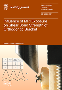
Cover Story (view full-size image): Magnetic resonance imaging (MRI) is widely used in diagnostics, but its effects on orthodontic materials remain a concern. This study aimed to evaluate the impact of MRI exposure at 1.5 T and 3 T on the shear bond strength (SBS) and adhesive remnant index (ARI) of different orthodontic bracket types (metal, self-ligating, and ceramic). The findings suggest that high-field MRI (3 T) has a more pronounced effect on SBS, particularly for self-ligating and ceramic brackets. The clinical importance of understanding these results is that both patients and clinicians must be aware of the inevitable changes that occur in SBS during MRI since exposure to high-field MRI may alter bond strength and adhesive failure characteristics. View this paper
  • Issues are regarded as officially published after their release is announced to the table of contents alert mailing list.
  • You may sign up for e-mail alerts to receive table of contents of newly released issues.
  • PDF is the official format for papers published in both, html and pdf forms. To view the papers in pdf format, click on the "PDF Full-text" link, and use the free Adobe Reader to open them.
Order results
Result details
Section
Select all
Export citation of selected articles as:
15 pages, 2196 KiB  
Article
Phenotypic Craniofacial and Upper Spine Characteristics in Patients with Obstructive Sleep Apnoea
by Anne Marie Aavang Arvidson and Liselotte Sonnesen
Dent. J. 2025, 13(3), 136; https://doi.org/10.3390/dj13030136 - 20 Mar 2025
Viewed by 350
Abstract
Background/Objectives: This study investigates differences in craniofacial morphology including skull thickness, sella turcica morphology, nasal bone length, and posterior cranial fossa dimensions, as well as differences in head posture and deviations in upper spine morphology, in adult OSA patients compared to healthy [...] Read more.
Background/Objectives: This study investigates differences in craniofacial morphology including skull thickness, sella turcica morphology, nasal bone length, and posterior cranial fossa dimensions, as well as differences in head posture and deviations in upper spine morphology, in adult OSA patients compared to healthy controls with neutral occlusion. Methods: 51 OSA patients (34 men, 17 women, mean age 51.9 ± 11.3 years) and 74 healthy controls (19 men, 55 women, mean age 38.7 years ± 14.0 years) with neutral occlusion were included. Craniofacial morphology and head posture were investigated using cephalometric measurements on lateral cephalograms and morphological deviations in sella turcica and upper spine were assessed through visual description of lateral cephalograms. Results: OSA patients had significantly more retrognathic maxilla (p = 0.02) and mandible (p = 0.032 and p = 0.009), significantly larger beta-angle (p = 0.006), and significantly smaller jaw angle (p = 0.045) compared to controls. OSA patients had significantly larger length (p = 0.003, p = 0.001, p = 0.044) and depth of the posterior cranial fossa (p < 0.001) compared to controls. OSA patients had a significantly more extended (p < 0.001) and forward-inclined head posture (p < 0.001) and morphological deviations in the upper spine occurred significantly more often in OSA patients compared to controls (p = 0.05). No significant differences in skull thickness, nasal bone length, and morphological deviations in the sella turcica (p = 0.235) were found between the groups. Conclusions: Significant deviations were found in craniofacial morphology, head posture, and morphological deviations in the upper spine. The results may prove valuable in the diagnostics of OSA patients and in considerations regarding etiology and the phenotypic differentiation of OSA patients. Full article
(This article belongs to the Special Issue Dentistry in the 21st Century: Challenges and Opportunities)
Show Figures

Figure 1

12 pages, 789 KiB  
Systematic Review
Managing the Leeway Space in Mixed Dentition Using a Passive Lingual Arch: A Systematic Review
by Alberto De Stefani, Giovanni Bruno, Valentina Montanari, Ayoub Boutarbouche, Patrizio Bollero, Antonio Gracco and Michele Basilicata
Dent. J. 2025, 13(3), 135; https://doi.org/10.3390/dj13030135 - 20 Mar 2025
Viewed by 564
Abstract
Background/Objectives: Dental crowding and the premature loss of one or more deciduous teeth are common issues during the growth phase that accompanies the transition from mixed to permanent dentition. The aim of this systematic review is to examine the effectiveness of using a [...] Read more.
Background/Objectives: Dental crowding and the premature loss of one or more deciduous teeth are common issues during the growth phase that accompanies the transition from mixed to permanent dentition. The aim of this systematic review is to examine the effectiveness of using a passive lingual arch in preserving the length of the lower arch and managing the leeway space, analyzing the effects on the linear and angular positions of the permanent teeth. Methods: A systematic review of the literature was conducted using the PubMed, Web of Science, Scopus, and Cochrane Library database. After an initial selection of 306 articles, seven studies that met the defined selection criteria were included. These articles were used to compile the PICO table. Results: The studies examined agree that the application of the passive lingual arch is useful in preserving the length of the lower arch during the transition from mixed to permanent dentition. The observed changes in the linear and angular positions of the permanent teeth, particularly the distoinclination of the permanent molars and the proclination of the incisors, were considered indicative of the effectiveness of this technique. However, one author did not observe these changes, noting only a prevention of mesioinclination and lingualization of the molars and incisors. Conclusions: The use of the passive lingual arch in the transition from mixed to permanent dentition proves to be advantageous for correcting mild anterior crowding, maintaining residual spaces after the premature loss of deciduous molars, and preventing the impaction of permanent premolars. This simple and effective orthodontic device can be applied in clinical practice, always based on an accurate diagnosis and a well-defined treatment plan. Full article
(This article belongs to the Special Issue Tradition and Innovation in Orthodontics)
Show Figures

Figure 1

20 pages, 12143 KiB  
Case Report
A Novel Intraoral Optical Scan-Transfer Device for Full-Arch Implant Reconstruction
by Cemal Ucer, Rabia Sannam Khan and Gwyn Jones
Dent. J. 2025, 13(3), 134; https://doi.org/10.3390/dj13030134 - 19 Mar 2025
Viewed by 562
Abstract
Background: Dental implantology has undergone significant advancements with the integration of digital workflows, transforming the processes of planning, designing, surgical delivery, and prosthetic rehabilitation. Among these innovations, intraoral optical scanning (IOS) has emerged as a preferred method over traditional analogue impressions. This [...] Read more.
Background: Dental implantology has undergone significant advancements with the integration of digital workflows, transforming the processes of planning, designing, surgical delivery, and prosthetic rehabilitation. Among these innovations, intraoral optical scanning (IOS) has emerged as a preferred method over traditional analogue impressions. This preference is due to its cost-effectiveness, efficiency, and streamlined patient-friendly use while producing clinically acceptable results in terms of trueness and precision, particularly for short-span implant prostheses. Methods: However, the clinical utility of intraoral scanning is significantly affected by the lack of reference points and difficulties in moisture and bleeding control at the time of immediate implant placement surgery in the fully edentulous arch. Current evidence supports the general consensus that the traditional analog impression technique still provides superior trueness and precision compared to IOS, specifically in full-arch implant cases. Results: The continuous quest for precision in dental implantology has led to the introduction of photogrammetry, which is now considered the most accurate technique for the digital scanning of dental implants. Photogrammetry has demonstrated superior results compared to those obtained using the analog technique. Conclusions: The aim of this case report is to provide an overview of analog techniques, digital intraoral optical scanning, and photogrammetry, setting the stage for the introduction of a novel technique involving a dedicated optical scan-transfer device (IPD®) that can be scanned with ease using IOS, either intra- or extra-orally, due to its unique design features and digital properties. Full article
Show Figures

Figure 1

23 pages, 2628 KiB  
Systematic Review
The Role of Genetics in Human Oral Health: A Systematic–Narrative Review
by Anita Joy-Thomas, Zarna Lalwani, Leticia Guajardo, John Valenza and Walid D. Fakhouri
Dent. J. 2025, 13(3), 133; https://doi.org/10.3390/dj13030133 - 16 Mar 2025
Viewed by 932
Abstract
Background/Objectives: An individual’s genetic makeup influences their organ development, orofacial structures, and overall health. Though many studies have been conducted to determine the inheritance of oral diseases and conditions, there is a lack of comprehensive research classifying these disorders based on the [...] Read more.
Background/Objectives: An individual’s genetic makeup influences their organ development, orofacial structures, and overall health. Though many studies have been conducted to determine the inheritance of oral diseases and conditions, there is a lack of comprehensive research classifying these disorders based on the genetic and environmental etiology. Methods: This systematic review aimed to analyze the existing body of literature using the PubMed and Cochrane databases and answer the following question: “What evidence exists supporting the role of genetic factors in oral conditions?” This systematic–narrative review methodically categorizes oral diseases and conditions based on their genetic or environmental linkages. Each classification is rigorously supported by the peer-reviewed articles and evidence strength, affirming the sufficient validity of the identified associations. Results: This study provides an overview of how genetics can influence oral health, from predisposition to susceptibility to various oral diseases, and the impact of genetic alterations on dental and oral conditions. Additionally, this study discusses the importance of understanding the interplay between genetic and environmental factors to improve oral health outcomes. An enhanced understanding of the impact of genetics on oral health will provide a better understanding of the implications of inherited or de novo genetic mutations and their potential interactions with environmental factors. Conclusions: The data collection and analysis indicate 25 oral conditions with strong genetic components and 2 with moderate genetic contributions (fibrous dysplasia and impacted teeth), while 14 oral conditions seem to have weak genetic contributions. Treatment planning that includes genetic testing and counseling as an approach of precision oral healthcare is encouraged to develop appropriate preventative and timely treatment plans to provide the effective management of patients’ symptoms. Full article
Show Figures

Graphical abstract

18 pages, 1410 KiB  
Review
Effects of Bromelain and Papain in Tooth Whitening and Caries Removal: A Literature Review
by Stanca Cuc, Amalia Moldovan, Marioara Moldovan, Codruta Sarosi, Smaranda Buduru, Cecilia Bacali, Doina Prodan, Viorica Lazar and Sorin Claudiu Man
Dent. J. 2025, 13(3), 132; https://doi.org/10.3390/dj13030132 - 16 Mar 2025
Viewed by 1397
Abstract
Background/Objectives: The objective of this review is to establish a solid base of information regarding the use of proteolytic enzymes to replace hydrogen peroxide/carbamide in teeth whitening products. The use of proteolytic enzymes, such as bromelain and papain, can provide surprising results for [...] Read more.
Background/Objectives: The objective of this review is to establish a solid base of information regarding the use of proteolytic enzymes to replace hydrogen peroxide/carbamide in teeth whitening products. The use of proteolytic enzymes, such as bromelain and papain, can provide surprising results for solving two important aspects related to dental aesthetics: tooth whitening and the chemo-mechanical removal of damaged dental tissue. Due to their ability to degrade salivary proteins, these enzymes can be used successfully as active agents in tooth whitening and in the atraumatic treatment of caries without being accompanied by other side effects on dental components. Methods: Random-effects meta-analyses were performed with enzymes (bromelain, papain) used in dentistry. A keyword search of scientific publications was conducted using the Google Academic, Web of Science and PubMed search engines. Results: The results were systematized in the present work in two parts: bromelain and papain effects in tooth whitening and chemo-mechanical/atraumatic removal of damaged dental tissues. Conclusions: The findings from different studies and clinical reports indicate that bromelain and papain could be considered efficient and safe therapeutic agents not only in various medical conditions but also in dental problems. Full article
Show Figures

Figure 1

14 pages, 15064 KiB  
Case Report
Successful Correction of Crossbite with Multi-Segment Le Fort I Osteotomy in a Patient with Cleft Lip and Palate
by Naoko Nemoto, Hitoshi Kawanabe and Akihiko Oyama
Dent. J. 2025, 13(3), 131; https://doi.org/10.3390/dj13030131 - 14 Mar 2025
Viewed by 412
Abstract
Objectives: Cleft lip and palate is a multifactorial disease that causes various problems, such as maxillary and facial morphological abnormalities, oral dysfunction, and postoperative scarring due to lip and palate formation after birth. This condition can easily cause obstruction and may require [...] Read more.
Objectives: Cleft lip and palate is a multifactorial disease that causes various problems, such as maxillary and facial morphological abnormalities, oral dysfunction, and postoperative scarring due to lip and palate formation after birth. This condition can easily cause obstruction and may require surgical orthodontic treatment in the future. Methods: In this study, we performed multi-segment Le Fort type 1 osteotomy on a patient with a cleft lip and palate who presented with a crossbite, horizontal inclination of the occlusal plane due to dental arch stenosis on the left side of the maxilla, and deviation of the mandible. Results: In this case, close occlusion was achieved by improving the patient’s facial appearance and occlusal relationship by combining sagittal division of the mandibular ramus, and the stability of the occlusion was measured without relapse 1 year after the surgery. Conclusions: This case is considered of great medical significance, as there have been few reports of cases showing a stable course. Full article
Show Figures

Figure 1

15 pages, 2312 KiB  
Article
Effectiveness of Lighting Conditions on Shade Matching Accuracy Among Dental Students
by Christina Perou, Andrianos Petalas, Michaella Stoupi and Christina Hadjichristou
Dent. J. 2025, 13(3), 130; https://doi.org/10.3390/dj13030130 - 14 Mar 2025
Viewed by 541
Abstract
Background: In prosthodontics, achieving esthetic success requires precise shade matching of restorations to natural teeth. This study evaluated the shade-matching abilities of fourth-year dental students using different tools and conditions in laboratory (LB) and clinical (CL) settings. Methods: In the LB [...] Read more.
Background: In prosthodontics, achieving esthetic success requires precise shade matching of restorations to natural teeth. This study evaluated the shade-matching abilities of fourth-year dental students using different tools and conditions in laboratory (LB) and clinical (CL) settings. Methods: In the LB setting, students matched blinded shade tabs to the VITA classical shade guide under natural daylight (ND), artificial light (AL), and a polarized filter (PF). In the CL setting, they determined the shades of patients’ central incisors using the same conditions. Participants also completed a questionnaire about their experience. Results: Quantitative analysis showed better shade matching in the LB setting, particularly with the PF (52% correct), compared to ND (50%) and AL (43%). In the CL setting, overall accuracy dropped to 32% across all conditions. No results were statistically significant. Qualitative feedback revealed that students found the spectrophotometer to have improved their accuracy and reliability compared to the ND and AL conditions. Conclusions: This study highlights the importance of incorporating technology as a validation tool in dental practice. Leveraging tools like spectrophotometers may enhance shade-matching accuracy, streamline processes, and improve patient satisfaction while balancing traditional methods with technological advancements. Full article
Show Figures

Figure 1

11 pages, 919 KiB  
Review
Correlation Between Angular Position and Pathological Changes in Impacted Lower Third Molars: A Systematic Review and Meta-Analysis
by Shaniko Kaleci, Pierantonio Bellini, Giacomo Setti, Giulia Melloni, Matilde Ruozzi and Ugo Consolo
Dent. J. 2025, 13(3), 129; https://doi.org/10.3390/dj13030129 - 14 Mar 2025
Viewed by 394
Abstract
Background: The extraction of impacted third molars presents anatomical challenges and surgical risks, prompting debate over prophylactic removal, particularly for impacted lower molars. Studies highlight associated pathologies and complications that influence treatment decisions. Objective: This study aims to systematically review and analyze the [...] Read more.
Background: The extraction of impacted third molars presents anatomical challenges and surgical risks, prompting debate over prophylactic removal, particularly for impacted lower molars. Studies highlight associated pathologies and complications that influence treatment decisions. Objective: This study aims to systematically review and analyze the correlation between the angular positions of impacted lower third molars and their association with pathological changes, including periodontal defects, alveolar bone loss, and cystic degeneration. Methods: This systematic review was conducted according to PRISMA guidelines, including studies from 2000 to 2024. Studies reporting the angular position and associated pathologies of impacted lower third molars were included. The Newcastle-Ottawa Scale (NOS) was used to assess the risk of bias. A meta-analysis of the proportion of pathological changes related to specific angular positions was performed. Results: Of the 2943 studies initially identified, six studies (including 2222 patients and 3276 impacted lower third molars) met the inclusion criteria and were included in the review, while four studies were included in the proportional meta-analysis. The most common angular positions observed were mesioangular (34.0–66.1%), followed by vertical (12.8–48.3%), horizontal (8.1–23.4%), and distoangular (3.2–14.0%). Pathological changes were reported in 8.1–75.0% of cases, with horizontal impactions demonstrating the highest correlation (41.1%, 95% CI: 20.9–63.0). Conclusions: The angular position of impactions of the lower third molars significantly influences the risk of pathological changes. Horizontal impactions exhibit the highest associated with pathological changes, whereas distoangular impactions show the lowest prevalence of complications. Standardized diagnostic and reporting practices are needed to improve clinical decision-making. Further research should focus on long-term outcomes and the impact of clinical management strategies. Full article
Show Figures

Figure 1

14 pages, 9800 KiB  
Article
Photobiomodulation in Patients Taking Denosumab: Case Report and Literature Review
by Gianluigi Caccianiga, Antonio Barbarisi, Paolo Caccianiga, Dorina Lauritano and Saverio Ceraulo
Dent. J. 2025, 13(3), 128; https://doi.org/10.3390/dj13030128 - 13 Mar 2025
Viewed by 553
Abstract
Background: Denosumab is a human monoclonal antibody playing a central role in bone resorption. The impaired bone healing observed in patients on denosumab is linked to the drug’s inhibition of osteoclast activity. Photobiomodulation (PBM) has garnered attention as a potential adjunctive therapy for [...] Read more.
Background: Denosumab is a human monoclonal antibody playing a central role in bone resorption. The impaired bone healing observed in patients on denosumab is linked to the drug’s inhibition of osteoclast activity. Photobiomodulation (PBM) has garnered attention as a potential adjunctive therapy for managing oral complications in patients on denosumab therapy. The aim of this study is to provide a review of the literature regarding the benefits of photobiomodulation therapy in patients taking denosumab while providing a case report of a patient treated with this therapy. Materials and Methods: Key terms were used to search PubMed (MEDLINE), Scopus, and Web of Science, and at last, 25 articles were compared. Following the proposed review, a case of a patient is illustrated. Results: Based on our literature findings, there are no papers regarding the benefits of photobiomodulation therapy in patients taking denosumab specifically, but there are articles regarding photobiomodulation therapy and MRONJ osteonecrosis patients, which can be caused by denosumab. Discussion: Despite all the limitations of the data in the literature, it can be deduced that there are evident benefits of photobiomodulation therapy in patients taking denosumab. The integration of laser-assisted techniques and photobiomodulation into MRONJ management protocols represents a significant evolution in treatment strategies. Conclusions: Further studies are needed to better understand a potential association between odontoclasts (which can cause external root resorption) and neoplastic disease or medication, as well as to explore the role of photobiomodulation in the therapeutic rehabilitation process. Full article
Show Figures

Figure 1

13 pages, 2340 KiB  
Opinion
Oral Medicine and Oral Clinical Chemistry Game Changers for Future Plaque Control and Maintenance: PerioSafe® aMMP-8 POCT, Lumoral® 2× PDT- and Lingora® Fermented Lingonberry Oral Rinse-Treatments
by Nur Rahman Ahmad Seno Aji, Vaibhav Sahni, Miika T. Penttala, Dimitra Sakellari, Andreas Grigoriadis, Tommi Pätilä, Pirjo Pärnänen, Dirk Neefs, Andreas Pfützner, Shipra Gupta, Timo Sorsa and Ismo T. Räisänen
Dent. J. 2025, 13(3), 127; https://doi.org/10.3390/dj13030127 - 13 Mar 2025
Viewed by 747
Abstract
Background: Periodontitis is a global health crisis that affects almost half of the world’s population and commonly goes unnoticed because of its asymptomatic and pain-free nature. For early and easy detection and treatment, safe and non-invasive chair-side oral fluid biomarker (aMMP-8) diagnostics [...] Read more.
Background: Periodontitis is a global health crisis that affects almost half of the world’s population and commonly goes unnoticed because of its asymptomatic and pain-free nature. For early and easy detection and treatment, safe and non-invasive chair-side oral fluid biomarker (aMMP-8) diagnostics and new anti-microbial, anti-inflammatory and anti-proteolytic treatment modalities have been developed, which this review aims to introduce. Methods: For convenient diagnosis and tackling of periodontitis, adoption of an oral fluid aMMP-8 chair-side point-of-care rapid diagnostic test (POCT) has been proposed, comparable to home pregnancy and COVID-19 antigen tests, to be conveniently used by healthcare professionals and by patients themselves. To improve treatment of detected periodontitis, Finnish scientists have also developed a potentially industry-altering, biofilm-modulating, anti-microbial, anti-inflammatory, and anti-proteolytic (i) dual-light-activated photodynamic-therapy (2×PDT) and (ii) fermented lingonberry juice (FLJ) oral rinse designed for home personalized medicine and professional use. These new oral medicine technologies are reviewed and some unpublished results are presented. Results: aMMP-8 is the superior biomarker for grade of periodontitis (progression rate) when compared to the total latent/proform MMP-8 (total-MMP-8) and microbial lipopolysaccharide (LPS/LAL) activity. Cut-off 20 ng/mL is the optimal cut-off for aMMP-8 POCT and does not make false positives. Antibacterial 2× PDT light and anti-microbial FLJ treatments can eliminate and reduce problem-causing bacteria and Candida-yeasts from the mouth. Conclusions: These new oral medicine technologies have shown promising results and could have the potential to revolutionize diagnosis, prevention, oral care, plaque control and maintenance. These new game-changer oral medicine technologies have launched a new clinical field in dentistry: oral clinical chemistry. Full article
Show Figures

Figure 1

10 pages, 240 KiB  
Article
Impact of High-Intensity Sports Practice on Stomatognathic System Function: An Observational Study
by Evandro Marianetti Fioco, Marcelo Palinkas, Natália de Moraes Barbosa, Edson Donizetti Verri, Luciano Maia Alves Ferreira, Danilo Henrique Lattaro, Gabriella Simi Gariba Silva, Selma Siéssere and Simone Cecilio Hallak Regalo
Dent. J. 2025, 13(3), 126; https://doi.org/10.3390/dj13030126 - 13 Mar 2025
Viewed by 611
Abstract
Background: Physical activity improves quality of life, but competitive sports emphasize performance, leading to intense training and restrictive diets that increase injury risk. This affects the stomatognathic system, underscoring the role of sports dentistry in preventing injuries and orofacial functional changes. This observational [...] Read more.
Background: Physical activity improves quality of life, but competitive sports emphasize performance, leading to intense training and restrictive diets that increase injury risk. This affects the stomatognathic system, underscoring the role of sports dentistry in preventing injuries and orofacial functional changes. This observational study analyzed the stomatognathic system’s functionality in male high-intensity athletes (n = 18) compared to a sedentary group (n = 18). Methods: Functional parameters were analyzed: electromyographic activity during mandibular tasks, masseter and temporal muscles thickness, and molar bite force. Results: Student’s t-test was used for analysis. At mandibular rest, male high-performance athletes showed lower electromyographic activity in the right and left masseter muscles, suggesting adaptation to training. During maximum voluntary contraction, activity was higher in the temporal and masseter muscles, especially in the left masseter, indicating hypertrophy. Electromyographic activity increased during protrusion and lateral movements, particularly in the masseter and temporal muscles, demonstrating a greater functional efficiency group of athletes. They also exhibited greater masseter muscle thickness and thinner temporal muscle thickness, with a significant difference in the right masseter muscle at rest. The group of athletes showed greater molar bite force, with significant differences, indicating greater muscle capacity. Conclusions: Sports practice promotes adaptations in the stomatognathic system, improving its functionality. Full article
(This article belongs to the Special Issue Dentistry in the 21st Century: Challenges and Opportunities)
15 pages, 2406 KiB  
Article
Load-Bearing Capacity of Incisors Restored Using Fiber-Reinforced Composite Post-Core Systems
by Keiichiro Uchikura, Sufyan Garoushi, Kohji Nagata, Pekka K. Vallittu, Noriyuki Wakabayashi and Lippo Lassila
Dent. J. 2025, 13(3), 125; https://doi.org/10.3390/dj13030125 - 13 Mar 2025
Viewed by 625
Abstract
Objectives: This study aimed to analyze the load-bearing performance of upper incisors and evaluate the curing of the luting polymer composite at various depths within the canal. Methods: A total of one hundred maxillary central incisors (10 groups, n = 10/group) were subjected [...] Read more.
Objectives: This study aimed to analyze the load-bearing performance of upper incisors and evaluate the curing of the luting polymer composite at various depths within the canal. Methods: A total of one hundred maxillary central incisors (10 groups, n = 10/group) were subjected to various restorative techniques. Approach A used Gradia Core for post-core and crown; Approach B employed prefabricated fiber posts (4 mm or 8 mm) with Gradia for luting and core build-up; Approach C used short-fiber composite (everX Flow) for post-core build-up; and Approach D used fiber posts with everX Flow for luting and core build-up. Restorations underwent cyclic fatigue (40,000 cycles at 95 N) and quasi-static fracture testing. Surface hardness of luting polymer composites was also measured. Results: Data showed that restorations with additional fiber posts (Approaches B and D) had significantly higher load-bearing capacity (p < 0.05), while post material and length had no significant impact (p > 0.05). Short-fiber composite as luting and core material (Approach D) enhanced load-bearing performance compared to Gradia-based restorations (Approach B, p < 0.05). Conclusions: The use of short-fiber composite as both the post luting and core material in restoring compromised incisors, along with a conventional fiber post, demonstrated favorable results in terms of load-bearing capacity. Full article
Show Figures

Figure 1

13 pages, 2228 KiB  
Opinion
Identifying Key Factors in Papilla Growth Around Implants: Focus on Intraoral Negative Pressure
by Daniele Botticelli, Ivo Agabiti, Rihito Yamada, Nozomi Maniwa, Karol Alí Apaza Alccayhuaman and Yasushi Nakajima
Dent. J. 2025, 13(3), 124; https://doi.org/10.3390/dj13030124 - 13 Mar 2025
Viewed by 1056
Abstract
The absence of interdental papillae in dental prosthetics often leads to unsatisfactory esthetic outcomes, such as black triangles and elongated clinical crowns. While previous research has demonstrated that papillae can regenerate in a coronal direction, the underlying mechanisms remain incompletely understood. Several theories [...] Read more.
The absence of interdental papillae in dental prosthetics often leads to unsatisfactory esthetic outcomes, such as black triangles and elongated clinical crowns. While previous research has demonstrated that papillae can regenerate in a coronal direction, the underlying mechanisms remain incompletely understood. Several theories have been proposed to explain this phenomenon, but no clear cause–effect relationship has been established among the various factors involved in spontaneous papilla growth around implants. This study aims to identify and classify the factors influencing this process. Various potential contributors were analyzed, including adjacent elements, buccal–lingual papilla width, contact point position, convergent neck design, crown overcontour, intraoral negative pressure, and others. To systematically organize these factors, a modified Overton Window and a mind map were employed. The factors were categorized as cause-related, essential, or influencing based on the collective opinion of the research group following a comprehensive review of the relevant literature. In the absence of clear evidence supporting a definitive cause–effect relationship, Occam’s Razor (the principle of parsimony) was applied to identify the most plausible cause-related factors. Full article
(This article belongs to the Special Issue Dentistry in the 21st Century: Challenges and Opportunities)
Show Figures

Figure 1

11 pages, 212 KiB  
Article
Assessment of Oral Health-Related Quality of Life of the United Arab Emirates’ Elderly Population: Observational Prospective Cross-Sectional Study
by Faris El-Dahiyat, Ammar Abdulrahman Jairoun, Obaida Jairoun, Islam Eljilany and Mohammed Alsbou
Dent. J. 2025, 13(3), 123; https://doi.org/10.3390/dj13030123 - 11 Mar 2025
Viewed by 534
Abstract
Background/Objectives: The current study aimed to evaluate the oral health self-perception on quality of life in the elderly using the Geriatric Oral Health Assessment Index (GOHAI) to assess the impact of demographic and oral health factors on oral health-related quality of life. Methods: [...] Read more.
Background/Objectives: The current study aimed to evaluate the oral health self-perception on quality of life in the elderly using the Geriatric Oral Health Assessment Index (GOHAI) to assess the impact of demographic and oral health factors on oral health-related quality of life. Methods: An observational prospective cross-sectional study for the entire six-month period was conducted in a dental health care center in the United Arab Emirates. The principal inclusion criterion is being aged 60 and over. The GOHAI questionnaire is composed of 4 domains of 12 items that address functional limitation, pain and discomfort, psychological impacts, and behavioral impacts. Patients were questioned about the frequency at which they experience any of the 12 listed problems. Results: A total of 318 respondents participated in the study and completed the whole questionnaire. Among these participants, 63.5% (n = 202) were male and 86.8% (n = 276) were ≤70 years. The average GOHAI score was 13.25, with a 95% confidence interval (CI) [12.4%, 14%], indicating a low self-perception of oral health by the allocated sample. Statistical modeling identified dry mouth (OR = 2.21, 95% CI 1.40–3.48) and chewing problems (OR = 1.87, 95% CI 1.09–3.20) as the strongest determinants of poor oral health-related quality of life (OHRQoL) in the elderly population. Conclusions: Healthcare professionals should develop targeted strategies to address the specific needs of this population, ensuring sustained improvements in their quality of life. Full article
(This article belongs to the Special Issue Preventive Dentistry and Dental Public Health)
12 pages, 1213 KiB  
Article
Dental Morphology in Restorative Dentistry: A Pilot Study on Morphological Consistency and Variability in Human Upper First Molars
by Gregorio Oxilia, Mauro Tomasella and Alberto Cecere
Dent. J. 2025, 13(3), 122; https://doi.org/10.3390/dj13030122 - 11 Mar 2025
Viewed by 900
Abstract
Background: Geometric morphometric analysis, a methodology traditionally used in evolutionary studies, offers unprecedented precision in quantifying the morphological traits of human organs and tissues by identifying specific anatomical landmarks. Despite its potential, this approach has not yet been applied in medical or dental [...] Read more.
Background: Geometric morphometric analysis, a methodology traditionally used in evolutionary studies, offers unprecedented precision in quantifying the morphological traits of human organs and tissues by identifying specific anatomical landmarks. Despite its potential, this approach has not yet been applied in medical or dental research. Materials and Methods: This pilot study analyzed 76 permanent upper first molars (M1) from Italian individuals (38 males and 38 females), selected for their completeness, minimal wear (stage 1), and absence of pathological conditions or non-metric traits. Each individual contributed two contralateral molars: the left molar (Group A) and the right molar (Group B). The molars were scanned using a Sinergia Scan Advanced Plus optical scanner with 5 μm accuracy. The scans were processed with the Dental Scan 7.0 software to generate high-resolution STL files, followed by refinement in Meshlab to preserve the morphological integrity of the 3D models. The geometric morphometric analysis was performed using the Viewbox software, thereby enabling the placement of anatomical landmarks and semilandmarks to quantify and compare molar morphology with exceptional precision. Results: The analysis confirmed that contralateral molars exhibit high morphological similarity, with significantly smaller variability compared to molars from different individuals. Among the specific traits analyzed, the distolingual cusp (hypocone) showed the greatest variation, followed by the mesiolingual cusp (protocone). No significant differences were observed between males and females in the degree of similarity between contralateral molars. Conclusions: This pilot study highlights the potential of geometric morphometric approaches to enhance our understanding of the dental variation between sexes and across human populations, thereby improving clinical applications and advancing toward personalized medicine. Full article
Show Figures

Figure 1

9 pages, 192 KiB  
Article
Awareness of Traumatic Dental Injuries and Impact of Educational Intervention Among Croatian Soccer Coaches and Players
by Dina Bursać, Lovro Marinović, Marta Horvat and Kristina Goršeta
Dent. J. 2025, 13(3), 121; https://doi.org/10.3390/dj13030121 - 10 Mar 2025
Viewed by 451
Abstract
Background/Objectives: This study aimed to assess the knowledge of dental injuries in both soccer coaches and players, as well as the impact of educational intervention on coaches and the prevalence of traumatic dental injuries and mouthguard usage among soccer players. Methods: [...] Read more.
Background/Objectives: This study aimed to assess the knowledge of dental injuries in both soccer coaches and players, as well as the impact of educational intervention on coaches and the prevalence of traumatic dental injuries and mouthguard usage among soccer players. Methods: The study involved 94 male soccer players (median age 18, IQR: 12–19) and 39 coaches. Data were collected through questionnaires covering age, gender, years of experience, injury history, mouthguard use, and knowledge of tooth rescue kits and tooth repositioning. Results: Of the players, 34 (36.2%) experienced orofacial injuries during soccer, while only 7 (7.4%) used mouthguards. Reasons for not using mouthguards included discomfort (26.1%), belief they are unnecessary (26.1%), and difficulty purchasing them (8.0%). Among coaches, 24 (61.5%) reported handling dental injuries during training, including crown fractures (41.7%), tooth avulsions (33.3%), and tooth luxations (25.0%). Only four (10.3%) coaches personally used mouthguards. Conclusions: The study identified a significant lack of knowledge about dental injuries and limited use of mouthguards among Croatian soccer players. Coaches, while influential in athlete development, should also focus on injury prevention, including dental trauma. Full article
(This article belongs to the Special Issue Oral Health Care in Paediatric Dentistry Volume 2)
15 pages, 265 KiB  
Article
Knowledge of and Attitudes Toward Erosive Tooth Wear Among Students of a Portuguese University: Pilot Survey
by Patrícia Manarte-Monteiro, Maria Vittoria Buscemi, Joana Domingues, Liliana Teixeira, Bernardo Lemos and Lígia Pereira da Silva
Dent. J. 2025, 13(3), 120; https://doi.org/10.3390/dj13030120 - 10 Mar 2025
Viewed by 551
Abstract
Background/Objectives: To assess and compare the knowledge of and attitudes toward erosive tooth wear (ETW) among university students at the Portuguese University Fernando Pessoa (UFP). Methods: Cross-sectional, prospective pilot study approved by the UFP-Ethics Committee for a bilingual (English and Portuguese) online self-administered [...] Read more.
Background/Objectives: To assess and compare the knowledge of and attitudes toward erosive tooth wear (ETW) among university students at the Portuguese University Fernando Pessoa (UFP). Methods: Cross-sectional, prospective pilot study approved by the UFP-Ethics Committee for a bilingual (English and Portuguese) online self-administered questionnaire displayed to all UFP students of the 2023–2024 academic year. Two groups were recruited, dental students (DSs) and non-medical students (NMSs), based on a convenience sample of 344 students. Participants voluntarily answered demographic questions, 15 true/false/do not know questions between knowledge of ETW, and 10 positive statements to score the attitude toward ETW based on a 5-point Likert scale. Differences in scores for DSs and NMSs groups by non-parametric tests and the correlation between knowledge of and attitudes by the Pearson coefficient were considered significant for p < 0.05. Results: A total of 251 (72.9%) students participated, but only 245 (71.2%) fulfilled the survey. The knowledge and attitude scores of the DSs (Md = 12.0; IQR 11.0–13.0 and Md = 43.0; IQR 40.0–48.0) were higher (p < 0.001; p = 0.019) than those of NMSs (Md = 8.0; IQR 6.0–10.0 and Md = 41.0; IQR 38.0–46.0). The attitudes score showed similar (p > 0.05) results for gender, age, nationality, and curricular year. Moderate level of knowledge was higher (p < 0.001) for DSs (Md = 12.0, IQR 11.0–12.0) than for NMSs (Md = 10.0, IQR 9.0–10.0) students. DSs and NMSs revealed differences in attitude level distribution (p < 0.001). Conclusions: This survey highlighted the need for knowledge on ETW among NMSs, but especially the high need for measures to promote more positive attitudes toward ETW among all university students, DSs, and NMSs. It provided valuable insights into the demographic characteristics, response rate, knowledge, and attitude scores of ETW with the implementation of multicenter designs applied to similar populations being important for future research. Full article
18 pages, 626 KiB  
Article
Knowledge and Perceptions of Dentists Regarding E-Cigarettes: Implications for Oral Health and Public Awareness and Education
by Donika B. Shabani, Linda J. Dula, Zana Sllamniku Dalipi, Mirlinda S. Krasniqi and Aida Meto
Dent. J. 2025, 13(3), 119; https://doi.org/10.3390/dj13030119 - 10 Mar 2025
Viewed by 1182
Abstract
Background/Objectives: The rising popularity of e-cigarette use is creating new challenges for oral healthcare. Factors such as targeted marketing, appealing flavors, and the perception that e-cigarettes are a safer alternative to traditional smoking have contributed to their widespread adoption. This trend is particularly [...] Read more.
Background/Objectives: The rising popularity of e-cigarette use is creating new challenges for oral healthcare. Factors such as targeted marketing, appealing flavors, and the perception that e-cigarettes are a safer alternative to traditional smoking have contributed to their widespread adoption. This trend is particularly prevalent among young adults and teenagers, many of whom turn to e-cigarettes despite having no prior history of regular cigarette use. This study aimed to evaluate dentists’ knowledge and perceptions regarding e-cigarettes, focusing on their health effects, regulatory status, and potential risks to oral health. Assessing dentists’ knowledge of e-cigarette risks is important, as they guide patients on the potential oral health dangers of vaping. E-cigarette use has been linked to several oral health problems. Methods: A cross-sectional survey was conducted among 189 dentists in Kosovo, comprising both general dentists and specialists. The questionnaire assessed participants’ awareness of e-cigarettes’ FDA approval status, their perceptions of health impacts, and their understanding of e-cigarettes’ association with oral conditions such as caries, periodontal disease, and oral cancer. Data were analyzed using SPSS 22, with significance set at p < 0.05. Results: The findings revealed significant knowledge gaps, with 74.1% of respondents being uncertain about the FDA approval status of e-cigarettes and 82.0% recognizing their harmful effects on general and oral health. Nearly half (49.7%) of the participants identified e-cigarettes as a potential risk factor for caries, while 66.1% and 64.6% associated their use with oral cancer and periodontal disease, respectively. Gender and professional specialization did not significantly influence these perceptions. Conclusions: Dentists need better education on the risks of e-cigarette use, including their harmful effects on oral health. Incorporating this knowledge into dental curricula and professional training is essential to give dentists the information needed to advise patients effectively. Raising public awareness through dental health professionals can help mitigate the oral health risks associated with e-cigarettes. Full article
(This article belongs to the Special Issue Preventive Dentistry and Dental Public Health)
Show Figures

Figure 1

14 pages, 2538 KiB  
Article
Ablative and Expansive Protocols for Bone Osteotomy in Rabbits
by Kazuhisa Kuwano, Luigi Canullo, Daniele Botticelli, Samuel Porfirio Xavier, Erick Ricardo Silva, Kaoru Kusano and Shunsuke Baba
Dent. J. 2025, 13(3), 118; https://doi.org/10.3390/dj13030118 - 7 Mar 2025
Viewed by 1072
Abstract
Background: Cortical and marrow bone layer have different histomorphometric features. The traditional implant insertion technique provides for fixture stabilization through the cortical area. However, this approach has been found to result in an overstress of this bone layer, which may lead to resorption. [...] Read more.
Background: Cortical and marrow bone layer have different histomorphometric features. The traditional implant insertion technique provides for fixture stabilization through the cortical area. However, this approach has been found to result in an overstress of this bone layer, which may lead to resorption. Therefore, the aim of this study was to evaluate bone healing by applying two different implant site preparation protocols across various bone densities. Materials and Methods: One implant was placed in each femur and tibia of the rabbits (four implants per animal), using two distinct site preparation methods: drilling alone or drilling followed by osteotomes (funnel technique). Three regions around the implant were evaluated: cervical, marrow, and apical. The study included 12 rabbits, divided into two groups of 6 animals each, which were euthanized at 3 and 6 weeks, respectively (n = 6 per group). Results: In the cervical region of both femur and tibia, no marginal bone resorption could be detected. Similar BIC% (bone-to-implant contact percentages) were observed for funnel and drill sites after 3 weeks and 6 weeks of healing. Differences, though not statistically significant, ranged between 2.8% and 4.7%. However, higher BIC% values were observed in the femora compared to the tibia in both periods. Conclusions: No marginal bone loss was observed in both techniques. No statistically significant differences in bone resorption or bone-to-implant contact around the implant collar were observed when comparing two implant site preparation protocols across various bone densities. The use of osteotome did not influence the healing in the marrow region. Full article
Show Figures

Figure 1

16 pages, 5823 KiB  
Article
Mechanical Performance and Surface Roughness of Lithium Disilicate and Zirconia-Reinforced Lithium Silicate Ceramics Before and After Exposure to Acidic Challenge
by Ahmed Elsherbini, Salma M. Fathy, Walid Al-Zordk, Mutlu Özcan and Amal A. Sakrana
Dent. J. 2025, 13(3), 117; https://doi.org/10.3390/dj13030117 - 6 Mar 2025
Viewed by 685
Abstract
Background/Objectives: Fabrication of ceramic restorations with higher performance, biocompatibility, and mechanical durability, as well as excellent optical properties, is challenging. Therefore, this study was designed to investigate the mechanical properties and surface roughness of different glass ceramics manufactured with CAD/CAM and pressed [...] Read more.
Background/Objectives: Fabrication of ceramic restorations with higher performance, biocompatibility, and mechanical durability, as well as excellent optical properties, is challenging. Therefore, this study was designed to investigate the mechanical properties and surface roughness of different glass ceramics manufactured with CAD/CAM and pressed techniques before and after exposure to simulated gastric acidic challenge. Methods: Lithium disilicate-based (LDS) ceramic, advanced lithium disilicate (ALDS), and zirconia-reinforced lithium silicate (ZLSC), were manufactured with two techniques. Disc-shaped (N = 336) specimens were used to test the biaxial flexural strength (BFS), while surface microhardness and surface roughness were evaluated before and after exposure to hydrochloric acid (34–37% concentration and 1.2 pH for 24 h). The results were statistically evaluated using two- and one-way ANOVA, Tukey’s post-hoc and Student’s t-test. Results: Both CAD and pressed types of LDS ceramic IPS e.max, showed significantly highest BFS and microhardness before and after acidic challenge (p = 0.01 and <0.01, respectively). ALDS and ZLSC showed significantly lower roughness before acidic aging, while all groups showed no significant difference after aging. Conclusion: Pressed groups showed better mechanical performance than CAD groups. LSD (IPS e.max types) continuously showed a better mechanical performance than modified LDS. ALDS and ZLSC showed the best performance in terms of surface smoothness, but after acidic exposure, no significant difference was observed compared to the other groups. Full article
Show Figures

Figure 1

9 pages, 468 KiB  
Article
Pulp Responsiveness of Healthy Non-Pathological Teeth Following Surgical Enucleation of Cysts
by Syed Nabil, Muhd Fazlynizam Rashdi and Abd Jabar Nazimi
Dent. J. 2025, 13(3), 116; https://doi.org/10.3390/dj13030116 - 5 Mar 2025
Viewed by 439
Abstract
Background/Objectives: Odontogenic cysts are pathological cavities lined by cells arising from odontogenic epithelial cells, occurring mostly on the tooth-bearing areas of the jaws. It is common to find that the apices of the teeth around the cyst are within the cyst’s cavities [...] Read more.
Background/Objectives: Odontogenic cysts are pathological cavities lined by cells arising from odontogenic epithelial cells, occurring mostly on the tooth-bearing areas of the jaws. It is common to find that the apices of the teeth around the cyst are within the cyst’s cavities due to its expansion. This study aims to assess the outcome of cyst enucleation on the associated teeth, specifically the latter’s responsiveness after cyst enucleation. Methods: This retrospective study examined a sample of patients who had been previously treated for odontogenic cysts from 1 January 2000 to 31 December 2021. A list of patients was obtained and included whether they met the imposed inclusion criteria. The data collected included the patients’ preoperative and postoperative electric pulp testing readings and their timings. Results: In total, 77 individual teeth from 19 patients were included after meeting the inclusion/exclusion criteria. Overall, 57 out of the 77 (74%) teeth were responsive following long-term follow-up. Among the 57 teeth with a positive response, 8 teeth were initially non-responsive and regained their responsiveness after a period of time. Pulp responsiveness recovery was seen even 300 days after surgery. Conclusions: It is not certain that a tooth with apices involved in a cyst cavity will be non-vital following enucleation. It is recommended that these teeth be reassessed for a minimum of 10 months postoperatively before proceeding with root canal treatment. Full article
(This article belongs to the Special Issue Dentinal Hypersensitivity)
Show Figures

Figure 1

11 pages, 236 KiB  
Article
Oral Hygiene Management of Hematologic Patients Undergoing Auto-Transplantation: An Observational Study
by Marco Miceli, Elisabetta Carli, Enrico Orciolo, Maria Rita Giuca and Rossana Izzetti
Dent. J. 2025, 13(3), 115; https://doi.org/10.3390/dj13030115 - 5 Mar 2025
Viewed by 543
Abstract
Background/Objectives: The present study evaluated the effectiveness of a prevention protocol involving oral hygiene, instructions, and motivation in patients eligible for hematopoietic stem cell transplantation (HSCT). Methods: Consecutive patients scheduled for HSCT were enrolled. All patients received a complete dental examination aimed at [...] Read more.
Background/Objectives: The present study evaluated the effectiveness of a prevention protocol involving oral hygiene, instructions, and motivation in patients eligible for hematopoietic stem cell transplantation (HSCT). Methods: Consecutive patients scheduled for HSCT were enrolled. All patients received a complete dental examination aimed at assessing periodontal health status through periodontal charting and registration of the levels of plaque and inflammation. Subsequently, patients received professional oral hygiene care and were instructed on and motivated toward oral prophylaxis prior to HSCT. Upon engraftment, patients received dedicated instructions to improve oral hygiene maneuvers. After HSCT, patients were re-evaluated for periodontal clinical parameters. The Oral Health Impact Profile-14 (OHIP-14) questionnaire was also administered to evaluate perceived oral health status. Results: Thirty-five patients (18 males and 17 females, with a mean age of 43.28 ± 11.73 years) were enrolled. An overall improvement was noted in periodontal parameters. However, despite plaque reduction, inflammation was still present because of the treatment. The OHIP-14 score slightly worsened after HSCT. Conclusions: Professional oral hygiene and prophylaxis proved effective in improving the oral health status of patients undergoing HSCT, potentially impacting post-treatment outcomes. Further assessment is needed to validate these results. Full article
(This article belongs to the Special Issue Dentistry in the 21st Century: Challenges and Opportunities)
18 pages, 306 KiB  
Article
Is Sustainability Part of the Drill? Examining Knowledge and Awareness Among Dental Students in Bucharest, Romania
by Ana Maria Cristina Țâncu, Marina Imre, Laura Iosif, Silviu Mirel Pițuru, Mihaela Pantea, Ruxandra Sfeatcu, Radu Ilinca, Dana Cristina Bodnar and Andreea Cristiana Didilescu
Dent. J. 2025, 13(3), 114; https://doi.org/10.3390/dj13030114 - 5 Mar 2025
Viewed by 843
Abstract
Background. Despite dentistry’s alarmingly high energy use, plastic waste, and travel emissions, research on Romanian dental students’ sustainability awareness is absent. This study aimed to assess their knowledge of the environmental impact of dental materials and practices, hypothesizing that early exposure to sustainability [...] Read more.
Background. Despite dentistry’s alarmingly high energy use, plastic waste, and travel emissions, research on Romanian dental students’ sustainability awareness is absent. This study aimed to assess their knowledge of the environmental impact of dental materials and practices, hypothesizing that early exposure to sustainability education would benefit preclinical students most. Materials and Methods. A cross-sectional survey using a form questionnaire with 15 items was conducted on 1800 dental students at Carol Davila University of Medicine and Pharmacy, Bucharest, Romania, for one week in March 2022. The questionnaire, consisting of socio-demographics, students’ perspectives on sustainability in dentistry, and personal sustainability, was analyzed using SPSS 26. Data analysis included the Shapiro–Wilk test for normality, Fisher’s exact test for categorical variables, the Mann–Whitney U test for non-parametric quantitative comparisons, and Z-tests with Bonferroni correction for contingency tables. Results. A response rate of 26.06% was achieved, with 469 participants. The majority (51.1%), particularly males (66.1%), perceived sustainability as promoting durability. The most common definition of sustainability (33.8%) was related to environmental protection, with significantly higher agreement among female students (39.4%) (p = 0.001). While 49.3% of participants identified single-use plastics in patient care as having the greatest environmental impact in dental practices, 39.2% of female students, primarily from clinical study years (50%), ranked patient paperwork and records as the most significant factor (p = 0.031). The highest-carbon-footprint dental procedures were considered to be amalgam and composite fillings (50.7%), with clinical year students indicating this as the most relevant issue (62.8% vs. 47.7%) (p = 0.011). Students aged 25–30 were more actively engaged in sustainability initiatives compared to the younger group (p = 0.005), while all students over 30 identified scaling and polishing as the most impactful procedure (p < 0.001). A majority of students supported future university sustainability initiatives (62.7%) and an elective course on sustainability in dentistry (65%). Female students showed significantly greater interest than male students in both initiatives (66.3% vs. 52.7%, p = 0.003 and 70.8% vs. 49.6%, p < 0.001, respectively). Conclusions. Greater awareness of sustainability was found in preclinical-year dental students and among female students, with knowledge gaps in clinical-year students, particularly regarding the environmental impact of dental practices and materials. Introducing sustainability courses could better prepare future dentists for sustainable practices in dentistry. Research collaborations and curriculum reforms to further promote sustainability would also be beneficial. Full article
(This article belongs to the Special Issue Dental Education: Innovation and Challenge)
12 pages, 14668 KiB  
Brief Report
Novel Roles of Nestin in Postnatal Root Formation
by Yan Jing, Jinqiu Wu, Ying Liu, Xiaohua Liu and Chi Ma
Dent. J. 2025, 13(3), 113; https://doi.org/10.3390/dj13030113 - 4 Mar 2025
Viewed by 461
Abstract
Background/Objectives: Nestin is an intermediate filament protein and a marker of odontoblasts, but its function in tooth and bone formation is largely unknown. This study aimed to determine whether Nestin plays a role in postnatal tooth formation. Methods: 4-week-old Nestin knockout (KO) mice [...] Read more.
Background/Objectives: Nestin is an intermediate filament protein and a marker of odontoblasts, but its function in tooth and bone formation is largely unknown. This study aimed to determine whether Nestin plays a role in postnatal tooth formation. Methods: 4-week-old Nestin knockout (KO) mice were analyzed with a range of techniques, including X-ray imaging, uCT, backscattered and acid-etched casted SEM, FITC-confocal microscopy, H&E and TRAP staining, and immunohistochemistry. Results: The KO mice had no apparent difference in crown formation compared to age-matched wild-type (WT) but showed delayed molar eruption with reduced TRAP+ osteoclasts. More importantly, KO mice developed expanded predentin and shorter, thinner roots with irregular and shortened dentin tubules. Additionally, the Nestin KO mice exhibited a reduced cellular cementum mass with sharp reductions in DMP1, OPN, and BSP. Conclusions: These findings suggest that Nestin plays a critical role in the postnatal development of root dentin and cellular cementum. Full article
Show Figures

Figure 1

13 pages, 544 KiB  
Article
Color Change and Color Stability of White Spot Lesions Treated with Resin Infiltration, Microabrasion, or Nano-Hydroxyapatite Remineralization: An In Vitro Study
by Nina Novozhilova, Anastasia Mun, Maria Polyakova, Anna Mikheikina, Alexandr Zaytsev and Ksenia Babina
Dent. J. 2025, 13(3), 112; https://doi.org/10.3390/dj13030112 - 3 Mar 2025
Viewed by 1314
Abstract
Background: We compared the camouflage effect of three white spot lesion (WSL) treatments (infiltration, nano-hydroxyapatite (nHAP) remineralization, and microabrasion) and color stability of the treated surfaces. Methods: Fifty sound extracted teeth were used in the study. WSLs were created on 40 [...] Read more.
Background: We compared the camouflage effect of three white spot lesion (WSL) treatments (infiltration, nano-hydroxyapatite (nHAP) remineralization, and microabrasion) and color stability of the treated surfaces. Methods: Fifty sound extracted teeth were used in the study. WSLs were created on 40 buccal dento-enamel specimens through the use of acidic methylcellulose gel. These specimens were randomly assigned to treatment groups (n = 10 per group): negative controls, nano-hydroxyapatite (nHAP), resin infiltration, and microabrasion. After the treatment, all 50 specimens were immersed in coffee for 7 days. Color measurements were performed four times: at baseline (T0), after the demineralization procedure (T1), after the treatments (T2), and after immersion in coffee (T3). Results: No restoration of the initial enamel color was observed in any of the groups. The ICON and MA groups exhibited the highest masking effect, with the mean ΔET0-T2 = 7.46, although the differences among the study groups were insignificant. All three treatments increased the resistance of WSLs to discoloration in coffee compared to the negative control group; however, infiltration (∆ET2-T3 = 4.13) and microabrasion (∆ET2-T3 = 3.49) showed a better color stability tendency than nHAP remineralization (∆ET2-T3 = 7.26). Conclusions: Despite its well-known remineralizing and desensitizing effects, nHAP showed the least masking effect for WSLs and lower color stability compared to resin infiltration and microabrasion. However, none of the methods allowed for complete restoration of the original color. After the discoloration procedure, the color changes in the white spots treated with microabrasion and infiltration were comparable to those of the sound enamel. Full article
(This article belongs to the Special Issue Preventive Dentistry and Dental Public Health)
Show Figures

Graphical abstract

27 pages, 255 KiB  
Article
Impact of Marital Status, Education, and Family Size on Parental Behaviors Toward Early Childhood Caries in Romania
by Abel Emanuel Moca, Ioan Andrei Țig, Jessica Olivia Cherecheș, Rahela Tabita Moca and Raluca Iurcov
Dent. J. 2025, 13(3), 111; https://doi.org/10.3390/dj13030111 - 3 Mar 2025
Viewed by 498
Abstract
Background and Objectives: This study aimed to examine the influence of socio-demographic factors (marital status, number of children, and education level) on the knowledge, attitudes, and practices (KAP) concerning Early Childhood Caries (ECC) prevention among parents in Bihor, Romania. This research seeks [...] Read more.
Background and Objectives: This study aimed to examine the influence of socio-demographic factors (marital status, number of children, and education level) on the knowledge, attitudes, and practices (KAP) concerning Early Childhood Caries (ECC) prevention among parents in Bihor, Romania. This research seeks to address the lack of regional data on the influence of socio-demographic factors, such as marital status, number of children, and education level, on parental knowledge, attitudes, and practices regarding ECC prevention. These insights are essential for developing targeted public health interventions in Romania. Materials and Methods: Conducted from March to September 2024, this cross-sectional study utilized a KAP questionnaire distributed online to parents of children under six. The survey was adapted to local contexts and included sections on demographic data and ECC-specific knowledge, attitudes, and practices. Statistical analyses, including Chi-square tests, were performed to evaluate the relationship between socio-demographic factors and KAP outcomes, ensuring robust data interpretation under ethical standards set by the Declaration of Helsinki. Results: Of the 798 respondents who accessed the questionnaire, 419 completed it, resulting in a completion rate of 52.5%. The participants had a mean age of 33.8 years. In terms of gender distribution, 348 (83.1%) were female and 71 (16.9%) were male. Higher educational levels were strongly correlated with better ECC knowledge and preventive practices; parents with university degrees demonstrated significantly better understanding and engagement in ECC prevention (p < 0.05). Married parents participated more actively in ECC prevention than unmarried ones, with 61.1% adhering to recommended practices compared to significantly lower rates among unmarried parents (p = 0.020). While this difference was statistically significant, the lower representation of unmarried parents in the sample should be considered when interpreting this finding. Families with fewer children showed more effective ECC preventive practices (p = 0.001). Conclusions: The study suggests that higher education and smaller family sizes are associated with better ECC prevention behaviors, emphasizing the need for targeted public health interventions. These could include parental education campaigns on ECC prevention, community-based oral health workshops, subsidized fluoride programs, and increased accessibility to pediatric dental services for underprivileged families. Full article
8 pages, 327 KiB  
Article
Dissolution of Bovine Palatal Tissue with Degassed Sodium Hypochlorite
by Justin Fang, Zhen Shen, Ransome van der Hoeven and David E. Jaramillo
Dent. J. 2025, 13(3), 110; https://doi.org/10.3390/dj13030110 - 28 Feb 2025
Viewed by 549
Abstract
Objective: To investigate the tissue dissolution efficacy of different concentrations of sodium hypochlorite (NaOCl) using the GentleWave® technology (GW) and compare it with that of passive ultrasonic irrigation. This study will provide a novel in vitro model used to evaluate the tissue [...] Read more.
Objective: To investigate the tissue dissolution efficacy of different concentrations of sodium hypochlorite (NaOCl) using the GentleWave® technology (GW) and compare it with that of passive ultrasonic irrigation. This study will provide a novel in vitro model used to evaluate the tissue dissolution efficacy of different concentrations of NaOCl using the GentleWave® procedure. Materials and Methods: Palatal bovine tissue was standardized by weight at 70–75 mg (average of 74.4 mg). The samples were divided into three groups of n = 10, Group 1: GW + 0.5% NaOCl, Group 2: GW + 3% NaOCl, and Group 3: ultrasonic group (US) + 6% NaOCl, and a control group of n = 2 with GW + H2O. A specialized CleanFlow instrument was manufactured for the GW groups. For the US group, an apparatus was developed to allow the tissue to be in close contact with a freely operating ultrasonic tip. Each group was operated with its specific irrigants and dissolution times were obtained unless the samples were not visually dissolved within 5 min, and the dissolution rates were calculated. Dissolution times and rates were analyzed using univariate analysis of variance followed by a t-test. Results: The GW groups with 0.5% and 3% NaOCl dissolved entire tissue samples within 5 min and had average dissolution times of 227.5 s (Group 1) and 81.5 s (Group 2). GW with water had a higher tissue dissolution rate than ultrasonics with 6% NaOCl. Conclusions: The GentleWave® using a lower concentration of NaOCl showed a similar clinical efficacy of dissolving tissues but displayed a significantly faster rate when compared with passive ultrasonic agitation. Full article
(This article belongs to the Special Issue Endodontics: From Technique to Regeneration)
Show Figures

Figure 1

13 pages, 1782 KiB  
Article
Breaking Barriers in Orthodontics: An Experimental Study on How Stabilization Discs Improve Mini-Implant Outcomes
by Tinela Panaite, Cristian Liviu Romanec, Mihnea Iacob, Carina Balcos, Carmen Savin, Nicolae Daniel Olteanu, Raluca-Maria Vieriu, Chehab Alice and Irina Nicoleta Zetu
Dent. J. 2025, 13(3), 109; https://doi.org/10.3390/dj13030109 - 28 Feb 2025
Viewed by 407
Abstract
Background/Objectives: The stabilization disc (SD) for orthodontic mini-implants is a novel device designed to enhance anchorage stability and minimize the risk of mini-implant mobility. The disc features a flat structure with four prongs and is crafted from biocompatible materials such as titanium or [...] Read more.
Background/Objectives: The stabilization disc (SD) for orthodontic mini-implants is a novel device designed to enhance anchorage stability and minimize the risk of mini-implant mobility. The disc features a flat structure with four prongs and is crafted from biocompatible materials such as titanium or stainless steel. It provides additional support to mini-implants by improving force distribution and reducing stress concentration around the insertion site. This study aims to evaluate the biomechanical performance of mini-implants with an SD compared to without-SD mini-implants, with a specific focus on their ability to maintain anchorage under orthodontic loading conditions. Methods: A finite element analysis (FEA) model was created for a commercially available mini-implant (2.0 mm in diameter and 12 mm in length). The mandible’s anatomical structure was reconstructed in 3D from computed tomography (CT) scans using SpaceClaim software 2023.1. To simulate real-world orthodontic conditions, forces of 10 N were applied at an angle of 30°. This retrospective study explores the role of SDs in enhancing mini-implant stability by reducing displacement and optimizing stress distribution. The evaluation included analyzing von Mises stress, cortical bone deformation, and mini-implant movement under simulated orthodontic loading. Results: The results demonstrate that the SD significantly reduces maximum total displacements by over 41% and redistributes von Mises stresses more evenly across the mini-implant and surrounding bone. Cortical bone stress and deformation were reduced in cases utilizing the SD, indicating enhanced implant stability and durability. Conclusions: The stabilization disc enhances mini-implant stability by improving stress distribution and reducing deformation without requiring permanent implant modifications. Its adaptability makes it a valuable solution for managing variable bone density and high orthodontic forces, offering a promising advancement in orthodontic anchorage. Full article
(This article belongs to the Special Issue Orthodontics and New Technologies: 2nd Edition)
Show Figures

Figure 1

11 pages, 496 KiB  
Article
The Effect of MRI Exposure on the Shear Bond Strength and Adhesive Remnant Index of Different Bracket Types
by Luka Šimunović, Jakov Stojanović, Katarina Tečić, Dijana Zadravec and Senka Meštrović
Dent. J. 2025, 13(3), 108; https://doi.org/10.3390/dj13030108 - 28 Feb 2025
Viewed by 448
Abstract
Background/Objectives: Magnetic resonance imaging (MRI) is widely used in diagnostics, but its effects on orthodontic materials remain a concern. This study aimed to evaluate the impact of MRI exposure at 1.5 T and 3 T on the shear bond strength (SBS) and [...] Read more.
Background/Objectives: Magnetic resonance imaging (MRI) is widely used in diagnostics, but its effects on orthodontic materials remain a concern. This study aimed to evaluate the impact of MRI exposure at 1.5 T and 3 T on the shear bond strength (SBS) and adhesive remnant index (ARI) of different orthodontic bracket types (metal, self-ligating, and ceramic). Methods: A total of 90 extracted human premolars were divided into three groups (control, 1.5 T, and 3 T MRI exposure). The three bracket types were bonded using Transbond XT adhesive and subjected to standardized polymerization. MRI scans were conducted using 1.5 T and 3 T machines with clinically relevant sequences. SBS was measured using a universal testing machine, and the ARI was assessed under a stereomicroscope. Statistical analysis was performed using Kruskal–Wallis and chi-square tests. Results: MRI exposure influenced SBS and the ARI differently across bracket types. Firstly, 3 T MRI exposure significantly reduced SBS in self-ligating (p = 0.017) and ceramic brackets (p = 0.014) compared to the control, whereas metal brackets showed no significant changes. ARI scores varied across MRI conditions, with metal and self-ligating brackets showing increased adhesive retention at higher field strengths. No significant differences were observed in ARI scores for ceramic brackets across MRI conditions. Conclusions: The clinical importance of understanding these results is that both patients and clinicians must be aware of inevitable changes that occur in SBS during MRI, since exposure to high-field MRI, particularly 3 T, may alter bond strength and adhesive failure characteristics. Full article
(This article belongs to the Special Issue Orthodontics and New Technologies: 2nd Edition)
Show Figures

Figure 1

17 pages, 1363 KiB  
Systematic Review
Antibiotic Prescription for the Prevention of Postoperative Complications After Third-Molar Extractions: A Systematic Review
by Nicola De Angelis, Lorenzo Denegri, Ioana Cristina Miron, Catherine Yumang, Paolo Pesce, Domenico Baldi, Francesca Delucchi, Francesco Bagnasco and Maria Menini
Dent. J. 2025, 13(3), 107; https://doi.org/10.3390/dj13030107 - 28 Feb 2025
Viewed by 1117
Abstract
Background: Third-molar extractions are common procedures often complicated by infections and alveolitis. The use of antibiotics as prophylaxis to prevent these complications is debated due to potential risks and side effects. Therefore, the aim of the present systematic review was to determine the [...] Read more.
Background: Third-molar extractions are common procedures often complicated by infections and alveolitis. The use of antibiotics as prophylaxis to prevent these complications is debated due to potential risks and side effects. Therefore, the aim of the present systematic review was to determine the efficacy of antibiotic prescription for the prevention of these complications. Methods: A comprehensive literature search was conducted in MEDLINE/PubMed, Cochrane, and SCOPUS databases up until June 2024. The focused question was “Does the antibiotic prescription influence the incidence of postoperative complications following third-molar extractions in healthy patients?” Systematic reviews assessing complications after third-molar extractions were included. Results: A total of 16 studies were included, revealing that antibiotic use significantly reduces infection risk and dry socket incidence compared to no prescription. Amoxicillin–clavulanic acid was particularly effective. Conclusions: Antibiotics, especially amoxicillin–clavulanic acid, are effective in preventing postoperative infections and alveolitis after third-molar extraction. However, their administration should be carefully considered to balance benefits against potential risks. Evidence supports the judicious use of antibiotics in dental surgery to optimize patient outcomes, minimizing possible adverse effects and the risk of developing antibiotic resistance. Full article
(This article belongs to the Special Issue Dentistry in the 21st Century: Challenges and Opportunities)
Show Figures

Figure 1

Previous Issue
Next Issue
Back to TopTop