


Abstract
We report a 1100 nm all-polarization-maintaining (all-PM) fiber laser based on gain-managed nonlinear amplification (GMNA), and demonstrate its capability for in vivo two-photon imaging. The home-built fiber oscillator functioned using a nonlinear amplification loop mirror (NALM), delivering a 38.1 MHz, 13.8 mW, 1024 nm signal laser. The pre-chirp management (PCM) was incorporated with GMNA to enable efficient nonlinear amplification. The system ultimately generated pulses with an energy of 110 nJ and a duration of 56 fs, with the corresponding 10 dB spectral range spanning from 1041 nm to 1117 nm. This 1100 nm ultrafast fiber laser provides a convenient light source for multi-photon microscopy (MPM).
1. Introduction
Multi-photon microscopy (MPM) has been widely researched in vivo and ex vivo due to its advantages such as low phototoxicity and deep imaging depth, better signal-to-noise ratio, multiple imaging processes, etc. [1]. Processes involved in MPM are based on high-order nonlinear light–matter interactions, which mainly include fluorescence, harmonic generation and vibrational scattering [1,2]. However, the material information provided by a single imaging mode has limitations, so researchers usually combine the signals of multi-model imaging for histopathological analysis [3,4,5,6,7,8]. Light sources in the near-infrared short-wavelength region (700–1000 nm) are often used for two-photon excitation (TPE) and second-harmonic generation (SHG) [9,10]. As for the near-infrared long-wave region (1200–1700 nm), light sources are often used for three-photon excitation (3PE) and third-harmonic generation (THG) [11]. However, most fiber lasers lack spectral tunability, making it desirable to achieve multi-photon and multi-mode imaging with a single excitation wavelength.
Further investigations indicate 1100 nm light sources are particularly suitable for such applications. Based on the excitation of a 1100 nm laser pulse produced by the supercontinuum spectrum (SCG), Tu et al. collected TPE, 3PE, SHG, and THG signals simultaneously, which demonstrated the multifunctionality and efficiency of unlabeled multimode microscopy with images and real-time video from in vivo rat mammary tumors [12].
Due to the limited gain bandwidth of conventional Yb fibers, femtosecond pulses at 1100 nm cannot be directly generated from standard gain media. The method of nonlinear frequency conversion is usually used for high-power femtoseconds lasers with a 1100 nm band [8,13,14]. Liu et al. demonstrated a 6.2 nJ, 1100 nm fiber laser functioning via a self-phase modulation (SPM) effect with a Yb-doped mode-locked fiber laser [8]. Nevertheless, the instability of fiber-coupled systems as well as low conversion efficiency are the main disadvantages. Another way to generate a 1100 nm fiber laser is double-pass pre-chirp-managed amplification (PCMA). Gao et al. demonstrated a 58.7 fs, 24.4 MHz, 19.8 nJ, 1100 nm fiber laser using this method [15]. However, the average output power with double-pass PCMA is affected by Raman scattering and it is difficult to obtain higher average power.
Further, Sidorenko et al. report a new amplification regime in which nonlinear spectral broadening is balanced by strong, longitudinally evolving gain-shaping [16]. Tomaszewska-Rolla et al. produced a 30 MHz, 33-fs, 80.5-nJ, 1100 nm ultrafast laser via gain-managed nonlinearity amplification (GMNA) [17]. However, GMNA requires strong nonlinear interaction and precise control of the interplay among gain, dispersion, and SPM, placing stringent demands on the seed pulse conditions. Nevertheless, further investigations are still necessary for developing a high-power, 1100 nm femtosecond fiber laser for multimode multi-photon microscopy.
The combination of PCMA and GMNA is a new way of generating a high-power 1100 nm all-fiber femtosecond laser. In this study, we built a high-power, 1100 nm fiber laser based on pre-chirp-managed and gain-managed nonlinear amplification (PCM-GMNA). The home-built all-PM fiber oscillator can deliver a 38.1 MHz, 13.8 mW, 1024 nm pulse laser, employing a nonlinear amplification loop mirror (NALM). Between the two stages of amplifiers, a pre-chirp management module was used for adjusting the nonlinear phase during the nonlinear amplification process, which can conjugate with the high-order dispersion accumulated inside the fiber amplifier. The optimized output result was generated by changing the position of the pre-chirper and the length of the gain fiber. The corresponding slope efficiency was 28%. The system can deliver a 110 nJ, 60 fs, amplified signal with a 10 dB spectrum range spanning from 1041 nm to 1117 nm. After spectral filtering, a 38.1 MHz, 39 nJ, 125 fs, 1100 nm femtosecond laser was generated. Such a high pulse energy and an ultrashort duration at 1100 nm are ideally suited for high-fidelity two-photon excitation of red fluorophores (e.g., tdTomato) while minimizing tissue scattering. The practical utility of this source was further validated through in vivo deep-tissue imaging of a live mouse brain, reaching a maximum depth of 750 μm.
2. Materials and Methods
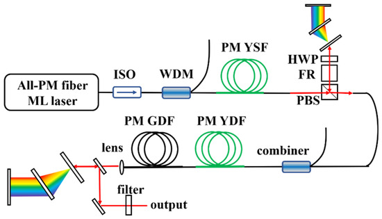
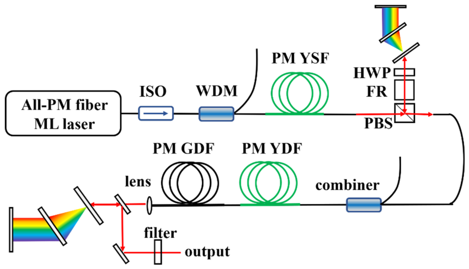
The schematic design of the 1100 nm fiber system is illustrated in Figure 1. This fiber laser system consists of a home-built all-PM fiber oscillator, a PM single-mode fiber (PMSMF) amplifier, and PCM-GMNA. The 38.1 MHz, 1024 nm fiber oscillator was built with a mirror, a grating pair, a filter (950–1047 nm), a PM Yb-doped fiber (Yb401-PM), a PM wavelength-division multiplexer (WDM), a 10:90 PM fiber coupler, two PM fiber collimators, a Faraday rotator, a λ/8 wave plate (WP) and a λ/2 WP between the two polarizing beam splitters (PBSs), which serves as a nonreciprocal phase shifter to introduce a phase bias. The intra-cavity net dispersion was maintained at +82,521 fs2 with a grating separation of 19.3 mm, indicating the oscillator operates with the stretched-pulse mode-locking scheme. The mode-locking signal laser was delivered by the 90% port of the fiber coupler. The corresponding output power was 10.6 mW with a 3 dB spectral bandwidth of 8 nm. The output signal pulses had a pulse duration of 2.628 ps based on the Gaussian assumption [18]. The PMSMF amplifier consisted of a PM fiber wavelength division multiplexer (WDM), a PM Yb-doped fiber (YB401-PM, Coractive, Quebec, QC, Canada), and a wavelength-stabilized fiber-coupled 976 nm laser diode (LD) with a maximum output power of 600 mW. It was utilized to compensate for the loss of PCM. The average power of signal pulses was amplified to 54.1 mW, with a slope efficiency of 18.03%.
Figure 1.
The schematic design of the 1100 nm fiber laser system (ISO: isolator; YSF: Yb-doped single-mode fiber; PBS: polarizing beam splitter; FR: Faraday rotator; YDF: Yb-doped double-clad fiber).
The PCM-GMNA module consisted of a pre-chirp management (PCM) section and a GMNA section. In the PCM stage, the amplified signal pulses passed the half-wave plate (HWP) and the polarizing beam splitter (PBS), which were used to control the beam splitter ratio of the PCM. The reflected pulses were compressed utilizing a transmission grating pair (1600 lines/mm, Lightsmyth, Eugene, OR, USA). A silver mirror was used to reflect the light back into the GMNA stage, passing another HWP, the Faraday rotator (FR) and the PBS in turn. The compression efficiency of the whole PCM stage was 75.92%. The subsequent main amplifier was built with a fiber combiner, a 5 µm core-diameter PM-YDF (PM-YDF-5/130-VIII, Nufern, East Granby, CT, USA), and a 2.5 m passive-fiber PM-GDF (PLMA-GDF-15/130, Nufern, East Granby, CT, USA). The average power of the signal pulses was scaled up to 4.43 W under a pump power of 18 W, corresponding to a slope efficiency of 28.44%. The PM-GDF was used to remove the residual pump laser and launch the output laser. The amplified signal pulses were compressed to 56 fs based on the Lorentzian assumption, using a grating pair (1000 lines/mm, Lightsmyth).
3. Results
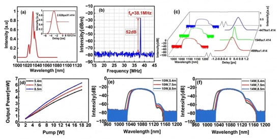
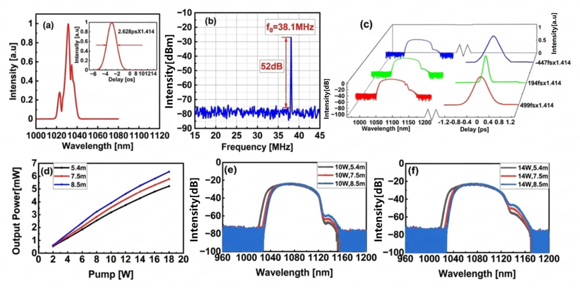
Figure 2a shows the optical spectra and the corresponding autocorrelation traces of the mode-locked signal pulses. The 3 dB spectral bandwidth was 8 nm. The corresponding pulse duration of the measured autocorrelation traces was 2.628 × 1.414 ps based on the Gaussian assumption. Figure 2b plots the measured radio frequency (RF) spectrum. The measured repetition rate was 38.1 MHz, and the corresponding RF signal-to-noise ratio was 50 dB.
Figure 2.
(a) The output spectra of the mode-locking fiber laser. The corresponding autocorrelation trace is plotted as the inset. (b) The corresponding output RF spectrum of the mode-locking signal pulses. (c) The output spectra after GMNA, based on the positive (the green curve), the transform-limited (the red curve) and the negative (the blue curve) pre-chirp management. The corresponding autocorrelation traces are on the right. (d) The average output power as a function of pump power at PM-YDF lengths of 5.4 m (the black curve), 7.5 m (the red curve) and 8.5 m (the blue curve). (e,f) The output spectra of corresponding PM-YDF lengths with pump powers of 10 W and 14 W.
The output spectra after PCM-GMNA with a pump power of 14 W are shown in Figure 2c, with near-zero-chirped and pre-chirped (positive and negative) input signal pulses. The pre-chirp signal pulse was controlled by adjusting the distance between the two transmission gratings. The amplified output spectra exhibited a similar flat range from 1041 nm to 1115 nm, with near-zero-chirped and pre-chirped input signal pulses. However, negative pre-chirped pulses resulted in the emergence of Raman components beyond 1115 nm, accompanied by reduced signal intensity compared with the near-zero-chirped pulses and positive pre-chirped pulses. The corresponding autocorrelation traces of near-zero-chirped and pre-chirped (positive and negative) pulses are embedded on the right of the image.
Figure 2d illustrates the influence of PM-YDF fiber lengths on GMNA. The average output power is shown as a function of pump power for PM-YDF lengths of 5.4 m (the blue curve), 7.5 m (the red curve) and 8.5 m (the black curve), and the corresponding slope efficiencies were 30%, 33.7% and 37%, respectively. Figure 2e,f show the corresponding output spectra at pump powers of 10 W and 14 W. As the length of PM-YDF fiber increased, the short-wavelength edge of the spectrum shifted toward longer wavelengths, while the long-wavelength edge remained nearly unchanged but exhibited increased Raman components. Considering both output power and spectral bandwidth, the optimized condition was obtained using a pre-chirp group-delay dispersion (GDD) of −40,000 fs2 and a 7.5 m PM-YDF.
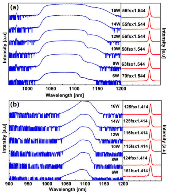
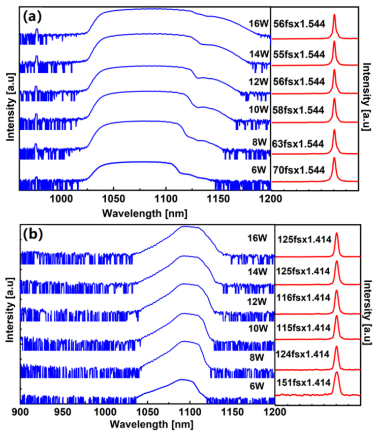
Figure 3a shows the output spectra after GMNA and the corresponding compressed autocorrelation traces. As the pump power increased from 6 W to 16 W, the long-wavelength edges of output spectra moved towards longer bands. Limited by pump maximum output power, the 10 dB spectral bandwidth of the widest amplified output spectrum was 87 nm, with the output power of 4.43 W after compression. The corresponding pulse duration of the compressed signal pulse was 56 × 1.544 fs, based on the Lorentzian assumption. A bandpass filter with a central wavelength of 1100 nm and a bandwidth of 25 nm was used to filter the 1100 nm pulses. The filtered spectra and the corresponding autocorrelation traces are shown in Figure 3b. With a pump power of 16 W, the filtered pulse delivered an average power of 1.27 W. The corresponding pulse duration was 125 × 1.414 fs based on the Gaussian assumption, which is 1.26 times the calculated Fourier transform-limited (FTL) pulse duration.
Figure 3.
(a) The out spectra after GMNA with different pump power levels. The corresponding autocorrelation traces are inset on the right. (b) The outer spectra after the 1100 nm filter with different pump power levels. The corresponding autocorrelation traces are inset on the right.
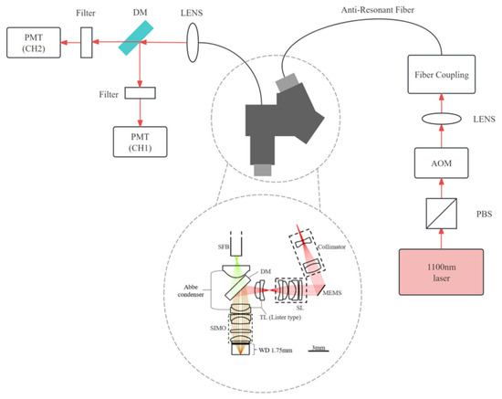
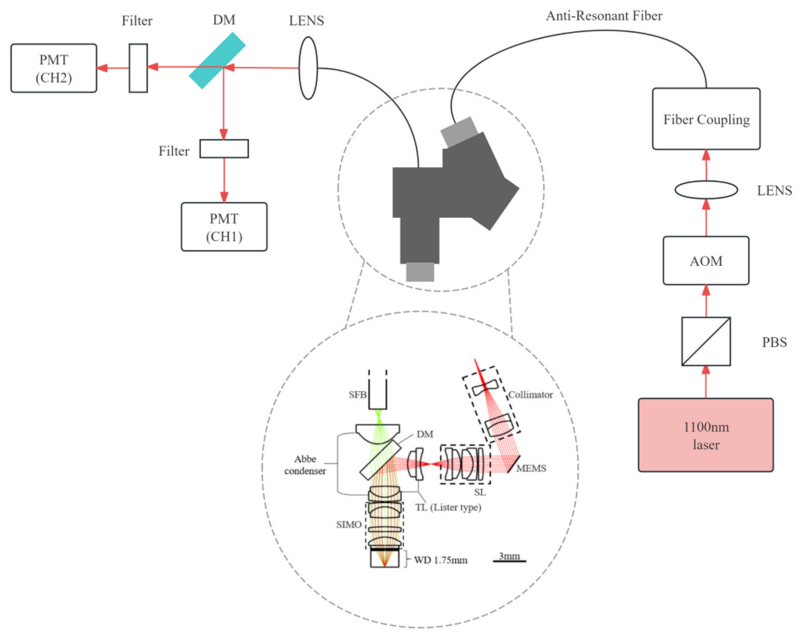
To further validate the application performance of the proposed laser in multi-photon imaging, a multi-photon imaging system was constructed as illustrated in Figure 4. The efficacy of the laser for multi-photon imaging was comprehensively validated through imaging experiments on standardized targets, hibiscus pollen grains, and living mouse brain tissue.
Figure 4.
The schematic construction of the home-built miniature multi-photon microscope. PBS, polarization beam splitter; MEMS, MEMS galvanometer; SL, scan lens; TL, tube lens; DM, dichroic mirror; PMT, photomultiplier tube.
The laser output was power-modulated via an acousto-optic modulator (AOM), with automatic power adjustment at varying imaging depths based on pre-calibrated compensation curves to ensure consistent imaging quality. The laser beam was adjusted to an appropriate spot size, with collimation performed using a lens assembly, then coupled into an anti-resonant fiber (ARF) through a coupling module. The laser was transmitted through the ARF to a miniaturized microscope comprising a collimator, a MEMS galvanometer, a scan lens, a tube lens, and an imaging objective [19]. Fluorescent signals emitted from the sample were filtered and collected into a quartz fiber bundle. These signals propagated through the fiber bundle to the detection module, underwent secondary filtering, and were ultimately acquired by a PMT.
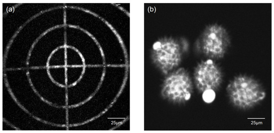
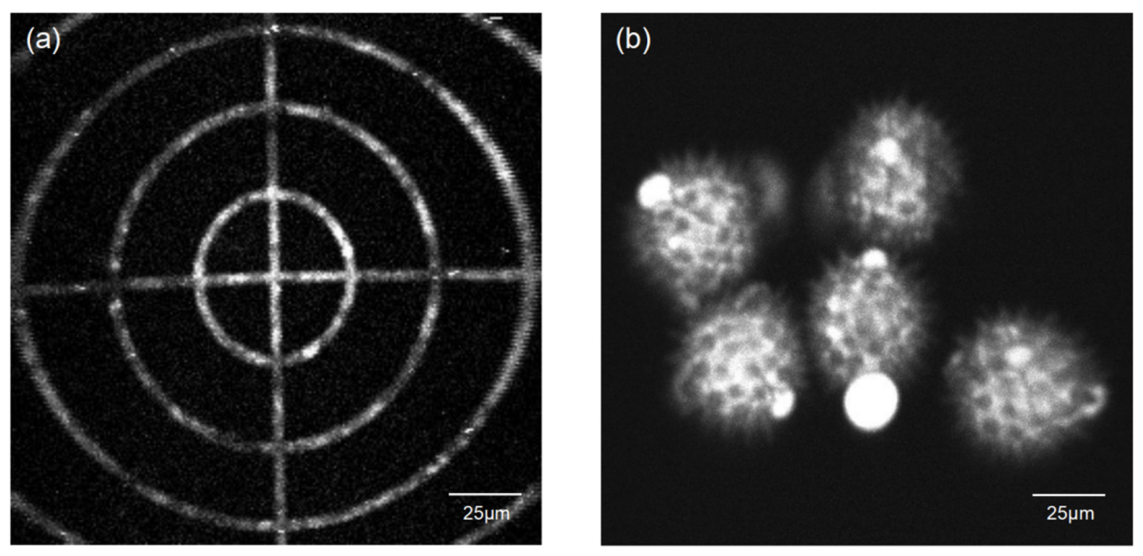
Following system integration, the multi-photon imaging capability was initially validated through fluorescence calibration targets and pollen specimens. The targets were fabricated by creating micron-scale grooves with specific patterns on glass substrates via photolithographic etching, followed by infusion of red fluorescent beads (Figure 5a). The demonstrated concentric ring pattern features 25 μm inter-ring spacing. The imaging objective, with a designed excitation numerical aperture (NA) of 0.5, provides a maximum field of view (FOV) of 400 × 400 μm2. To achieve optimal structural resolution of pollen features, the MEMS galvanometer’s driving voltage and frequency were adjusted to configure the operational FOV and frame rate. System calibration using the target yielded an effective FOV of 150 × 150 μm2 with a 10 Hz frame rate.
Figure 5.
Two-photon imaging characterization. (a) Imaging results of calibration target featuring concentric rings with 25 μm inter-ring spacing. (b) Two-photon imaging of hibiscus pollen grains acquired at 20 mW objective power.
As shown in Figure 5b, two-photon imaging of hibiscus pollen grains was conducted using the 1100 nm laser system. With 20 mW power at the objective, spine-like surface structures were clearly resolved, validating the laser’s imaging efficacy at the biological mesoscale.
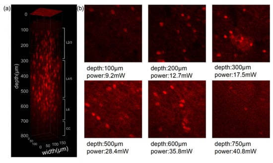
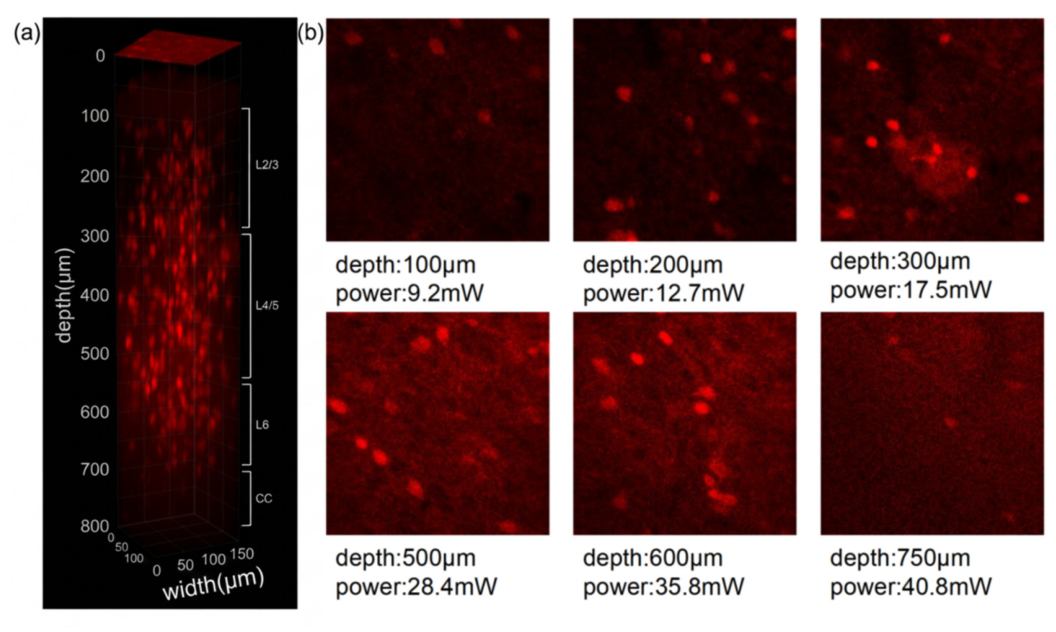
Finally, to systematically evaluate the laser’s in vivo imaging capability, we conducted two-photon serial sectioning imaging of the entire cerebral cortex in live tdTomato-expressing mice. Each mouse was secured on a head holder stage, with a high-precision Z-axis translation stage controlling the descent of the miniaturized microscopy. The zero-depth reference (0 μm) was defined as the z-position where fluorescence signals from the quartz window first became undetectable. Continuous downward displacement at 4 μm increments generated sequential optical sections, with 20 frames captured per section for subsequent multi-frame averaging. The AOM dynamically regulated objective power across depths to maintain consistent imaging quality.
As shown in Figure 6, full-cortex imaging revealed neuronal structures at a 750 μm depth (reaching the corpus callosum boundary), where 40 mW objective power sustained detectable two-photon fluorescence signals. This demonstrates the laser’s capability for whole-cortex imaging in live mouse brains.
Figure 6.
(a) 3D reconstruction of the entire cerebral cortex structure. (b) Objective power used at different imaging depths.
In conclusion, we demonstrated a high-power, 1100 nm fiber laser based on PCM-GMNA. The system comprised a 38.1 MHz, 1024 nm all-PM fiber oscillator, two-stage amplification and a pre-chirp management module. The influence of pre-chirped pulses on the GMNA process was systematically investigated. The optimized system delivered pulses with 110 nJ energy and a 60 fs pulse duration, with a 10 dB spectral bandwidth spanning from 1041 nm to 1117 nm. After spectral filtering, a 38.1 MHz, 1.17 W, 125 fs, 1100 nm femtosecond laser was obtained. The superior peak power and high pulse energy of this source enable efficient two-photon excitation of red fluorophores with enhanced penetration depth and reduced scattering. The proposed PCM-GMNA-based fiber laser provides a robust and practical light source for deep-tissue, multi-mode, and multi-photon microscopy.
4. Discussion
In this work, we have successfully demonstrated a high-power, 1100 nm all-polarization-maintaining (all-PM) femtosecond fiber laser utilizing a novel pre-chirp-managed and gain-managed nonlinear amplification (PCM-GMNA) scheme. Combining pre-chirp management with longitudinally evolving gain-shaping can efficiently manipulate nonlinear phases and suppress higher-order dispersion, thereby unlocking high pulse energies and ultrashort pulse durations in the 1100 nm spectral band that are inaccessible through conventional standard gain media.
The interpretation of our results highlights a significant breakthrough compared to previous studies. Traditional 1100 nm generation methods, such as those relying solely on self-phase modulation (SPM) or standard double-pass pre-chirp-managed amplification (PCMA), have been severely constrained by low conversion efficiency, system instabilities, or power limitations imposed by stimulated Raman scattering. While isolated gain-managed nonlinear amplification (GMNA) offers a partial remedy, it imposes exceptionally stringent demands on seed pulse conditions due to the intricate balance required between gain and dispersion. By introducing a pre-chirp management module between the two amplification stages, our system circumvents these bottlenecks. It achieves an exceptional compressed pulse duration of 56 fs and a remarkable pulse energy of 110 nJ (yielding 39 nJ at 1100 nm after spectral filtering). This represents a substantial enhancement in output power and peak intensity over previously reported configurations.
From the systematic investigation of the pre-chirp, gain fiber length, and pump power, a general optimization strategy for PCM-GMNA systems can be distilled. The pre-chirp should be established first, as it determines the longitudinal position at which SPM initiates; the optimal value delays SPM to the gain-saturated region where the pulse energy is highest, maximizing spectral broadening while preserving the linearly chirped character of the output. The gain fiber length must then balance pump absorption, nonlinear phase accumulation, and gain saturation: at 5.4 m, incomplete pump absorption limits the slope efficiency to approximately 30% and constrains spectral broadening, whereas at 8.5 m, excessive nonlinear phase accumulation promotes SRS and degrades pulse quality despite a higher slope efficiency of approximately 37%. The 7.5 m fiber achieves the optimal trade-off, yielding the widest 10 dB bandwidth of 87 nm. The pump power should then be adjusted to drive sufficient SPM for gain-managed broadening while remaining below the SRS threshold; the optimal window of 12–14 W maximizes spectral broadening, whereas pump powers exceeding 16 W lead to uncontrolled Raman growth. Because these three parameters are strongly coupled—increasing the fiber length reduces the pump power required to reach a given nonlinear phase, and increasing the pre-chirp shifts the SPM onset deeper into the gain fiber—the optimization should be performed iteratively rather than independently.
When evaluated within the broadest context of biomedical imaging, the implications of this light source are profound. The near-infrared long-wavelength region, particularly around 1100 nm, occupies a strategic niche for multi-photon microscopy (MPM). It enables high-fidelity, label-free multimode imaging (simultaneously capturing TPE, 3PE, SHG, and THG signals) and provides optimal two-photon excitation for red fluorophores like tdTomato. Crucially, our in vivo deep-tissue imaging of a live mouse brain, reaching a maximum depth of 750 μm (extending to the corpus callosum boundary), demonstrates that this system drastically minimizes tissue scattering while sustaining high-contrast fluorescence signals deep within living biological mesoscales. This capability bridges the gap between traditional shallow-tissue imaging and the rigorous demands of deep-tissue functional neuroimaging. Compared with conventional 920 nm excitation, the tissue scattering coefficient at 1100 nm is reduced by approximately 25–35% in cortical tissue, following the wavelength-dependent relationship μ_s(λ) ∝ λ−b (b ≈ 1.2–1.5 for brain tissue in the near-infrared regime). At an imaging depth of 750 μm, this scattering reduction translates to a round-trip attenuation improvement of approximately 1.5–2× relative to 920 nm excitation, directly enhancing the fluorescence signal-to-noise ratio at depth. Moreover, 1100 nm aligns with the peak two-photon action cross-section of red-shifted fluorophores such as tdTomato (~1050–1100 nm), compared with 1030 nm sources, while the scattering reduction is more modest (~5–10%), the 1100 nm wavelength offers superior spectral overlap with tdTomato and enables simultaneous two-photon excitation and third-harmonic generation, making it a more versatile choice for multimode deep-tissue imaging.
Moving forward, several compelling future research directions emerge from this study. While the current filtering method provides a high-peak-power 1100 nm output, optimizing the fiber parameters or seed pulse shapes could further scale up the direct average output power and efficiency. Expanding the biological application of this 1100 nm source to longitudinal studies of diseased models—such as tracking tumor microenvironments or deep-brain neurodegenerative progressions in vivo—will fully unlock its clinical and histopathological potential.
Author Contributions
Conceptualization, Q.S. and Z.F.; methodology, Q.S. and Z.F.; validation, Q.S. and Z.F.; formal analysis, Q.S. and Z.F.; investigation, Q.S. and Z.F.; resources, C.Z. and A.W.; data curation, Q.S. and Z.F.; writing—original draft preparation, Q.S. and Z.F.; writing—review and editing, R.X.; supervision, C.Z. and A.W.; project administration, C.Z. and A.W.; funding acquisition, C.Z. and A.W. All authors have read and agreed to the published version of the manuscript.
Funding
This research received no external funding.
Institutional Review Board Statement
This study was conducted in accordance with the Declaration of Helsinki, and approved by the Ethics Committee of Peking University (protocol code: IMM-ChengHP-15; date of approval: 2 July 2024).
Data Availability Statement
The data presented in this study are available in this article.
Acknowledgments
During the preparation of this manuscript, the authors used Gemini (May 2026 version) for the purposes of language proofreading. The authors have reviewed and edited the output and take full responsibility for the content of this publication.
Conflicts of Interest
The authors declare no conflicts of interest. The funders had no role in the design of the study; in the collection, analyses, or interpretation of data; in the writing of the manuscript; or in the decision to publish the results.
Abbreviations
The following abbreviations are used in this manuscript:
| all-PM | All-polarization-maintaining |
| GMNA | Gain-managed nonlinear amplification |
| NALM | Nonlinear amplification loop mirror |
| PCM | Pre-chirp management |
| PCM-GMNA | Pre-chirp-managed and gain-managed nonlinear amplification |
| WDM | Wavelength division multiplexer |
| YSF | Yb-doped single-mode fiber |
| PBS | Polarizing beam splitter |
| FR | Faraday rotator |
| YDF | Yb-doped double-clad fiber |
| PMSMF | Polarization-maintaining single-mode fiber |
| AOM | Acousto-optic modulator |
| ARF | Anti-resonant fiber |
| MEMS | MEMS galvanometer (Micro-Electro-Mechanical Systems) |
| SL | Scan lens |
| TL | Tube lens |
| DM | Dichroic mirror |
| PMT | Photomultiplier tube |
| FOV | Field of view |
| NA | Numerical aperture |
| GDD | Group-delay dispersion |
References
- Lefort, C. A review of biomedical multiphoton microscopy and its laser sources. J. Phys. D Appl. Phys. 2017, 50, 423001. [Google Scholar]
- Wang, K.; Horton, N.G.; Xu, C. Going deep: Brain imaging with multi-photon microscopy. Opt. Photon-News 2013, 24, 32. [Google Scholar]
- Wu, X.; Chen, G.; Lu, J.; Zhu, W.; Qiu, J.; Chen, J.; Xie, S.; Zhuo, S.; Yan, J. Label-Free Detection of Breast Masses Using Multiphoton Microscopy. PLoS ONE 2013, 8, e65933. [Google Scholar] [CrossRef] [Scilit] [PubMed]
- Borile, G.; Sandrin, D.; Filippi, A.; Anderson, K.I.; Romanato, F. Label-Free Multiphoton Microscopy: Much More Than Fancy Images. Int. J. Mol. Sci. 2021, 22, 2657. [Google Scholar] [CrossRef] [Scilit] [PubMed]
- He, K.; Zhao, L.; Chen, Y.; Huang, X.; Ding, Y.; Hua, H.; Liu, L.; Wang, X.; Wang, M.; Zhang, Y.; et al. Label-free multiphoton microscopic imaging as a novel real-time approach for discriminating colorectal lesions: A preliminary study. J. Gastroenterol. Hepatol. 2019, 34, 2144–2151. [Google Scholar] [CrossRef] [Scilit] [PubMed]
- Jiang, L.; Wang, X.; Wu, Z.; Du, H.; Wang, S.; Li, L.; Fang, N.; Lin, P.; Chen, J.; Kang, D.; et al. Label-free imaging of brain and brain tumor specimens with combined two-photon excited fluorescence and second harmonic generation microscopy. Laser Phys. Lett. 2017, 14, 105401. [Google Scholar] [CrossRef] [Scilit]
- Lee, J.H.; Rico-Jimenez, J.J.; Zhang, C.; Alex, A.; Chaney, E.J.; Barkalifa, R.; Spillman, D.R.; Marjanovic, M.; Arp, Z.; Hood, S.R.; et al. Simultaneous label-free autofluorescence and multi-harmonic imaging reveals in vivo structural and metabolic changes in murine skin. Biomed. Opt. Express 2019, 10, 5431–5444. [Google Scholar] [CrossRef] [Scilit] [PubMed]
- Liu, W.; Chia, S.-H.; Chung, H.-Y.; Greinert, R.; Kärtner, F.X.; Chang, G. Energetic ultrafast fiber laser sources tunable in 1030–1215 nm for deep tissue multi-photon microscopy. Opt. Express 2017, 25, 6822–6831. [Google Scholar] [PubMed]
- Chen, W.-L.; Hu, P.-S.; Ghazaryan, A.; Chen, S.-J.; Tsai, T.-H.; Dong, C.-Y. Quantitative analysis of multiphoton excitation autofluorescence and second harmonic generation imaging for medical diagnosis. Comput. Med. Imaging Graph. 2012, 36, 519–526. [Google Scholar] [CrossRef] [Scilit] [PubMed]
- Goh, G.B.-B.; Leow, W.Q.; Liang, S.; Wan, W.K.; Lim, T.K.H.; Tan, C.K.; Chang, P.E. Quantification of hepatic steatosis in chronic liver disease using novel automated method of second harmonic generation and two-photon excited fluorescence. Sci. Rep. 2019, 9, 2975. [Google Scholar] [CrossRef] [Scilit] [PubMed]
- Sun, C.; Kao, C.; Wei, M.; Chia, S.; Kärtner, F.X.; Ivanov, A.; Liao, Y. Slide-free imaging of hematoxylin-eosin stained whole-mount tissues using combined third-harmonic generation and three-photon fluorescence microscopy. J. Biophotonics 2019, 12, e201800341. [Google Scholar] [CrossRef] [Scilit] [PubMed]
- Tu, H.; Liu, Y.; Turchinovich, D.; Marjanovic, M.; Lyngsø, J.K.; Lægsgaard, J.; Chaney, E.J.; Zhao, Y.; You, S.; Wilson, W.L.; et al. Stain-free histopathology by programmable supercontinuum pulses. Nat. Photonics 2016, 10, 534–540. [Google Scholar] [CrossRef] [Scilit] [PubMed]
- Chou, L.-T.; Zhong, D.-L.; Liu, Y.-C.; Lee, J.-C.; Chen, Y.-S.; Chia, S.-H. Widely-tunable femtosecond source tunable between 770–1180 nm for two-photon fluorescence microscopy. Presented at the Frontiers in Ultrafast Optics: Biomedical, Scientific, and Industrial Applications XX2020, San Francisco, CA, USA, 1–6 February 2020. [Google Scholar]
- Chou, L.-T.; Liu, Y.-C.; Zhong, D.-L.; Lin, W.-Z.; Hung, H.-H.; Chan, C.-J.; Chen, Z.-P.; Chia, S.-H. Low noise, self-phase-modulation-enabled femtosecond fiber sources tunable in 740–1236 nm for wide two-photon fluorescence microscopy applications. Biomed. Opt. Express 2021, 12, 2888–2901. [Google Scholar] [CrossRef] [Scilit] [PubMed]
- Gao, K.; Liu, Y.; Qiao, W.; Xu, R.; Feng, T.; Xuan, H.; Li, D.; Zhao, X.; Wang, A.; Li, T. Sub-60-fs, compact 1100-nm fiber laser system based on double-pass pre-chirp managed amplification. Opt. Lett. 2022, 47, 5016–5019. [Google Scholar] [CrossRef] [Scilit] [PubMed]
- Sidorenko, P.; Fu, W.; Wise, F. Nonlinear ultrafast fiber amplifiers beyond the gain-narrowing limit. Optica 2019, 6, 1328–1333. [Google Scholar] [CrossRef] [Scilit] [PubMed]
- Tomaszewska-Rolla, D.; Lindberg, R.; Pasiskevicius, V.; Laurell, F.; Soboń, G. A comparative study of an Yb-doped fiber gain-managed nonlinear amplifier seeded by femtosecond fiber lasers. Sci. Rep. 2022, 12, 404. [Google Scholar] [CrossRef] [Scilit] [PubMed]
- Edelmann, M.; Hua, Y.; Koch, A.; Kärtner, F.X. Generation of 64 fs, 10 kW peak-power, transform-limited pulses directly from an Yb-doped Figure-9 fiber laser. Presented at the CLEO: Science and Innovations, San Jose, CA, USA, 10–15 May 2020; p. SW4R.3. [Google Scholar]
- Zhao, C.; Chen, S.; Zhang, L.; Zhang, D.; Wu, R.; Hu, Y.; Zeng, F.; Li, Y.; Wu, D.; Yu, F.; et al. Miniature three-photon microscopy maximized for scattered fluorescence collection. Nat. Methods 2023, 20, 617–622. [Google Scholar] [CrossRef] [Scilit] [PubMed]
Disclaimer/Publisher’s Note: The statements, opinions and data contained in all publications are solely those of the individual author(s) and contributor(s) and not of MDPI and/or the editor(s). MDPI and/or the editor(s) disclaim responsibility for any injury to people or property resulting from any ideas, methods, instructions or products referred to in the content. |
© 2026 by the authors. Licensee MDPI, Basel, Switzerland. This article is an open access article distributed under the terms and conditions of the Creative Commons Attribution (CC BY) license.