


Abstract
The complex relationship between epilepsy and sleep has been early recognised based on behavioural studies showing that the majority of epileptic seizures occur during sleep. In particular, during NREM (slow wave) sleep, neuronal circuits oscillate between their active and inactive state, facilitating epileptic discharges. Conversely, epileptic activity during sleep can disrupt the physiological sleep microstructure and sleep architecture, and may cause sleep deprivation and daytime sleepiness, which further enhances seizure probability. Besides this mutual interaction, the clinical differentiation between epileptic and non-epileptic events during sleep can be very challenging. In this article, we aim to give an overview of the intertwined relationship between sleep and epilepsy, discuss the differential diagnosis between epileptic and non-epileptic nocturnal episodes and give insights to novel techniques of sleep modulation to treat epilepsy.

The temporal association between sleep and epilepsy
Hippocrates early recognised the predominant occurrence of seizures during sleep and the negative effect of sleep deprivation in patients with epilepsy in the 5th century B.C. In 1880, Féré was among the first to recognise that up to 70% of all epileptic seizures occur during sleep [1]. Gowers proposed a classification of generalized tonic-clonic seizures based on the appearance of the seizures “mostly at night”, “during the day”, or “randomly throughout the sleep-wake cycle” [2]. His study also provided evidence that around one fifth of all patients with epilepsy experience seizures exclusively at night. More recently, many groups confirmed that a high proportion of seizures (40–70%) occur during the night [3] and about 12% of all patients with epilepsy suffer exclusively from sleep-related seizures [4–7]. Later, Janz recognised a further differentiation between patients with seizures while asleep and others who experienced their seizures mainly upon awakening, in particular for patients with genetic epilepsies [4]. Various circadian studies revealed that peaks of seizure activity often occur in response to falling asleep or upon awakening [8], and others confirmed the same influence of circadian patterns and sleep on epileptiform discharges [9]. The periodicity of the internal “Zeitgeber” (rhythm) regulating recurrent seizure activity might, however, be more complex, and interestingly seizure clusters also show a multi-day (multidien) periodicity, as shown in a recent and very comprehensive long-term invasive electroencephalogram (EEG) study by Baud et al. [9]. The circadian analysis in this cohort revealed a high variability, but also confirmed peak epileptic activity during phases of reduced wakefulness in the sleep/wake cycle. In particular, late afternoon, early night and early morning were identified as the phases of peak epileptic activity [9].
Epilepsy and sleep macrostructure
These behavioural and circadian patterns corroborate the hypothesis that most seizures have their onset in phases of drowsiness and sleep. According to the current classification, sleep is divided in four stages: nonrapid eye movement sleep (NREM stages 1, 2 and 3) and rapid eye movement sleep (REM). Over one night, multiple cycles of NREM and REM sleep occur (one cycle = NREM stage 1 – stage 2 – stage 3 – REM), each cycle having a duration of approximately 90–110 minutes [10,11]. NREM stage 1 represents the stage of transition between wakefulness and sleep and lasts only a few minutes. Heartbeat, breathing and eye movements slow down, the muscles tend to relax and the brain activity starts to slow down in comparison to wakefulness. NREM stage 2 is marked by deeper sleep and brief bursts of electrical activity such as sleep spindles and K-complexes, but no consolidated slow wave sleep [10]. Sleep spindles are generated in thalamic circuits as a response to cortical neuronal firing and are therefore thought to result from a cortico-thalamic network. K-complexes are hypothesised to represent microarousals caused by either internal or external stimulation, for example provoked by a sudden noise, which does not reach the threshold for EEG arousals or clinical awakenings [12]. NREM stage 3 represents deep sleep (Delta sleep). Brain activity is characterised by high amplitude, low frequency waves between 0.5 and 4 Hz. Over the course of the night, the duration of NREM deep sleep decreases. REM sleep typically occurs around 90 minutes after falling asleep, but might occur earlier in sleep deprived patients. Brain activity and vital parameters are similar to those observed during wakefulness, characteristic rapid eye movements appear and muscles are paralysed to avoid acting out the dreams [10]. Sleep is regulated by several structures mainly localised in the diencephalon (hypothalamus, thalamus and epiphysis) and the brain stem in response to exogenous (light–darkness cycles) and endogenous factors (circadian rhythms, homeostatic processes) [13]. Interestingly, neurones in sleep and wake states can coexist in the brain at the same moment, described by Krueger et al. as “local sleep” [14]. Importantly, the regulation of local sleep in a given neuronal/glial network seems to be use-dependent [14]. Huber et al. showed in a simple, but very comprehensive study that motor activity during wakefulness affects local cortical depth of sleep [15]. Intriguingly, the local slow wave activity may co-localise with the ictal onset zone in patients with epilepsy, corroborating the interaction of slow wave activity and epileptogenicity also form a local (“bottom-up”) perspective [16].
In line with this, an analysis of 613 seizures in a video-EEG monitoring study revealed that 91% of all seizures occurred in phases of “light” NREM sleep (NREM 1 and 2) [3]. Furthermore, the authors found that frontal lobe seizures are most likely to occur exclusively during sleep, whereas patients with temporal onset seizures have intermediate sleep seizure rates. Conversely, seizures with occipital and parietal onset are rarely observed during sleep [3].
Interictal epileptiform discharges can be detected most frequently during NREM sleep stages 2 and 3, and are rare phenomena during REM sleep. Slow wave sleep is regulated homeostatically and the prevalence and amplitude of sleep slow waves depend directly on the waking time before sleep registration. Thus, slow wave activity and delta power is highest at sleep onset and in the first half of the night. Sleep deprivation leads to a further increase of slow wave activity [17]. Over the course of a sleep cycle, tends to decrease, which is thought to be a marker for synaptic renormalisation and its beneficial effect [18,19]. Now, the high neuronal synchronisation typical of NREM sleep apparently represents a suitable ground for the enhancement of epileptic activity during deep sleep and there is a significant correlation of interictal discharges with slow wave sleep. Furthermore, in patients with epilepsy, increased slow wave activity can persist for days after generalised tonic-clonic seizures. As a result, the physiological modulation of the sleep cycle does not occur, which might interfere with sleep quality and learning. Finally, the increase can be limited to the region comprehending the epileptic focus in patients with focal seizures [20].
Epilepsy and sleep microstructure
Intriguingly, the frontal predominance of delta activity in the sleep EEG is mirrored by the frontal predominance of sleep-related seizures. This suggests that also in terms of EEG microstructure a relevant coupling between sleep and epileptic patterns occurs.
Indeed, delta waves in NREM sleep offer suitable conditions for epileptic discharges. Delta waves show a slow oscillating pattern corresponding presumably to an active “up” (depolarised, surface positive to physical reference) or silent “down” (hyperpolarised, surface negative to physical reference) state [21,22]. A recent analysis assessed the phase locking of epileptiform spikes to the delta wave depending on its micro state (up/down). Whereas most of the physiological patterns typical of NREM sleep (e.g., sleep spindles and ripples) are coupled to the active “up” half-wave, epileptiform spikes are predominantly observed in the “down”, silent phase, and during the transition from “up” to “down” [23]. This somehow counterintuitive finding could be explained by the fact that, in the “up” state, not only summarised excitatory neurones but also inhibitory inter-neurones are firing. Contrarily, the neuronal silence of the “down” state may result rather from a lack of inhibition [23]. K complexes have been interpreted as isolated “down” states [24] and, interestingly, the association of epileptic spikes with a K complex is a rare but well described phenomenon [25].
Further evidence for coupling of sleep and epilepsy in terms of EEG structure stems from studies on high frequency oscillations. Physiological high frequency oscillations can be observed both in REM and NREM sleep, but pathological epileptiform high frequency oscillations in the form of ripples (80–250 Hz) and fast ripples (>250 Hz) as novel possible electrophysiological biomarkers of epileptogenicity are mainly coupled to NREM sleep [26]. Similarly, in mesio-temporal lobe epilepsy, epileptiform potentials can associate with normal sharp wave ripples involved in memory consolidation and to pathological high frequency oscillations in epilepsy [27]. In absence epilepsy and juvenile myoclonic epilepsy, the cortico-thalamic network responsible for the generation of sleep-spindles is suspected to enhance epileptic bilateral synchronous spike-waves, interfering with the normal homeostatic processes of NREM sleep [27].
Effect of anticonvulsant drugs on sleep
Not only nocturnal seizures, but also anticonvulsant drugs have a relevant influence on sleep, mainly through the alteration of the sleep architecture. For instance, among older antiepileptic drugs, carbamazepine and phenobarbital influence sleep wake regulation and sleep stage distribution. At the beginning of treatment, this leads to an increase of deep sleep, but also affects sleep though reduction of REM sleep and a marked increase of the number of sleep stage transitions, leading to sleep fragmentation [28]. In contrast, phenobarbital is associated with a dose-dependent decrease of both slow wave sleep and REM sleep and patients frequently suffer from excessive daytime sleepiness, resulting from its GABA-ergic effect on the one hand and reduced sleep efficiency on the other [29]. Benzodiazepines also have a negative influence on sleep through reduction of slow wave sleep and consequent disruption of the homeostatic sleep/wake processes [30]. Sodium valproate has little to no influence on sleep architecture [31], whereas pregabalin [32] and gabapentin [33] enhance slow wave sleep. Lamotrigine does not show relevant modifications of sleep architecture and does not cause daytime sleepiness, but has rather a stimulating effect [34]. In contrast, patients treated with levetiracetam show an increase of daytime naps and the total sleep time during the day [35], along with an increase of NREM sleep stage 2 [36]. Importantly, subjective and objective daytime sleepiness under treatment with levetiracetam is most prominent in the first weeks after treatment onset.
Epileptic vs non-epileptic sleep disorders
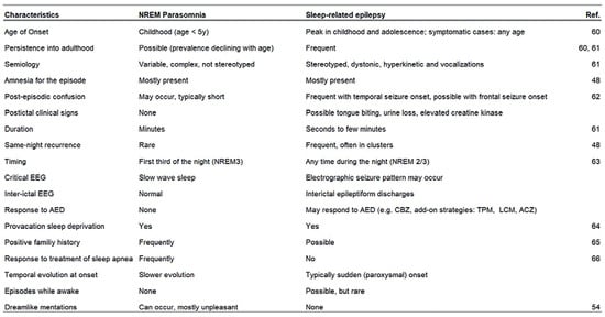
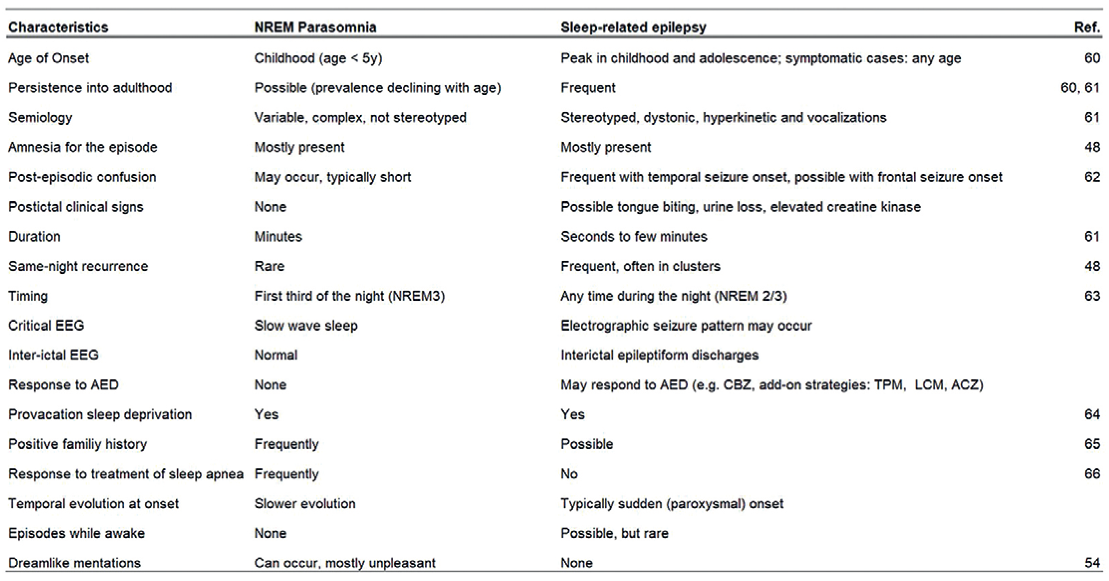
Based on the patient’ s history or observations by family members it is often challenging to differentiate nocturnal seizures from parasomnias, in particular disorders of arousal occurring during NREM sleep (e.g., sleep terrors, sleepwalking, confusional arousals). Parasomnia is defined as an umbrella term for “clinical disorders that are not abnormalities of the processes responsible for sleep and awake states, but are undesirable physical phenomena that occur predominantly during sleep” [48]. Their pathogenesis is unknown in most cases, but an underlying impairment of the arousal mechanisms resulting in a dissociation between motor control and EEG activity has been postulated [49]. Thus, some experts consider parasomnia a disorder of arousal, rather than a sleep disorder. Parasomnia can be related both to REM and NREM sleep and cause bizarre, even violent behaviours during sleep [50]. Even though many semiological and paraclinical aspects regarding parasomnias and sleep related epilepsies have been described over the last years and clinical scores might add to the differential diagnosis (i.e., by using the FLEP scale), the differential diagnosis still is challenging in many cases and usually video-EEG monitoring is indicated for confirming the diagnosis. The semiology of parasomnias can vary from ictal-like dystonic posturing, choreatic movements or ballistic movements to minor motor events such as paroxysmal arousals or bruxism [51]. A common neuronal pathway with involvement of the anterior cingulate, orbito-polar and temporal regions could explain the similarity of the motor patterns in epileptic and non-epileptic events [52]. Moreover, an overlap in the genetic predisposition for sleep related hypermotor epilepsy and parasomnias was suggested, as the prevalence of arousal parasomnias seems to be higher not only in subjects with SHE, but also in their relatives [53]. The gold standard for the differential diagnosis is video-polysomnography using a 10/20 montage, but some clinical and anamnestic information can help differentiate the two conditions. Arousal disorders for instance mostly appear in the first third of the night and tend to have longer duration, lower same-night recurrence and lower tendency to postictal confusion than SHE, while the motor patterns tend to be more variable and less stereotyped [53]. Dreamlike mentations, mostly unpleasant (i.e., apprehension, misfortune, aggression) and often experienced during episodes of sleepwalking or sleep terrors, are rarely reported after epileptic seizures [54]. Some exemplary clinical features of epileptic and non-epileptic nocturnal events are summarised in Figure 1.
Figure 1.
Comparison of sleep-related epilepsy and NREM-parasomnia based on clinical features and EEG patterns.
Novel experimental approaches of sleep modulation in epilepsy
Considering the overwhelming mutual interactions between sleep and epilepsy on a behavioural and electrophysiological level, it seems a reasonable approach to modulate sleep on a microstructural level to increase the threshold for nocturnal epileptic activity. Recently, acoustic stimulation during sleep has been tested in children to modulate this interaction [55]. The effect of the acoustic stimulus on the slow waves depends on the exact timing of the tone’ s onset in relationship to the wave’ s phase: stimulation time-locked to the “up” phase leads to an enhancement of the [56–58], whereas stimulation time-locked to the “down” phase tends to reduce the slow wave activity [59]. As the epileptic spike-wave activity is mainly linked to the down-phase of slow-wave activity during sleep, closed loop and time-locked acoustic stimuli should in principle allow for modulation of nocturnal spikewave activity. So far, a pilot study performed on children showed the feasibility and tolerability of this method, while the effect of the acoustic stimulation on spike-wave activity is still under debate [55]. Further studies are ongoing and planned in our centre to extend and further explore the potential of this novel approach, which could represent a safe option to treat pharmaco-resistent sleep related epilepsies.
Conclusion
Sleep and epilepsy are connected in many ways: sleep has a seizure promoting effect, which seems to be primarily fuelled by the diffuse bilateral hypersynchronicity during NREM sleep, leading to an increase of interictal spikes and seizures. Conversely, seizures and anticonvulsant medication may lead to altered sleep structure, sleep fragmentation and daytime sleepiness. From a diagnostic perspective, the differentiation between parasomnias and nocturnal hypermotor seizures is a common clinical challenge. Finally, novel therapeutic approaches aim to use the tight interaction between sleep and epilepsy to modulate sleep microstructure and nocturnal epileptic activity simultaneously.
References
- You find the complete bibliography in the online version of the article at http://doi.org/10.4414/sanp.2021.10044.
This is an open-access article distributed under the terms of the Creative Commons Attribution Non-Commercial No Derivatives (CC BY-NC-ND) 4.0 License.