


Summary
Fabry disease is a rare X-linked lysosomal storage disorder caused by the absence or deficiency of the hydrolase alpha-galactosidase A activity. As a consequence, accumulation of globotriaosylceramide occurs in a wide variety of cells throughout the human body. Specific gene mutations determine disease severity and different phenotypes. Fabry disease is a multisystemic disease with nonspecific initial manifestations. Neuropathic pain and acropraesthesia are one of the earliest symptoms, already reported in childhood or adolescence. Later signs and symptoms involve the heart, kidney and brain, resulting in life-threatening complications such as cardiac and renal failure as well as cerebral strokes. Early treatment initiation can ameliorate disease progression and potentially prevents long-term complications. Based on its diverse and nonspecific manifestation, it can take up to 15 years between the onset of the first symptoms and the final diagnosis of Fabry disease. Recognition of early symptoms, such as neuropathic pain and acropraesthesia, and considering Fabry disease in young patients with strokes, is important. As such, neurologists may play a key role in early diagnosis of this disease.
Introduction
Fabry disease, also called angiokeratoma corporis diffusum, ceramide trihexosidosis, or Anderson-Fabry disease, is a rare lysosomal storage disorder due to the absence or deficiency of hydrolase alpha-galactosidase A (alpha-Gal A) activity in lysosomes. This dysfunction results in progressive accumulation of glycosphingolipids, especially of globotriaosylceramide (Gb3), within lysosomes in a wide variety of cells throughout the human body: vascular endothelial cells, neurons, astrocytes, meningeal cells, smooth muscle cells, as well as podocytes and other kidney cells, myocytes, etc., resulting in major organ system damage [1,2,3].
More than 400 mutations have been identified in the alpha-Gal A gene (GLA gene) situated on the long arm of the X chromosome. For decades, Fabry disease was believed to be an X-linked recessive disorder. However, actual data suggest an X-linked dominant inheritance with variable penetrance. Specific gene mutations determine disease phenotypes and severity [4]. Classical or typical Fabry disease is present in 1:40 000 to 1:60 000 men and usually occurs when alpha-Gal A activity is less than 1%. Milder phenotypes occur when gene mutations result in residual alpha-Gal A activity [4]. Heterozygous women, 1:6000 to 1:40 000, may also be affected with variable penetrance and manifestations. They can exhibit severe disease manifestation similar to that seen in men [4,5]. Hwu et al. reported results from a programme for newborn screening for Fabry disease in 171 977 infants. They found an incidence of Fabry disease of approximately 1 in 1250 males, of whom 86% carried mutations associated with a later-onset phenotype [6]. Underdiagnosed atypical phenotypes and mutations with limited alpha-Gal A activity might indicate that the actual incidence could be higher.
Measuring alpha-Gal A enzyme activity in plasma, leucocytes, cultured skin fibroblasts, biopsied tissue or a dried blood spot is a diagnostic tool in men. In contrast, women with Fabry disease may have enzyme activity levels within the normal range, as a result of random X chromosome inactivation [7]. Molecular genetic testing is thus the most reliable method to establish the diagnosis in females who are symptomatic or carriers. Prenatal testing is possible by means of DNA testing using chorionic villus sampling (at 9-10 weeks) or cultured amniotic cells (amniocentesis at approximately 15 weeks) [8]. 
Fabry disease is a multisystemic disorder, and signs and symptoms may appear in childhood and adolescence. Among the earliest and most important organ manifestations are those in the nervous system. The focus of this article is, therefore, to review the neurological manifestations and to enhance awareness of this entity. We reviewed the current literature using a MedLine search and congress abstracts.
Clinical manifestation
Neurological involvement
The sphingolipid deposition can affect the entire peripheral (PNS) and central (CNS) nervous systems involving small and large cerebral vessels, neurons, ganglia, nerve sheaths and perineurium.
Involvement of the peripheral nervous system
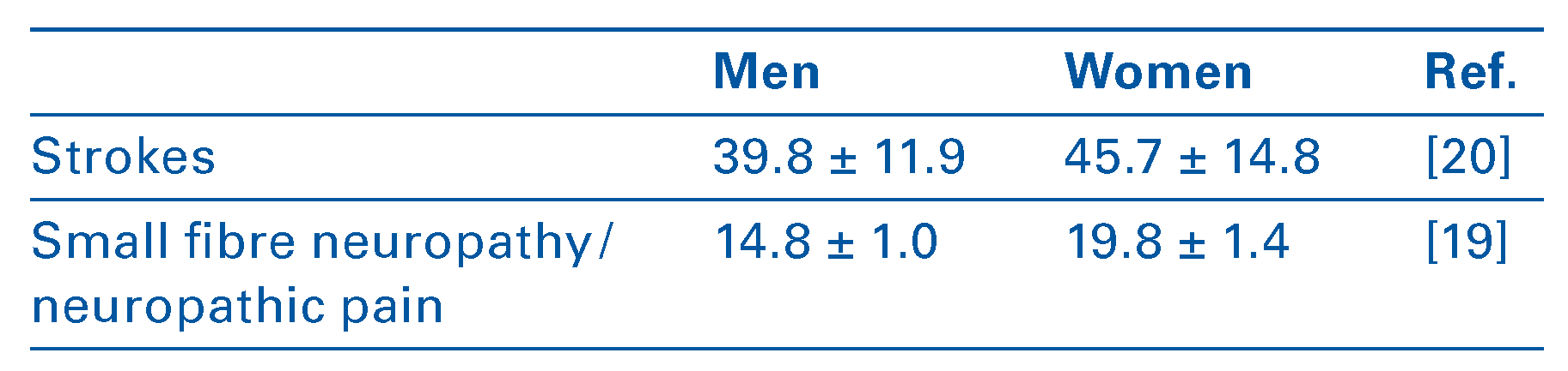
Small fibre neuropathy (SFN) is the main manifestation of Fabry disease in the PNS [9]. The pathophysiology of SFN is not fully understood. Gb3 accumulation has been demonstrated in dermal vascular endothelial and perineural cells of thin myelinated Aδ and unmyelinated C fibres involved in mediating pain and temperature misperception [10,11,12,13]. Electron microscopy of skin biopsies revealed a decreased density of Aδ and C fibres [13]. With progression of the disease, large fibre involvement and nerve conduction abnormalities may develop [14]. Patients with SFN present with somatosensory discomfort and neuropathic pain as prominent manifestations. Typically, burning pain dysaesthesia, and paraesthesia, defined as acroparaesthesia, of the hands and feet occur. They may present as recurrent painful episodic crises triggered by stress, fever, heat, fatigue or exercise [15,16]. The duration may vary from a few minutes up to several days and extreme pain attacks known as "Fabry crises" may be accompanied by fever, joint pain and elevated erythrocyte sedimentation rate, and may require hospitalisation [17]. During a crisis, the distal extremities may be livid and swollen. Neuropathic pain with acroparaesthesia is often the earliest manifestation of Fabry disease reported by patients. Pain has been reported as early as 3 years of age in boys and 6 years in girls [18]; mean age at onset is 14.8 years in men, 19.8 years in women [19] (Table 1). The Fabry Outcome Survey (FOS) reports a high frequency of neuropathic pain in Fabry disease (76% men, 64% women) [3,20]. Intolerance of cold and warmth is another manifestation of SFN. Quantitative sensory testing may show increased or unmeasurable thermal and, specifically, cold detection thresholds, as well as increased vibration detection thresholds [12,21]. Up to 63–100% of men and 16–33% of women have impaired sensory perception [3,9,22,23].
Hypohidrosis, reduced saliva and tear production, impaired pupillary constriction, heart conduction defects, sexual dysfunctions such as priapism, and gastrointestinal disturbances may be signs and symptoms of peripheral autonomic dysfunction in Fabry disease [3,24,25].
Table 1.
Natural course of neurological involvement in Fabry disease (age in years, mean ± SD).
Involvement of the central nervous system
Diffuse glycosphingolipid deposits in endothelial cells of small and large vessels and vascular smooth muscle cells may result in stenosis and occlusion as well as vasodilatation and, hence, lead to impaired cerebral perfusion [3,26,27,28,29]. The vertebrobasilar territory is preferentially affected and increased basilar artery diameter has been shown to be a sensitive indicator of Fabry disease [30]. The higher frequency of ischaemic lesions in this area, furthermore, explains the typical CNS manifestations of vertigo, anopsia, diplopia, nystagmus, dysarthria, hemiparesis, hemiataxia, and gait ataxia [26].
The mechanism linking glycosphingolipid accumulation to ischaemic tissue damage is not fully understood. Endothelial dysfunction, vessel wall irregularities leading to compromised cerebral blood flow velocities and cerebral autoregulation, and immunological and cellular changes leading to, among others, a prothrombotic state probably contribute to cerebral ischaemia [26,27,28,29,31,32]. Fabry disease predisposes to valvular heart disease and arrhythmia, and kidney disease, cardiogenic embolism and hypertension could also be contributory factors.
Data from FOS revealed that 13.2% (15.1% men, 11.5% women) of Fabry patients developed an ischaemic stroke or transient ischaemic attacks (TIA), and that cerebrovascular events typically occur at an earlier age in Fabry patients than in the general population [20]. Median age of first stroke in men was 39.8 years and in women 45.7 years (Table 1). The observed number of ischaemic strokes in men aged between 25 and 44 years was about 12 times higher than expected in a comparable general population [20,33]. Most Fabry patients did not experience hypertension and atherosclerotic plaques before their first stroke. Approximately 50% of men and 38.5% of women experienced their first stroke before renal or cardiac events and diagnosis of Fabry disease [3,34,35,36]. In a recent meta-analysis of nine studies, the prevalence of Fabry disease ranged from 0.6 to 11.1% among patients with cryptogenic strokes and from 0.4 to 3.5% in all stroke aetiologies [37].
Development of neuropsychiatric symptoms such as depression and memory deficits are frequently observed in Fabry disease patients. Unlike the general population, men with Fabry disease report a higher prevalence of severe depression than women (36% men; 22% women) [38]. The mechanism is unclear and, besides a primary origin, a secondary cause due to the persistence of neuropathic pain has been discussed [38]. A large proportion of patients with Fabry disease also report progressive hearing impairment, tinnitus and dizziness and/or vertigo, presumably due to the involvement of central and peripheral vestibulocochlear system [39,40].
Neuro-imaging
Nonspecific white and grey matter lesions, as well as vascular abnormalities, are often seen in brain magnetic resonance imaging (MRI). The most common structural abnormalities are found in the deep white matter, mostly in the posterior periventricular and centrum semi-ovale region [32,41], and occur at a comparable frequency in male and female patients [33]. It is hypothesised that glycosphingolipid deposition leads to increased interstitial pressure in deep white matter and to metabolic alterations resulting in gliosis, demyelination and increased interstitial water content [42]. Calcification in the pulvinar and posterior thalamic regions, on the other hand, has been shown to result from cerebral hyperperfusion [43,44]. Calcification of the pulvinar, best seen in T1-weighted MRI (Figure 1), represents a characteristic, although not pathognomonic, neuroradiological feature of Fabry disease. Additional structures to be involved by dystrophic calcification are the dentate nucleus and basal ganglia [43]. Dolichoectasia (Figure 2) is detectable with MRI and magnetic resonance angiography. A vessel diameter exceeding 3.2 mm for the basilar artery is able to distinguish between Fabry disease patients and normal controls with a sensitivity of 87% and specificity of 86% p <0.001 [45]. With the relative risk of stroke elevated by 12.2 for men and 4.2 for women in the 35 to 45 year age category, strokes with either lacunar infarcts (Figure 3) or territorial infarction are usually the first serious complication seen in Fabry disease patients [35,36].
Other organ involvement (Figure 4)
Kidney involvement is reported in about 50% of patients. Kidney lesions result from deposition of Gb3 and other sphingolipids in the podocytes, mesangial and glomerular endothelial cells, tubular cells, vessels and interstitial cells. It results in cellular changes, typically in prominently vacuolated podocytes and tubular cells by light microscopy (Figure 5A). Electron microscopy reveals lysosomal inclusions with lamellated structure, called myelin or zebra bodies (Figure 5B). There may be peritubular capillary inclusions, vascular intimal and medial inclusions and vascular smooth muscle cell hypertrophy in the kidneys. Vasculopathy has also been observed, probably due to local upregulation of the renin-angiotensin system [46]. Glomerular sclerosis and tubulointerstitial fibrosis are the histological features that best correlate with progression of kidney disease in humans with Fabry disease [47]. Kidney impairment often begins with microalbuminuria and proteinuria in the second to third decade of life [8,10]. Disease-related tubular and glomerular alterations lead to proteinuria and loss of glomerular filtration rate, resulting in chronic kidney disease. It should be noted that glomerular compensation (hyperfiltration) can mask impairment of kidney function for years. For this reason, gradual deterioration of kidney function usually becomes apparent in the third to fifth decades of life. At this stage, fibrosis, sclerosis and tubular atrophy dominate the disease activity. By the age of 40 years, end-stage renal disease has usually been reached [36,48].
Cardiac involvement is common, affecting more than 50% of all Fabry patients [49,50]. It may affect all cardiac structures. Intracellular accumulation of Gb3 occurs within myocytes, valves and vascular endothelium of the heart [51]. Interstitial remodelling, due to intracellular lysosomal storage of Gb3 and an increase of trophic factor such as lyso-Gb3, is an important feature of Fabry cardiomyopathy [51]. In most Fabry patients with cardiomyopathy, concentric left ventricular hypertrophy without left ventricle outflow tract obstruction and normal left ventricular systolic function is found [36,52]. Other findings in Fabry cardiomyopathy are prominent papillary muscles and early stages of diastolic dysfunction. Few Fabry patients exhibit mild aortic, mitral and tricuspid insufficiency, especially in end-stage cardiomyopathy [50,52]. End-stage Fabry cardiomyopathy is characterised by intramural fibrosis, detectable by late gadolinium-enhanced MRI [53] and echocardiography [51]. This fibrosis leads to wall motion abnormalities and arrhythmias [51,54].
Glycosphingolipid deposition in intestinal ganglia and small vessels causes gastrointestinal dysmotility: episodic diarrhoea, nausea, vomiting, bloating, cramping abdominal pain and/or malabsorption. The median age of onset for these symptoms is before 15 years and, overall, 50% of patients with Fabry disease complain about gastrointestinal symptoms [55].
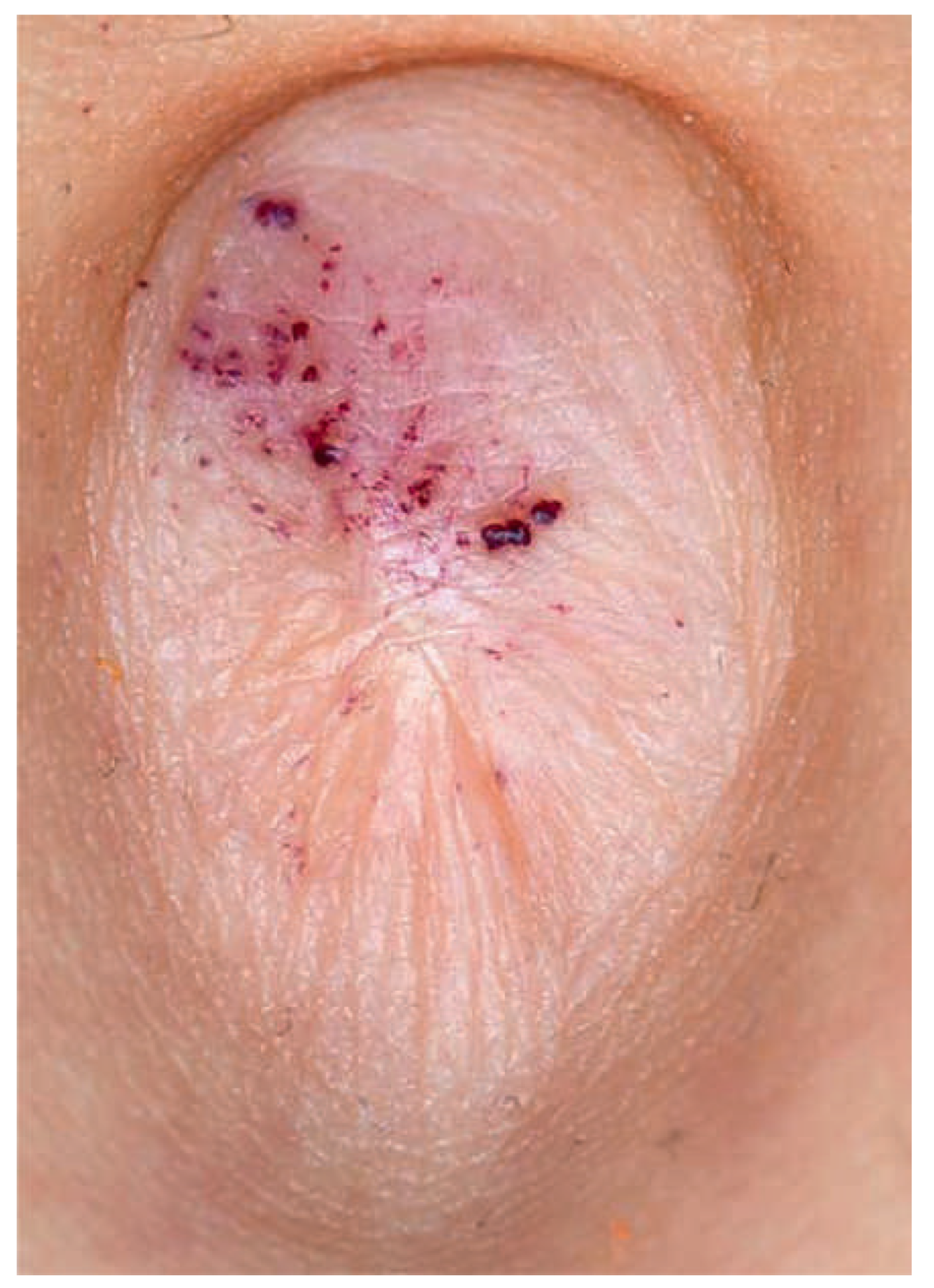
Skin involvement with angiokeratomas, clusters of individual punctate (Figure 6) or scattered (Figure 7) dark red to blue-black angioectases, is a hallmark of Fabry disease [56]. These benign tumors, characterised by ectasia of blood vessels in the papillary dermis and possibly associated with acanthosis and hyperkeratosis of the epidermis, are commonly localised on the lower trunk, palms, around the mouth and umbilicus. They are observed in 40–60% of patients and may appear already during childhood [57]. Hypo/anhidrosis, rarely hyperhidrosis, occurs in Fabry disease [58].
Cornea verticillata is a typical ophthalmological manifestation with bilateral keratopathy occurring in over 70% patients (Figure 8). It is identifiable during slit-lamp examination as whirl-like white-to-golden-brown opacities extending from centre to periphery of the cornea and corresponds to glycosphingolipid accumulation in the basal epithelium of the cornea [59]. Two types of lenticular changes may also be present in Fabry disease: anterior capsular or subcapsular cataract, and radial posterior subcapsular cataract [60]. Posterior subcapsular cataract is rare and specific for Fabry disease, and hence called "Fabry cataract". Other possible ocular abnormalities are retinal and conjunctival vessel tortuosity [60].
Lung involvement with mild to severe airway obstruction, manifesting as chronic bronchitis, wheezing or dyspnoea, is present in 25% of women and up to 60% of men [61].
Although Fabry disease may present in childhood or puberty, given the rarity of this disease and the nonspecific presenting symptoms, the disease is often not even considered by clinicians. As a consequence, diagnosis of this deadly disease is delayed in a majority of affected patients until the underlying pathology is already advanced [36,62].
Figure 1, 2.
T1 magnetic resonance image (Figure 1) of a 33-year-old male with typical hyperintense pulvinar sign indicating hyperperfusion-induced dystrophic calcification and magnetic resonance angiography (Figure 2) with mild elongation and dolichoectasia of the basilar artery (diameter 3.8 mm).
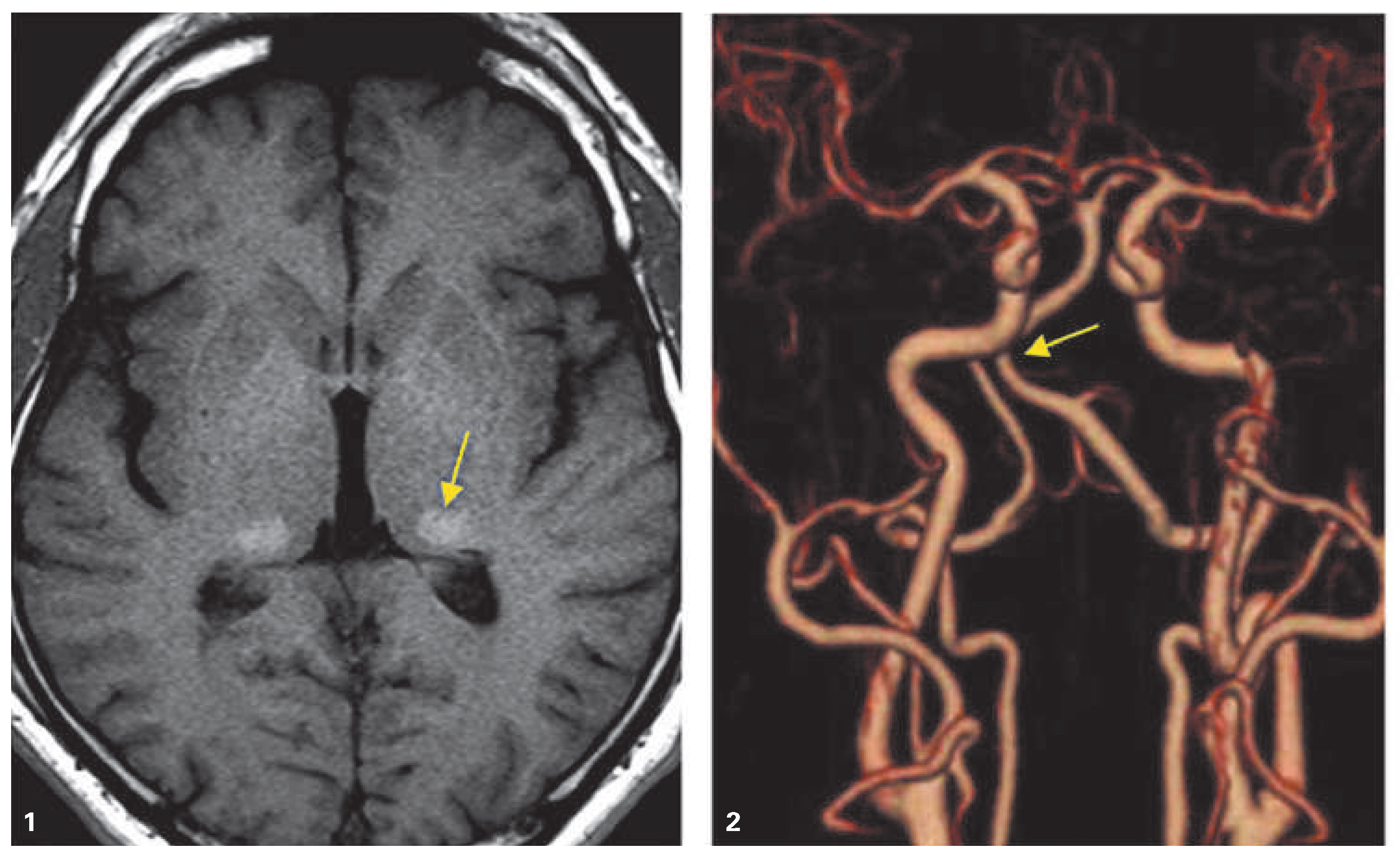
Figure 3.
Axial T2w magnetic resonance image of a 38-year-old female with left thalamic and subinsular infarctions.
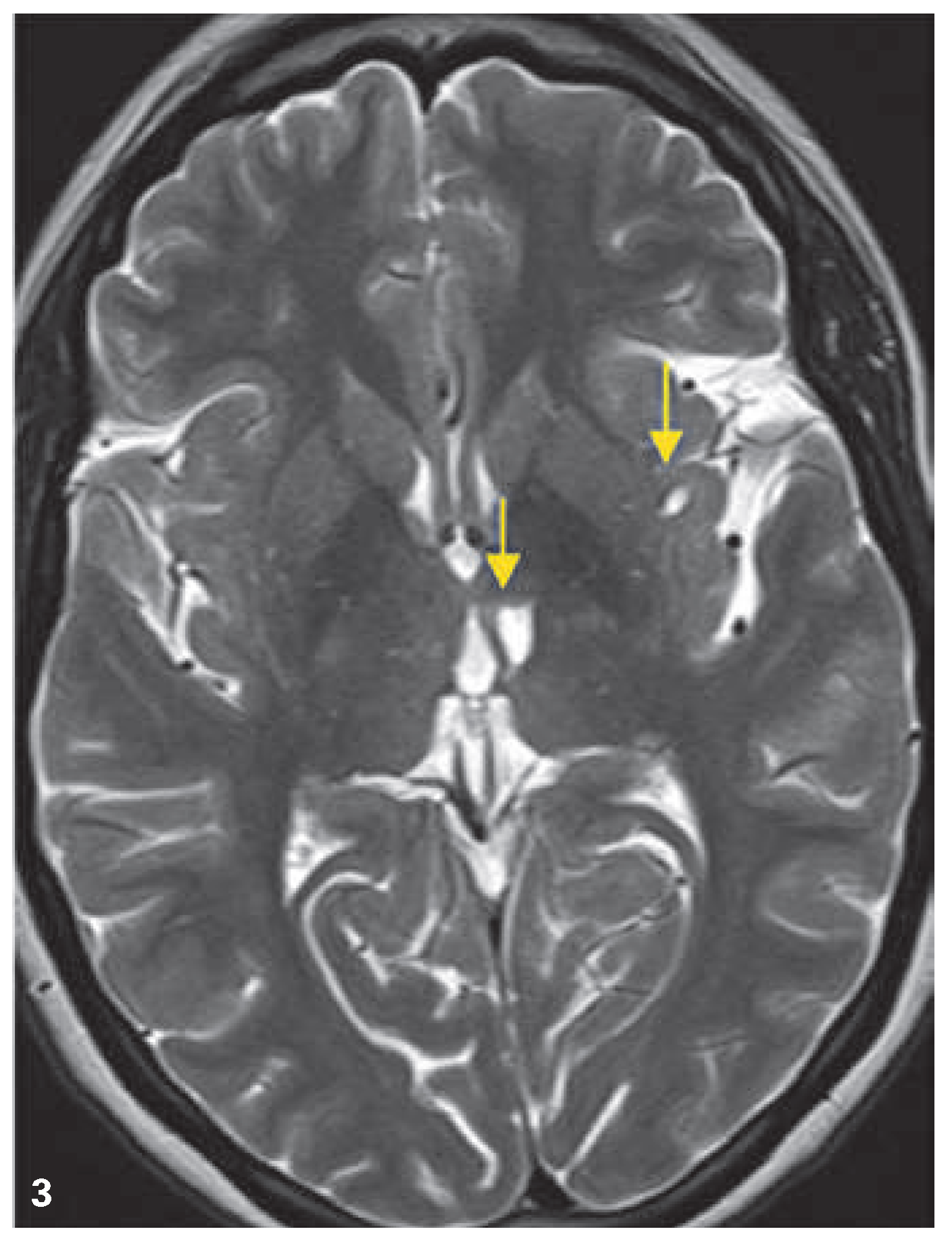
Figure 4.
Multi-organ involvement in children (A) and adults (B) [174, 175]. * Frequent manifestations. GFR = glomerular filtration rate; TIA = transient ischaemic attack.
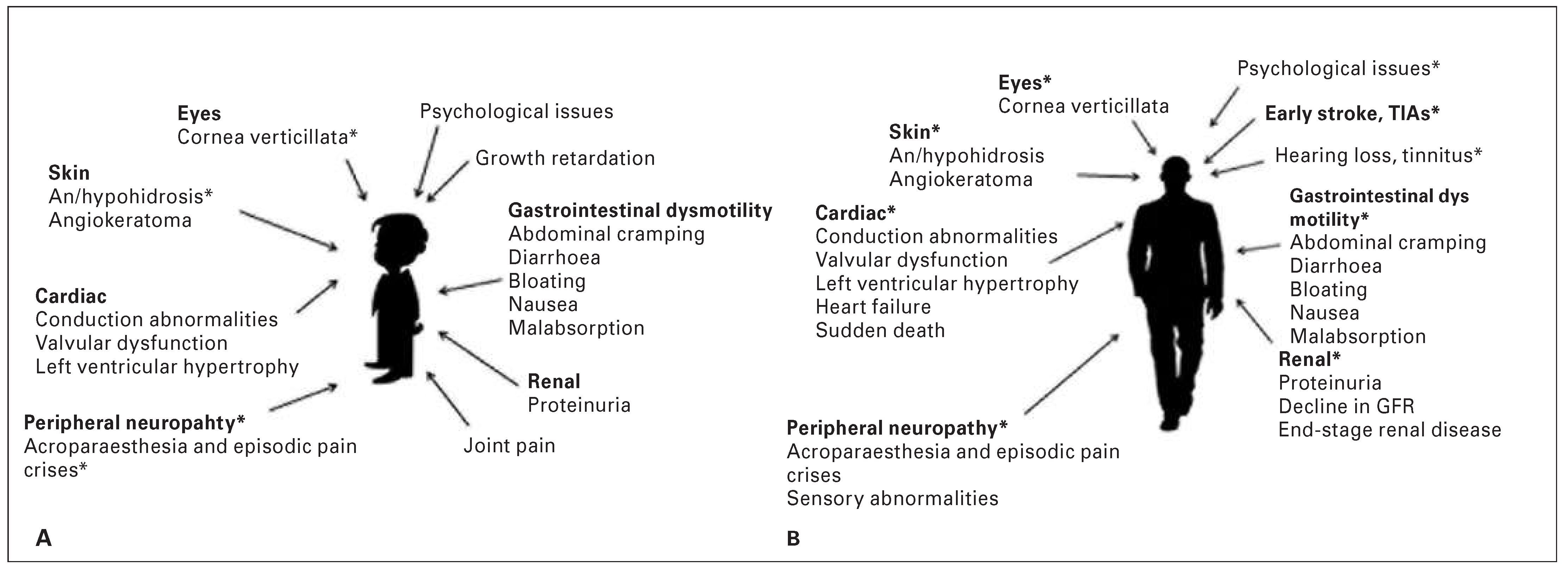
Figure 5.
Histological findings in a kidney biopsy: (A) Glomerulus with prominent, vacuolated podocytes (arrows) (H&E, ×200). (B) Lysosomal inclusions with lamellated structure – myeloid or zebra bodies in podocytes (TEM, ×2800).
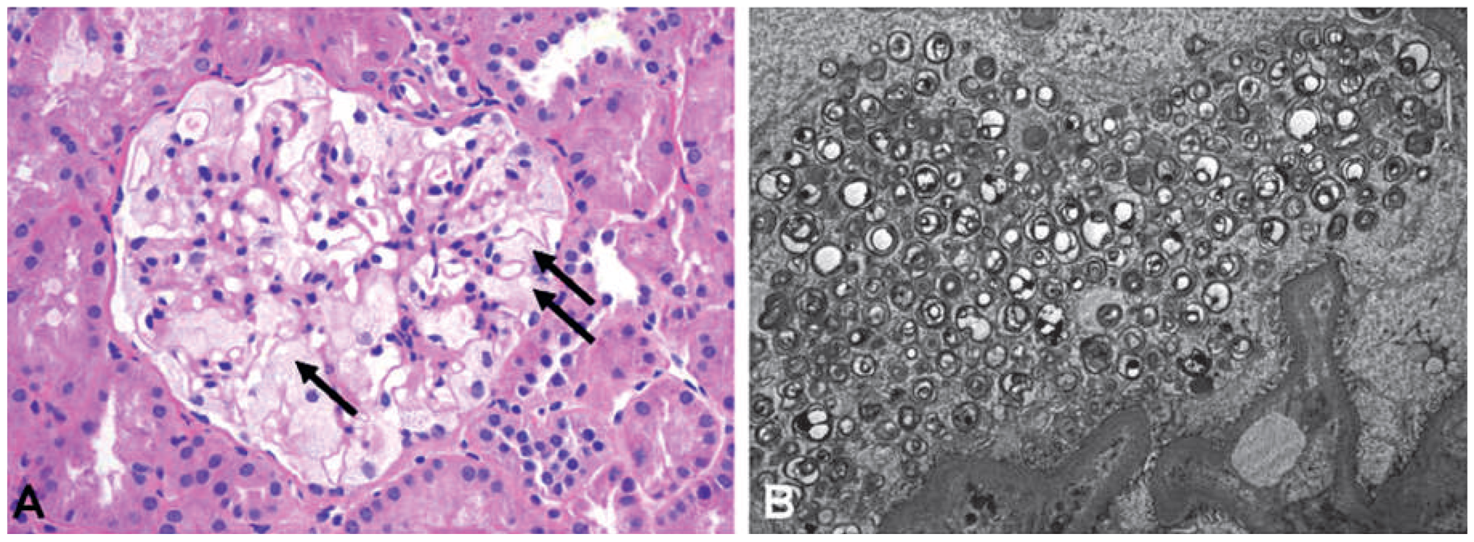
Figure 6.
Angiokeratomas.
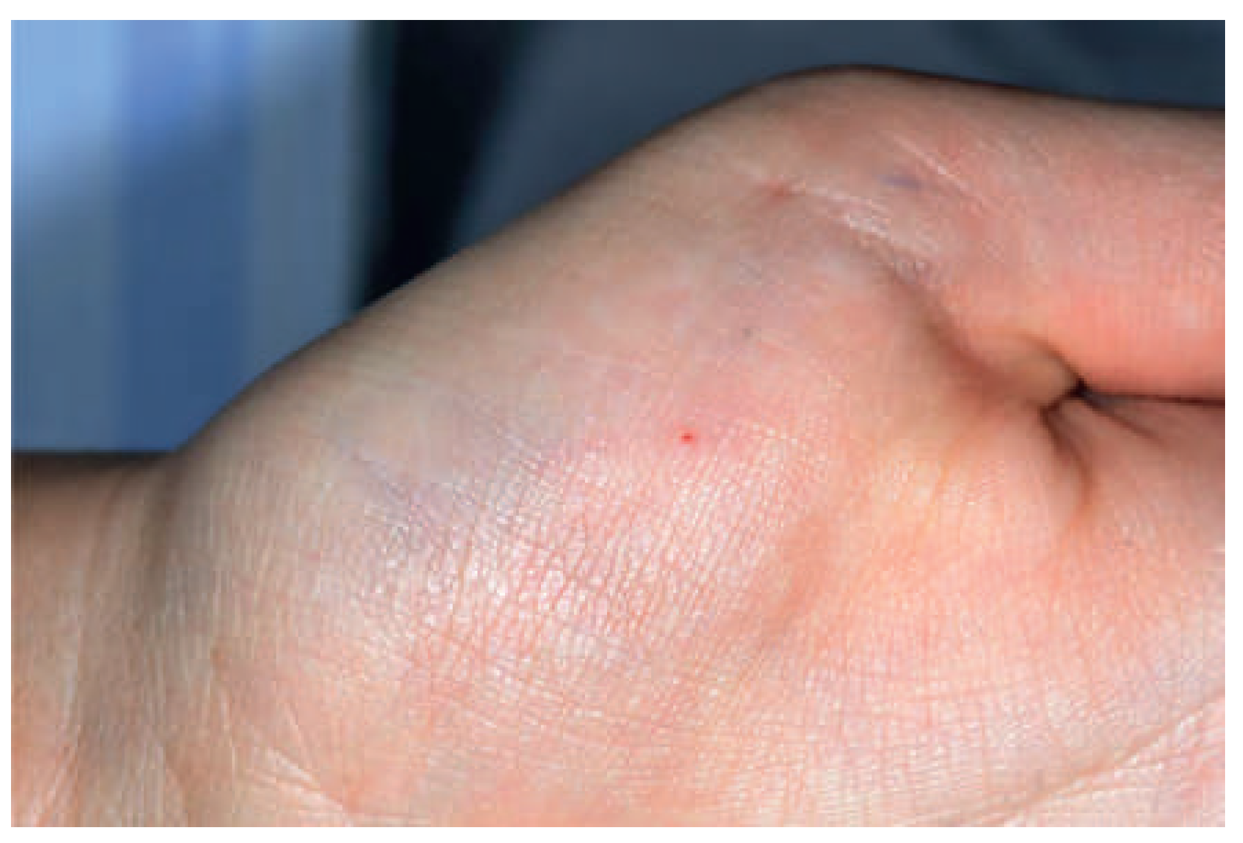
Figure 7.
Angiokeratoma in the navel.
Management
Treatment of patients with Fabry disease primarily focuses upon substitution of alpha-Gal A enzyme.
Enzyme replacement therapy (ERT) with alpha-Gal A has been used to treat Fabry disease in Europe since 2001 and in the USA since 2003. Recombinant human alpha-Gal A is available in two forms: agalsidase-alfa (Replagal®, Shire HGT) and agalsidase-beta (Fabrazyme®, Genzyme Corp.). Both proteins have the same amino acid sequence as the native human enzyme. They differ in glycosylation pattern. Agalsidase-alfa is purified from a stable transfected line of cultured human skin fibroblasts and is infused at a dose of 0.2 mg/kg every 2 weeks [63]. Agalsidase-beta is produced by expression of human alpha-Gal A DNA in Chinese hamster ovary cells and is infused at a dose of 1.0 mg/kg every 2 weeks [64]. The use of a lower maintenance dose of agalsidase-beta, 0.3 mg/kg has been evaluated and has been shown to maintain Gb3 clearance in some, but not in all patients. Long-term clinical effects of this lower dose of agalsidase-beta, however, have not been evaluated. Common side effects with ERT include infusion reactions and seroconversion (i.e., development of antibodies to either agalsidase-alfa or agalsidase-beta) [65,66]. Studies suggest that there are no significant differences between the two forms in terms of efficacy and safety profiles [67].
Neurological symptoms ameliorate with ERT significantly. An improvement of small nerve fibre function has been shown, resulting in decreased pain and consequently less analgesic consumption [16,65,68,69]. Improvement in intradermal vibration and temperature perception thresholds is reported [70]. Intraepidermal innervation density does not increase, suggesting that epidermal nerve fibres do not regenerate during ERT treatment. A hypothesis is that structural damage to small fibres is irreversible, whereas function of remaining fibres may improve on ERT.
CNS studies have demonstrated significant functional vascular changes under ERT treatment, with a tendency to normalisation of cerebral vessel autoregulation and regional perfusion [32,71]. However, effects of ERT on white matter lesions or incidence of cerebrovascular events have not yet been demonstrated [67,68,72]. Studies have reported recurrence of strokes despite long-term ERT [73,74]. The explanation may be the inability of ERT to pass the blood-brain barrier [75].
ERT leads to decreased left ventricular mass, heart rate variability and Gb3 in cardiac endothelium, as well as improved left ventricular function [76,77]. It has been shown to stabilise kidney function and to reduce proteinuria [74,78,79,80].
Although there is, to date, no consensus on treatment initiation, studies have shown that ERT reverses Gb3 accumulation in cells and tissues particularly when started at an earlier stage of the disease, whereas advanced disease stages such as advanced nephropathy and myocardial fibrosis do not respond to ERT [65,72,78]. Furthermore, ERT does not cure Fabry disease and adjunctive conventional medical treatment and therapy may be required. Symptomatic neuropathic pain management includes acetaminophen (paracetamol), gabapentin, carbamazepine and lamotrigine as well as tricyclic antidepressants such as amitriptyline. Opioids should be used carefully in Fabry disease patients, in particular those with gastrointestinal motility disorders; nonsteroidal inflammatory drugs are not recommended because of their nephrotoxicity. Additional measures include lifestyle changes with avoidance of stimuli such as stress, intense physical exertion and extreme temperature changes. Prophylactic therapy is particularly important in stroke prevention. It could include the use of antiplatelet agents (i.e., aspirin, clopidogrel, or a combination of aspirin with dipyridamole), antihypertensive agents and statins. It is important to note, however, that the efficacy of antiplatelet and anticoagulant therapy has not been proven at the present time. Avoidance of smoking, obesity, dyslipidaemia and arterial hypertension is likely to reduce the risk of stroke as in the general population. They also prevent heart failure and pulmonary symptoms. Angiotensin converting-enzyme (ACE) inhibitors and angiotensin receptor blockers (ARBs) are effective in reducing proteinuria and controlling hypertension [80]. Patients may require renal replacement therapy, such as dialysis or kidney transplantation. Diuretics, ACE inhibitors, ARBs, beta-blockers, and implantable cardiac devices need to be considered in patients with cardiovascular involvement. Heart transplantation may be necessary in cases of advanced congestive heart failure.
Ongoing trials are investigating novel approaches such as chaperone therapy [81], gene therapy [82] and substrate reduction therapy [83]. Further evaluations and pharmacological studies are needed as efficacy and safety in humans need to be demonstrated.
Figure 8.
Cornea verticillata. Courtesy Dr. R. Kovacs, Ophthalmology Department, University Hospital Zürich.
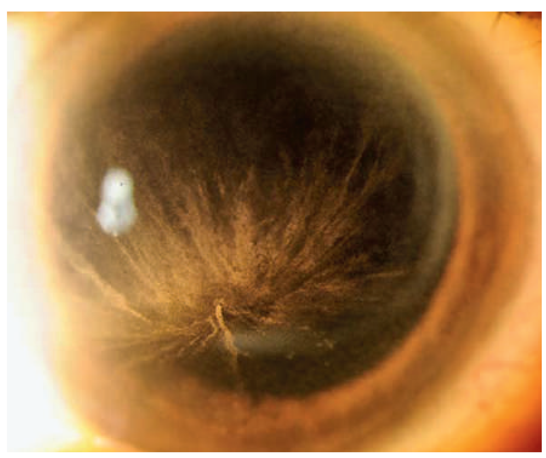
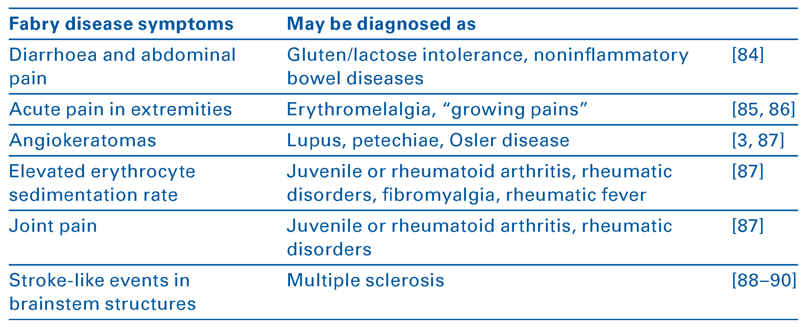
Table 2.
Common misdiagnoses of Fabry disease (not an exhaustive list).
Conclusions
Fabry disease is a rare multisystemic genetic disorder characterised by progressive accumulation of glycosphingolipids in tissues, leading to ischaemia and irreversible vital organ damage. Neurological involvement by Fabry disease, presenting at the end of the first decade of life or during puberty, is comparable in men and women and is one of the most frequent and earliest manifestations of Fabry disease reported by patients. Neurological involvement is also one of the earliest, potentially life-threatening, complications of Fabry disease with an average age of onset for cerebrovascular ischaemia and premature stroke around 35–40 years.
Because of its variable nonspecific symptoms, diagnosis of Fabry disease is challenging. Misdiagnoses, especially with rheumatic disorders and neurological disease, are frequent (Table 2) and the delay between first symptoms and diagnosis could reach 10–15 years. Adequate care management and treatment are promising to slow or even reverse disease manifestation. However, organ damage is usually irreversible if treatment is started too late, underscoring the importance of early recognition of this devastating disease.
Among patients enrolled in FOS – the Fabry Outcome Survey – the medical specialists who most often establish the diagnosis of Fabry disease were nephrologists followed by dermatologists and paediatricians; fewer cases are diagnosed by neurologists [3]. The earlier the diagnosis is suspected and confirmed and the earlier treatment, including ERT, is initiated, the more effective is the therapy.
Recognition of early neurological symptoms, in particular unexplained peripheral neuropathic pain typically in the hands and feet, and considering Fabry disease in the case of unexplained stroke in young patients is important.
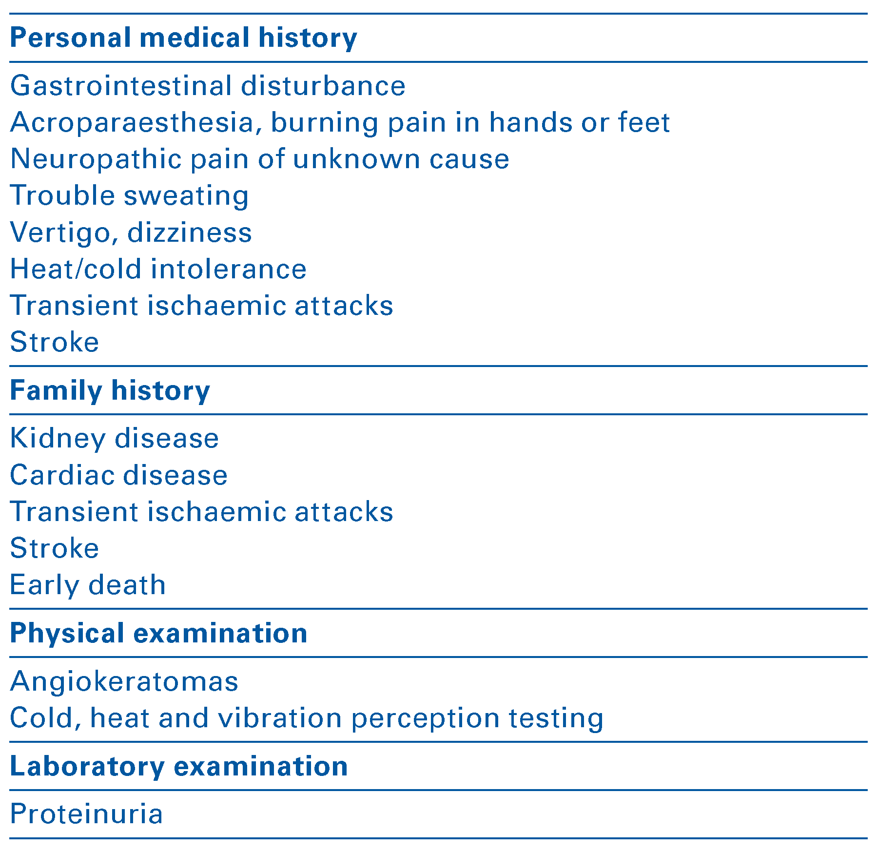
To avoid misdiagnosis and to start treatment early, young patients with unexplained stroke should be evaluated for presence of other systemic problems in their personal medical and family history such as (i) gastrointestinal disturbance, (ii) recurrent tingling or burning in hands or feet, (iii) pain crises of pain possibly accompanied by fever remaining of unexplained origin, (iv) trouble sweating, (v) difficulties to tolerate heat or cold, and (vi) a family history of kidney/cardiac failure, ischaemic attack or stroke and early death. A complete physical examination should be performed to look for angiokeratomas, reddish-purple spots particularly in the bathing trunk area. Neurological examination should focus on sensations mediated by peripheral small fibres, such as cold, heat, light touch, pressure and vibration perception testing. Proteinuria has to be sought (Table 3).
Table 3.
Signs and symptoms suggestive of Fabry disease.
In conclusion, Fabry disease requires a multidisciplinary approach, including neurologists, nephrologists, cardiologists, dermatologists, gastroenterologists, geneticists, pathologists and clinical pharmacologists. Neurologists play a key role in the early diagnosis of this deadly disease and may be the first clinicians to identify Fabry disease.
Disclosure statement
A.N.: publication support for this article and speaker honoraria from Genzyme; research support from Genzyme and Shire. M.R.: speaker honoraria from Genzyme and Shire. The other authors have no conflict of interest to declare.
References
The full list of references is included in the online version of the article on www.sanp.ch.
References
- Kint JA. Fabry's disease: alpha-galactosidase deficiency. Science. 1970;167(3922):1268-9.
- Germain DP. Fabry disease. Orphanet J Rare Dis. 2010;5:30.
- Mehta A, Beck M, Sunder-Plassmann G. Fabry disease: perspectives from 5 years of FOS. 2006: Oxford PharmaGenesis, Oxford (2006).
- Germain DP, Shabbeer J, Cotigny S, Desnick RJ. Fabry disease: twenty novel alpha-galactosidase A mutations and genotype-phenotype correlations in classical and variant phenotypes. Mol Med. 2002;8(6):306-12.
- Deegan PB, Baehner AF, Barba Romero MA, Hughes DA, Kampmann C, Beck M. Natural history of Fabry disease in females in the Fabry Outcome Survey. J Med Genet. 2006;43(4):347-52.
- Hwu WL, Chien YH, Lee NC, Chiang SC, Dobrovolny R, Huang AC, et al. Newborn screening for Fabry disease in Taiwan reveals a high incidence of the later-onset GLA mutation c.936+919G>A (IVS4+190G>A). Hum Mutat. 2009;30(10):1397-405.
- Lyon MF. Gene action in the X-chromosome of the mouse (Mus musculus L.). Nature. 1961;190:372-3.
- Desnick RJ. Prenatal diagnosis of Fabry disease. Prenat Diagn. 2007;27(8):693-4.
- Dutsch M, Marthol H, Stemper B, Brys M, Haendl T, Hilz MJ. Small fiber dysfunction predominates in Fabry neuropathy. J Clin Neurophysiol. 2002;19(6):575-86.
- Pellissier JF, Van Hoof F, Bourdet-Bonerandi D, Monier-Faugere MC, Toga M. Morphological and biochemical changes in muscle and peripheral nerve in Fabry's disease. Muscle Nerve. 1981;4(5):381-7.
- Scott LJ, Griffin JW, Luciano C, Barton NW, Banerjee T, Crawford T, et al. Quantitative analysis of epidermal innervation in Fabry disease. Neurology. 1999;52(6):1249-54.
- Torvin Moller A, Winther Bach F, Feldt-Rasmussen U, Rasmussen A, Hasholt L, Lan H, et al. Functional and structural nerve fiber findings in heterozygote patients with Fabry disease. Pain. 2009;145(1-2):237-45.
- Vital A, Vital C, Maleville J. Fabry's disease: an ultrastructural study of muscle and peripheral nerve. Clin Neuropathol. 1984;3(4):168-72.
- Burlina AP, Sims KB, Politei JM, Bennett GJ, Baron R, Sommer C, et al. Early diagnosis of peripheral nervous system involvement in Fabry disease and treatment of neuropathic pain: the report of an expert panel. BMC Neurol. 2011;11:61.
- Moller AT, Jensen TS. Neurological manifestations in Fabry's disease. Nat Clin Pract Neurol. 2007;3(2):95-106.
- Schiffmann R, Scott LJ. Pathophysiology and assessment of neuropathic pain in Fabry disease. Acta Paediatr Suppl. 2002;91(439):48-52.
- Kolodny EH, Pastores GM. Anderson-Fabry disease: extraneural, neurologic manifestations. J Am Soc Nephrol. 2002;13(Suppl 2):S150-3.
- Ries M, Ramaswami U, Parini R, Lindblad B, Whybra C, Willers I, et al. The early clinical phenotype of Fabry disease: a study on 35 European children and adolescents. Eur J Pediatr. 2003;162(11):767-72.
- Hoffmann B, Beck M, Sunder-Plassmann G, Borsini W, Ricci R, Mehta A. Nature and prevalence of pain in Fabry disease and its response to enzyme replacement therapy – a retrospective analysis from the Fabry Outcome Survey. Clin J Pain. 2007;23(6):535-42.
- Mehta A, Ginsberg L. Natural history of the cerebrovascular complications of Fabry disease. Acta Paediatr Suppl. 2005;94(447):24-7; discussion 9-10.
- Uceyler N, He L, Schonfeld D, Kahn AK, Reiners K, Hilz MJ, et al. Small fibers in Fabry disease: baseline and follow-up data under enzyme replacement therapy. J Peripher Nerv Syst. 2011;16(4):304-14.
- Luciano CA, Russell JW, Banerjee TK, Quirk JM, Scott LJ, Dambrosia JM, et al. Physiological characterization of neuropathy in Fabry's disease. Muscle Nerve. 2002;26(5):622-9.
- Maag R, Binder A, Maier C, Scherers A, Toelle T, Treede RD, et al. Detection of a characteristic painful neuropathy in Fabry disease: a pilot study. Pain Med. 2008;9(8):1217-23.
- Cable WJ, Kolodny EH, Adams RD. Fabry disease: impaired autonomic function. Neurology. 1982;32(5):498-502.
- Toyooka K. Fabry disease. Curr Opin Neurol. 2012;25(5):463-8.
- Rolfs A, Bottcher T, Zschiesche M, Morris P, Winchester B, Bauer P, et al. Prevalence of Fabry disease in patients with cryptogenic stroke: a prospective study. Lancet. 2005;366(9499):1794-6.
- Hilz MJ, Brys M, Marthol H, Stemper B, Dutsch M. Enzyme replacement therapy improves function of C-, Adelta-, and Abeta-nerve fibers in Fabry neuropathy. Neurology. 2004;62(7):1066-72.
- Moore DF, Altarescu G, Ling GS, Jeffries N, Frei KP, Weibel T, et al. Elevated cerebral blood flow velocities in Fabry disease with reversal after enzyme replacement. Stroke. 2002;33(2):525-31.
- Moore DF, Herscovitch P, Schiffmann R. Selective arterial distribution of cerebral hyperperfusion in Fabry disease. J Neuroimaging. 2001;11(3):303-7.
- Fellgiebel A, Keller I, Marin D, Muller MJ, Schermuly I, Yakushev I, et al. Diagnostic utility of different MRI and MR angiography measures in Fabry disease. Neurology. 2009;72(1):63-8.
- Hilz MJ, Kolodny EH, Brys M, Stemper B, Haendl T, Marthol H. Reduced cerebral blood flow velocity and impaired cerebral autoregulation in patients with Fabry disease. J Neurol. 2004;251(5):564-70.
- Moore DF, Scott LT, Gladwin MT, Altarescu G, Kaneski C, Suzuki K, et al. Regional cerebral hyperperfusion and nitric oxide pathway dysregulation in Fabry disease: reversal by enzyme replacement therapy. Circulation. 2001;104(13):1506-12.
- Ginsberg L. Nervous system manifestations of Fabry disease: data from FOS – the Fabry Outcome Survey, in Fabry Disease: Perspectives from 5 Years of FOS, A. Mehta, M. Beck, and G. Sunder-Plassmann, Editors. 2006, Oxford PharmaGenesis: Oxford.
- Sims K, Politei J, Banikazemi M, Lee P. Stroke in Fabry disease frequently occurs before diagnosis and in the absence of other clinical events: natural history data from the Fabry Registry. Stroke. 2009;40(3):788-94.
- Kolodny E, Fellgiebel A, Hilz MJ, Sims K, Caruso P, Phan TG, et al. Cerebrovascular involvement in Fabry disease: current status of knowledge. Stroke. 2015;46(1):302-13.
- Mehta A, Ricci R, Widmer U, Dehout F, Garcia de Lorenzo A, Kampmann C, et al. Fabry disease defined: baseline clinical manifestations of 366 patients in the Fabry Outcome Survey. Eur J Clin Invest. 2004;34(3):236-42.
- Shi Q, Chen J, Pongmoragot J, Lanthier S, Saposnik G. Prevalence of Fabry disease in stroke patients – a systematic review and meta-analysis. J Stroke Cerebrovasc Dis. 2014;23(5):985-92.
- Cole AL, Lee PJ, Hughes DA, Deegan PB, Waldek S, Lachmann RH. Depression in adults with Fabry disease: a common and under-diagnosed problem. J Inherit Metab Dis. 2007;30(6):943-51.
- Malinvaud D, Germain DP, Benistan K, Bonfils P. Vestibular and cochlear manifestations in Fabry's disease. Rev Med Interne. 2010;31(Suppl 2):S251-6.
- Palla A, Hegemann S, Widmer U, Straumann D. Vestibular and auditory deficits in Fabry disease and their response to enzyme replacement therapy. J Neurol. 2007;254(10):1433-42.
- Crutchfield KE, Patronas NJ, Dambrosia JM, Frei KP, Banerjee TK, Barton NW, et al. Quantitative analysis of cerebral vasculopathy in patients with Fabry disease. Neurology. 1998;50(6):1746-9.
- Fellgiebel A, Muller MJ, Ginsberg L. CNS manifestations of Fabry's disease. Lancet Neurol. 2006;5(9):791-5.
- Moore DF, Ye F, Schiffmann R, Butman J. Increased signal intensity in the pulvinar on T1-weighted images: a pathognomonic MR imaging sign of Fabry disease. AJNR Am J Neuroradiol. 2003;24(6):1096-101.
- Takanashi J, Barkovich AJ, Dillon WP, Sherr EH, Hart KA, Packman S. T1 hyperintensity in the pulvinar: key imaging feature for diagnosis of Fabry disease. AJNR Am J Neuroradiol. 2003;24(5):916-21.
- Uceyler N, Homola GA, Guerrero Gonzalez H, Kramer D, Wanner C, Weidemann F, et al. Increased arterial diameters in the posterior cerebral circulation in men with Fabry disease. PLoS One. 2014;9(1):e87054.
- Rombach SM, Twickler TB, Aerts JM, Linthorst GE, Wijburg FA, Hollak CE. Vasculopathy in patients with Fabry disease: current controversies and research directions. Mol Genet Metab. 2010;99(2):99-108.
- Fogo AB, Bostad L, Svarstad E, Cook WJ, Moll S, Barbey F, et al. Scoring system for renal pathology in Fabry disease: report of the International Study Group of Fabry Nephropathy (ISGFN). Nephrol Dial Transplant. 2010;25(7):2168-77.
- Schiffmann R, Warnock DG, Banikazemi M, Bultas J, Linthorst GE, Packman S, et al. Fabry disease: progression of nephropathy, and prevalence of cardiac and cerebrovascular events before enzyme replacement therapy. Nephrol Dial Transplant. 2009;24(7):2102-11.
- Seydelmann N, Wanner C, Stork S, Ertl G, Weidemann F. Fabry disease and the heart. Best Pract Res Clin Endocrinol Metab. 2015;29(2):195-204.
- Linhart A, Kampmann C, Zamorano JL, Sunder-Plassmann G, Beck M, Mehta A, et al. Cardiac manifestations of Anderson-Fabry disease: results from the international Fabry outcome survey. Eur Heart J. 2007;28(10):1228-35.
- Kramer J, Niemann M, Liu D, Hu K, Machann W, Beer M, et al. Two-dimensional speckle tracking as a non-invasive tool for identification of myocardial fibrosis in Fabry disease. Eur Heart J. 2013;34(21):1587-96.
- Anastasakis A, Papatheodorou E, Steriotis AK. Fabry disease and cardiovascular involvement. Curr Pharm Des. 2013;19(33):5997-6008.
- Moon JC, Sachdev B, Elkington AG, McKenna WJ, Mehta A, Pennell DJ, et al. Gadolinium enhanced cardiovascular magnetic resonance in Anderson-Fabry disease. Evidence for a disease-specific abnormality of the myocardial interstitium. Eur Heart J. 2003;24(23):2151-5.
- Weidemann F, Niemann M, Breunig F, Herrmann S, Beer M, Stork S, et al. Long-term effects of enzyme replacement therapy on fabry cardiomyopathy: evidence for a better outcome with early treatment. Circulation. 2009;119(4):524-9.
- Hoffmann B, Schwarz M, Mehta A, Keshav S. Gastrointestinal symptoms in 342 patients with Fabry disease: prevalence and response to enzyme replacement therapy. Clin Gastroenterol Hepatol. 2007;5(12):1447-53.
- Zampetti A, Orteu CH, Antuzzi D, Bongiorno MR, Manco S, Gnarra M, et al. Angiokeratoma: decision-making aid for the diagnosis of Fabry disease. Br J Dermatol. 2012;166(4):712-20.
- Falke K, Buttner A, Schittkowski M, Stachs O, Kraak R, Zhivov A, et al. The microstructure of cornea verticillata in Fabry disease and amiodarone-induced keratopathy: a confocal laser-scanning microscopy study. Graefes Arch Clin Exp Ophthalmol. 2009;247(4):523-34.
- Orteu CH, Jansen T, Lidove O, Jausaud R, Hughes DA, Pintos-Morell G, et al. Fabry disease and the skin: data from FOS, the Fabry outcome survey. Br J Dermatol. 2007;157(2):331-7.
- Sivley MD. Fabry disease: a review of ophthalmic and systemic manifestations. Optom Vis Sci. 2013;90(2):e63-78.
- Sodi A, Ioannidis AS, Mehta A, Davey C, Beck M, Pitz S. Ocular manifestations of Fabry's disease: data from the Fabry Outcome Survey. Br J Ophthalmol. 2007;91(2):210-4.
- Magage S, Lubanda JC, Susa Z, Bultas J, Karetova D, Dobrovolny R, et al. Natural history of the respiratory involvement in Anderson-Fabry disease. J Inherit Metab Dis. 2007;30(5):790-9.
- Meikle PJ, Hopwood JJ, Clague AE, Carey WF. Prevalence of lysosomal storage disorders. JAMA. 1999;281(3):249-54.
- Beck M. Agalsidase alfa – a preparation for enzyme replacement therapy in Anderson-Fabry disease. Expert Opin Investig Drugs. 2002;11(6):851-8.
- Banikazemi M, Bultas J, Waldek S, Wilcox WR, Whitley CB, McDonald M, et al. Agalsidase-beta therapy for advanced Fabry disease: a randomized trial. Ann Intern Med. 2007;146(2):77-86.
- Eng CM, Guffon N, Wilcox WR, Germain DP, Lee P, Waldek S, et al. Safety and efficacy of recombinant human alpha-galactosidase A replacement therapy in Fabry's disease. N Engl J Med. 2001;345(1):9-16.
- Schiffmann R, Ries M, Timmons M, Flaherty JT, Brady RO. Long-term therapy with agalsidase alfa for Fabry disease: safety and effects on renal function in a home infusion setting. Nephrol Dial Transplant. 2006;21(2):345-54.
- Vedder AC, Linthorst GE, Houge G, Groener JE, Ormel EE, Bouma BJ, et al. Treatment of Fabry disease: outcome of a comparative trial with agalsidase alfa or beta at a dose of 0.2 mg/kg. PLoS One. 2007;2(7):e598.
- Schiffmann R, Kopp JB, Austin HA, 3rd, Sabnis S, Moore DF, Weibel T, et al. Enzyme replacement therapy in Fabry disease: a randomized controlled trial. JAMA. 2001;285(21):2743-9.
- El Dib RP, Nascimento P, Pastores GM. Enzyme replacement therapy for Anderson-Fabry disease. Cochrane Database Syst Rev. 2013;2:CD006663.
- Schiffmann R, Floeter MK, Dambrosia JM, Gupta S, Moore DF, Sharabi Y, et al. Enzyme replacement therapy improves peripheral nerve and sweat function in Fabry disease. Muscle Nerve. 2003;28(6):703-10.
- Moore DF, Altarescu G, Herscovitch P, Schiffmann R. Enzyme replacement reverses abnormal cerebrovascular responses in Fabry disease. BMC Neurol. 2002;2:4.
- Wilcox WR, Banikazemi M, Guffon N, Waldek S, Lee P, Linthorst GE, et al. Long-term safety and efficacy of enzyme replacement therapy for Fabry disease. Am J Hum Genet. 2004;75(1):65-74.
- Rombach SM, Smid BE, Bouwman MG, Linthorst GE, Dijkgraaf MG, Hollak CE. Long term enzyme replacement therapy for Fabry disease: effectiveness on kidney, heart and brain. Orphanet J Rare Dis. 2013;8:47.
- Anderson LJ, Wyatt KM, Henley W, Nikolauo V, Waldek S, Hughes DA, et al. Long-term effectiveness of enzyme replacement therapy in Fabry disease: results from the NCS-LSD cohort study. J Inherit Metab Dis. 2014;37(6):969-78.
- Begley DJ, Pontikis CC, Scarpa M. Lysosomal storage diseases and the blood-brain barrier. Curr Pharm Des. 2008;14(16):1566-80.
- Kalliokoski RJ, Kantola I, Kalliokoski KK, Engblom E, Sundell J, Hannukainen JC, et al. The effect of 12-month enzyme replacement therapy on myocardial perfusion in patients with Fabry disease. J Inherit Metab Dis. 2006;29(1):12-8.
- Weidemann F, Breunig F, Beer M, Sandstede J, Turschner O, Voelker W, et al. Improvement of cardiac function during enzyme replacement therapy in patients with Fabry disease: a prospective strain rate imaging study. Circulation. 2003;108(11):1299-301.
- Germain DP, Charrow J, Desnick RJ, Guffon N, Kempf J, Lachmann RH, et al. Ten-year outcome of enzyme replacement therapy with agalsidase beta in patients with Fabry disease. J Med Genet. 2015;52(5):353-8.
- Mehta A, Beck M, Elliott P, Giugliani R, Linhart A, Sunder-Plassmann G, et al. Enzyme replacement therapy with agalsidase alfa in patients with Fabry's disease: an analysis of registry data. Lancet. 2009;374(9706):1986-96.
- Ortiz A, Oliveira JP, Wanner C, Brenner BM, Waldek S, Warnock DG. Recommendations and guidelines for the diagnosis and treatment of Fabry nephropathy in adults. Nat Clin Pract Nephrol. 2008;4(6):327-36.
- Ishii S. Pharmacological chaperone therapy for Fabry disease. Proc Jpn Acad Ser B Phys Biol Sci. 2012;88(1):18-30.
- Ogawa K, Hirai Y, Ishizaki M, Takahashi H, Hanawa H, Fukunaga Y, et al. Long-term inhibition of glycosphingolipid accumulation in Fabry model mice by a single systemic injection of AAV1 vector in the neonatal period. Mol Genet Metab. 2009;96(3):91-6.
- Ashe KM, Budman E, Bangari DS, Siegel CS, Nietupski JB, Wang B, et al. Efficacy of enzyme and substrate reduction therapy with a novel antagonist of glucosylceramide synthase for Fabry disease. Mol Med. 2015;21:389-99.
- Buda P, Wieteska-Klimczak A, Ksiazyk J, Smorczewska-Kiljan A, Gietka P, Czartoryska B, et al. Diagnostic problems in a 17-year-old patient with gastrointestinal manifestations of Fabry disease. Med Wieku Rozwoj. 2011;15(1):69-72.
- Cailleux N, Levesque H, Joly P, Thomine E, Courtois H. Childhood acromelalgia a propos of a case revealing Fabry's disease. J Mal Vasc. 1995;20(2):142-5.
- Lazareth I. False erythemalgia. J Mal Vasc. 1996;21(2):84-7.
- Marchesoni CL, Roa N, Pardal AM, Neumann P, Caceres G, Martinez P, et al. Misdiagnosis in Fabry disease. J Pediatr. 2010;156(5):828-31.
- Bottcher T, Rolfs A, Tanislav C, Bitsch A, Kohler W, Gaedeke J, et al. Fabry disease – underestimated in the differential diagnosis of multiple sclerosis? PLoS One. 2013;8(8):e71894.
- Callegaro D, Kaimen-Maciel DR. Fabry's disease as a differential diagnosis of MS. Int MS J. 2006;13(1):27-30.
- Saip S, Uluduz D, Erkol G. Fabry disease mimicking multiple sclerosis. Clin Neurol Neurosurg. 2007;109(4):361-3.
This is an open-access article distributed under the terms of the Creative Commons Attribution Non-Commercial No Derivatives (CC BY-NC-ND) 4.0 License.