


Abstract
Polycythaemia is characterised by excessive erythropoiesis and a low serum concentration of erythropoietin. It is a vascular risk factor. We underlined its relevance in the aetiological investigation of a stroke in a young patient without any prior major vascular risk factor, who presented with headaches, vomiting and four-limb pyramidal syndrome. The red blood cell count was 6.15 × 10⁶/μl; the haemoglobin was 16.5 g/dl and the haematocrit 48.3%. The mean corpuscular volume was 78.5 fl, the mean corpuscular haemoglobin was 26.8 pg and the platelet count was 614,000/μl. The diagnosis of the polycythaemia was confirmed. A brain computed tomography scan displayed a right frontoparietal haematoma with a left posterior superficial sylvian territory haemorrhagic infarction. Angioscan of the brain and supra-aortic arteries was normal. The thrombotic and haemorrhagic complications of polycythaemia are known. However, the association of a brain haematoma with a hemorrhagic infarction is rare.
Introduction
Vaquez disease is characterised by excessive erythropoiesis and a low concentration of erythropoietin in serum [1]. Two erythrocyte precursor cells have been identified in the bone marrow. One of them reacts normally to erythropoietin and the other, a malignant clone, displays an excessive reaction to it. Haemorrhagic and thrombotic complications are the most serious, interms of frequency and severity. They are often correlated with the haematocrit [2]. Ischaemic stroke is one of the principal nervous system complications of polycythaemia [3]. Our goal was to present the diagnostic pertinence of the association of a haemorrhagic infarction and a spontaneous haematoma with polycythaemia, and to underline the relevance of an aetiological investigation of stroke in a young patient without major vascular risk factors.
Case report
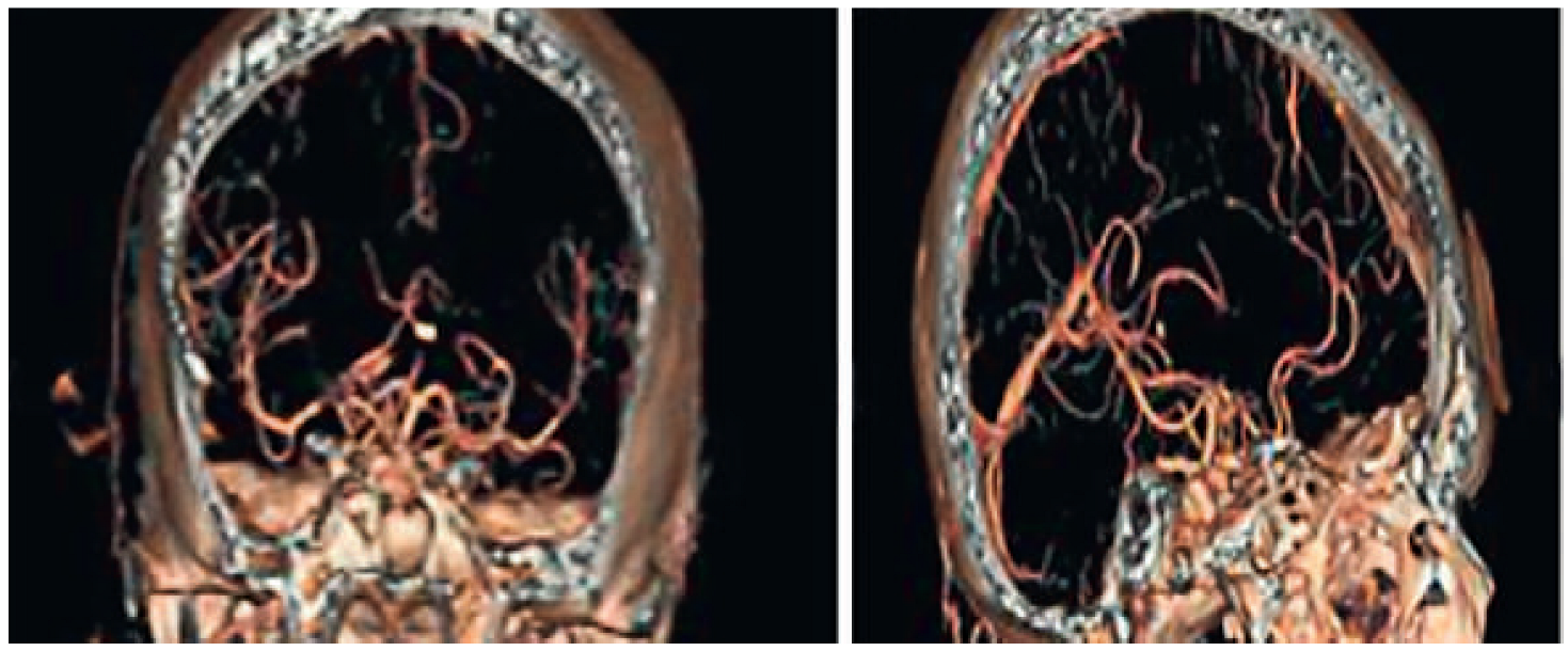
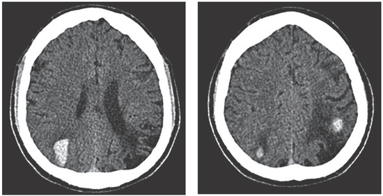
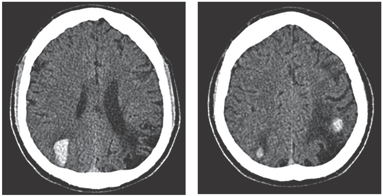
The patient was a right-handed 38-year-old high school teacher without hypertension, diabetes and smoking history. He was admitted to the neurology department for a sudden right hemiplegia, dysarthria and a state of confusion with headaches on 1 October 2010. During the admission, the patient displayed a worsening of headache, vomiting and left hemiplegia 3 hours after the onset of the first symptoms. The onset of symptoms was without particular circumstances; the patient was at rest. The clinical examination displayed a normal blood pressure and a pyramidal syndrome of the four limbs. The Rankin modified scale (RMS) score was of 4/6 and there was a score of 17 on the National Institutes of Health Stroke Scale (NIHSS). Spontaneous speech was very poor, but with a good comprehension of oral language. There was a dysexecutive syndrome with disorientation, bucco-facial apraxia and lack of attention. The brain computed tomography (CT) scan displayed a right frontoparietal haematoma and a left posterior superficial sylvian territory haemorrhagic infarction (Figure 1). An angioscan of the brain and supra-aortic arteries did not display any vascular abnormality (fig. 2). The electrocardiogram was normal. Doppler scanning displayed a moderate cardiomegaly without any systolic or diastolic dysfunction. (Lung flux = 0.6 m/s; on the aorta = 0.65 m/s.) The red blood cell count was 6.15 × 10⁶/μl, the haemoglobin was 16.5 g/dl and the haematocrit 48.3%. The mean corpuscular volume was 78.5 fl, the mean corpuscular haemoglobin was 26.8 pg, the platelet count was 614,000/μl and the leucocyte count 12,300/μl. Human immunodeficiency virus antibody testing was negative. The polycythaemia was treated hydroxycarbamid 500 mg twice per day. We used paracetamol against headaches and omeprazole to prevent stress ulcer during the acute period. Functional restoration was three sessions per week in addition to the nursing care. Three months after the beginning of the treatment, there was no improvement of the functional state (RMS 4/6, NIHSS 17). The red blood cell count was 5.01 × 10⁶/μl, the haemoglobin was 14.4 g/dl, the mean corpuscular volume was 86.4 fl, the mean corpuscular hemoglobin was 28.7 pg, and the platelet count was 376,000/μl. The polycythaemia stabilised. At 6 months the RMS remained at 4/6, and the NIHSS score 17.
Figure 1.
Right fronto-parietal haematoma associated to left posterior sylvian haemorrhagic infarction.
Figure 3.
Normal brain angioscan.
Discussion
Polycythaemia is a rare vascular risk factor [4]. Thrombotic and haemorrhagic complications are the main vascular complications of polycythaemia. The thrombosis polycythaemia is the result of the thrombocytosis which triggers platelet aggregation, and explains the ischaemic event [5]. The haemor rhage occurring in polycythaemia might be the consequence of a thrombopenia related to chronic disseminated intravascular coagulation, an abnormal aggregation of platelets, and anomalies in the platelets [6]. The mechanism of haemorrhagic infarction, often described as a complication of ischaemic stroke and anticoagulation treatment, is not clearly explained. Several early theories were advanced to explain the pathogenesis of secondary bleeding into a zone of infarction. A sufficient volume of blood must flow through the vessels distal to the site of occlusion to produce a haemorrhagic area; there must be sufficient alteration in the permeability of the vessel wall to permit the escape of blood into the tissue [7]. The current classification of haemorrhagic transformation includes secondary bleeding ranging from small areas of petechial haemorrhage to massive space-occupying haematomas; the distinction between haemorrhagic infarction and a parenchymatous haematoma is important, as the clinical outcome and the pathogenesis of these two types of haemorrhage may differ [8]. Haemorrhagic infarction in this patient appeared as a haematoma within the infarction in the left posterior superficial sylvian territory whereas the right frontoparietal haematoma appeared clearly in normal parenchymatous (Figure 1). In the case of polycythaemia, the thrombosis caused by the excessive aggregation of platelets leads to a haemorrhagic process [9]. The clinical symptom in haemorrhagic infarction may vary according to the volume of blood within the infarction territory. A small haemorrhage can be asymptomatic, according to the function of the anatomical zone of the brain concerned [10]. But small hemorrhages in critical areas of the brain can also be devastating as the most significant haemorrhagic transformation. The decline in the patient’s condition was the result of the bilateral attack and the topography of the lesions. According to Christoforidis [11], haemorrhage can be defined by volume and size. The treatment of such a patient raises very serious issues owing to the double association of haemorrhage and ischaemia standing against the use, in the acute stage, of thrombolytic and anti-platelet drugs, considering the risk of escalation of the haemorrhage. The treatment of polycythaemia depends on risk stratification (age >60 years, or previous thrombosis, cardiovascular risk factors: hypertension, hypercholesterolaemia, diabetes and smoking) [12]. Patients in the low-risk or intermediate-risk category and without any symptoms do not need therapy. Aspirin is recommended to prevent microvascular disturbances if there are no major bleeding contraindications [12]. The patient was a high-risk subject. High-risk is an indication for the use of hydroxycarbamid, which inhibits thrombocyte, erythrocyte and leucocyte production [13]. Low-molecular heparin is administered unless contraindicated, in the case of venous or arterial thrombosis in combination with a vitamin K antagonist (warfarin, acenocoumarol) to achieve an international normalised ratio between 2.0 and 3.0 [13]. The prognosis of polycythaemia in this patient should be better, but the disability state and cognitive function remain uncertain because, despite the stabilisation of the polycythaemia, at 6 months there was no improvement on the RMS and NIHSS scales.
Conclusion
Thrombotic and haemorrhagic complications are well-known in polycythaemia. The association of a spontaneous brain haematoma and a haemorrhagic infarction is rare. The association of ischaemia and haemorrhage in the context of polycythaemia raise serious issues regarding anti-aggregation and anti-coagulation during the acute phase. The final outcome of the patient was determined by the initial combination of stroke lesions as, despite adequate management of the polycythaemia and stroke, the patient had not improved clinically after 6 months.
Disclosures
No financial support and no other potential conflict of interest relevant to this article was reported.
References
- Meng R, Zhou J, Ji XM, Li LM, Jia JP, Yang BF. The diagnosis and treatment of polycythemia rubra vera manifesting as acute cerebral stroke. Zhonghua Nei Ke Za Zhi. 2006;45:366–8.
- Arboix A, Besses C, Acín P, Massons JB, Florensa L, Oliveres M, et al. Ischemic stroke as a first manifestation of essential thrombocythemia. Report of six cases. Stroke. 1995;26:1463–6.
- Mosso M, Georgiadis D, Baumgartner RW. Progressive occlusive disease of large cerebral arteries and ischemic events in a patient with essential thrombocythemia. Neurol Res. 2004;26:702–3.
- Vemmos KN, Spengos K, Tsivgoulis G, Manios E. Progressive stroke due to essential thrombocythemia. Eur J Intern Med. 2004;15:390–2.
- De Stefano V, Za T, Rossi E, Vannucchi AM, Ruggeri M, Elli E, et al. Recurrent thrombosis in patients with polycythemia vera and essential thrombocythemia: incidence, risk factors, and effect of treatments. Haematologica. 2008;93:331–5.
- Ho Yoo S, Shik Shin H, Jae Shin J, Hong Kim T, Soon Hwang Y, Keun Park S. Intraventricular hemorrhage and ischemic heart disease as initial presentation of polycythemia Vera. Case report. Kor J Cerebrovasc Surg. 2010;12: 237–9.
- Faris AA, Hardin CA, Poser CM. Pathogenesis of Hemorrhagic Infarction of the Brain I. Experimental Investigations of Role of Hypertension and of Collateral Circulation. Arch Neurol. 1963;9: 468–72.
- Fiorelli M, Bastianello S, von Kummer R, del Zoppo GJ, Larrue V, Lesaffre E. Hemorrhagic Transformation Within 36 Hours of a Cerebral Infarct Relationships With Early Clinical Deterioration and 3–Month Outcome in the European Cooperative Acute Stroke Study I (ECASS I) Cohort. Stroke. 1999;30:2280–4.
- Ishihara S, Yasuhara H, Ogawa, S, Muto T. Successful surgical treatment for spontaneous hematoma in polycythemia vera: report of a case. Surg Today. 2000;30(2),199–201.
- Berger C, Fiorelli M, Steiner T, Schäbitz WR, Bozzao L, Bluhmki E, et al. Hemorrhagic Transformation of Ischemic Brain Tissue Asymptomatic or Symptomatic? Stroke. 2001;32:1330–5.
- Christoforidis GA, Slivka A, Mohammad Y, Karakasis C, Avutu B, Yang M. Size Matters: Hemorrhage Volume as an Objective Measure to Define Significant Intracranial Hemorrhage Associated with Thrombolysis. Stroke. 2007;38:1799–804.
- Tefferi A, Barbui T, Personalized management of essential thrombocythemia- application of recent evidence to clinical practice. Leukemia. 2013;27(8):1617–20.
- Tefferi A, Barbui T. Polycythemia vera and essential thrombocy themia: 2015 update on diagnosis, risk-stratification and management. Am J Hematol. 2015;90:162–73. doi: 10.1002/ajh.23895.
This is an open-access article distributed under the terms of the Creative Commons Attribution Non-Commercial No Derivatives (CC BY-NC-ND) 4.0 License.