


Simple Summary
Objectives: Arachnoid cysts are benign congenital malformations of the arachnoid. Many anecdotal reports as well as a few systematic studies indicate an association of various psychiatric symptoms and several aspects of cognitive problems in arachnoid cyst patients. Here we report to the best of our knowledge the first coincidence of bipolar disorder and the finding of an arachnoid cyst and discuss the clinical implications. Methods and results: The case of a patient with a long term history of bipolar disorder and cognitive impairment and the incidental finding of a large arachnoid cyst with adjacent chronic subdural haematoma is described and related to results of a literature review. Conclusions: There might be a causal relationship between the existence of an arachnoid cyst and the symptoms of bipolar disorders. Neurosurgical treatment to improve cognitive impairment and psychiatric symptoms should be discussed carefully. Further functional studies to allow evidence based treatment recommendations are necessary.
Introduction
Arachnoid cysts are benign congenital malformations arising from the arachnoid layer. They have been reported to occur in around 1% of the general population [1] and even more frequently in psychiatric patients [2,3]. Developmental delay and retardation are more common in patients with arachnoid cysts than in subjects without [4]. A congenital structural defect of the arachnoid layer is suggested. However, the development of arachnoid pockets seems to be dynamic and further changes may occur at any stage of life [5].
Clinical symptoms are determined by location and size of the cystic lesion. Arachnoid cysts can occur at any location in the cranium and spine. A strong predilection for the temporal fossa as well as a strong left hemispheric predominance has been described [6]. Symptoms are caused either by pressure of the cyst on the surrounding brain tissue, cranial nerves, and/or leptomeninges or interference with the functions of the surrounding brain tissue, including higher brain functions such as cognition and memory. Temporal cysts most often present with headaches and seizures in addition to contralateral motor symptoms [5]. A number of dynamic events such as spontaneous intracystic haemorrhage or subdural haematoma, after even minor trauma, have been reported to cause clinical symptoms.
There is an ongoing discussion on the association of arachnoid cysts with psychiatric symptoms and cognitive deficits and a possible causal relationship. While the neurosurgical treatment of these patients is a matter of controversy, focal neurological symptoms are a more likely indication for surgical treatment.
Case report
Ms A, a 51-year-old woman with a 30 year history of ICD-10 bipolar disorder (DSM-IV bipolar I disorder) presented to our general adult psychiatric clinic with a manic episode clearly resulting in a reduced general health condition. Severity was not quantified by dimensional assessment scales. Alcohol and other psychotropic substance abuse was negated. She reported no further medical conditions, including headaches, seizures or other neurological symptoms.
The patient was born at full term after a normal pregnancy and delivery. Childhood and adolescence were uneventful. Her first manic episode occurred at the age of 21 and led to hospitalisation and subsequent psychiatric (and psychotherapeutic) treatment. Since then she has experienced numerous depressive and manic episodes. Hospitalisation occurred primarily for manic episodes, partly with psychotic features (with a total of seven admissions to our institution). She has been on long term lithium therapy in addition to valproic acid and atypical neuroleptic therapy with long term psychotherapy. She had no other (psychiatric) diagnosis. At the age of 26, she had a cycling accident during a manic episode; there were no other accidents or injuries reported.
Her mother had a history of schizoaffective disorder and committed suicide when the patient was in her mid-twenties. Two of three siblings have been diagnosed with schizophrenia.
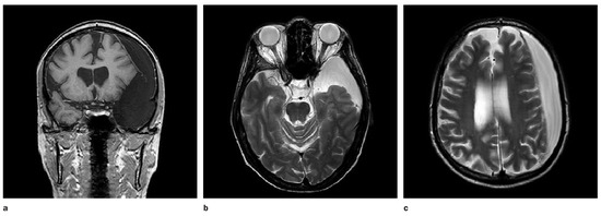
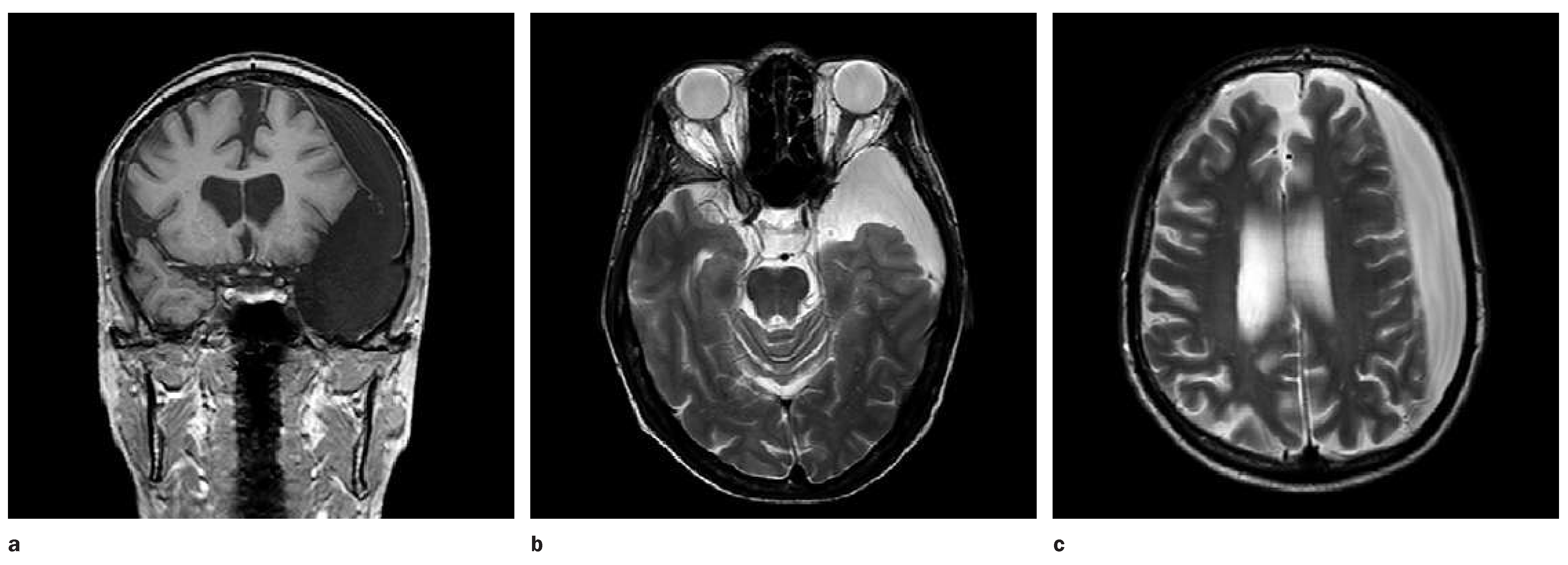
Ms A presented in reduced general health condition. Clinical and neurological examination revealed no focal finding, laboratory studies were all unremarkable. Since no cerebral imaging had been performed in the past, a MRI scan of the brain was carried out. Surprisingly, the MRI of the brain (Figure 1a–c) showed a previously unknown large left hemispheric arachnoid cyst with adjacent chronic subdural haematoma. It was located in front of the left temporal pole with extension to the insula cisterna. There was a midline shift but no other morphological signs of elevated intracranial pressure. The chronic subdural haematoma showed a consistent contrast agent acquisitioning membrane with fine septation. Overall both findings had a distinct left hemispheric space occupying effect with light subfaxical herniation and midline shift. Electroencephalography (EEG) was unremarkable. Neuropsychological testing performed at remission of the manic episode showed attention deficits such as impairment in different qualities of memory function, including short-term and working memory. In addition, the visual perception/ visual construction and reproduction of geometric patterns were impaired.
Figure 1.
MRI of the brain (coronal T1-weighted [T1w FFE; a] and axial T2-weighted [T2w TSE; b, c]): Large left hemispheric arachnoid cyst with adjacent chronic subdural haematoma.
The findings were discussed in an interdisciplinary panel including neurological and neurosurgical experts. However, the patient refused any surgical intervention.
The patient improved under valproic acid and atypical neuroleptic medication according to the national guidelines for bipolar disorders and psychosocial therapy. She was discharged after three months on valproic acid and quetiapin therapy to an ambulatory follow-up treatment.
Discussion
Increased prevalence of arachnoid cysts in psychiatric patients is not proof of an aetiological association with psychiatric syndromes. However, indications of a causal relationship come from neuroimaging studies, as well as case reports and systematic investigations.
Neuroimaging studies have shown that there might be functional reorganisation or displacement of cortical areas within the left hemisphere [7]. Thinning of cortical tissue in the vicinity of an arachnoid cyst was observed by Hund-Georgiadis et al. in 2002 [8]. Zaatreh et al. 2002 described that the temporal lobe adjacent to an arachnoid cyst is smaller and less metabolically active than the contra-lateral temporal region [9]. Furthermore, it has been shown that the surrounding cortical regions of an arachnoid cyst not only exhibit a reduced perfusion and metabolism but that these changes are reversible after neurosurgical treatment, paralleling an improvement in cognitive impairment in these patients [10,11,12]. Many case reports describe various psychiatric symptoms and cognitive problems in arachnoid cyst patients. Interestingly there are a few publications pointing to a probable association between cystic formations in the left temporal area and psychotic clinical presentation, which would be at least hypothetically conform with the family history of our patient [13,14,15]. The majority of reports on cyst decompression document a great improvement and even normalisation of symptoms and/or cognition. Strong evidence of a causal relationship was added from systematic, prospective studies in large series of arachnoid cyst patients. All studies documented impairment of a wide spectrum of cognitive functions after surgery [16].
The appropriate treatment of these patients is a matter of ongoing discussion. There is a general academic consent that focal neurological symptoms can justify the indication for neurosurgical treatment. However the recommendations for patients with or without a psychiatric history and the incidental finding of an arachnoid cyst remain vague. It might be reasonable to approach the therapeutic decision with a systematic work-up including a detailed past medical history, a neurological examination, a neurophysiological testing to reveal slight clinical symptoms, an EEG and (functional) MRI imaging. At present decision remains individual.
One can only speculate about the causal contribution of the cyst to our patient’s complaints. In this context, due to limited longitudinal data, we are left with the challenging question concerning time and mode of cyst genesis. Does it result from an early developmental event or is it acquired later on—and if the second is true, is it linked to the clinical onset of the affective disorder? However, since there is an elevated genetic predisposition for bipolar disorder due to the patient’s family history of schizophrenia [17], the aetiological importance of the cystic lesion in our case seems to be diminished. Arachnoid cyst patients appear to be prone to develop chronic subdural haematomas even after minor head trauma [18]. The only traumatic event in her past medical history was a cycling accident at the age of 26 without imaging at this time.
In conclusion, there is accumulating evidence that arachnoid cysts may cause cognitive and psychiatric symptoms and following this case report, there is a likelihood of parallel findings in bipolar patients. Symptoms are often reversible after cyst decompression. The difficulty, however, is to identify patients who will profit from surgical intervention. There are only vague recommendations for patients with apparent (focal) neurologic or psychiatric deficits, leaving all incidental findings associated with psychiatric diagnosis at the doctors individual judgement in terms of therapeutic implication.
In the future there is a need for more functional studies highlighting the relevance of cystic lesions in patients with psychiatric symptoms and cognitive problems, including patients with bipolar disorders, since at present only descriptive case reports are available. On this basis the discussion on surgical treatment in patients without apparent focal central deficits should be renewed.
Funding
This research received no external funding.
Conflicts of Interest
No financial support and no other potential conflict of interest relevant to this article was reported.
References
- Vernooij, M.W.; Ikram, M.A.; Tanghe, H.L.; Vincent, A.J.; Hofman, A.; et al. Incidental findings on brain MRI in the general population. N Engl J Med. 2007, 357, 1821–1828. [Google Scholar] [CrossRef] [Scilit] [PubMed]
- Gewirtz, G.; Squires-Wheeler, E.; Sharif, Z.; Honer, W.G. Results of computerised tomography during first admission for psychosis. Br J Psychiatry 1994, 164, 789–795. [Google Scholar] [CrossRef] [Scilit] [PubMed]
- Becker, T.; Lanczik, M.; Hofmann, E.; Müller, T.; Warmuth-Metz, M.; et al. Erhöhte Prävalenz von Arachnoidalzysten bei psychiatrischen Patienten: Eine retrospektive CTund MR-Untersuchung. Klin Neuroradiol. 1991, 1, 154–160. [Google Scholar]
- Arai, H.; Sato, K.; Wachi, A.; Okuda, O.; Takeda, N. Arachnoid cysts of the middle cranial fossa: experience with 77 patients who were treated with cystoperitoneal shunting. Neurosurgery 1996, 39, 1108–1112. [Google Scholar] [CrossRef] [Scilit] [PubMed]
- Westermaier, T.; Schweitzer, T.; Ernestus, R.I. Arachnoid cysts. Adv Exp Med Biol. 2012, 724, 37–50. [Google Scholar] [PubMed]
- Helland, C.A.; Lund-Johansen, M.; Wester, K. Location, sidedness, and sex distribution of intracranial arachnoid cysts in a population-based sample. J Neurosurg. 2010, 113, 934–939. [Google Scholar] [CrossRef] [Scilit] [PubMed]
- Stowe, L.A.; Go, K.G.; Pruim, K.; den Dunnen, W.; Meiners, L.C.; Paans, A.M. Language localization in cases of left temporal lobe arachnoid cyst: evidence against interhemispheric reorganization. Brain Lang. 2000, 75, 347–358. [Google Scholar] [CrossRef] [Scilit] [PubMed]
- Hund-Georgiadis, M.; Yves Von Cramon, D.; Kruggel, F.; Preul, C. Do quiescent arachnoid cysts alter CNS functional organization?: A fMRI and morphometric study. Neurology 2002, 59, 1935–1939. [Google Scholar] [CrossRef] [Scilit] [PubMed]
- Zaatreh, M.; Bates, E.R.; Hooper, S.R.; Palmer, G.; Elmenshawi, E.E.; Courvoisie, H.E.; Greenwood, R.S. Morphometric and neuropsychologic studies in children with arachnoid cysts. Pediatr Neurol. 2002, 26, 134–138. [Google Scholar] [CrossRef] [Scilit] [PubMed]
- Martínez-Lage, J.F.; Valentí, J.A.; Piqueras, C.; Ruiz-Espejo, A.M.; Román, F.; Nuño de la Rosa, J.A. Functional assessment of intracranial arachnoid cysts with TC99 m-HMPAO SPECT: a preliminary report. Childs Nerv Syst. 2006, 22, 1091–1097. [Google Scholar] [CrossRef] [Scilit]
- Sgouros, S.; Chapman, S. Congenital middle fossa arachnoid cysts may cause global brain ischaemia: a study with 99Tc-hexamethylpropyleneamineoxime single photon emission computerised tomography scans. Pediatr Neurosurg. 2001, 35, 188–194. [Google Scholar] [CrossRef] [Scilit] [PubMed]
- Tsurushima, H.; Harakuni, T.; Saito, A.; Tominaga, D.; Hyodo, A.; Yoshii, Y. Symptomatic arachnoid cyst of the left frontal convexity presenting with memory disturbance—case report. Neurol Med Chir (Tokyo) 2000, 40, 339–341. [Google Scholar] [CrossRef] [Scilit]
- Da Silva, J.A.; Alves, A.; Talina, M.; Carreiro, S.; Guimarães, J.; Xavier, M. Arachnoid cyst in a patient with psychosis: Case report. Ann Gen Psychiatry 2007, 6, 16. [Google Scholar] [CrossRef] [Scilit] [PubMed]
- Kuloglu, M.; Caykoylu, A.; Yilmaz, E.; Ekinci, O. A left temporal lobe arachnoid cyst in a patient with schizophrenia-like psychosis: a case report. Prog. Neuro-Psychopharmacol Biol. Psychiatry 2008, 32, 1353–1354. [Google Scholar] [CrossRef] [Scilit]
- Bahk, W.M.; Pae, C.U.; Chae, J.H.; Jun, T.Y.; Kim, K.S. A case of brief psychosis associated with an arachnoid cyst. Psychiatry Clin Neurosci. 2002, 56, 203–205. [Google Scholar] [CrossRef] [Scilit] [PubMed]
- Wester, K. Intracranial arachnoid cysts--do they impair mental functions? J. Neurol. 2008, 255, 1113–1120. [Google Scholar] [CrossRef] [Scilit] [PubMed]
- Gejman, P.V.; Sanders, A.R.; Kendler, K.S. Genetics of schizophrenia: new findings and challenges. Annu Rev. Genomics Hum Genet. 2011, 12, 121–144. [Google Scholar] [CrossRef] [Scilit] [PubMed]
- Gelabert-González, M.; Castro-Bouzas, D.; Arcos-Algaba, A.; Santín-Amo, J.M.; Díaz-Cabanas, L.; et al. Chronic subdural hematoma associated with arachnoid cyst. Report of 12 cases. Neurocirugia (Astur) 2010, 21, 222–227. [Google Scholar] [CrossRef] [Scilit]
This is an open-access article distributed under the terms of the Creative Commons Attribution Non-Commercial No Derivatives (CC BY-NC-ND) 4.0 License.