


Summary
Background: Palinopsia is the persistence of visual images after removal of the exciting stimulus. It is commonly caused by occipital epileptic stati, focal cerebral lesions, migraine. Case: A 28-year-old man experienced two episodes of acute headache with negative scotoma and palinopsia. All symptoms recovered spontaneously after one hour but palinopsia still persisted. The MRI showed a cavernous haemangioma in the left occipital lobe and the EEG showed no sign of epileptic activity, but this data did not exclude an epilepsy. The fluctuating manner and stereotypy of the symptom was, in fact, attributed to an epileptic aetiology and palinopsia disappeared after initiation of antiepileptic therapy. This study addresses the relevance of properly considering a visual symptom which can occur after occipital lesions and which can be easily missed or misinterpreted as a symptom belonging to a migraine with aura.
Introduction
Palinopsia (from Greek πάλιν, again, and ὄψις, vision) is the persistence or recurrence of visual images after removal of the exciting stimulus [1,2]. Several pathologies can cause palinopsia such as seizures [3], focal cerebral lesions [4], migraine [5], multiple sclerosis [4], Charles Bonnet syndrome [4], and psychiatric disorders [4]. Occurrence of palinopsia during treatment with psychoactive drugs [6,7,8,9] and topiramate [10] have also been reported. Palinopsia typically localises in the occipito-parieto-temporal cortex of the non-dominant hemisphere [11,12]: fMRI studies have suggested that it may be caused by an increase of activity (foci epileptici, cerebral hyperperfusion adjacent to areas of cortical damage) [13] in the parietal projections of the dorsal pathway from the occipital lobe; this hypothesis is supported by the typical localisation of the phenomenon in the peripheral visual field, organisational feature of the dorsal projections [14,15]. Usually there is no emotional response or confusion accompanying the phenomenon.
Case report
We present the case of a 28-year-old man who experienced an episode of acute cephalalgia in the left occipital region with no photophobia, phonophobia, osmophobia nor nausea, but with a progressive loss of vision (an initially punctiform scotoma spreading to the right part of visual field) and a peculiar visual symptom described as the persistence of objects or anatomical details in the visual field lasting a few minutes after looking away, referrable to palinopsia (for example the patient kept on seeing the scalp of a friend he was talking to after looking away from him). The attack of cephalalgia lasted about one hour, then disappeared with spontaneous and full regression of pain and negative scotoma. By contrast, the palinopsia persisted in a fluctuating manner and no triggers were reported. No further diseases were reported. Palinopsia was initially interpreted by the family doctor as the first episode of migraine with aura and, therefore, the patient was treated with aspirin, without benefit.
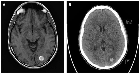
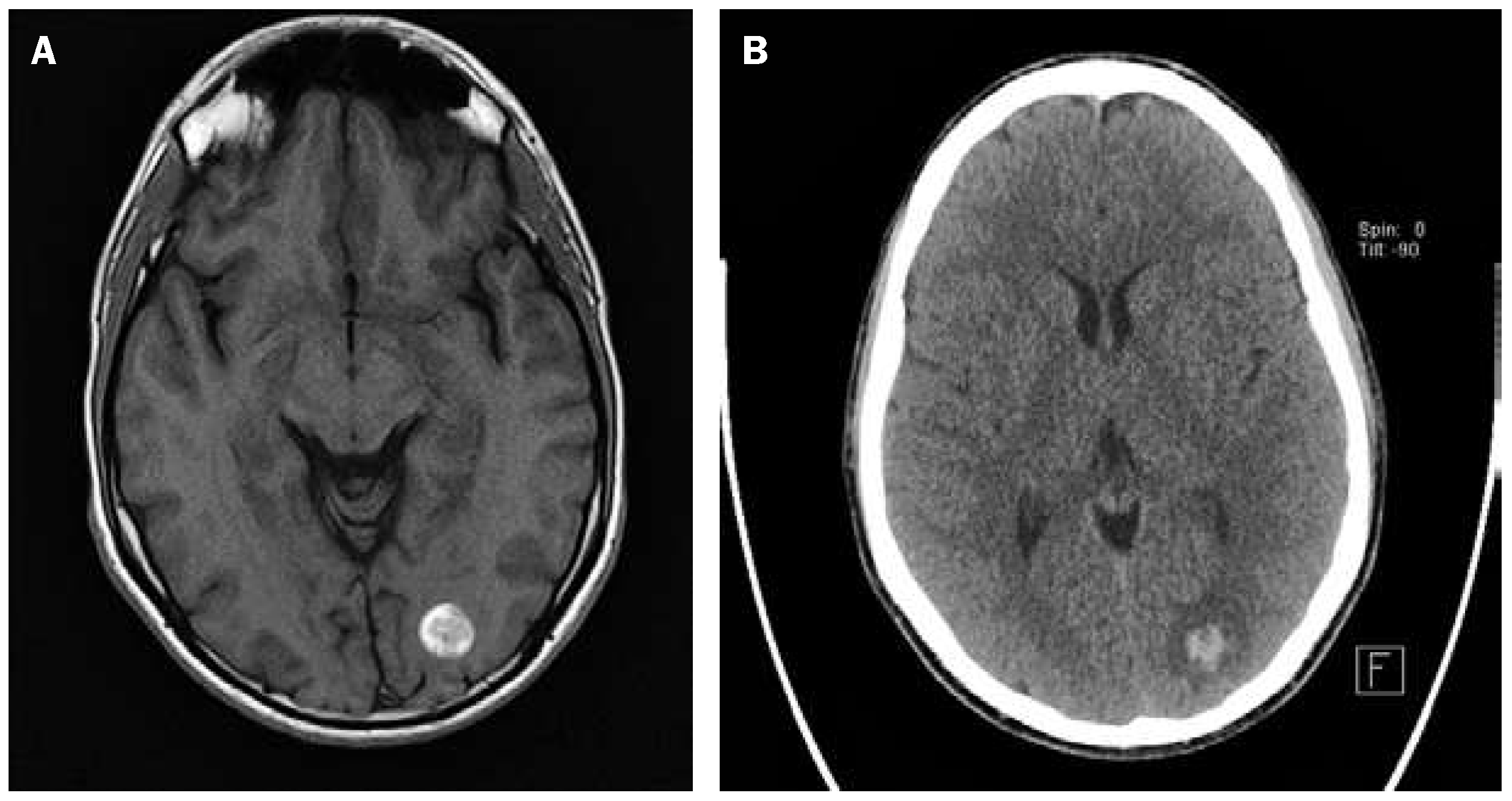
Five days later, another cephalalgia occurred, so the patient was admitted to the emergency room and he underwent both cerebral CT and MRI (fig. 1), which showed the presence of a cavernous haemangioma in the left occipital lobe under the calcarine sulcus, with a subacute haemorrhage. No alteration at the angio MRI was found. So, the patient was admitted to the neurological unit. During his hospitalisation the neurological examination was normal (in particular no sign of visual field deficit was reported) and no signs of agnosia emerged in tasks assessing visual perception (Montréal-Toulouse battery for visual agnosia) [16]. The only symptom that lasted in a fluctuating manner during the hospitalisation was a persistent vision of objects or faces after the patient looked away from the original image. The EEG did not show epileptic alteration and it was also performed during the manifestation of the symptoms. Despite this data we started a therapy with levetiracetam 1000 mg/die and the symptom disappeared after a week.
Figure 1.
MRI (A) and CT (B) scan of the brain showing the occipital cavernous haemangioma.
Discussion
This case focused our attention on the problem related to the comprehension of the cause of the palinopsia and the importance of researching it in the patient history, although rarely occuring.
Although the bleeding of the cavernoma justified the headache and the scotoma as a lesional symptom, the persistence of palinopsia in a fluctuating manner between the attacks, evoked the possibility of a simple partial epilepsy. A long duration EEG, however, revealed no signs of epileptic activity. An EEG with surface electrodes is not very sensitive for epileptic activity in deep cortical structures. Therefore an EEG showing no epileptic activity makes the diagnosis of partial seizures less probable, yet does not exclude them. Furthermore, the fluctuation and the stereotypic positive symptoms are in favour of a diagnosis of partial seizure than of a deficit caused by oedema or by haemorrhage.
Referring in particular to migraine with aura the components that make this possibility less likely were the simultaneous occurrence of headache and scotoma at the onset and the duration of the migraine with aura, which typically develops gradually over ≥5 minutes lasting no more than 60 minutes usually preceding the attacks. Another issue that reduces the possibility of migraine with aura is the simultaneous occurrence of headache and scotoma at the onset and the fluctuation of this visual symptom.
In conclusion this study addresses the relevance of properly considering a visual symptom like palinopsia: it can occur after occipital lesions and it can be easily missed or misinterpreted as a symptom belonging to a migraine with aura.
Funding / potential competing interests
No financial support and no other potential conflict of interest relevant to this article were reported.
References
- Pötzl, O. Über Palinopsia. Wien Z Nerv. 1954, 8, 161–86. [Google Scholar]
- Meadows, JC; Munro, SS. Palinopsia. J Neurol Neurosurg Psychiatry 1977, 40, 5–8. [Google Scholar] [CrossRef] [Scilit] [PubMed]
- Ossola, M; et al. Epileptic mechanisms in Charles Bonnet syndrome. Epilepsy Behav. 2010, 18(1–2), 119–22. [Google Scholar] [CrossRef] [Scilit] [PubMed]
- Evans, RW. Reversible palinopsia and the Alice in Wonderland syndrome associated with topiramate use in migraineurs. Headache 2006, 46(5), 815–8. [Google Scholar] [CrossRef] [Scilit] [PubMed]
- Belcastro, V; et al. Palinopsia in patients with migraine: a case-control study. Cephalalgia 2011, 31(9), 999–1004. [Google Scholar] [CrossRef] [Scilit] [PubMed]
- Hughes, MS; Lessell, S. Trazodone-induced palinopsia. Arch Ophthalmol. 1990, 108(3), 399–400. [Google Scholar] [CrossRef] [Scilit] [PubMed]
- Ihde-Scholl, T; Jefferson, JV. Mitrazapine-associated palinopsia. J Clin Psychiatry 2001, 62, 373. [Google Scholar] [CrossRef] [Scilit] [PubMed]
- Hori, H; Terao, T; Nakamura, J. Visual perseveration: a new side effect of maprotiline. Acta Psychiatr Scand. 2000, 101(6), 476–7. [Google Scholar] [CrossRef] [Scilit] [PubMed]
- Faber, RA; Benzick, JM. Nafazodone-induced palinopsia. J Clin Psychopharmacol. 2000, 20(2), 275–6. [Google Scholar] [CrossRef] [Scilit] [PubMed]
- Fontenelle, LF. Topiramate-induced palinopsia. J Neuropsychiatry Clin Neurosci. 2008, 20, 249–50. [Google Scholar] [CrossRef]
- Norton, JW; Corbett, JJ. Visual perceptual abnormalities: hallucinations and illusions. Semin Neurol. 2000, 20, 111–21. [Google Scholar] [CrossRef] [Scilit]
- Santhouse, AM; et al. The functional significance of perinatal corpus callosum damage: an fMRI study in young adults. Brain 2002, 125(8), 1782–92. [Google Scholar] [CrossRef] [Scilit]
- Hayashi, R; et al. Palinopsia and perilesional hyperperfusion following subcortical hemorrhage. Acta Neurol Scand. 2002, 105(3), 228–31. [Google Scholar] [CrossRef] [Scilit] [PubMed]
- Baizer, JS; Ungerleider, LG; Desimone, R. Organization of visual inputs to the inferior temporal and posterior parietal cortex in macaques. J Neurosci. 1991, 11(1), 168–90. [Google Scholar] [CrossRef] [Scilit] [PubMed]
- Santhouse, AM; Howard, RJ; Ffytche, DH. Visual hallucinatory syndromes and the anatomy of the visual brain. Brain 2000, 123(10), 2055–64. [Google Scholar] [CrossRef] [Scilit] [PubMed]
- Agniel A, Joanette Y, Doyon B, Duchein C. Protocole Montréal–Toulouse. Evaluation des gnosies visuelles et auditives. Paris: L’Ortho-Edition; 1992.
This is an open-access article distributed under the terms of the Creative Commons Attribution Non-Commercial No Derivatives (CC BY-NC-ND) 4.0 License.