


Abstract
Fluri F, Lyrer PA, Steck AJ, Engelter S. Internal carotid artery dissection with occlusion during heparin therapy. Schweiz Arch Neurol Psychiatr. 2007;158:331–3. Internal carotid artery dissection (ICAD) is frequently treated with anticoagulation. Objections against anticoagulation are based on solely theoretical concerns of progressive intramural haematoma.We describe a patient with progressive stenoocclusion of the internal carotid artery (ICA) due to intramural haematoma as a possible complication of immediate heparin treatment in ICAD. Heparin dosage was based on estimates of body weight due to aphasia. The later determined body mass index (15 kg/m2) showed that the initial dosage was 20% too high. Follow-up neurosonology disclosed haematoma-related ICA occlusion. Thus, in ICAD heparin treatment bears the risk of overanticoagulation leading to progressive mural haematoma, especially in low-weight patients.
Introduction
Dissection of the extracranial internal carotid artery (ICAD) is frequently treated with anticoagulation [1]. This regimen has been questioned [2,3]. Objections against anticoagulation are based on the general risk of intracranial haemorrhage [4] and the theoretical concern of worsening the mural haematoma [5]. Published reports about such complications are sparse. We report on an ICAD patient who suffered from progressive stenoocclusion of the internal carotid artery (ICA) associated with immediate heparinisation.
Case report
A 53-year-old woman presented with a oneday history of left-sided headache, followed by right-sided weakness and speech disturbances for 8 hours. Vascular risk factors were absent.
The neurological examination at admission revealed a Horner’s syndrome on the left, a mild hemiparesis on the right side and a severe aphasia (NIH-Stroke scale score equals 6).
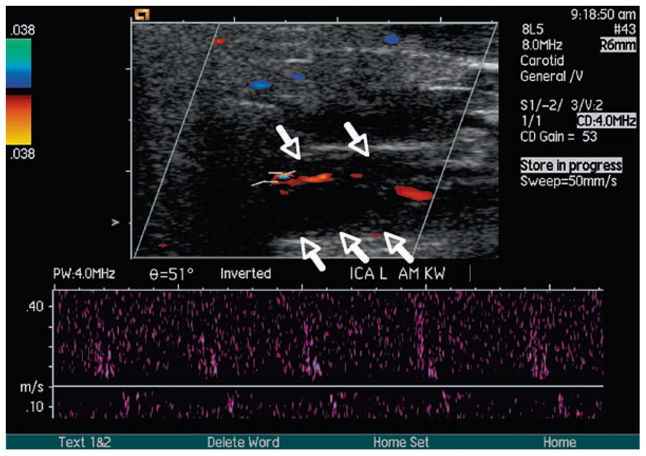
The clinically assumed acute stroke due to left extracranial ICAD was verified by magnetic resonance imaging (MRI) and neurosonology findings. Diffusion-weighted MRI visualised acute ischaemic lesions within the middle cerebral artery territory (not shown). MRI of the neck (not shown) showed intramural haematoma. The haematoma was also visible by colour-coded duplex sonography (fig. 1A) which showed that the haematoma compromised the lumen of the ICA causing an at least moderate-grade stenosis. Signs of atherosclerosis were absent. Transcranial Doppler sonography of the anterior circulation was normal.
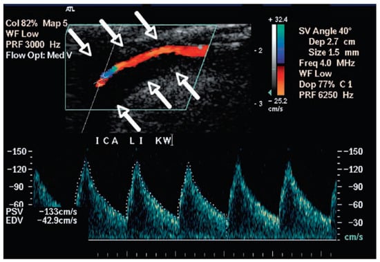
Figure 1A.
Internal carotid artery dissection with heparin-associated occlusion during follow-up.
Partial thrombin time (33 s; norm 25–40), INR (0.9; norm <1.1), platelet count (165 × 109/l; norm 150–450), creatinine level (48 mmol/l; norm 45–93) and liver function tests were normal. History and clinical examination showed no evidence for a connective tissue disorder or an underlying disposition to bleedings.
The patient received intravenous heparin without bolus (16 800 IU/24 h). Heparin dosage was based on estimates of the body weight (approximately 45–50 kg, i.e., 350 IU/kg/24 h) due to severe aphasia of the patient and absence of relatives. Further heparin treatment was titrated using thrombin time as monitoring parameter. Control after 6 hours indicated that anticoagulation was above the therapeutic range (i.e., thrombin time-2 >120 s; norm 4–10 s). Heparin was reduced to 12 000 IU/24 h. On day 3 and 4, anticoagulation was again slightly above the target range (i.e., thrombin time-2 was 14 and 12 s respectively rather than 4–10 s). In the meantime weighing of the patient revealed that the real weight was 38 kg (BMI: 15 (norm 18.5–24.9) kg/m2). During this period creatinine level and platelet count were still in the normal range.
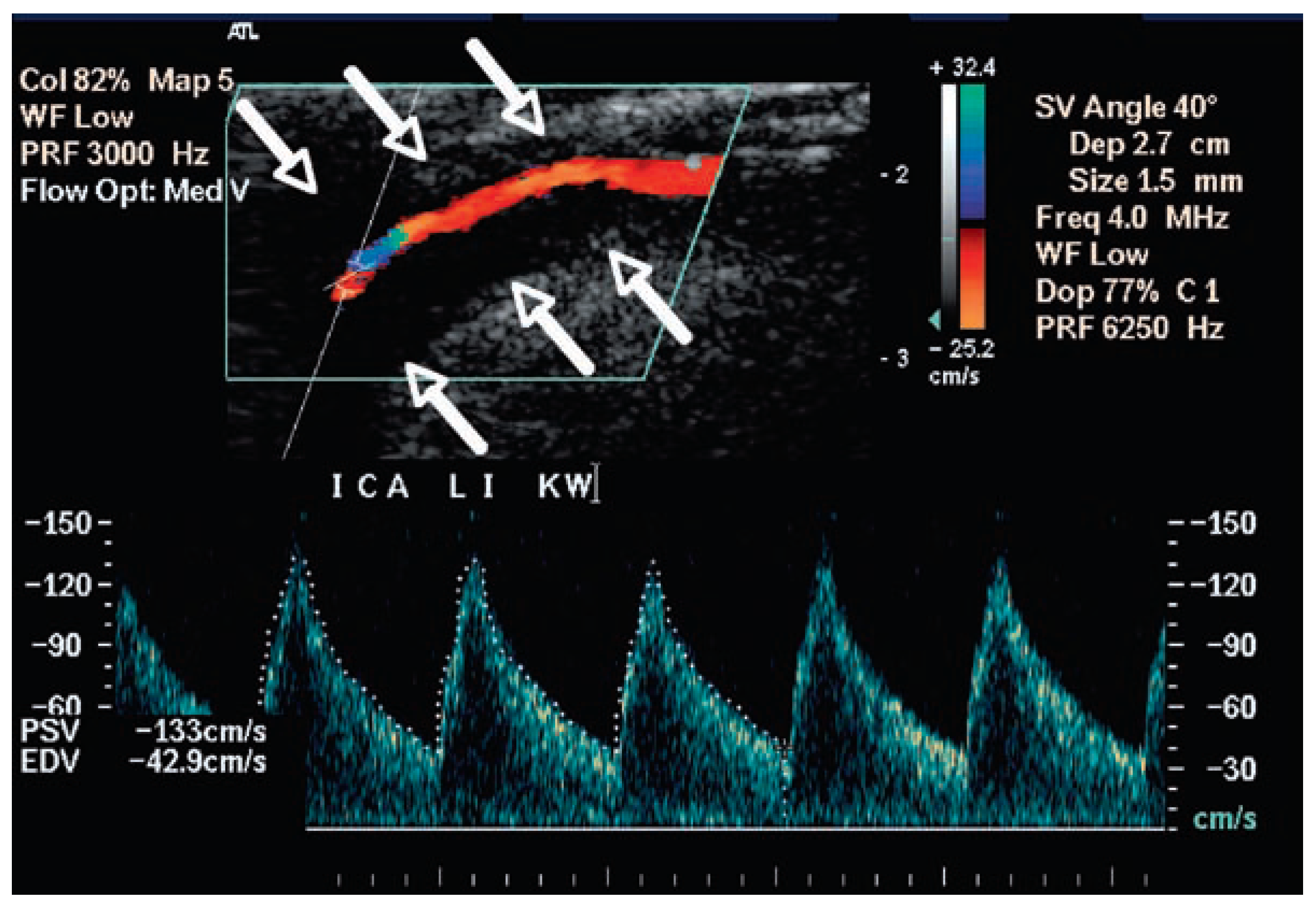
On day 7, follow-up duplex sonography (fig. 1B) revealed a newly occluded ICA with persisting large intramural haematoma. A bidirectional flow pattern (Doppler spectrum fig. 2) indicated that the occlusion was located distally. Transcranial Doppler sonography revealed a marked right-to- left cross-flow via anterior communicating artery.
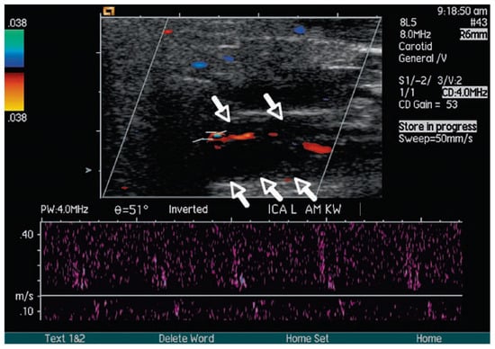
Figure 1B.
A 53-year-old patient with extracranial internal artery dissection on the left. Colour-coded duplex sonography shows circular hypoechogenicity indicating acute intramural haematoma (arrows) on day 1 (A) and on day 7 (B). While the left internal carotid artery had (at least) a moderategrade stenosis on day 1 (A), it was distally occluded on day 7 (B), as indicated by the bidirectional flow pattern (Doppler spectrum).
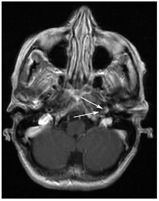
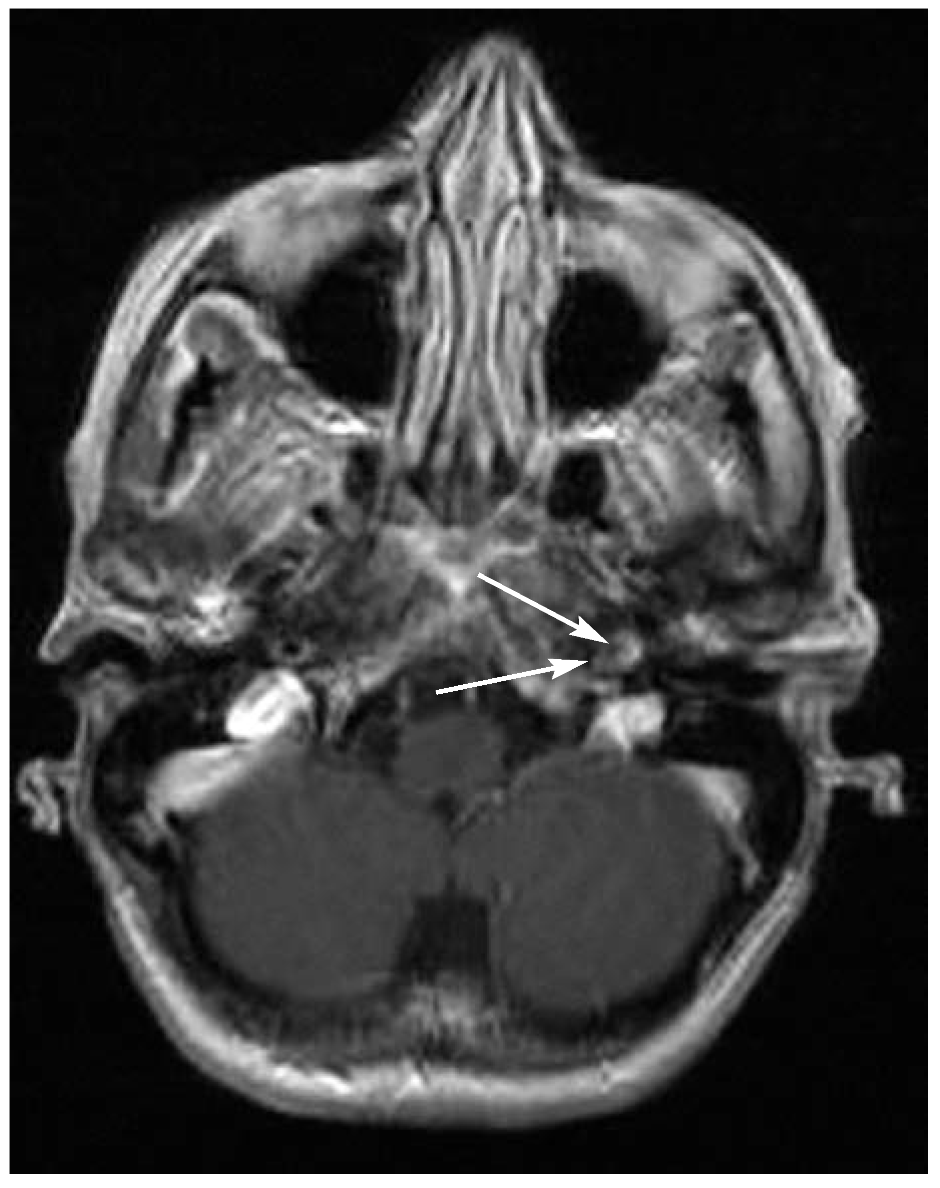
Figure 2.
T1-weighted MRI shows intramural haematoma of the left internal carotid artery (arrows).
Neither clinical examination nor follow-up brain MRI including diffusion-weighted sequences showed evidence for recurrent infarction.
Discussion and conclusion
In this ICAD patient we observed ICA occlusion during intravenous heparin treatment. Though ICAD-triggered intraluminal thrombus formation as differential diagnosis is not definitively excluded, the most likely reason for our observation is enlargement of the intramural haematoma. A causal relationship between heparin treatment and worsening of the haemodynamics is at least possible, indicating that heparin-associated enlargement of the intramural bleeding with progression of haemodynamic compromise is not a merely theoretical risk [5]. Clinically, the occlusion remained asymptomatic, probably because of a sufficient intracranial collateralisation. In addition, our case report illustrates that overheparinisation is a risk especially in patients in whom heparin dosage is based on estimates of the body weight, e.g., because of severe aphasia and absence of relatives. As a result of the wrong weight estimates the initial heparin dosage was about 20% higher than recommended. Other reasons for overheparinisation such as renal insufficiency or thrombocytopenia were not present in our case. Nevertheless, our case should not be interpreted as evidence that heparin is harmful in ICAD patients in general. Such a conclusion is inappropriate taking into account that even thrombolysis with rtPA has been reported safe in ICAD patients [6]. Though, our observation urges to cautiousness; as practical consequences measuring coagulation parameter should be thought about after 3 hours rather than after 6 hours. In addition, weighing patients rather than estimating the body weight should be considered in patients in whom body weight is not known.
In our ICAD patient carotid occlusion during heparin therapy was clinically asymptomatic, while another ICAD patient in a similar situation suffered from a watershed infarct [7]. In contrast to our case, this patient had no anterior and posterior communicating arteries, prompting haemodynamic stroke due to lacking collaterals. Our observation shows that occlusion of the dissected carotid artery can be asymptomatic if collateralisation is sufficient.
The BMI of our patient was low (15 [norm 18.5–24.9] kg/m2). Low BMI has been shown to be a risk factor for intraparenchymal haemorrhage [8]. Our case suggests that ICAD patients with low BMI are at risk to suffer from progression of intramural bleeding.
Both findings indicate that anticoagulation might be harmful for some individual ICAD patients, especially for those with insufficient collateralisation of the circle of Willis. Furthermore, these observations raise the question whether all ICAD patients should receive immediate anticoagulation. Such treatment is recommended by experts [1], though its efficacy is unproven so far [2,9]. A controlled randomised trial comparing benefits and risks of antiplatelets versus anticoagulants is needed. In the UK such a pilot trial (i.e., CADISS) has indeed started (see www. sgul.ac.uk/index.cfm?27D0604F-9A88-39E4-033B-B68CD9284AB6). For UK centres participation in this trial is recommended. For others, antithrombotic treatment decisions may be tailored individually until more evidence is available [10].
Learning points
- In ICAD patients immediate heparin treat- ment bears the risk of overanticoagulation leading to progressive mural haematoma.
- ICAD patients with low BMI may be at a particular risk of progressive intramural bleeding.
- Antithrombotic treatment decisions in ICAD patients should be tailored individually balancing potential risks and benefits until more evidence is available.
References
- Norris JW. Extracranial arterial dissection: anticoagulation is the treatment of choice: for. Stroke. 2005;36(9):2041–2.
- Lyrer P, Engelter S. Antithrombotic drugs for carotid artery dissection. Cochrane Database Syst Rev. 2003;(3):CD000255.
- Lyrer PA. Extracranial arterial dissection: anticoagulation is the treatment of choice: against. Stroke. 2005;36(9):2042–3.
- International Stroke Trial Collaborative Group. The International Stroke Trial (IST): a randomized trial of aspirin, subcutaneous heparin, both or neither among 19 435 patients with acute ischemic stroke. Lancet. 1997;349:1569–81.
- Touze E, Gauvrit JY, Meder JF, Mas JL. Prognosis of cervical artery dissection. In: Baumgartner RW, Bogousslavsky J, Caso V, Paciaroni M, editors. Handbook on Cerebral Artery Dissection. Basel: Karger; 2005. p. 129–39.
- Georgiadis D, Lanczik O, Schwab S, Engelter S, Arnold M, Sieber M, et al. IV thrombolysis in patients with acute stroke due to spontaneous carotid dissection. Neurology. 2005;64:1612–4.
- Dreier JP, Lurtzing F, Kappmeier M, Bohner G, Klingebiel R, Leistner S, et al. Delayed occlusion after internal carotid artery dissection under heparin. Cerebrovasc Dis. 2004;18(4):296–303.
- Cui R, Iso H, Toyoshima H, Date C, Yamamoto A, Kikuchi S, et al. Body mass index and mortality from cardiovascular disease among Japanese men and women: the JACC study. Stroke. 2005;36:1377–82.
- Schievink WI. Spontaneous dissection of the carotid and vertebral arteries. N Engl J Med. 2001;344(12):898–906.
- Engelter S, Lyrer PA. Antithrombotic therapy for cervical artery dissection. In: Baumgartner RW, Bogousslavsky J, Caso V, Paciaroni M, editors. Handbook on Cerebral Artery Dissection. Basel: Karger; 2005. p. 147–59.
This is an open-access article distributed under the terms of the Creative Commons Attribution Non-Commercial No Derivatives (CC BY-NC-ND) 4.0 License.