


Abstract
Dreaming is the mental activity of REM (and less commonly NREM) sleep. It is characterised by a perception of self, inner and outer world that is distinct from that of wakefulness. Neurophysiological and neuroimaging studies suggest that dreaming arises from activation of brainstem, thalamo-limbic and medial temporo-occipital areas and inactivation of dorsolateral prefrontal areas. Dream abnormalities in neurological disorders have poorly been studied. They include a reduction/cessation of dreaming (Charcot-Wilbrand syndrome),an increase in dream frequency, changes in dream contents and accompaniments (including REM sleep behaviour disorder) and the occurrence of dream-like experiences (hallucinations) during wakefulness and wake-sleep/sleep-wake transitions. Dream changes in association with neurological conditions including primary sleep disorders (e.g. narcolepsy, REM sleep behaviour disorder [RBD], nightmares), stroke and Parkinson’s syndrome are reviewed. Assessment of altered dreaming and dreamlike experiences can be of clinical significance because of their diagnostic and therapeutic implications (RBD, hallucinations), prognostic significance (hallucinations) and distress that they can have for patients and their bed partners (e.g. nightmares, hallucinations, RBD). In addition, systematic studies of dream and dream-like experiences are expected to offer unique insights into the brain, mind and emotions of neurological patients.
Introduction
- Dream theories before the discovery of REM sleep in 1953
In ancient culture dreams were often considered to be of supernatural source and, while allowing a direct communication with the gods, capable of providing hints of the coming future (prophetic and premonitory dreams). Among Egyptians (the oldest known book about dreams dates back to the 12th dynasty, 2000 BC), Greeks and Romans dream interpreters, working in such places as the oracles of Delphi, were appreciated and powerful. Natural theories of dreaming were less common. Hippocrates and particularly Aristotle (3–4th century BC) recognised psychological and physiological elements of dreaming.Aristotle differentiated between dreaming and dream recall and asked, “whether the truth is that sleepers always dream but not always remember their dreams”. Lucretius (1st century BC) observed the twitching leg movements of a sleeping dog and linked them to a presumed ongoing dream about a chase. In the 2nd century Artemidorus of Daldis made in his Onirocriticon a detailed description and a first classification of dreams, supporting the idea of a premonitory value of dreaming. Galen wrote in his Diagnosis from Dreams that dreams can have a premonitory character (“somnia”) but also reflect natural processes, changes in the body “humours” (“insomnia”).
Natural and supernatural theories of dreaming were often discussed in combination in the Middle Ages. Augustine recognised the existence of corporal, intellectual and spirital sources of dreams. He as well as other Christian thinkers, beginning with Tertullian and Isidore of Seville (author of De tentamentis somniorum), condemned the practice of dream interpretation because of the difficulty in differentiating between internal and external, as well as corporeal and noncorporeal contents and sources of dreams (revelatio divina vs illusio demonica) [1].
Gerolamo Cardano (1501–1576), while strongly referring to Artemidorus’ theories, further elaborated laic and scientific views of dreaming. He described what are now called the lucid dreams (“rendersi conto di stare sognando indica forza mentale”, “vegliare nel sonno”) [2]. The first experimental works on dreaming began in France in the second half of the 19th century. Alfred Maury (1817–1892) systematically recorded his dreams after having been awakened by his assistant. He found that the presentation of specific sensory, visual or acoustic stimuli during sleep tended to be incorporated into his dreams. He was also among the first, in 1848, to describe sleep onset (hypnagogic) hallucinations. Hervey de Saint-Denis (1822– 1892) filled a dream journal of 22 volumes, becoming soon a “lucid dreamer”.Both,Maury and Saint-Denis, were convinced that dreams arise from external stimuli leading to a partial awakening during sleep.Hermann von Helmholtz (1821–1894) speculated that the perception of movements during dreams is related to an activation of the motor cortex during sleep. A similar concept was also put forward by his pupil Wilhelm Wundt (1832–1920), the founder of experimental psychology, who recognised in dreams the coexistence of excitation (“autoactivation” of sensory and particularly visual cortices) and inhibition (of “volition” and “attention”) of cortical brain areas. Maria Manasseina (1843–1903), one of the first Russian woman doctors, wrote in her monography that during sleep the brain is in an active state. She regarded dreams as evidence for an ongoing psychic life of sleep generated by the brain [3]. Mary Calkins (1863–1930) performed the first quantitative analysis of dream contents pointing to the preponderance of visual experiences [4].
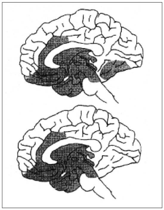
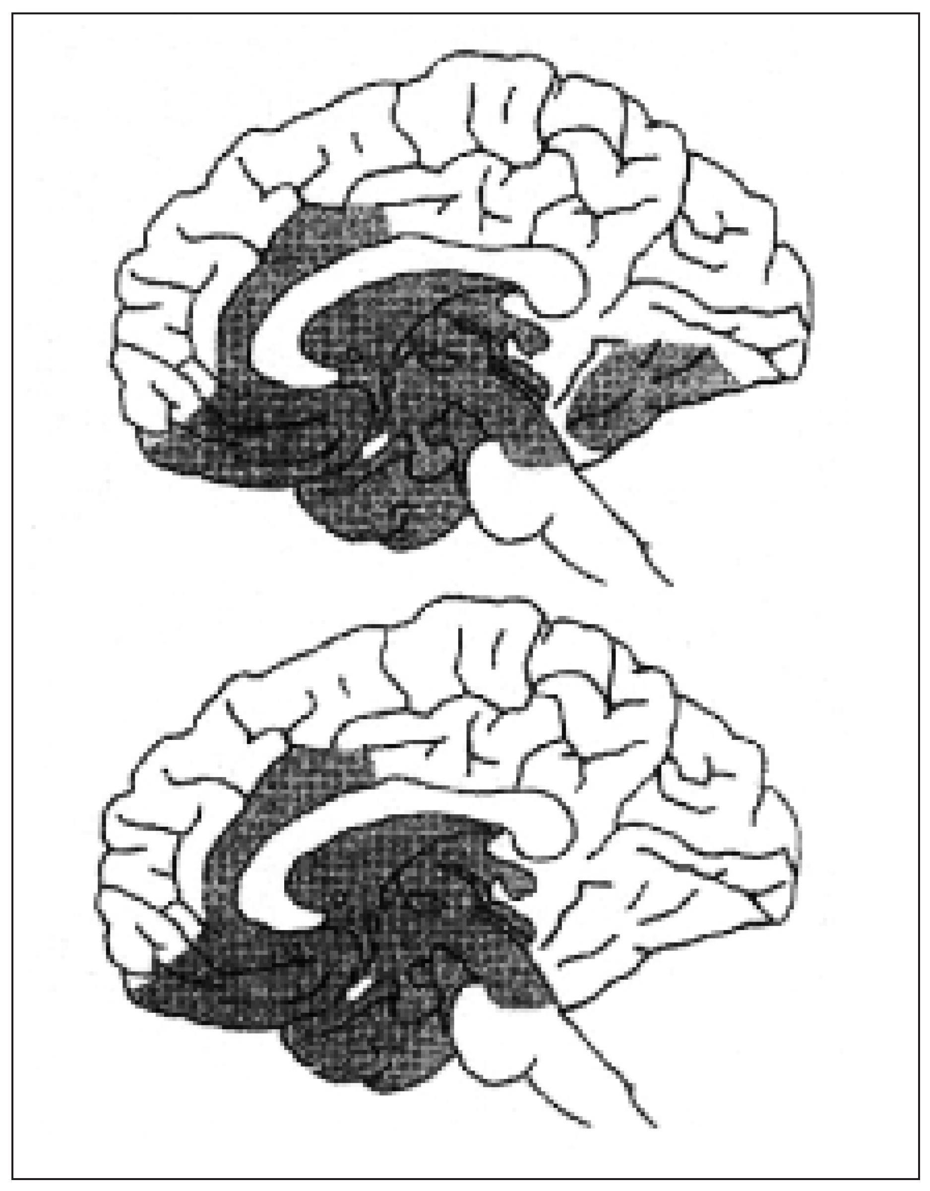
Freud’s contribution to dream theories is presented in other articles in this special issue. He was right, also in light of recent discoveries on the neurobiology of dreams (see below and fig. 1), in pointing to an analogy between the dreaming brain and the emotional brain and in stressing the importance of memory traces in determining the contents of dreams.
Figure 1.
The dreaming brain, as estimated by modern neuroimaging studies, includes the pontine tegmentum, thalamus, limbic/ paralimbic structures (including the amygdala) and temporooccipital areas (with permission from: M. Solms, O. Turnbull. Il cervello e il mondo interno. Milano: Raffello Cortina Editore; 2002).
The first all-night observations of sleeping humans were made by the French physician Nicholas Vaschide (1874–1907), who noted periodic motor activation in association with dreaming.He also described alternating periods of vasoconstriction and vasodilatation throughout the night and changing periods with good and poor dream recall at sleep interruption [5]. De Sanctis 1899 and de Lisi in the 1930s also described the typical limb twitches of REM sleep and linked them with dream experiences. In 1944 sleep-related periodic penile erections were described by Ohlmeyer et al. [6]. Following the first recordings of the EEG by Hans Berger, several authors including Loomis et al. noted in the 1930s that sleep is associated with a slowing of the electroencephalographic activity [7]. In 1953 Hess, Koella and Akert described the activated EEG of REM sleep in the cat, giving for this observation – in the absence of data on eye movements and muscle tone –, however, the wrong interpretation [8].
- REM sleep and its brainstem control: the “REM-dream equation”
In the search of an objective measure of the process of falling asleep and motivated by an article on blinking in the wake-sleep transition [9], in 1953 Kleitman and Aserinsky described the presence of rapid eye movements (REMs) in deep sleep [10,11]. They observed that REMs appear approximately 90 minutes after sleep onset, are associated with a desynchronised cortical low-voltage fast EEG activity and a significant increase and instability of heart and respiratory rates, and occur at regular intervals throughout the whole night. To test the hypothesis that REM sleep may be linked with dreaming, 10 probands were awakened during periods with and without REMs. Vivid dreams were reported in 74% when awakened during periods with REMs, but only in 17% when awakened in the absence of REMs.
The neurosurgeon and neurophysiologist Jouvet discovered muscle atonia of REM sleep in 1959 [12]. The combination of REMs, activated EEG and muscle atonia suggested the term “paradoxical sleep” as synonym of REM-sleep. Jouvet demonstrated also that the generation of REM sleep depends on an intact pontine tegmentum [13] and that REM atonia is due to an inhibition of motor centres in the medulla oblongata. Cats with lesions around the locus pericoeruleus exhibit during REM sleep a variety of complex behaviours including motor patterns suggesting attack, defence and exploration [14].
Another co-worker of Kleitman, Dement, further elaborated on the relationship between REM sleep and dreaming. He was convinced that dreams only occur during REM sleep and suggested a direct connection between dream content, REMs and motor activities during REM sleep [15,16]. In line with Freud’s theory he also suggested, based on a REM-sleep deprivation study (which was subsequently going to be strongly criticised), that REM sleep was essential for the psychological health [17].
In the 1970s, following the pioneer work of Jouvet, Hobson et al. further elucidated the brainstem neurophysiologic and neurochemical machinery of REM sleep.They described cholinergic “REM-on” neurons and monoaminergic (noradrenaline and serotonin-producing) “REM-off” neurons in the pontine tegmentum, and REM sleep as the results of their reciprocal interaction [18]. These authors developed a new dream theory called the “activation-synthesis hypothesis”, according to which dreams are the result of a random/nonspecific activation provided by the brainstem, and a subsequent, more or less coherent interpretation (synthesis) by the forebrain [19].
- NREM sleep and dreaming
Already the awakening studies of Aserinski and Kleitman have shown that subjects even when awakened from NREM sleep can report dreams or dream-like experiences. This observation was confirmed by several other authors and consequently challenged the “REM-dream equation” [20,21,22]. The persistence of dream experiences despite complete pharmacological REM-sleep suppression gives further support for the existence of a dream-like mentation in NREM sleep [23].
Dream reports from patients awakened from REM sleep are more frequent, more real, more emotional and more bizarre, and their content is more often visual and/or motor [24,25].
- Brain metabolism/activation during sleep and dreaming
Neuroimaging studies with PET, SPECT and fMRI [26,27,28,29] have shown, in line with neurophysiological observations, different patterns of brain activation during the different states of being (wakefulness, NREM sleep, REM sleep). In most studies a deactivation of the dorsolateral prefrontal cortex and associative parietal areas during both NREM and REM sleep, and an activation of pontine tegmentum, thalamus, limbic/paralimbic structures (including the amygdala) and temporooccipital areas (other than primary visual cortex) during REM sleep were reported (see the article by Dang-Vu et al. in this issue for further details) (fig. 1).
These profiles of activation and deactivation are thought to explain the characteristics of mental activity during wakefulness, sleep onset,NREM and REM sleep. Deactivation of the prefrontal cortex may explain the lack of insight and the decrease in volitional control typical of oneiric experiences in general. Activation of the limbic/ paralimbic structures (including the amygdala and the anterior cingulum and orbito-frontal cortex) and tempo-occipital cortices, on the other hand, could explain the strong emotional and visual character of REM-sleep dreaming and the frequent translation of new and old memories into dreams (see the article by Schwartz et al. in this issue for further details). The sleep-associated functional connectivity (coherent neuronal firing) of neurons within the mesio-temporal lobe may underlie the variable ability – in normal subjects and possibly also in patients – to recall dream experiences [30].
The exact profile of brain activation (and deactivation) underlying the different mental activities across the states of being (thinking, day dreaming, sleep-onset hallucinations, NREM and REM dreams, sleep-end hallucinations) remains essentially unknown at this point.
Dreaming in neurological patients
The frequency and extent of dream changes in neurological patients has rarely been assessed in a systematic fashion. The available data in the literature suggest nevertheless a relatively high frequency of altered dreaming in neurological disorders.
The brain mechanisms involved in dream changes are, with the exception of REM-sleep behaviour (see below), essentially unknown. Neurochemical (Cholinergic and monaminergic (e.g. dopaminergic, serotoninergic, noradrenergic) transmissions, which are known to regulate sleep-wake functions, are probably implicated also in the modulation of dream experiences.) and/or structural changes in the above-mentioned thalamo-limbic-temporo-occipital “dreaming circuits” can generally be postulated. In many instances clinical symptomatology can be understood as a “dissociated state” arising from the loss of the physiological boundary between the different states of being (wakefulness, NREM and REM sleep).
The exact pathophysiology is expected to vary according to the specific form of altered dreaming. In patients with narcolepsy and Parkinson’s disease visual hallucinations may represent, at least on some occasions, the intrusion of REMsleep mentation into wakefulness [31]. In “out-ofbody experiences” a dysfunction of body- and selfprocessing at the temporo-parietal junction has been suggested [32].
In addition to brain dysfunction per se, other factors such as age, personality, mood, current physical and psychic status, stress, fever, medications (e.g. antidepressants, beta-blockers, acetylcholinesterase inhibitors), alcohol and presence of sleep disorders may influence dreaming in neurological patients [33].
- Decreased/cessation of dreaming
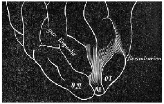
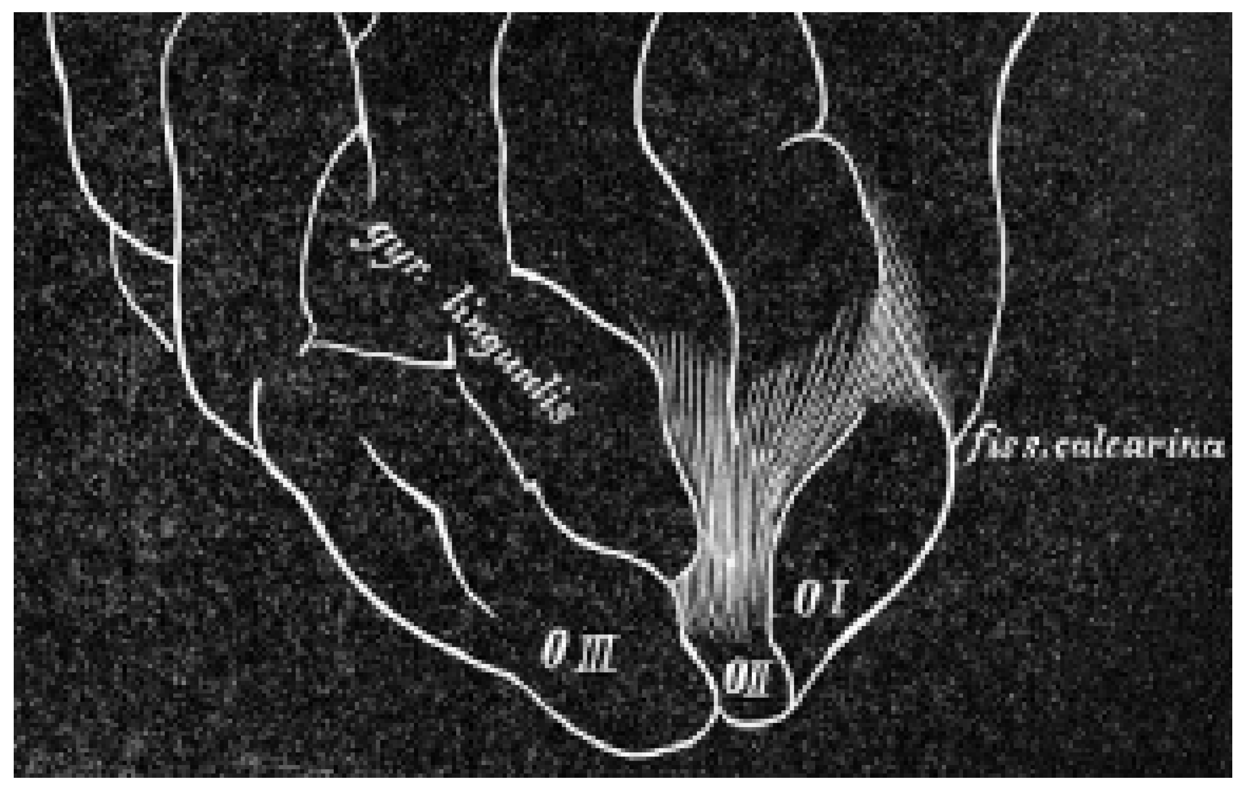
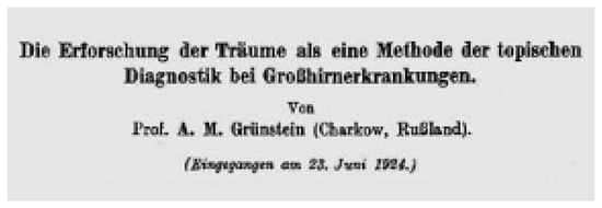
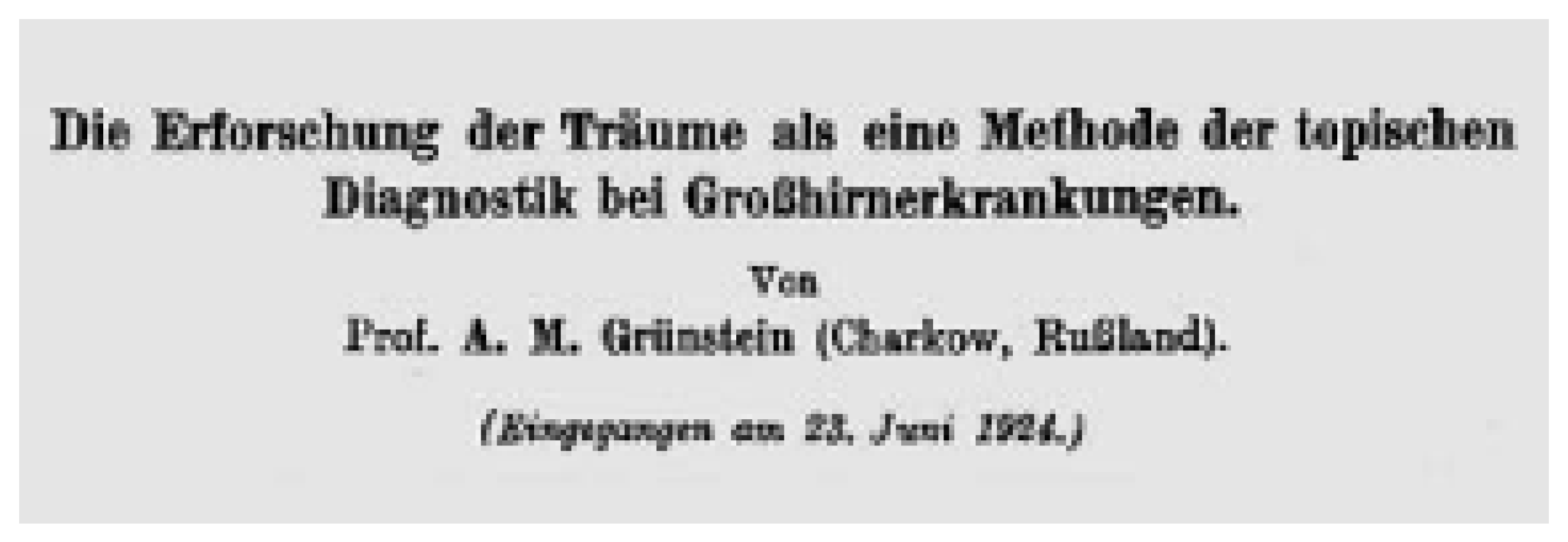
In 1883 Charcot described a patient who lost the ability to recall the visual mental images of his dreams in association with deficit in revisualisation during wakefulness (visual irreminiscence) [34]. In 1887 Wilbrand presented a case of global cessation of dreaming in association with prosopagnosia (inability to recognise familiar faces) after infarction in the occipito-temporal region (fig. 2) [35]. The terms “Charcot-Wilbrand syndrome” (CWS) and anoneira were used to describe the cessation of dreaming as a consequence of focal brain damage. Grünstein noted that CWS is associated with visual agnosia and an irreminiscence but not with cortical blindness, correctly suggesting a role of the extrastriate visual cortices in normal dreaming (fig. 3) [36]. Later studies confirmed the strong association (in 91% of cases reviewed by Solms) between CWS and irreminiscence [37].
Figure 2.
The brain autopsy of the patient described by Wilbrand in 1887 with loss of visual dreaming following a cerebral infarction in association with prosopagnosia. This precise drawing shows the infarcted area in the right deep occipital lobe, just beside the inferior lingual gyrus.
Figure 3.
The publication of Grünstein from the year 1924 with the title “Die Erforschung der Träume als eine Methode der topischen Diagnostik bei Großhirnerkrankungen” [The study of dreaming as a method for topical diagnosis in cerebral diseases].
A decrease and even (transient) cessation of dreaming is frequently seen as a nonspecific symptom of acute neurological disorders, particularly of those affecting the central nervous system.
A persistent cessation of dreaming (anoneira, CWS) has been reported following focal lesions (e.g. stroke, traumatic brain injury) in the inferior parietal, medial temporo-occipital or bifrontal brain areas [37,38,39,40,41]. In a study of 53 patients with acute unilateral brain lesions (stroke, neoplasia) those with posterior lesions showed a frequent loss of dream recall while those with anterior lesions rarely did so (61–75% vs 11–15%, respectively). The cessation of dream recall was associated with defective performance in tests for visuoperceptive functions. There were no right-left differences, whereas aphasic patients were more commonly non-recallers that patients without aphasia (83 vs 33%, respectively) [40]. In Solms’ study of 322 patients with focal brain lesions anoneira was found in 35% of cases and represented the most common form of dream changes, with a tendency to chronicity particularly with right-sided lesions but without any association with amnesia or aphasia [37].
Dementia and amnesic syndromes can lead to a reduction in frequency and verbal expression of dreams [42,43]. It is noteworthy that even in the presence of profound declarative amnesia patients with extensive bilateral medial temporal lobe damage report at sleep onset, as normal controls do, intrusive, stereotypical, visual images following a prolonged play with the computer game Tetris [44].
- Increased dreaming
Increased dreaming is seen with anterior limbic damage [37] and, rarely, as an epileptic phenomenon in temporal lobe seizures (epileptic dreams, see below).Often,but not always,increased dreaming is accompanied by appearance of dream-like experiences during wakefulness and by a difficulty in differentiating between dreams and reality.
- Changes in dream contents/accompaniments
Dreams with a strong anxiety component (terrifying dreams) can be seen as REM-related parasomnia (nightmare) and in the course of focal, neurodegenerative (e.g. Parkinson’s disease) brain diseases and, rarely, as epileptic equivalent.
Dreams with increased vividness, violent/aggressive contents and increased/uncontrolled motor activity (enacted dreams) suggest the presence of a REM-sleep behaviour disorder (RBD). The diagnosis is proven by the polysomnographic demonstration of a loss of physiologic atonia and increased motor phasic activity during REM sleep. RBD can be observed in the course of neurodegenerative disorders, brainstem lesions, narcolepsy, drug/alcohol abuse and in the so-called parasomnia overlap syndrome.
A cessation of visual dream imagery (“nonvisual dreaming”) has been reported following medial occipito-temporal lesions [37].
Patients with unilateral medial temporo-occipital lesions have been reported to have less expressive activity and distorted settings in their dreams [45].
Dreams with unaltered perception of the self as a critical observer of the oneiric experience and a unique capability of influencing their contents (lucid dreams) are occasionally reported by patients with narcolepsy.
- Dream-like experiences during wakefulness (hallucinations)
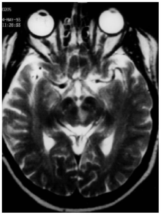
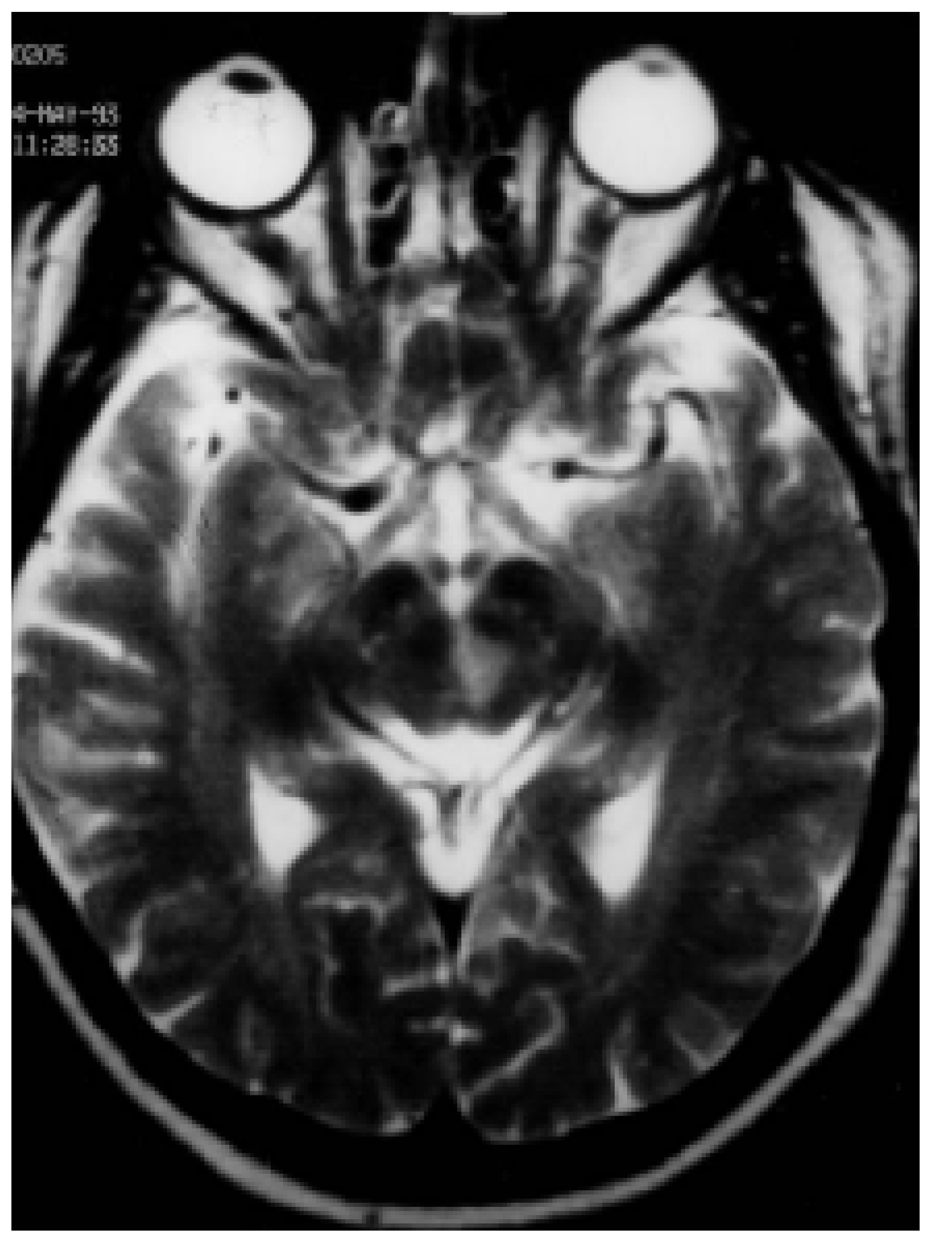
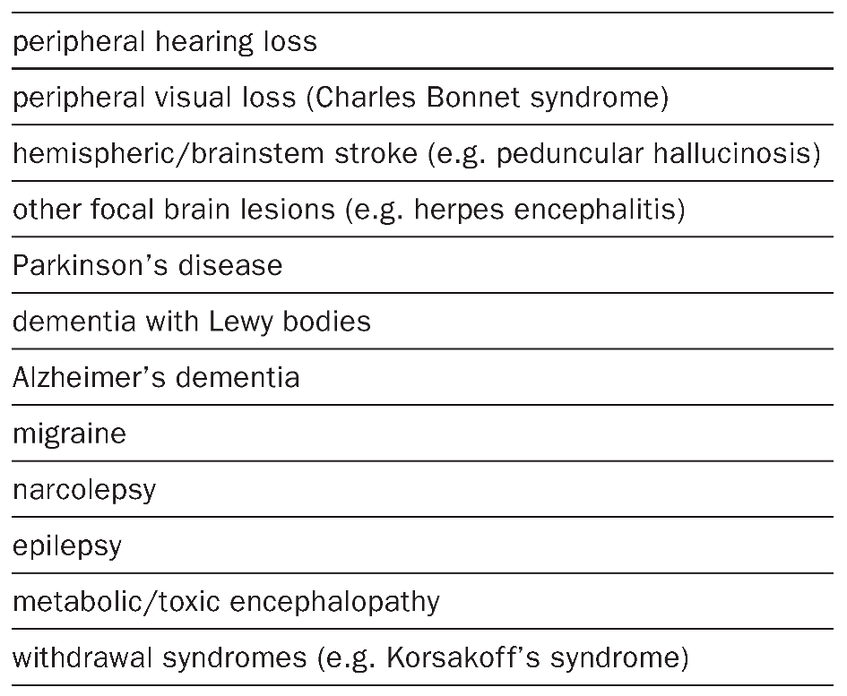
Visual hallucinations during wakefulness, at sleep onset (hypnagogic hallucinations) and on awakening (hypnopompic hallucinations) can occur in normal subjects as well as in patients with migraine, epilepsy, midbrain/thalamic lesions (peduncular hallucinosis), lesions causing visual loss (Charles Bonnet syndrome), sleep paralysis, narcolepsy or Parkinson’s syndromes [46]. Visual hallucinations can sometimes be highly complex and colourful (“movie-like”).This is well illustrated by the following episode (audio-taped) reported by a 48-yearold man with peduncular hallucinosis due to a tegmental midbrain stroke that had appeared after coronarography (fig. 4):
Figure 4.
Brain MRI of a 48-year-old man with peduncular hallucinosis, due to a unilateral tegmental midbrain stroke that appeared after coronarography (see text).
I suddenly lost the feeling of my own body weight. In front of me I saw – on the right side of the wall – a parade of young and old people that I could not recognise. There was a little girl in front that attracted my attention. She was wearing a very bright dress with red-orange flowers on it like in a painting by Gaugain. I was ready to leave for an adventure … when suddenly the voice of the cardiologist woke me up.
Patients are often aware that these hallucinatory phenomena are not real. They may be indifferent to, amused or disturbed by them. In some patients, however, difficulties in differentiating dreaming from hallucinations may be reported. The occurrence of both, hallucinations and increased dreaming, is also possible [47]. The existence of such a continuum had already been suggested by Lasègue (“le délire alcoolique n’est pas un délire, mais un rêve”) [48].
Simple and complex acoustic hallucinations can complicate peripheral or central hearing loss. Somaesthetic hallucinations are reported following amputation of an extremity or profound sensory loss (phantom limb syndrome). Polymodal hallucinations are typical for delirium tremens, but can also be seen with focal brain damage, as reported by a 49-year-old patient with tegmental pontine stroke (and history of alcohol abuse):
I suddenly saw the face of a madonna, like on a church’s window, on my left side, lasting for about one minute … I found in front of me a large white snow ball … on my right side the face of a madonna … I heard for hours in my sleep school songs in my left ear and a bird singing.
Hallucinations not related to a false perception (so-called psychic hallucinations after Baillarger) include the sense of a presence of persons (often relatives standing in the room, sitting on the edge of the bed). This type of hallucination (“sensed presence”) is particularly common in patients with narcolepsy, sleep paralysis and Parkinson’s disease [49,50]. Less commonly patients may project/feel the own body image and/or the self in the external space (“out-of-body” and autoscopic experiences) [32].
- Unaltered dreaming
Dreaming can remain unaltered despite dorsolateral prefrontal lesions, right hemispherectomy [51], split-brain procedures [52] and brainstem lesions [37], with and without REM sleep changes [37].
Dream changes in specific neurological disorders
- Sleep disorders
- Narcolepsy
Narcolepsy is a chronic debilitating disorder characterised by excessive daytime sleepiness, cataplexy, hallucinations and sleep paralysis [49]. Narcolepsy is believed to be caused by an autoimmune destruction of hypothalamic hypocretinproducing neurons [53].
Hallucinations,dreams,nightmares, arousal disorders and RBD are more frequent in narcoleptics than in the normal population (fig. 5). In two recent systematic studies nightmares (54%) and lifelong hallucinations (81%) were found to be more common than in normal controls (5 and 37%, respectively) [49,54]. Not infrequently patients may experience a variety of hallucinations and dream-like experiences, as is well illustrated by the following report of a 19-year-old patient:
Figure 5.
Painting of a 56-year-old man with narcolepsy depicting frightening and vivid visual hallucinations.
During the day I often had the feeling as if somebody was holding my left leg when passing a dark passage … At sleep onset I often saw a dark, silent, black shadow on the right side of my bed,which vanished immediately when turning the light on … While sleeping I often had strange dreams. I would see myself as acting in the dream action, and at the same time I would feel myself as separated from it, ‘floating’ over the scene and realising the illusionary nature of the dream.
In narcoleptics, hallucinations and dreaming may accompany or follow attacks of cataplexy and sleep paralysis. Dreams can occur at sleep onset during daytime (napping) and nighttime, as well as on awakening (Rosenthal’s syndrome). Typical of narcoleptic dreams is the presence of elements of the normal awake mentation in form of volitional control of mentation and some awareness of the environment. Fantastic and absurd dreams with religious,mysterious or sexual components are possible. Hallucinations are often visual,less commonly somatosensory, vestibular, gustatory/olfactory or vegetative. The hallucinations of the presence of someone nearby (people, intruders, animals, “sensed presence”), of a pressure on the chest with breathing difficulties (“incubus/succubus”) and of floating/flying sensations with out-of-body experiences are typically associated with sleep paralysis [55].
Dahmen et al. stressed the occasional difficulty in differentiating between narcoleptic and schizophrenic hallucinations [54]. They investigated the different aspects of hallucinations in 148 patients with narcolepsy, 21 patients with exacerbated acute schizophrenia and 128 healthy controls. Auditory hallucinations were twice as frequent in the schizophrenic group compared to narcoleptics, whereas 83% of narcoleptic patients reported visual hallucinations compared to only 29% among schizophrenics.
In a study of 15 narcoleptics and 9 controls, Fosse et al. found emotions to be more common and more intense in narcoleptic sleep-onset REM (SOREM) periods than in nighttime REM of either narcoleptic or normal subjects, with anxiety/ fear exhibiting the strongest increase, followed by joy/elation. Comparing nighttime REM in narcoleptic and normal subjects, narcoleptics were also found to have more intense feelings of anxiety/fear and joy/elation but to have a less frequent experience of surprise and anger [56].
Sturzenegger and Bassetti collected dream reports of 57 narcoleptics with cataplexy and compared them with patients with nonnarcoleptic hypersomnia and normal controls [49]. Dreams of narcoleptics more often included such contents as flying, being chased, crawling in a tube, monsters and the presence of persons in the patient’s room (“sensed presence”). Moreover, dream reports of narcoleptics contained more negative emotions such as sorrow, anger, fear, guilt, disgust and disdain. No link was found between emotions in the dreams and emotions triggering cataplectic attacks.
The possibility that emotions in the dreams of narcoleptics may precipitate symptoms at awakening was suggested by Wilson in 1928. One of his patients had a terrifying dream about a murder and immediately thereafter woke up with an unusually pronounced sleep paralysis [57].
Attarian et al. reported a case of RBD in a narcoleptic patient arising from cataplexy and wakeful dreaming, probably facilitated by his use of tricyclic antidepressants to treat cataplexy [58].
- Sleep terrors and sleepwalking (NREM parasomnias / arousal disorders)
Sleep terror (pavor nocturnus) and sleepwalking are parasomnias of the first part of the night.These parasomnias are so-called disorders of arousal because they represent a sudden but incomplete arousal from slow-wave sleep (stages 3 and 4 of NREM sleep) [59]. Arousal disorders have for a long time erroneously been regarded as behavioural expressions of ongoing dreams [60,61]. In fact, oneiric activity is usually absent or consists in a brief fragmentary imagery [62,63].The possibility to induce sleep terrors by forced awakenings during slow-wave sleep also argues against the old belief that sleep terrors might be triggered by some fearful dream.
Particularly in adults oneiric experience in conjunction with sleepwalking may be reported and amnesia for the episodes can be incomplete [64]. The presence of oneiric experiences in sleepwalking has also been suggested by the fight-or-flight imitating behaviour occasionally observable in sleepwalkers [65].
In sleep terrors subjects abruptly sit up, producing a loud scream in association with a facial expression of extreme fear.The attacks are accompanied by a sudden and pronounced increase in heart and respiratory rate and muscle tone. Most characteristically the utterly worried parents usually cannot arouse and console their child.The combination of features of arousal disorders with REM sleep behaviour disorder has been called “overlap parasomnia”.
Sleep terrors and sleepwalking are more common in children but may be present in up to a few per cent of the adult population. They are not associated with specific physical findings. Developmental, genetic, psychological and organic factors can contribute to their occurrence.About one third of individuals with frequent nightmares have a positive family history and an association with the HLA haplotype DQB1*05 has been suggested. Sleepwalking can be triggered by fever, stress,sleep deprivation, acute brain disorder (stroke, trauma) and treatment/withdrawal from drugs.
- Nightmares/posttraumatic stress disorder
Nightmares are parasomnias of the middle of the night or early morning when REM sleep is more common. Nightmares are typically dreams of physical danger (e.g. being chased) or psychological threat (e.g. being teased). Typical contents include monsters, ghosts, animals and bad individuals. Vocalisations, movement and autonomic symptoms are typically minimal.When awakened, subjects are rapidly oriented, can be calmed and usually recall the details of the dream. These features differentiate nightmares from sleep terrors (see above) and REM sleep behaviour disorder (see below).
Nightmares are more common in children and are not associated with specific physical findings. Developmental,genetic, psychological and organic factors can contribute to their occurrence. About 10% of individuals with frequent nightmares have family history of nightmares. Nightmares are more common in subjects with mental retardation, depression and CNS diseases; as well as in an association with fever and treatment/withdrawal from drugs. Nightmares may result from a severe traumatic event and indicate posttraumatic stress disorder.
- Epic dreaming and complex nocturnal visual hallucinations
Epic dreaming has been described by Schenck and Mahowald in 1995 and represents a dreaming disorder characterised by endless and exhausting dreams associated with morning tiredness and chronic daytime fatigue [66]. The condition shows a strong female predominance (85%). The dream content typically consists in a relentless, mostly banal physical activity reoccurring nightly in 90% of affected patients. A remarkable feature is the poor affective tone of the dreams and the absence of emotional arousals. Polysomnographic recordings do not reveal any particular abnormalities,and the underlying disturbance of the disorder remains unknown.
Silber et al. have recently reported a series of 12 patients (11 of whom were females) with vivid, silent, often distorted images of people and animals occurring after waking, disappearing with increased light. Idiopathic hypersomnia, betablocker use, dementia with Lewy bodies, macular degeneration and anxiety were associated factors [67].
- REM sleep behaviour disorder (RBD)
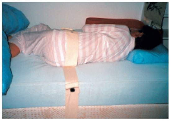
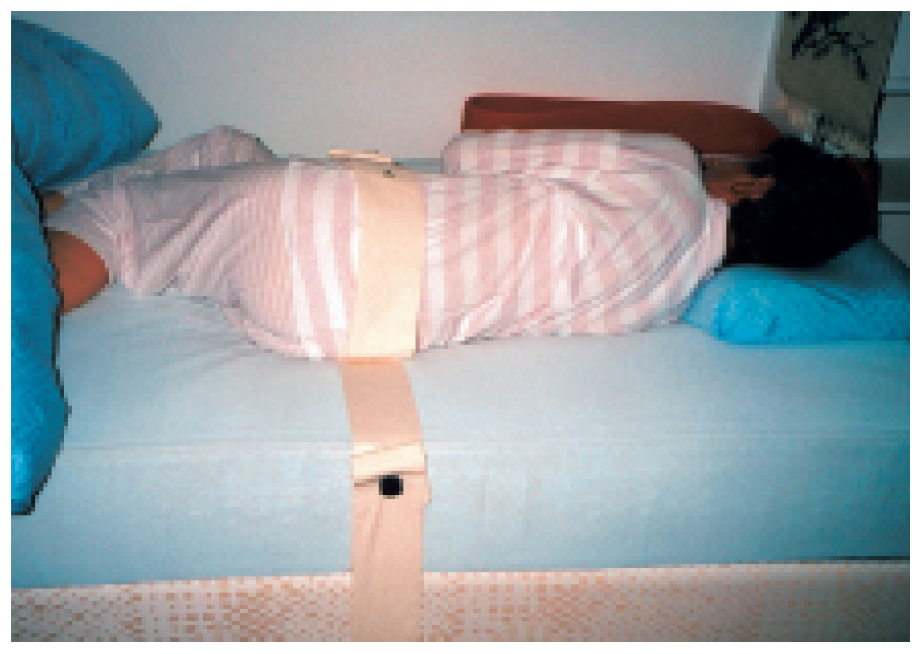
This disorder was formally first described in 1986 [68], although single reports had described features of this entity already before. RBD presents with dramatic, often violent, motor behaviours occurring during sleep in association with vivid, frightening dreams.Since RBD occurs during REM sleep, it typically appears in the second half of nighttime sleep when REM sleep predominates. Injuries and assaults to bed partners have been reported to occur in up to 32% and 64% of patients (fig. 6) [69]. In a study of 49 polysomnographic-confirmed RBD patients Fantini et al. found dreams in these patients to be characterised by an elevated proportion of aggressive contents, despite normal levels of daytime aggressiveness [70].
Figure 6.
Picture of a 65-year-old woman with an idiopathic REM sleep behaviour disorder who restrained herself for years with ropes, in order to prevent her from falling out of bed.
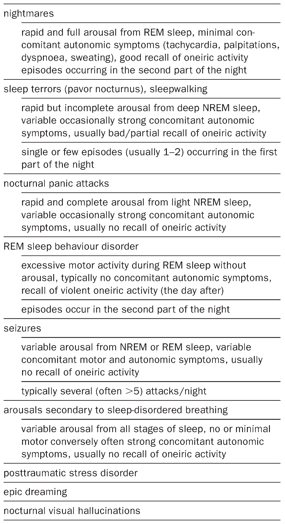
Polysomnography demonstrates an excessive muscle tone during REM sleep (e.g. in the chin EMG), which may represent the first sign of RBD [71], an enhanced phasic muscle activity with corresponding excessive/abnormal motor behaviour and vocalisations. Patients with an idiopathic RBD have an increase in slow-wave sleep and delta power and an increased frequency of periodic limb movements during all stages of sleep [72,73]. The differential diagnosis of RBD includes sleeprelated seizures, confusional arousals, sleepwalking, sleep terrors, nightmares, panic attacks and arousals related to obstructive sleep apnoea.
The pathophysiology of RBD corresponds, as first shown experimentally in the cat by Jouvet [13], to a dysfunction of ponto-medullary REM mechanisms of atonia. Approximately 60% of RBDs are idiopathic. Idiopathic RBD usually presents in the sixth or seventh decade and predominates in males. In these patients cognitive (visuospatial, constructive) disturbances, slowing of occipital EEG activity and a decreased striatal dopaminergic availability during wakefulness have been reported [74,75,76,77]. A significant proportion of patients with idiopathic RBD develop in the later course a Parkinsonian disorder [78]. In about 40% of cases RBD is due to narcolepsy or neurodegenerative disorders such as Parkinson’s disease, multisystem atrophy and,less commonly,brainstem dysfunction of other origin (e.g. multiple sclerosis,stroke,limbic encephalitis).Together with olfactory disturbances, RBD can be the first clinical symptom of neurodegeneration of Parkinson’s disease [79].An acute/ transient form of RBD can be seen in patients discontinuing the consumption of alcohol and REMsuppressing medications (e.g. antidepressants). Most cases of RBD respond to small doses of clonazepam. Melatonin, dopaminergic drugs and donepezil can also be used.
- Insomnia
Insomnia is the most common sleep-related complaint and affects up to 47% of the patients consulting general practices [80]. A substantial number of investigations have suggested that in healthy individuals a significant correlation exists between the frequency of dream recall, frequency of nocturnal awakenings and low sleep quality [81,82,83]. Schredl et al. have recently confirmed that insomniacs’ dream-recall frequency is elevated and dream reports are longer [84]. Dreams of insomniacs are characterised by more negative emotions, frequent reports on health themes, depression and aggression and a reduced degree of explicit visual perception.
If on the one hand insomnia can increase dream-recall frequency, there might exist on the other hand a subcategory of insomnia that is explicitly produced by dreaming.This condition is called “dream-interruption insomnia” [85]. Patients complain about repetitive nocturnal awakenings and typically mention frequent episodes of nightmares prior to the onset of their insomnia.
- Sleep-disordered breathing
Schredl et al. found an elevated dream-recall frequency in patients with sleep-disordered breathing (SDB) without any correlation, however, with the frequency of nocturnal awakenings [86]. A strong negative correlation was found between the frequency of respiratory events and dream bizarreness, suggesting that apnoea-induced arousals may interrupt the (REM-)dream process.
Gross and Lavie reported that dreams of patients with sleep-disordered breathing were completely devoid of any feelings of suffocation and did not contain peculiar signs of fear or anxiety [87]. More recently, Iranzo and Santamaria have, however, reported the occurrence of agitated, violent dreams with abnormal motor behaviours mimicking RBD in severe SDB patients [88].
- Restless legs syndrome
In patients with restless legs syndrome no increased dream frequency was found compared to healthy controls [89].
- Stroke and other focal brain diseases
Following the seminal papers by Charcot and Wilbrand on cessation of dreaming [34,35], a few authors reported on dream characteristics of patients with stroke [36,38,40,90,91,92]. More recently, Hobson has given a first-person account of “abnormal visual percepts” in association with total insomnia and dream suppression following a lateral medullary stroke [93]. The most extensive work on dreaming in patients with focal brain damage has been performed by Solms [37]. Out of the 332 patients with cerebral lesions included in his systematic analysis 83 received the diagnosis of cerebrovascular disease and 79 that of neoplasm. The four main changes in dreaming were the following: (1) global cessation of dreaming (35% of cases), usually with parietal or frontal lesions, and persistence over one year, particularly with rightsided lesions; (2) cessation of visual dreaming (1%), usually with medial occipito-temporal lesions; (3) dream-reality confusion (5%), usually with anterior limbic lesions; (4) recurrent nightmares (8%), without a specific topographic association.
An example of increased dreaming and nightmares is the following report of a 68-year-old woman seen in our department with severe rightsided hemispheric stroke in the middle cerebral artery distribution:
I saw little Appenzeller dogs entering my room through the window, at the same time my motherin-law was sitting on my bedside. I tried to touch her with my right hand … I saw a dark snake … and then suddenly a bouquet of flowers of different colours which was offered to me by a chimpanzee …
Solms’ work is of paramount value but has some limitations. First, no awakenings from REM sleep were undertaken.The possibility that CWS may in some cases be due to amnesia therefore cannot be ruled out. Second, the anatomical data are based mainly on CT scans, which do not always allow – particularly in the posterior fossa – to determine the exact extent of the underlying lesion. Third, the patients suffered from a variety of diseases and were examined at different intervals following disease onset, factors which can significantly influence oneiric experiences.
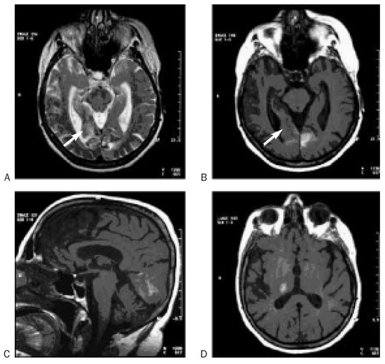
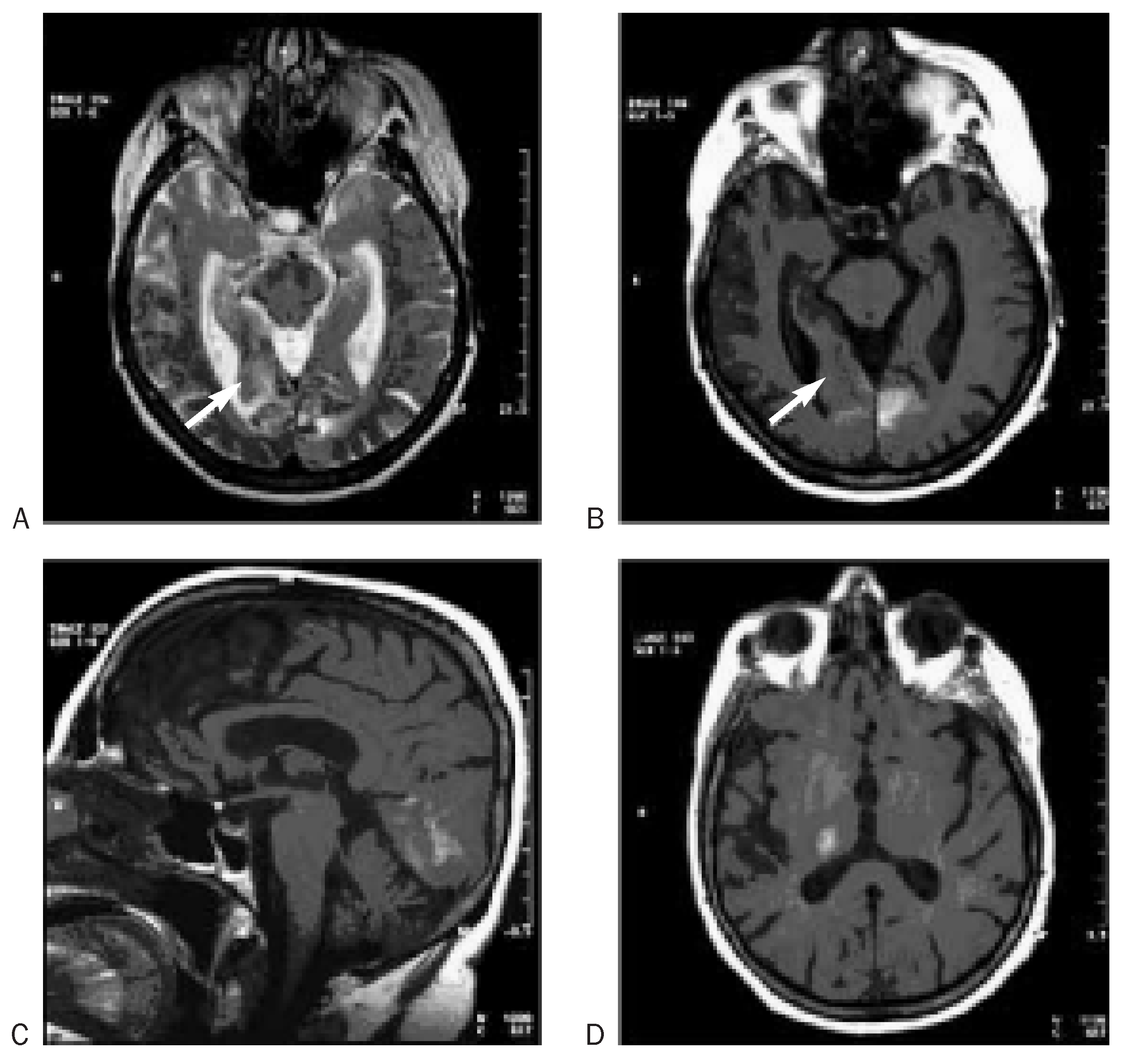
In a personal observation of CWS we tried to be aware of these limitations [41]. This previously healthy 73-year-old woman was hospitalised because of the sudden visual loss and weakness of the left side of her body. Brain magnetic resonance imaging showed an acute ischaemic infarction of both deep occipital lobes (including the lingual gyrus on the right side) and the right posterolateral thalamus (fig. 7). Clinical examination revealed a left homonymous hemianopsia, a right inferior quadrantanopsia, achromatopsia and bilateral decrease of visual acuity. The third night after the stroke, the patient reported an unusual, very vivid and colourful dream:
Figure 7.
Brain MRI (T2 [A] and native T1 [B–D]) of a 73-year-old woman with total dream loss, performed one day after stroke onset (see text). The images show a subacute infarction of both occipital lobes and the right posterolateral thalamus (D) with slight bleeding in the infarcted area. On the right side the inferior lingual gyrus (arrows) is involved.
An unknown person gives me a huge piece of cotton with a drawing on it of many dwarfs dressed in bright colours … Later the same person shows a second … larger piece of cotton with hundreds of tiny men on it. I try to find the first group of dwarfs but I don’t succeed. I am upset.At this point the nurse wakes me up.
Following this unusual dream,the patient – who used to recall dreams three to four times per week before the stroke – stopped dreaming for months. A detailed neuropsychological examination was unrevealing. In particular, the patient could imagine well-known surroundings and places, was able to recognise familiar faces and displayed normal topographic orientation. Sleep studies were also essentially unremarkable with, especially, normal REM amounts, REM density and REM latency. Dream experiences were also denied after four awakenings from REM sleep undertaken during one polysomnographic recording. This report is interesting because it demonstrates the following three points: (1) the possible association of initial vivid dreaming with CWS (we postulated in this patient a release phenomenon of the partially damaged visual areas being responsible for the episode of vivid dreaming); (2) the existence of CWS as a distinct neuropsychological dysfunction, in the absence of other cognitive deficits (and in particular of a so-called irreminiscence, see above); (3) the independency of dream and REM-sleep generators.
- Traumatic brain injury
Traumatic brain injury may lead to visual hallucinations, anxiety dreams and posttraumatic stress disorder [94,95,96]. Also, one systematic study overall found only small differences in dreaming between the pre- and posttraumatic period [97].
Depending on the presence and topography of the underlying brain damage, traumatic brain injury has, however, been shown to cause dream changes (see above) [37,40]. It is noteworthy that out of the 361 patients included in Solms’ systematic analysis 108 carried the diagnosis of head trauma.
- Parkinson’s syndrome
Changes in dream frequency and contents and the occurrence of nightmares, hallucinations and RBD (see above) are frequently seen in patients with Parkinson’s syndrome.A correlation with the presence of other sleep-wake disturbances, cognitive changes,axial motor symptoms and medication (dopaminergic drugs, anticholinergics) has been recognised [47,98,99].
Visual hallucinations are reported by about 25–50% of patients with Parkinon’s syndrome and predict a Lewy-body pathology with involvement of the parieto-occipital and limbic cortices [50,99]. In Parkinson’s disease (PD) hallucinations typically occur in the second half of the disease course, whereas they can occur early in the course of dementia with Lewy bodies (DLB). Visual hallucinations in Parkinson’s disease have been linked with a higher risk of institutionalisation [100]. Reduction of dopaminergic drugs and prescription of atypical neuroleptics (e.g. clozapine, quetiapine) and acetylcholinesterase inhibitors may improve hallucinations in Parkinson’s disease [101,102].
REM sleep behaviour disorder is seen in about 25–50% of patients with Parkinson’s disease and in 50% of patients with DLB and multisystem atrophy [69,71,103,104,105,106].In non-synucleinopathies (progressive supranuclear palsy, corticobasal degeneration) RBD is conversely rare. RBD can precede the onset of these conditions by years to decades and often exhibits a fluctuating course after its onset.
- Dementia
Visual hallucinations are common in Alzheimer’s disease (about 20% of cases) and dementia with Lewy bodies (50% of cases) [99,107,108]. They correlate with cognitive decline and worse outcome (institutionalisation, mortality) [109].
Alzheimer’s disease can also lead to a reduction in frequency and contents of dreaming [42,43]. A few patients are observed to show improved language skills during sleep compared to wakefulness [110]. Treatment with acetylcholinesterase inhibitors may induce vivid dreams [33].
A dementia syndrome with hypersomnia, enacted dreams and loss of slow-wave sleep has recently been reported [111].
- Epilepsy
Epileptic hallucinations are usually brief, simple, stereotyped and fragmentary associated with other seizure manifestations such as altered awareness, motor activity and automatisms [46]. Less commonly, epileptic hallucinations are complex (“dream-like”) or, in seizures of temporal or parietal origin, consist of autoscopic experiences [112].
Recurrent epileptic nightmares have been reported in temporal lobe epilepsy as well as a poststroke event [113,114]. Epstein reported a patient with recurrent partial seizures in form of painful dreaming of dying [115]. Occasionally, patients report similar contents in dreams and daytime epileptic seizures [116]. In 1954 Penfield and Jasper described in their book Epilepsy and the Functional Anatomy of the Brain the report of dream-like experiences in a patient following cortical (temporo-parietal) stimulation.
Patients with complex partial seizures have a higher frequency of dream recall than those with generalised seizures [117]. In a sleep laboratory study the frequency of dream recall was lower in patients with complex partial seizures after awakening from stage 2 NREM sleep than REM sleep, regardless of the side of epileptic focus, while the length and structural organisation of the dreams did not significantly differ in REM and NREM sleep [118].
- Migraine
Migraine aura is well recognised as a cause of simple visual hallucinations; complex dream-like visual hallucinations are conversely less common. They are more frequently seen in migraine coma and familial hemiplegic migraine.
Levitan analysed the content of 23 dreams that immediately preceded a migraine attack, finding a significant predominance of powerful negative emotions [119].
Recurrent dreams and nightmares have been reported as aura equivalent [120].
- Creutzfeldt-Jakob disease
Fatal familial insomnia (FFI) is a hereditary, occasionally sporadic prion disease with prominent neurodegenerative changes affecting mainly the antero-ventral and medio-dorsal thalamic nuclei; the disease has been described in 1986 [121]. The clinical features of FFI consist in progressive insomnia, sympathetic overactivation (perspiration, tachycardia, hypertension, evening pyrexia) and motor disturbances (dysarthria, myoclonus, disturbed coordination). Patients typically show peculiar episodes of vivid dream enactment that may spontaneously intrude into wakefulness, during which they are in a stupor-like state (“oneiric stuporous state”) and perform complex jerklike movements, corresponding to the dream content reported afterwards [122,123]. Polysomnography reveals profound sleep EEG changes with progressive disappearance of the hallmarks of NREM sleep including sleep spindles, K-complexes and slow-wave sleep [124]. Recently, Landolt et al. have described similar sleep-wake disturbances in seven patients with sporadic Creutzfeldt-Jakob disease (sCJD); three patients also showed dream-reality confusion and had visual and/or acoustic hallucinations [125].
- Encephalitis and other CNS infections
Dream-like hallucinations can be observed in patients with herpes encephalitis and other CNS infections involving the mesiotemporal areas (personal observation).
- Delirium tremens, other withdrawal syndromes
Delirium tremens complicates sudden alcohol withdrawal after chronic alcohol abuse and has a mortality of 5 to 15% [126]. The main symptoms are agitation, tremor, increased autonomic activity and an acute confusional state with polymodal hallucinations and dream enactment.
Polysomnography in acute alcohol withdrawal syndrome shows a markedly reduced sleep efficiency, reduction or absence of NREM sleep and long periods of wakefulness alternating with abruptly emerging periods of REM sleep without atonia [127].This patient repeatedly presented with intense enacting dreams with violent fighting behaviour, jerks and talking throughout the night and also during daytime episodes of drowsiness.Whenever awakened, he reported vivid dream content.
Acute withdrawal from barbiturate and benzodiazepine can also be complicated by visual hallucinations and vivid dreaming.
- Autoimmune disorders
- Morvan’s fibrillary chorea
This is a rare disorder which has been linked with autoantibodies against voltage-gated potassium channels [128]. Severe agrypnia usually lasts only a few weeks in about 90% of cases, but occasionally may persist and lead to death; it is associated in most cases with intense anxiety, delirium and often remarkable hallucinatory behaviour with enacted dreams [129]. In addition, patients exhibit symptoms of autonomic hypertension and peculiar motor disturbances with agitation, cramps, myoclonic jerks and fasciculations (“fibrillary chorea”).
- Limbic encephalitis
This is a rare disorder which has been linked with autoantibodies against voltage-gated potassium channels and with the presence of REM sleep behaviour disorder [130,131].
- Guillain-Barré syndrome
In a series of 139 patients with Guillain-Barré syndrome (GBS) mental status changes were found in 31% of patients including vivid dreams (19%), illusions (30%, including an illusory body tilt), hallucinations (60%, mainly visual) and delusions (70%, mostly paranoid). They appeared with a median of 9 days after disease onset and lasted for a median of 8 days. Autonomic dysfunction, assisted ventilation and high CSF protein levels were significant risk factors for abnormal mental status in GBS patients, whereas CSF hypocretin-1 levels were normal [132]. The origin of these disturbances remains speculative at this point.
- Others
Patients with motor deficits, acquired blindness or aphasia do not infrequently dream of moving, seeing and speaking normally (personal observation). Intraindividual differences in the latency of “incorporation” of daytime deficits in oneiric experiences are present.
Conversely, changes in dream frequency and contents have also been observed in patients with disorders of the peripheral nervous system such as myasthenia gravis [133].
Patients with phantom limbs often experience similar but painless sensations in their dreams [134].
Conclusions
Dream changes in patients with neurological disorders have been studied poorly, although their frequency is not negligible (e.g. in Parkinon’s syndrome, stroke and narcolepsy). Despite the little attention that neurologists typically pay to dream reports, their assessment can be of diagnostic interest (e.g. REM sleep behaviour disorder, visual hallucinations), have prognostic implications (e.g. visual hallucinations in Parkinson’s disease) and be a source of major distress for patients and their bed partners (e.g. nightmares, RBD). In addition, specific and effective management of “anxiety dreams” and disturbing “dream-like experiences” relies upon the knowledge of their phenomenological and aetiological differential diagnosis (Table 1 and Table 2). Finally, systematic studies of dream experiences are expected to offer unique insights into the brain, mind and emotions of neurological patients.
Table 1.
Differential diagnosis of anxiety/violent dreams.
Table 2.
Neurological causes of dream-like hallucinations.
References
- Kruger, SE. Dreaming in the Middle Ages; Cambridge University Press: Cambridge, 1992. [Google Scholar]
- Cardano G. Sul sonno e sul sognare. Supertascabili Marsilio; 1989.
- De Manacéïne, M; Manasseina, M. Sleep: Its Physiology, Pathology, Hygiene, and Psychology; Walter Scott, Ltd: London, 1897. [Google Scholar]
- Calkins, MW. Statistics of dreams. Am J Psychol 1893, 5, 311–43. [Google Scholar] [CrossRef] [Scilit]
- Vaschide, N. Le sommeil et les rêves; Ernest Flammarion: Paris, 1920. [Google Scholar]
- Ohlmeyer, P; Brilmayer, H; Hüllstrung, H. Periodische Vorgänge im Schlaf. Pflügers Arch 1944, 248, 559–60. [Google Scholar] [CrossRef] [Scilit]
- Loomis, AL; Harvey, EN; Hobart, G. Potential rhythms of the cerebral cortex during sleep. Science 1935, 81, 597–8. [Google Scholar] [CrossRef] [Scilit]
- Hess, R; Koella, WP; Akert, K. Cortical and subcortical recordings in natural and artificially induced sleep in cats. Electroencephalogr Clin Neurophysiol Suppl 1953, 5, 75–90. [Google Scholar] [CrossRef] [Scilit]
- Lawson, RW. Blinking and sleep. Nature 1950, 165, 81–2. [Google Scholar] [CrossRef] [Scilit]
- Aserinsky, E; Kleitman, N. Regularly occurring periods of eye motility, and concomitant phenomena, during sleep. Sience 1953, 118, 273–4. [Google Scholar] [CrossRef] [Scilit]
- Aserinsky, E; Kleitman, N. Two types of ocular motility occurring in sleep. J Appl Physiol 1955, 8, 11–8. [Google Scholar] [CrossRef] [Scilit]
- Jouvet, M; Michel, F. Corrélations éléctromyographiques du sommeil chez le chat decortiqué et mésencéphalique chronique. C R Séances Soc Biol Fil 1959, 153, 422–5. [Google Scholar]
- Jouvet, M; Michel, F. Release of the “paradoxal phase” of sleep by stimulation of the brain stem in the intact and chronic mesencephalic cat. C R Soc Biol 1960, 154, 636–41. [Google Scholar]
- Jouvet, M; Delorme, F. Locus coeruleus et sommeil paradoxal. C R Soc Biol 1965, 159, 895–9. [Google Scholar]
- Dement, W; Kleitman, N. The relation of eye movements during sleep to dream activity: an objective method for the study of dreaming. J Exp Psychol 1957, 53, 339–46. [Google Scholar] [CrossRef] [Scilit]
- Dement, W; Wolpert, EA. The relation of eye movements, body motility, and external stimuli to dream content. J Exp Psychol 1958, 55, 543–53. [Google Scholar] [CrossRef] [Scilit]
- Dement, W. The effect of dream deprivation. Science 1960, 131, 1705–7. [Google Scholar] [CrossRef] [Scilit]
- Hobson, JA; McCarley, RW; Wyzinski, PW. Sleep cycle oscillation: reciprocal discharge by two brainstem neuronal groups. Science 1975, 189, 55–8. [Google Scholar] [CrossRef] [Scilit]
- Hobson, JA; McCarley, RW. The brain as a dream state generator: an activation-synthesis hypothesis of the dream process. Am J Psychiatry 1977, 134, 1335–48. [Google Scholar] [PubMed]
- Foulkes, D. Dream reports from different stages of sleep. J Abnorm Soc Psychol 1962, 65, 14–25. [Google Scholar] [CrossRef] [Scilit] [PubMed]
- Rechtschaffen, A; Verdone, P; Wheaton, J. Reports of mental activity during sleep. Can Psychiatr Assoc J 1963, 257, 409–14. [Google Scholar] [CrossRef] [Scilit] [PubMed]
- Cavallero, C; Cicogna, P; Natale, V; Occhionero, M; Zito, A. Slow-wave sleep dreaming. Sleep 1992, 15, 562–6. [Google Scholar] [CrossRef] [Scilit] [PubMed]
- Landolt, HP; de Boer, LP. Effect of chronic phenelzine treatment on REM sleep: report of three patients. Neuropsychopharmacology 2001, 25, S63–7. [Google Scholar] [CrossRef] [Scilit]
- Antrobus, JS. REM and NREM sleep reports. Psychophysiology 1983, 20, 562–8. [Google Scholar] [CrossRef] [Scilit]
- Antrobus, JS. Dreaming: cognitive processes during cortical activation and high afferent thresholds. Psychol Rev 1991, 98, 96–121. [Google Scholar] [CrossRef]
- Braun, AR; Balkin, TJ; Wesensten, NJ; Carson, RE; Varga, M; Baldwin, P; et al. Regional cerebral blood flow throughout the sleep-wake cycle. An H215O PET study. Brain 1997, 120, 1173–97. [Google Scholar] [CrossRef] [Scilit] [PubMed]
- Maquet, P; Peters, J; Aerts, J; Delfiore, G; Degueldre, C; Luxen, A; et al. Functional neuroanatomy of human rap-id-eye-movement sleep and dreaming. Nature 1996, 383, 163–6. [Google Scholar] [CrossRef] [Scilit] [PubMed]
- Braun, AR; Balkin, TJ; Wesensten, NJ; Gwadry, F; Carson, RE; Varga, M; et al. Dissociated pattern of activity in visual cortices and their projections during human rapid-eyemovement sleep. Science 1998, 279, 91–5. [Google Scholar] [CrossRef] [Scilit]
- Lövblad, KO; Remonda, I; Heid, O; Schneider, J; Gonner, F; Schroth, G. Silent functional magnetic resonance imaging demonstrates focal activation in rapid-eye-movement sleep. Neurology 1999, 53, 2193–5. [Google Scholar] [CrossRef] [Scilit] [PubMed]
- Fell, J; Fernandez, G; Lutz, MT; Kockelmann, E; Burr, W; Schaller, C; et al. Rhinal-hippocampal connectivity determines memory formation during sleep. Brain 2006, 129, 108–14. [Google Scholar] [CrossRef] [Scilit]
- Arnulf, I; Bonnet, AM; Damier, P; Bejjani, BP; Seilhean, D; Derenne, JP; et al. Hallucinations, REM sleep, and Parkinson’s disease: a medical hypothesis. Neurology 2000, 55, 281–8. [Google Scholar] [CrossRef] [Scilit]
- Blanke, O; Landis, T; Spinelli, L; Seeck, M. Out-of-body experience and autoscopy of neurological origin. Brain 2004, 127, 243–58. [Google Scholar] [CrossRef] [Scilit]
- Singer, M; Romero, B; Koenig, E; Forstl, H; Brunner, H. Nightmares in patients with Alzheimer’s disease caused by donepezil. Therapeutic effect depends on the time of intake. Nervenarzt 2005, 76, 1127–9. [Google Scholar] [CrossRef] [Scilit]
- Charcot, JM. Un cas de suppression brusque et isolée de la vision mentale des signes et des objets (formes et couleurs). Progr Méd 1883, 2, 568–71. [Google Scholar]
- Wilbrand, H. Ein Fall von Seelenblindheit und Hemianopsie mit Sectionsbefund. Dtsch Z Nervenheilkd 1887, 2, 361–87. [Google Scholar] [CrossRef] [Scilit]
- Grünstein, AM. Die Erforschung der Träume als eine Methode der topischen Diagnostik bei Großhirnerkrankungen. Zeit Ges Neurol Psychiatr 1924, 93, 416–20. [Google Scholar] [CrossRef] [Scilit]
- Solms, M. The Neuropsychology of Dreams; Erlbaum: Mahwah, NJ, 1997. [Google Scholar]
- Humphrey, M; Zangwill, O. Cessation of dreaming after brain injury. J Neurol Neurosurg Psychiatr 1951, 14, 322–5. [Google Scholar] [CrossRef] [Scilit]
- Cathala, HP; Laffont, F; Siksou, M; Esnault, S; Gilbert, A; Minz, M; et al. Sleep and dreams in patients with parietal and frontal lobe lesions. Rev Neurol (Paris) 1983, 139, 497–508. [Google Scholar]
- Murri, l; Arena, R; Siciliano, G; Mazzotta, R; Muratorio, A. Dream recall in patients with focal cerebral lesions. Arch Neurol 1984, 41, 183–5. [Google Scholar] [CrossRef] [Scilit]
- Bischof, M; Bassetti, CL. Total dream loss: a distinct neuropsychological dysfunction after bilateral PCA stroke. Ann Neurol 2004, 56, 583–6. [Google Scholar] [CrossRef] [Scilit] [PubMed]
- Greenberg, R. Cerebral cortex lesions: the dream process and sleep spindles. Arch Gen Psychiatry 1968, 18, 203–9. [Google Scholar] [CrossRef] [Scilit]
- Cathala, HP; Laffont, F; Esnault, S; Sereni, C; Siksou, M. Sleep and dreams in Korsakoff’s amnesia due to alcoholism. Neurophysiol Clin 1988, 18, 173–86. [Google Scholar] [CrossRef] [Scilit] [PubMed]
- Stickgold, R; Malia, A; Maguire, D; Roddenberry, D; O’Connor, M. Replaying the game: hypnagogic images in normals and amnesics. Science 2000, 290, 350–3. [Google Scholar] [CrossRef] [Scilit] [PubMed]
- Peralta, R; Gomes, A; Costa, A; Santos, R; Paiva, T. Dream recall and dream content in posterior cerebral artery (PCA) stroke patients. Sleep Med 2005, Suppl 2, S207–8. [Google Scholar]
- Manford, M; Andermann, F. Complex visual hallucinations. Clinical and neurobiological insights. Brain 1998, 121, 1819–40. [Google Scholar] [CrossRef] [Scilit] [PubMed]
- Goetz, CG; Wuu, J; Curgian, LM; Leurgans, S. Hallucinations and sleep disorders in PD: six-year prospective longitudinal study. Neurology 2005, 64, 81–6. [Google Scholar] [CrossRef] [Scilit]
- Lasègue, C. Délire alcoolique n’est pas un délire, mais un rève. Arch Gen Méd 1881, 88, 513–36. [Google Scholar] [CrossRef] [Scilit]
- Sturzenegger, C; Bassetti, C. The clinical spectrum of narcolepsy with cataplexy: a reappraisal. J Sleep Res 2004, 13, 395–406. [Google Scholar] [CrossRef] [Scilit]
- Fenelon, G; Mahieux, F; Huon, R; Ziegler, M. Hallucinations in Parkinson’s disease: prevalence, phenomenology and risk factors. Brain 2000, 123, 733–45. [Google Scholar] [CrossRef] [Scilit] [PubMed]
- McCormick, L; Nielsen, T; Ptito, M; Hassainia, F; Ptito, A; Villemure, JG; et al. REM sleep dream mentation in right hemispherectomized patients. Neuropsychologia 1997, 35, 695–701. [Google Scholar] [CrossRef] [Scilit]
- Greenwood, P; Wilson, DH; Gazzaniga, MS. Dream report following commissurotomy. Cortex 1977, 13, 311–6. [Google Scholar] [CrossRef] [Scilit]
- Bassetti, C. Narcolepsy: selective hypocretin (orexin) neuronal loss and multiple signaling deficiencies. Neurology 2005, 65, 1152–3. [Google Scholar] [CrossRef] [Scilit]
- Dahmen, M; Kasten, N; Mittag, K; Muller, MJ. Narcoleptic and schizophrenic hallucinations. Implications for differential diagnosis and pathophysiology. Eur J Health Econ 2002, 3 Suppl 2, S94–8. [Google Scholar] [CrossRef] [Scilit]
- Cheyne, JA; Rueffer, SD; Newby-Clark, IR. Hypnagogic and hypnopompic hallucinations during sleep paralysis: neurological and cultural construction of the nightmare. Consc Cogn 1999, 8, 319–37. [Google Scholar] [CrossRef] [Scilit]
- Fosse, R; Stickgold, R; Hobson, JA. Emotional experience during rapid-eye-movement sleep in narcolepsy. Sleep 2002, 25, 724–32. [Google Scholar] [CrossRef] [Scilit]
- Wilson, SAK. The narcolepsies. Brain 1928, 51, 63–109. [Google Scholar] [CrossRef] [Scilit]
- Attarian, HP; Schenck, CH; Mahowald, MW. Presumed REM sleep behavior disorder arising from cataplexy and wakeful dreaming. Sleep Med 2000, 1, 131–3. [Google Scholar] [CrossRef] [Scilit]
- Broughton, RJ. Sleep disorders: disorders of arousal? Enuresis, somnambulism, and nightmares occur in confusional states of arousal, not in “dreaming sleep”. Science 1968, 159, 1070–8. [Google Scholar] [CrossRef] [Scilit]
- Roger, H. Les Troubles du Sommeil-Hypersomnies, Insomnies, Parasomnies; Masson & Cie: Paris, 1932. [Google Scholar]
- Seashore, CE. The frequency of dreams. Sci Monthly 1916, 2, 467–74. [Google Scholar]
- Gastaut, H; Broughton, RJ. A clinical and polygraphic study of episodic phenomena during sleep. Biol Psychiatry 1965, 7, 197–221. [Google Scholar]
- Jacobson, A; Kales, A; Lehmann, D; Zweizig, JR. Somnambulism: all-night electroencephalographic studies. Science 1965, 148, 975–7. [Google Scholar] [CrossRef] [Scilit]
- Bassetti, CL; Vadilonga, D; Tafti, M; Hess, CW. Adult sleepwalking: a study of 74 patients. Neurology 2002, 58, S3,A509–10. [Google Scholar]
- Schenck, CH; Mahowald, MW. A polysomnographically documented case of adult somnambulism with longdistance automobile driving and frequent nocturnal violence: parasomnia with continuing danger as a noninsane automatism? Sleep 1995, 18, 765–72. [Google Scholar] [CrossRef] [Scilit]
- Schenck, CH; Mahowald, MW. A disorder of epic dreaming with daytime fatigue, usually without polysomnographic abnormalities, that predominantly affects women. Sleep Res 1995, 24, 137. [Google Scholar]
- Silber, MH; Hansen, MR; Girish, M. Complex nocturnal visual hallucinations. Sleep Med 2005, 6, 363–6. [Google Scholar] [CrossRef] [Scilit]
- Schenck, CH; Bundlie, SR; Ettinger, MG; Mahowald, MW. Chronic behavioral disorders of human REM sleep: a new category of parasomnia. Sleep 1986, 9, 293–308. [Google Scholar] [CrossRef] [Scilit] [PubMed]
- Olsen, EJ; Boeve, BF; Silber, MH. Rapid-eye-movement sleep behaviour disorder: demographic, clinical and laboratory findings in 93 cases. Brain 2000, 123, 331–9. [Google Scholar]
- Fantini, ML; Corona, A; Clerici, S; Ferini-Strambi, L. Aggressive dream content without daytime aggressiveness in REM sleep behavior disorder. Neurology 2005, 65, 1010–5. [Google Scholar]
- Gagnon, JF; Bedard, MA; Fantini, ML; Petit, D; Panisset, M; Rompre, S; et al. REM sleep behavior disorder and REM sleep without atonia in Parkinson’s disease. Neurology 2002, 59, 585–9. [Google Scholar] [CrossRef] [Scilit] [PubMed]
- Massicotte-Marquez, J; Carrier, J; Decary, A; Mathieu, A; Vendette, M; Petit, D; et al. Slow-wave sleep and delta power in rapid-eye-movement sleep behavior disorder. Ann Neurol 2005, 57, 277–82. [Google Scholar]
- Fantini, ML; Michaud, M; Gosselin, N; Lavigne, G; Montplaisir, J. Periodic leg movements in REM sleep behavior disorder and related autonomic and EEG activation. Neurology 2002, 59, 1889–94. [Google Scholar] [CrossRef] [Scilit] [PubMed]
- Ferini-Strambi, L; Di Gioia, MR; Castronovo, V; Oldani, A; Zucconi, M; Cappa, SF. Neuropsychological assessment in idiopathic REM sleep behavior disorder (RBD): does the idiopathic form of RBD really exist? Neurology 2004, 62, 41–5. [Google Scholar]
- Fantini, ML; Gagnon, JF; Petit, D; Rompre, S; Decary, A; Carrier, J; et al. Slowing of electroencephalogram in rapideye-movement sleep behavior disorder. Ann Neurol 2003, 53, 774–80. [Google Scholar] [CrossRef] [Scilit]
- Eisensehr, I; Linke, R; Noachtar, S; Schwarz, J; Gildehaus, FJ; Tatsch, K. Reduced striatal dopamine transporters in idiopathic rapid-eye-movement sleep behaviour disorder. Comparison with Parkinson’s disease and controls. Brain 2000, 123, 1155–60. [Google Scholar] [CrossRef] [Scilit]
- Eisensehr, I; Linke, R; Tatsch, K; Kharraz, B; Gildehaus, FJ; Wetter, CT; et al. Increased muscle activity during rapideye-movement sleep correlates with decrease of striatal presynaptic dopamine transporters. IPT and IBZM SPECT imaging in subclinical and clinically manifest idiopathic REM sleep behavior disorder, Parkinson’s disease, and controls. Sleep 2003, 26, 507–12. [Google Scholar]
- Schenck, CH; Bundlie, ST; Mahowald, MW. Delayed emergence of a parkinsonian disorder in 38% of 29 older men initially diagnosed with idiopathic rapideye-movement sleep behaviour disorder. Neurology 1996, 46, 388–93. [Google Scholar] [CrossRef] [Scilit]
- Stiasny-Kolster, K; Doerr, Y; Moller, JC; Hoffken, H; Behr, TM; Oertel, WH; et al. Combination of ‘idiopathic’ REM sleep behaviour disorder and olfactory dysfunction as possible indicator for alpha-synucleinopathy demonstrated by dopamine transporter FP-CIT-SPECT. Brain 2005, 128, 126–37. [Google Scholar] [CrossRef] [Scilit]
- Hohagen, F; Rink, K; Kappler, C; Schramm, E; Riemann, D; Weyerer, S; et al. Prevalence and treatment of insomnia in general practice. A longitudinal study. Eur Arch Psychiatry Clin Neurosci 1993, 242, 329–36. [Google Scholar] [CrossRef] [Scilit]
- Cory, TL; Ormiston, DW. Predicting the frequency of dream recall. J Abnorm Psychol 1975, 84, 261–6. [Google Scholar] [CrossRef] [Scilit]
- Halliday, G. Relationship of spontaneous awakenings to dreams and nightmares. Assoc Study Dreams Newslett 1988, 5, 4. [Google Scholar]
- Schredl M, Montasser A. Dream recall: state or trait variable? Part II: state factors, investigations, and final conclusions. Imagination, Cognition and Personality 1996–97;16:231–61.
- Schredl, M; Schafer, G; Weber, B; Heuser, I. Dreaming and insomnia: dream recall and dream content of patients with insomnia. J Sleep Res 1998, 7, 191–8. [Google Scholar] [CrossRef] [Scilit]
- Greenberg, R. Dream interruption insomnia. J Nerv Ment Dis 1967, 144, 18–21. [Google Scholar] [CrossRef] [Scilit] [PubMed]
- Schredl, M; Kraft-Schneider, B; Kröger, H; Heuser, I. Dream content of patients with sleep apnea. Somnologie 1999, 3, 319–23. [Google Scholar] [CrossRef] [Scilit]
- Gross, M; Lavie, P. Dreams in sleep apnea patients. Dreaming 1994, 4, 195–204. [Google Scholar] [CrossRef] [Scilit]
- Iranzo, A; Santamaria, J. Severe obstructive sleep apnea/hypopnea mimicking REM sleep behavior disorder. Sleep 2005, 28, 203–6. [Google Scholar] [CrossRef] [Scilit]
- Schredl, M. Dream recall frequency and sleep quality of patients with restless legs syndrome. Eur J Neurol 2001, 8, 185–9. [Google Scholar] [CrossRef] [Scilit]
- Adler, A. Course and outcome of visual agnosia. J Nerv Ment Dis 1959, 111, 41–51. [Google Scholar] [CrossRef] [Scilit] [PubMed]
- Gloning, K; Sternbach, I. The dreams in case of cerebral focal lesions. Wien Z Nervenheilkd Grenzgeb 1953, 6, 302–29. [Google Scholar]
- Greenberg, R. Cerebral cortex lesions: the dream process and sleep spindles. Cortex 1966, 2, 357–66. [Google Scholar] [CrossRef] [Scilit]
- Hobson, JA. Sleep and dream suppression following a lateral medullary infarct: a first-person account. Conscious Cogn 2002, 11, 377–90. [Google Scholar] [CrossRef] [Scilit]
- Prigatano, GP; Stahl, ML; Orr, WC; Zeiner, HK. Sleep and dreaming disturbances in closed head injury patients. J Neurol Neurosurg Psychiatry 1982, 45, 78–80. [Google Scholar] [CrossRef] [Scilit] [PubMed]
- Askenasy, JJ; Gruskiewicz, J; Braun, J; Hackett, P. Repetitive visual images in severe war head injuries. Resuscitation 1986, 13, 191–201. [Google Scholar] [CrossRef] [Scilit] [PubMed]
- Glaesser, J; Neuner, F; Lutgehetmann, R; Schmidt, R; Elbert, T. Posttraumatic Stress Disorder in patients with traumatic brain injury. BMC Psychiatry 2004, 4, 5. [Google Scholar] [CrossRef] [Scilit] [PubMed]
- Benyakar, M; Tadir, M; Groswasser, Z; Stern, MJ. Dreams in head-injured patients. Brain Inj 1988, 2, 351–6. [Google Scholar] [CrossRef] [Scilit]
- Levy, G; Tang, MX; Cote, LJ; Louis, ED; Alfaro, B; Meija, H; et al. Motor impairment in PD: relationship to incident dementia and age. Neu-rology 2000, 55, 539–44. [Google Scholar] [CrossRef] [Scilit]
- Williams, DR; Lees, AJ. Visual hallucinations in the diagnosis of idiopathic Parkinson’s disease: a retrospective autopsy study. Lancet Neurol 2005, 4, 605–10. [Google Scholar] [CrossRef] [Scilit]
- Goetz, CG; Leurgans, S; Pappert, EJ; Raman, R; Stemer, AB. Prospective longitudinal assessment of hallucinations in Parkinson’s disease. Neurology 2001, 57, 2078–82. [Google Scholar] [CrossRef] [Scilit]
- The French Clozapine Parkinson Study Group. Clozapine in drug-induced psychosis in Parkinson’s disease. Lancet 1999, 353, 2041–2. [Google Scholar] [CrossRef] [Scilit]
- Emre, M; Aarsland, D; Albanese, A; Byrne, EJ; Deuschl, G; De Deyn, PP; et al. Rivastigmine for dementia associated with Parkinson’s disease. N Engl J Med 2004, 351, 2509–18. [Google Scholar] [CrossRef] [Scilit] [PubMed]
- Comella, CL; Nardine, TM; Diederich, NJ; Stebbins, GT. Sleep-related violence, injury, and REM sleep behavior disorder in Parkinson’s disease. Neurology 1998, 51, 526–9. [Google Scholar] [CrossRef] [Scilit]
- Plazzi, G; Cortelli, P; Montagna, P; De Monte, A; Corsini, R; Contin, M; et al. REM sleep behaviour disorder differentiates pure autonomic failure from multiple system atrophy with autonomic failure. J Neurol Neurosurg Psychiatry 1998, 64, 683–5. [Google Scholar] [CrossRef] [Scilit]
- Boeve, BF; Silber, MH; Ferman, TJ; Kokmen, E; Smith, GE; Ivnik, RJ; et al. REM sleep behavior disorder and degenerative dementia: an association likely reflecting Lewy body disease. Neurology 1998, 51, 363–70. [Google Scholar] [CrossRef] [Scilit] [PubMed]
- Scaglione, C; Vignatelli, L; Plazzi, G; Marchese, R; Negrotti, A; Rizzo, G; et al. REM sleep behaviour disorder in Parkinson’s disease: a questionnaire-based study. Neurol Sci 2005, 25, 316–21. [Google Scholar] [CrossRef] [Scilit]
- Cummings, JL; Kaufer, D. Neuropsychiatric aspects of Alzheimer’s disease: the cholinergic hypothesis revisited. Neurology 1996, 47, 876–83. [Google Scholar] [CrossRef] [Scilit]
- Ropacki, SA; Jeste, DV. Epidemiology of and risk factors for psychosis of Alzheimer’s disease: a review of 55 studies published from 1990 to 2003. Am J Psychiatry 2005, 162, 2022–30. [Google Scholar] [CrossRef] [Scilit] [PubMed]
- Scarmeas, N; Brandt, J; Albert, M; Hadjigeorgiou, G; Papadimitriou, A; Dubois, B; et al. Delusions and hallucinations are associated with worse outcome in Alzheimer’s disease. Arch Neurol 2005, 62, 1601–8. [Google Scholar] [CrossRef] [Scilit]
- Fisher, CM. Neurologic fragments. I. Clinical observations in demented patients. Neurology 1988, 38, 1868–73. [Google Scholar] [CrossRef] [Scilit]
- Cibula, JE; Eisenschenk, S; Gold, M; Eskin, TA; Gilmore, RL; Heilman, KM. Progressive dementia and hypersomnolence with dream-enacting behavior: oneiric dementia. Arch Neurol 2002, 59, 630–4. [Google Scholar] [CrossRef] [Scilit]
- Maillard, L; Vignal, JP; Gavaret, M; Guye, M; Biraben, A; McGonigal, A. Semiologic and electrophysiologic correlations in temporal lobe seizure subtypes. Epilepsia 2004, 45, 1590–9. [Google Scholar] [CrossRef] [Scilit] [PubMed]
- Silvestri, R; Bromfield, E. Recurrent nightmares and disorders of arousal in temporal lobe epilepsy. Brain Res Bull 2004, 63, 369–76. [Google Scholar] [CrossRef] [Scilit]
- Boller, F; Wright, DG; Cavalieri, R; Mitsumoto, H. Paroxysmal “nightmares”. Sequel of a stroke responsive to diphenylhy-dantoin. Neurology 1975, 25, 1026–8. [Google Scholar] [CrossRef] [Scilit]
- Epstein, AW. Effect of certain cerebral hemispheric diseases on dreaming. Biol Psychiatry 1979, 14, 77–93. [Google Scholar]
- Reami, DO; Silva, DF; Albuquerque, M; Campos, CJ. Dreams and epilepsy. Epilepsia 1991, 32, 51–3. [Google Scholar] [CrossRef] [Scilit]
- Bonanni, E; Cipolli, C; Iudice, A; Mazzetti, M; Murri, L. Dream recall frequency in epilepsy patients with partial and generalized seizures: a dream diary study. Epilepsia 2002, 43, 889–95. [Google Scholar] [CrossRef] [Scilit] [PubMed]
- Cipolli, C; Bonanni, E; Maestri, M; Mazzetti, M; Murri, L. Dream experience during REM and NREM sleep of patients with complex partial seizures. Brain Res Bull 2004, 63, 407–13. [Google Scholar]
- Levitan, H. Dreams which culminate in migraine headaches. Psychother Psychosom 1984, 41, 161–6. [Google Scholar] [CrossRef] [Scilit] [PubMed]
- Podoll, K; Topper, R; Robinson, D; Sass, H. Recurrent dreams as migraine aura symptoms. Fortschr Neurol Psychiatr 2000, 68, 145–9. [Google Scholar] [PubMed]
- Lugaresi, E; Medori, R; Montagna, P; Baruzzi, A; Cotelli, P; Lugaresi, A; et al. Fatal familial insomnia and dysautonomia with selective degeneration of thalamic nuclei. N Engl J Med 1986, 315, 997–1003. [Google Scholar] [PubMed]
- Tinuper, P; Montagna, P; Medori, R; Cortelli, P; Zucconi, M; Baruzzi, A; et al. The thalamus participates in the regulation of the sleep-waking cycle. A clinico-pathological study in fatal familial thalamic degeneration. Electroencephalogr Clin Neurophysiol 1989, 73, 117–23. [Google Scholar] [CrossRef] [Scilit]
- Montagna, P. Fatal familial insomnia: a model disease in sleep physiopathology. Sleep Med Rev 2005, 9, 339–53. [Google Scholar] [CrossRef] [Scilit]
- Ferrillo, F; Plazzi, G; Nobili, L; Beelke, M; De Carli, F; Cortelli, P; et al. Absence of sleep EEG markers in fatal familial insomnia healthy carriers: a spectral analysis study. Clin Neurophysiol 2001, 112, 1888–92. [Google Scholar] [CrossRef] [Scilit]
- Landolt, HP; Glatzel, M; Blättler, T; Achermann, P; Roth, C; Mathis, J; et al. Sleep-wake disturbances in sporadic Creutzfeldt-Jakob disease. Neurology 2005, (in press).
- Erwin, WE; Williams, DB; Speir, WA. Delirium tremens. South Med J 1998, 91, 425–32. [Google Scholar] [CrossRef] [Scilit]
- Plazzi, G; Montagna, P; Meletti, S; Lugaresi, E. Polysomnographic study of sleeplessness and oneiricisms in the alcohol withdrawal syndrome. Sleep Med 2002, 3, 279–82. [Google Scholar]
- Liguori, R; Vincent, A; Clover, L; Avoni, P; Plazzi, G; Cortelli, P; et al. Morvan’s syndrome: peripheral and central nervous system and cardiac involvement with antibodies to voltage-gated potassium channels. Brain 2001, 124, 2417–26. [Google Scholar] [CrossRef] [Scilit] [PubMed]
- Provini, F; Lombardi, C; Lugaresi, E. Insomnia in neurological diseases. Semin Neurol 2005, 25, 81–9. [Google Scholar] [CrossRef] [Scilit]
- Lee, EK; Maselli, RA; Ellis, WG; Agius, MA. Morvan’s fibrillary chorea: a paraneoplastic manifestation of thymoma. J Neurol Neurosurg Psychiatry 1998, 65, 857–62. [Google Scholar]
- Iranzo, A; Graus, F; Clover, L; Morera, J; Bruna, J; Vilar, C; et al. Rapid-eye-movement sleep behavior disorder and potassium channel antibody-associated limbic encephalitis. Ann Neurol 2006, 59, 178–81. [Google Scholar] [CrossRef] [Scilit] [PubMed]
- Cochen, V; Arnulf, I; Demeret, S; Neulat, ML; Gourlet, V; Drouot, X; et al. Vivid dreams, hallucinations, psychosis and REM sleep in Guillain-Barré syndrome. Brain 2005, 128, 2535–45. [Google Scholar] [CrossRef] [Scilit] [PubMed]
- Happe, S; Klosch, G; Zeitlhofer, J. Perception of dreams and subjective sleep quality in patients with myasthenia gravis. Neuropsychobiology 2004, 50, 21–7. [Google Scholar] [CrossRef] [Scilit]
- Frank, B; Lorenzoni, E. Experiences of phantom limb sensations in dreams. Psychopathology 1989, 22, 182–7. [Google Scholar] [CrossRef] [Scilit]



This is an open-access article distributed under the terms of the Creative Commons Attribution Non-Commercial No Derivatives (CC BY-NC-ND) 4.0 License.