


Summary
A number of autoantibodies that induce inflammation on autoimmune peripheral neuropathies have been described. We review the techniques to measure autoantibodies and assess the usefulness of antibody assays in acquired acute demyelinating neuropathies such as Guillain-Barré syndrome (GBS) and chronic acquired demyelinating neuropathies including CIDP, multifocal motor neuropathy and MGUS neuropathy. In acute acquired demyelinating neuropathies associations of clinical characteristics and specific infections and the presence of anti-ganglioside antibodies have been found. Since diagnostic criteria have been available to subclassify Guillain-Barré syndrome in different clinical variants including clinical, electrophysiological, pathological and immunological findings, several varieties have been described: pure motor forms, sensory forms, primary axonal and primary demyelinating varieties. However, further studies are necessary to validate the usefulness of this subclassification with respect to treatment and prognosis.This is particularly important if subgrouping of GBS patients may lead to more individualised treatment. It has been suggested that for the IgG anti-GM1-positive subgroup of GBS patients, IVIg therapy may be the more efficacious treatment than plasmapheresis. The spectrum of chronic acquired demyelinating polyneuropathies cover different entities that have recently been categorised in three major groups: chronic inflammatory demyelinating polyneuropathy (CIDP), multifocal motor neuropathy (MMN) and MGUS neuropathy. Though the majority of CIDP patients respond well to immunotherapy, no constant autoantibody activity has been reported. In MMN serum antibodies, mostly IgM to the ganglioside GM1 or less frequently to asialo-GM1, GD1a or GM2 have been reported by ELISA in a variable proportion of MMN patients with a prevalence for anti-GM1 IgM in most large series, ranging from 30 to 60%. The reasons for these discrepant figures are still unclear but may be related to differences in the ELISA procedure or in the controls used to establish normal reference values. Antibodies to the myelin-associated glycoprotein (MAG) are detected in 50 to 60% of patients with neuropathy and immunoglobulin M (IgM) monoclonal gammopathy. Most patients have a slowly progressive, sensory or sensorimotor, demyelinating polyneuropathy. A causal relation between anti-MAG antibodies and neuropathy is supported because pathologic studies of sural nerve biopsies of patients with neuropathy and anti-MAG IgM monoclonal gammopathy show demyelination associated with IgM deposits on the affected myelin sheaths. We conclude that testing for serum autoantibodies should never be the first step in the work-up of peripheral neuropathy but an additional diagnostic measure after careful clinical and electrophysiological evaluation. High quality standards for a diagnostic antibody test should be applied. In our experience anti-MAG antibodies are a valuable marker that is relevant for diagnosis. Patients with chronic demyelinating polyneuropathies should be screened for anti-MAG antibodies. We test all our patients with lower motor neuron disease or motor neuropathies for anti-GM1 antibodies. We do not routinely test for anti-GM1 or related gangliosides in Guillain-Barré syndrome as the results have not yet a definite impact on the diagnosis or treatment regimen.
Introduction
Since a number of autoantibodies that might induce inflammation or autoimmune peripheral neuropathies have been described, autoantibody assays have been used to diagnose specific polyneuropathy syndromes and proposed to explain their pathogenesis [1,2,3]. However, there is often only partial correlation between the presence of autoantibodies and the development or severity of peripheral nerve damage. For example, anti-ganglioside antibodies are found in a variable proportion of patients with multifocal motor neuropathy, a neuropathy of suspected autoimmune origin; therefore, in practice, autoantibody assays are not always predictive of autoimmune diseases but can reflect autoimmune mediated damage.
Detection of specific autoantibodies
There are several ways to measure autoantibodies to neuronal antigens. They differ in terms of sensitivity and specificity and the choice of the appropriate test has to be carefully made [4].
Immunohistochemistry
In-situ analysis of tissue sections is particularly useful as a first screen for antibodies and a sensitive technique to pick up anti-myelin antibodies. Immunohistochemistry is, however, of limited use for diagnostic purposes because it does not allow to identify the specific epitope and should be followed by ELISA or Western Blotting to identify the relevant antigen.
ELISA
ELISAs using purified gangliosides (glycolipids) are the most widely used techniques for measuring antibody in peripheral neuropathy since they are easily handled and can be performed on a large scale. The majority of ELISA are performed in polystyrene microtitre plates, using commercially available equipments, facilitating easy and automatic handling of the system. Ideally a diagnostic test should demonstrate a relatively high accuracy, i.e. sensitivity and specificity. Holloway and Feasby [5] have pointed out that diagnostic tests are seldom systematically evaluated before their introduction into clinical use and that there are no standards for diagnostic tests, similar to those for clinical trials. While some of the discrepancies in results may be explained by variation in laboratory procedures, other biases such as basic demographic and clinical features are involved.
Western Blotting
Western Blotting of tissue extracts or recombinant proteins is useful when an antibody against an unknown antigen is searched for. A limitation of these tests, leading to false-negative results, is that pretreatments may denaturate the proteins with subsequent loss of conformational epitopes on a protein which might be crucial for antibody binding. On the other hand, some antibodies, like anti-MAG antibodies, that are not reacting with a conformational epitope are very easy to detect with Western Blotting. A similar method to Western Blotting is thin-layer chromatography (TLC) followed by blotting. This method is used for the detection of anti-ganglioside antibodies.
Anti-ganglioside antibodies: when, which and for what?
The past decade has seen the emergence of a large field of research investigating the occurrence of anti-ganglioside antibodies in peripheral neuropathy. Defining the clinical serological associations as aid in diagnosis by anti-glycolipid antibody measurement is an important issue for patient management. Gallardo et al. [6] have recently analysed 275 sera: 78 Guillain-Barré syndromes (GBS), 37 Miller-Fisher syndromes (MFS), 17 chronic inflammatory demyelinating polyneuropathies (CIDP), 42 multifocal motor neuropathies (MMN), 84 chronic axonal polyneuropathies (PNP), 28 amyotrophic lateral scleroses (ALS) and 17 lower motor neuron syndrome (LMND). Results, measuring IgG and IgM antibodies to 9 gangliosides using ELISA and TLC, were the following: anti-GQ1b antibodies in 36/37 of patients (97.3%) with Miller-Fisher syndrome, after 4 weeks undetectable in 83% of patients, positivity against several gangliosides in 26/78 GBS patients (34%), the most frequent specificity (54%) being GalGalNAc. IgM class anti-GM1 antibodies were positive in 10/12 patients with multifocal motor neuropathy, while only 3–9% of patients with amyotrophic lateral sclerosis, CIDP and lower motor neuron syndrome presented anti-ganglioside antibodies. These findings confirm a reasonable correlation between specific neurological syndromes and ganglioside antibodies.The question of how the quality of patient care is affected by antibody tests in these patients, however, remains a matter of debate.
Autoantibodies in acute acquired demyelinating neuropathies
Since diagnostic criteria have been available to subclassify Guillain-Barré syndrome in different clinical variants including clinical, electrophysiological, pathological and immunological findings [7], several varieties have been described: pure motor forms, sensory forms, primary axonal and primary demyelinating varieties. Associations of clinical characteristics and specific infections and the presence of anti-ganglioside antibodies have been found. However, further studies are necessary to validate the usefulness of this subclassification with respect to treatment and prognosis. This is particularly important if subgrouping of GBS patients may lead to more individualised treatment. It has been suggested that for the IgG anti-GM1-positive subgroup of GBS patients, IVIg therapy may be the more efficacious treatment than plasmapheresis [8].
Motor-sensory Guillain-Barré syndrome (about 75% of Guillain-Barré syndrome in Western Countries): In comparison with pure motor Guillain-Barré syndrome, distribution of weakness is more often global or proximal than distal. Cranial nerves are involved in the majority of patients.Autonomic dysfunction is more frequent than in pure motor form. Sensory deficit is present. CMV infections occur in about 20% (antibodies to ganglioside GM2 are significantly associated with CMV evidence) [9,10].
Pure motor Guillain-Barré syndrome (about 20% of Guillain-Barré syndrome in Western Countries): In the majority weakness starts in the distal muscles. Myotatic reflexes disappear relatively late and autonomic dysfunction is less frequent than in motor sensory Guillain-Barré syndrome. Paresthesias may occur. C. jejuni infection occurs in about 65% and anti-GM1 antibodies significantly associated with the evidence of preceding C. jejuni infection [11]. In these patients, IVIg may be a more efficacious treatment than plasmapheresis. In China a pattern called acute motor axonal neuropathy (AMAN) has been described [12]. Antibodies to the ganglioside GD1a have been shown to be associated with AMAN [13].
Miller-Fisher variant of Guillain-Barré syndrome (about 3% of Guillain-Barré syndrome in Western Countries): Weakness starts in the external eye muscles, together with ataxia. Weakness of the facial muscles and lower bulbar muscles occur in half of the patients. IgG antibodies are present in about 85% of these patients. It has been suggested that Miller-Fisher syndrome, Guillain-Barré syndrome with ophthalmoplegia, Bickerstaff’s brain stem encephatitis and acute ophthalmoparesis without ataxia are forming a continuous range of disorders associated with anti-GQ1b antibodies [14].
Bulbar variant of Guillain-Barré syndrome (about 2% of Guillain-Barré syndrome in Western Countries): Onset of weakness in facial and lower bulbar muscles spreading to the extremities in about half of the patients. Anti-GT1a antibodies may be a marker for this condition [15].
Autoantibodies in chronic acquired demyelinating neuropathies
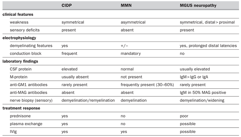
The spectrum of chronic acquired demyelinating polyneuropathies cover different entities that have recently been categorised in three major groups according to clinical, electrophysiological, pathophysiological and immunological data (table 1). These three major clinical syndromes are CIDP, multifocal motor neuropathy (MMN) and MGUS neuropathy. MGUS neuropathies may be categorised in MGUS IgM, IgG and IgA neuropathies. Regrouping MGUS neuropathies under the category of DADS (distal acquired demyelinating neuropathy) neuropathy [16] may be useful in characterisation of the disorder, but it should not be thought of as a separate disease [17].
Table 1.
Features of chronic acquired immune-mediated demyelinating polyneuropathies.
Chronic inflammatory demyelinating polyneuropathy (CIDP)
Chronic inflammatory demyelinating polyneuropathy (CIDP) should be separated from other chronic polyradiculopathies:
- –
- monoclonal proteins of undetermined significance (MGUS);
- –
- infections, such as those due to HIV-1, hepatitis C, cytomegalovirus, Epstein-Barr virus;
- –
- metabolic diseases (diabetes mellitus, uraemia, intermittant porphyria);
- –
- other immune neuropathies, e.g. anti-MAG neuropathy, non-systemic vasculitis, LED.
Though the majority of CIDP patients respond well to immunotherapy [18], no constant autoantibody activity has been reported.
Multifocal motor neuropathy (MMN)
Serum antibodies, mostly IgM to the ganglioside GM1 or less frequently to asialo-GM1, GD1a or GM2, have been reported by ELISA in a variable proportion of MMN patients with a prevalence for anti-GM1 IgM in most large series, ranging from 30 to 60% [19,20,21,22,23].The reasons for these discrepant figures are still unclear but may be related to differences in the ELISA procedure or in the controls used to establish normal reference values. Pestronk and Choksi [24], using a new ELISA technique based on the covalent linkage of GM1 to the secondary amino groups on Covalink ELISA plates, show the presence of anti-GM1 IgM antibodies in 85% of MMN patients.These results have not been confirmed. Carpo et al. [21] showed that the Covalink technique detected only a slightly higher proportion of MMN patients (35%) as compared with the standard ELISA (31%). Overall, these studies indicate that testing for anti-GM1 IgM may help confirm the diagnosis of multifocal motor neuropathy in positive patients even if a negative test does not exclude this diagnosis. GM1 is highly represented in the peripheral nervous system and several studies with antibodies to GM1 localised the presence of GM1 to the nodes of Ranvier [25], myelin [26] and the motor end plate of neuromuscular junction [27]. The higher concentration of GM1 (and GD1a) in motor nerve than in sensory nerve myelin was also considered consistent with the selective impairment of motor nerves observed in multifocal motor neuropathy [28]. Intraneural injection or exposure to sera from patients with high anti-GM1 antibodies and multifocal motor neuropathy was able to induce focal conduction block both in vivo and in vitro [29,30]. However, C. jejuni is unlikely to be involved in the pathogenesis of multifocal motor neuropathy in most patients [31].
MGUS neuropathy
There are theoretical and practical reasons for separating patients with MGUS neuropathy (also called paraproteinaemic neuropathy) from those without MGUS such as CIDP. The reasons are the following:
- –
- Patients with MGUS need to be carefully assessed and followed up for the presence of an associated plasma- or lympho-proliferative disorder.
- –
- MGUS IgM differ from CIDP or from MGUS IgG and IgA neuropathies in clinical, electrophysiological, histological and immunological features and response to treatment [32]. Risk for haematological malignancy in MGUS neuropathy is 25% after a median follow-up of 22 years [33]. The reported risk factors include M-protein level [34], increase of M-protein level during follow-up, light chain proteinuria, age >70 years [35]. Progression of the polyneuropathy, unexplained weight loss, and M-protein level >1 g/l are independent predictors for an underlying malignancy [36].
Antibodies to the myelin-associated glycoprotein (MAG) are detected in 50 to 60% of patients with neuropathy and immunoglobulin M (IgM) monoclonal gammopathy. Most patients have a slowly progressive, sensory or sensorimotor, demyelinating polyneuropathy [37].A causal relation between anti-MAG antibodies and the neuropathy is supported because pathologic studies of sural nerve biopsies of patients with neuropathy and anti-MAG IgM monoclonal gammopathy show demyelination associated with IgM deposits on the affected myelin sheaths [38]. The reactive epitope is the carbohydrate part of MAG and it is shared with a number of other neural glycoconjugates, including the major P0 glycoprotein of myelin, PMP22 as well as sulfoglycuronyl paragloboside (SGPG) [39,40]. Electrophysiologically there is a distal accentuation of conduction slowing in the anti-MAG polyneuropathy, suggesting a lengthdependent neuropathic process that begins distally [41]. IgM deposits are found on the basement membrane, but major areas of IgM deposits are localised to regions of non-compact myelin, such as Schmidt-Lantemann incisurves and paranodal loops [42]. The IgM deposits in myelin are proportional to the myelin widening [43], suggesting that the IgM induces the separation of the myelin leaflets by blocking or interfering with the adhesive properties of MAG or other HNK-1-containing glycoconjugates.
A recent study has addressed the diagnostic and prognostic value of anti-MAG antibodies in terms of future neurologic outcome or progression [44]. In a population of 65 patients with polyneuropathy associated with IgM monoclonal gammopathy, a slowly progressive disease course was associated with sensory symptoms, k-light chain of the IgM monoclonal protein, deposition of IgM in the sural nerve biopsy as well as elevated anti-MAG antibodies. Tremor was associated with malignant monoclonal gammopathy. However, in a multivariate analysis of outcome only initial symptoms and electrophysiological studies are independent prognostic factors: initial sensory symptoms of the feet are prognostic for a benign course at 4 years, while evidence of demyelination is prognostic for development of weakness of the upper extremities at 4 years. Addition of anti-MAG antibody tests did not yield any further prediction of outcome. Nobile-Orazio et al. [45] found no difference in neurological impairment between patients with moderate and very high titres of anti-MAG antibody in a large number of patients at study entry and at last follow-up after treatment. Investigators have suggested a strong correlation between reduced titres and clinical improvement [46,47] but these reports were limited to small series of cases. It has also been shown that serial measurement of CD57 positive lymphocytes (NK cells) is useful for monitoring treatment effect in the anti-MAG neuropathy [48]. In clinical practice, elevated anti-MAG anti-SGPG or antisulfatide antibody titres do not change management or prognosis in terms of future neurologic deficit or outcome of MGUS neuropathy [44].
Conclusion
When should one test for autoantibodies in the diagnostic work-up of a neuropathy? As outlined above, testing for serum autoantibodies is never the first step but an additional diagnostic measure after careful clinical and electrophysiological evaluation. High quality standards for a diagnostic antibody test should be applied. In our experience anti-MAG antibodies are a valuable marker that is relevant for diagnosis. Patients with chronic demyelinating polyneuropathies should be screened for anti-MAG antibodies. We test all our patients with lower motor neuron disease or motor neuropathies for anti-GM1 antibodies. We do not routinely test for anti-GM1 or related gangliosides in Guillain-Barré syndrome as the results have not yet a definite impact on the diagnosis or treatment regimen. Nevertheless, future studies are needed to address the usefulness of antiglycolipid testing with respect to progression or response to treatment in Guillain-Barré syndrome. If a Miller-Fisher syndrome is suspected, we test for anti-GQ1b antibodies to clarify the diagnosis.
Acknowledgments
The authors thank Mrs Maya Rolli for skilful secretarial help.
References
- Quarles, RH; Weiss, MD. Autoantibodies associated with peripheral neuropathy. Muscle Nerve 1999, 22, 800–22. [Google Scholar] [CrossRef]
- Renaud S, Leppert D, Steck AJ. Clinical and biological aspects of autoantibodies in immune neuropathies. In: Conrad K, Humbel RL, Meurer M, Shoenfeld Y, Tan EM, eds. Autoantigens and Autoantibodies: Diagnostic Tools and Clues to Understanding Autoimmunity. Lengerich: Pabst Science; 2000.
- Steck, AJ. Auto-antibody tests in peripheral neuropathies: pros and cons. J Neurol 2000, 247, 423–8. [Google Scholar] [CrossRef] [Scilit]
- Vincent A, Martins G, editors. Autoantibodies in Neurological Diseases. Milano: Springer-Verlag; 2002.
- Holloway, RG; Feasby, TE. To test or not to test – that is the question. Neurology 1999, 53, 1905–7. [Google Scholar] [CrossRef] [Scilit]
- Gallardo, E; Rojas-Garcia, R; Belvis, R. Antiganglioside antibodies: when, which and for what. Neurologia 2001, 16, 293–7. [Google Scholar]
- Van der Meche, FG; Van Doorn, PA; Meulstee, J; Jennekens, FG. Diagnostic and classification criteria for the Guillain-Barré syndrome. Eur Neurol 2001, 45, 133–9. [Google Scholar] [CrossRef] [Scilit]
- Kuwabara, S; Mori, M; Ogawara, K; Hattori, T; Oda, S; Koga, M; et al. Intravenous immunoglobulin therapy for Guillain-Barré syndrome with IgG anti-GM1 antibody. Muscle Nerve 2001, 24, 54–8. [Google Scholar] [CrossRef] [Scilit] [PubMed]
- Jacobs, BC; van Doorn, PA; Groeneveld, JH; Tio-Gillen, AP; van der Meche, FG. Cytomegalovirus infections and anti-GM2 antibodies in Guillain-Barré syndrome. J Neurol Neurosurg Psychiatry 1997, 62, 641–3. [Google Scholar] [CrossRef] [Scilit]
- Visser, LH; van der Meche, FG; Meulstee, J; Rothbarth, PP; Jacobs, BC; Schmitz, PI; et al. Cytomegalovirus infection and Guillain-Barré syndrome: the clinical, electrophysiologic, and prognostic features. Dutch Guillain-Barré Stud Group. Neurology 1996, 47, 668–73. [Google Scholar] [CrossRef] [Scilit]
- Jacobs, BC; van Doorn, PA; Schmitz, PI; Tio-Gillen, AP; Herbrink, P; Visser, LH; et al. Campylobacter jejuni infections and anti-GM1 antibodies in Guillain-Barré syndrome. Ann Neurol 1996, 40, 181–7. [Google Scholar] [CrossRef] [Scilit]
- McKhann, GM; Cornblath, DR; Griffin, JW; Ho, TW; Li, CY; Jiang, Z; et al. Acute motor axonal neuropathy: a frequent cause of acute flaccid paralysis in China. Ann Neurol 1993, 33, 333–42. [Google Scholar] [CrossRef] [Scilit] [PubMed]
- Ho, TW; Willison, HJ; Nachamkin, I; Li, CY; Veitch, J; Ung, H; et al. Anti-GD1a antibody is associated with axonal but not demyelinating forms of Guillain-Barré syndrome. Ann Neurol 1999, 45, 168–73. [Google Scholar] [CrossRef] [Scilit] [PubMed]
- Odaka, M; Yuki, N; Hirata, K. Anti-GQ1b IgG antibody syndrome: clinical and immunological range. J Neurol Neurosurg Psychiatry 2001, 70, 50–5. [Google Scholar] [CrossRef] [Scilit]
- Koga, M; Yuki, N; Ariga, T; Morimatsu, M; Hirata, K. Is IgG anti-GT1a antibody associated with pharyngealcervical-brachial weakness or oropharyngeal palsy in Guillain-Barré syndrome? J Neuroimmunol 1998, 86, 74–9. [Google Scholar] [CrossRef] [Scilit]
- Katz, JS; Saperstein, DS; Gronseth, G; Amato, AA; Barohn, RJ. Distal acquired demyelinating symmetric neuropathy. Neurology 2000, 54, 615–20. [Google Scholar] [CrossRef] [Scilit] [PubMed]
- Dyck, PJ; Dick, PJ. Atypical varieties of chronic demyelinating neuropathies. Lancet 2000, 355, 1293–4. [Google Scholar] [CrossRef] [Scilit]
- Hahn, AF. Treatment of chronic inflammatory demyelinating polyneuropathy with intravenous immunoglobulin. Neurology 1998, 51 6 Suppl 5, S16–S21. [Google Scholar] [CrossRef] [Scilit]
- Adams, D; Kuntzer, T; Steck, AJ; Burger, D; Chofflon, M; Magistris, MR; et al. Predictive value of anti-GM1 ganglioside antibodies in neuromuscular diseases: a study of 180 sera. J Neuroimmunol 1991, 32, 223–30. [Google Scholar] [CrossRef] [Scilit]
- Azulay, JP; Rihet, P; Pouget, J; Cador, F; Blin, O; Boucraut, J; et al. Long-term follow-up of multifocal motor neuropathy with conduction block under treatment. J Neurol Neurosurg Psychiatry 1997, 62, 391–4. [Google Scholar] [CrossRef] [Scilit]
- Carpo, M; Allaria, S; Scarlato, G; Nobile-Orazio, E. Marginally improved detection of GM1 antibodies by Covalink ELISA in multifocal motor neuropathy. Neurology 1999, 53, 2206–7. [Google Scholar] [CrossRef] [Scilit]
- Sadiq, SA; Thomas, FP; Kilidireas, K; Protopsaltis, S; Hays, AP; Lee, KW; et al. The spectrum of neurological disease associated with anti-GM1 antibodies. Neurology 1990, 40, 1067–72. [Google Scholar] [CrossRef] [Scilit] [PubMed]
- Taylor, BV; Wright, RA; Harper, CM; Dyck, PJ. Natural history of 46 patients with multifocal motor neuropathy with conduction block. Muscle Nerve 2000, 23, 900–8. [Google Scholar] [CrossRef] [Scilit]
- Pestronk, A; Choksi, R. Multifocal motor neuropathy. Serum IgM anti-GM1 ganglioside antibodies in most patients detected using covalent linkage of GM1 to ELISA plates. Neurology 1997, 49, 1289–92. [Google Scholar] [CrossRef] [Scilit]
- Santoro, M; Uncini, A; Corbo, M; Staugaitis, SM; Thomas, FP; Hays, AP; et al. Experimental conduction block induced by serum from a patient with anti-GM1 antibodies. Ann Neurol 1992, 31, 385–90. [Google Scholar] [CrossRef] [Scilit] [PubMed]
- Corbo, M; Quattrini, A; Latov, N; Hays, AP. Localization of GM1 and Gal(β1–3)GalNAc antigenic determinants in peripheral nerve. Neurology 1993, 43, 809–14. [Google Scholar] [CrossRef] [Scilit]
- Schluep, M; Steck, AJ. Immunostaining of motor nerve terminals by IgM M-protein with activity against gangliosides GM1 and GD1b from a patient with motor neuron disease. Neurology 1988, 38, 1890–2. [Google Scholar] [CrossRef] [Scilit] [PubMed]
- Ogawa-Goto, K; Funamoto, N; Ohta, Y; Abe, T; Nagashima, K. Myelin gangliosides of human peripheral nervous system: an enrichment of GM1 in the motor nerve myelin isolated from cauda equina. J Neurochem 1992, 59, 1844–9. [Google Scholar] [CrossRef] [Scilit]
- Arasaki, K; Kusunoki, S; Kudo, N; Kanazawa, J. Acute conduction block in vitro following exposure to anti-ganglioside sera. Muscle Nerve 1993, 16, 587–93. [Google Scholar] [CrossRef] [Scilit]
- Uncini, A; Santoro, M; Corbo, M; Lugaresi, A; Latov, N. Conduction abnormalities induced by sera of patients with multifocal motor neuropathy and anti-GM1 antibodies. Muscle Nerve 1993, 16, 610–5. [Google Scholar] [CrossRef] [Scilit]
- Terenghi, F; Allaria, S; Scarlato, G; Nobile-Orazio, E. Multifocal motor neuropathy and Campylobacter jejuni reactivity. Neurology 2002, 59, 282–4. [Google Scholar] [CrossRef] [Scilit]
- Yeung, KB; Thomas, PK; King, RH; Waddy, H; Will, RG; Hughes, RA; et al. The clinical spectrum of peripheral neuropathies associated with benign monoclonal IgM, IgG and IgA paraproteinaemia. Comparative clinical, immunological and nerve biopsy findings. J Neurol 1991, 238, 383–91. [Google Scholar] [CrossRef] [Scilit]
- Kyle, RA. Monoclonal proteins in neuropathy. Neurol Clin 1992, 10, 713–34. [Google Scholar] [CrossRef] [Scilit] [PubMed]
- Van der Poel, HMW; Coebergh, JWW; Hillen, HFP. Malignant transformation of monoclonal gammopathy of undetermined significance among out-patients of a community hospital in Southeastern Netherlands. Br J Haematol 1995, 91, 121–5. [Google Scholar] [CrossRef] [Scilit]
- Baldini, L; Guffanti, A; Cesana, BM. Role of different hematologic variables in defining the risk of malignant transformation in monoclonal gammopathy. Blood 1996, 87, 912. [Google Scholar] [CrossRef] [Scilit] [PubMed]
- Eurelings, M; Notermans, NC; Van de Donk, NW; Lokhorst, HM. Risk factors for hematological malignancy in polyneuropathy associated with monoclonal gammopathy. Muscle Nerve 2001, 24, 1295–302. [Google Scholar] [CrossRef] [Scilit]
- Ropper, AH; Gorson, KC. Neuropathies associated with paraproteinemia. N Engl J Med 1998, 338, 1601–7. [Google Scholar] [CrossRef] [Scilit]
- Miescher, GC; Steck, AJ. Paraproteinaemic neuropathies. Baillieres Clin Neurol 1996, 5, 219–32. [Google Scholar]
- Bollensen, E; Steck, AJ; Schachner, M. Reactivity with the peripheral myelin glycoprotein P0 in serum from patients with monoclonal gammopathy and polyneuropathy. Neurology 1988, 38, 1266–70. [Google Scholar] [CrossRef] [Scilit]
- Chou, KH; Ilyas, AA; Evans, BA; Costello, C; Quarles, RH; Jungalwala, FB. Structure of sulfated glucuronyl glycolipids in the nervous system reacting with HNK-1 antibody and some IgM paraproteins in neuropathy. J Biol Chem 1986, 261, 11717–25. [Google Scholar] [CrossRef] [Scilit]
- Radziwill, AJ; Renaud, S; Steck, AJ. Is distal motor latency a sensitive marker of anti-MAG polyneuropathy? Neurology 2001, 56 8 Suppl 3, A399. [Google Scholar]
- Gabriel, JM; Erne, B; Miescher, GC; Miller, SL; Vital, A; Vital, C; et al. Selective loss of myelin-associated glycoprotein from myelin correlates with anti-MAG antibody titer in demyelinating paraproteinaemic polyneuropathy. Brain 1996, 119, 775–87. [Google Scholar] [CrossRef] [Scilit] [PubMed]
- Ritz, M; Erne, B; Ferracin, F. Anti-MAG IgM penetration into myelinated fibers correlates with the extent of myelin widening. Muscle Nerve 1999, 22, 1030–7. [Google Scholar] [CrossRef] [Scilit]
- Eurelings, M; Moons, KG; Notermans, NC; Sasker, LD; De Jager, AE; Wintzen, AR; et al. Neuropathy and IgM M-proteins. Prognostic value of antibodies to MAG, SGPG, and sulfatide. Neurology 2001, 56, 228–33. [Google Scholar] [CrossRef] [Scilit]
- Nobile-Orazio, E; Meucci, N; Baldini, L; Di Troia, A; Scarlato, G. Long-term prognosis of neuropathy associated with anti-MAG IgM M-proteins and its relationship to immune therapies. Brain 2000, 123, 710–7. [Google Scholar] [CrossRef] [Scilit] [PubMed]
- Blume, G; Pestronk, A; Goodnought, LT. Anti-MAG antibody associated polyneuropathies: improvement following immunotherapy with monthly plasma exchange and IV cyclophosphamide. Neurology 1995, 45, 1577–80. [Google Scholar] [CrossRef] [Scilit] [PubMed]
- Gorson, KC; Ropper, AH; Weinberg, DH. Treatment experience in patients with anti-myelin-associated glycoprotein neuropathy. Musle Nerve 2001, 24, 778–86. [Google Scholar] [CrossRef] [Scilit] [PubMed]
- Tagawa, Y; Yuki, N; Ohnishi, A; Hirata, K; Hosokawa, S. Parameters for monitoring treatment effects in CIDP with anti-MAG/SGPG IgM antibody. Muscle Nerve 2001, 24, 701–4. [Google Scholar] [CrossRef] [Scilit]

This is an open-access article distributed under the terms of the Creative Commons Attribution Non-Commercial No Derivatives (CC BY-NC-ND) 4.0 License.