


Summary
Hypersomnolent state, severe amnesia, transient coma and vertical gaze abnormalities are characteristic features of bilateral paramedian thalamic infarcts. Vergence disorders or blepharospasm has recently been reported but there is no study reporting blepharospasm together with exotropia. In this paper, we present a case with bilateral paramedian thalamic infarct who had blepharospasm and acute exotropia. A 60-year-old, right-handed, male patient who was unconscious was admitted to the hospital. His history revealed a speech disorder and a headache preceding his unconsciousness. On physical examination the pulse rate was 86/min/regular, the blood pressure 200/120 mm Hg, and respirations 14 per minute. On neurologic examination, he was stuporous and his pupils were equal and myotic. Bilateral light reaction was diminished. There was bilateral persistent tonic spasm of the eyelids and the eyes were in divergence position. Plantar reflexes were bilateral extensor. Cranial CT disclosed bilateral hypodense infarct areas in the paramedian thalamic localisation. The patient was treated with an antiaggregant, dexamethasone and antihypertensive. No change was found in blepharospasm and divergence position during therapy. The patient died on the seventh day after stroke onset. The supranuclear organisation of the vergence system is not well-known, though conjugate eye movements are mostly understood. Dysfunction of the vergence in structures contributing to neural integration may be due to disordered convergence secondary to organic conditions. Although the anatomic pathway of these signals has not yet been identified, divergence eye movements have been assumed to result from a complementary input from divergence neurons to the lateral rectus motoneurons. Divergence position in our case might be related to convergence paralysis or excessive stimulation of divergence neurons due to involvement of vergence pathways. Data from previous studies indicate that convergence paralysis might be more responsible for acute exotropia in bilateral paramedian thalamic lesions. The pathophysiological mechanism for disorders of eyelid movements is not well known. Bilateral eyelid opening in awake patients by unilateral stimulation in the prefrontal, occipital and rarely precentral cortices has been demonstrated. Reflex blepharospasm or excessive eyelid closure that is provoked by attempts to hold the eyelids open is seen with nondominant hemispheric infarction, brainstem lesions or rarely in thalamic vascular events. In conclusion, we propose that vergence and eyelid functions are already closely linked with the paramedian thalamic area and the descending cortical pathways which pass through the paramedian thalamus and exert an inhibitory input to premotor vergence neurons in the midbrain. We would like to point out the fact that although paramedian thalamic infarcts are considered not to be fatal, our patient died on the seventh day after stroke onset. This case raises the question of whether acute vergence together with eyelid function abnormalities could be a negative prognostic sign.
Introduction
The thalamus is a crossing point for ocular pathways. Various ocular abnormalities such as vertical gaze paralysis, skew deviations, pseudoabducens palsy, convergence-retraction nystagmus, wronggoing eyes and Horner’s syndrome may be observed due to vascular thalamic lesions [1,2]. Vertical gaze paralysis is the most frequent ocular finding of bilateral paramedian thalamic infarct [1,2,3,4,5,6,7,8,9]. Although vergence abnormalities or blepharospasm has recently been reported, the supranuclear pathways for vergence eye movements are poorly understood [3,6,7,8,9].
In this paper, we present a case with bilateral paramedian thalamic infarct who had blepharospasm and acute exotropia.
Case report
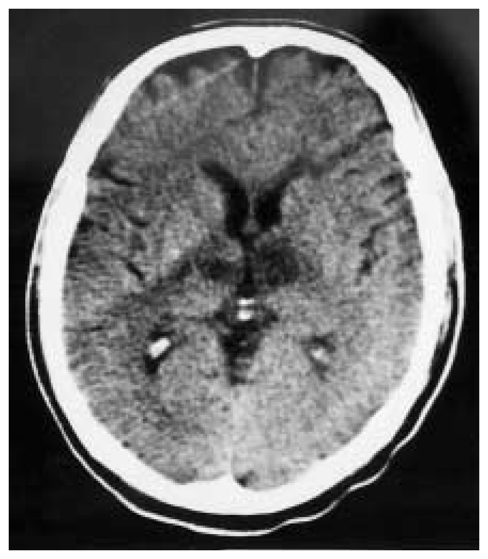
A 60-year-old, right-handed, male patient who was unconscious was admitted to the hospital. His history revealed a speech disorder and a headache preceding his unconsciousness. In his past history, there was no cerebrovascular or other neurological diseases. He was hypertensive and on irregular antihypertensive agent. On physical examination the pulse rate was 86/min/regular, the blood pressure 200/120 mm Hg, and respirations 14 per minute. There were no cervical bruits. On neurologic examination he was stuporous and his pupils were equal and myotic. Bilateral light reaction was diminished. Fundoscopic examination was normal bilaterally. Vertical oculocephalic movements were full. There was bilateral persistent tonic spasm of the eyelids and the eyes were in divergence position (fig. 1). Plantar reflexes were bilateral extensor. Routine blood-urine investigations were normal.The electrocardiogram revealed left ventricular hypertrophy due to hypertension. In carotid artery Doppler examination, intimal thickening and small plaques were found but vertebral arteries were normal. Cranial CT disclosed bilateral hypodense infarct areas in the paramedian thalamic localisation (fig. 2).
The patient was treated with an antiaggregant (acetylsalicylic acid 300 mg/day), dexamethasone (16 mg/day/intravenously) and antihypertensive agent (enalapril maleat 20 mg/day). No change was found in blepharospasm and divergence position during therapy. The patient died on the seventh day after stroke onset.
Figure 1.
Divergence position in the eyes of the patient.
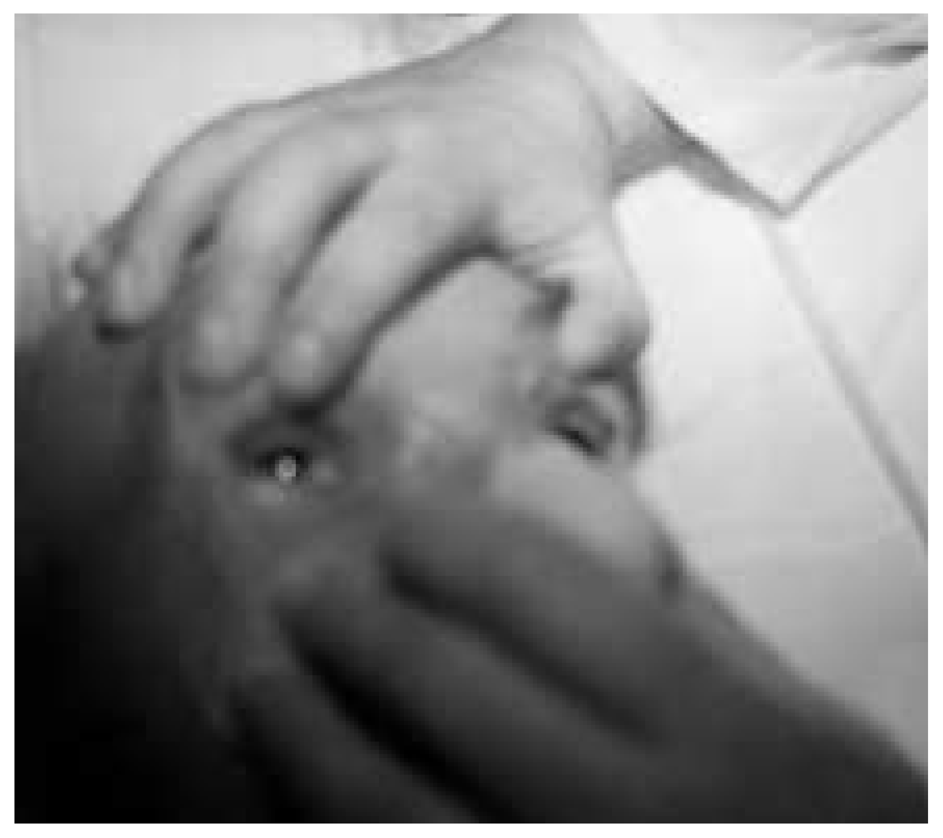
Figure 2.
Bilateral paramedian thalamic infarct on CT scan.
Discussion
The supranuclear organisation of the vergence system is not well-known though conjugate eye movements are mostly understood [6]. The vergence pathways have been defined in the monkey as lying dorsal and lateral to the oculomotor nucleus but the human analogue is unknown [1,6,8,9]. In the monkey, “vergence neurons” specifically involved in the control of vergence have been found 1 to 2 mm dorsal and dorsolateral to the oculomotor nucleus. There are three main types of neurons: vergence tonic cells that discharge in association with vergence angle vergence burst cells that discharge in association with vergence velocity and vergence burst-tonic cells that discharge in association with both angle and velocity. Most vergence tonic cells discharge in relation to the angle of convergence but some discharge in association with the angle of divergence. Dysfunction of the vergence in structures contributing to neural integration may be due to disordered convergence secondary to organic conditions [3,6,7]. Although the anatomic pathway of these signals has not yet been identified, divergence eye movements have been assumed to result from a complementary input from divergence neurons to the lateral rectus motoneurons [3,7]. The divergence position in our case might be related to convergence paralysis or excessive stimulation of divergence neurons due to involvement of vergence pathways. Data from previous studies indicate that convergence paralysis might be more responsible for acute exotropia in bilateral paramedian thalamic lesions [6,7].
The pathophysiological mechanism for disorders of eyelid movements is not well known. Bilateral eyelid opening in awake patients by unilateral stimulation in the prefrontal, occipital, and rarely precentral cortices has been demonstrated. Reflex blepharospasm or excessive eyelid closure that is provoked by attempts to hold the eyelids open is seen with nondominant hemispheric infarction, brainstem lesions or rarely in thalamic vascular events [8].
In conclusion, we propose that vergence and eyelid functions are closely linked with the paramedian thalamic area and the descending cortical pathways which pass through the paramedian thalamus and exert an inhibitory input to premotor vergence neurons in the midbrain. We would like to point out the fact that although paramedian thalamic infarcts are considered not to be fatal, our patient died on the seventh day after stroke onset. This case raises the question of whether acute vergence together with eyelid function abnormalities could be a negative prognostic sign.
References
- Guberman A, Stuss D. The syndrome of bilateral paramedian thalamic infarction. Neurology 1983;33:540–6. [CrossRef] [Scilit]
- Gentilini M, Renzi ED, Crisi G. Bilateral paramedian thalamic artery infarcts: report of eight cases. J Neurol Neurosurg Psychiatry 1987;50:900–9. [CrossRef] [Scilit] [PubMed]
- Pullicino P, Lincoff N, Truax BT. Abnormal vergence with upper brainstem infarcts. Neurology 2000;55:352–8. [CrossRef] [Scilit] [PubMed]
- Biller J, Sand JJ, Corbett JJ, Adams HP, Dunn V. Syndrome of the paramedian thalamic arteries: clinical and neuroimaging correlation. J Clin Neuroophthalmol 1985;5:217–23. [PubMed]
- Gerber O, Gudesblatt M. Bilateral paramedian thalamic infarctions: a CT study. Neuroradiology 1986;28:128–31. [CrossRef] [Scilit] [PubMed]
- Wiest G, Mallek R, Baumgartner C. Selective loss of vergence control secondary to bilateral paramedian thalamic infarction. Neurology 2000;54:1997–9. [CrossRef] [Scilit] [PubMed]
- Gomez CR, Gomez SM, Selhort JB. Acute thalamic esotropia. Neurology 1988;38:1759–62. [CrossRef] [Scilit] [PubMed]
- Verghese J, Miling C, Rosenbaum DM. Ptosis, blepharospasm, and apraxia of eyelid opening secondary to putaminal hemorrhage. Neurology 1999;53:652. [CrossRef] [Scilit] [PubMed]
- 9. Deleu D. Selective vertical saccadic palsy from unilateral medial thalamic infarction: clinical, neurophysiologic and MRI correlates. Acta Neurol Scand 1997;96:332–6. [CrossRef] [Scilit] [PubMed]
This is an open-access article distributed under the terms of the Creative Commons Attribution Non-Commercial No Derivatives (CC BY-NC-ND) 4.0 License.