


Abstract
Background: Candida species are among the most frequently isolated fungi from respiratory specimens in hospitalised patients, yet the clinical significance of their presence—infection versus colonisation, treatment versus observation—remains an unresolved challenge in clinical microbiology. Data from dedicated pulmonology centres in Eastern Europe are scarce. This study aimed to characterise the three-year epidemiology, species distribution, temporal trends, colony-load profiles, and longitudinal mycological trajectories of fungal respiratory isolates at a Romanian tertiary pulmonology hospital. Methods: We retrospectively analysed 1000 microbiological records from bronchial aspirates and other respiratory specimens collected between January 2022 and December 2024. Variables included patient demographics, clinical ward, primary diagnosis, fungal species, and semi-quantitative colony counts. Patients with multiple records were identified and analysed as a longitudinal sub-cohort. Results: Candida albicans predominated (45.5%), with a declining proportional trend across the study period. Candida krusei—intrinsically resistant to fluconazole—increased from 5.3% of isolates in 2022 to 9.7% in 2024. The intensive care unit (ICU) accounted for 25.8% of all records. Patients were predominantly male (67.5%) with a mean age of 63.4 years; acute respiratory failure was the most frequent underlying diagnosis (41.7%). High-load colonisation (>100 colony-forming units/mL) was documented in 19.0% of quantified specimens. Among 91 patients with serial cultures, 26 demonstrated inter-sample species shifts—several consistent with antifungal selection pressure—and 29 showed escalating colony loads, suggesting progression from colonisation toward probable infection. Conclusions: In a clinical setting where fluconazole has historically served as the default empirical antifungal agent, a C. krusei proportion approaching 10% of annual isolates constitutes a clinically actionable finding. Species-level identification of all fungal respiratory isolates is strongly warranted, as it directly influences therapeutic decision-making in approximately one in ten cases.
1. Introduction
Candida species represent the most common opportunistic fungal pathogens encountered in hospital settings worldwide. Their detection in respiratory specimens from patients with respiratory disease is a frequent and clinically consequential finding. Despite decades of research, the interpretation of a positive bronchial aspirate culture remains genuinely challenging: distinguishing true pulmonary infection from airway colonisation—and determining which patients need antifungal therapy—continues to represent one of the more difficult diagnostic dilemmas in clinical microbiology [1,2]. The absence of validated quantitative thresholds equivalent to those established for bacterial ventilator-associated pneumonia, combined with the non-specific clinical and radiological presentation of pulmonary candidiasis, means that microbiological results must always be interpreted within their clinical context rather than in isolation [3,4].
This ambiguity is particularly pronounced in pulmonology. Patients with chronic obstructive pulmonary disease (COPD), old tuberculosis scars, lung cancer, haemoptysis, and those requiring prolonged mechanical ventilation have risk factors that encourage Candida to thrive in the airways. Disrupted mucociliary clearance, inhaled and systemic corticosteroids, prior antibiotic courses, and varying degrees of immune compromise create favourable conditions for colonisation [5,6]. At the same time, these are patients in whom unnecessary antifungal therapy carries real risks: drug toxicity, cost, promotion of resistance, and treatment for what may be a harmless bystander organism.
What makes the current moment particularly important is the quiet but steady increase in non-albicans Candida (NAC) species. Candida krusei has always been intrinsically resistant to fluconazole—but when it was rare, that resistance rarely mattered in practice [7,8,9]. As its proportion grows, the stakes of misidentification rise accordingly. C. glabrata navigates azoles with dose-dependent susceptibility that can turn into resistance under treatment pressure [10]. The global emergence of Candida auris adds a further dimension of urgency to mycological epidemiological surveillance efforts. First described in 2009, this species has since been reported across six continents and is now classified by the World Health Organization as a critical-priority fungal pathogen [11,12]. This is mostly due to its capacity for multidrug resistance, including pan-resistance in some isolates; its exceptional environmental persistence on surfaces and medical equipment; and its demonstrated ability to cause sustained nosocomial transmission chains in healthcare settings, which would not be expected from other, more common Candida species [11,12]. Identifying these species reliably and promptly is no longer a specialist curiosity; it is a clinical necessity.
Despite all this, published epidemiological surveillance data from dedicated pulmonology hospitals in Eastern Europe remain limited. The region carries a disproportionate burden of respiratory disease—tuberculosis sequelae, post-coronavirus disease 2019 (COVID-19) lung damage, and high rates of smoking-related COPD—yet its mycological epidemiology rarely makes it into the literature. The present study attempts to fill part of that gap, describing three years of fungal respiratory epidemiological surveillance at a Romanian tertiary pulmonology hospital across 1000 records, with special attention to species trends, ward-level patterns, patient demographics, colony-load dynamics, and—uniquely—the longitudinal trajectories of the 91 patients who were sampled more than once.
2. Materials and Methods
2.1. Study Design and Setting
This is a single-centre retrospective observational study at a tertiary-level pulmonology hospital in Romania, comprising a medical ICU, a pulmonary surgery ward, and seven pneumology wards with a combined inpatient capacity of approximately 500 beds. Electronic microbiological and clinical records were extracted for January 2022 through December 2024. This retrospective study was conducted in accordance with the Declaration of Helsinki (revised 2013). All data were fully anonymised prior to analysis. The study protocol was reviewed and approved by the Institutional Ethics Committee of the Marius Nasta Institute of Pneumology, Bucharest, Romania (approval no. 23343, 17 October 2023). The requirement for individual informed consent was formally waived by the same committee, given the retrospective, fully anonymised nature of the data.
2.2. Study Population, Inclusion and Exclusion Criteria
All records—inpatient and outpatient—with at least one respiratory specimen yielding fungal growth were included. No lower or upper age limit was applied; patients of any age with a qualifying respiratory specimen were eligible, including nine paediatric cases (age range 1–92 years). No sex-based restriction was applied. Eligible clinical contexts included all inpatient wards (ICU, pulmonary surgery, and the seven pneumology wards) as well as outpatient consultations, provided a qualifying respiratory specimen was obtained. The primary specimen type was bronchial aspirate obtained during flexible bronchoscopy or via endotracheal/tracheostomy tube aspiration; pleural fluid and bronchoalveolar lavage samples were included where available. Duplicate isolates from the same patient within the same admission were deliberately retained as separate records: in the ICU, a patient sampled weekly across a six-week stay generates cultures that each reflect a clinically distinct moment, and collapsing them into a single record would obscure the exact longitudinal patterns we were trying to characterise. Regarding the exclusion criteria, specimens from non-respiratory sources (e.g., urine, wound, ear, nasal, vaginal, or catheter-tip swabs) were excluded, as were records with incomplete or missing essential data (patient demographics, specimen type, or organism identification) and cultures judged to represent likely contaminants rather than clinically relevant isolates. For the cross-sectional analyses, all 1000 records were used; for the longitudinal analysis, patient identity was established by name-matching across the full dataset before anonymisation and codification of data, ensuring confidentiality.
2.3. Study Population Characteristics
The 1000 respiratory specimens analysed comprised bronchial aspirates obtained via endotracheal or tracheostomy tube suction (n = 989), pleural fluid (n = 16), tracheal aspirate (n = 4), and bronchoalveolar lavage (n = 1). For mycological analysis, a standardised 0.01 mL calibrated inoculation loop was used for specimen plating, in accordance with internal laboratory protocol. These 1000 records were derived from 888 unique patients: 794 patients (89.4%) contributed a single specimen, while 94 patients contributed multiple specimens across their admission (range 2–8 samples per patient; mean 1.14 samples/patient overall). As detailed in Section 2.2, duplicate specimens from the same patient were retained as separate records rather than collapsed, since each reflects a distinct clinical time point; this multi-sample subgroup forms the basis of the longitudinal analysis presented in Section 3.7. For each record, the following variables were extracted: year and month of admission, ward, patient sex and age, primary admitting diagnosis, portage designation (where documented), specimen type, yeast species, semi-quantitative colony count, co-isolated filamentous fungi, and identification method.
2.4. Mycological Identification
According to internal protocols and manufacturer recommendations, fungal isolation was performed using a standardised 0.01 mL calibrated inoculation loop to plate each specimen onto Sabouraud dextrose agar with chloramphenicol (PO0161A Thermo Fisher Scientific, Waltham, MA, USA), followed by incubation for 48 h at 35 °C and for an additional 72 h at 30 °C. The initial 48 h incubation at 35 °C favoured rapid recovery of yeast species, while the extended 72 h incubation at the lower temperature of 30 °C allowed for the growth and sporulation of slower-growing filamentous fungi, which typically require lower temperatures for morphological development and reliable identification. Species identification relied on chromogenic medium (Brilliance Candida agar, PO1034A; Thermo Fisher Scientific, USA), based on the manufacturer’s colour-reaction chart, for all 1000 isolates. For isolates originating from the ICU (n = 258), identification was additionally confirmed using VITEK® 2 automated identification (bioMérieux, Marcy-l’Étoile, France); in all cases, VITEK® 2 results were concordant with the chromogenic medium results. For isolates from other wards (n = 742), identification relied on chromogenic medium alone, without VITEK® 2 confirmation. All six Cryptococcus laurentii isolates were confirmed by VITEK® 2 automated identification, given the atypical nature of this finding in a Candida-focused surveillance dataset and its established association with underlying immunosuppression, warranting definitive rather than presumptive identification. This distinction reflects internal clinical protocol, under which ICU patients—considered at higher risk of invasive disease—routinely underwent confirmatory identification alongside antifungal susceptibility testing, while isolates from other wards, more often reflecting colonisation in a non-critical setting, did not. Throughout this manuscript, fungal species are referred to by their previously established nomenclature—for example, Candida krusei rather than Pichia kudriavzevii and Candida glabrata rather than Nakaseomyces glabrata—in the interest of clinical clarity and to maintain consistency with the terminology most familiar to the intended readership, including clinicians, microbiologists, and infection control practitioners. It should be noted, however, that all epidemiological, microbiological, and clinical data presented refer to organisms as currently defined under their accepted taxonomic classification, and the use of legacy nomenclature throughout this work does not imply any departure from their correct phylogenetic assignment.
Colony counts were recorded semi-quantitatively: exact values for sparse growth (<30 colony-forming units [CFU]/mL) and tiered estimates (>30 through >100 CFU/mL) for heavier yields.
2.5. Data Variables and Statistical Analysis
Using the variables described in Section 2.3, the analysis is descriptive throughout. Categorical variables were expressed as frequencies and percentages; continuous variables (age) were summarised as mean ± standard deviation (SD), median, and range. For the longitudinal subset, patient trajectories were classified by species identity, colony-load direction, and ward-transfer pattern. To identify risk factors associated with non-albicans Candida species (versus C. albicans), a multivariate binary logistic regression was performed, with species category (non-albicans vs. albicans) as the outcome and patient age, sex, care area (ICU vs. other wards), and semi-quantitative colony load (log-transformed) as predictors. Records with unspeciated Candida spp., filamentous fungi only, or mixed two-species results were excluded from this analysis, as were records with missing age, sex, or colony-load data (n = 595 of 1000 records were analysed). Odds ratios (OR) with 95% confidence intervals (CI) were calculated using the Wald method; p < 0.05 was considered statistically significant.
To assess whether retaining duplicate isolates from the same patient (Section 2.2) introduced bias into the species distribution, a sensitivity analysis was performed on the 989 of 1000 records for which patient-level deduplication could be verified against the source laboratory database, comparing the full set of these records against a deduplicated version retaining only the first chronological isolate per patient (871 unique patients). Species-level percentages differed by no more than 0.7 percentage points between the two approaches, with the exception of Cryptococcus laurentii, a low-frequency species concentrated in a small number of multi-sample patients, for which the relative proportion was more sensitive to duplicate retention. Given the very small proportion of records not covered by this check (11 of 1000; the specimen subtypes described in Section 2.2), we consider these findings representative of the full cohort. Analyses were performed using Python 3.11.
3. Results
3.1. Patient Demographics
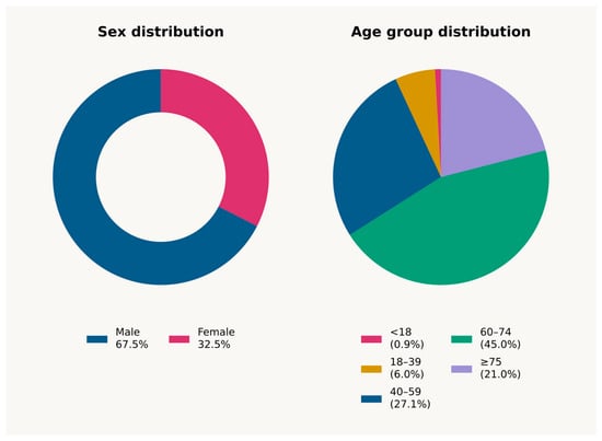
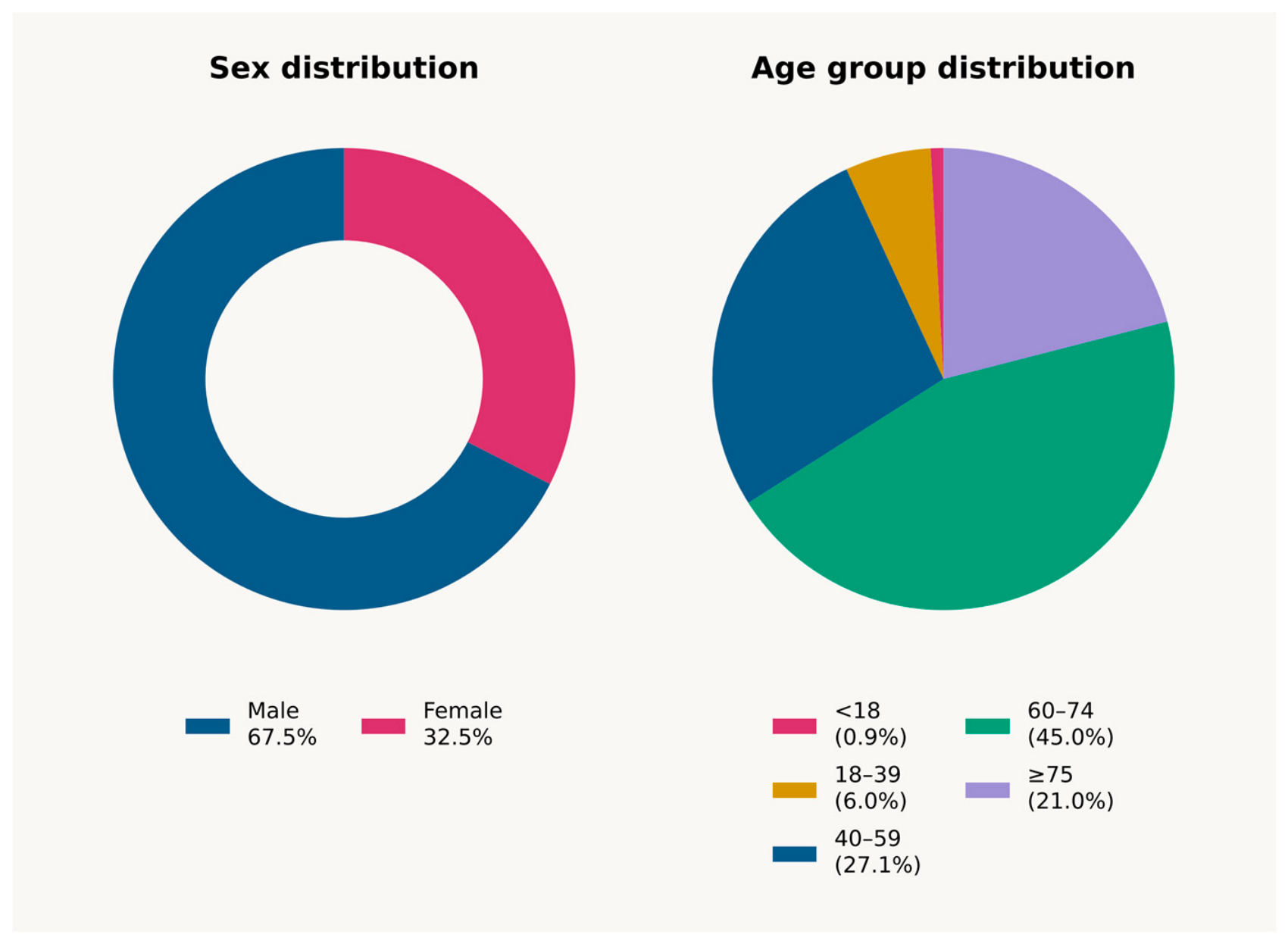
More than half of the records (67.5%) were from male patients, and 32.5% were from female patients (Figure 1). The overall mean age was 63.4 ± 14.5 years (median 66; range 1–92). Two-thirds of all patients were aged 60 or older. Nine paediatric cases were identified.
Figure 1.
Sex (doughnut, left) and age group (pie, right) distribution of sex/age data. Patients aged 60–74 years constitute 45.0% of the cohort; together with the ≥75 group (21.0%), patients aged 60 or older account for 66.0% of all records.
3.2. Seasonal Distribution
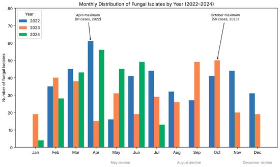
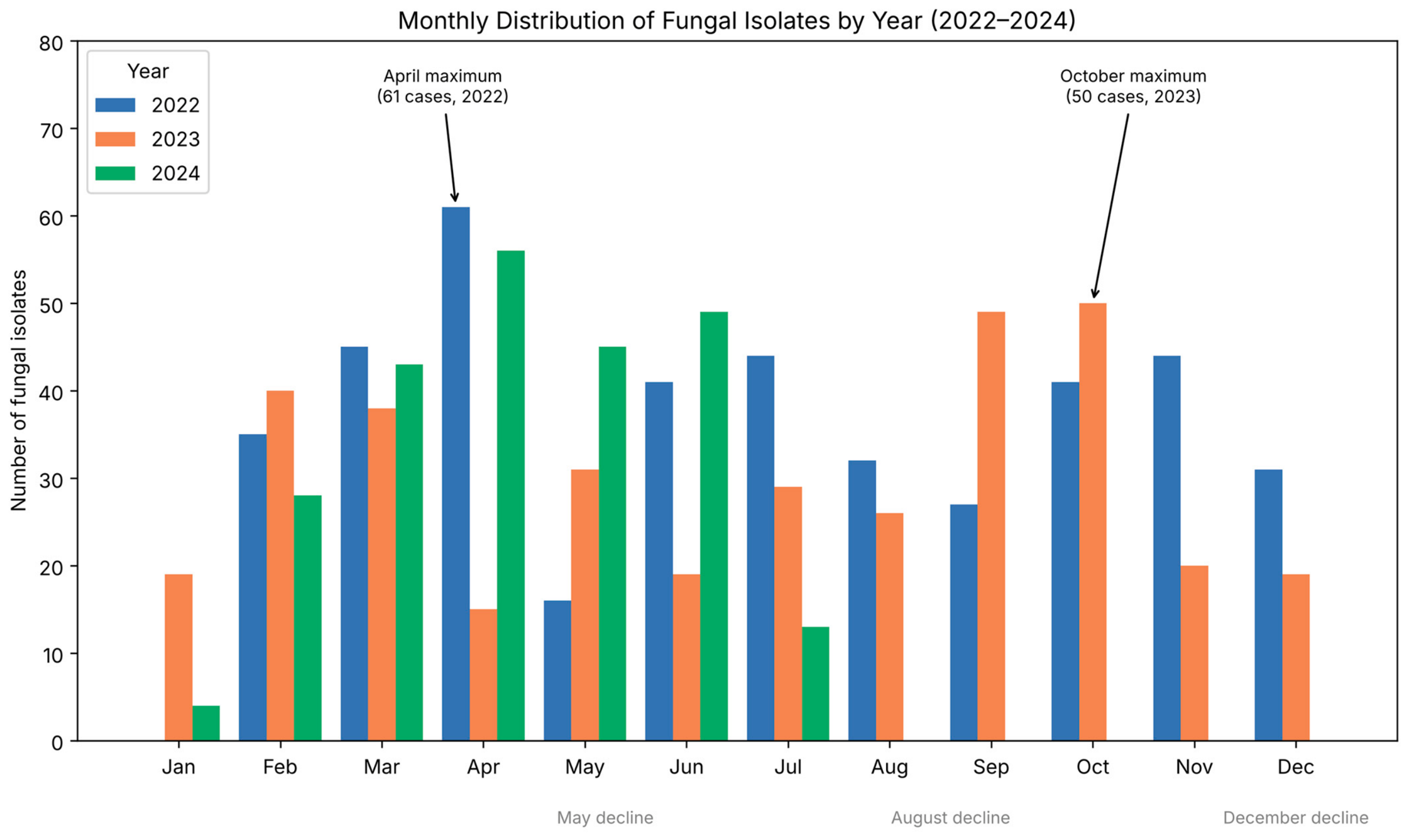
Monthly analysis across all three years revealed the highest monthly counts in spring (April: 131 cases; March: 123 cases), a secondary elevation in June (107 cases), and a distinct autumn maximum in October (91 cases combined across years), which was in fact the single highest month of 2023 (50 cases), exceeding even the spring months of that year. Three notable declines were observed: May (92 cases, following the April maximum), August (57 cases, the lowest summer month), and December (50 cases, the lowest month of the year excluding the partial-data month of January). This seasonal pattern aligns with the post-winter cluster of acute respiratory infections and COPD exacerbations that fill the wards each year, with the October maximum likely reflecting a secondary autumn wave of respiratory decompensation (Figure 2, Supplementary Table S1). Because absolute isolate counts, rather than positivity rates, are reported, month-to-month and year-to-year differences may partly reflect variation in overall patient volume and specimen submission rates rather than fungal epidemiology alone; this is discussed further in the Limitations Section.
Figure 2.
Monthly distribution of fungal isolates by year (2022–2024). The highest counts in spring (March–April) are consistent across all three years, while October 2023 shows a distinct secondary maximum exceeding the spring months of that year. May, August, and December show consistent declines relative to adjacent months. The data for 2024 are available for January–July only (partial-year capture).
3.3. Ward-Level Distribution
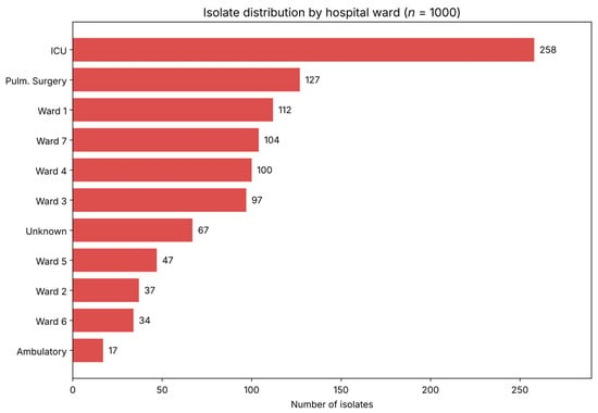
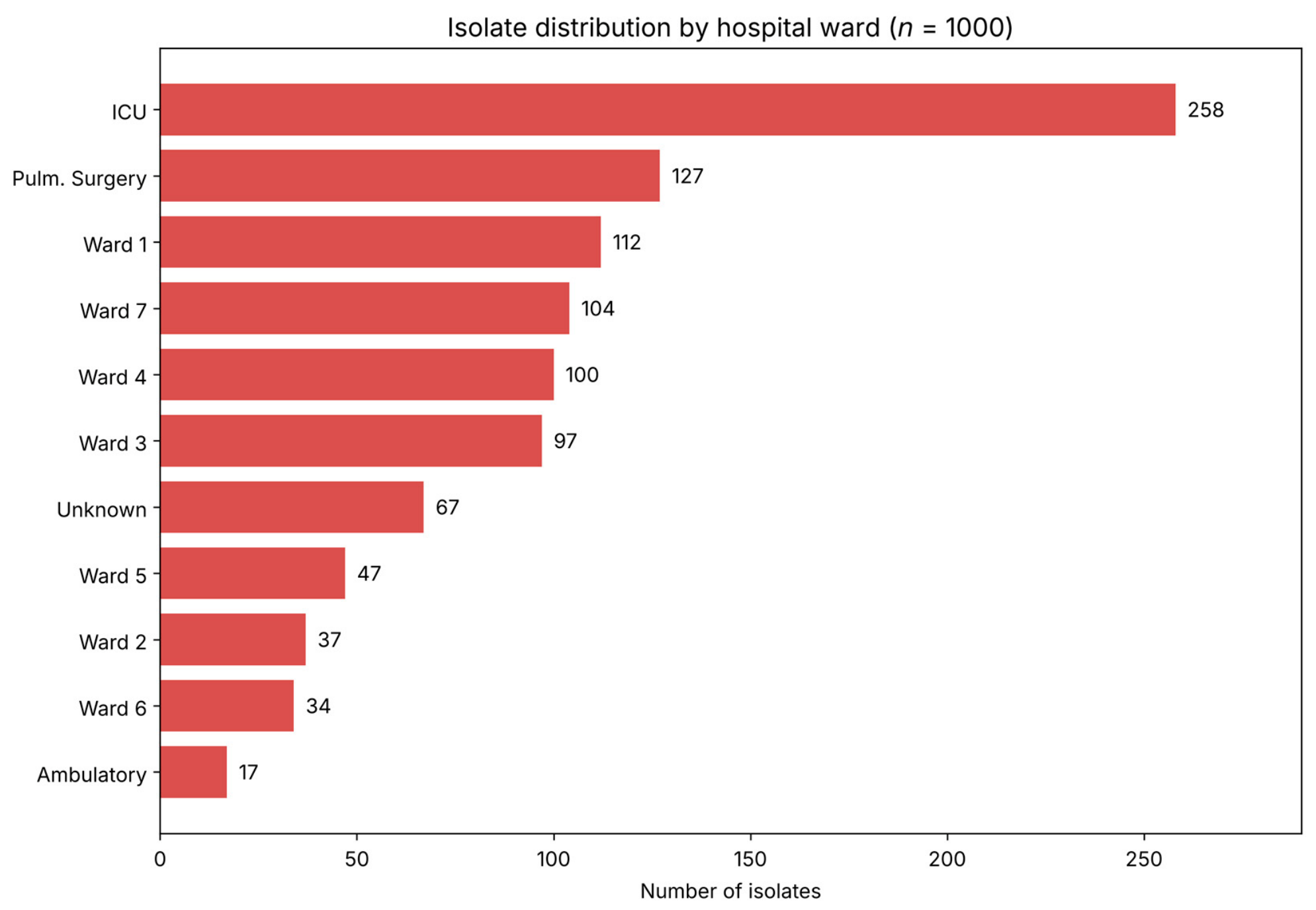
The ICU generated 258 of 1000 isolates—one in four—from a ward that represents a small fraction of total bed-days (Figure 3). The distribution across wards 1–7 was relatively even (34–112 cases per ward). An additional 127 isolates originated from pulmonary and general surgical wards combined, 17 from ambulatory and emergency consultations, and ward attribution could not be determined for 67 records due to undocumented or non-standard laboratory entries. Ward-level counts are reported in absolute terms; patient-days per ward could not be reliably reconstructed for this retrospective period (see Limitations) and are therefore not presented as a denominator.
Figure 3.
Distribution of fungal isolates by hospital ward (n = 1000). The ICU accounts for 25.8% of all records—disproportionate relative to bed-days. Wards 1–7 show a relatively even distribution (34–112 cases). Pulmonary/general surgery and ambulatory/emergency contexts are shown separately; ward information could not be verified for 6.7% of records.
3.4. Fungal Species Distribution
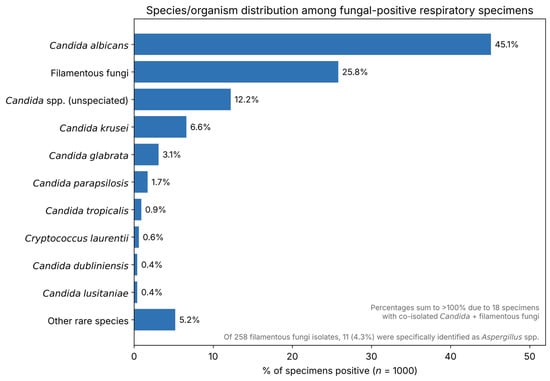
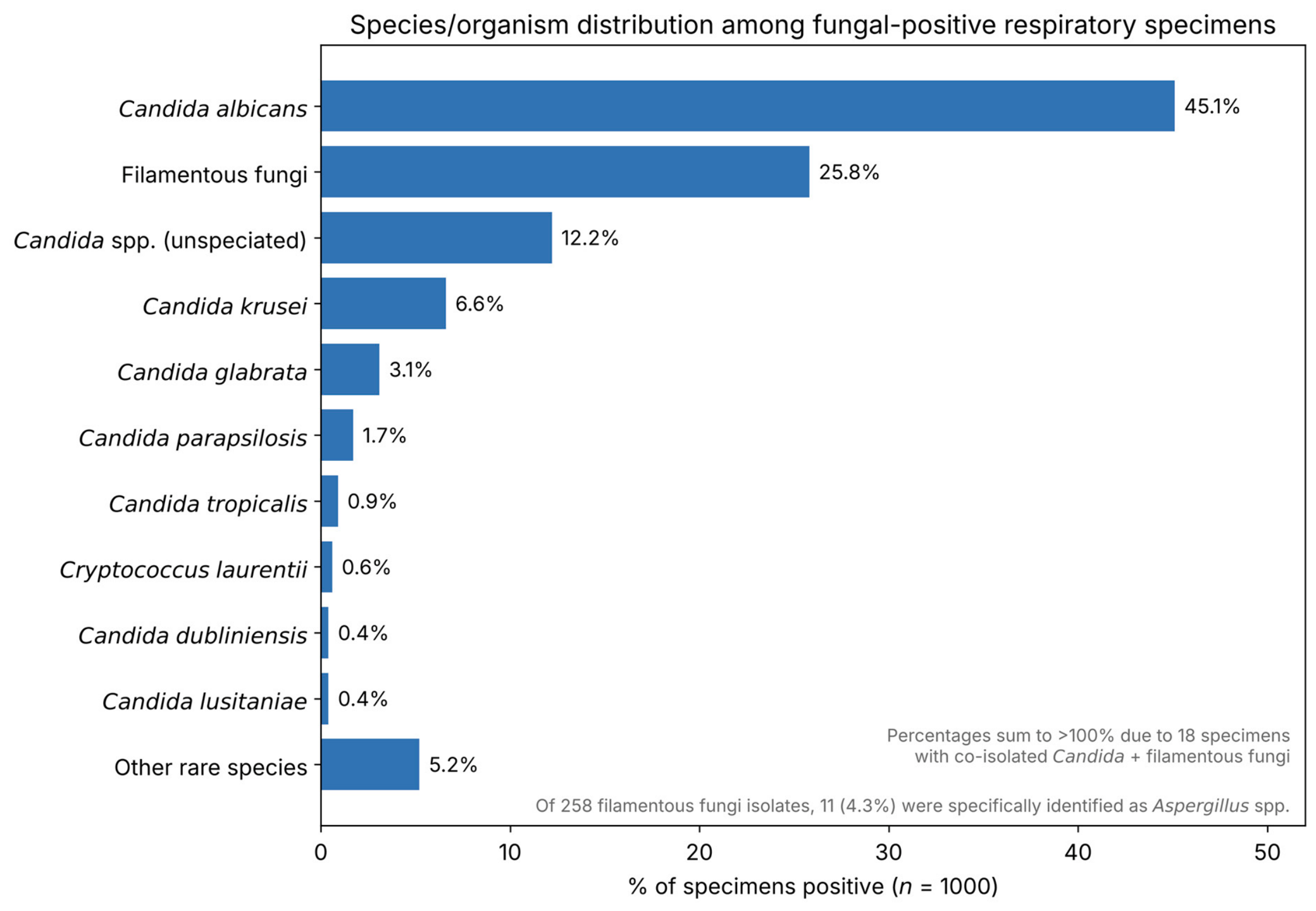
Candida albicans was identified in 451 of 1000 respiratory specimens (45.1%). A further 122 records (12.2%) yielded unspeciated Candida spp., predominantly from early 2022. Filamentous fungi (predominantly Aspergillus spp.) were identified in 258 specimens (25.8%)—either alone or, in 18 cases, co-isolated alongside a Candida species from the same specimen. Among identified non-albicans species, C. krusei led with 66 cases (6.6%), followed by C. glabrata (31; 3.1%), C. parapsilosis (17; 1.7%), and C. tropicalis (9; 0.9%) (Figure 4, Supplementary Table S2). Six records yielded Cryptococcus laurentii. Saprochaete capitata appeared once, and one case of Candida auris was identified in 2023. A sensitivity analysis comparing the full dataset against a deduplicated dataset (one isolate per patient, first chronological culture), covering 989 of the 1000 records, showed that species-level percentages differed by at most 0.7 percentage points for all species except Cryptococcus laurentii, indicating that the retention of duplicate isolates did not materially bias the overall species distribution. A comparative overview of these findings alongside previously published Candida surveillance data is presented in Table 1 (Section 4).
Figure 4.
Organism distribution among fungal-positive respiratory specimens (n = 1000), horizontal bar chart. C. albicans dominates at 45.1%, followed by filamentous fungi (predominantly Aspergillus spp.) at 25.8%. Percentages sum to more than 100% because 18 specimens (1.8%) yielded co-isolation of a Candida species together with filamentous fungi in the same specimen. ‘Other rare species’ encompasses C. tropicalis, C. kefyr, C. ciferrii, C. spherica, Candida auris, and other rare species.
Table 1.
Comparative overview of published Candida epidemiological surveillance data alongside the present study.
3.5. Temporal Trends in Species Composition
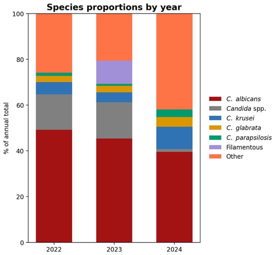
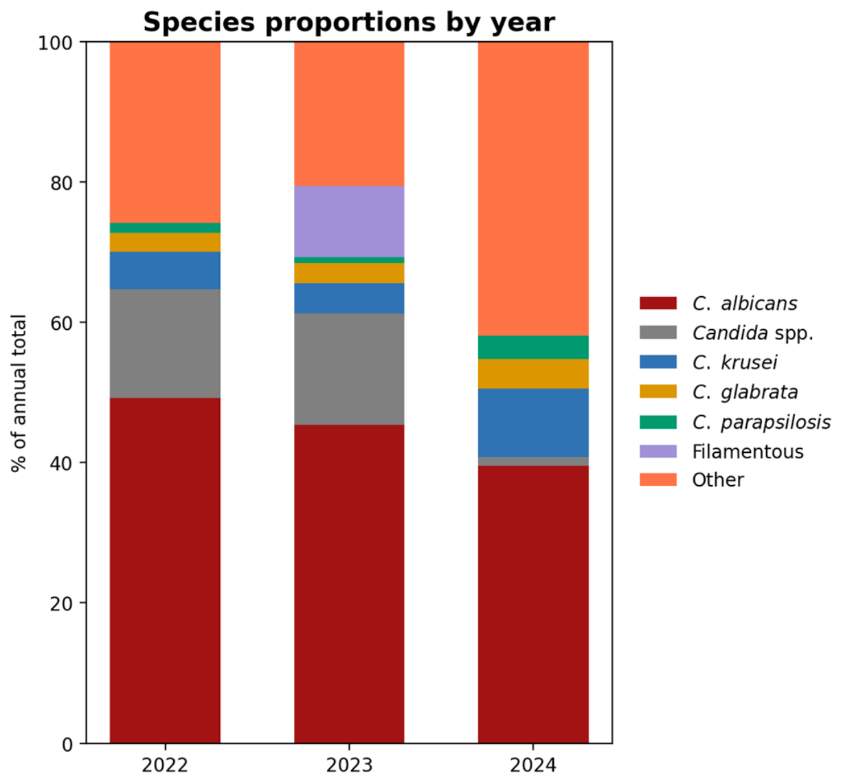
The year-on-year shift in species proportions is where the most clinically relevant trends lie (Figure 5). C. albicans held at roughly 49% in 2022 and slipped to 39.5% by 2024—not a steep decline, but a consistent one. C. krusei moved in the opposite direction: 5.3% in 2022, a dip to 4.3% in 2023, followed by a rise to 9.7% in 2024.
Figure 5.
Species proportions by year (% of annual total; 2022 n = 413, 2023 n = 348, 2024 n = 238). Each stacked bar sums to 100%. The gradual decline of C. albicans (49.2% → 39.5%) and the annotated increase in C. krusei (5.3% → 9.7%) are the two trends with the clearest clinical implications. The near-disappearance of unspeciated Candida spp. reflects methodological improvement, not a biological shift. Filamentous fungi in 2023 likely reflect post-COVID-19 pulmonary aspergillosis.
The decline in unspeciated Candida spp.—from 15.5% in 2022 to 1.3% in 2024—reflects the progressive improvements in diagnostic methods. Filamentous fungi appearing as primary isolates exclusively in 2023 (10.1% that year) represent an intriguing anomaly; we suspect this reflects the cohort of post-COVID-19 patients with Aspergillus-related airway disease admitted in that year, though retrospective confirmation is not possible from our records.
3.6. Species × Ward Heatmap
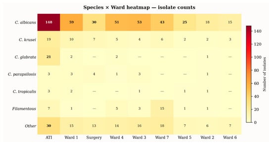
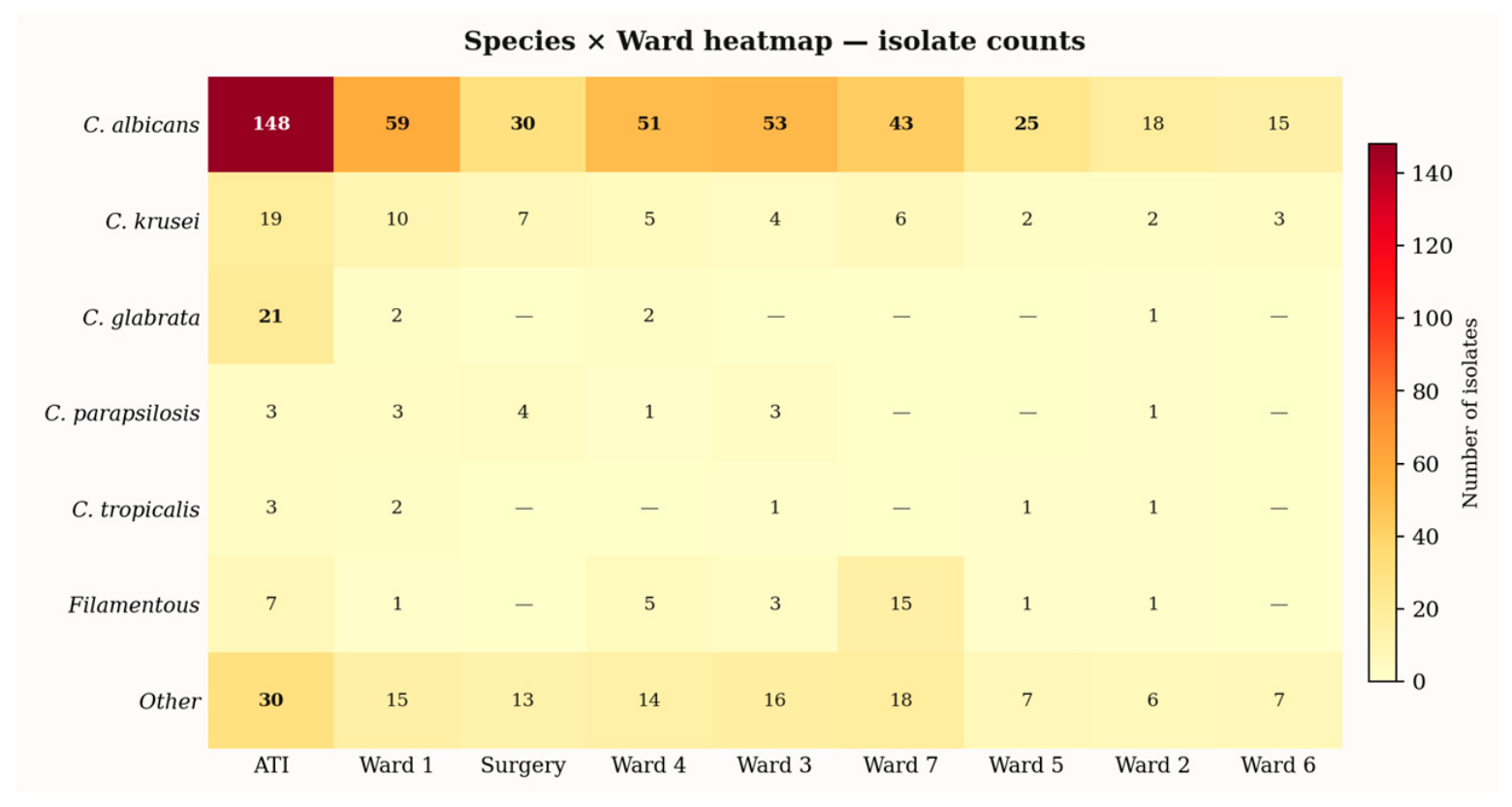
Looking at species through the lens of ward distribution (Figure 6) reveals patterns that aggregated data miss. C. glabrata is not evenly distributed: 21 of its 31 total isolates came from the ICU—67.7% of the species total from a single ward.
Figure 6.
Species × ward heatmap (absolute isolate counts per species-ward combination). C. albicans dominates across all wards (highest count in the ICU: n = 148). C. glabrata is disproportionately concentrated in the ICU (21/31 total isolates; 67.7%), consistent with severity-related risk. Filamentous fungi cluster in Ward 7 (n = 15), a pattern unexplained by current data. C. parapsilosis is most prevalent in pulmonary surgery, consistent with its catheter-related risk profile. Red-to-yellow shading indicates increasing counts; ‘—’ = zero isolates.
3.7. Primary Diagnoses
Acute respiratory failure (ARF/IRA) was the primary diagnosis in 417 cases (41.7%). General medical examination accounted for 158 cases (15.8%)—admission screening bronchoscopies where fungi were found incidentally. Other diagnoses included haemoptysis (29; 2.9%), bronchiectasis (17; 1.7%), tuberculosis (17; 1.7%), fever (24; 2.4%), COPD (16; 1.6%), and lung malignancy (16; 1.6%). Comparing the ICU to the rest of the hospital (Table 2, Supplementary Table S3) shows that ARF accounts for 67.8% of ICU-associated diagnoses versus 32.6% across other wards—a stark divergence that has direct implications for how positive cultures should be interpreted.
Table 2.
Primary diagnoses, overall and by care area (n = 1000).
3.8. Semi-Quantitative Colony Counts
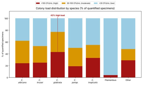
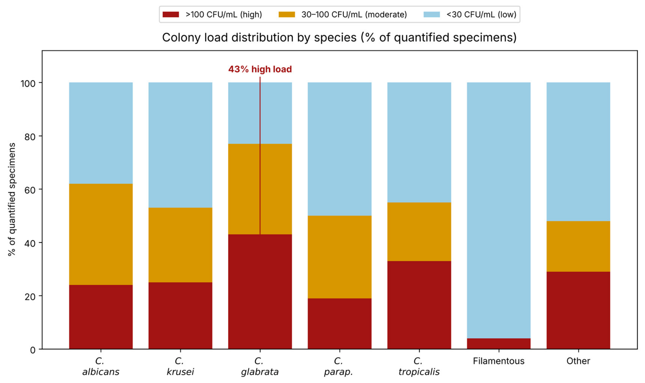
Colony count data were available for 768 of 1000 records (76.8%). High-load colonisation (>100 CFU/mL) was recorded in 190 cases (19.0%); moderate loads (30–100 CFU/mL) in 133 (13.3%); low-to-moderate loads (10–30 CFU/mL) in 203 (20.3%); and low loads (<10 CFU/mL) in 242 (24.2%). The 232 records without colony counts came almost entirely from 2022, before semi-quantitative reporting became standard practice. Breaking down colony load by species (Figure 7), C. glabrata showed the highest proportion of high-load specimens (~42% of its quantified records), compared with ~24% for C. albicans (Supplementary Tables S4 and S5).
Figure 7.
Colony load by species, as 100% stacked bars (quantified specimens only). C. glabrata shows the highest relative proportion of high-load isolates (~42%), roughly double that of C. albicans (~24%). This ICU-concentrated species is the one most likely associated with genuinely heavy fungal airway burden in the critically ill. Filamentous fungi are predominantly low-count, consistent with environmental contamination. Species with small sample sizes (C. tropicalis n = 9; C. parapsilosis n = 16) should be interpreted cautiously.
3.9. Age, Sex, and Species Correlates
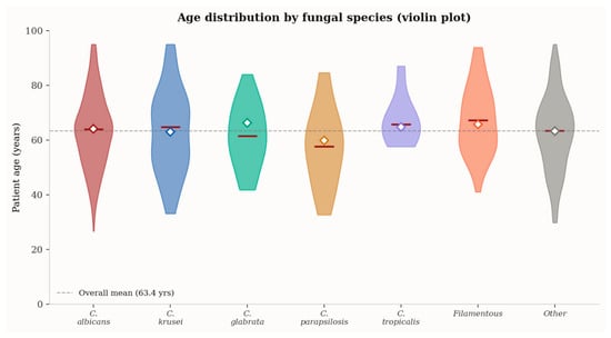
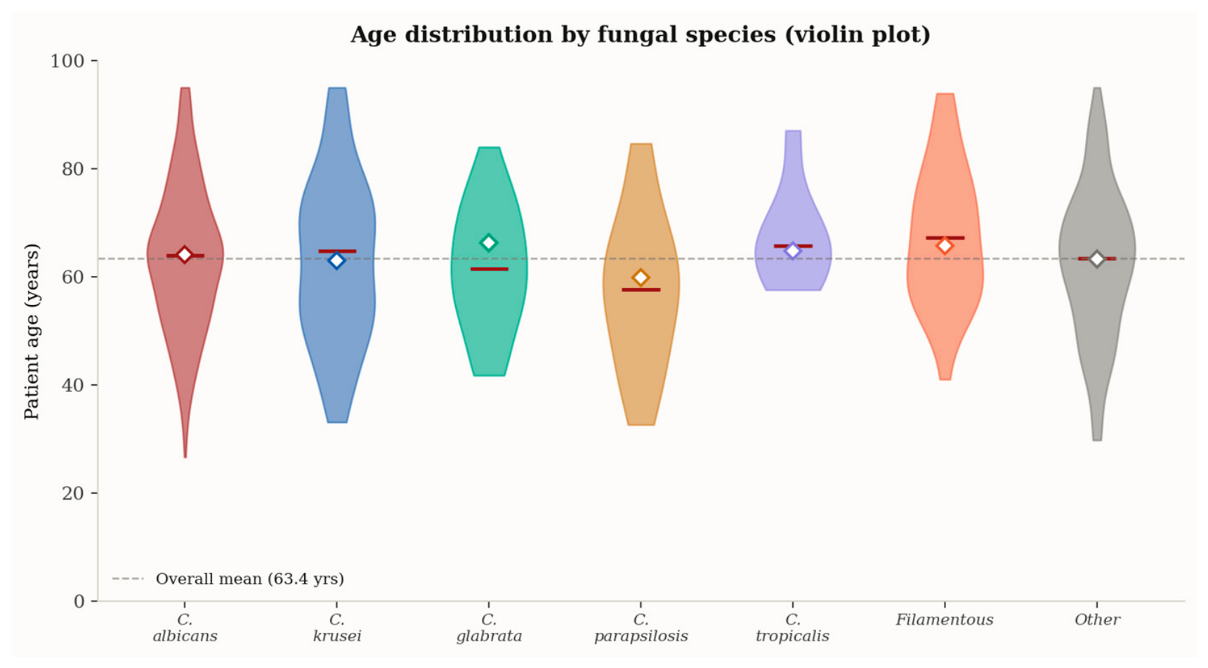
Age distributions were broadly similar across species, reflecting the predominantly elderly patient population (Figure 8). C. glabrata sits at the oldest end (mean 66.2 years; median 69), while C. parapsilosis skews younger (mean 59.8 years)—a pattern seen across multiple published series and attributed to its catheter-related risk profile spanning a wider adult age range. The violin shapes for C. parapsilosis (n = 16) and C. tropicalis (n = 9) are narrow; they are informative as patterns but should not be over-interpreted.
Figure 8.
Patient age by fungal species, as violin plots. Violin width represents observation density; white diamonds = species means; dashed line = overall cohort mean (63.4 years). C. glabrata is associated with the oldest patients (mean 66.2 years); C. parapsilosis skews younger (mean 59.8 years). Narrow violins for C. parapsilosis and C. tropicalis reflect small sample sizes—interpret cautiously.
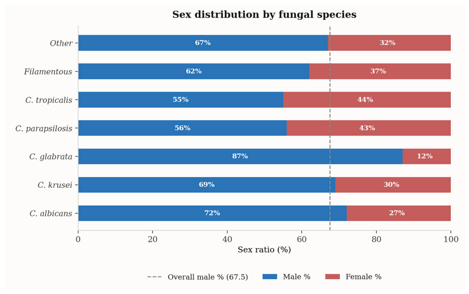
Male predominance was observed across all species but varied substantially (Figure 9). C. glabrata showed the most extreme male skew (87% male), likely reflecting the sex composition of the ICU population rather than any biological sex-predisposition effect. C. parapsilosis and C. tropicalis approached near-equal sex ratios (56% and 55% male).
Figure 9.
Sex distribution by species, as 100% stacked horizontal bars. The dashed line marks the overall cohort male proportion (67.5%). C. glabrata shows the most extreme male skew (87%), likely reflecting the ICU population demographics. C. parapsilosis (56%) and C. tropicalis (55%) approach near-equal sex ratios.
3.10. Co-Isolation of Filamentous Fungi
Filamentous fungi were co-identified alongside a Candida species in 18 of 1000 records (1.8%) (Supplementary Table S6). This high co-isolation rate likely reflects the structural lung damage prevalent in this cohort—post-tuberculosis cavities, bronchiectatic airways, emphysematous bullae—providing ecological niches where yeasts and moulds coexist in ways that healthy lungs simply do not permit.
3.11. Serial Culture Dynamics in the Multi-Sample Cohort
Of the 1000 records, 207 (20.7%) originated from 91 patients with more than one culture, identified by name-matching across the full dataset before anonymisation and coding. The remaining 793 patients had a single episode.
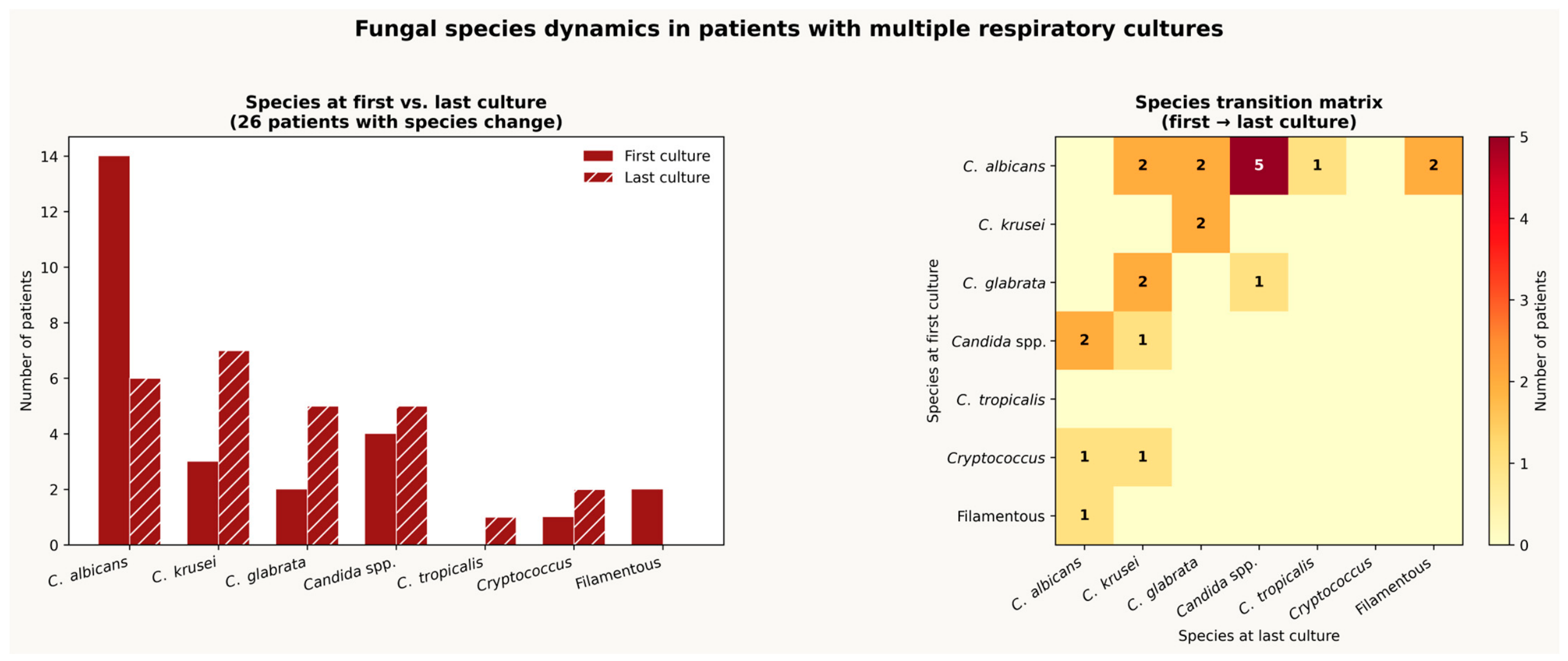
The 91 multi-sample patients do not represent a random cross-section. The ICU accounts for 68 of their 207 records (32.9%), substantially above the 25.8% ICU share in the full cohort. Twenty-six of the 91 patients showed a species change between their first and last culture; 29 showed load escalation; and 24 changed wards between samples. A representative, anonymised subset of ten of these 91 patients is presented in Supplementary Table S7 for illustration; the percentages above reflect analysis of the complete cohort of 91 multi-sample patients. The species transition matrix (Figure 10) maps which species gave way to which across the 26 patients with a change. The most clinically significant transitions were those involving C. krusei emergence as a late isolate—occurring in at least five patients after an initial C. albicans culture. In several of these cases, the sequence is suggestive of antifungal selection pressure: C. albicans was detected first, fluconazole was likely initiated, and C. krusei—intrinsically resistant—appeared on the follow-up culture. The three patients who progressed to Cryptococcus laurentii as a late isolate (Table 3) represent a distinct and unexplained pattern; in all three cases, the Cryptococcus appeared at high load in the ICU, raising the possibility of unrecognised underlying immunosuppression.
Figure 10.
Species dynamics in the 26 patients with species change. Left: species at first culture (solid) versus last culture (hatched). C. albicans dominance at first culture (14 patients) gives way to a more diverse last-culture distribution, with C. krusei rising from 3 to 7 patients and C. glabrata from 2 to 5 patients. Right: transition matrix (first → last culture species). The five cases of C. albicans → C. krusei shift may reflect antifungal selection pressure; three C. albicans → Cryptococcus laurentii cases require investigation for immunosuppression.
Table 3.
Selected patients with species change across serial respiratory cultures.
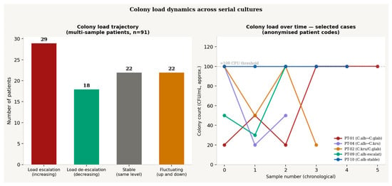
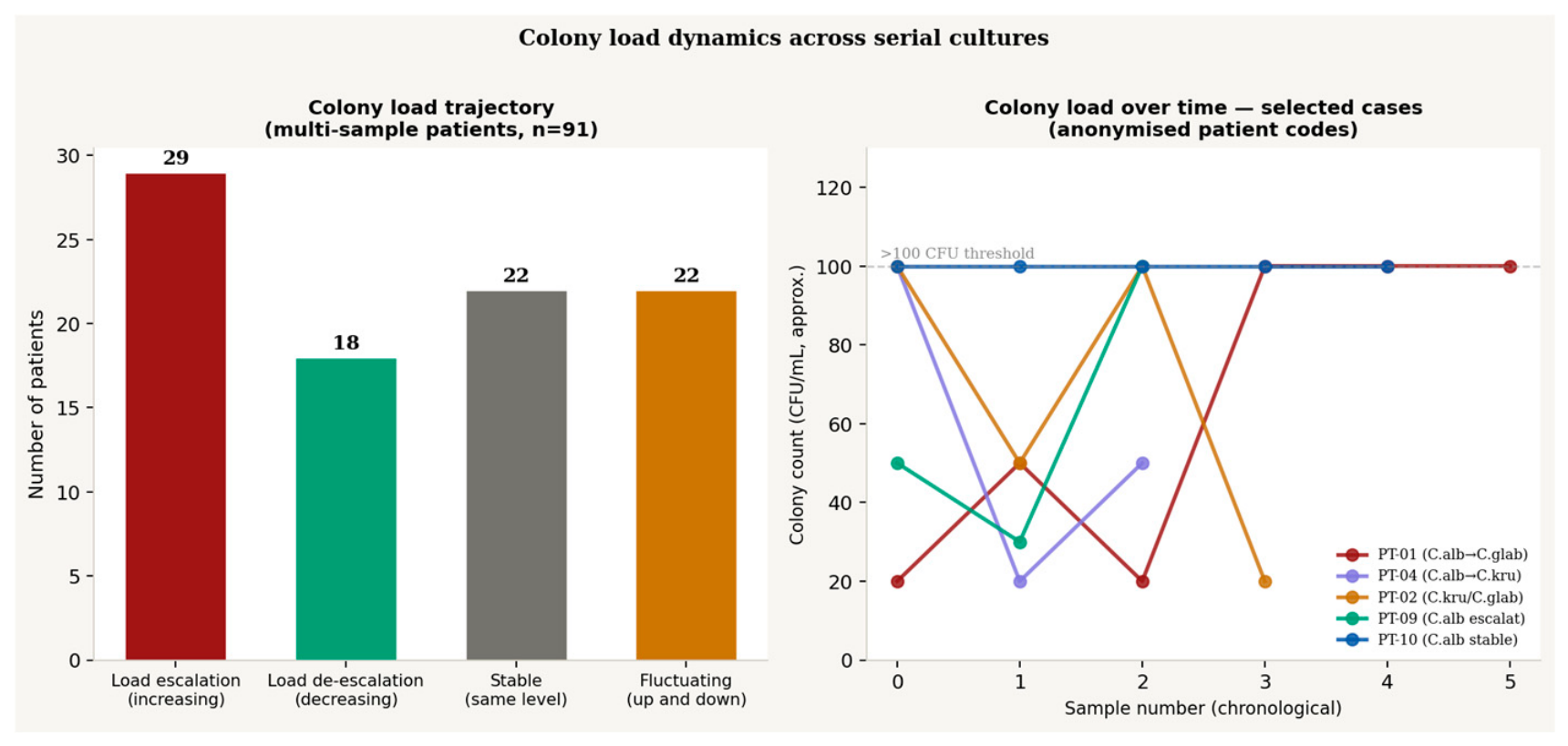
Among the multi-sample patients with quantified colony data, 29 (31.9%) showed load escalation, 18 showed de-escalation, 22 showed stable loads, and 22 showed a fluctuating pattern (Figure 11). Patient-01 (PT-01)—an 80-year-old male with eight ICU cultures across three months—is the most compelling case: his C. albicans counts climbed from >20 CFU/mL in October 2022 to >100 CFU/mL in November, at which point C. glabrata appeared alongside and eventually displaced the albicans strain (Figure S1). This is a textbook trajectory for prolonged ICU Candida colonisation progressing toward probable infection. PT-08 (55 years, female) shows the reverse: C. albicans at >100 CFU/mL in March 2024, C. tropicalis at low load in May, and a negative culture in June—consistent with either successful treatment response or spontaneous resolution.
Figure 11.
Colony-load trajectories. Left: classification of 91 multi-sample patients by load pattern. Load escalation (n = 29; 31.9%) and de-escalation (n = 18; 19.8%) each represent potentially significant clinical patterns; ‘stable’ (n = 22) reflects sustained colonisation; ‘fluctuating’ (n = 22) shows irregular patterns without consistent direction. Right: individual load trajectories for five key cases, plotted chronologically by sample number. Dashed line = >100 CFU/mL threshold. PT-01 (red) shows the most pronounced trajectory: escalating counts with C. glabrata emergence; PT-04 (purple) shows threshold crossing before C. krusei emergence. Colony counts are semi-quantitative estimates; the y-axis represents approximate CFU/mL.
3.12. Risk Factors for Non-Albicans Candida Species
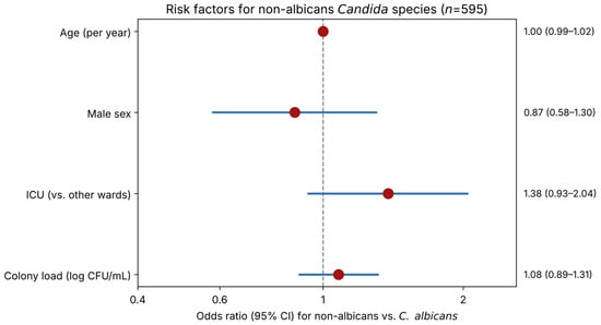
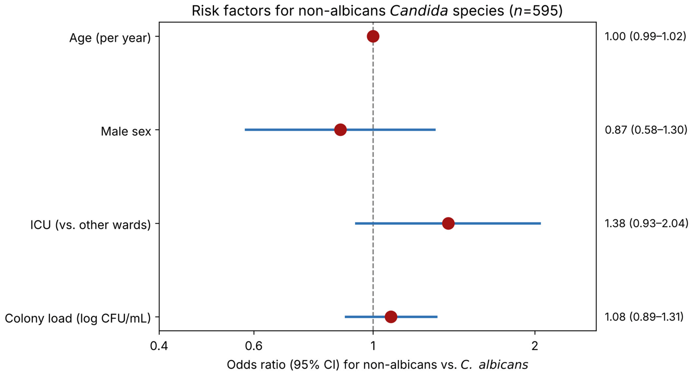
In multivariate logistic regression (n = 595), none of the evaluated variables were significantly associated with non-albicans Candida species. The care area showed a non-significant trend toward association (ICU vs. other wards: OR 1.38, 95% CI 0.93–2.04, p = 0.109), while age (OR 1.00 per year, 95% CI 0.99–1.02, p = 0.600), male sex (OR 0.87, 95% CI 0.58–1.30, p = 0.495), and colony load (OR 1.08 per log-unit, 95% CI 0.89–1.32, p = 0.434) showed no meaningful association (Figure 12, Table 4).
Figure 12.
Forest plot of odds ratios (95% CI) for non-albicans Candida species (vs. C. albicans) from multivariate logistic regression (n = 595). None of the evaluated predictors reached statistical significance.
Table 4.
Multivariate logistic regression for non-albicans vs. C. albicans (n = 595).
4. Discussion
The overall species landscape described in this study is broadly consistent with published respiratory mycology data from comparable settings [18,19,20,21,22], with Candida albicans maintaining its expected dominance. Several findings, however, warrant closer attention. The proportional decline of C. albicans across the study period, the concurrent increase in C. krusei, the identification of a single Candida auris isolate, and the cluster of Cryptococcus laurentii detections collectively suggest a mycological profile that, while not alarming in aggregate, points to trends with direct clinical and therapeutic implications.
Beyond these headline trends, several individual species carry specific clinical implications worth highlighting. C. parapsilosis, identified in 17 specimens, is a recognised biofilm producer with a predilection for catheter-associated infection and documented variability in echinocandin minimum inhibitory concentration (MIC), both of which are relevant should invasive disease be suspected in a patient carrying this species. C. tropicalis (9 cases) is associated with higher virulence in neutropenic hosts, a consideration for the haematology-oncology overlap seen in some of our paediatric and adult cases. C. dubliniensis (4 cases) is phenotypically similar to C. albicans and may be misidentified as such without chromogenic differential media, a risk directly relevant to the 92.7% of records in our dataset for which the identification method was not explicitly documented (Supplementary Table S5). C. lusitaniae (4 cases) carries a well-described potential for intrinsic, strain-dependent amphotericin B resistance, relevant to empirical therapy choices in refractory cases. The single case of Saprochaete capitata, though rare, is noteworthy for its disproportionately high mortality in immunocompromised hosts relative to its low incidence. Finally, the co-isolation of filamentous fungi (predominantly Aspergillus spp.) in 25.8% of specimens, including 18 specimens with simultaneous Candida co-isolation, underscores that respiratory specimens from this population frequently harbour a polymicrobial fungal burden; as with Candida, isolation from a non-sterile specimen does not by itself establish invasive disease, and correlation with clinical and radiological findings remains essential.
The 2:1 male predominance observed in our cohort likely reflects the higher burden of chronic respiratory disease among men in Romania, driven by decades of higher smoking rates, occupational exposures, and historically higher tuberculosis incidence. The nine paediatric cases identified may represent children with cystic fibrosis or haematological malignancy, though the dataset did not capture underlying diagnoses with sufficient granularity to confirm this.
The disproportionate concentration of isolates in the ICU (25.8% from a single ward), the dominance of ARF as the primary diagnosis (67.8% of ICU-associated records), and the specific concentration of C. glabrata in the ICU (67.7% of all C. glabrata isolates) confirm the mechanically ventilated critically ill patient as the highest-risk phenotype in this institution. This is consistent with the known pathophysiology: endotracheal intubation bypasses laryngeal defences, universal antibiotic therapy eliminates competing bacteria, and the metabolic stress of critical illness suppresses immune clearance [23,24,25]. The longitudinal data add a further dimension: the multi-sample ICU patients show the highest rates of load escalation and species shift, making them the subgroup most in need of a structured monitoring protocol.
Using a framework that integrates colony load, species virulence, ward context, and temporal pattern (Figure S2), approximately 45 of the 91 multi-sample patients show features most consistent with persistent colonisation: same species, stable moderate loads, and non-ICU diagnoses. Around 29 fall into the escalating colonisation category—at highest risk of transitioning to infection. A smaller group of approximately 14 patients show patterns most consistent with probable infection: high loads (>100 CFU/mL) sustained across multiple samples with NAC species in the ICU context. The 26 patients with species changes present a distinct challenge. Some transitions almost certainly reflect antifungal selection pressure. Others may reflect co-colonisation with simultaneous detection of a minor population that later becomes dominant.
The interpretive framework applied in this study—integrating colony load, species identity, clinical symptoms, and radiological findings to distinguish colonisation from probable infection—reflects the current state of a debate that remains, despite decades of investigation, genuinely unresolved. Unlike bacterial ventilator-associated pneumonia, for which validated CFU/mL thresholds exist (103 CFU/mL for protected specimen brush, 104 CFU/mL for bronchoalveolar lavage [BAL]), no equivalent quantitative standard has been established for Candida in respiratory specimens [26,27,28], meaning that definitive diagnosis of pulmonary candidiasis continues to require histopathological confirmation—a standard that is almost never achievable in living patients outside of post-mortem studies. There is, however, growing evidence that Candida is not simply an innocent bystander in the airways of mechanically ventilated patients: bronchial colonisation has been independently associated with prolonged mechanical ventilation, increased ICU length of stay, and elevated mortality, as well as with a significantly increased risk of subsequent Pseudomonas aeruginosa pneumonia [3,29]. The most recent global guidance on candidiasis management—the ECMM/ISHAM/ASM Global Guideline (2025)—does not recommend antifungal therapy based on respiratory isolates alone in the absence of histological confirmation [1], a standard that, as our data illustrate, is almost never achievable in routine clinical practice. In the absence of validated clinical criteria specific to bronchial aspirates, the approach reflected in our clinical decision framework—high colony load combined with a non-albicans species and compatible clinical findings as a composite trigger for treatment consideration—represents a pragmatic synthesis of published evidence rather than a consensus-endorsed protocol and should be interpreted as such.
The published literature on respiratory Candida epidemiological surveillance from dedicated pulmonology hospitals is thinner than one might expect—most epidemiological surveillance studies focus on candidaemia from blood cultures, not on bronchial aspirates from inpatient wards. This scarcity is itself one of the arguments for the present study. Table 1 collects the most directly comparable published datasets alongside our own figures.
The C. albicans proportion in our cohort (45.5% overall; 39.5% in 2024) is strikingly close to the 45.1% reported in the multi-centre Southern European CANCoVEU candidaemia study [13], which pooled 1203 blood culture isolates from six tertiary hospitals across Italy, Portugal, Greece, and Spain during 2020–2021 [13]. This convergence is noteworthy precisely because the specimen types are so different: blood cultures and bronchial aspirates capture fundamentally distinct patient populations and clinical presentations. That both datasets arrive at roughly the same C. albicans proportion suggests that this species’ dominance is a genuinely robust biological characteristic—not merely an artefact of specimen type—across the spectrum from colonisation to invasive disease.
The ICU-specific respiratory data from France [3,30]—the FUNGIBACT study—reported C. albicans at 68.7% of endotracheal aspirate isolates from mechanically ventilated patients, substantially higher than our 45.5% and our ICU-specific proportion. This difference almost certainly reflects the era of that study (pre-VITEK® 2, when species identification was less complete) and the exclusively ICU-based sampling frame—patient populations in our wards and outpatient services dilute the C. albicans proportion relative to a purely critical care dataset. The Terraneo et al. (2012) Spanish series [15], also ICU-based and respiratory, found a similar ~62% C. albicans proportion, reinforcing this interpretation. Our data, covering all inpatient wards and outpatients rather than the ICU alone, represent a fuller institutional picture.
The proportional increase in C. krusei from 5.3% to 9.7% in our cohort between 2022 and 2024 is the finding that most clearly distinguishes our dataset from the European average. This species is intrinsically resistant to fluconazole—the most widely used empirical antifungal agent in our institution—and requires echinocandin or voriconazole therapy regardless of in vitro testing [10,31]. Published candidaemia data across Europe have consistently placed C. krusei at 2–5% of total isolates [7], a figure that has remained broadly stable over the past two decades [18,32]. A comparable Iranian respiratory cohort [15] found 5.4% C. krusei across COVID-19-era respiratory specimens—still below our 2024 figure of 9.7%. Our C. krusei proportion in 2024 is thus approximately double the European candidaemia benchmark and approaching double the published figure from a comparable respiratory epidemiological surveillance study. Whether this reflects a genuine local epidemiological shift, therapeutic pressure from empirical fluconazole prescribing, or a statistical fluctuation magnified by the smaller 2024 sample cannot be determined retrospectively—but the direction and magnitude of the trend are clinically consequential enough to warrant prospective epidemiological surveillance.
The bubble chart of antifungal susceptibility profiles (Figure S3) frames the clinical stakes. Of 1000 isolates, a minimum of 61—C. krusei plus the single C. auris—represent organisms where fluconazole cannot be expected to work. A further 31 C. glabrata isolates require at minimum a higher dose or an alternative agent. That is 9.2% of all records where the choice between fluconazole and something else is not a matter of preference but of pharmacological necessity.
Figure S4 summarises the decision pathway that emerges from this dataset: species identification and colony count quantification are the two non-negotiable inputs; integration with clinical context determines the output. When high colony loads, NAC species, and clinical symptoms are present, antifungal therapy should be initiated, and susceptibility data should be obtained. For everything else, monitoring and repeat testing are recommended. The framework is not a protocol; individual clinical judgement is irreplaceable.
The 2018 Romanian candidaemia study from an Eastern European University Hospital [16] found a strikingly different species hierarchy from ours: C. parapsilosis dominated at 37.8%—a pattern more consistent with Mediterranean and Southern European epidemiological surveillance programmes and distinctly different from our C. albicans-dominated respiratory cohort. This divergence almost certainly reflects the known epidemiological distinction between candidaemia (where C. parapsilosis is overrepresented due to its biofilm-forming propensity on vascular catheters) and respiratory colonisation (where C. albicans reasserts its dominance) [12,32]. The two studies are not in conflict—they capture different clinical phenomena in the same country.
Of greater concern for the current epidemiological surveillance context is the Romanian C. auris data. The first Romanian C. auris cases were reported in early 2022 in Bucharest hospitals [33]. A subsequent outbreak report from Agrippa Ionescu Clinical Emergency Hospital [17] described 13 patients colonised or infected in a cardiovascular surgery ICU between October 2022 and July 2023, with whole-genome sequencing confirming South Asian Clade I lineage [17]. Our 2023 C. auris isolate from a pulmonology ICU bronchial aspirate fits exactly into this national epidemiological timeline: the species was not present in our institution before 2023, appeared coinciding with the period of documented national spread, and was identified in the ICU consistently associated with C. auris acquisition. The ECDC 2024 survey documented over 4000 cumulative EU/EEA C. auris cases between 2013 and 2023, with 1346 cases in 2023 alone. Romania is now classified among countries with documented nosocomial transmission [19]. In this context, our single isolate is not a curiosity: it is confirmation that C. auris has penetrated the respiratory mycological landscape of Romanian tertiary hospitals. The Candida auris case and the six Cryptococcus laurentii isolates—three of which appeared as late species shifts in ICU multi-sample trajectories—demand attention beyond their small numbers. All six Cryptococcus laurentii isolates in this cohort were confirmed by VITEK® 2 identification, rather than relying on chromogenic medium alone, given the clinical significance of this unusual finding and its association with possible underlying immunosuppression. C. auris is classified by major health agencies as a critical-priority pathogen due to its capacity for multidrug resistance and sustained nosocomial transmission [11]. Each new case should trigger immediate infection control notification and environmental sampling. The C. laurentii cluster is a different kind of signal: this organism is not a typical airway resident in immunocompetent patients, and its repeated appearance at high loads in the ICU—as a late isolate following conventional Candida species—raises serious questions about immune status that the available records do not fully answer [34].
The filamentous fungus co-isolation rate in our cohort (22.3%) is higher than typically reported in European respiratory epidemiological surveillance studies, which generally describe co-isolation rates of 5–15% in general ICU populations. The pulmonary surgery ward rate of 48.7% is particularly elevated and reflects the structural lung damage—resection margins, bronchial anastomoses, post-operative airways—that creates an unusually favourable niche for fungal colonisation. To our knowledge, ward-specific filamentous co-isolation rates of this magnitude have not been published from comparable European settings; this finding may represent a genuine epidemiological signal specific to this patient cohort.
Collectively, these comparisons argue for the relevance of this dataset beyond its local context. Romania has historically been under-represented in European fungal epidemiological surveillance networks, despite carrying a substantial burden of respiratory disease and, increasingly, a documented C. auris transmission chain. The present study provides what, to our knowledge, is the first systematic three-year bronchial aspirate epidemiological surveillance dataset from a Romanian pulmonology hospital—a dataset that is both qualitatively consistent with and quantitatively distinct from Western European benchmarks in ways that matter clinically.
Several practical improvements to institutional surveillance follow directly from the patterns identified in this study. First, laboratory data capture would benefit from standardisation: a substantial minority of records in the source database carried inconsistent or non-standard ward and specimen-type codes, which complicated ward-level and specimen-level analysis and likely affects real-time surveillance reporting as well; adopting a controlled, structured vocabulary within the laboratory information system would reduce this ambiguity going forward. Second, the substantial proportion of unspeciated Candida spp. isolates, concentrated in the earliest years of the study period, indicates that reflex species-level identification—now largely achieved by 2024—should be formalised as standard practice for all positive respiratory cultures, given the direct implications of species identity for antifungal choice. Third, given the rising proportion of C. krusei and the confirmed presence of C. auris, we recommend that routine antifungal susceptibility testing be extended to all non-albicans isolates, rather than performed selectively, to allow timely detection of emerging resistance patterns. Fourth, the single C. auris isolate identified in this cohort, while numerically minor, warrants a disproportionate institutional response: given the well-documented potential of C. auris for nosocomial transmission and multidrug resistance, we recommend that any future C. auris detection trigger immediate contact precautions, environmental screening, and notification of infection control teams, rather than being processed as a routine culture result. Fifth, the ICU-concentrated burden of high colony loads and species turnover identified in the multi-sample cohort supports the value of a structured, prospective monitoring protocol for patients with prolonged ICU stays, with serial cultures interpreted jointly by clinical and microbiology teams rather than in isolation. Finally, prospective linkage of microbiological data with clinical outcome data—unavailable in the present retrospective dataset—would allow future surveillance to move from descriptive species tracking toward outcome-validated risk stratification, and we recommend this as a priority for the institution’s electronic data infrastructure going forward.
Limitations. This study’s retrospective design precludes clinical outcome assessment. This study was conducted at a single tertiary pulmonology centre in Bucharest, Romania. Consequently, the species distribution, resistance patterns, and temporal trends reported here reflect the specific patient population, referral pattern, and antifungal prescribing practices of this institution and may not be generalisable to other centres, regions, or healthcare systems with different case mixes, laboratory protocols, or antifungal stewardship policies. Multi-centre studies would be needed to confirm whether the trends identified here—particularly the rising proportion of fluconazole-resistant non-albicans species—reflect a broader regional or national pattern. Monthly and seasonal comparisons in this study are based on absolute counts of fungal-positive respiratory isolates, not on positivity rates. The source laboratory database captured only fungal-positive results; total respiratory specimen volume (including fungal-negative results) was not retained in the exported dataset and could not be reconstructed retrospectively. Consequently, we cannot determine what proportion of the observed monthly and seasonal variation reflects true differences in fungal epidemiology versus differences in overall hospital admission volume, testing frequency, or specimen submission practices across months and years. This limitation is compounded by the incomplete 2024 data (available for January–July only), which precludes any conclusion about whether additional seasonal maxima occurred in August–December of that year. Future surveillance would benefit from capturing total specimen volume (or, at minimum, total admissions) alongside fungal-positive results to allow calculation of a true positivity rate rather than absolute counts. We were unable to include ward-level patient-days as a denominator for the isolate counts presented in Figure 4. Over the three-year study period, bed capacity across wards was highly variable and not reliably reconstructable retrospectively: the hospital underwent partial facility closures, ward renovations, and temporary ward closures for infection-control sanitation following various outbreaks, all of which altered effective bed-days in ways not systematically recorded in a form usable for this analysis. In addition, length of stay varied substantially by patient population—from admissions of only a few days to several weeks for patients with active or sequelae of tuberculosis—further complicating any attempt to reconstruct a reliable patient-days denominator retrospectively. As a result, isolate counts in Figure 4 are reported in absolute terms and as a proportion of the total cohort, rather than as a rate per patient-day, and this should be considered when interpreting ward-level differences, particularly the disproportionate ICU contribution discussed in Section 3.3/Discussion. The distinction between Candida colonisation and true pulmonary infection cannot be made reliably from microbiological records alone; histopathological confirmation was not available. Key confounders—antibiotic exposure, steroid dose, ventilation duration, immunosuppression status—were not systematically captured. A further limitation concerns species identification methodology. All isolates were identified using chromogenic medium, based on the manufacturer’s colour-reaction chart, which provides presumptive rather than definitive species identification. Confirmatory VITEK® 2 testing was performed only for the subset of isolates for which antifungal susceptibility testing was also clinically indicated—in practice, predominantly ICU isolates (25.8% of the cohort)—and in this subset, VITEK® 2 results were fully concordant with the chromogenic medium findings. However, this concordance was observed in a non-random, higher-acuity subset of isolates and does not by itself establish that the remaining 74.2% of isolates, identified by chromogenic medium alone and never subjected to confirmatory testing, were correctly identified at the species level. Without confirmatory testing, we cannot rule out species-level misidentification among non-ICU isolates, particularly for species less reliably distinguished by colour reaction alone (e.g., C. glabrata, C. parapsilosis, and rarer species). This is a genuine limitation of the retrospective dataset rather than one that can be resolved analytically, and it should be borne in mind when interpreting the non-albicans species distribution reported in this study. We recommend that future surveillance extend confirmatory identification (VITEK® 2, MALDI-TOF, or molecular methods) beyond the ICU to all wards, regardless of susceptibility-testing indication. Species-level identification of filamentous fungi was performed in only a small minority of cases (11 of 258, 4.3%), all confirmed as Aspergillus spp.; the remaining filamentous isolates were recorded only at the morphological level, without genus- or species-level confirmation. Consequently, while Aspergillus is the most plausible predominant genus given regional epidemiology and the confirmed subset, this cannot be stated definitively for the cohort as a whole. Antifungal susceptibility testing was not routinely performed; species identity served as a proxy for susceptibility categorisation.
5. Conclusions
C. albicans is still the dominant fungus in our bronchial aspirates and probably will remain so for the foreseeable future. However, the non-albicans fraction—and C. krusei in particular—is growing, and that growth has direct therapeutic consequences. In a setting where fluconazole has long been the default empirical antifungal, a 9.7% C. krusei proportion is a number that should give clinical teams pause.
For roughly one in ten patients, species identity changes the antifungal of choice. For patients with C. auris, it changes not just the treatment but the entire infection control response. For the 26 patients in our cohort who changed species between serial cultures—several following a pattern consistent with antifungal selection pressure—species identity at follow-up is literally the most important result in the report.
Systematic species-level identification of all fungal respiratory isolates is strongly recommended as a baseline standard. Routine antifungal susceptibility testing, defined criteria for treatment initiation that integrate colony load with clinical and radiological findings, and active epidemiological surveillance for emerging resistant species represent the logical next steps.
Supplementary Materials
The following supporting information can be downloaded at https://www.mdpi.com/article/10.3390/germs16040026/s1, Table S1. Monthly raw isolate counts by year. Table S2. Complete species frequency table with 95% confidence intervals. Table S3. Complete diagnostic categories. Table S4. Specimen-type distribution. Table S5. Semi-quantitative colony load distribution stratified by year. Table S6. Filamentous fungal co-isolation. Table S7. Anonymised trajectory summaries for the ten multi-sample patients. Figure S1. Individual patient timelines for five key cases. Figure S2. Clinical interpretation framework for multi-sample patients. Figure S3. Antifungal susceptibility profiles by species. Figure S4. Proposed clinical decision framework for Candida respiratory isolates in a pulmonology setting.
Author Contributions
Conceptualisation, M.S., M.C.R. and L.S.C.M.; methodology, M.C.R.; software, M.C.R.; validation, M.S., M.C.R., O.P., L.S., D.V., D.T., B.M., J.A., R.-E.H., E.-M.C. and L.S.C.M.; formal analysis, M.S. and L.S.C.M.; investigation, M.S., M.C.R., O.P., L.S., D.V., D.T., B.M., J.A., R.-E.H. and L.S.C.M.; resources, M.S., O.P., L.S., D.V., D.T., B.M. and L.S.C.M.; data curation, M.S., M.C.R., O.P., L.S. and L.S.C.M.; writing—original draft preparation, M.S., E.-M.C. and L.S.C.M.; writing—review and editing, M.S., M.C.R., O.P., L.S., D.V., D.T., B.M., J.A., R.-E.H., E.-M.C. and L.S.C.M.; visualisation, M.S. and L.S.C.M.; supervision, M.S. and L.S.C.M.; project administration, M.S. and L.S.C.M.; funding acquisition, M.C.R. All authors have read and agreed to the published version of the manuscript.
Funding
This work was supported by the Carol Davila University of Medicine and Pharmacy, Bucharest, Romania, postdoctoral programme contract number 28567/2 October 2023.
Institutional Review Board Statement
The study was conducted in accordance with the Declaration of Helsinki and approved by the Institutional Ethics Committee of Marius Nasta Institute of Pneumology (23343/17 October 2023).
Informed Consent Statement
Patient consent was waived by the Institutional Ethics Committee of the Marius Nasta Institute of Pneumology (approval no. 23343, 17 October 2023), due to the retrospective, fully anonymised nature of the study, in accordance with the Declaration of Helsinki.
Data Availability Statement
The data presented in this study are available on request from the corresponding author due to privacy restrictions.
Acknowledgements
We thank Carol Davila University of Medicine and Pharmacy, Bucharest, Romania, for the support offered for this research during the postdoctoral programme.
Conflicts of Interest
The authors declare no conflicts of interest.
References
- Pappas, P.G.; Kauffman, C.A.; Andes, D.R.; Clancy, C.J.; Marr, K.A.; Ostrosky-Zeichner, L.; Reboli, A.C.; Schuster, M.G.; Vazquez, J.A.; Walsh, T.J.; et al. Clinical Practice Guideline for the Management of Candidiasis: 2016 Update by the Infectious Diseases Society of America. Clin. Infect. Dis. 2016, 62, e1–e50. [Google Scholar] [CrossRef] [Scilit]
- Wisplinghoff, H.; Bischoff, T.; Tallent, S.M.; Seifert, H.; Wenzel, R.P.; Edmond, M.B. Nosocomial Bloodstream Infections in US Hospitals: Analysis of 24,179 Cases from a Prospective Nationwide Surveillance Study. Clin. Infect. Dis. 2004, 39, 309–317. [Google Scholar] [CrossRef] [Scilit]
- Azoulay, E.; Timsit, J.-F.; Tafflet, M.; de Lassence, A.; Darmon, M.; Zahar, J.-R.; Adrie, C.; Garrouste-Orgeas, M.; Cohen, Y.; Mourvillier, B.; et al. Candida Colonization of the Respiratory Tract and Subsequent Pseudomonas Ventilator-Associated Pneumonia. Chest 2006, 129, 110–117. [Google Scholar] [CrossRef] [Scilit]
- Arendrup, M.C.; Arikan-Akdagli, S.; Jørgensen, K.M.; Barac, A.; Steinmann, J.; Toscano, C.; Arsenijevic, V.A.; Sartor, A.; Lass-Flörl, C.; Hamprecht, A.; et al. European Candidaemia Is Characterised by Notable Differential Epidemiology and Susceptibility Pattern: Results from the ECMM Candida III Study. J. Infect. 2023, 87, 428–437. [Google Scholar] [CrossRef] [Scilit]
- Pfaller, M.A.; Diekema, D.J. Epidemiology of Invasive Candidiasis: A Persistent Public Health Problem. Clin. Microbiol. Rev. 2007, 20, 133–163. [Google Scholar] [CrossRef] [Scilit]
- Beardsley, J.; Kim, H.Y.; Dao, A.; Kidd, S.; Alastruey-Izquierdo, A.; Sorrell, T.C.; Tacconelli, E.; Chakrabarti, A.; Harrison, T.S.; Bongomin, F.; et al. Candida Glabrata (Nakaseomyces Glabrata): A Systematic Review of Clinical and Microbiological Data from 2011 to 2021 to Inform the World Health Organization Fungal Priority Pathogens List. Med. Mycol. 2024, 62, myae041. [Google Scholar] [CrossRef] [Scilit]
- Solomon, M.; Mahler, B.; Popescu, O.; Ditu, L.-M.; Gheorghe-Barbu, I.; Chivu, L.I.; Manolescu, L.S.C. Shifting Epidemiology and Antifungal Susceptibility Patterns of Clinical Fungal Isolates in an Intensive Care Unit (ICU) from Bucharest, Romania: A Retrospective Observational Study. Antibiotics 2026, 15, 440. [Google Scholar] [CrossRef] [Scilit]
- Arendrup, M.C. Update on Antifungal Resistance in Aspergillus and Candida. Clin. Microbiol. Infect. 2014, 20, 42–48. [Google Scholar] [CrossRef] [Scilit]
- Calvo, M.; Scalia, G.; Trovato, L. Antifungal Susceptibility Data and Epidemiological Distribution of Candida Spp.: An In Vitro Five-Year Evaluation at University Hospital Policlinico of Catania and a Comprehensive Literature Review. Antibiotics 2024, 13, 914. [Google Scholar] [CrossRef] [Scilit]
- Rex, J.H.; Rinaldi, M.G.; Pfaller, M.A. Resistance of Candida Species to Fluconazole. Antimicrob. Agents Chemother. 1995, 39, 1–8. [Google Scholar] [CrossRef] [Scilit]
- Chowdhary, A.; Sharma, C.; Meis, J.F. Candida Auris: A Rapidly Emerging Cause of Hospital-Acquired Multidrug-Resistant Fungal Infections Globally. PLoS Pathog. 2017, 13, e1006290. [Google Scholar] [CrossRef] [Scilit]
- Preda, M.; Chivu, R.D.; Ditu, L.M.; Popescu, O.; Manolescu, L.S.C. Pathogenesis, Prophylaxis, and Treatment of Candida Auris. Biomedicines 2024, 12, 561. [Google Scholar] [CrossRef] [Scilit]
- Boattini, M.; Pinto, M.F.; Christaki, E.; Fasciana, T.; Falces-Romero, I.; Tofarides, A.; Bianco, G.; Cendejas-Bueno, E.; Tricoli, M.R.; Tsiolakkis, G.; et al. Multicentre Surveillance of Candida Species from Blood Cultures during the SARS-CoV-2 Pandemic in Southern Europe (CANCoVEU Project). Microorganisms 2023, 11, 560. [Google Scholar] [CrossRef] [Scilit]
- Terraneo, S.; Ferrer, M.; Martín-Loeches, I.; Esperatti, M.; Pasquale, M.D.; Giunta, V.; Rinaudo, M.; de Rosa, F.; Bassi, G.L.; Centanni, S.; et al. Impact of Candida Spp. Isolation in the Respiratory Tract in Patients with Intensive Care Unit-Acquired Pneumonia. Clin. Microbiol. Infect. 2016, 22, 94.e1–94.e8. [Google Scholar] [CrossRef] [Scilit]
- Rouhi, F.; Soltani, S.; Sadeghi, S.; Nasri, E.; Hosseini, M.; Ghafel, S.; Aboutalebian, S.; Fakhim, H.; Mirhendi, H. Yeast Species in the Respiratory Samples of COVID-19 Patients; Molecular Tracking of Candida Auris. Front. Cell. Infect. Microbiol. 2024, 14, 1295841. [Google Scholar] [CrossRef] [Scilit]
- Şular, F.-L.; Szekely, E.; Cristea, V.C.; Dobreanu, M. Invasive Fungal Infection in Romania: Changing Incidence and Epidemiology During Six Years of Surveillance in a Tertiary Hospital. Mycopathologia 2018, 183, 967–972. [Google Scholar] [CrossRef] [Scilit]
- Melinte, V.; Tudor, A.D.; Bujoi, A.G.; Radu, M.-A.; Văcăriou, M.C.; Cismaru, I.M.; Holban, T.S.; Mîrzan, C.L.; Popescu, R.; Ciupan, R.C.; et al. Candida Auris Outbreak in a Multidisciplinary Hospital in Romania during the Post-Pandemic Era: Potential Solutions and Challenges in Surveillance and Epidemiological Control. Antibiotics 2024, 13, 325. [Google Scholar] [CrossRef] [Scilit]
- Guinea, J. Global Trends in the Distribution of Candida Species Causing Candidemia. Clin. Microbiol. Infect. 2014, 20, 5–10. [Google Scholar] [CrossRef] [Scilit]
- European Centre for Disease Prevention and Control. Survey on the Epidemiological Situation, Laboratory Capacity and Preparedness for Candidozyma (Candida) Auris, 2024; European Centre for Disease Prevention and Control: Stockholm, Sweden, 2025.
- Kullberg, B.J.; Arendrup, M.C. Invasive Candidiasis. N. Engl. J. Med. 2015, 373, 1445–1456. [Google Scholar] [CrossRef] [Scilit]
- Clancy, C.J.; Pappas, P.G.; Vazquez, J.; Judson, M.A.; Kontoyiannis, D.P.; Thompson, G.R., III; Garey, K.W.; Reboli, A.; Greenberg, R.N.; Apewokin, S.; et al. Detecting Infections Rapidly and Easily for Candidemia Trial, Part 2 (DIRECT2): A Prospective, Multicenter Study of the T2Candida Panel. Clin. Infect. Dis. 2018, 66, 1678–1686. [Google Scholar] [CrossRef] [Scilit]
- Casadevall, A.; Kontoyiannis, D.P.; Robert, V. On the Emergence of Candida Auris: Climate Change, Azoles, Swamps, and Birds. mBio 2019, 10, e01397-19. [Google Scholar] [CrossRef] [Scilit]
- Nseir, S.; Jozefowicz, E.; Cavestri, B.; Sendid, B.; Di Pompeo, C.; Dewavrin, F.; Favory, R.; Roussel-Delvallez, M.; Durocher, A. Impact of Antifungal Treatment on Candida–Pseudomonas Interaction: A Preliminary Retrospective Case–Control Study. Intensive Care Med. 2007, 33, 137–142. [Google Scholar] [CrossRef] [Scilit]
- Arendrup, M.C.; Patterson, T.F. Multidrug-Resistant Candida: Epidemiology, Molecular Mechanisms, and Treatment. J. Infect. Dis. 2017, 216, S445–S451. [Google Scholar] [CrossRef] [Scilit]
- d’Enfert, C.; Kaune, A.-K.; Alaban, L.-R.; Chakraborty, S.; Cole, N.; Delavy, M.; Kosmala, D.; Marsaux, B.; Fróis-Martins, R.; Morelli, M.; et al. The Impact of the Fungus-Host-Microbiota Interplay upon Candida Albicans Infections: Current Knowledge and New Perspectives. FEMS Microbiol. Rev. 2020, 45, fuaa060. [Google Scholar] [CrossRef] [Scilit]
- Liu, J.; Yu, Y.-T.; Xu, C.-H.; Chen, D.-C. Candida Colonization in the Respiratory Tract: What Is the Significance? Front. Med. 2021, 7, 598037. [Google Scholar] [CrossRef] [Scilit]
- Cavalheiro, M.; Teixeira, M.C. Candida Biofilms: Threats, Challenges, and Promising Strategies. Front. Med. 2018, 5, 28. [Google Scholar] [CrossRef] [Scilit]
- de Melo, C.C.; de Sousa, B.R.; da Costa, G.L.; Oliveira, M.M.E.; de Lima-Neto, R.G. Colonized Patients by Candida Auris: Third and Largest Outbreak in Brazil and Impact of Biofilm Formation. Front. Cell Infect. Microbiol. 2023, 13, 1033707. [Google Scholar] [CrossRef] [Scilit]
- De Pascale, G.; Antonelli, M. Candida Colonization of Respiratory Tract: To Treat or Not to Treat, Will We Ever Get an Answer? Intensive Care Med. 2014, 40, 1381–1384. [Google Scholar] [CrossRef] [Scilit]
- Timsit, J.-F.; Schwebel, C.; Styfalova, L.; Cornet, M.; Poirier, P.; Forrestier, C.; Ruckly, S.; Jacob, M.-C.; Souweine, B. Impact of Bronchial Colonization with Candida Spp. on the Risk of Bacterial Ventilator-Associated Pneumonia in the ICU: The FUNGIBACT Prospective Cohort Study. Intensive Care Med. 2019, 45, 834–843. [Google Scholar] [CrossRef] [Scilit]
- Delma, F.Z.; Melchers, W.J.G.; Verweij, P.E.; Buil, J.B. Wild-Type MIC Distributions and Epidemiological Cutoff Values for 5-Flucytosine and Candida Species as Determined by EUCAST Broth Microdilution. JAC Antimicrob. Resist. 2024, 6, dlae153. [Google Scholar] [CrossRef] [Scilit]
- Cernat, E.M.; Dima, A.; Popescu, C.; Neagu, A.; Betianu, C.; Moga, M.; Manolescu, L.S.C.; Barbilian, A. Anterior Intercondylar Notch Geometry in Relation to the Native Anterior Cruciate Ligament Size. J. Clin. Med. 2024, 13, 309. [Google Scholar] [CrossRef] [Scilit]
- Stanciu, A.M.; Florea, D.; Surleac, M.; Paraschiv, S.; Oțelea, D.; Tălăpan, D.; Popescu, G.A. First Report of Candida Auris in Romania: Clinical and Molecular Aspects. Antimicrob. Resist. Infect. Control 2023, 12, 91. [Google Scholar] [CrossRef] [Scilit]
- Lockhart, S.R. Candida Auris and Multidrug Resistance: Defining the New Normal. Fungal Genet. Biol. 2019, 131, 103243. [Google Scholar] [CrossRef] [Scilit]

Disclaimer/Publisher’s Note: The statements, opinions and data contained in all publications are solely those of the individual author(s) and contributor(s) and not of MDPI and/or the editor(s). MDPI and/or the editor(s) disclaim responsibility for any injury to people or property resulting from any ideas, methods, instructions or products referred to in the content. |
© 2026 by the authors. Published by MDPI on behalf of the Infection Science Forum. Licensee MDPI, Basel, Switzerland. This article is an open access article distributed under the terms and conditions of the Creative Commons Attribution (CC BY) license.