


Abstract
Background: Carbapenem-resistant Acinetobacter baumannii (CRAB) is a World Health Organization (WHO) critical-priority pathogen. Its role in chronic otologic infection is poorly characterised. In Romania and most Central-Eastern European settings, the practical management of CRAB is constrained by uneven access to last-line agents. Case Presentation: A 16-year-old male adolescent presented with 9 months of left otorrhoea and conductive hearing loss from attic cholesteatoma, refractory to four ambulatory antimicrobial courses. Two superficial aural-discharge swabs yielded Pseudomonas aeruginosa susceptible to ciprofloxacin; sterile intraoperative sampling identified an extensively drug-resistant (XDR) A. baumannii, non-susceptible in five of the six antimicrobial categories tested and retaining activity only against polymyxins. Transcanal endoscopic extended atticotomy with complete cholesteatoma removal provided source control. Topical colistin was then applied to an endaural pack for a defined 14-day course. Otorrhoea resolved, and epithelialisation was complete at 6 weeks, by which time the mean air–bone gap had fallen from 27.5 dB to 2.5 dB and the pure-tone average from 32.5 to 12.5 dB HL, without recurrence at 8-month follow-up. Conclusions: Complete transcanal endoscopic excision of the cholesteatoma was the decisive intervention, with topical colistin given as a short adjunctive course on the operated surface; otorrhoea resolved, the reconstruction epithelialised, and the air–bone gap closed. Local availability of last-line agents shaped the options that were considered but did not determine the outcome of this localised, surgically controlled infection.
1. Introduction
Acinetobacter baumannii is a Gram-negative coccobacillus that has progressed, over four decades, from an environmental opportunist to one of the most consequential nosocomial pathogens worldwide [1,2]. Its clinical importance is driven less by intrinsic virulence than by an extraordinary capacity for environmental persistence, biofilm formation, and the rapid acquisition of resistance determinants spanning every major antimicrobial class [3,4]. The 2024 update of the WHO Bacterial Priority Pathogens List retained carbapenem-resistant A. baumannii (CRAB) within the critical-priority tier—a categorisation reflecting both the high mortality associated with these infections and the structural lack of effective treatment options [5]. The Global Research on Antimicrobial Resistance systematic analysis estimated 57,700 deaths globally attributable to carbapenem resistance in A. baumannii in 2019, and recent reviews place the annual figure above 50,000 [6,7].
Romania occupies an unenviable position on the European resistance map. EARS-Net surveillance and recent multicentre Romanian studies consistently report carbapenem resistance rates exceeding 90% among clinical A. baumannii isolates, with endemic intra-hospital and inter-facility transmission of OXA-23-producing clones [8,9,10]. An 8-year analysis of bloodstream isolates at the National Institute of Infectious Diseases “Prof. Dr. Matei Balș” in Bucharest documented an aminoglycoside- and carbapenem-resistance plateau approaching 100% by 2024, with concurrent emergence of colistin resistance [10]. ESKAPE-pathogen surveillance from a Romanian intensive care unit confirms the same pattern, with A. baumannii consistently dominating the multidrug-resistant burden [11].
While most of the contemporary literature on CRAB centres on bloodstream infection, ventilator-associated pneumonia, and complicated urinary infection, the role of A. baumannii in chronic otologic disease is markedly underreported. A small case series from China described chronic otitis media complicated by Acinetobacter infection in adults with comorbidities, often with cholesteatoma and bone destruction [12]; further evidence comes from a few reports of multidrug-resistant A. baumannii in necrotising otitis externa [13,14]. Microbiome studies of cholesteatoma tissue demonstrate that A. baumannii is recoverable from a non-trivial fraction of chronic suppurative otitis media (CSOM) specimens, although superficial cultures often miss it [15].
A further consideration in CRAB management is access. Several agents with documented activity against carbapenem-resistant A. baumannii have entered practice over the past decade, and are positioned by the 2024 IDSA guidance for serious infection [16,17,18,19,20]. Regulatory authorisation, however, does not translate uniformly into bedside availability, and analyses of patient access to newly approved antibacterials across the European Union repeatedly identify Central-Eastern European countries, Romania included, as the lowest-access tier [21,22,23]. This background is given here as context for the options that were weighed in the present case, and not as the subject of this report.
We report the case of an adolescent with cholesteatomatous chronic otomastoiditis complicated by an XDR A. baumannii infection, managed by complete endoscopic excision of the cholesteatoma with a short adjunctive course of topical colistin. We describe the discrepancy between the superficial and the intraoperative cultures, the resistance phenotype of the isolate, the reasoning behind the choice of agent and route, and the clinical and audiometric outcome at 8 months. We state at the outset that this patient recovered on an accessible agent and that we do not contend he required a novel one; the local availability of last-line antimicrobials is described only as the context within which the therapeutic options were weighed.
2. Case Presentation
2.1. Patient Information, Clinical Course and Risk Factors
A 16-year-old male adolescent was referred to our tertiary otology centre with a 9-month history of persistent left otorrhoea, progressive conductive hearing loss, and intermittent otalgia. There was a documented history of four prior oral and topical antibiotic courses, detailed below with only transient symptomatic improvement. Onset and course of the disease: Symptoms began insidiously, with intermittent, scant and malodorous left-sided otorrhoea that was initially attributed to recurrent otitis externa. Over the following months, the discharge became continuous and mucopurulent, a change dated by the patient to approximately 3 months before referral, and he reported progressive muffling of hearing on the affected side, without tinnitus. Intermittent dull retro-auricular otalgia was a late feature, appearing only 7 days before referral. At no point during the course of the disease or during follow-up did the patient develop vertigo, facial nerve weakness, fever, headache, post-auricular swelling or any other symptom suggestive of intracranial or intratemporal complication.
Prior antimicrobial exposure: Before referral, the patient had received four courses of ambulatory antimicrobial therapy, each combining a systemic and a topical agent. The first comprised amoxicillin–clavulanate 1 g every 12 h for 7 days with topical ciprofloxacin–dexamethasone for 7 days; the second, ciprofloxacin 500 mg every 12 h for 7 days with topical ciprofloxacin for 7 days; the third, levofloxacin 500 mg every 24 h for 7 days with topical tobramycin–dexamethasone for 7 days; and the fourth, levofloxacin 500 mg daily for 10 days with topical moxifloxacin for 7 days. Each course was followed by transient reduction of otorrhoea and by relapse between 5 and 30 days after completion. Microbiological sampling during this period consisted of two superficial aural-discharge swabs. The first, obtained after the initial course, was reported as Pseudomonas aeruginosa susceptible to ciprofloxacin, and that result directed the three subsequent courses, each of which contained a fluoroquinolone. The second was obtained 7 days before surgery and again yielded P. aeruginosa susceptible to ciprofloxacin (Section 2.2). Two features of this exposure history are relevant to what followed. First, two superficial specimens taken on separate occasions were concordant with one another and discordant with the organism ultimately recovered from the middle ear at operation, so the consistency of the surface result offered no protection against its being wrong. Second, one course contained a topical aminoglycoside.
Risk factors: The patient had no systemic comorbidity, no diabetes mellitus, no known primary or acquired immunodeficiency, no immunosuppressive therapy, no previous hospital admission, no previous ear surgery, no tympanostomy tube insertion, no recent travel and no occupational or household exposure to a healthcare environment. Nutritional status and growth were normal. The relevant risk profile was therefore local and iatrogenic rather than systemic, and comprised (i) long-standing cholesteatomatous chronic otitis media, providing a keratinised, avascular, biofilm-permissive niche shielded from both host immunity and systemic antimicrobial penetration; (ii) repeated and incompletely effective antimicrobial exposure—four consecutive fluoroquinolone-containing courses directed by a single superficial swab that had not recovered the causative organism—exerting sustained selective pressure on the canal and middle-ear flora; and (iii) repeated ambulatory aural toilet and microsuction performed in outpatient ENT settings within a national healthcare system with a documented endemic burden of carbapenem-resistant A. baumannii [8,9,10], which constitutes the most plausible route of acquisition of a nosocomial organism in a patient with no admission history.
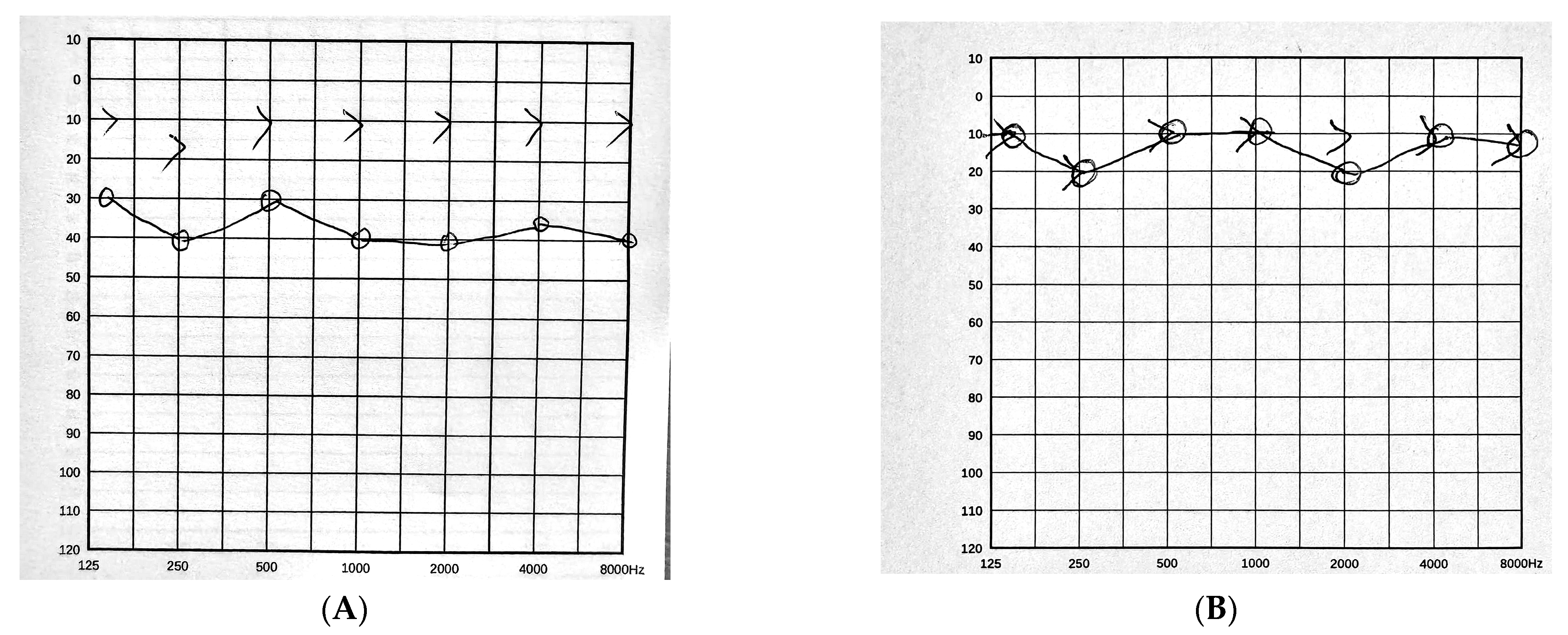
Otoendoscopic examination revealed an attic retraction with associated keratin debris and purulent discharge consistent with cholesteatoma. Pure-tone audiometry of the affected ear showed a mild conductive hearing loss present across the tested range rather than confined to particular frequencies. Air-conduction thresholds were 30 dB HL at 0.5 kHz, 35 dB HL at 1 kHz, 35 dB HL at 2 kHz and 30 dB HL at 4 kHz, giving a four-frequency pure-tone average of 32.5 dB HL, against bone-conduction thresholds of 5 dB HL at each of these frequencies. The air–bone gap was therefore 25–30 dB at every frequency tested, with a four-frequency mean of 27.5 dB. Bone-conduction thresholds were within normal limits, excluding a pre-existing sensorineural component. The tympanic membrane was intact, without perforation, throughout the pre-operative period.
2.2. Imaging and Pre-Operative Microbiology
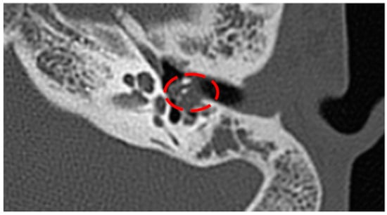
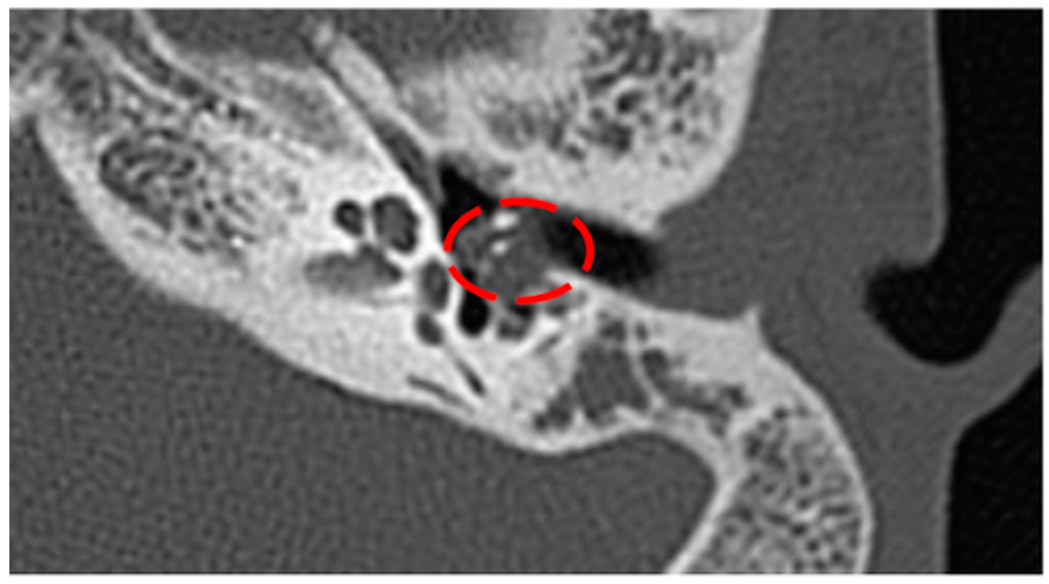
High-resolution computed tomography (HRCT) of the temporal bone demonstrated soft-tissue opacification of the epitympanum and mastoid air cells, with focal bony erosion compatible with cholesteatoma (Figure 1).
Figure 1.
Axial HRCT of the left temporal bone showing soft-tissue opacification within the middle ear (dashed red circle), with possible involvement of the ossicular chain—the radiological correlate of attic cholesteatoma. Original clinical image obtained at our institution and reproduced with identifying data removed; no generative artificial-intelligence tool was used to generate, enhance or otherwise process this image, as stated in the Acknowledgments.
A second aural-discharge culture, obtained 7 days prior to surgery, was again reported as Pseudomonas aeruginosa, susceptible to ciprofloxacin—the same organism and the same susceptibility pattern as the specimen taken months earlier, after the first ambulatory course (Section 2.1). Because this regimen was selected on the basis of an available culture and antibiogram, it constituted culture-directed (targeted) therapy rather than empirical therapy, and consisted of oral ciprofloxacin together with topical otic ciprofloxacin drops for 7 days. This regimen produced partial symptomatic relief, consistent with the clinical observation that surface cultures in chronic biofilm-protected otologic disease frequently fail to capture the dominant pathogenic flora present in deeper compartments. It is worth noting that the two superficial specimens agreed with each other. Reproducibility of a surface result across months is easily read as confirmation, and in this case it was not: both specimens recovered an organism that the intraoperative sample did not.
2.3. Microbiological Identification and Antimicrobial Susceptibility
Sterile intraoperative sampling of the middle-ear cavity and the cholesteatoma matrix was performed at the time of surgery. The isolate was identified as Acinetobacter baumannii by matrix-assisted laser desorption/ionization time-of-flight (MALDI-TOF) mass spectrometry.
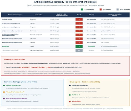
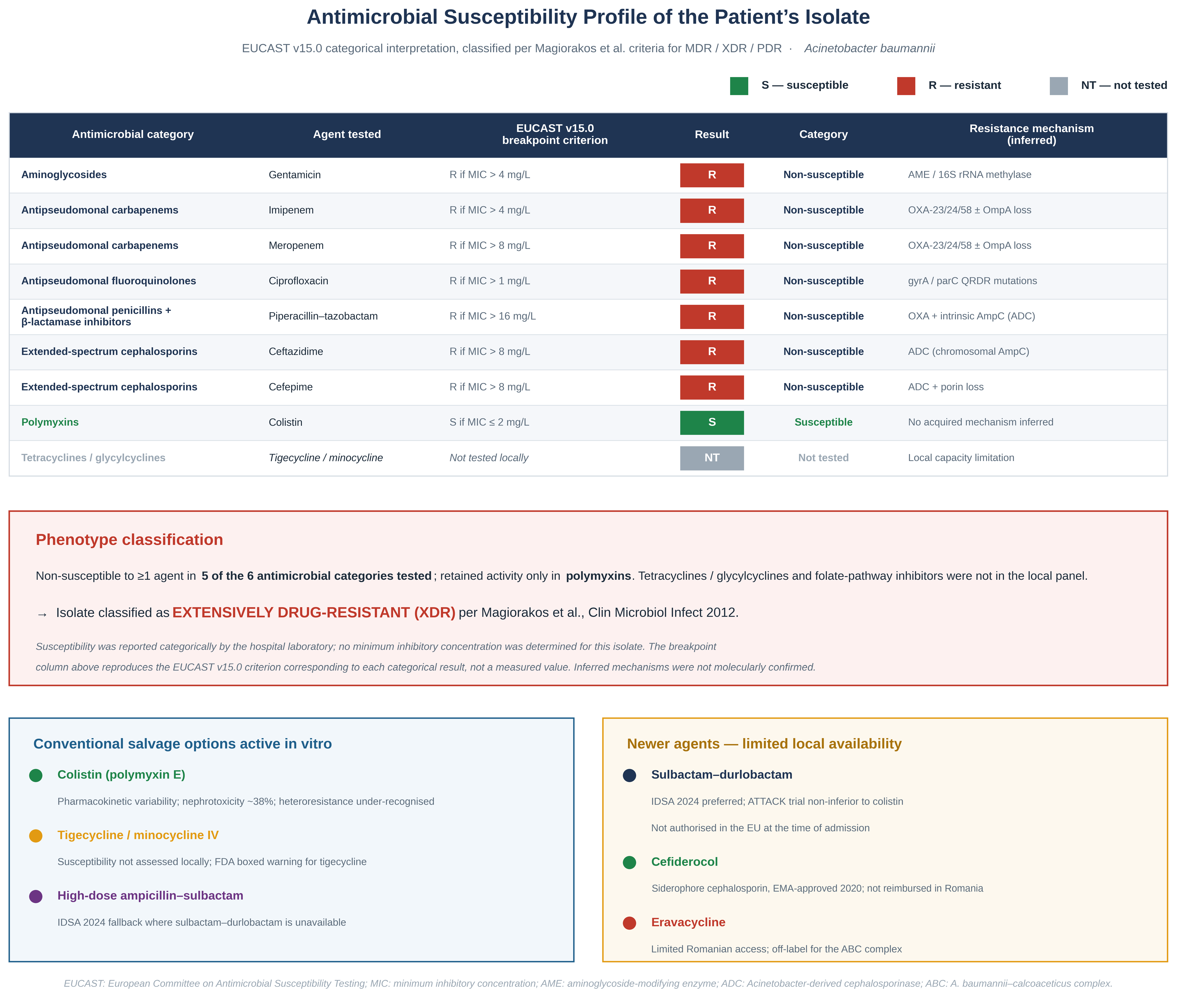
Antimicrobial susceptibility testing was performed in the hospital microbiology laboratory and interpreted according to the clinical breakpoints of the European Committee on Antimicrobial Susceptibility Testing (EUCAST), Breakpoint Tables version 15.0, valid from 1 January 2025 [24]. Results were issued categorically, as susceptible or non-susceptible for each agent tested; no minimum inhibitory concentration was quantified, including for colistin. EUCAST, following the joint EUCAST–CLSI Polymyxin Breakpoints Working Group, regards reference broth microdilution as the only acceptable method for determining polymyxin minimum inhibitory concentrations and advises against gradient diffusion and disc testing [24]; a colistin minimum inhibitory concentration obtained by that method was not available for this isolate. The isolate was non-susceptible to gentamicin, imipenem, meropenem, ciprofloxacin, piperacillin–tazobactam, ceftazidime, and cefepime, and susceptible only to colistin (Figure 2). Because the local laboratory does not routinely report tigecycline, minocycline, or cefiderocol minimum inhibitory concentrations, susceptibility for these agents could not be obtained.
Figure 2.
Antimicrobial susceptibility profile of the patient’s A. baumannii isolate, interpreted using EUCAST clinical breakpoints version 15.0 [24] and classified according to the criteria of Magiorakos et al. [25]. Susceptibility was reported categorically by the hospital laboratory; the column headed “EUCAST v15.0 breakpoint criterion” reproduces the breakpoint corresponding to each categorical result and does not report a minimum inhibitory concentration measured for this isolate, none of which was determined (Section 2.3). The isolate was non-susceptible to at least one agent in five of the six antimicrobial categories for which susceptibility was determined, with retained activity only in polymyxins; tetracyclines/glycylcyclines and folate-pathway inhibitors were not included in the local panel, and the extensively drug-resistant classification follows Magiorakos et al. [25] on that basis. The mechanisms shown in the right-hand column are inferred from the published resistance ecology of Romanian A. baumannii [3,4,8,9,26] and were not confirmed by molecular testing in this patient. The lower panels summarise the conventional salvage options [18,20,27,28,29,30,31] and the newer agents that were not procurable in our centre at the time of the case [16,17,18,19,20,21,22,23]. The scientific content of this figure—the categories, the isolate data and the classification—was defined and verified by the authors. In the upper table, the coloured result boxes give the categorical interpretation of each agent: green, susceptible (S); red, resistant/non-susceptible (R); grey, not tested (NT). In the two lower panels, the coloured bullets serve only to distinguish the listed agents and carry no additional meaning. The graphical layout was produced with the assistance of a generative artificial-intelligence tool (ChatGPT, OpenAI; version stated in the Acknowledgments), as stated in the Acknowledgments.
An important interpretive caveat applies to the colistin result. The EUCAST breakpoint for colistin in Acinetobacter spp. (S ≤ 2 mg/L) is derived from the epidemiological cut-off value rather than from clinical outcome data [24]. An in vitro “susceptible” result therefore identifies an isolate without an acquired resistance mechanism; it does not, in itself, predict clinical success. In the present case, that limitation is compounded, and we state it plainly: the susceptibility of this isolate to the only agent retaining activity was established categorically, against a breakpoint that is epidemiological rather than clinical, and without a quantified minimum inhibitory concentration to show where within the susceptible range the isolate lay. The evidential base for the therapeutic decision was therefore narrower than the word “susceptible” conveys. This distinction is material to the therapeutic reasoning set out in Section 3.6.
Applying the criteria of Magiorakos et al. [25], the isolate was classified as extensively drug-resistant (XDR): non-susceptible to at least one agent in five of the six antimicrobial categories tested for A. baumannii, with retained activity restricted to a single category (polymyxins); tetracyclines/glycylcyclines and folate-pathway inhibitors were not included in the local panel, so the classification rests on the six categories for which susceptibility was determined. Although whole-genome sequencing was not performed, the resistance profile is highly characteristic of the OXA-23-positive CRAB clones endemic to Romanian healthcare facilities [8,9], with carbapenem resistance plausibly driven by class D β-lactamase production reinforced by porin alterations and intrinsic ADC-type AmpC, aminoglycoside resistance attributable to modifying enzymes and/or 16S rRNA methylases, and fluoroquinolone resistance reflecting QRDR mutations in gyrA and parC [3,4,26].
2.4. Surgical and Antimicrobial Management
Definitive surgical source control was undertaken as the primary therapeutic intervention. The patient underwent transcanal endoscopic exploration with an extended atticotomy, complete removal of the cholesteatoma sac with preservation of the ossicular chain, and reconstruction of the lateral attic wall using tragal cartilage with perichondrium. Intraoperative irrigation of the tympanic cavity with rifampicin solution was performed, and Gelaspon fragments impregnated with dexamethasone and rifampicin were placed at the reconstruction site. Both are standing practice in our department for procedures of this type and were not selected in response to this patient’s microbiology: the causative organism was unknown at the time of surgery and was identified only on post-operative day 7. The only antimicrobial decision made in response to the isolate was the escalation to topical colistin described below. The tympanic membrane was intact at presentation, was not perforated during the procedure, and was confirmed intact at every post-operative review. The extended atticotomy converted the epitympanum into a compartment that communicated directly with the external auditory canal, and the reconstructed lateral attic wall—cartilage and perichondrium rather than bone—formed the medial boundary of that communication. This anatomical configuration is central to the choice of route discussed below and in Section 3.4. The approach was transcanal and endoscopic throughout; no post-auricular incision was made, and no mastoidectomy cavity was created, so the operated field consisted of the epitympanum, the reconstruction and the adjacent canal skin rather than an open mastoid bowl.
Choice of antimicrobial agent and route. The isolate retained in vitro activity only against polymyxins. Three considerations determined that colistin was administered topically rather than systemically, and as monotherapy rather than in combination.
First, the infection was anatomically confined and surgically controlled. There was no bacteraemia, no systemic inflammatory response, no fever, and inflammatory markers that were normal or at most marginally raised (C-reactive protein 6 mg/L; white blood cell count 12,350/µL); the presentation did not meet the definition of a serious CRAB infection for which the 2024 IDSA guidance recommends systemic therapy [20]. Systemic colistin carries dose-limiting nephrotoxicity—38% in the colistin arm of the ATTACK trial, conducted in serious systemic A. baumannii infection [18]—together with marked inter-individual pharmacokinetic variability and unfavourable tissue penetration [32,33,34]. Exposing an otherwise healthy adolescent to that toxicity in the absence of systemic infection would not have been proportionate.
Second, the pharmacological target was a debrided, keratin-adjacent surface directly accessible from the external auditory canal, not a deep tissue compartment reached through the bloodstream. Topical instillation delivers local concentrations orders of magnitude above the MIC at that surface, which no tolerable systemic regimen could achieve at the same site. Colistin-containing otic preparations carry a specific regulatory indication for the treatment of infections of mastoidectomy and fenestration cavities [35], which is the closest licensed analogue to the operated attic in this case—not because a mastoid cavity was created here, but because the pharmacological situation is the same: an epithelially raw, infected surface reachable directly from the external canal.
Third, combination therapy would have added toxicity without evidence of benefit. Randomised evidence in serious systemic A. baumannii infection has not demonstrated that polymyxin-based combinations improve survival, and its limited transferability to the present setting is discussed in Section 3.6. Adding a second agent—to which the isolate was in any case not susceptible—in a localised, surgically controlled infection would have exposed the patient to avoidable toxicity for no demonstrable gain.
Post-operative topical ciprofloxacin—continued from the pre-operative culture-directed regimen—was replaced by topical colistin once antimicrobial susceptibility results became available on post-operative day 7, and continued for 14 days [32,33,34]. Two considerations converged at that point. The susceptibility report established that the organism recovered intra-operatively was non-susceptible to the agent then in use, and the operated canal was healing slowly, with persistent congestion of the canal skin at review. Adjuvant topical therapy was introduced on that combined basis—a resistant organism documented in the operated compartment together with delayed epithelialisation of the surface from which it had been recovered—rather than as routine post-operative prophylaxis. The agent was not instilled as free drops. It was applied to an endaural pack placed within the external auditory canal against the operated attic and the canal aspect of the reconstruction, after confirmation at each change that the tympanic membrane was intact. Delivery on a pack was chosen deliberately: an impregnated pack holds the agent in sustained contact with the debrided surface, whereas instilled drops drain from the canal within minutes. Administration was therefore confined to the external auditory canal and the operated attic communicating with it, and at no point was trans-tympanic delivery to the middle-ear cleft intended or required. Because the isolate was non-susceptible to ciprofloxacin, this change constituted therapeutic escalation to the only agent with retained in vitro activity, and not de-escalation; we use the term escalation throughout for this reason.
2.5. Outcome and Follow-Up
Post-operative evolution was favourable. Otoendoscopy at ear-pack removal showed only mild canal congestion, treated with silver nitrate cauterisation. Otorrhoea resolved completely. Complete epithelialisation of the attic reconstruction and the adjacent canal skin was documented at 6 weeks. The chronology of the clinical course, from symptom onset to the 8-month review, is summarised in Table 1.
Table 1.
Timeline of the clinical course, prepared in accordance with the CARE reporting guideline for case reports.
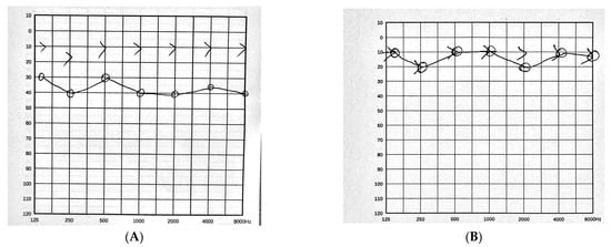
Post-operative pure-tone audiometry of the operated ear, performed 6 weeks after surgery according to ISO 8253-1 [36], demonstrated near-complete closure of the conductive air–bone gap (Figure 3A,B). Air-conduction thresholds were 10 dB HL at 0.5 kHz, 10 dB HL at 1 kHz, 20 dB HL at 2 kHz and 10 dB HL at 4 kHz, giving a four-frequency pure-tone average of 12.5 dB HL. Bone-conduction thresholds were 10 dB HL at each of these frequencies. The air–bone gap was therefore closed at 0.5, 1 and 4 kHz, with a residual gap of 10 dB at 2 kHz alone, and the mean air–bone gap across the four frequencies was 2.5 dB. Bone-conduction thresholds were unchanged from the pre-operative study, confirming the absence of an acquired sensorineural component.
Figure 3.
Pure-tone audiometry of the affected (left) ear before and after surgery, reproduced from the clinical audiograms held in the patient’s hospital record, with identifying data removed. Thresholds were obtained according to ISO 8253-1 [36]. In both panels the horizontal axis gives the test frequency in Hz and the vertical axis the hearing level in dB HL. Circles mark the air-conduction thresholds of the left ear and are joined by a solid line; the “>” symbols mark the unmasked bone-conduction thresholds of the left ear and are, by convention, not joined. No generative artificial-intelligence tool was used to generate, enhance or otherwise process these images, as stated in the Acknowledgments. (A) Pre-operative study: four-frequency air-conduction pure-tone average 32.5 dB HL, with an air–bone gap of 25–30 dB at every frequency tested and a four-frequency mean of 27.5 dB. (B) Post-operative study at 6 weeks: pure-tone average 12.5 dB HL, air–bone gap closed at 0.5, 1 and 4 kHz with a residual 10 dB gap at 2 kHz, and a four-frequency mean of 2.5 dB. Bone-conduction thresholds are unchanged between the two studies. Both panels are original clinical records obtained at our institution.
Against the pre-operative measurements reported in Section 2.1, this represents a reduction of the four-frequency mean air–bone gap from 27.5 dB to 2.5 dB, a closure of 25 dB, and an improvement in the four-frequency air-conduction pure-tone average from 32.5 dB HL to 12.5 dB HL, a gain of 20 dB. Every threshold in both studies lies on the conventional 5 dB grid.
Auditory surveillance for ototoxicity. Because polymyxins are cochleotoxic when applied directly to the middle ear in experimental models [37,38], auditory function was monitored during and after topical therapy. Monitoring was confined to conventional pure-tone audiometry performed according to ISO 8253-1 [36]. Bone-conduction thresholds in the conventional frequency range showed no deterioration relative to the pre-operative study, and the patient reported no tinnitus, aural fullness, vertigo or imbalance at any point during treatment or follow-up.
No extended high-frequency audiometry and no otoacoustic-emission data are available for this patient. This is a material limitation rather than an incidental omission, because conventional pure-tone audiometry is insensitive to early cochleotoxic injury, which characteristically appears first above 8 kHz. The surveillance reported here can therefore exclude a clinically evident cochleotoxic effect in the conventional range, but it cannot exclude a subclinical high-frequency shift. We state the point explicitly here, return to it in Section 3.5, and list it among the limitations in Section 3.10.
At 8-month follow-up, otoendoscopic examination revealed stable attic reconstruction without granulation tissue or recurrent infection. No adverse effects attributable to topical colistin were identified. Non–echo-planar diffusion-weighted magnetic resonance imaging (non-EPI DWI-MRI) is scheduled at 12 months in accordance with current recommendations for the detection of residual or recurrent cholesteatoma.
3. Discussion
3.1. Mechanisms of Resistance and Phenotypic Classification
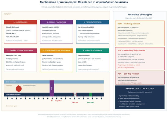
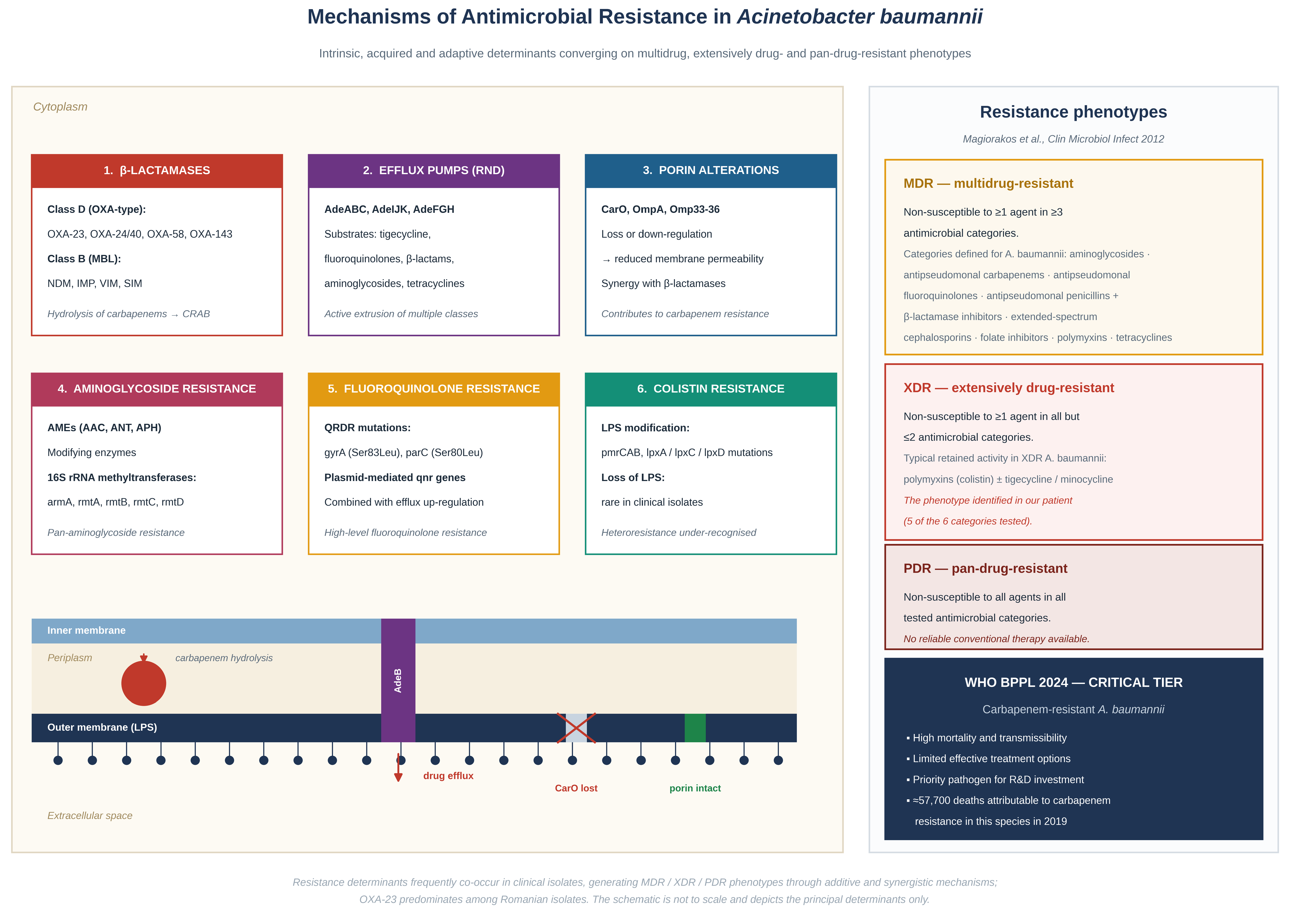
The XDR phenotype documented in our patient is congruent with the resistance ecology of A. baumannii endemic to Romanian hospital settings, where the OXA-23 carbapenemase has become the dominant class D β-lactamase, often co-existing with chromosomal AmpC (ADC) cephalosporinases and a battery of efflux and target-modification determinants [8,9,26]. Six categories of mechanism are clinically relevant in this species and are summarised in Figure 4: β-lactamases, with OXA-23, OXA-24/40 and OXA-58 dominant in Europe; RND-family efflux pumps; outer-membrane porin loss or down-regulation; aminoglycoside-modifying enzymes and 16S rRNA methyltransferases; QRDR mutations in gyrA and parC; and lipopolysaccharide modification or loss as the principal route to colistin resistance [3,4,26]. Their frequent co-occurrence explains the rapidity with which clinical isolates traverse from MDR to XDR and, occasionally, to pan-drug-resistant phenotypes [25].
Figure 4.
Principal mechanisms of antimicrobial resistance in Acinetobacter baumannii. The left-hand panel groups the six mechanism classes of clinical relevance in this species—β-lactamases, RND-family efflux pumps, porin loss or down-regulation, aminoglycoside-modifying enzymes and 16S rRNA methyltransferases, QRDR mutations, and lipopolysaccharide modification or loss—and relates them schematically to the outer membrane, periplasm and inner membrane; the content is compiled from the cited sources [3,4,26], with the Romanian OXA-23 context from [8,9] and colistin heteroresistance from [31]. The six numbered, colour-coded boxes (1–6) correspond to the six mechanism classes. In the membrane schematic beneath them, the red sphere and arrow in the periplasm denote carbapenem hydrolysis by β-lactamases, the purple channel with a downward arrow denotes drug efflux through an RND pump (AdeB), the red “X” marks loss of the CarO porin and the green segment an intact porin; colours serve to distinguish the elements and carry no quantitative meaning. The right-hand panel reproduces the MDR/XDR/PDR definitions and the antimicrobial categories defined for A. baumannii by Magiorakos et al. [25], together with the WHO Bacterial Priority Pathogens List 2024 critical-tier designation of carbapenem-resistant A. baumannii [5] and the global mortality estimate attributable to carbapenem resistance in this species [6,7]. The mechanistic content was compiled and verified by the authors from the cited sources; the graphical layout was produced with the assistance of a generative artificial-intelligence tool (ChatGPT, OpenAI; version stated in the Acknowledgments), as stated in the Acknowledgments.
3.2. The Diagnostic Pitfall of Superficial Cultures in Biofilm-Associated Otologic Disease
Our case illustrates a discrepancy that is both diagnostically instructive and clinically dangerous, and it does so more forcefully than a single discordant culture would. Two superficial aural-discharge specimens were obtained from this patient on separate occasions, months apart and separated by three antimicrobial courses. Both were reported as P. aeruginosa susceptible to ciprofloxacin—the organism most frequently recovered from otorrhoea in chronic suppurative otitis media [39]. Sterile intraoperative sampling identified an XDR A. baumannii as the dominant deep pathogen. The reproducibility of the surface result is the instructive part. A clinician confronted with a single unexpected culture may reasonably repeat it; here, repetition returned the same answer, and the same wrong answer. Concordance between successive superficial specimens is intuitively read as confirmation, and in biofilm-associated otologic disease, it is not: two samples drawn from the same accessible compartment interrogate the same compartment twice. What they reproduce is the composition of the canal surface, not the composition of the disease. In this patient, the consequence was four antimicrobial courses—three of them directed by that surface result—none of which could have been active against the organism that was actually present. This pattern is not idiosyncratic: biofilms anchored to keratin debris, bone and granulation tissue create protected sanctuaries that are poorly represented in superficial discharge specimens [40], and microbiome and metagenomic analyses of cholesteatoma confirm distinct bacterial communities in the discharge, the middle-ear mucosa and the matrix itself [15,41]. This argues for routine sterile intraoperative sampling in any chronic otologic infection that is refractory to first-line therapy, and for caution in interpreting antibiotic susceptibility data derived solely from canal swabs.
3.3. Colonisation Versus Infection, and the Respective Contributions of Surgery and Topical Therapy
A legitimate question in any cholesteatoma harbouring a resistant Gram-negative organism is whether that organism represents true tissue infection or merely colonisation of the keratin sac—and, by extension, whether complete excision of the sac renders adjunctive topical therapy superfluous. Our position is deliberately conservative, and we state it plainly: we regard the surgery as the curative intervention and the topical colistin as consolidation of a surgical result, not as the treatment that cured the infection.
Three considerations nonetheless argue against dismissing the isolate as innocuous colonisation. First, the clinical phenotype was that of infection rather than colonisation: continuous mucopurulent otorrhoea over 9 months, mucosal hypertrophy and granulation at operation, and radiologically documented bony erosion, a finding that metagenomic work has related to bacterial burden in acquired cholesteatoma [41]. Second, the cholesteatoma sac and the adjacent hypertrophic mucosa constitute a demonstrated biofilm niche [42,43], and biofilm-resident organisms tolerate concentrations far above the planktonic MIC [44]—which is precisely what makes surgical removal, rather than antimicrobial therapy, the decisive manoeuvre. Third, the only comparable published series of Acinetobacter chronic otitis media reported bone destruction, required surgery combined with susceptibility-directed therapy, and still recorded recurrence in two of six patients [12].
The related premise—that the operated field is healed, and therefore no longer a microbial niche, within approximately 1 week—is not supported by the healing literature, nor by this case. Epithelialisation after middle-ear surgery is measured in weeks to months rather than days: in a controlled surgical series, conventionally managed mastoid cavities required more than 80 days to epithelialise and become dry [45]. A transcanal endoscopic approach that creates no mastoid cavity heals considerably faster than that, and it did so here—but complete epithelialisation of the reconstruction and the adjacent canal skin was nonetheless documented only at 6 weeks, six times the interval the premise assumes. Throughout that interval, the reconstruction site is a raw, keratin-adjacent surface lying within a canal that had been colonised for months by an XDR organism.
The healing interval is the reason for giving adjuvant topical therapy at all; it is not the reason for giving it for any particular length of time. Our patient received a defined 14-day course, directed at a documented organism and timed to the phase of greatest vulnerability—an actively discharging, freshly debrided surface carrying the highest bioburden—after which healing continued under clinical and otoendoscopic surveillance without further antimicrobial cover. We did not attempt to maintain antimicrobial exposure across the whole 6-week epithelialisation period, and we would not advocate doing so.
The limits of this argument should be stated plainly. There is no randomised evidence that adjunctive topical antimicrobial therapy after cholesteatoma surgery improves outcome; the closest available trial, in clean elective myringoplasty rather than a colonised operated field, was negative [46]. Nor can a single case establish that surgery alone would have failed. Pooled data show cholesteatoma recurrence rising from 7% at 12 months to 39% at 15 years [47], so neither the surgery nor its adjunct can be declared definitively curative on an 8-month follow-up.
3.4. Rationale for the Topical Route in an Ear with an Intact Tympanic Membrane
At first inspection, instilling an antimicrobial into the external auditory canal of an ear with an intact tympanic membrane appears an ineffective way to treat a middle-ear infection, and in an unoperated ear, that objection would be well founded: the intact drum is a substantial diffusion barrier, and passive instillation does not reliably generate therapeutic middle-ear concentrations [48].
The anatomy in the present case, however, was not that of an unoperated ear. The extended atticotomy converted the epitympanum into a compartment communicating with the external auditory canal, and the lateral attic wall was reconstructed with tragal cartilage and perichondrium rather than sealed with bone. The target surface for topical therapy was therefore not the middle-ear cleft lying behind an intact drum, but the exposed attic and the canal aspect of the reconstruction—precisely the surface from which the cholesteatoma matrix and its associated biofilm had been removed, and precisely the surface against which the medicated pack was placed. This is the same anatomical rationale that underpins the licensed indication of colistin-containing otic preparations for the treatment of infections of mastoidectomy and fenestration cavities [35].
Two consequences follow. First, the target of therapy was the operated field itself—the canal, the exposed attic and the reconstruction—which was the surface colonised by the organism, rather than the middle-ear cleft; this is topical therapy directed at a colonised operated field, not trans-tympanic drug delivery. Second, delivery on an endaural pack rather than as free drops kept the agent in sustained contact with that debrided surface over a defined course. The intact tympanic membrane was accordingly not a barrier that had to be crossed, but the structure that made the route safe, excluding the drug from the middle-ear cleft and from the round-window membrane, where the experimental cochleotoxicity of the polymyxins has been demonstrated.
3.5. Ototoxicity Considerations
Polymyxins warrant respect as potential cochleotoxins, and the concern is not theoretical. In the defining animal experiment, polymyxin B applied to the round window produced greater cochlear damage than neomycin [37], and systematic review confirms the signal across agents applied into the open middle ear [38]. The human evidence is considerably weaker, and the reported cases occur essentially always in ears with a non-intact tympanic membrane [49]—but the risk is not zero, and it is concentrated precisely where the round-window membrane is accessible to the drug.
Two features of the present case place it outside that risk window. First, the tympanic membrane was intact and was confirmed intact before each application, so the round window was not accessible to the applied preparation. Second, in licensed colistin-containing otic products, the ototoxicity warning is attributed by the manufacturer to the neomycin component rather than to colistin sulfate [35], and contemporary reviews of systemic colistin toxicity describe neurotoxicity—paraesthesiae, ataxia, neuromuscular blockade—without listing sensorineural hearing loss among recognised adverse effects [50].
The adequacy of the surveillance actually performed must nonetheless be addressed candidly. Monitoring was limited to conventional pure-tone audiometry, which is insensitive for this purpose: extended high-frequency testing detects ototoxic threshold shifts substantially earlier [51] and is recommended in children receiving potentially cochleotoxic antimicrobials [52,53]. The reassuring auditory outcome reported here is therefore reassuring only within the conventional range.
A further caution applies specifically to this patient: one of the four pre-referral courses included topical tobramycin, given to a chronically discharging ear, so any high-frequency deficit that extended monitoring might have revealed could as plausibly have predated our intervention as followed it.
Prospectively, extended high-frequency audiometry with otoacoustic emissions, obtained before the first application and again after completion of therapy and interpreted against the patient’s own baseline [53], would have allowed this question to be answered directly rather than by inference. We regard it as the appropriate surveillance wherever topical polymyxins are applied to an operated ear beyond a short course.
3.6. Therapeutic Context and the Limits of Extrapolation from Systemic-Infection Evidence
This patient did not require, and did not receive, any of the newer anti-CRAB agents. His infection was localised, was controlled surgically, and resolved on the accessible pharmacopoeia. The agents positioned by the 2024 IDSA guidance for carbapenem-resistant A. baumannii—sulbactam–durlobactam with a background carbapenem, with cefiderocol and high-dose ampicillin–sulbactam as alternatives [16,17,18,19,20]—are recommended for serious systemic infection, a category the present case did not meet (Section 2.4). They are mentioned here only to situate the isolate within the current treatment landscape, and not to suggest that any of them was indicated in this patient.
The randomised evidence on polymyxin-based therapy in A. baumannii infection comes entirely from severe systemic disease and cannot be transferred to the present case. AIDA randomised patients with severe carbapenem-resistant Gram-negative infection [27]; OVERCOME had a comparable case mix [28]; and ATTACK enrolled patients with serious infections caused by the A. baumannii–calcoaceticus complex [18]. All three report 28- or 30-day mortality in critically ill populations with bacteraemia, ventilator-associated pneumonia or comparable syndromes, and their findings—no survival advantage for colistin-based combinations over colistin monotherapy [27,28,29], and the nephrotoxicity of systemic colistin [18]—bore on only two of our decisions: not to give colistin systemically, and not to add a second agent (Section 2.4). They carry no implication, in either direction, for topically applied colistin on a debrided, surgically controlled otologic surface, for which no comparable trial evidence exists. The reported prevalence of colistin heteroresistance and its association with mortality in bacteraemic patients [31] is likewise a systemic-infection observation: it is a reason for caution in reading a categorical susceptibility report (Section 2.3), not evidence bearing on the outcome achieved here.
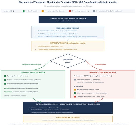
Access to newly approved antibacterials across the European Union correlates with national health-economic indicators rather than with epidemiological need, the bottleneck lying between centralised authorisation and national reimbursement [21,22,23]. Of the agents potentially active against XDR A. baumannii, only two were routinely available and reimbursed in our setting at the time of this case; the decision pathway actually followed is summarised in Figure 5, and the agent-by-agent access status is provided as Supplementary Figure S1. This is the context in which the options were weighed. It did not alter the management of this patient, whose infection was controlled surgically and who recovered on an agent that was available.
Figure 5.
Diagnostic and therapeutic algorithm for suspected MDR/XDR Gram-negative chronic otologic infection, with access-tier annotations reflecting Romanian availability of each candidate agent at the time of this case. The antimicrobial pathway is adapted from the 2024 IDSA guidance on the treatment of antimicrobial-resistant Gram-negative infections [20]; the positions assigned to sulbactam–durlobactam and cefiderocol derive from [16,17,18,19]; identification and susceptibility testing follow EUCAST v15.0 [24]; the polymyxin data underlying the colistin option derive from [32,33,34]; the access tiers A–D were assigned by the authors from the European access analyses [21,22,23] and are set out agent by agent in Supplementary Figure S1; and the surgical source-control step, which does not form part of the IDSA algorithm, was added by the authors on the basis of the biofilm and source-control evidence discussed in Section 3.3 and Section 3.7 [41,42,43,44,54,55,56,57]. Arrows indicate the sequence of decisions, the green arrow leading to the branch for isolates susceptible to at least one first-line agent and the red arrow to the MDR/XDR branch. The coloured squares denote the Romanian access tier of each agent, as defined in the key at the top of the figure: green, tier A (broadly available); amber, tier B (restricted or partial reimbursement); red, tier C (nominal procurement only); grey, tier D (not authorised in the EU/Romania at the time of admission). The algorithm is a synthesis prepared by the authors for this report and is not a validated clinical decision instrument. The scientific content was defined and verified by the authors; the graphical layout was produced with the assistance of a generative artificial-intelligence tool (ChatGPT, OpenAI; version stated in the Acknowledgments), as stated in the Acknowledgments.
3.7. Surgical Source Control as the Principal Therapeutic Intervention
Because the isolate retained in vitro activity only against polymyxins, the accessible pharmacological options were narrow, and systemic colistin carries high inter-individual variability in plasma concentrations, dose-limiting nephrotoxicity and underrecognised heteroresistance [31,32,33,34]. Topical administration within the external auditory canal circumvented systemic toxicity but offered no parenteral coverage—a limitation that would have mattered had the infection not been anatomically confined.
In this constrained setting, surgical source control assumed primary therapeutic responsibility. Complete cholesteatoma removal achieves three pharmacodynamic objectives that no antibiotic regimen alone can replicate: a reduction in bacterial inoculum by several orders of magnitude, mechanical disruption of the biofilm matrix that protects sessile A. baumannii populations from antimicrobial penetration, and elimination of the keratinised epithelial niche that supports persistent colonisation [54,55,56]. Experimental work in three-dimensional human skin models has shown that A. baumannii can persist on stratum corneum and form biofilms supported by surface-associated proteins such as Bap, providing a biological rationale for the empirical observation that source control is decisive in keratin-rich infectious niches [56,57]. Published experience in XDR A. baumannii osteomyelitis, in which repeated debridement alone did not clear the infection [58], is a reminder that source control is decisive where the infected compartment can be excised in its entirety and insufficient where it cannot. Surgical eradication of the cholesteatoma is therefore not an adjunct to antimicrobial therapy here—it is the principal intervention, and antimicrobial therapy consolidates the surgical result.
Excision also changes the task the antimicrobial is required to perform. Removing the sac eliminates the great majority of the bacterial population together with the matrix that shields it [44], leaving a thin residual burden on a debrided surface where a topical agent reaches concentrations no systemic regimen could achieve. This reasoning is specific to infections that are surgically accessible and completely excisable—a description that fits attic cholesteatoma and fits most serious CRAB infections not at all.
One practical consequence follows for the operating surgeon: the completeness of excision matters, and intraoperative endoscopic verification of complete removal, as performed in this case, is part of the therapeutic strategy rather than a refinement of it.
3.8. Stewardship Implications
Two stewardship lessons follow directly from the case. First, the discordance between superficial and deep cultures should reframe the empirical approach to refractory chronic otologic infection: clinicians should expect a wider pathogen spectrum than ciprofloxacin-susceptible P. aeruginosa in patients who have already received multiple antibiotic courses, and should not select definitive therapy on the basis of canal swab data alone. This is consistent with contemporary guidance on chronic suppurative otitis media, in which topical therapy is the mainstay but microbiological targeting requires specimens representative of the deep compartment [59]. Second, the local susceptibility panel did not extend to tigecycline, minocycline or cefiderocol, which limited the precision with which this isolate could be characterised.
3.9. Patient Perspective
The patient, reviewed with his legal guardian at the 8-month visit, described the preceding year as dominated less by pain than by the persistence of the discharge and by the repeated cycle of treatment and relapse. He reported that each course of antibiotics produced a few days of improvement, that the discharge always returned, and that the recurring question in the family was whether anything would work. He identified the resolution of the otorrhoea, rather than the improvement in hearing, as the change that mattered most to him, and noted that he had not appreciated before surgery that an operation was likely to be the definitive step. Asked what he would want other patients to know, he said that he wished the decision to operate had been reached sooner.
3.10. Limitations
This is a single-patient report with an 8-month follow-up from a tertiary referral centre. Whole-genome sequencing was not performed, so the inferred resistance mechanisms remain phenotypically supported but not molecularly proven. The choice of topical colistin in an adolescent ear was a pragmatic decision and should not be extrapolated as standard practice. We should also be consistent about the intraoperative rifampicin, since this report argues for culture-directed prescribing: rifampicin irrigation and rifampicin-impregnated dressings are used routinely in our department rather than for an indication established in the individual patient, they contributed no demonstrable component to the outcome described, and we regard the practice as a legitimate target for review in our own unit. Because the patient recovered after combined surgical and topical management, the independent contribution of each component cannot be separated; we regard the surgery as decisive, but this attribution is inferential. No randomised evidence supports adjuvant topical antimicrobial therapy after cholesteatoma surgery [46]. Documentation of the pre-referral courses derived from ambulatory records and patient report and may be incomplete. Susceptibility was reported categorically without quantified minimum inhibitory concentrations, so we cannot state how far below the colistin breakpoint this isolate lay, nor exclude heteroresistance, for which testing is not routinely available in our setting. Auditory surveillance was limited to conventional pure-tone audiometry, so a subclinical cochleotoxic effect above 8 kHz cannot be excluded. The cost figures in Supplementary Figure S1 are illustrative wholesale ranges intended to convey orders of magnitude; precise pricing in Romania is contractual and varies between institutions. Larger prospective series are required to delineate the incidence of CRAB in chronic otomastoiditis.
4. Conclusions
This report documents an extensively drug-resistant A. baumannii infection complicating cholesteatomatous chronic otomastoiditis in an adolescent, an organism and a site rarely reported together. Two observations follow from the case itself. First, two concordant superficial swabs identified an organism that sterile intraoperative sampling did not confirm, and four antimicrobial courses were directed by that surface result; in chronic otologic infection refractory to first-line therapy, deep sampling at operation should not be omitted. Second, complete transcanal endoscopic excision of the cholesteatoma was the decisive intervention, with a defined 14-day course of topical colistin applied to the operated surface as an adjunct: otorrhoea resolved, the reconstruction epithelialised by 6 weeks, the mean air–bone gap fell from 27.5 dB to 2.5 dB, and there was no recurrence at 8 months. In a single case, the independent contribution of the topical agent cannot be separated from that of the surgery, and an 8-month follow-up cannot establish cure. The limited local availability of newer anti-CRAB agents shaped the options that were weighed rather than the outcome obtained, since the infected compartment could be excised in its entirety—a condition that does not hold for most serious CRAB infections.
Supplementary Materials
The following supporting information can be downloaded at: https://www.mdpi.com/article/10.3390/germs16030022/s1, Figure S1: Agent-by-agent access status in Romania, at the time of the case, of the antimicrobial agents with potential activity against extensively drug-resistant Acinetobacter baumannii, with the access tiers (A–D) used in Figure 5 and illustrative wholesale cost ranges; Table S1: Completed CARE (CAse REport) checklist for this case report.
Author Contributions
Conceptualization, C.D.N. and L.-M.G.; methodology, L.-M.G. and S.M.A.; clinical management, C.D.N. and L.-M.G.; literature review and infectious-disease perspective, S.M.A. and P.A.; writing—original draft preparation, L.-M.G. and S.M.A.; writing—review and editing, all authors; supervision, P.A. All authors have read and agreed to the published version of the manuscript.
Funding
This research received no external funding.
Institutional Review Board Statement
The case report was conducted in accordance with the Declaration of Helsinki. Institutional ethical review was waived because this is a single-patient retrospective case report and informed consent for publication was obtained.
Informed Consent Statement
Written informed consent was obtained from the patient (and the patient’s legal guardian, given the patient’s minor status) for publication of this case report and any accompanying images.
Data Availability Statement
Anonymised clinical data supporting the findings of this study are available from the corresponding author on reasonable request, in accordance with applicable data-protection regulations.
Acknowledgments
Publication of this paper was supported by the “Carol Davila” University of Medicine and Pharmacy, through the institutional programme “Publish not Perish”. This case report was prepared in accordance with the CARE (CAse REport) guideline; the completed CARE checklist is provided as Supplementary Table S1. During the preparation of this manuscript, the authors used ChatGPT (GPT-5.6, OpenAI, 2026) to produce the graphical layout of Figure 2, Figure 4 and Figure 5. The scientific content of these figures—the isolate data, the resistance classifications, the algorithmic steps and the access-tier assignments—was defined, verified and edited by the authors from the patient’s microbiological record and from the cited primary sources; the tool was used for visual presentation only and did not generate, infer or alter any datum. Figure 1 and Figure 3 are original clinical records obtained at our institution—a computed-tomography image and a reproduction of the patient’s clinical audiograms—and were not generated, enhanced or otherwise processed with any artificial-intelligence tool. No generative artificial-intelligence tool was used to produce the scientific text, the analysis or the interpretation presented in this manuscript. The authors have reviewed and edited all output and take full responsibility for the content of this publication.
Conflicts of Interest
The authors declare no conflicts of interest.
Abbreviations
| ABC | Acinetobacter baumannii–calcoaceticus complex |
| ADC | Acinetobacter-derived cephalosporinase |
| AME | aminoglycoside-modifying enzyme |
| BPPL | Bacterial Priority Pathogens List |
| CEE | Central-Eastern European |
| CNAS | Casa Națională de Asigurări de Sănătate (Romanian national health insurance fund) |
| CRAB | carbapenem-resistant Acinetobacter baumannii |
| CSOM | chronic suppurative otitis media |
| EMA | European Medicines Agency |
| ESKAPE | Enterococcus faecium, Staphylococcus aureus, Klebsiella pneumoniae, Acinetobacter baumannii, Pseudomonas aeruginosa, and Enterobacter spp. |
| EUCAST | European Committee on Antimicrobial Susceptibility Testing |
| FDA | US Food and Drug Administration |
| HRCT | high-resolution computed tomography |
| IDSA | Infectious Diseases Society of America |
| MALDI-TOF | matrix-assisted laser desorption/ionization time-of-flight |
| MBL | metallo-β-lactamase |
| MDR | multidrug-resistant |
| MIC | minimum inhibitory concentration |
| non-EPI DWI-MRI | non–echo-planar diffusion-weighted magnetic resonance imaging |
| OXA | oxacillinase |
| PDR | pan-drug-resistant |
| QRDR | quinolone resistance-determining region |
| WHO | World Health Organization |
| XDR | extensively drug-resistant |
References
- Ayoub Moubareck, C.; Hammoudi Halat, D. Insights into Acinetobacter baumannii: A review of microbiological, virulence, and resistance traits in a threatening nosocomial pathogen. Antibiotics 2020, 9, 119. [Google Scholar] [CrossRef] [Scilit] [PubMed]
- Yehya, A.; Ezzeddine, Z.; Chakkour, M.; Dhaini, Z.; Bou Saba, M.S.; Bou Saba, A.S.; Nohra, L.; Nassar, N.B.; Yassine, M.; Bahmad, H.F.; et al. The intricacies of Acinetobacter baumannii: A multifaceted comprehensive review of a multidrug-resistant pathogen and its clinical significance and implications. Front. Microbiol. 2025, 16, 1565965. [Google Scholar] [CrossRef] [Scilit] [PubMed]
- Vrancianu, C.O.; Gheorghe, I.; Czobor, I.B.; Chifiriuc, M.C. Antibiotic resistance profiles, molecular mechanisms and innovative treatment strategies of Acinetobacter baumannii. Microorganisms 2020, 8, 935. [Google Scholar] [CrossRef] [Scilit] [PubMed]
- Castanheira, M.; Mendes, R.E.; Gales, A.C. Global Epidemiology and Mechanisms of Resistance of Acinetobacter baumannii-calcoaceticus Complex. Clin. Infect. Dis. 2023, 76, S166–S178. [Google Scholar] [CrossRef] [Scilit] [PubMed]
- Sati, H.; Carrara, E.; Savoldi, A.; Hansen, P.; Garlasco, J.; Campagnaro, E.; Boccia, S.; Castillo-Polo, J.A.; Magrini, E.; Garcia-Vello, P.; et al. The WHO Bacterial Priority Pathogens List 2024: A prioritisation study to guide research, development, and public health strategies against antimicrobial resistance. Lancet Infect. Dis. 2025, 25, 1033–1043. [Google Scholar] [CrossRef] [Scilit] [PubMed]
- Antimicrobial Resistance Collaborators. Global burden of bacterial antimicrobial resistance in 2019: A systematic analysis. Lancet 2022, 399, 629–655. [Google Scholar] [CrossRef] [Scilit] [PubMed]
- Boutzoukas, A.E.; Doi, Y. The global epidemiology of carbapenem-resistant Acinetobacter baumannii. JAC Antimicrob. Resist. 2025, 7, dlaf134. [Google Scholar] [CrossRef] [Scilit] [PubMed]
- Gheorghe, I.; Czobor Barbu, I.; Surleac, M.; Sârbu, I.; Popa, L.I.; Paraschiv, S.; Feng, Y.; Lazăr, V.; Chifiriuc, M.C.; Oțelea, D.; et al. Subtypes, resistance and virulence platforms in extended-drug resistant Acinetobacter baumannii Romanian isolates. Sci. Rep. 2021, 11, 13288. [Google Scholar] [CrossRef] [Scilit] [PubMed]
- Pană, A.G.; Șchiopu, P.; Țoc, D.A.; Neculicioiu, V.S.; Butiuc-Keul, A.; Farkas, A.; Dobrescu, M.S.; Flonta, M.; Costache, C.; Szász, I.É.; et al. Clonality and the phenotype-genotype correlation of antimicrobial resistance in Acinetobacter baumannii isolates: A multicenter study of clinical isolates from Romania. Microorganisms 2025, 13, 176. [Google Scholar] [CrossRef] [Scilit] [PubMed]
- Borcan, A.M.; Rotaru, E.; Caravia, L.G.; Filipescu, M.C.; Simoiu, M. Trends in Antimicrobial Resistance of Acinetobacter baumannii and Pseudomonas aeruginosa from Bloodstream Infections: An Eight-Year Study in a Romanian Tertiary Hospital. Pharmaceuticals 2025, 18, 948. [Google Scholar] [CrossRef] [Scilit] [PubMed]
- Sava, M.; Codru, I.R.; Bereanu, A.S.; Stoia, O.; Vintila, B.I. Resistance dynamics in a Romanian critical care unit: Four years of ESKAPE pathogen surveillance. Medicina 2025, 61, 2114. [Google Scholar] [CrossRef] [Scilit] [PubMed]
- Zhang, K.; Qu, P.; Jia, N.; Wang, Y.; Tian, J.; Chen, Z.; Xu, A.; Fu, T. Infection of chronic otitis media with Acinetobacter: 6 cases report. J. Clin. Otorhinolaryngol. Head Neck Surg. 2016, 30, 1885–1887. [Google Scholar] [CrossRef] [PubMed]
- Treviño González, J.L.; Reyes Suárez, L.L.; Hernández de León, J.E. Malignant otitis externa: An updated review. Am. J. Otolaryngol. 2021, 42, 102894. [Google Scholar] [CrossRef] [Scilit] [PubMed]
- Marra, P.; Trusio, A.; De Cristofaro, G.; Filosa, B.; Colacurcio, V.; De Luca, P.; Malafronte, G. Multiresistant Acinetobacter baumannii causing malignant otitis externa: A case report. Clin. Ter. 2023, 174, 318–321. [Google Scholar] [CrossRef] [PubMed]
- Frank, D.N.; Magno, J.P.M.; Velasco, K.J.S.; Bootpetch, T.C.; Salud, J.E.D.; David, K.J.V.; Miller, A.L.; Yee, E.C.; Dulnuan, H.P.; Pyles, R.B.; et al. Microbiota associated with cholesteatoma tissue in chronic suppurative otitis media. Front. Cell. Infect. Microbiol. 2022, 12, 746428. [Google Scholar] [CrossRef] [Scilit] [PubMed]
- Bassetti, M.; Echols, R.; Matsunaga, Y.; Ariyasu, M.; Doi, Y.; Ferrer, R.; Lodise, T.P.; Naas, T.; Niki, Y.; Paterson, D.L.; et al. Efficacy and safety of cefiderocol or best available therapy for the treatment of serious infections caused by carbapenem-resistant Gram-negative bacteria (CREDIBLE-CR): A randomised, open-label, multicentre, pathogen-focused, descriptive, phase 3 trial. Lancet Infect. Dis. 2021, 21, 226–240. [Google Scholar] [CrossRef] [Scilit] [PubMed]
- Onorato, L.; de Luca, I.; Monari, C.; Coppola, N. Cefiderocol either in monotherapy or combination versus best available therapy in the treatment of carbapenem-resistant Acinetobacter baumannii infections: A systematic review and meta-analysis. J. Infect. 2024, 88, 106113. [Google Scholar] [CrossRef] [Scilit] [PubMed]
- Kaye, K.S.; Shorr, A.F.; Wunderink, R.G.; Du, B.; Poirier, G.E.; Rana, K.; Miller, A.; Lewis, D.; O’Donnell, J.; Chen, L.; et al. Efficacy and safety of sulbactam–durlobactam versus colistin for the treatment of patients with serious infections caused by Acinetobacter baumannii–calcoaceticus complex: A multicentre, randomised, active-controlled, phase 3, non-inferiority clinical trial (ATTACK). Lancet Infect. Dis. 2023, 23, 1072–1084. [Google Scholar] [CrossRef] [Scilit] [PubMed]
- Karlowsky, J.A.; Hackel, M.A.; McLeod, S.M.; Miller, A.A. In Vitro Activity of Sulbactam-Durlobactam against Global Isolates of Acinetobacter baumannii-calcoaceticus Complex Collected from 2016 to 2021. Antimicrob. Agents Chemother. 2022, 66, e00781-22. [Google Scholar] [CrossRef] [Scilit] [PubMed]
- Tamma, P.D.; Heil, E.L.; Justo, J.A.; Mathers, A.J.; Satlin, M.J.; Bonomo, R.A. Infectious Diseases Society of America 2024 guidance on the treatment of antimicrobial-resistant Gram-negative infections. Clin. Infect. Dis. 2024, ciae403. [Google Scholar] [CrossRef] [Scilit] [PubMed]
- Zasheva, A.; Batcheva, E.; Ivanova, K.D.; Yanakieva, A. Differences in patient access to newly approved antibacterial drugs in EU/EEA countries. Antibiotics 2024, 13, 1077. [Google Scholar] [CrossRef] [Scilit] [PubMed]
- Tängdén, T.; Carrara, E.; Hellou, M.M.; Yahav, D.; Paul, M. Introducing new antibiotics for multidrug-resistant bacteria: Obstacles and the way forward. Clin. Microbiol. Infect. 2025, 31, 354–359. [Google Scholar] [CrossRef] [Scilit] [PubMed]
- Outterson, K.; Orubu, E.S.F.; Rex, J.; Årdal, C.; Zaman, M.H. Patient access in 14 high-income countries to new antibacterials approved by the US Food and Drug Administration, European Medicines Agency, Japan Pharmaceuticals and Medical Devices Agency, or Health Canada, 2010–2020. Clin. Infect. Dis. 2022, 74, 1183–1190. [Google Scholar] [CrossRef] [Scilit] [PubMed]
- The European Committee on Antimicrobial Susceptibility Testing. Breakpoint Tables for Interpretation of MICs and Zone Diameters, Version 15.0, 2025. Available online: https://www.eucast.org (accessed on 31 July 2026).
- Magiorakos, A.P.; Srinivasan, A.; Carey, R.B.; Carmeli, Y.; Falagas, M.E.; Giske, C.G.; Harbarth, S.; Hindler, J.F.; Kahlmeter, G.; Olsson-Liljequist, B.; et al. Multidrug-resistant, extensively drug-resistant and pandrug-resistant bacteria: An international expert proposal for interim standard definitions for acquired resistance. Clin. Microbiol. Infect. 2012, 18, 268–281. [Google Scholar] [CrossRef] [Scilit] [PubMed]
- Wong, D.; Nielsen, T.B.; Bonomo, R.A.; Pantapalangkoor, P.; Luna, B.; Spellberg, B. Clinical and pathophysiological overview of Acinetobacter infections: A century of challenges. Clin. Microbiol. Rev. 2017, 30, 409–447. [Google Scholar] [CrossRef] [Scilit] [PubMed]
- Paul, M.; Daikos, G.L.; Durante-Mangoni, E.; Yahav, D.; Carmeli, Y.; Benattar, Y.D.; Skiada, A.; Andini, R.; Eliakim-Raz, N.; Nutman, A.; et al. Colistin alone versus colistin plus meropenem for treatment of severe infections caused by carbapenem-resistant Gram-negative bacteria: An open-label, randomised controlled trial. Lancet Infect. Dis. 2018, 18, 391–400. [Google Scholar] [CrossRef] [Scilit] [PubMed]
- Kaye, K.S.; Marchaim, D.; Thamlikitkul, V.; Carmeli, Y.; Feng, J.; Chakrabarti, A.; Bhattacharya, S.; Bhaumik, S.; Bonomo, R.A.; Perez, F.; et al. Colistin monotherapy versus combination therapy for carbapenem-resistant organisms. NEJM Evid. 2023, 2, EVIDoa2200131. [Google Scholar] [CrossRef] [Scilit] [PubMed]
- Alwazzeh, M.J.; Algazaq, J.; Al-Salem, F.A.; Alnimr, A.; Alwarthan, S.; Alhajri, M.; AlShehail, B.M.; Almuhanna, F.A. Mortality and clinical outcomes of colistin versus colistin-based combination therapy for infections caused by multidrug-resistant Acinetobacter baumannii in critically ill patients. BMC Infect. Dis. 2025, 25, 416. [Google Scholar] [CrossRef] [Scilit] [PubMed]
- U.S. Food and Drug Administration. TYGACIL (Tigecycline) for Injection—Prescribing Information, Boxed Warning: All-Cause Mortality. Available online: https://www.accessdata.fda.gov/drugsatfda_docs/label/2013/021821s026s031lbl.pdf (accessed on 31 July 2026).
- Kon, H.; Hameir, A.; Nutman, A.; Temkin, E.; Carmeli, Y.; Lellouche, J.; Schwartz, D.; Weiss, D.S.; Kaye, K.S.; Daikos, G.L.; et al. Prevalence and clinical consequences of colistin heteroresistance and evolution into full resistance in carbapenem-resistant Acinetobacter baumannii. Microbiol. Spectr. 2023, 11, e05093-22. [Google Scholar] [CrossRef] [Scilit] [PubMed]
- Tsuji, B.T.; Pogue, J.M.; Zavascki, A.P.; Paul, M.; Daikos, G.L.; Forrest, A.; Giacobbe, D.R.; Viscoli, C.; Giamarellou, H.; Karaiskos, I.; et al. International consensus guidelines for the optimal use of the polymyxins: Endorsed by the American College of Clinical Pharmacy (ACCP), European Society of Clinical Microbiology and Infectious Diseases (ESCMID), Infectious Diseases Society of America (IDSA), International Society for Anti-infective Pharmacology (ISAP), Society of Critical Care Medicine (SCCM), and Society of Infectious Diseases Pharmacists (SIDP). Pharmacotherapy 2019, 39, 10–39. [Google Scholar] [CrossRef] [Scilit] [PubMed]
- Nation, R.L.; Velkov, T.; Li, J. Colistin and polymyxin B: Peas in a pod, or chalk and cheese? Clin. Infect. Dis. 2014, 59, 88–94. [Google Scholar] [CrossRef] [Scilit] [PubMed]
- Nang, S.C.; Azad, M.A.K.; Velkov, T.; Zhou, Q.T.; Li, J. Rescuing the last-line polymyxins: Achievements and challenges. Pharmacol. Rev. 2021, 73, 679–728. [Google Scholar] [CrossRef] [Scilit] [PubMed]
- U.S. Food and Drug Administration. Coly-Mycin S Otic (Neomycin and Colistin Sulfates, Thonzonium Bromide and Hydrocortisone Acetate) Otic Suspension—Prescribing Information, NDA 050356. Available online: https://www.accessdata.fda.gov/ (accessed on 31 July 2026).
- ISO 8253-1:2010; Acoustics—Audiometric Test Methods—Part 1: Pure-Tone Air and Bone Conduction Audiometry. 2nd ed. International Organization for Standardization: Geneva, Switzerland, 2010.
- Wright, C.G.; Meyerhoff, W.L.; Halama, A.R. Ototoxicity of neomycin and polymyxin B following middle ear application in the chinchilla and baboon. Am. J. Otol. 1987, 8, 495–499. [Google Scholar] [PubMed]
- Roland, P.S.; Rybak, L.; Hannley, M.; Matz, G.; Stewart, M.G.; Manolidis, S.; Bauman, N.; Goldman, J.L.; Sismanis, A. Animal ototoxicity of topical antibiotics and the relevance to clinical treatment of human subjects. Otolaryngol. Head Neck Surg. 2004, 130, S57–S78. [Google Scholar] [CrossRef] [Scilit] [PubMed]
- Shilpa, C.; Sandeep, S.; Thanzeemunisa, U.; Prakash, B.G.; Radhika, S.; Virender, S. Current microbiological trends of chronic suppurative otitis media in a tertiary care centre, Mysuru, India. Indian J. Otolaryngol. Head Neck Surg. 2019, 71, 1449–1452. [Google Scholar] [CrossRef] [Scilit] [PubMed]
- Hall-Stoodley, L.; Hu, F.Z.; Gieseke, A.; Nistico, L.; Nguyen, D.; Hayes, J.; Forbes, M.; Greenberg, D.P.; Dice, B.; Burrows, A.; et al. Direct detection of bacterial biofilms on the middle ear mucosa of children with chronic otitis media. JAMA 2006, 296, 202–211. [Google Scholar] [CrossRef] [Scilit] [PubMed]
- Jiang, H.; Wu, C.; Xu, J.; Wang, Q.; Shen, L.; Ou, X.; Liu, W.; Deng, Y.; Yang, F.; Zhang, Q.; et al. Bacterial and fungal infections promote the bone erosion progression in acquired cholesteatoma revealed by metagenomic next-generation sequencing. Front. Microbiol. 2021, 12, 761111. [Google Scholar] [CrossRef] [Scilit] [PubMed]
- Kaya, E.; Dag, I.; Incesulu, A.; Gurbuz, M.K.; Acar, M.; Birdane, L. Investigation of the presence of biofilms in chronic suppurative otitis media, nonsuppurative otitis media, and chronic otitis media with cholesteatoma by scanning electron microscopy. Sci. World J. 2013, 2013, 638715. [Google Scholar] [CrossRef] [Scilit] [PubMed]
- Galli, J.; Calò, L.; Giuliani, M.; Sergi, B.; Lucidi, D.; Meucci, D.; Bassotti, E.; Sanguinetti, M.; Paludetti, G. Biofilm’s role in chronic cholesteatomatous otitis media: A pilot study. Otolaryngol. Head Neck Surg. 2016, 154, 914–916. [Google Scholar] [CrossRef] [Scilit] [PubMed]
- Ciofu, O.; Moser, C.; Jensen, P.Ø.; Høiby, N. Tolerance and resistance of microbial biofilms. Nat. Rev. Microbiol. 2022, 20, 621–635. [Google Scholar] [CrossRef] [Scilit] [PubMed]
- Kanemaru, S.; Hiraumi, H.; Omori, K.; Takahashi, H.; Ito, J. An early mastoid cavity epithelialization technique using a postauricular pedicle periosteal flap for canal wall-down tympanomastoidectomy. Acta Otolaryngol. Suppl. 2010, 563, 20–23. [Google Scholar] [CrossRef] [Scilit] [PubMed]
- Lou, Z.; Lou, Z.; Lv, T.; Chen, Z. Role of topical antibiotic ointment in the lateral graft following underlay myringoplasty: A prospective randomised study. J. Otolaryngol. Head Neck Surg. 2023, 52, 80. [Google Scholar] [CrossRef] [PubMed]
- Massoud, S.; Farhat, R.; Abd Elhadi, U.; Abu Abed, B.; Merchavy, S.; Safia, A. Long-term recurrence of cholesteatoma after surgery: Pooled rates and determinants. BJS Open 2025, 9, zraf131. [Google Scholar] [CrossRef] [Scilit] [PubMed]
- Early, S.; Yang, R.; Li, X.; Zhang, Z.; van der Valk, J.C.; Ma, X.; Kohane, D.S.; Stankovic, K.M. Initial method for characterization of tympanic membrane drug permeability in human temporal bones in situ. Front. Neurol. 2021, 12, 580392. [Google Scholar] [CrossRef] [Scilit] [PubMed]
- Matz, G.; Rybak, L.; Roland, P.S.; Hannley, M.; Friedman, R.; Manolidis, S.; Stewart, M.G.; Weber, P.; Owens, F. Ototoxicity of ototopical antibiotic drops in humans. Otolaryngol. Head Neck Surg. 2004, 130, S79–S82. [Google Scholar] [CrossRef] [Scilit] [PubMed]
- Wagenlehner, F.; Lucenteforte, E.; Pea, F.; Soriano, A.; Tavoschi, L.; Steele, V.R.; Henriksen, A.S.; Longshaw, C.; Manissero, D.; Pecini, R.; et al. Systematic review on estimated rates of nephrotoxicity and neurotoxicity in patients treated with polymyxins. Clin. Microbiol. Infect. 2021, 27, 671–686. [Google Scholar] [CrossRef] [Scilit] [PubMed]
- Stevenson, L.J.; Biagio-de Jager, L.; Graham, M.A.; Swanepoel, D.W. Extended high-frequency audiometry for ototoxicity monitoring: A longitudinal evaluation of drug-resistant tuberculosis treatment. Am. J. Audiol. 2023, 32, 70–80. [Google Scholar] [CrossRef] [Scilit] [PubMed]
- Al-Malky, G.; Dawson, S.J.; Sirimanna, T.; Bagkeris, E.; Suri, R. High-frequency audiometry reveals high prevalence of aminoglycoside ototoxicity in children with cystic fibrosis. J. Cyst. Fibros. 2015, 14, 248–254. [Google Scholar] [CrossRef] [Scilit] [PubMed]
- Campbell, K.C.M.; Le Prell, C.G. Drug-induced ototoxicity: Diagnosis and monitoring. Drug Saf. 2018, 41, 451–464. [Google Scholar] [CrossRef] [Scilit] [PubMed]
- Mittal, R.; Lisi, C.V.; Gerring, R.; Mittal, J.; Mathee, K.; Narasimhan, G.; Azad, R.K.; Yao, Q.; Eshraghi, A.A.; Liu, X.Z. Current concepts in the pathogenesis and treatment of chronic suppurative otitis media. J. Med. Microbiol. 2015, 64, 1103–1116. [Google Scholar] [CrossRef] [Scilit] [PubMed]
- Gedefie, A.; Demsis, W.; Ashagrie, M.; Kassa, Y.; Tesfaw, G.; Tilahun, M.; Bisetegn, H.; Sahle, Z. Acinetobacter baumannii biofilm formation and its role in disease pathogenesis: A review. Infect. Drug Resist. 2021, 14, 3711–3719. [Google Scholar] [CrossRef] [Scilit] [PubMed]
- Brossard, K.A.; Campagnari, A.A. The Acinetobacter baumannii biofilm-associated protein plays a role in adherence to human epithelial cells. Infect. Immun. 2012, 80, 228–233. [Google Scholar] [CrossRef] [Scilit] [PubMed]
- de Breij, A.; Haisma, E.M.; Rietveld, M.; El Ghalbzouri, A.; van den Broek, P.J.; Dijkshoorn, L.; Nibbering, P.H. Three-dimensional human skin equivalent as a tool to study Acinetobacter baumannii colonization. Antimicrob. Agents Chemother. 2012, 56, 2459–2464. [Google Scholar] [CrossRef] [Scilit] [PubMed]
- Dagher, M.; Ruffin, F.; Marshall, S.; Taracila, M.; Bonomo, R.A.; Reilly, R.; Fowler, V.G., Jr.; Thaden, J.T. Successful rescue therapy with cefiderocol for carbapenem-resistant Acinetobacter baumannii osteomyelitis. Open Forum Infect. Dis. 2020, 7, ofaa150. [Google Scholar] [CrossRef] [Scilit] [PubMed]
- Bhutta, M.F.; Leach, A.J.; Brennan-Jones, C.G. Chronic suppurative otitis media. Lancet 2024, 403, 2339–2348. [Google Scholar] [CrossRef] [Scilit] [PubMed]

Disclaimer/Publisher’s Note: The statements, opinions and data contained in all publications are solely those of the individual author(s) and contributor(s) and not of MDPI and/or the editor(s). MDPI and/or the editor(s) disclaim responsibility for any injury to people or property resulting from any ideas, methods, instructions or products referred to in the content. |
© 2026 by the authors. Published by MDPI on behalf of the Infection Science Forum. Licensee MDPI, Basel, Switzerland. This article is an open access article distributed under the terms and conditions of the Creative Commons Attribution (CC BY) license.