
Case Report

Enterococcal Infective Endocarditis with Meningocerebral and Articular Involvement: An Unusual Diagnostic Presentation—A Case Report


by
Victoria Birlutiu
1,2,
Ioana Maria Cobirje
2 and
Rares-Mircea Birlutiu
3,4,*
1
Faculty of Medicine, Lucian Blaga University of Sibiu, 550169 Sibiu, Romania
2
County Clinical Emergency Hospital, 550245 Sibiu, Romania
3
Faculty of Medicine, Department 14, The “Carol Davila” University of Medicine and Pharmacy, 050474 Bucharest, Romania
4
Department of Orthopaedics, “Foisor” Clinical Hospital of Orthopaedics, Traumatology and Osteoarticular TB, 021382 Bucharest, Romania
*
Author to whom correspondence should be addressed.
Germs 2026, 16(3), 21; https://doi.org/10.3390/germs16030021
Submission received: 16 April 2026 / Revised: 4 July 2026 / Accepted: 31 July 2026 / Published: 21 August 2026

Abstract

Background: Enterococcus faecalis is the third leading cause of infective endocarditis (IE) and remains the predominant enterococcal species involved in this condition, particularly among older patients with multiple comorbidities and healthcare-associated exposure. Its diagnosis may be challenging because the disease often follows a subacute course and may initially present with atypical extracardiac manifestations. Case presentation: We report the case of a 76-year-old man admitted with a 5-day history of fever, chills, profuse diaphoresis, myalgia, abdominal pain, and progressive confusion. On admission, he was febrile, hypotensive, disoriented, and presented with neck stiffness, a grade IV aortic murmur, suprapubic tenderness, hepatomegaly, and hemorrhagic lesions of the left hand. Initial transthoracic echocardiography did not reveal valvular vegetations. Cerebrospinal fluid analysis showed inflammatory changes with marked granulocytic predominance, whereas the multiplex PCR panel for meningitis/encephalitis pathogens and conventional cerebrospinal fluid cultures remained negative. In contrast, all blood cultures yielded E. faecalis. Repeat cranial computed tomography demonstrated a focal parasagittal frontal lesion involving the rostrum and genu of the corpus callosum, consistent with septic embolic involvement. Transesophageal echocardiography performed on hospital day 5 confirmed aortic valve IE, showing vegetative lesions on all cusps, the largest measuring approximately 8 mm, and severe aortic regurgitation. The clinical course was further complicated by cardiac decompensation and acute inflammatory involvement of the left knee, with synovial fluid analysis demonstrating a neutrophil-predominant inflammatory effusion, although cultures remained sterile. The patient received ampicillin plus vancomycin, followed by ampicillin plus linezolid after vancomycin-associated renal impairment. Follow-up blood cultures became negative after 3 weeks of treatment, but persistent severe aortic valve involvement required referral for cardiac surgical evaluation. The patient was readmitted to the cardiology department and subsequently developed multi-organ system failure, resulting in death one week after discharge. Conclusions: This case highlights the diagnostic complexity of. E. faecalis IE when meningocerebral and osteoarticular manifestations dominate the initial presentation, emphasizing the importance of repeated blood cultures and early transesophageal echocardiography.

1. Introduction

Enterococcus faecalis is the third leading cause of infective endocarditis (IE) accounting for 10–15% of cases, after Staphylococcus aureus and streptococci, and accounts for most enterococcal IE cases, Enterococcus faecalis being responsible for nearly 90% of enterococcal infections [1,2,3,4]. In contemporary cohorts, it is increasingly diagnosed in older and medically complex patients, particularly in those with prosthetic valves, multiple comorbidities, or healthcare-associated exposure [1,4]. This epidemiologic shift makes diagnosis more difficult, as E. faecalis IE often follows a subacute course and may initially present with nonspecific constitutional symptoms rather than a classic septic syndrome [1,5].
Neurological involvement may further obscure recognition of the underlying infection. Stroke, meningitis, seizures, and brain abscess are well-described complications of IE and may dominate the initial presentation, especially in elderly population in whom concomitant cerebrovascular disease can confound the clinical picture [6,7,8]. Although uncommon, E. faecalis endocarditis presenting as meningitis has also been reported, highlighting the need to consider underlying endocardial infection in patients with enterococcal bloodstream or central nervous system infection [9]. Management remains challenging because E. faecalis can form biofilm, has limited susceptibility to bactericidal monotherapy, and carries a substantial risk of relapse even after apparently appropriate therapy [2,3].
Having this background, we present the case of an elderly patient with Enterococcus faecalis infective endocarditis whose initial clinical picture was dominated by neurological manifestations, creating a complex diagnostic and therapeutic challenge.

2. Case Presentation

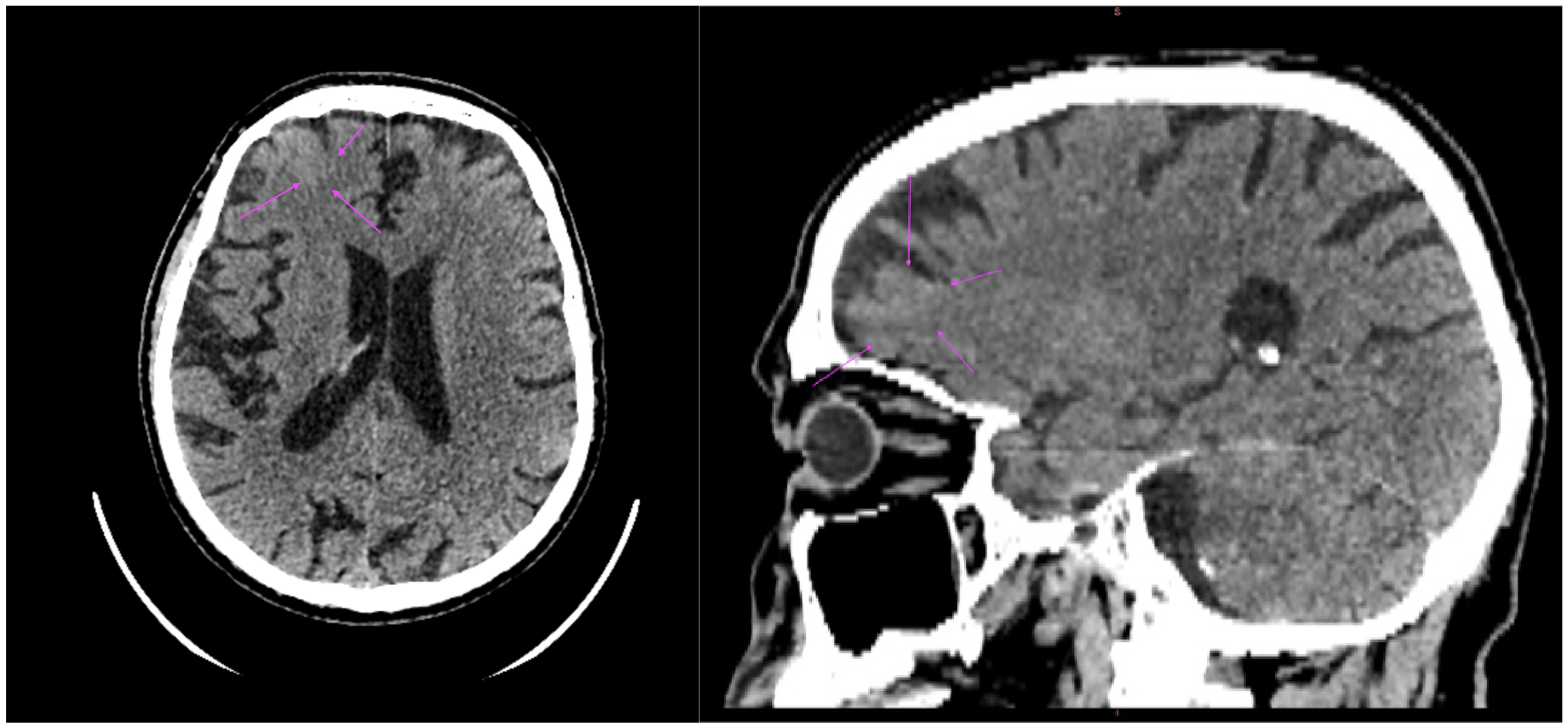
A 76-year-old man was admitted to the emergency department with a 5-day history of chills, fever (38.2 °C), profuse diaphoresis, myalgia, abdominal pain, and progressive confusion. Before presentation, he had received cefuroxime axetil 500 mg twice daily at home for three doses, without clinical improvement. His medical history was significant for bilateral hip osteoarthritis, prior surgery for L4–L5 disc herniation (2 years before the current admission), permanent dual-chamber pacemaker implantation (3 years before the current admission), paroxysmal atrial fibrillation, aortic stenosis, and grade II mitral regurgitation. His long-term treatment included apixaban 5 mg twice daily, atorvastatin 20 mg once daily, and bisoprolol 2.5 mg once daily. On admission, he appeared acutely ill, febrile (38.0 °C), confused, and disoriented to time and place. Physical examination revealed neck stiffness, atrial fibrillation with an irregular pulse of 88 bpm, a grade IV holosystolic murmur best heard over the aortic area, hypotension (90/60 mmHg), hepatomegaly, suprapubic tenderness, and hemorrhagic lesions on the left hand. Acute phase reactants at admission were markedly elevated, with a C-reactive protein of 252.9 mg/L (normal range 1–5 mg/L), a leukocyte count of 9930/mm3, a fibrinogen level of 874.5 mg/dL (normal range 170–420 mg/dL), and an erythrocyte sedimentation rate of 85 mm/h (normal range 0–15 mm/h). Three sets of blood cultures were obtained at the time of presentation. Cranial computed tomography scan (CT scan), performed to exclude an acute cerebrovascular event, demonstrated a hyperdense serpiginous lesion in the high right frontal region, considered most suggestive of a developmental venous anomaly. A cranial magnetic resonance imaging (MRI) was recommended for further characterization (Figure 1). Cranial MRI, though recommended, was not subsequently performed, as the patient’s clinical and neurological status did not permit the procedure. Thoracoabdominal CT scan highlighted the following: bilateral basal fibro-atelectatic bands, without pleural or pericardial effusion, hepatomegaly with a right hepatic lobe measuring 132 mm, and an enlarged heterogeneous prostate with mild stranding of the adjacent fat. This finding was considered incidental; urological consultation was obtained, and given the patient’s acute systemic infection, the urologist recommended deferring further evaluation until after resolution of the infective endocarditis.
Initial transthoracic echocardiography showed no evidence of valvular vegetations. A lumbar puncture was performed and the cerebrospinal fluid was clear, colorless, under increased pressure, with mildly elevated protein levels (0.59 g/L; reference range, 0.15–0.45 g/L), chloride 124 mEq/L (reference range, 110–130 mEq/L), glucose 73 mg/dL (reference range, 40–70 mg/dL), and lactate 29.7 mg/dL (reference range, 10–22 mg/dL). Cytological analysis revealed 460 cells/mm3, with marked granulocytic predominance (84.8%) and 15.2% mononuclear cells. A multiplex PCR meningitis/encephalitis panel was negative for Escherichia coli K1, Haemophilus influenzae, Listeria monocytogenes, Neisseria meningitidis, Streptococcus agalactiae, Streptococcus pneumoniae, cytomegalovirus, enterovirus, herpes simplex virus 1, herpes simplex virus 2, human herpesvirus 6, human parechovirus, varicella-zoster virus, and Cryptococcus neoformans/gattii. Conventional cerebrospinal fluid cultures remained sterile after 3 days of incubation, and the etiology of the meningeal involvement initially remained undetermined. In contrast, all blood cultures obtained at the time of the admission yielded Enterococcus faecalis at 48 h after collection, AST are reported in Table 1 and were assessed according to the EUCAST (European Committee on Antimicrobial Susceptibility Testing version 16.0, valid from 1 January 2026) breakpoints [10]. Empirical antibiotic therapy with ceftriaxone 2 g twice daily and vancomycin 1 g twice daily was continued for a total of 3 days until the complete antibiogram was available, after which targeted therapy was initiated.
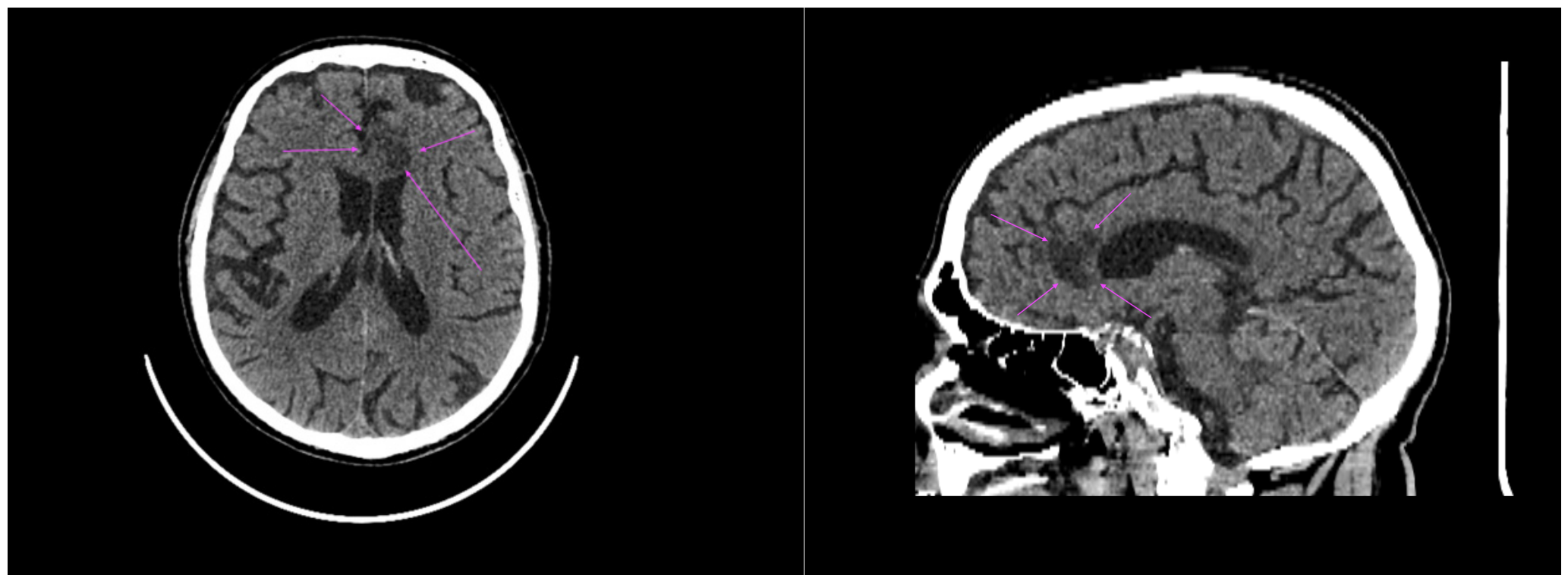
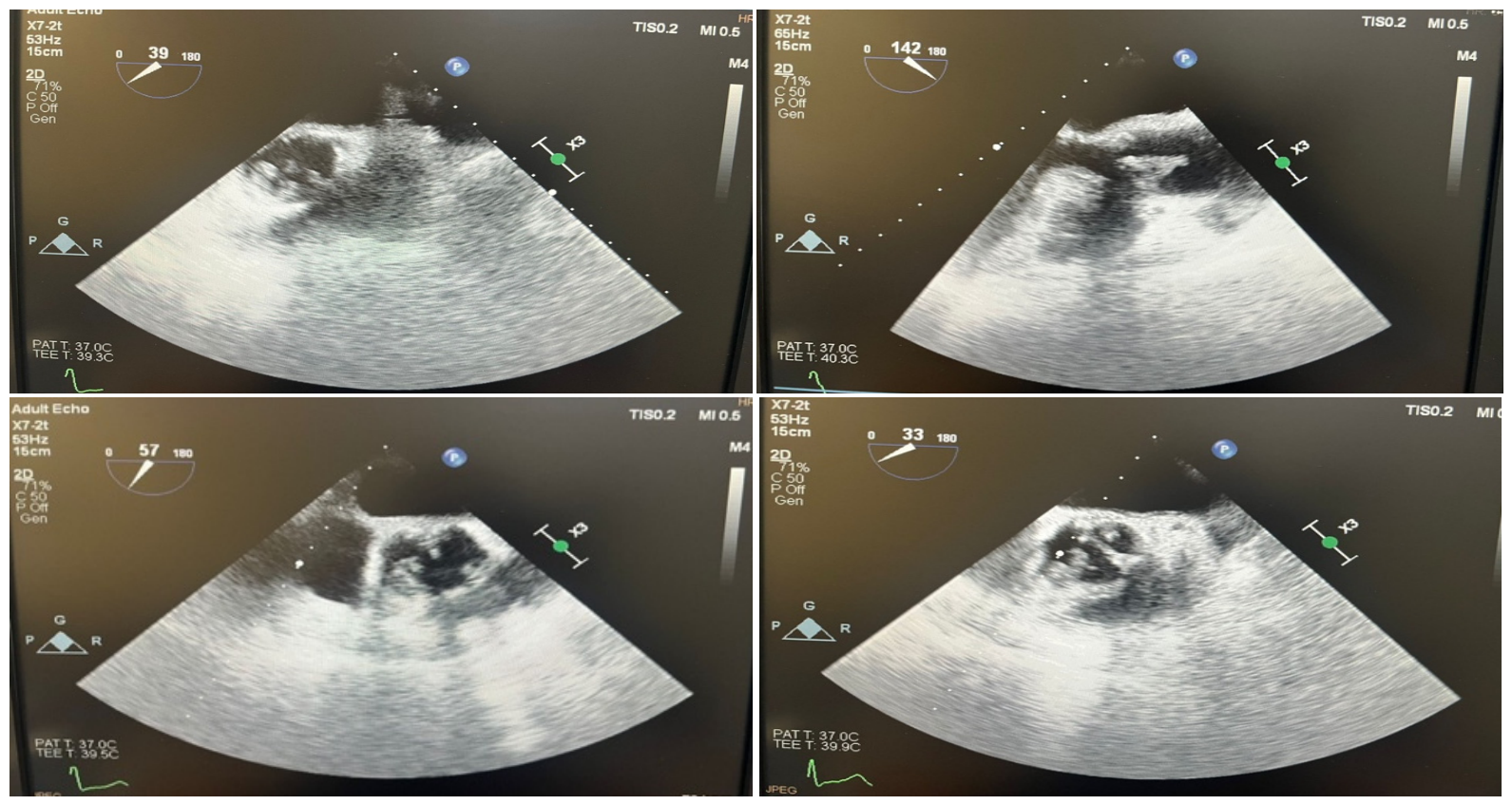
A repeat cranial CT scan, performed on hospital day 6 (5 days after the initial study, and after 6 days of antibiotic therapy), was subsequently requested and, unlike the initial examination, highlighted the presence of a focal parasagittal frontal lesion involving the rostrum and genu of the corpus callosum (Figure 2). Transesophageal echocardiography, performed on day 6 of hospitalization, confirmed infective endocarditis (Figure 3). The left atrium was nondilated. The multilobulated left atrial appendage was free of thrombus, although spontaneous echo contrast was present. Appendage emptying velocity was 0.6 m/s. The mitral valve was fibrotic, with mild mitral regurgitation and a small echogenic image adherent to the posterior mitral annulus, suspicious for vegetation. The aortic valve was heavily calcified, with attached vegetative masses involving all cusps and resulting in severe aortic regurgitation. The aortic annulus measured 21 mm, and the ascending aorta was dilated to 45 mm. No vegetations were seen on the tricuspid or pulmonary valves, but a mild tricuspid regurgitation was noted. Left ventricular systolic function was preserved, with a homogeneous contractile pattern and an ejection fraction of 55%. There was no pericardial effusion. These findings supported the diagnosis of aortic valve infective endocarditis with severe aortic regurgitation, associated ascending aortic dilatation, and mild degenerative mitral regurgitation. The main laboratory examinations that were performed during the hospitalization period are reported in Table 2. Urine cultures, obtained on repeated occasions during the hospitalization, remained sterile.
During hospitalization, the clinical course was further complicated by cardiac decompensation on day 7, manifested by marked lower-extremity edema, followed on day 12 by acute inflammatory involvement of the left knee. An arthrocentesis was performed, yielding a turbid orange synovial fluid. Synovial fluid analysis showed a glucose level of 24 mg/L, amylase 37 U/L, total protein 4.1 g/dL, total nucleated cell count 41.852 × 10³/µL, erythrocytes 0.039 × 10⁶/µL, mononuclear cells 1.304 × 10³/µL, and granulocytes 40.548 × 10³/µL, corresponding to 3.1% mononuclear cells and 96.9% granulocytes. Gram staining highlighting abundant polymorphonuclear leukocytes and fibrin, whereas synovial fluid cultures remained negative. Synovial fluid analysis showed a glucose level of 24 mg/dL, amylase 37 U/L, total protein 4.1 g/dL, total nucleated cell count 41.852 × 10³/µL, erythrocytes 0.039 × 10⁶/µL, mononuclear cells 1.304 × 10³/µL, and granulocytes 40.548 × 10³/µL, corresponding to 3.1% mononuclear cells and 96.9% granulocytes. Our institution’s laboratory does not report standard reference intervals for synovial fluid parameters; the ranges cited below are derived from the published literature. Reference values: total nucleated cell count—non-inflammatory < 2000/mm3, inflammatory 2000–10,000/mm3, strongly inflammatory 10,000–50,000/mm3, sepsis suspicion > 50,000/mm3 with >90% PMN; glucose—within 10 mg/dL of a concurrent fasting serum value, ≥25 mg/dL below serum in bacterial infection; total protein—<2.5 g/dL normal, >4.5 g/dL indicates significant inflammation; amylase—no established reference range in synovial fluid) [11]. The arthritis subsequently evolved favorably, with resolution of pain and return of full mobility by one week after diagnosis.
The patient received vancomycin 1 g twice daily in association with ampicillin 12 g per day for a total of 3 weeks. Vancomycin was subsequently discontinued because of the development of an acute renal impairment. Antibiotic treatment was then continued with linezolid 600 mg twice daily in combination with ampicillin 12 g per day for an additional 2 weeks. Supportive therapy also included diuretics, albumin, antifungal treatment, and symptomatic medication. Follow-up blood cultures obtained after 3 weeks of treatment were negative. Biological monitoring was performed every 3 days to allow early detection of vancomycin-associated nephrotoxicity and, subsequently, linezolid-related myelotoxicity.
On follow-up, transesophageal echocardiography showed largely unchanged findings. The left atrium remained nondilated. The multilobulated left atrial appendage appeared free of thrombus and no longer exhibited spontaneous echo contrast. Peak appendage emptying velocity was 0.6 m/s. The mitral valve remained fibrotic, with mild central mitral regurgitation and no evidence of vegetation. The aortic valve continued to show diffuse calcification of all cusps, with hypoechoic vegetative formations attached to all leaflets, resulting in severe aortic regurgitation. The aortic annulus measured 21 mm, and the ascending aorta remained dilated at 45 mm. No vegetations were identified on the tricuspid or pulmonary valves; mild tricuspid regurgitation persisted. Left ventricular systolic function was preserved, with homogeneous wall motion and a left ventricular ejection fraction of 55%. A small pericardial effusion was noted. Given the persistence of severe aortic valve involvement, the patient was referred for cardiac surgical assessment and consideration of operative management.
Following discharge, the patient was readmitted to the cardiology department and subsequently developed multi-organ system failure, resulting in death one week after discharge.

3. Discussion

The presented case illustrates enterococcal IE in an elderly patient with multiple pre-existing cardiovascular conditions (aortic stenosis, mitral regurgitation, paroxysmal atrial fibrillation) and a permanent dual-chamber pacemaker, who presented with neurological manifestations in a febrile context, initially orienting clinical and imaging investigations toward a diagnosis of meningoencephalitis. This case also illustrates a limitation of transthoracic echocardiography for aortic valve pathology: the initial study excluded valvular vegetations, a false-negative result subsequently clarified by transesophageal echocardiography. The decision to pursue transesophageal imaging was prompted by the identification of Enterococcus faecalis in blood cultures together with cerebrospinal fluid findings suggestive of meningeal involvement despite negative CSF cultures; the latter is unlikely to reflect the antibiotic therapy received at home prior to admission, as cefuroxime axetil has no activity against enterococci.
Enterococcal IE represents the third leading etiologic category after staphylococcal and streptococcal IE. It is responsible for approximately 10% of cases, although higher rates, ranging from 13% to 18% of all IE episodes, have also been reported [12].
Among enterococcal species, Enterococcus faecalis is the pathogen most frequently associated with infective endocarditis [5,13,14,15,16,17,18]. Its predominance is largely explained by its ability to form biofilm [5,13,14,15,16,17,18,19], persist within host tissues, and adhere to previously damaged valves or structurally abnormal cardiac surfaces, thereby facilitating sustained infection and therapeutic difficulty [20].
Over the last two decades, enterococci have been increasingly identified as important causative agents of infective endocarditis, particularly in elderly patients, but also in association with interventional and medical procedures, including catheter-based techniques and dental surgery, which may favor healthcare-associated acquisition [21]. This trend is especially evident in vulnerable populations, such as patients with HIV infection, malignancy—particularly colorectal neoplasia—diabetes mellitus, and people who inject drugs. Concomitantly, rising resistance to commonly used antimicrobial agents has further increased the clinical relevance of enterococcal endocarditis and reinforced its status as a persistent diagnostic and therapeutic challenge [1,22,23,24,25].
Among the 15 species within the genus Enterococcus, the organisms most commonly implicated in infective endocarditis are Enterococcus faecalis, which accounts for approximately 90% of cases, and Enterococcus faecium [5,26]. The gastrointestinal tract is generally considered the principal portal of entry [27,28], although the urinary tract may also serve as the primary source of infection.
Regarding the portal of entry, a gastrointestinal source was considered unlikely, as the patient reported no digestive symptoms and was completely edentulous. A source associated with the cardiac implantable electronic device could not be excluded on clinical grounds; confirmation would have required device extraction with bacteriological culture, which was not performed. The patient’s lumbar discectomy, performed 2 years before this admission, was also considered; the absence of spinal symptoms or findings during the current hospitalization argues against this as the source. A prostatic origin was suspected clinically, based on the patient’s reported scrotal pain, suprapubic tenderness, elevated prostate-specific antigen (PSA) levels, and a free PSA/PSA ratio and urinary symptoms together with the imaging finding of prostatic enlargement with adjacent fat stranding. However, urine cultures were repeatedly sterile, no imaging evidence of cystitis or pyelonephritis was identified, and a seminal or expressed prostatic secretion culture could not be obtained; this origin therefore could not be microbiologically confirmed.
Meningocerebral involvement in infective endocarditis is rare, whether related to septic cerebral embolization or to direct bacterial passage across the blood–brain barrier, and carries a substantial risk of death. The pathophysiology of meningocerebral involvement in enterococcal IE likely reflects a combination of overlapping mechanisms rather than a single process. Septic embolization from valvular vegetations can obstruct the cerebral arterial lumen or vasa vasorum, producing a dual ischemic and infectious insult; when embolic material lodges in smaller vessels, the resulting focal arteritis may extend to a septic encephalopathy, meningitis, or cerebritis. Direct bacterial translocation across the blood–brain barrier represents an additional potential mechanism, alongside an immune-mediated vasculitic component driven by an intense systemic host response and circulating immune complexes [29].
In our patient, the imaging findings were consistent with ischemic injury secondary to septic emboli, whereas the cerebrospinal fluid profile additionally raised the possibility of associated meningeal invasion. Such neurological complications are reported more commonly in streptococcal endocarditis, including pneumococcal infection, and in Staphylococcus aureus disease, but appear to be distinctly uncommon in enterococcal infective endocarditis. Although enterococcal etiology accounts for up to 3% of these cases, the reported mortality remains high, reaching 28.6% [30].
Osteoarticular involvement is a rare complication of enterococcal infective endocarditis, while its concurrence with meningocerebral manifestations appears to be even more exceptional. Reported osteoarticular localizations include lumbar intervertebral abscess, L5–S1 osteomyelitis, and septic arthritis of the knee [31,32,33]. Importantly, the 2023 ESC guideline update includes spondylodiscitis among the minor Duke criteria for infective endocarditis, highlighting the growing recognition of musculoskeletal manifestations within the diagnostic spectrum of this disease [8].
A multicenter study of 633 patients with infective endocarditis followed for 6 months after the acute episode found, in both univariate and multivariate analyses, that vertebral osteomyelitis occurred in 36 cases and was significantly more strongly associated with enterococcal endocarditis than with streptococcal, Staphylococcus aureus, or coagulase-negative staphylococcal etiologies [34]. Accordingly, the combination of enterococcal infection, advanced age, and infective endocarditis should prompt a high index of suspicion for concomitant vertebral infection in patients with spinal pain. Complementary imaging studies, including PET/CT, may play an important role in establishing an accurate diagnosis of osteoarticular involvement in this setting.
With regard to anatomical localization, enterococcal infective endocarditis most frequently affects the aortic valve [35,36]. It occurs predominantly in elderly men, usually follows a subacute course, and only infrequently manifests with the typical cutaneous stigmata of infective endocarditis, this case, by contrast, evolved acutely over five days.
This case illustrates two diagnostic pitfalls. First, initial transthoracic echocardiography showed no evidence of valvular vegetations, a false-negative result later corrected by transesophageal echocardiography—a recognized limitation of TTE for aortic valve pathology, particularly in the setting of heavily calcified valves, which can obscure smaller or early vegetations. Second, the initial cranial CT identified a lesion interpreted as a developmental venous anomaly, a benign incidental finding; only the repeat CT, performed five days later, revealed the true parasagittal frontal lesion.
The markedly inflammatory but culture-negative CSF profile should be interpreted with caution. A purely reactive or chemical meningitis related to adjacent ischemic injury seems less likely, although it cannot be completely excluded. In our case, the pronounced neutrophilic predominance and overall inflammatory CSF pattern favor a bacterial or septic inflammatory process rather than a nonspecific reactive response. Prior antibiotic exposure alone is also unlikely to fully explain the negative cultures for the typical pathogens included in the multiplex meningitis/encephalitis panel. The patient had received oral cefuroxime axetil at home, which would not be expected to provide adequate CNS coverage or reliably sterilize the CSF, and molecular testing is generally less affected by previous antibiotic use than culture. Nevertheless, a negative multiplex result does not exclude off-panel organisms or low pathogen burden. Overall, septic embolization from left-sided infective endocarditis appears to be the most plausible mechanism, given the infected aortic valve and the potential for embolic spread to the cerebral circulation. An associated immune-mediated or vasculitic component cannot be ruled out. The absence of erythrocytes in the CSF argues against overt hemorrhagic meningeal involvement at the time of lumbar puncture, but does not exclude nonhemorrhagic vascular inflammation.
The antibiotic regimen selected in this case departed from the combinations most commonly used for ampicillin-susceptible, non-HLAR E. faecalis IE, for reasons specific to this patient’s multi-site disease. Ampicillin combined with gentamicin, though guideline-supported for susceptible strains, was not selected because aminoglycosides achieve limited concentrations within the central nervous system even in the presence of meningeal inflammation, making this combination poorly suited to a patient with a confirmed cerebral septic focus. Vancomycin, which penetrates the blood–brain barrier more reliably, was therefore continued alongside ampicillin. The coexistence of cerebral and articular septic foci, compartments in which ampicillin alone achieves less consistent penetration, further supported this combination over reliance on ampicillin as a single agent. Renal function was monitored every 3 days throughout treatment given the recognized nephrotoxic potential of vancomycin in an elderly patient. Combination therapy was also intended to limit the risk of enterococcal resistance emerging over a prolonged treatment course. Vancomycin was ultimately discontinued after the development of acute renal impairment, and linezolid was substituted; current guidelines recognize linezolid as a therapeutic option for enterococcal IE, albeit with a more limited and variable evidence base than first-line regimens, reflecting its use here as a necessitated alternative rather than a primary choice [8].

4. Conclusions

This case of enterococcal infective endocarditis in a patient with multiple comorbidities is notable for the concomitant cerebral and articular septic localizations, and for a severe, unfavorable outcome despite a fully ampicillin-susceptible isolate treated with combination antibiotic therapy specifically to maximize the chance of survival. The patient received a total of 37 days of continuous intravenous antibiotic therapy, yet surgical intervention, delayed, would likely have offered the best chance of resolving the case. The patient was readmitted after discharge in the cardiology department and died one week after discharge from multi-organ failure. This outcome underscores that antimicrobial susceptibility and appropriately chosen combination therapy alone may be insufficient in enterococcal IE complicated by embolic phenomena, and that early surgical evaluation should not be delayed when clinically feasible.

Author Contributions

Conceptualization, R.-M.B. and V.B.; methodology, R.-M.B., I.M.C. and V.B.; investigation, data curation, R.-M.B., I.M.C. and V.B.; writing—original draft preparation, R.-M.B. and V.B.; writing—review and editing, R.-M.B., I.M.C. and V.B.; visualization, R.-M.B., I.M.C. and V.B.; supervision, V.B.; project administration, R.-M.B. and V.B.; critical review and final approval, R.-M.B. and V.B. All authors have read and agreed to the published version of the manuscript.

Funding

This research received no external funding.

Institutional Review Board Statement

This study was conducted in accordance with the guidelines of the Declaration of Helsinki. The study was accepted by the County Clinical Emergency Hospital, Sibiu, Romania, and they encouraged the publication of the article (file number 7973/23 March 2026). All methods were carried out following relevant guidelines and regulations.

Informed Consent Statement

Written informed consent has been obtained from the patient’s next of kin to publish this paper.

Data Availability Statement

The original contributions presented in this study are included in the article. Further inquiries can be directed to the corresponding authors.

Conflicts of Interest

The authors declare no conflicts of interest.

References

  1. Herrera-Hidalgo, L.; Fernández-Rubio, B.; Luque-Márquez, R.; López-Cortés, L.E.; Gil-Navarro, M.V.; de Alarcón, A. Treatment of Enterococcus faecalis Infective Endocarditis: A Continuing Challenge. Antibiotics 2023, 12, 704. [Google Scholar] [CrossRef] [Scilit] [PubMed]
  2. Muñoz, P.; Kestler, M.; De Alarcon, A.; Miro, J.M.; Bermejo, J.; Rodríguez-Abella, H.; Fariñas, M.C.; Cobo Belaustegui, M.; Mestres, C.; Llinares, P.; et al. Current Epidemiology and Outcome of Infective Endocarditis. Medicine 2015, 94, e1816. [Google Scholar] [CrossRef] [Scilit] [PubMed]
  3. Tzoumas, A.; Sagris, M.; Xenos, D.; Ntoumaziou, A.; Kyriakoulis, I.; Kakargias, F.; Liaqat, W.; Nagraj, S.; Patel, R.; Korosoglou, G.; et al. Epidemiological Profile and Mortality of Infective Endocarditis over the Past Decade: A Systematic Review and Meta-Analysis of 133 Studies. Am. J. Cardiol. 2025, 244, 67–88. [Google Scholar] [CrossRef] [Scilit] [PubMed]
  4. Murdoch, D.R. Clinical Presentation, Etiology, and Outcome of Infective Endocarditis in the 21st Century. Arch. Intern. Med. 2009, 169, 463–473. [Google Scholar] [CrossRef] [Scilit] [PubMed]
  5. Chirouze, C.; Athan, E.; Alla, F.; Chu, V.H.; Ralph Corey, G.; Selton-Suty, C.; Erpelding, M.-L.; Miro, J.M.; Olaison, L.; Hoen, B. Enterococcal Endocarditis in the Beginning of the 21st Century: Analysis from the International Collaboration on Endocarditis-Prospective Cohort Study. Clin. Microbiol. Infect. 2013, 19, 1140–1147. [Google Scholar] [CrossRef] [Scilit] [PubMed]
  6. Dahl, A.; Fowler, V.G.; Miro, J.M.; Bruun, N.E. Sign of the Times: Updating Infective Endocarditis Diagnostic Criteria to Recognize Enterococcus faecalis as a Typical Endocarditis Bacterium. Clin. Infect. Dis. 2022, 75, 1097–1102. [Google Scholar] [CrossRef] [Scilit] [PubMed]
  7. Sanguettoli, F.; Marchini, F.; Frascaro, F.; Zanarelli, L.; Campo, G.; Sinning, C.; Tan, T.C.; Pavasini, R. The Impact of Neurological Complications in Endocarditis: A Systematic Review and Meta-Analysis. J. Clin. Med. 2024, 13, 7053. [Google Scholar] [CrossRef] [Scilit] [PubMed]
  8. Delgado, V.; Ajmone Marsan, N.; de Waha, S.; Bonaros, N.; Brida, M.; Burri, H.; Caselli, S.; Doenst, T.; Ederhy, S.; Erba, P.A.; et al. 2023 ESC Guidelines for the Management of Endocarditis. Eur. Heart J. 2023, 44, 3948–4042. [Google Scholar] [CrossRef] [Scilit] [PubMed]
  9. Fernández-Guerrero, M.L.; Herrero, L.; Bellver, M.; Gadea, I.; Roblas, R.F.; De Górgolas, M. Nosocomial Enterococcal Endocarditis: A Serious Hazard for Hospitalized Patients with Enterococcal Bacteraemia. J. Intern. Med. 2002, 252, 510–515. [Google Scholar] [CrossRef] [Scilit] [PubMed]
  10. European Committee on Antimicrobial Susceptibility Testing. Clinical Breakpoints and Interpretation of Clinical MICs, Version 16. Available online: https://www.eucast.org/bacteria/clinical-breakpoints-and-interpretation/clinical-breakpoint-tables/ (accessed on 1 July 2026).
  11. Oliviero, F.; Mandell, B.F. Synovial Fluid Analysis: Relevance for Daily Clinical Practice. Best Pract. Res. Clin. Rheumatol. 2023, 37, 101848. [Google Scholar] [CrossRef] [Scilit] [PubMed]
  12. Danneels, P.; Hamel, J.-F.; Picard, L.; Rezig, S.; Martinet, P.; Lorleac’h, A.; Talarmin, J.-P.; Buzelé, R.; Guimard, T.; Le Moal, G.; et al. Impact of Enterococcus faecalis Endocarditis Treatment on Risk of Relapse. Clin. Infect. Dis. 2023, 76, 281–290. [Google Scholar] [CrossRef] [Scilit] [PubMed]
  13. Nappi, F.; Avtaar Singh, S.S.; Jitendra, V.; Fiore, A. Bridging Molecular and Clinical Sciences to Achieve the Best Treatment of Enterococcus faecalis Endocarditis. Microorganisms 2023, 11, 2604. [Google Scholar] [CrossRef] [Scilit] [PubMed]
  14. Barnes, A.M.T.; Frank, K.L.; Dale, J.L.; Manias, D.A.; Powers, J.L.; Dunny, G.M. Enterococcus faecalis colonizes and forms persistent biofilm microcolonies on undamaged endothelial surfaces in a rabbit endovascular infection model. FEMS Microbes 2021, 2, xtab014. [Google Scholar] [CrossRef] [Scilit] [PubMed]
  15. Di Domenico, E.G.; Rimoldi, S.G.; Cavallo, I.; D’Agosto, G.; Trento, E.; Cagnoni, G.; Palazzin, A.; Pagani, C.; Romeri, F.; De Vecchi, E.; et al. Microbial biofilm correlates with an increased antibiotic tolerance and poor therapeutic outcome in infective endocarditis. BMC Microbiol. 2019, 19, 228. [Google Scholar] [CrossRef] [Scilit] [PubMed]
  16. Nallapareddy, S.R.·.; Singh, K.V.; Sillanpää, J.; Garsin, D.A.; Höök, M.; Erlandsen, S.L.; Murray, B.E. Endocarditis and biofilm-associated pili of Enterococcus faecalis. J. Clin. Investig. 2006, 116, 2799–2807. [Google Scholar] [CrossRef] [Scilit] [PubMed]
  17. Ramos, S.; Silva, V.; Dapkevicius, M.; Igrejas, G.; Poeta, P. Enterococci, from Harmless Bacteria to a Pathogen. Microorganisms 2020, 8, 1118. [Google Scholar] [CrossRef] [Scilit] [PubMed]
  18. Holland, T.L.; Baddour, L.M.; Bayer, A.S.; Hoen, B.; Miro, J.M.; Fowler, V.G. Infective Endocarditis. Nat. Rev. Dis. Primers 2016, 2, 16059. [Google Scholar] [CrossRef] [Scilit] [PubMed]
  19. Barnes, A.M.T.; Frank, K.L.; Dunny, G.M. Enterococcal Endocarditis: Hiding in Plain Sight. Front. Cell. Infect. Microbiol. 2021, 11, 722482. [Google Scholar] [CrossRef] [Scilit] [PubMed]
  20. Toh, C.C.S.; Bali, K.P. Natural History of Streptococcus faecalis Endocarditis. BMJ 1960, 2, 640–644. [Google Scholar] [CrossRef] [Scilit] [PubMed]
  21. Nappi, F. Current Knowledge of Enterococcal Endocarditis: A Disease Lurking in Plain Sight of Health Providers. Pathogens 2024, 13, 235. [Google Scholar] [CrossRef] [Scilit] [PubMed]
  22. Fernández-Hidalgo, N.; Escolà-Vergé, L.; Pericàs, J.M. Enterococcus faecalis Endocarditis: What’s Next? Future Microbiol. 2020, 15, 349–364. [Google Scholar] [CrossRef] [Scilit] [PubMed]
  23. Escolà-Vergé, L.; Fernández-Hidalgo, N.; Larrosa, M.N.; Fernandez-Galera, R.; Almirante, B. Secular Trends in the Epidemiology and Clinical Characteristics of Enterococcus faecalis Infective Endocarditis at a Referral Center (2007–2018). Eur. J. Clin. Microbiol. Infect. Dis. 2021, 40, 1137–1148. [Google Scholar] [CrossRef] [Scilit] [PubMed]
  24. Bashore, T.M.; Turner, N.A. Addressing the Menace of Enterococcal Endocarditis. J. Am. Coll. Cardiol. 2020, 75, 495–497. [Google Scholar] [CrossRef] [Scilit] [PubMed]
  25. Ramos-Martínez, A.; Domínguez, F.; Muñoz, P.; Marín, M.; Pedraz, Á.; Fariñas, M.C.; Tascón, V.; de Alarcón, A.; Rodríguez-García, R.; Miró, J.M.; et al. Clinical Presentation, Microbiology, and Prognostic Factors of Prosthetic Valve Endocarditis. Lessons Learned from a Large Prospective Registry. PLoS ONE 2023, 18, e0290998. [Google Scholar] [CrossRef] [Scilit] [PubMed]
  26. Zimmermann, V.·.; Fourré, N.; Ledergerber, B.; Epprecht, J.; Siedentop, B.; Monney, P.; Tzimas, G.; Frank, M.; Senn, L.; Ianculescu, N.; et al. Evaluation of Different Versions of the Duke Criteria for the Diagnosis of Infective Endocarditis Among Patients with Enterococcal Bacteremia; a Multicenter Study. Open Forum Infect. Dis. 2025, 12, ofaf408. [Google Scholar] [CrossRef] [Scilit] [PubMed]
  27. Escolà-Vergé, L.; Peghin, M.; Givone, F.; Pérez-Rodríguez, M.T.; Suárez-Varela, M.; Meije, Y.; Abelenda, G.; Almirante, B.; Fernández-Hidalgo, N. Prevalence of Colorectal Disease in Enterococcus faecalis Infective Endocarditis: Results of an Observational Multicenter Study. Rev. Esp. Cardiol. (Engl. Ed.) 2020, 73, 711–717. [Google Scholar] [CrossRef] [Scilit] [PubMed]
  28. Lebreton, F.; Willems, R.J.L.; Gilmore, M.S. Enterococcus Diversity, Origins in Nature, and Gut Colonization. In Enterococci: From Commensals to Leading Causes of Drug Resistant Infection [Internet]; Massachusetts Eye and Ear Infirmary: Boston, MA, USA, 2014. [Google Scholar]
  29. Pruitt, A.A. Neurologic Complications of Infective Endocarditis. Curr. Treat. Options Neurol. 2013, 15, 465–476. [Google Scholar] [CrossRef] [Scilit] [PubMed]
  30. Béraud, G.; Tubiana, S.; Erpelding, M.-L.; Le Moing, V.; Chirouze, C.; Gorenne, I.; Manchon, P.; Tattevin, P.; Vernet, V.; Varon, E.; et al. Combined Bacterial Meningitis and Infective Endocarditis: When Should We Search for the Other When Either One Is Diagnosed? Infect. Dis. Ther. 2022, 11, 1521–1540. [Google Scholar] [CrossRef] [Scilit] [PubMed]
  31. Vlahakis, N.E.; Temesgen, Z.; Berbari, E.F.; Steckelberg, J.M. Osteoarticular Infection Complicating Enterococcal Endocarditis. Mayo Clin. Proc. 2003, 78, 623–628. [Google Scholar] [CrossRef] [Scilit] [PubMed]
  32. Devarakonda, P.K.; Dhulipala, V.R.; Karki, M.; Ayala-Rodriguez, C.; Reddy, S. A Rarely Reported Case of Enterococcus faecalis Bacteremia Causing Infective Endocarditis and Osteomyelitis. Cureus 2022, 14, e22522. [Google Scholar] [CrossRef] [Scilit] [PubMed]
  33. Spomer, A.; Ho, G. Bacterial Endocarditis and Septic Arthritis Presenting as Polymyalgia Rheumatica. R. I. Med. 1994, 77, 5–6. [Google Scholar] [PubMed]
  34. Douiyeb, S.; Sigaloff, K.C.E.; Ulas, E.G.; Duffels, M.G.J.; Drexhage, O.; Germans, T.; Wagenaar, J.F.P.; Buis, D.T.P.; van der Vaart, T.W.; van Werkhoven, C.H.; et al. Vertebral Osteomyelitis in Patients with Infective Endocarditis: Prevalence, Risk Factors and Mortality. Eur. J. Clin. Microbiol. Infect. Dis. 2025, 44, 819–825. [Google Scholar] [CrossRef] [Scilit] [PubMed]
  35. McDonald, J.R.; Olaison, L.; Anderson, D.J.; Hoen, B.; Miro, J.M.; Eykyn, S.; Abrutyn, E.; Fowler, V.G.; Habib, G.; Selton-Suty, C.; et al. Enterococcal Endocarditis: 107 Cases from the International Collaboration on Endocarditis Merged Database. Am. J. Med. 2005, 118, 759–766. [Google Scholar] [CrossRef] [Scilit] [PubMed]
  36. Stevens, M.P.; Edmond, M.B. Endocarditis Due to Vancomycin-Resistant Enterococci: Case Report and Review of the Literature. Clin. Infect. Dis. 2005, 41, 1134–1142. [Google Scholar] [CrossRef] [Scilit] [PubMed]
Figure 1. Non-contrast cranial CT obtained during the initial diagnostic work-up for suspected acute cerebrovascular disease shown in axial and sagittal planes demonstrated a hyperdense serpiginous lesion in the high right frontal region (pink arrows), with imaging features most consistent with a developmental venous anomaly.
Germs 16 00021 g001
Figure 2. Repeat cranial CT, performed during the subsequent clinical course, shown in axial and sagittal planes, demonstrated a focal parasagittal frontal lesion involving the rostrum and genu of the corpus callosum (pink arrows), a finding not evident on the initial examination.
Germs 16 00021 g002
Figure 3. Transthoracic echocardiography showed a heavily calcified aortic valve, with hypoechoic vegetative formations attached to all cusps. The largest, measuring approximately 8 mm, was located on the free edge of the right coronary cusp and was highly suggestive of vegetation. The valvular involvement was hemodynamically significant and associated with severe aortic regurgitation.
Germs 16 00021 g003
Table 1. Antibiotic susceptibility profile of the E. faecalis isolate (Vitek automated system, EUCAST breakpoints).
AntibioticMIC (mg/L)Interpretation
Amoxicillin/clavulanic acid≤2.0Susceptible
Ampicillin/sulbactam≤2.0Susceptible
Ampicillin≤2.0Susceptible
Ciprofloxacin≤0.5Susceptible
Nitrofurantoin≤16.0Susceptible
Vancomycin1.0Susceptible
Levofloxacin1.0Susceptible
Linezolid2.0Susceptible
Teicoplanin≤0.5Susceptible
Tigecycline≤0.12Susceptible
Streptomycin (high-level synergy)Susceptible
Kanamycin (high-level synergy)Susceptible
Gentamicin (high-level resistance)Negative
Quinupristin/dalfopristin4.0Resistant
Imipenem≤1.0Intermediate
Table 2. Laboratory examinations performed during the admission period.
ParameterNormal Range4 Feb 265 Feb 269 Feb 2611 Feb 2612 Feb 2617 Feb 2624 Feb 2627 Feb 261 Mar 262 Mar 263 Mar 264 Mar 266 Mar 2613 Mar 26
PCT (ng/mL)<0.050.06
CRP (mg/L)1–5252.983.5107.861.772.643.915.9
ESR (mm/h)0–158597
Fibrinogen (mg/dL)170–420874.5600.5
WBC (/mm3)4200–9500 993010,3406790537053806470
Neutrophils (/mm3)4500–7500743076104320315029205340
NLRno fixed range6.4055.3222.5562.1581.708
Hematocrit (%)40–5030.126.628.328.827.428.2
Hemoglobin (g/dL)13–1710.49.09.79.69.29.2
Platelets (/mm3)150,000–400,000182,000221,000215,000171,000113,00061,000
Urea18–55553731695842424152125
Creatinine (mg/dL)0.72–1.250.660.800.671.731.941.631.501.381.551.97
AST/TGO (U/L)11–343112531
ALT/TGP (U/L)0–451131317
ALP (U/L)40–150228
GGT (U/L)8–61210
INR0.86–1.11.38
aPTT (s)25.1–37.739.9
Blood glucose (mg/dL)80–1151941311029082175
CRP, C-reactive protein; WBC, white blood cell count; NLR, neutrophil-to-lymphocyte ratio; AST, aspartate aminotransferase; ALT, alanine aminotransferase; INR, international normalized ratio; aPTT, activated partial thromboplastin time; ALP, alkaline phosphatase; GGT, gamma-glutamyl transferase; ESR, erythrocyte sedimentation rate.
Disclaimer/Publisher’s Note: The statements, opinions and data contained in all publications are solely those of the individual author(s) and contributor(s) and not of MDPI and/or the editor(s). MDPI and/or the editor(s) disclaim responsibility for any injury to people or property resulting from any ideas, methods, instructions or products referred to in the content.