


Abstract
Two hemostatic bionanocomposite dressings were developed using natural, semi-natural (or semi-synthetic) and synthetic polymers. The first system consisted of chitosan (CS), polyvinyl alcohol (PVA), and carboxymethyl cellulose (CMC) (CS/PVA/CMC), while the second was based on CS, PVA, and starch (SR) (CS/PVA/SR). Zinc oxide (ZnO) nanoparticles and bicyclic monoterpene camphor (CP) ketone were incorporated as bioactive agents in order to enhance antimicrobial and hemostatic performance. FTIR spectroscopy confirmed the successful solvent casting synthesis of the dressings and the interactions between the biopolymers and additives. XRD analysis indicated a predominantly amorphous structure, while SEM images and EDS analysis revealed uniform dispersion of ZnO particles within the polymer matrices without aggregation. Furthermore, the CS/PVA/CMC-1ZnO/CP sample exhibited a water sorption of 12,666 ± 126%, while CS/PVA/SR-1ZnO/CP reached 7013 ± 215%. ZnO incorporation also improved mechanical performance, with CS/PVA/SR-2ZnO/CP displaying the highest tensile strength (39.18 ± 0.2 MPa) and elongation at break (9.54 ± 1.04%). ZnO incorporation also led to a concentration-dependent increase in antibacterial activity, with SR-based dressings achieving near-complete bacterial reduction at higher ZnO loadings. All the dressings demonstrated good biocompatibility, while CS/PVA/SR-1ZnOCP showed the fastest clotting time (420 s ± 40), highlighting its potential for hemostatic applications.
1. Introduction
The human body’s largest and outermost organ, the skin, provides a natural barrier that protects the internal organs from the outside world. Any injury to the skin causes an abrupt change in how the skin functions and may result in the creation of wounds and hemorrhage [1]. Once the skin is injured, the initial stage of the wound-healing mechanism is hemostasis, which involves controlling bleeding and forming a protective barrier [2]. Intravascular blood loss from hemorrhage can induce organ and tissue malfunction and even death if it is above a certain threshold. Almost half of soldier deaths on the battlefield are brought on by excessive bleeding, while over 15% of deaths are entirely avoidable, according to US military statistics [3]. Therefore, the most crucial aspect of patient treatment is emergency bleeding management. Over the past years, research has focused on the development of advanced hemostasis-stimulating materials that ensure rapid hemostasis without thermal effects, while offering excellent biocompatibility, flexibility, adaptability to the wound shape, biodegradability after use, and low cost [4]. Recently, several hemostatic products have been commercially available, with different forms, chemical compositions, and hemostatic mechanism activity, including oxidized regenerated cellulose (Surgicel) [5], kaolin (Combat Gauze®) [6], chitosan (Chitoflex®, ChitoGauze®, HemCon®, and Celox®) [7], and gelatin (Gelfoam®) [8]. However, they have various side effects, as most are moderately effective for rapid hemostasis, expensive, and raise safety concerns [9]. For instance, some hemostatic powders, such as PerClot®, Arista®, and SuperClot®, have demonstrated in vitro cytotoxic effects, potentially affecting surrounding tissues during clinical use [10]. Certain chitosan-based products also show low effectiveness, such as Chitodine®, which can lead to hypersensitivity reactions due to its elemental iodine content [11]. Furthermore, some dressings exhibit limitations in their mechanical properties and clinical handling. For instance, HemCon®, a chitosan-based hemostatic gauze, has been reported to be relatively stiff and brittle, making its effective application on irregular wound surfaces difficult, and due to its limited performance, it has been progressively replaced by more advanced products [12]. In addition, clinical and experimental studies have reported cases of rebleeding and reduced efficacy [13]. Safety concerns have also been raised in experimental models. Animal studies for WoundStat® and Celox® hemostatic products have reported the presence of intraluminal particles, thrombus formation, and tissue damage several hours after treatment [14]. Moreover, severe macroscopic and microscopic alterations, and produce damage in vital organs such as liver, lungs, kidneys, and heart, have been observed following the use of Combat Gauze®, ChitoGauze®, and Celox Gauze® in vascular injury models [15].
Chitin-derived chitosan (CS) is a widely used hemostatic biopolymer due to its intrinsic properties such as high biocompatibility, antibacterial activity, hemostatic nature, and biodegradability (Figure 1) [16]. Moreover, it promotes biosorption, coagulation, and flocculation. CS’s hemostatic properties result from direct electrostatic interactions between CS’s positively charged amino groups and negatively surface-charged platelets and red blood cells [1]. In addition, other negatively charged blood components, including plasma and blood proteins, can interact with CS. It has been reported that CS’s hemostatic mechanism is not primarily governed by the classical coagulation cascade but by the contact activation cascade, which is involved in adhesion [17]. Further evidence supporting this mechanism has reported that the accessibility of amino groups plays a critical role in blood coagulation. Specifically, a study showed that the use of CS induced the formation of a thick blood coagulum, whereas chemically modified derivatives with reduced availability of amino groups exhibited significantly diminished or no coagulation. This behavior was attributed to steric hindrance, which limits electrostatic interactions between CS and blood components [18]. The research community and the pharmacy industry are focusing on developing CS-based hemostatic products due to the enhanced hemostatic properties of CS. However, CS presents low water solubility at physiological pH and low mechanical properties, which limit its use [19]. Starch (SR) is a plant-derived, highly polar polysaccharide composed of amylose and hyperbranched amylopectin [20], exhibits excellent water uptake properties, and is also inexpensive, readily available, absorbable, and nontoxic (Figure 1) [21]. SR-based hemostatic agents rapidly absorb plasma and concentrate blood cells, clotting factors, and platelets at the injury site. This accelerates clot formation and reduces bleeding time [22]. Carboxymethyl cellulose (CMC) is a derivative of the natural cellulose polysaccharide in which some of its hydroxyl groups have been modified into carboxymethyl groups (Figure 1) [23]. CMC is a biocompatible, non-toxic material with enhanced water solubility compared to cellulose [24]. Moreover, CMC exhibits good water retention ability and promotes platelet coagulation [25]. Nonetheless, due to their poor chemical stability, natural and biopolymers have less mechanical strength than synthetic polymeric films. Hence, blending natural polymers with synthetic polymers exhibiting high biocompatibility, hydrophilicity and elasticity, such as poly (vinyl alcohol) (PVA), greatly improves their mechanical strength (Figure 1) [4].
Figure 1.
Chemical structures of PVA, CS, CMC and SR.
Moreover, efficient hemostatic dressings must also prevent the potential infection at the bleeding area. Although CS presents antibacterial properties, it frequently falls short of completely preventing infection [26]. To improve infection prevention, research recommends adding metal oxide nanoparticles (MNPs), like zinc oxide (ZnO), to the polymer dressing [27]. Because of their distinct physicochemical properties and high surface-area-to-volume ratio, ZnO NPs are especially effective. ZnO NPs’ main antibacterial mechanism involves the production of reactive oxygen species (ROS), which harm and kill bacteria by inducing lipid peroxidation of membrane lipids and cellular leakage. Complementary to their antibacterial activity, the positive charge of ZnO NPs disrupts the negatively charged bacterial membrane, causing their death [28]. ZnO NPs can also promote the coagulation cascade, further enhancing hemostatic efficacy. However, in some cases, ZnO NPs demonstrated toxicity to human cells [29]. As an alternative, camphor (CP), a natural compound derived from Cinnamomum camphora, has attracted considerable attention due to its diverse bioactivities. It exhibits significant antimicrobial properties, including antibacterial and antifungal effects, and has been reported to be effective against pathogens such as Escherichia coli, Staphylococcus aureus, and Pseudomonas aeruginosa [30]. CP has also been incorporated into Pickering emulsions, demonstrating sustained antibacterial activity for up to 10 days [31]. In addition, CP demonstrates notable anti-inflammatory activity by hindering the production of both inflammation mediators and chemotactic cytokines. It is also known to promote wound healing by reducing edema, enhancing tissue regeneration, and accelerating the repair of damaged skin [32]. Furthermore, CP has been shown to increase collagen and elastin synthesis, thereby improving skin structure and promoting anti-aging effects. These properties highlight the potential of CP as a multifunctional agent in biomedical and pharmaceutical applications [25].
Despite the extensive research on CS/PVA/ZnO-based systems, most studies focus on single polymer blends and the incorporation of ZnO as a standalone antibacterial agent, without exploring synergistic multifunctional systems or comparative polymer matrix effects. In particular, the combined use of ZnO nanoparticles with natural bioactive compounds and the systematic evaluation of different polysaccharide matrices remain limited. In this context, the present study aims for the development and comprehensive characterization of two distinct CS-based nanocomposite wound dressings (CS/PVA/CMC and CS/PVA/SR), introducing a dual-functional system that integrates ZnO nanoparticles with CP as a synergistic antimicrobial and hemostatic strategy. The proposed systems are engineered to form structurally stable hydrogel networks upon interaction with water and/or blood without undergoing dissolution, providing important functional advantages for wound management. The resulting hydrogels are intended to promote rapid hemostasis while acting as protective barriers that prevent microbial infiltration, retain moisture within the wound environment, and facilitate the absorption of excess exudates [26]. In addition, the study investigates the potential synergistic antimicrobial activity between ZnO NPs and CP, aiming to enhance antimicrobial efficacy while maintaining minimal cytotoxicity. To the best of our knowledge, this study is the first that (i) combines ZnO nanoparticles with CP as a dual-functional antimicrobial system; (ii) systematically compares CMC- and SR-based polymeric matrices within CS/PVA systems; and (iii) correlates structural properties with hemostatic performance, mechanical behavior, and water sorption capacity. Collectively, these characteristics highlight the potential of the proposed nanocomposite systems as promising candidates for next-generation hemostatic wound dressings with significant clinical potential.
2. Materials and Methods
2.1. Materials
PVA (No. 9002-89-5, Mw 89,000–98,000, 99 + % hydrolyzed) and CP (No. 76-22-2, 96%, Mw = 152.24 g/mol) were obtained from Sigma-Aldrich, Darmstadt, Germany. CMC (No. 9004-32-4, average Mw ≈ 700,000), CS (No. 9012-76-4, high Mw, deacetylation degree (310,000–375,000 Da, >75% deacetylation), SR (No. 9005-84-9) from potatoes (purity ≥ 80%), glacial acetic acid, and phosphate-buffered saline solution (PBS) were also procured from Sigma-Aldrich. ZnO nanopowder (Product no. NG04SO3803, 99.5%, particle size 18 nm) was obtained from Nanografi Nano Technology, Ankara, Turkey.
2.2. Fabrication of CS/PVA/CMC and CS/PVA/SR Dressings
2.2.1. Preparation of CS/PVA Films
PVA was initially dissolved in deionized water at 60 °C under continuous magnetic stirring until complete dissolution was achieved. CS was then gradually dispersed into the solution, followed by the addition of acetic acid until the desired concentration of 2% v/v was reached. The binary polymer solution was stirred continuously until a homogeneous solution was obtained. The resulting solution was poured into an ovenproof dish and dried at 60 °C for 48 h in order to ensure complete evaporation of the solvent. The prepared CS/PVA film was used as a control to investigate the hemostatic dressings. A total volume of 50 mL of the final solution was poured into an ovenproof dish (diameter: 8.5 cm; height: 1.5 cm) and dried at 60 °C for 48 h to remove residual solvent.
2.2.2. CS/PVA/CMC Dressings’ Preparation
The preparation of the CS/PVA/CMC dressings, as it is listed in Table 1, followed a protocol similar to that of the CS/PVA system. Initially, PVA was dissolved in deionized water at 60 °C under magnetic stirring. CMC was afterwards incorporated into the solution, followed by the addition of CS. Once dispersion was complete, acetic acid was added to achieve a final concentration of 2% v/v in the total solution, ensuring uniform mixing. A total of 50 mL of the final solution was poured into an ovenproof dish (diameter: 8.5 cm; height: 1.5 cm) and dried at 60 °C for 48 h in order to completely remove the residual solvent.
Table 1.
Abbreviations of the prepared dressings, their composition in terms of PVA, CS, CMC, and SR, and the corresponding thickness values. All the concentrations (w/w) are expressed relative to CS.
2.2.3. CS/PVA/SR Dressings’ Preparation
CS/PVA/SR dressings’ synthesis, as it is listed in Table 1, was conducted using a procedure analogous to that of CS/PVA/CMC dressings, with the primary distinction being the substitution of CMC with SR. PVA was initially dissolved in water at 60 °C under magnetic stirring, after which SR was added and allowed to dissolve in the aqueous solution. Subsequently, CS was incorporated into the solution, and acetic acid was introduced to achieve homogenization. The solution (50 mL) was then poured into an ovenproof dish (diameter: 8.5 cm; height: 1.5 cm) and dried at 60 °C for 48 h under identical conditions to obtain the final CS/PVA/SR dressings.
2.3. Preparation of CS/PVA/CMC and CS/PVA/SR Dressings Incorporating ZnO NPs and CP
To prepare the CS/PVA/CMC-ZnO/CP dressing according to Table 2, 1% w/v of PVA was magnetically stirred for 1 h in water at 60 °C in order to dissolve completely. Then, a solution of ZnO NPs in ethanol was stirred vigorously and sonicated, and then added to the previously prepared PVA solution. Subsequently, 0.5 w/w CP, 1.5 w/w CMC, and finally, 1% w/v CS were dissolved in the PVA-ZnO solution at 60 °C under continuous magnetic stirring. Eventually, glacial acetic acid was added until a 2% v/v concentration was reached, and the mixture was stirred continuously until complete homogenization. A total of 50 mL of the final solution was poured into an ovenproof dish (diameter: 8.5 cm; height: 1.5 cm) and dried at 60 °C for 48 h to remove all the solvent. The preparation of the CS/PVA/SR-ZnO/CP dressings followed the same protocol as the CS/PVA/CMC-ZnO/CP system, with the only modification being the replacement of CMC with 1.5% w/v SR. The drying process remained identical, ensuring complete solvent removal and the formation of structurally stable dressings (Scheme 1). Furthermore, the CS/PVA/SR-CP and CS/PVA/CMC-CP dressings were prepared using the same protocol, without the addition of ZnO nanoparticles, and were utilized as control samples. The thickness of the CS/PVA/CMC-ZnO/CP, CS/PVA/SR-ZnO/CP, CS/PVA/SR-CP and CS/PVA/CMC-CP dressings is presented in Figure 9c.
Table 2.
Abbreviations of dressings and their contents of PVA, CS, CMC, SR, ZnO NPs, and CP. All concentrations (w/w) are expressed relative to CS.
Scheme 1.
Preparation of nanocomposite dressings.
2.4. Prepared Dressings’ Characterization
Hemostatic nanocomposite dressings’ chemical structure was investigated using the Frontier attenuated total reflectance Fourier-transform infrared spectrometer (ATR-FTIR) by Perkin-Elmer (Shelton, CT, USA) at a nominal resolution of 2 cm−1, and spectral range 4000–550 cm−1. The crystallinity of dressings was studied by a D8 FOCUS X-ray diffractometer (XRD) by Bruker AXS LLC, Madison, WI, USA) equipped with CuKa radiation (λ = 0.1541 nm), over a 2θ range of 5–90°. The morphology of the hemostatic dressings was characterized using a JSM6390LV by JEOL Ltd. (Tokyo, Japan) scanning electron microscope (SEM). Energy-dispersive X-ray spectroscopy (EDS) was conducted using a QUANTAX system (Brucker, Billerica, MA, USA) equipped with an XFlash410 silicon drift detector. The system was attached to the scanning electron microscope operating at 15 kV with a 35° takeoff angle. Sample spectra were collected with a live time of 60 s and processed using the ESPRIT 2025 R1 software. Quantification was performed using standardless ZAF correction.
2.5. Study of Water Sorption and Water Content Capacity
The water sorption capacity and stability of the fabricated hemostatic dressings were measured using either simulated body fluid (SBF) (pH = 7.4) or phosphate buffer (pH = 5.6) at 37 °C. Each sample was initially weighed in its dry state (W0) before being immersed in a buffer. The films were removed at the predetermined time intervals of 30, 60, 90, 120, 180, 360, 1440, 2880, 4320, 5760, 7200, and 8640 min, and any excess surface buffer was gently blotted using filter paper. The swollen films were weighed (Wt) until a constant weight was achieved. The water sorption (WS) and water content (WC) of the tested patches were calculated from the following expressions (Equations (1) and (2)) [33]:
2.6. In Vitro Hydrolysis
The dressings were incubated at 37 °C in SBF (pH = 7.4). Τhe samples were removed, rinsed with deionized water, and weighed at predetermined time intervals (3, 4, 5, 6, 7, and 8 days). The mass remaining (MR) was calculated by comparing the mass after the incubation in SBF (Mt) with the initial mass (M0) according to the following equation (Equation (3)) [34]:
2.7. Mechanical Properties
Each film’s thickness was determined via a thickness dial gauge. The mean film thickness was calculated from four measurements made on the outside of the film and one in the center. The prepared hemostatic dressings’ tensile strength and elongation at break were investigated using the ASTM standard test method D882-10 (ASTM, 2010 [35]). A 100 kN Computerized Testing Machine (WDW–100E, Jinan, China) was employed to evaluate their mechanical properties. Each sample was cut into 60 mm-long, 10 mm-wide rectangles, while each film’s thickness was measured three times. Setting the ends of the samples in the equipment at a distance of 30 mm and the head speed to 5 mm min−1 at room temperature (RT: 25 ± 2 °C) enabled measurement of strength and elongation. Mechanical testing was performed under both dry and hydrated conditions. For hydrated testing, samples were immersed for 30 min in SBF at 37 °C in order to simulate physiological conditions and evaluate the performance of the dressings in a realistic wound environment. The relatively short immersion time (30 min) was intentionally selected to reflect the intended hemostatic application of the dressings, where rapid blood absorption and clot formation occur within a short time frame. This approach allows comparison between intrinsic mechanical properties (dry state) and application-relevant behavior (hydrated state).
2.8. Biological Studies
2.8.1. MTT Assay
In order to evaluate the cytocompatibility of the test materials with human periodontal ligament (hPDL) cells, an MTT assay was performed. hPDL cells were isolated from healthy donor biopsies obtained during routine third molar extractions and cultured as described previously [36]. In detail, small tissue fragments were minced and transferred into tissue culture flasks containing 5 mL of DMEM supplemented with 10% fetal bovine serum (FBS; Invitrogen) and antibiotics (100 U/mL penicillin and 100 µg/mL streptomycin; Invitrogen). The cultures were maintained at 37 °C in a humidified incubator with 95% air and 5% CO2. Once the hPDL cells’ outgrowth reached approximately 80% confluence, the cells were detached by trypsinization using 0.25% trypsin/1 mM EDTA, subcultured under standard conditions [35]. The study protocol was approved by the Institutional Ethical Committee (approval no. 110/10-2-2021).
The materials (100 mg) were first sterilized for 30 min by UV-light exposure. Then, the hPDL cells at passages 5–6 were seeded in 96-well plates at a density of 1 × 10⁴ cells/well and allowed to adhere for 24 h. The metabolic activity of the cells in contact with the materials was assessed after 24 h of incubation. All the experiments were conducted in quintuplicate. Mitochondrial activity, as an indicator of cell metabolism and proliferation, was measured using the MTT (3-(4,5-dimethylthiazol-2-yl)-2,5-diphenyltetrazolium bromide) assay, and optical density was recorded at 570 nm, with a reference wavelength of 630 nm. The outcomes were given as a percentage of the control average optical density.
2.8.2. Blood Clotting Time (BCT)
The clotting time of blood was determined according to a previously described protocol [37], with the Ethics Committee of the General Hospital of Naousa’s approval (ID 233205922). Healthy donors’ whole blood was used, and the same materials tested in the MTT assay were evaluated. Each material (100 mg) was put in an Eppendorf tube and pre-incubated for 10 min in a water bath at 37 °C. Subsequently, 340 µL of donors’ blood and 20 µL of 0.2 M CaCl2 were introduced to each tube. The samples were maintained in a 37 °C water bath, and the tubes were inverted for 1 s every 10 s. The clotting time was defined as the time point at which no blood flow was observed during inversion. Control measurements were performed using blood without the addition of any test material. All the clotting times were determined independently by three observers.
The washed red blood cells were fixed for 1 h in 2.5% glutaric dialdehyde prepared in 0.1 M sodium cacodylate buffer (pH 7.2) at 4 °C. Following primary fixation, the samples were washed thrice with the same buffer for 5 min each at 1500× g and 4 °C. Post-fixation was carried out with 1% osmium (VIII) oxide in 0.1 M sodium cacodylate buffer solution for 30 min at room temperature, protected from light. The cells were then dehydrated through a graded ethanol series (30%, 50%, 70%, 90%, and 100%), each step lasting 10 min with gentle resuspension, followed by two additional washes in absolute ethanol. After dehydration, the samples were air-dried at room temperature to preserve cellular morphology. Finally, the morphology of dried cells was investigated using SEM.
2.8.3. Antibacterial Activity
The dressings’ antibacterial activity against the aerobic Gram-positive bacterium Staphylococcus aureus was evaluated as previously described [35]. The clinically isolated bacterial strain was identified using standard biochemical tests according to the procedures proposed by Cowan and Steel. A S. aureus culture was prepared in tryptic soy broth supplemented with 0.5% yeast extract (TSBY) and was left overnight at 37 °C, and the bacterial suspension was adjusted to approximately 107 CFU/mL. The dressings were suspended in sterile tryptic soy broth at a 100 g L−1 dose and were subjected before use to UV-mediated sterilization for 30 min. Next, the suspensions were inoculated with 1% (v/v) of the bacterial culture and incubated in an atmosphere of 5% CO2 at 37 °C for 24 h. Following incubation, the plate count method was employed in order to determine the bacterial viability. Afterwards, serial dilution of each culture’s aliquots in tryptic soy broth to 10−6 and their spreading onto TSY broth plates containing 1.5% agar was performed. After the plates’ incubation under identical conditions for 24 h, the viable bacteria number was determined by colony-forming unit (CFU) counting by three independent observers. The cultures grown in tryptic soy broth without the incorporation of the dressings served as positive controls. All the experiments were conducted in triplicate, and the antibacterial activity was expressed as the relative change between the mean CFU counts of the culture containing each dressing (N(treated)) and that of the control culture (N(control)) (Equation (4)).
2.9. Statistical Analysis
Statistical analysis was performed using an independent t-test. Differences were considered statistically significant at p < 0.05. Different letters indicate statistically significant differences between the samples.
3. Results and Discussion
3.1. CS/PVA/CMC and CS/PVA/SR Dressings
3.1.1. Characterization of the Prepared Dressings
The interactions among the polymers in the hemostatic dressings were analyzed using FTIR spectroscopy. Figure 2 presents the FTIR spectra of PVA, CS, CMC, SR (Figure 2a), CS/PVA/CMC (Figure 2b), and CS/PVA/SR films (Figure 2c). The FTIR spectrum of PVA features a broad band at 3264 cm−1, corresponding to O–H stretching vibrations of hydroxyl groups [38]. The peaks at 2924 cm−1 and 1724 cm−1 are attributed to C–H and non-hydrolyzed acetate groups’ C=O stretching [39], while the symmetric bending and rocking vibration peaks of CH2 are observed at 1416 cm−1 and 918 cm−1, respectively. A strong absorption peak at 1080 cm−1 corresponds to C–O stretching, while the peak at 1322 cm−1 is associated with CH and OH bending vibrations. The PVA backbone’s C–C stretching vibrations are observed at 834 cm−1 [40]. The fingerprint 900–1200 cm−1 region of SR [41] includes hydrogen bonds of hydrated anhydroglucose units, indicating helical backbone conformations, C–O–C (α-1,4 glycosidic linkage) skeletal mode vibrations, C–O–C bending and C–O/C–C stretching vibrations, which were observed at 928, 993, 1077 and 1148 cm−1, respectively [42]. The peaks located at 762, 858, 1246, 1364, and 1622 cm−1 correspond to the C–C stretching, C–H bending, side chain CH2OH , C–O–H bending/CH2 twisting, and adsorbed water bending vibrations [43,44]. The characteristic symmetric and asymmetric stretching vibration peaks of CMC carboxylate groups appear at 1413 cm−1 and 1591 cm−1, respectively [45]. The peaks of C–O–C (ether linkage) stretching [46] and –C–CH and O–CH- in-plane bending vibrations [47] were observed at 1022 cm−1 and 1324 cm−1, respectively. As far as CS is concerned, the characteristic broad band peaking at 3302 cm−1 corresponds to overlapping O–H and N–H stretching vibrations, while the amide I (C=O stretching) and amide II (C–N stretching and N–H bending) vibrations appear at 1652 cm−1 and 1526 cm−1, respectively [48]. In-plane bending vibrations of –CH and –CH2 groups were observed at 1376 cm−1 [49], while the peaks located at 900 cm−1 and 1024 cm−1 correspond to saccharide ring’s C–H and asymmetric C–O stretching, respectively, and that at 1312 cm−1 to amide III vibrations of the remaining N-acetyl groups [50].
Figure 2.
The FTIR spectra of (a) PVA, CS, CMC, SR polymers, as well as of their (b) CS/PVA/CMC and (c) CS/PVA/SR blends. Their characteristic peaks were indicated by short, perpendicular, red lines.
The spectra of CS/PVA/CMC and CS/PVA/SR (Figure 2b,c) films contain the characteristic peaks of the corresponding polymers, indicating the successful incorporation of each polymer in the blends. Focusing on CS/PVA, depicted in both Figure 2b,c for comparison purposes with the ternary polymer blends, the broad overlapping O–H and N–H stretching bands peak at 3270 cm−1, which value lies between that of CS (3302 cm−1) and PVA (3264 cm−1), caused by the formation of intermolecular hydrogen bonds between them [51,52]. Atomistic simulations by Freire et al. on binary CS:PVA blends having also a weight ratio of 1:1 revealed that the contribution of the hydrogen bonds arising between PVA’s hydroxyl groups and CS’s amine groups to the overall polymer-polymer interactions is similar to that between the former and CS’s hydroxymethyl groups [53]. The peaks at 1718 cm−1 and 1626 cm−1 originate from carbonyl stretching vibrations of non-hydrolyzed acetate groups of PVA and non-deacetylated glucosamine groups of CS (amide I), respectively, with both of them moving to smaller wavenumbers (red-shift) indicating the formation of new hydrogen bonds where carbonyl groups act as H-bond acceptors [54]. Amide II peak blue-shifts from 1526 cm−1 of CS to 1530 cm−1 upon blending with PVA, which is in agreement with the results οf Stachowiak et al. [51] also on binary films with a CS:PVA weight ratio of 1:1. These results indicate the good miscibility of CS/PVA blends due to the increased intermolecular hydrogen bonding interactions [55,56,57].
In Table 3, the peak location of bond vibration bands of polar groups that strongly participate in hydrogen bonding formation in the tertiary CS/PVA/CMC and CS/PVA/SR blends is presented. More specifically, we focus on the OH and NH stretching, C=O stretching and NH bending vibration band shifts [58] with respect to the binary CS/PVA film in order to identify interaction intensity changes. Upon increasing the CMC content in the blend, the acetate group C=O stretching peak red-shifts, while the amide I and II vibration peaks blue-shift. Additionally, the OH and NH stretching peak at 3270 cm−1 red-shifts in the case of CS/PVA/CMC0.5 and, more prominently, for CS/PVA/CMC1, while a further increase in the CMC content both blue-shifts the peak and increases the band intensity, especially for CS/PVA/CMC2. This means that the incorporation of the hydroxyl and carboxyl group-bearing CMC at small amounts creates new hydrogen bonds with CS and PVA, while the addition of extra CMC in the blend increases the free OH groups in the blend and decreases the strength of the amide I- and II-mediated hydrogen bonds. The increment of SR content moves the band of O–H and N–H stretching and amide II bands to higher wavenumbers, while the C=O stretching and amide I peaks were observed at smaller wavenumbers. The enhanced intermolecular interactions between CS and SR as the PVA content increases have been reported by Kochkina et al. [59]. The results indicate that the addition of CMC or SR to CS/PVA blends alters the chemical environment of the hydrogen bonding-involved groups, affecting their bond strength, while it introduces new functional groups capable of forming hydrogen bonds.
Table 3.
FTIR characteristic band peaks of the polymer blend films.
XRD was used to examine the crystal properties of the dressings (Figure 3). The XRD pattern of CS exhibited two characteristic diffraction peaks at 2θ = 9° and 2θ = 22°, corresponding to two crystal types [60]. PVA is a semicrystalline polymer with four typical diffraction peaks at 11°, 19°, 22.5°, and a compound peak of crystalline planes at around 40.5° [61]. SR is a polysaccharide that exhibits a semicrystalline nature, with major peaks at 15°, 18°, and 23° and with weak diffraction peaks at 2θ = 11° and 26° as illustrated in Figure 3 [62]. CMC, however, does not show any crystalline peak and exhibits a broad diffraction peak around 2θ ≈ 20°, which indicates its mostly amorphous structure due to the disruption of hydrogen bonding in cellulose by carboxymethylation [63]. The XRD patterns of CS/PVA/CMC and CS/PVA/SR composite dressings exhibited a single broad peak at 2θ ≈ 20° and a low-intensity peak at 40°. However, increasing the amount of CMC and SR from 0.5 to 2 wt%, a slight enhancement in the intensity of these characteristic peaks was exhibited. The overall reduction in peak intensity, the disappearance of characteristic diffraction peaks of individual polymers, and the slight increase at 2θ ≈ 20° indicate the formation of intermolecular interactions between polymer chains, resulting in a predominantly amorphous structure in the composite films [64].
Figure 3.
XRD patterns of the used polymers (a) PVA, CS, CMC, SR, and of their ternary blends (b) CS/PVA/CMC, and (c) CS/PVA/SR. The exact position of the main characteristic peaks of the polymers is indicated by arrows, while reference positions used to compare peak shifts among their blends are represented by dotted lines.
3.1.2. Study of Water Sorption, Water Content Capacity and Hydrolysis Capacity
The WS capacity of wound dressings is critical in the hemostatic process. By absorbing substantial amounts of wound exudate, the polymer dressings form a hydrogel layer that acts as a physical barrier at the injury site, effectively aiding in the cessation of bleeding. This localized hydrogel environment also facilitates the coagulation factor concentration, thereby promoting the coagulation cascade’s activation and accelerating the hemostatic response [65]. The WS capacity of the prepared dressings (Figure 4) was investigated in different conditions at pH 7.4 using SBF buffer and pH 5.6 using a phosphate buffer, at 37 °C. The buffer solution at pH 5.6 was used to mimic the intact skin pH, and the buffer solution at pH 7.4 was used to simulate the wound-healing phase, during which cellular proliferation and granulation occur [66].
Figure 4.
(a) WS capacity of CS/PVA/CMC dressings, (b) WS capacity of CS/PVA/SR dressings, (c) WC capacity of CS/PVA/CMC, and (d) WC capacity of CS/PVA/SR dressings at pH = 7.4 and pH = 5.6. (e) MR of CS/PVA/CMC dressings and (f) MR of CS/PVA/SR dressings at pH = 7.4; different letters (a–e) suggest statistically significant differences with p-value < 0.05 among the dressing samples’ capacity measurements.
All the samples exhibited progressive WS until equilibrium was reached at approximately 4320 min (3 days). The polymers CS, PVA, CMC, and SR incorporated in the film contain amino and hydroxyl functional groups, which serve as binding sites for water molecules and contribute to their hydrophilic properties. The CS/PVA film displayed the lowest WS values (4338 ± 183% at pH 5.6 and 3347 ± 82% at pH 7.4), consistent with the limited number of polar groups available for water binding. The progressive incorporation of CMC (0.5–1.5%) into the CS/PVA film increased WS and WC capacity in the CS/PVA/CMC dressing. The maximum WS among CMC-containing films was observed for CS/PVA/CMC1.5 (6738 ± 128% at pH 5.6). This enhancement is attributed to the hydrophilic nature of CMC, as H2O molecules form hydrogen bonds with the polar groups on the CMC chains, thereby increasing water absorption [67]. However, all the dressings present extremely high WS due to the highly hydrophilic nature of the polymeric matrices (CS, PVA, CMC, and SR), which contain abundant hydroxyl and amino groups; extensive hydrogen bonding with water molecules occurs, leading to significant fluid uptake. Moreover, these values are attributed to the hydrogel-forming behavior of the prepared films. Upon immersion, CS/PVA/CMC and CS/PVA/SR dressings form loosely crosslinked, hydrogel-like networks with high free volume and porosity, allowing the absorption of large amounts of fluid relative to their low initial dry weight. The WS values were calculated based on the absorbed fluid relative to the dry weight of the samples, which can lead to very high swelling percentages. Such behavior is typical of highly hydrophilic, loosely crosslinked polymer networks used in wound-dressing applications [23,68].
Interestingly, a reduction in WS was observed for CS/PVA/CMC2 compared to CS/PVA/CMC1.5, suggesting that the increased CMC content promotes stronger interpolymer interactions and the formation of a denser network. This effect is attributed to enhanced hydrogen bonding between amino and hydroxyl groups, as confirmed by FTIR analysis. The resulting compact structure reduces polymer chain mobility and limits free volume within the matrix, thereby hindering water penetration. Furthermore, the increased hydrogen-bonding density restricts network expansion and reduces water diffusion through the polymer matrix. This non-linear behavior reflects a saturation threshold, where additional CMC content no longer contributes proportionally to WS due to physical constraints on polymer chain mobility [23]. Similarly, SR-containing films demonstrated enhanced water absorption compared to the base CS/PVA film. The improved WS capacity of CS/PVA/SR-based dressings is attributed to the polar hydroxyl groups on the SR macromolecular chains, which impart a high degree of hydrophilicity [69]. However, the slight decrease in WS observed for CS/PVA/SR2 suggests that excess SR also increases crystallinity and intermolecular hydrogen bonding, reducing available free volume for water diffusion. In addition, the increased hydrogen-bonding density reduces polymer chain flexibility, further limiting swelling capacity and water penetration. All samples exhibited a WC of approximately 98% at their maximum water sorption values.
Across all the formulations, WS was consistently higher at pH 5.6 than at pH 7.4. This increased WS in acidic conditions can be attributed to the CS content, as the primary amine groups of CS undergo protonation in an acidic environment, forming positively charged –NH3+ ions. The resulting electrostatic repulsion between CS chains enhances chain mobility and facilitates water molecule penetration, thereby increasing swelling capacity [70].
The hydrolytic degradation behavior of the CS/PVA/CMC and CS/PVA/SR dressings was evaluated under physiological conditions (37 °C, pH 7.4), and the results are presented in Figure 4e,f. As evidenced by the water sorption experiments, all the samples exhibited progressive water uptake up to day 3, reaching their maximum swelling capacity, and then after the fourth day, hydrolytic degradation was observed. The CS/PVA/CMC dressings displayed a relatively rapid degradation profile, with mass remaining values decreasing sharply between days 4 and 6, leading to nearly complete degradation by day 6 (≤1%). In contrast, CS/PVA/SR dressings demonstrated enhanced hydrolytic stability, retaining significantly higher mass at day 6 compared to the CS/PVA/CMC dressings and undergoing complete degradation by day 7. Furthermore, increasing the concentration of both CMC and SR resulted in progressively higher mass retention, suggesting the formation of denser polymeric networks due to the formation of stronger intermolecular interactions, such as hydrogen bonding among polymer chains.
3.2. ZnO Nanoparticle- and CP-Loaded CS/PVA/CMC and CS/PVA/SR Dressings
CS/PVA/CMC1.5 and CS/PVA/SR1.5 were selected for ZnO/CP incorporation due to their highest WS and WC capacities and physicochemical properties conducive to improved hemostatic properties. For clarity in subsequent results, these formulations are referred to as CS/PVA/CMC and CS/PVA/SR, respectively.
3.2.1. Characterization of the Prepared Dressings
Figure 5a,b show the FTIR spectra of the CS/PVA/CMC, CS/PVA/CMC-CP, CS/PVA/CMC-ZnO/CP, and CS/PVA/SR, CS/PVA/SR-CP, CS/PVA/SR-ZnO/CP nanocomposite films, respectively. The addition of CP to the blends blue-shifts both the O–H and N–H stretching and amide II (C–N stretching, N–H bending) vibrations. These shifts are greater in the case of CS/PVA/CMC-CP, indicating hydrogen bonding intensity reduction. The incorporation of ZnO in the blends causes a red-shifting of these peaks, with shifts increasing with the ZnO content, as shown in Table 4. This behavior can be attributed to the strengthening of intermolecular interactions involving hydroxyl, amine and amide groups. It has been reported [71] that OH/NH stretching and amide II band peaks shift to lower wavenumbers by increasing the ZnO NP amount in the CS/CMC films due to the formation of new intermolecular hydrogen bonds between the NPs and the polymer blend matrix. The red-shift in this band has also been associated with the formation of coordination bonds between these groups and NPs’ surface zinc cations, with the former acting as Lewis acid while the latter as Lewis base [72]. Regarding the systematic red-shift in the amide II band, quantum mechanical calculations accompanied by experimental FTIR measurements reveal that strong interfacial interactions between ZnO NPs and chitosan are present, while the overall results suggest the presence of synergistic effects of hydrogen and Zn–N/Zn–O coordination bonding [73]. Hence, the data presented in Table 4 indicate that by increasing the ZnO content of the nanocomposites, stronger intermolecular interactions between the polymer blend matrix and the NPs were observed. Interestingly, the SR containing composites exhibit stronger shifts in the OH/NH stretching region than the CMC composites. The results suggest that ZnO interacts more strongly with the OH/NH groups in the case of CS/PVA/SR-CP, probably due to the abundance of hydroxyl groups in SR’s structure. As the ZnO content in the composite increases, the interactions between the filler and the polymer blend matrix intensify by a combination of hydrogen/coordination bonding and ion-dipole interactions [74].
Figure 5.
FTIR spectra of (a) CS/PVA/CMC-ZnO/CP, and (b) CS/PVA/SR-ZnO/CP dressings. Nanocomposites’ characteristic peaks were indicated by short, perpendicular, red lines.
Table 4.
Nanocomposites’ FTIR OH/NH and amide II group band peaks and peak red-shifts with respect to the corresponding reference CS/PVA/CMC-CP and CS/PVA/SR-CP blend.
Figure 6 shows the XRD pattern of the ZnO nanoparticles and CP. The ZnO presented hexagonal crystal structure with sharp and intense diffraction peaks at 31.97°, 34.62°, 36.5°, 47.75°, 56.81°, 63.06°, 66.50°, 68.2°, 69.32°, 72.29°, and 77.12° correspond to (100), (002), (101), (102), (110), (103), (200), (112), (201), (004) and (202) crystal planes, respectively [75]. CP also exhibited a crystalline structure, as evidenced by characteristic XRD peaks at 14.6°, 15.3°, and 16.3° (2θ), which correspond to the (002), (200), and (220) crystallographic planes of its hexagonal phase, respectively [76]. In contrast, these characteristic peaks of ZnO and CP were not observed in the XRD patterns of the CS/PVA/CMC-ZnO/CP and CS/PVA/SR-ZnO/CP nanocomposite dressings (Figure 6b,c). Instead, the dressings displayed a broad peak at 2θ ≈ 20° and a diffraction peak at 40°, similar to the unloaded dressings (Figure 6) [52]. This behavior is further supported by SEM and EDS analyses. The SEM images revealed smooth and homogeneous surfaces without nanoparticle aggregation, indicating a uniform dispersion of ZnO within the polymer matrix. Such dispersion inhibits the formation of sufficiently large crystalline domains required for detectable XRD reflections. As a result, the characteristic diffraction peaks of ZnO disappeared, and may also be masked by the dominant amorphous nature of the polymer matrix. The resulting amorphous, crosslinked structure of the prepared dressings is advantageous for wound dressings, as it contributes to film elasticity and flexibility while supporting prolonged activity of the incorporated agents at the wound site [77,78].
Figure 6.
XRD patterns of the (a) ZnO NPs and CP additives, and (b) CS/PVA/CMC-ZnO/CP and (c) CS/PVA/SR-ZnO/CP nanocomposites. The exact position of the main characteristic peaks of the additives is indicated by arrows, while reference positions used to compare peak shifts among their dressings are represented by dotted lines.
Figure 7 shows the macroscopic and SEM images of the CS/PVA/CMC–ZnO/CP and CS/PVA/SR–ZnO/CP nanocomposite dressings. All the films exhibited smooth and homogeneous surfaces without visible pores or aggregates, indicating good structural integrity of the polymer networks. No distinct nanoparticles were observed on the film surfaces, suggesting that ZnO and CP are well dispersed within the polymer matrices. To further confirm the presence and distribution of ZnO, zinc elemental mapping and EDS analysis were performed. The zinc mapping (pink dots) demonstrates a uniform distribution of Zn throughout the films, while the EDS results confirm the elemental composition, verifying the successful incorporation of ZnO. Furthermore, an increase in ZnO content (0.5–2 wt%) within the polymer matrix is reflected by a corresponding increase in Zn atomic percentage in the EDS analysis, as shown in Figure 7. This outcome highlights a notable advancement in this study, as previous studies have shown that incorporating ZnO nanoparticles into CS-based dressings often leads to surface aggregation rather than uniform dispersion throughout the polymeric matrix [79,80].
Figure 7.
Macroscopic and SEM images of CS/PVA/CMC–ZnO/CP and CS/PVA/SR–ZnO/CP dressings, along with corresponding zinc elemental mapping and EDS analysis. The presence and distribution of ZnO are confirmed by zinc mapping (pink dots) and quantitative elemental analysis.
3.2.2. Study of Water Sorption, Water Content Capacity and Hydrolysis Capacity
The WS capacity of the nanocomposite dressings (Figure 8) is strongly influenced by the polymer network structure, including the content of hydrophilic groups, crosslinking density, network flexibility, and pH conditions. Consequently, any structural modification of the polymer matrix directly affects its WS behavior [81]. The incorporation of ZnO/CP enhanced the WS capacity. Dressings containing only CP (CS/PVA/CMC–CP and CS/PVA/SR–CP) exhibited higher WS capacity than their respective control films (CS/PVA/CMC and CS/PVA/SR), but lower values compared to ZnO/CP-containing systems. This behavior can be attributed to the interaction of CP, through its carbonyl (C=O) group, with hydroxyl and amino groups of the polymer chains via hydrogen bonding, which slightly increases free volume, thereby facilitating water uptake. The addition of ZnO further increases the WS capacity, which is attributed not only to the intrinsic water-binding ability of ZnO (via Zn–O–H interactions) but also to structural changes in the polymer network upon nanoparticle and CP incorporation [82]. Specifically, ZnO and CP disrupt polymer packing, due to the increased formation of new bonds [83], increasing free volume and promoting local segmental motion and chain mobility in both CS/PVA/CMC and CS/PVA/SR systems, thereby facilitating greater water uptake [84]. However, a significantly greater increase was observed in the CS/PVA/CMC films than in the CS/PVA/SR films. For instance, at pH 5.6, WS in the CS/PVA/CMC-1ZnO/CP sample reached 12,666 ± 126%, an 88% increase compared to the control (CS/PVA/CMC: 6738 ± 128%). In contrast, the CS/PVA/SR-1ZnO/CP sample reached 7013 ± 215%, only a 3.9% increase over its control counterpart (CS/PVA/SR: 6749 ± 69%). A similar trend was observed at pH 7.4. This difference is due to the intrinsic structural and chemical differences between CMC and SR. Due to its branched amylopectin structure, SR contains more hydroxyl groups, which can form denser hydrogen-bonding networks with the other polymers [85]. These stronger intermolecular interactions within the SR matrix led to a more compact structure, reducing free volume and limiting water molecules’ diffusion into the polymer matrix [86].
Figure 8.
(a) WS capacity of CS/PVA/CMC-ZnO/CP dressings, (b) WS capacity of CS/PVA/SR-ZnO/CP dressings, (c) WC capacity of CS/PVA/CMC-ZnO/CP, and (d) WC capacity of CS/PVA/SR-ZnO/CP dressings at pH = 7.4 and pH = 5.6. (e) MR of CS/PVA/CMC-ZnO/CP dressings and (f) MR of CS/PVA/SR-ZnO/CP dressings at pH = 7.4; different letters (a–f) suggest statistically significant differences with p-value < 0.05 among their capacity values.
The WS capacity peaked at a ZnO/CP concentration of 1% w/w in both systems. Further increasing the ZnO/CP content to 2% w/w resulted in a slight decrease in WS. This reduction is attributed to excessive hydrogen bonding between ZnO/CP and the polymer chains, which promotes additional crosslinking of the polymer matrix. As a result, the polymer network becomes more compact and structurally stable, which restricts further water penetration. This dual effect may also partially restrict water penetration within the polymer matrix, depending on the balance between increased free volume and crosslinking interactions. Each sample presented a WC of ~98% at its maximum sorption capacity. Furthermore, all ZnO/CP-containing samples reached their maximum WS capacity after six days of immersion. Compared to the control films, this water sorption suggests that ZnO/CP contributes to increased structural integrity and stability of the matrix, enabling sustained absorption over a more extended period [87].
The current results present a novel and contrasting behavior compared to the existing literature. For instance, R. Niranjan et al. reported a reduction in swelling capacity in PVA/CS films upon addition of ZnO-Curcumin, with swelling decreasing from 57 ± 2% in the control to 42 ± 1.4% after 5 days due to strong crosslinking effects [75]. Similarly, in CS-Pectin films, the incorporation of increasing amounts of ZnO nanoparticles resulted in a stepwise decrease in swelling from ~1010% to 465%, 384%, and 189% for ZnO loadings of 1%, 2%, and 3%, respectively [88]. Another study involving CMC-ZnO films reported a drop in WS capacity from 274.67 ± 43.59% in the neat CMC film to 152.81 ± 35.41% in the ZnO-loaded variant, again due to crosslink-induced matrix densification [89].
In contrast to these findings, the present study proposes a nanocomposite system that leverages ZnO/CP to improve WS capacity and retention duration. This outcome is likely due to the synergistic interaction among ZnO, CP, and the selected polymers, which optimizes the network structure and enhances WS.
The hydrolytic degradation behavior of the ZnO/CP-containing nanocomposite dressings (Figure 8e,f) was evaluated under physiological conditions (37 °C, pH 7.4) and was found to be influenced by the incorporation of ZnO and CP. According to the water sorption results, all the samples exhibited an initial sorption capacity until the third day, followed by a decrease in mass, indicating the onset of hydrolytic degradation from day 4 onward. The incorporation of ZnO/CP significantly enhanced the hydrolytic stability of the dressings compared to the respective CS/PVA/CMC, CS/PVA/SR, CS/PVA/CMC-CP, and CS/PVA/SR-CP dressings. In particular, the CS/PVA/CMC-ZnO/CP dressings demonstrated a more gradual degradation profile, with mass remaining values decreasing steadily over time, rather than exhibiting the abrupt hydrolysis observed in the control samples. This behavior suggests that ZnO and CP contribute to the formation of a more structurally stable network that can resist rapid hydrolytic degradation. Despite this effect, the CMC-based dressings exhibited faster degradation than the CS/PVA/SR-ZnO/CP dressings, reaching substantially lower mass remaining values by day 8. Although the incorporation of CP and the gradual increase in ZnO content enhance the water sorption capacity of the dressings, they simultaneously improve their structural stability, indicating the formation of a balanced polymer network that can absorb water molecules without promoting premature degradation of the matrix.
3.2.3. Mechanical Properties
An ideal wound dressing should be stable but flexible to provide patients ease of movement and allow for adaptation to various joints and folding areas [90]. Figure 9 presents the tensile strength, elongation at break, and thickness of the prepared dressings. The control CS/PVA/CMC film exhibited a moderate tensile strength (19.81 ± 0.3 MPa) and low elongation at break (2.4 ± 0.35%), indicative of limited flexibility and inherent brittleness of CS-based dressings. The addition of CP alone (CS/PVA/CMC–CP and CS/PVA/SR–CP) also improved both tensile strength and elongation at break compared to the control films, which can be attributed to hydrogen bonding interactions, enhancing intermolecular cohesion while maintaining chain mobility. Incorporating ZnO nanoparticles increased tensile strength and elongation at break. Specifically, the CS/PVA/CMC–2ZnO/CP dressing illustrated tensile strength at 26.55 ± 0.2 MPa and elongation at break of 18.5 ± 0.42%.
Figure 9.
(a) Strength, (b) elongation at break, and (c) thickness of prepared films; different letters (a–i) suggest statistically significant differences with p-value < 0.05 among the dressings.
Similarly, the CS/PVA/SR dressings demonstrated significantly higher mechanical properties with ZnO incorporation. The tensile strength of the CS/PVA/SR–0.5ZnO/CP was 30.45 ± 0.3 MPa and further increased to 39.18 ± 0.2 MPa at 2% ZnO loading, with elongation at break improving from 4.04 ± 0.36% to 9.54 ± 1.04%. This phenomenon is attributed to the enhanced filler-polymer matrix interactions and increased crosslinking density, which restricts the mobility of the polymer matrix’s chain segments [91]. It should be noted that the relatively high tensile strength values observed in this study are consistent with previously reported CS-based composite dressings, particularly in systems involving crosslinking or nanoparticle reinforcement [87,92,93]. Additionally, the WS study confirmed these results, in which the CS/PVA/SR dressings presented low WS values due to their compact structure. Moreover, the tensile strength of polymer composites depends significantly on the interface adhesion between filler and matrix. Good adhesion promotes an effective transfer, hence strengthening the matrix [94]. Moreover, the increase in elongation at break in the ZnO/CP-reinforced samples, particularly for CMC-based systems, suggests a synergistic effect of ZnO nanoparticles, CP, and the polymer blend in enhancing dressing ductility. These observations align with previous findings where low ZnO loadings (2–4 wt%) led to uniform nanoparticle dispersion, high aspect ratio, and optimal interfacial adhesion, contributing to improved mechanical performance [95]. Notably, the effect of ZnO nanoparticles varied depending on the polymer matrix. ZnO contributed more significantly to flexibility in CMC-containing films, as evidenced by greater elongation at break. In contrast, in SR-based films, ZnO nanoparticles played a more prominent role in enhancing tensile strength, which can be attributed to the intensified hydrogen and coordination bonding between ZnO and the rich in functional groups polymer matrix, and is in agreement with the FTIR results (Table 4).
It is also worth noting that the developed composite films exhibit relatively high mechanical properties compared to commercially available wound dressings such as Resolut LT® and Biofix®, which have reported tensile strengths of 11.72 and 14.49 MPa, respectively. The commercial hemostatic products were tested in the dry state using an Instron 4500 (UK) equipped with a 1 kN load cell at a strain rate of 1 mm/min and at room temperature (20 °C) until complete fracture occurred [96]. Upon application to the body, wound dressings inevitably come into contact with physiological fluids. Therefore, it is crucial to evaluate the mechanical properties of the nanocomposite dressings after prolonged exposure to aqueous environments. So, tensile properties were assessed after immersion in SBF at 37 °C for 30 min prior to testing to simulate realistic physiological conditions, and the results are presented in Figure 9. Under these conditions, water uptake does not reach equilibrium (maximum swelling was observed after approximately 3 days), and therefore, the materials do not behave as fully developed soft hydrogels. Instead, the dressings retain their film-like structure and exhibit characteristics of flexible nanocomposite films with enhanced mechanical strength. A comparison of the mechanical performance before and after SBF exposure showed a slight reduction in tensile strength, attributed to WS by the polymer matrix. This absorption is facilitated by hydrogen bonding between water molecules and the nanocomposite polymer matrix, as supported by the WS results (Figure 8). However, these reductions were limited to less than 10%, indicating that the dressings retain adequate mechanical integrity even after SBF immersion. In contrast, elongation at break increased post-exposure, suggesting improved flexibility due to the plasticizing effect of the absorbed water. This phenomenon is commonly observed in hydrophilic polymer systems, where moisture sorption enhances chain mobility. Additionally, the overall thickness of the dressings increased with higher ZnO content, and a slight increase was also observed following SBF exposure due to water absorption.
3.2.4. Biological Studies
MTT Assay
The results of the MTT assay (Figure 10) indicate that, after 24 h of incubation, all the tested materials exhibit increased enzymatic activity compared to the control group (only cells). All the materials at 100 mg exhibited increased enzymatic activity, indicating cell viability. Incorporating Zn into the material formulation appeared to affect cell viability without causing significant cytotoxic effects. Additionally, the CS/PVA/CMC-0.5ZnO/CP and CS/PVA/CMC-1ZnO/CP samples exhibited similar cell viability, while a slight decrease was observed at higher ZnO content (CS/PVA/CMC-2ZnO/CP). This behavior is attributed to the loosely crosslinked polymer networks of the CMC-based system, which enables comparable cell–material interactions at low ZnO concentrations, whereas increased ZnO loading may lead to stronger interactions within the matrix, slightly affecting cell viability [29]. In contrast, the CS/PVA/SR system shows a different trend, where CS/PVA/SR-1ZnO/CP exhibits higher cell viability compared to both CS/PVA/SR-0.5ZnO/CP and CS/PVA/SR-2ZnO/CP. This behavior can be attributed to the more compact structure and higher mechanical strength of SR-based dressings, which influence cell adhesion and proliferation. At intermediate ZnO content (1 wt%), an optimal balance between structural stability and surface activity was achieved, promoting enhanced biocompatibility, whereas lower or higher ZnO concentrations result in less favorable cell–material interactions.
Figure 10.
Biocompatibility results of the tested materials; different letters (a, b) suggest statistically significant differences (p < 0.05) among the wound dressings.
BCT Assay
Whole blood’s BCT in the absence of the materials was found to be 850 s, which was in agreement with previous results [37]. The difference between the clotting time in the presence of all the examined materials was statistically significant according to the t-test at α = 0.05. A statistically significant (p < 0.05) decrease in BCT was observed for all the formulations, with or without Zn ions, compared to the control. Figure 11 presents the surface morphology of the whole blood after contact with the tested materials. In detail, the cells were fixed before the onset of blood clot formation to allow observation of their morphology during the first two minutes, prior to the initiation of fibrin network development. Nanocomposite dressings, as observed in the SEM micrographs, revealed significant differences depending on the composition and the incorporation of ZnO/CP. The CS/PVA/CMC sample exhibited a relatively smooth and compact surface with limited platelet adhesion, which correlates with its long blood clotting time (approximately 850–900 s), indicating low thrombogenic potential. These observations should therefore be interpreted as qualitative and supportive, rather than definitive evidence of clot formation mechanisms. As reported in the literature, CS exhibits intrinsic hemostatic properties due to positively charged amino groups in its molecular structure, which can bind to platelets and blood cells, activating platelets and initiating the coagulation cascade [23]. In contrast, CMC and SR primarily enhance hemostasis through their mechanical properties and controlled fluid adsorption, rather than directly activating clotting factors [97,98].
Figure 11.
(a) Blood clotting time of the prepared hemostatic dressings; different letters (a–e) suggest statistically significant differences (p < 0.05) among the various materials. (b) SEM micrographs of CS/PVA-based composite samples showing red blood cell morphology and platelet adhesion after contact with whole blood.
The results demonstrated that the incorporation of CP, followed by ZnO nanoparticles, enhanced platelet adhesion and aggregation. These findings are consistent with the significantly reduced BCT values observed for the ZnO/CP-containing dressings. Notably, BCT decreased upon CP incorporation and further decreased progressively with increasing ZnO content in both the CMC and SR systems, indicating that higher ZnO concentrations promote platelet activation and fibrin network formation. In hemostatic applications, ZnO nanoparticles were incorporated into matrix polymeric materials in order to enhance their blood-holding capacity. They possess a strong positive surface charge and interact with negatively charged blood components, such as fibrin and cellular elements, facilitating adhesion to the material surface. These electrostatic interactions support the formation of a dense fibrin network, thereby accelerating clot formation [99,100].
Interestingly, the CS/PVA/CMC-ZnO/CP dressings exhibited longer BCT values than the corresponding CS/PVA/SR-ZnO/CP (around 400–500 s), indicating that SR incorporation promotes faster clotting. This behavior may be attributed to the higher water sorption capacity and lower mechanical strength of CMC-based dressings than SR-based dressings. High sorption capacity and rapid swelling can accelerate sample degradation, reducing hemostatic efficiency. Conversely, low-swelling, adhesive hydrogels maintain their mechanical integrity in highly hydrated tissue environments, leading to more rapid hemostasis [101,102]. These observations are consistent with previous studies demonstrating that hydrogels with low swelling and strong mechanical properties provide superior hemostatic performance [103]. For example, Zhang et al. developed a low-swelling hydrogel composed of Pluronic F127 diacrylate, quaternized CS diacrylate, silk fibroin, and tannic acid, which demonstrated enhanced mechanical strength, strong tissue adhesion, and antimicrobial activity. Importantly, this material achieved rapid blood coagulation within 176.3 s, highlighting its potential for effective wound-healing applications [103]. Similarly, Zhu et al. reported a double cross-linked low-swelling adhesive hydrogel consisting of gelatin methacrylate, nanoclay, and tannic acid. In vivo experiments in a rat liver injury model showed that this formulation achieved hemostasis within 60–120 s, reducing inflammation and accelerating the repair of full-thickness oral mucosal defects [101]. Moreover, the results showed that in the CS/PVA/CMC–ZnO/CP dressings, the BCT decreases progressively with increasing ZnO content, whereas in the CS/PVA/SR–ZnO/CP sys-tem, an initial decrease from CS/PVA/SR–0.5ZnO/CP to CS/PVA/SR–0.5ZnO/CP dressings followed by a slight increase at higher ZnO loading (CS/PVA/SR–2ZnO/CP) was observed. This behavior is also due to the formed compact structure due to SR and higher ZnO incorporation due to stronger intermolecular interactions, reducing surface accessibility and fluid penetration, partially limiting blood–material interactions and leading to a slight increase in BCT [104]. This effect is closely related to the water sorption behavior of the nanocomposites. Upon contact with blood, an ideal dressing should rapidly absorb plasma while maintaining structural integrity to concentrate platelets and clotting factors at the wound site. In highly swelling systems such as CS/PVA/CMC-based dressings, excessive water uptake leads to rapid expansion of the polymer network, increasing free volume but simultaneously diluting clotting factors and reducing effective platelet–surface interactions. This can result in weaker fibrin network formation and delayed clot stabilization [83]. In contrast, lower-swelling systems such as CS/PVA/SR–2ZnO/CP, which exhibit lower water sorption capacity, provide more controlled fluid uptake, preserving a compact, mechanically stable structure that promotes efficient platelet adhesion and the localized accumulation of coagulation components. Therefore, optimal hemostatic performance is achieved not by maximum absorb capacity, but by a balanced network structure that enables sufficient fluid absorption while maintaining surface accessibility and mechanical integrity.
The BCT results obtained in our study (approximately 420–680 s for the optimized formulations) were compared with values reported in the literature for commonly used commercial hemostatic products such as CS-based dressings (e.g., HemCon®) [105], kaolin-impregnated gauzes (e.g., QuikClot®) [106], and oxidized cellulose products (e.g., Surgicel®) [5]. These products typically demonstrate clotting times in the range of ~200–600 s depending on test conditions and models used. Although our formulations do not yet outperform all commercial standards, they exhibit comparable hemostatic performance to several CS-based materials while offering additional advantages such as tunable composition, increased antimicrobial activity, and improved biocompatibility (as evidenced by the cell viability results).
Antibacterial Activity
The antibacterial activity of the developed formulations in S. aureus, expressed as bacterial reduction (%), is presented in Figure 12. The CSPVA/CMC matrix showed a moderate reduction (~48%), which increased slightly upon incorporation of CP (~55%), although both samples remained within the same statistical group (a). The introduction of ZnO nanoparticles into the CSPVA/CMC/CP system resulted in a notable enhancement of antibacterial performance. Specifically, CSPVA/CMC-0.5ZnO/CP and CSPVA/CMC-1ZnO/CP achieved bacterial reductions of approximately 65% and 68%, respectively, both classified within statistical group (b). A further increase in ZnO content (CSPVA/CMC-2ZnO/CP) led to a significantly higher reduction (~80%), corresponding to group (c), indicating a clear concentration-dependent effect. For the SR-based systems, CSPVA/SR and CSPVA/SR-CP exhibited comparable antibacterial activity (~70–75%), both belonging to group (b). The incorporation of ZnO nanoparticles further improved performance, with CSPVA/SR-0.5ZnO/CP remaining within group (b), while CSPVA/SR-1ZnO/CP and CSPVA/SR-2ZnO/CP demonstrated significantly enhanced bacterial reduction (~98% and ~90%, respectively), both classified in group (d). A slight non-linear trend observed in the CS/PVA/SR–ZnO/CP dressings is consistent with their compact structure and lower water sorption, which affects Zn2+ diffusion and the availability of antibacterial active nanoparticles [80]. Overall, the results indicate that ZnO incorporation significantly improves antibacterial activity in a concentration-dependent manner. Furthermore, SR-based formulations consistently exhibited superior performance compared to their CMC counterparts at equivalent ZnO loadings [84].
Figure 12.
Antibacterial activity of prepared materials against S. aureus; different letters (a–d) suggest statistically significant differences (p < 0.05) among groups.
4. Conclusions
This study reports the development of CS/PVA/CMC and CS/PVA/SR nanocomposite dressings incorporating ZnO nanoparticles and CP as potential hemostatic materials. The FTIR analysis confirmed the formation of hydrogen bonding interactions between the CS/PVA matrix and CMC or SR, attributed to the presence of amino and hydroxyl groups, while ZnO incorporation contributed to a more crosslinked network structure. The XRD results revealed the predominantly amorphous nature of the dressings, and the SEM images and EDS analysis demonstrated the uniform dispersion of ZnO within the polymeric matrix. The CS/PVA/CMC dressings exhibited greater water-absorption capacity than the CS/PVA/SR systems, with the CS/PVA/CMC-1ZnO/CP dressing showing the highest water sorption (12,666 ± 126%). Additionally, ZnO significantly enhanced hydrolytic stability, with higher ZnO content (2 wt%) retaining greater mass (44.67% for CS/PVA/CMC–2ZnO/CP and 43.87% for CS/PVA/SR–2ZnO/CP at day 8) compared to both CP-containing and control dressings. Moreover, the CS/PVA/SR-based dressings demonstrated enhanced mechanical performance, with CS/PVA/SR-2ZnO/CP exhibiting the highest tensile strength (39.18 ± 0.2 MPa), while CS/PVA/CMC-2ZnO/CP showed the highest elongation at break (18.5 ± 0.42%). Following immersion in simulated body fluid, a slight reduction in tensile strength (<10%) was observed, accompanied by an increase in elongation at break, indicating improved flexibility due to the plasticizing effect of absorbed water. Regarding the antibacterial properties of the dressings, ZnO incorporation led to a concentration-dependent increase in antibacterial activity, with SR-based formulations exhibiting superior performance and achieving near-complete bacterial reduction at higher ZnO loadings. Furthermore, the MTT assays indicated that CS/PVA/CMC-ZnO/CP and CS/PVA/SR-ZnO/CP are biocompatible and reduce blood clotting time. Specifically, the CS/PVA/SR-ZnO/CP dressings exhibited the greatest decrease in blood clotting time, at 400–500 s, and high red blood cell and platelet adhesion after contact with whole blood, indicating their suitability as hemostatic bandages. Beyond the current findings, the developed nanocomposite systems demonstrate significant potential for further optimization and translation into practical hemostatic applications. Future studies should focus on in vivo evaluation, long-term biocompatibility, and the effectiveness of these materials in rapid bleeding control under clinically relevant conditions. Given their ability to promote fast clot formation while simultaneously preventing microbial contamination, these dressings are particularly promising for use in emergency care, trauma management, and surgical settings. Furthermore, the tunable composition of the polymeric matrix offers opportunities to tailor the hemostatic response and mechanical performance according to specific clinical requirements. Overall, this work provides a versatile platform for the design of advanced multifunctional hemostatic materials with combined rapid clotting and antibacterial capabilities.
Author Contributions
I.K.; writing—original draft preparation–review, editing, and methodology, T.A.; original draft preparation and investigation, M.A.F.; original draft preparation and investigation, P.E.; original draft preparation and investigation, and R.K.; original draft preparation and investigation, I.T.; writing—original draft preparation–review and editing, E.K.; writing—original draft preparation–review and editing, G.Z.K.; supervision, writing—original draft preparation–review and editing. All authors have read and agreed to the published version of the manuscript.
Funding
This research received no external funding.
Institutional Review Board Statement
The study was conducted in accordance with the Declaration of Helsinki, and the protocol was approved by the Institutional Ethics Committee of the School of Dentistry, Aristotle University of Thessaloniki (110/10-2-2021) on [27 February 2021].
Data Availability Statement
The original contributions presented in this study are included in the article. Further inquiries can be directed to the corresponding authors.
Conflicts of Interest
The authors declare no conflicts of interest.
References
- Fan, P.; Zeng, Y.; Zaldivar-silva, D.; Wang, S. Chitosan-Based Hemostatic Hydrogels: The Concept, Mechanism, Application, and Prospects. Molecules 2023, 28, 1473. [Google Scholar] [CrossRef] [Scilit]
- Montazerian, H.; Davoodi, E.; Baidya, A.; Baghdasarian, S.; Sarikhani, E.; Meyer, C.E.; Haghniaz, R.; Badv, M.; Annabi, N.; Khademhosseini, A.; et al. Engineered Hemostatic Biomaterials for Sealing Wounds. Chem. Rev. 2022, 122, 12864–12903. [Google Scholar] [CrossRef] [Scilit]
- Thoolen, S.J.J.; Kuypers, M.I. External Hemorrhage Control Techniques for Human Space Exploration: Lessons from the Battlefield. Wilderness Environ. Med. 2023, 34, 231–242. [Google Scholar] [CrossRef] [Scilit] [PubMed]
- Huang, Y.; Zhao, X.; Wang, C.; Chen, J.; Liang, Y.; Li, Z.; Han, Y.; Guo, B. High-strength anti-bacterial composite cryogel for lethal noncompressible hemorrhage hemostasis: Synergistic physical hemostasis and chemical hemostasis. Chem. Eng. J. 2022, 427, 131977. [Google Scholar] [CrossRef] [Scilit]
- Yoon, P.W.; Kim, H.J.; Yoon, J.Y.; Moon, J.K.; Lee, S. The efficacy of oxidised regenerated cellulose powder in reducing blood loss during primary total hip arthroplasty: A retrospective cohort study. J. Orthop. Surg. Res. 2025, 20, 368. [Google Scholar] [CrossRef] [Scilit]
- Elsabahy, M.; Hamad, M.A. Design and Preclinical Evaluation of Chitosan/Kaolin Nanocomposites with Enhanced Hemostatic Efficiency. Mar. Drugs 2021, 19, 50. [Google Scholar] [CrossRef] [Scilit]
- Cassano, R.; Perri, P.; Scarcello, E.; Piro, P.; Sole, R.; Curcio, F.; Trombino, S. Chitosan Hemostatic Dressings: Properties and Surgical Applications. Polymers 2024, 16, 1770. [Google Scholar] [CrossRef] [Scilit]
- Carretta, A.; Epskamp, M.; Ledermann, L.; Staartjes, V.E.; Neidert, M.C.; Regli, L.; Stienen, M.N. Collagen-bound fibrin sealant (TachoSil®) for dural closure in cranial surgery: Single-centre comparative cohort study and systematic review of the literature. Neurosurg. Rev. 2022, 45, 3779–3788. [Google Scholar] [CrossRef] [Scilit] [PubMed]
- Sanandiya, N.D.; Lee, S.; Rho, S.; Lee, H.; Kim, I.S.; Hwang, D.S. Tunichrome-inspired pyrogallol functionalized chitosan for tissue adhesion and hemostasis. Carbohydr. Polym. 2019, 208, 77–85. [Google Scholar] [CrossRef] [Scilit]
- Szymanski, L.; Gołaszewska, K.; Małkowska, J.; Kaczyńska, J.; Gołębiewska, M.; Gromadka, B.; Matak, D. Safety and Performance of Hemostatic Powders. Med. Devices Evid. Res. 2023, 16, 133–144. [Google Scholar] [CrossRef] [Scilit] [PubMed]
- Muzzarelli, R.A.A. Chitins and chitosans for the repair of wounded skin, nerve, cartilage and bone. Carbohydr. Polym. 2009, 76, 167–182. [Google Scholar] [CrossRef] [Scilit]
- Bennett, B.L.; Littlejohn, L. Review of new topical hemostatic dressings for combat casualty care. Mil. Med. 2014, 179, 497–514. [Google Scholar] [CrossRef] [Scilit]
- Kozen, B.G.; Kircher, S.J.; Henao, J.; Godinez, F.S.; Johnson, A.S. An alternative hemostatic dressing: Comparison of CELOX, HemCon, and QuikClot. Acad. Emerg. Med. 2008, 15, 74–81. [Google Scholar] [CrossRef] [Scilit]
- Kheirabadi, B.S.; Mace, J.E.; Terrazas, I.B.; Fedyk, C.G.; Estep, J.S.; Dubick, M.A.; Blackbourne, L.H. Safety evaluation of new hemostatic agents, smectite granules, and kaolin-coated gauze in a vascular injury wound model in swine. J. Trauma Inj. Infect. Crit. Care 2010, 68, 269–277. [Google Scholar] [CrossRef] [Scilit] [PubMed]
- Otrocka-Domagała, I.; Jastrzȩbski, P.; Adamiak, Z.; Paździor-Czapula, K.; Gesek, M.; Mikiewicz, M.; Rotkiewicz, T. Safety of the long-term application of QuikClot Combat Gauze, ChitoGauze PRO and Celox Gauze in a femoral artery injury model in swine—A preliminary study. Pol. J. Vet. Sci. 2016, 19, 337–343. [Google Scholar] [CrossRef] [Scilit]
- Guo, S.; Ren, Y.; Chang, R.; He, Y.; Zhang, D.; Guan, F.; Yao, M. Injectable Self-Healing Adhesive Chitosan Hydrogel with Antioxidative, Antibacterial, and Hemostatic Activities for Rapid Hemostasis and Skin Wound Healing. ACS Appl. Mater. Interfaces 2022, 14, 34455–34469. [Google Scholar] [CrossRef] [Scilit] [PubMed]
- Mahmood, A.; Maher, N.; Amin, F.; Alqutaibi, A.Y.; Kumar, N.; Zafar, M.S. Chitosan-based materials for dental implantology: A comprehensive review. Int. J. Biol. Macromol. 2024, 268, 131823. [Google Scholar] [CrossRef] [Scilit]
- Mehrizi, T.Z.; Ardestani, M.S.; Rezayat, S.M.; Javanmard, A. A review study of the use of modified chitosan as a new approach to increase the preservation of blood products (erythrocytes, platelets, and plasma products): 2010–2022. Nanomed. J. 2023, 10, 16–32. [Google Scholar] [CrossRef]
- Song, X.; Yu, S.; Deng, J.; Liu, J.; Xing, J. An Organic-Inorganic Hybrid Hydrogel Based on Chitosan for Effective Hemostasis. ACS Appl. Polym. Mater. 2024, 6, 8523–8534. [Google Scholar] [CrossRef] [Scilit]
- An, S.; Jeon, J.; Han, S.Y.; Lee, M.J.; Oh, T.G.; Jeon, E.J.; Joo, D.J.; Cho, S.W. Highly Absorbent Ultrafast Self-Gelling Starch Microparticles for Robust Wet-Tissue Adhesion and Instant Hemostasis. Adv. Sci. 2025, 12, 2501857. [Google Scholar] [CrossRef] [Scilit] [PubMed]
- Tithy, L.H.; Rahman, A.; Wong, S.Y.; Li, X.; Arafat, M.T. Chitosan/starch based unoxidized tannic acid modified microparticles for rapid hemostasis with broad spectrum antibacterial activity. Carbohydr. Polym. 2024, 336, 122111. [Google Scholar] [CrossRef] [Scilit]
- Zhao, Y.; Guo, J.; Peng, G.; Yao, M.; Wen, J.; He, Q.; Li, R.; Zhu, X.; Meng, Z.; Wang, S.; et al. Preparation and characterization of crosslinked kudzu porous starch hemostatic. Colloids Surf. B Biointerfaces 2025, 255, 114871. [Google Scholar] [CrossRef] [Scilit]
- Zhou, M.; Liao, J.; Li, G.; Yu, Z.; Xie, D.; Zhou, H.; Wang, F.; Ren, Y.; Xu, R.; Dai, Y.; et al. Expandable carboxymethyl chitosan/cellulose nanofiber composite sponge for traumatic hemostasis. Carbohydr. Polym. 2022, 294, 119805. [Google Scholar] [CrossRef] [Scilit] [PubMed]
- Bi, Z.; Teng, H.; Li, Q.; Zhang, S. Enhanced Carboxymethylcellulose Sponge for Hemostasis and Wound Repair. Front. Mater. 2022, 9, 944274. [Google Scholar] [CrossRef] [Scilit]
- Li, S.; Wu, X.; Bai, N.; Ni, J.; Liu, X.; Mao, W.; Jin, L.; Xiang, H.; Fu, H.; Shou, Q. Fabricating Oxidized Cellulose Sponge for Hemorrhage Control and Wound Healing. ACS Biomater. Sci. Eng. 2023, 9, 6398–6408. [Google Scholar] [CrossRef] [Scilit] [PubMed]
- Abedi, M.; Arbabi, M.; Gholampour, R.; Amini, J.; Barandeh, Z.; Hosseini, S.; Abedi, A.; Gholibegloo, E.; Zomorrodian, H.; Raoufi, M. Zinc oxide nanoparticle-embedded tannic acid/chitosan-based sponge: A highly absorbent hemostatic agent with enhanced antimicrobial activity. Int. J. Biol. Macromol. 2025, 300, 140337. [Google Scholar] [CrossRef] [Scilit] [PubMed]
- Susanti, D.; Haris, M.S.; Taher, M.; Khotib, J. Natural Products-Based Metallic Nanoparticles as Antimicrobial Agents. Front. Pharmacol. 2022, 13, 895616. [Google Scholar] [CrossRef] [Scilit]
- Rao, K.M.; Suneetha, M.; Zo, S.; Duck, K.H.; Han, S.S. One-pot synthesis of ZnO nanobelt-like structures in hyaluronan hydrogels for wound dressing applications. Carbohydr. Polym. 2019, 223, 115124. [Google Scholar] [CrossRef] [Scilit]
- Rao, K.M.; Suneetha, M.; Park, G.T.; Babu, A.G.; Han, S.S. Hemostatic, biocompatible, and antibacterial non-animal fungal mushroom-based carboxymethyl chitosan-ZnO nanocomposite for wound-healing applications. Int. J. Biol. Macromol. 2020, 155, 71–80. [Google Scholar] [CrossRef] [Scilit] [PubMed]
- Fazmiya, M.J.A.; Sultana, A.; Rahman, K.; Heyat, M.B.B.; Sumbul; Akhtar, F.; Khan, S.; Appiah, S.C.Y. Current Insights on Bioactive Molecules, Antioxidant, Anti-Inflammatory, and Other Pharmacological Activities of Cinnamomum camphora Linn. Oxid. Med. Cell. Longev. 2022, 2022, 9354555. [Google Scholar] [CrossRef] [Scilit]
- Dudhani, S.V.; Mavlankar, G.R.; Chavan, A.K.; Aade, D.B.; Patil, S.P. Mitigating Sublime Nature of Camphor via Pickering Emulsion Method for Sustained Antibacterial Efficacy. Int. J. Sci. Res. Sci. Technol. 2024, 11, 106–117. [Google Scholar]
- Shabbir, A.; Parvinzadeh Gashti, M. Camphor’s Therapeutic Uses and Potential Hazards: An In-Depth Review of Its Medicinal Applications. Molecules 2026, 31, 648. [Google Scholar] [CrossRef] [Scilit]
- Koumentakou, I.; Terzopoulou, Z.; Michopoulou, A.; Kalafatakis, I.; Theodorakis, K.; Tzetzis, D.; Bikiaris, D. Chitosan dressings containing inorganic additives and levofloxacin as potential wound care products with enhanced hemostatic properties. Int. J. Biol. Macromol. 2020, 162, 693–703. [Google Scholar] [CrossRef] [Scilit] [PubMed]
- Koumentakou, I.; Michopoulou, A.; Noordam, M.J.; Terzopoulou, Z.; Bikiaris, D.N. 3D-printed scaffolds for tissue engineering applications using thermosensitive hydrogels based on biopolymer blends. J. Mater. Sci. 2024, 59, 9021–9041. [Google Scholar] [CrossRef] [Scilit]
- Gkiliopoulos, D.; Tsamesidis, I.; Theocharidou, A.; Pouroutzidou, G.K.; Christodoulou, E.; Stalika, E.; Xanthopoulos, K.; Bikiaris, D.; Triantafyllidis, K.; Kontonasaki, E. SBA-15 Mesoporous Silica as Delivery Vehicle for rhBMP-2 Bone Morphogenic Protein for Dental Applications. Nanomaterials 2022, 12, 822. [Google Scholar] [CrossRef] [Scilit]
- Tsamesidis, I.; Tzika, P.; Georgiou, D.; Charisis, A.; Hans, S.; Lordan, R.; Zabetakis, I.; Kalogianni, E.P. Oil from Mullet Roe Byproducts: Effect of Oil Extraction Method on Human Erythrocytes and Platelets. Foods 2024, 13, 79. [Google Scholar] [CrossRef] [Scilit]
- Koumentakou, I.; Altantsidou, P.; Stefanidou, S.; Nikola, K.; Efthymiopoulos, P.; Tsamesidis, I.; Kontonasaki, E.; Kyzas, G.Z. Development of Chitosan Nanocomposite Films Reinforced with Metal Oxides and Lignin Derivatives for Sustainable Food Packaging. Polymers 2026, 18, 800. [Google Scholar] [CrossRef] [Scilit] [PubMed]
- Chikhaoui, E.; Cherif, E.; Ammar, M.; Chaste, J.; Bouville, D.; Herth, E. Rheological and film-forming properties of carboxymethylcellulose and polyvinyl alcohol (CMC/PVA) mixtures. J. Mol. Liq. 2025, 421, 126819. [Google Scholar] [CrossRef] [Scilit]
- Correa-Pacheco, Z.N.; Bautista-Baños, S.; Hernández-López, M.; Tapia-Maruri, D.; Jiménez-Pérez, J.L.; Ortega-Gudiño, P.; Cruz-Miranda, O.L. Characterization of nanostructured chitosan-PVA films and their effects on blueberries during storage. Future Foods 2025, 11, 100571. [Google Scholar] [CrossRef] [Scilit]
- Galarza, J.I.T.; González, L.G.C.; Luciani, C.; Chacón-Torres, J.C. Highly efficient caffeine-based PVA composite film for UV shielding. Carbon Trends 2025, 20, 100502. [Google Scholar] [CrossRef] [Scilit]
- Kong, X.; Yang, W.; Zuo, Y.; Dawood, M.; He, Z. Characteristics of physicochemical properties, structure and in vitro digestibility of seed starches from five loquat cultivars. Int. J. Biol. Macromol. 2023, 253, 126675. [Google Scholar] [CrossRef] [Scilit]
- Xiong, J.; Li, Q.; Shi, Z.; Ye, J. Interactions between wheat starch and cellulose derivatives in short-term retrogradation: Rheology and FTIR study. Food Res. Int. 2017, 100, 858–863. [Google Scholar] [CrossRef] [Scilit]
- Kizil, R.; Irudayaraj, J.; Seetharaman, K. Characterization of irradiated starches by using FT-Raman and FTIR spectroscopy. J. Agric. Food Chem. 2002, 50, 3912–3918. [Google Scholar] [CrossRef] [Scilit]
- Jingyi, Y.; Reddy, C.K.; Fan, Z.; Xu, B. Physicochemical and structural properties of starches from non-traditional sources in China. Food Sci. Hum. Wellness 2023, 12, 416–423. [Google Scholar] [CrossRef] [Scilit]
- Zhao, J.; Lin, D.; Zhang, R.; Wang, D.; Zhao, A.; Tian, H.; Yang, X. Preparation of W/W emulsion gels based on chitosan and carboxymethyl cellulose via associative phase separation. Food Hydrocoll. 2025, 166, 111348. [Google Scholar] [CrossRef] [Scilit]
- Ravi, S.N.; Balaji, Y.; Harshitha, K.; Snehithaa, K.; Thangaraj, S.; Moovendhan, M. Development of starch- based biodegradable films using carboxymethyl cellulose extracted from lignocellulosic biomass for food wrapping applications. Surf. Interfaces 2026, 87, 108824. [Google Scholar] [CrossRef] [Scilit]
- Riyajan, S.A.; Traitananan, K. Fabrication and properties of macrocellular modified natural rubber-poly (vinyl alcohol) foam for organic solvent/oil absorption. Ind. Crops Prod. 2020, 153, 112404. [Google Scholar] [CrossRef] [Scilit]
- Maroulas, K.N.; Tolkou, A.K.; Efthymiopoulos, P.; Kalavrouziotis, I.K.; Ntougias, S.; Katsoyiannis, I.A.; Kostoglou, M.; Kyzas, G.Z. Adsorption columns for the removal of hazardous hexavalent chromium, arsenite and fluoride ions from aqueous media by novel N, P—Doped graphene oxide/chitosan composites. Sep. Purif. Technol. 2026, 394, 137465. [Google Scholar] [CrossRef] [Scilit]
- Zaja̧c, A.; Hanuza, J.; Wandas, M.; Dymińska, L. Determination of N-acetylation degree in chitosan using Raman spectroscopy. Spectrochim. Acta Part A Mol. Biomol. Spectrosc. 2015, 134, 114–120. [Google Scholar] [CrossRef] [Scilit] [PubMed]
- Tapia, A.; Seña, R.; Zambrano, H.; Paredes, V. Extraction and characterization of chitosan obtained from shells of crab (Callinectes bocourti and Callinectes sapidus). Int. J. Biol. Macromol. 2025, 320, 145963. [Google Scholar] [CrossRef] [Scilit]
- Stachowiak, N.; Kowalonek, J.; Kozlowska, J. Effect of plasticizer and surfactant on the properties of poly(vinyl alcohol)/chitosan films. Int. J. Biol. Macromol. 2020, 164, 2100–2107. [Google Scholar] [CrossRef] [Scilit]
- Koumentakou, I.; Noordam, M.J.; Michopoulou, A.; Terzopoulou, Z.; Bikiaris, D.N. 3D-Printed Chitosan-Based Hydrogels Loaded with Levofloxacin for Tissue Engineering Applications. Biomacromolecules 2023, 24, 4019–4032. [Google Scholar] [CrossRef] [Scilit] [PubMed]
- Sandoval, C.; Castro, C.; Gargallo, L.; Radic, D.; Freire, J. Specific interactions in blends containing Chitosan and functionalized polymers. Molecular dynamics simulations. Polymer 2005, 46, 10437–10442. [Google Scholar] [CrossRef] [Scilit]
- Efthymiopoulos, P.; Siadimas, I.; Gkika, D.A.; Kavafaki, S.; Kosheleva, R.I.; Maliaris, G.; Kyzas, G.Z. 3D printed hierarchical porous amino-functionalized graphene oxide/activated carbon adsorbent composites for pharmaceuticals removal. Chem. Eng. J. 2026, 529, 172890. [Google Scholar] [CrossRef] [Scilit]
- El-Hefian, E.A.; Nasef, M.M.; Yahaya, A.H. The preparation and characterization of Chitosan/Poly (Vinyl Alcohol) blended films. J. Chem. 2010, 7, 1212–1219. [Google Scholar] [CrossRef] [Scilit]
- Zhang, S.; Zou, Y.; Wei, T.; Mu, C.; Liu, X.; Tong, Z. Pervaporation dehydration of binary and ternary mixtures of n-butyl acetate, n-butanol and water using PVA-CS blended membranes. Sep. Purif. Technol. 2017, 173, 314–322. [Google Scholar] [CrossRef] [Scilit]
- Sun, K.Q.; Li, F.Y.; Li, J.Y.; Li, J.F.; Zhang, C.W.; Chen, S.; Sun, X.; Cui, J.F. Optimisation of compatibility for improving elongation at break of chitosan/starch films. RSC Adv. 2019, 9, 24451–24459. [Google Scholar] [CrossRef] [Scilit]
- Kadir, M.F.Z.; Aspanut, Z.; Majid, S.R.; Arof, A.K. FTIR studies of plasticized poly(vinyl alcohol)-chitosan blend doped with NH4NO3 polymer electrolyte membrane. Spectrochim. Acta Part A Mol. Biomol. Spectrosc. 2011, 78, 1068–1074. [Google Scholar] [CrossRef] [Scilit]
- Kochkina, N.E.; Lukin, N.D. Structure and properties of biodegradable maize starch/chitosan composite films as affected by PVA additions. Int. J. Biol. Macromol. 2020, 157, 377–384. [Google Scholar] [CrossRef] [Scilit]
- El-araby, A.; El Ghadraoui, L.; Errachidi, F. Usage of biological chitosan against the contamination of post-harvest treatment of strawberries by Aspergillus niger. Front. Sustain. Food Syst. 2022, 6, 881434. [Google Scholar] [CrossRef] [Scilit]
- Sankarganesh, P.; Parthasarathy, V.; Ganesh Kumar, A.; Ragu, S.; Saraniya, M.; Udayakumari, N.; Anbarasan, R. Preparation of cellulose-PVA blended hydrogels for wound healing applications with controlled release of the antibacterial drug: An in vitro anticancer activity. Biomass Convers. Biorefin. 2024, 14, 3385–3395. [Google Scholar] [CrossRef] [Scilit]
- Pozo, C.; Rodríguez-Llamazares, S.; Bouza, R.; Barral, L.; Castaño, J.; Müller, N.; Restrepo, I. Study of the structural order of native starch granules using combined FTIR and XRD analysis. J. Polym. Res. 2018, 25, 266. [Google Scholar] [CrossRef] [Scilit]
- Damoom, M.M.; Saeed, A.; Alshammari, E.M.; Alhawsawi, A.M.; Yassin, A.Y.; Abdulwahed, J.A.M.; Al-Muntaser, A.A. The role of TiO2 nanoparticles in enhancing the structural, optical, and electrical properties of PVA/PVP/CMC ternary polymer blend: Nanocomposites for capacitive energy storage. J. Sol-Gel Sci. Technol. 2023, 108, 742–755. [Google Scholar] [CrossRef] [Scilit]
- Gautam, S.; Chou, C.F.; Dinda, A.K.; Potdar, P.D.; Mishra, N.C. Fabrication and characterization of PCL/gelatin/chitosan ternary nanofibrous composite scaffold for tissue engineering applications. J. Mater. Sci. 2014, 49, 1076–1089. [Google Scholar] [CrossRef] [Scilit]
- Li, M.; Dai, Q.; Zhu, S.; Feng, Q.; Qin, Z.; Gao, H.; Cao, X. An ultrafast water absorption composite cryogel containing Iron-doped bioactive glass with rapid hemostatic ability for non-compressible and coagulopathic bleeding. Chem. Eng. J. 2023, 469, 143758. [Google Scholar] [CrossRef] [Scilit]
- Miranda-Calderon, L.; Yus, C.; Landa, G.; Mendoza, G.; Arruebo, M.; Irusta, S. Pharmacokinetic control on the release of antimicrobial drugs from pH-responsive electrospun wound dressings. Int. J. Pharm. 2022, 624, 122003. [Google Scholar] [CrossRef] [Scilit]
- Hu, D.; Qiang, T.; Wang, L. Quaternized chitosan/polyvinyl alcohol/sodium carboxymethylcellulose blend film for potential wound dressing application. Wound Med. 2017, 16, 15–21. [Google Scholar] [CrossRef] [Scilit]
- Farid, E.; Kamoun, E.A.; Taha, T.H.; El-Dissouky, A.; Khalil, T.E. PVA/CMC/Attapulgite Clay Composite Hydrogel Membranes for Biomedical Applications: Factors Affecting Hydrogel Membranes Crosslinking and Bio-evaluation Tests. J. Polym. Environ. 2022, 30, 4675–4689. [Google Scholar] [CrossRef] [Scilit]
- Lee, J.; Choi, H.N.; Cha, H.J.; Yang, Y.J. Microporous Hemostatic Sponge Based on Silk Fibroin and Starch with Increased Structural Retentivity for Contact Activation of the Coagulation Cascade. Biomacromolecules 2023, 24, 1763–1773. [Google Scholar] [CrossRef] [Scilit]
- Becerra, J.; Sudre, G.; Royaud, I.; Montserret, R.; Verrier, B.; Rochas, C.; Delair, T.; David, L. Tuning the Hydrophilic/Hydrophobic Balance to Control the Structure of Chitosan Films and Their Protein Release Behavior. AAPS PharmSciTech 2017, 18, 1070–1083. [Google Scholar] [CrossRef] [Scilit] [PubMed]
- Noshirvani, N.; Ghanbarzadeh, B.; Mokarram, R.R.; Hashemi, M.; Coma, V. Preparation and characterization of active emulsified films based on chitosan-carboxymethyl cellulose containing zinc oxide nano particles. Int. J. Biol. Macromol. 2017, 99, 530–538. [Google Scholar] [CrossRef] [Scilit] [PubMed]
- Mostafa, M.H.; Elsawy, M.A.; Darwish, M.S.A.; Hussein, L.I.; Abdaleem, A.H. Microwave-Assisted preparation of Chitosan/ZnO nanocomposite and its application in dye removal. Mater. Chem. Phys. 2020, 248, 122914. [Google Scholar] [CrossRef] [Scilit]
- Elhaes, H.; Amin, K.S.; El Desouky, F.G.; Ibrahim, M.A. Modeling and experimental analyses for Chitosan/Zinc oxide nanocomposite. Sci. Rep. 2026, 16, 8942. [Google Scholar] [CrossRef] [Scilit]
- Heryanto, H.; Artasasta, M.A.; Tahir, D.; Alomari, A.; Partini, J.; Akouibaa, A. Development of a biodegradable smart packaging film with ZnO nanoparticles and anthocyanins for real-time freshness monitoring of shrimps. Food Biosci. 2025, 69, 106966. [Google Scholar] [CrossRef] [Scilit]
- Niranjan, R.; Kaushik, M.; Prakash, J.; Venkataprasanna, K.S.; Prema, D.; Christy, A.; Pannerselvam, B.; Devanand Venkatasubbu, G. Chitosan based wound dressing patch loaded with curcumin tagged ZnO nanoparticles for potential wound healing application. Inorg. Chem. Commun. 2023, 154, 110885. [Google Scholar] [CrossRef] [Scilit]
- Chen, J.; He, J.; Li, N.; Zheng, H.; Zhao, S. Determination and Correlation of Solubility of Borneol, Camphor, and Isoborneol in Different Solvents. J. Chem. Eng. Data 2019, 64, 1826–1833. [Google Scholar] [CrossRef] [Scilit]
- He, Y.; Yang, W.; Zhang, C.; Yang, M.; Yu, Y.; Zhao, H.; Guan, F.; Yao, M. ROS/pH dual responsive PRP-loaded multifunctional chitosan hydrogels with controlled release of growth factors for skin wound healing. Int. J. Biol. Macromol. 2024, 258, 128962. [Google Scholar] [CrossRef] [Scilit] [PubMed]
- Daya, R.; Xu, C.; Nguyen, N.Y.T.; Liu, H.H. Angiogenic Hyaluronic Acid Hydrogels with Curcumin-Coated Magnetic Nanoparticles for Tissue Repair. ACS Appl. Mater. Interfaces 2022, 14, 11051–11067. [Google Scholar] [CrossRef] [Scilit]
- Sani, I.K.; Pirsa, S.; Tağı, Ş. Preparation of chitosan/zinc oxide/Melissa officinalis essential oil nano-composite film and evaluation of physical, mechanical and antimicrobial properties by response surface method. Polym. Test. 2019, 79, 106004. [Google Scholar] [CrossRef] [Scilit]
- Al-Naamani, L.; Dobretsov, S.; Dutta, J. Chitosan-zinc oxide nanoparticle composite coating for active food packaging applications. Innov. Food Sci. Emerg. Technol. 2016, 38, 231–237. [Google Scholar] [CrossRef] [Scilit]
- Alvandi, H.; Rajati, H.; Naseriyeh, T.; Rahmatabadi, S.S.; Hosseinzadeh, L.; Arkan, E. Incorporation of Aloe vera and green synthesized ZnO nanoparticles into the chitosan/PVA nanocomposite hydrogel for wound dressing application. Polym. Bull. 2024, 81, 4123–4148. [Google Scholar] [CrossRef] [Scilit]
- Aykaç, A.; Ok, İ.; Bakay, E.; Topaloğlu, N. Chitosan-based hydrogel incorporating mesoporous silica/zinc oxide nanoparticles with controlled swelling and potent antibacterial activity. Int. J. Polym. Mater. Polym. Biomater. 2025, 74, 465–473. [Google Scholar] [CrossRef] [Scilit]
- Mousavifard, S.M.; Mohseni, M.; Yahyaei, H.; Makki, H. Water exchangeability between pore volumes: A key mechanism governing water diffusion in highly cross-linked polymer coatings. Prog. Org. Coat. 2025, 207, 109405. [Google Scholar] [CrossRef] [Scilit]
- Rawal, T.B.; Ozcan, A.; Liu, S.H.; Pingali, S.V.; Akbilgic, O.; Tetard, L.; O’Neill, H.; Santra, S.; Petridis, L. Interaction of Zinc Oxide Nanoparticles with Water: Implications for Catalytic Activity. ACS Appl. Nano Mater. 2019, 2, 4257–4266. [Google Scholar] [CrossRef] [Scilit]
- Cornejo-Ramírez, Y.I.; Martínez-Cruz, O.; Del Toro-Sánchez, C.L.; Wong-Corral, F.J.; Borboa-Flores, J.; Cinco-Moroyoqui, F.J. The structural characteristics of starches and their functional properties. CyTA J. Food 2018, 16, 1003–1017. [Google Scholar] [CrossRef] [Scilit]
- Cazón, P.; Velazquez, G.; Ramírez, J.A.; Vázquez, M. Polysaccharide-based films and coatings for food packaging: A review. Food Hydrocoll. 2017, 68, 136–148. [Google Scholar] [CrossRef] [Scilit]
- Zhang, M.; Yang, M.; Woo, M.W.; Li, Y.; Han, W.; Dang, X. High-mechanical strength carboxymethyl chitosan-based hydrogel film for antibacterial wound dressing. Carbohydr. Polym. 2021, 256, 117590. [Google Scholar] [CrossRef] [Scilit]
- Soubhagya, A.S.; Moorthi, A.; Prabaharan, M. Preparation and characterization of chitosan/pectin/ZnO porous films for wound healing. Int. J. Biol. Macromol. 2020, 157, 135–145. [Google Scholar] [CrossRef] [Scilit]
- Biswas, A.; Ahmed, T.; Rana, M.R.; Hoque, M.M.; Ahmed, M.F.; Sharma, M.; Sridhar, K.; Ara, R.; Stephen Inbaraj, B. Fabrication and Characterization of ZnO Nanoparticles-Based Biocomposite Films Prepared Using Carboxymethyl Cellulose, Taro Mucilage, and Black Cumin Seed Oil for Evaluation of Antioxidant and Antimicrobial Activities. Agronomy 2023, 13, 147. [Google Scholar] [CrossRef] [Scilit]
- Mallakpour, S.; Tukhani, M.; Hussain, C.M. Recent advancements in 3D bioprinting technology of carboxymethyl cellulose-based hydrogels: Utilization in tissue engineering. Adv. Colloid Interface Sci. 2021, 292, 102415. [Google Scholar] [CrossRef] [Scilit]
- Armynah, B.; Anugrahwidya, R.; Tahir, D. Composite cassava starch/chitosan/Pineapple Leaf Fiber (PALF)/Zinc Oxide (ZnO): Bioplastics with high mechanical properties and faster degradation in soil and seawater. Int. J. Biol. Macromol. 2022, 213, 814–823. [Google Scholar] [CrossRef] [Scilit]
- Rahmani, H.; Mahmoudi Najafi, S.H.; Ashori, A.; Arab Fashapoyeh, M.; Aziz Mohseni, F.; Torkaman, S. Preparation of chitosan-based composites with urethane cross linkage and evaluation of their properties for using as wound healing dressing. Carbohydr. Polym. 2020, 230, 115606. [Google Scholar] [CrossRef] [Scilit]
- Escárcega-Galaz, A.A.; Sánchez-Machado, D.I.; López-Cervantes, J.; Sanches-Silva, A.; Madera-Santana, T.J.; Paseiro-Losada, P. Characterization data of chitosan-based films: Antimicrobial activity, thermal analysis, elementary composition, tensile strength and degree crystallinity. Data Br. 2018, 21, 473–479. [Google Scholar] [CrossRef] [Scilit] [PubMed]
- Jabłońska, J.; Onyszko, M.; Konopacki, M.; Augustyniak, A.; Rakoczy, R.; Mijowska, E. Fabrication of paper sheets coatings based on chitosan/bacterial nanocellulose/zno with enhanced antibacterial and mechanical properties. Int. J. Mol. Sci. 2021, 22, 7383. [Google Scholar] [CrossRef] [Scilit]
- Matei, A.; Stoian, M.; Brincoveanu, O.; Ţucureanu, V. Preparation and characterization of nanocomposites based on chitosan with ZnO-Curcumin. Ceram. Int. 2023, 49, 19829–19839. [Google Scholar] [CrossRef] [Scilit]
- Milella, E.; Ramires, P.A.; Brescia, E.; La Sala, G.; Di Paola, L.; Bruno, V. Physicochemical, mechanical, and biological properties of commercial membranes for GTR. J. Biomed. Mater. Res. 2001, 58, 427–435. [Google Scholar] [CrossRef]
- Zheng, C.; Bai, Q.; Wu, W.; Han, K.; Zeng, Q.; Dong, K.; Zhang, Y.; Lu, T. Study on hemostatic effect and mechanism of starch-based nano-microporous particles. Int. J. Biol. Macromol. 2021, 179, 507–518. [Google Scholar] [CrossRef] [Scilit]
- Zhu, X.; Wang, J.; Wu, S.; Liu, T.; Lin, G.; Shang, B.; Ma, J.; Lu, W.; Zhang, F.; Li, J.; et al. Biological Application of Novel Biodegradable Cellulose Composite as a Hemostatic Material. Mediat. Inflamm. 2022, 2022, 4083477. [Google Scholar] [CrossRef] [Scilit]
- Tavakoli, S.; Kharaziha, M.; Nemati, S. Polydopamine coated ZnO rod-shaped nanoparticles with noticeable biocompatibility, hemostatic and antibacterial activity. Nano-Struct. Nano-Objects 2021, 25, 100639. [Google Scholar] [CrossRef] [Scilit]
- Atasoyu, B.K.; Bayrak, G.; Öztürk, S.; Süloğlu, A.K.; Özkahraman, B.; Perçin, I. ZnO nanoparticle-embedded Gellan Gum Based Sponge for Accelerated Wound Healing: An in vitro Study on Physicochemical, Antibacterial and Hemostatic Properties. J. Polym. Environ. 2025, 33, 210–229. [Google Scholar] [CrossRef] [Scilit]
- Zhu, J.; Li, Y.; Xie, W.; Yang, L.; Li, R.; Wang, Y.; Wan, Q.; Pei, X.; Chen, J.; Wang, J. Low-Swelling Adhesive Hydrogel with Rapid Hemostasis and Potent Anti-Inflammatory Capability for Full-Thickness Oral Mucosal Defect Repair. ACS Appl. Mater. Interfaces 2022, 14, 53575–53592. [Google Scholar] [CrossRef] [Scilit] [PubMed]
- Park, J.; Kim, T.Y.; Kim, Y.; An, S.; Kim, K.S.; Kang, M.; Kim, S.A.; Kim, J.; Lee, J.; Cho, S.W.; et al. A Mechanically Resilient and Tissue-Conformable Hydrogel with Hemostatic and Antibacterial Capabilities for Wound Care. Adv. Sci. 2023, 10, 2303651. [Google Scholar] [CrossRef] [Scilit] [PubMed]
- Zhang, L.; Zhang, Y.; Ma, F.; Liu, X.; Liu, Y.; Cao, Y.; Pei, R. A low-swelling and toughened adhesive hydrogel with anti-microbial and hemostatic capacities for wound healing. J. Mater. Chem. B 2022, 10, 915–926. [Google Scholar] [CrossRef] [Scilit]
- Huang, Y.; Feng, L.; Zhang, Y.; He, L.; Wang, C.; Xu, J.; Wu, J.; Brett, T. Hemostasis mechanism and applications of N-alkylated chitosan sponge. Polym. Adv. Technol. 2017, 28, 1107–1114. [Google Scholar] [CrossRef] [Scilit]
- Pieklarz, K.; Tylman, M.; Modrzejewska, Z. Applications of Chitosan–Graphene Oxide Nanocomposites in Medical Science: A Review. Prog. Chem. Appl. Chitin Deriv. 2018, 23, 5–24. [Google Scholar] [CrossRef] [Scilit]
- Li, H.; Wang, L.; Alwaal, A.; Lee, Y.C.; Reed-Maldonado, A.; Spangler, T.A.; Banie, L.; O’Hara, R.B.; Lin, G. Comparison of topical hemostatic agents in a swine model of extremity arterial hemorrhage: BloodSTOP iX Battle Matrix vs. Quikclot Combat Gauze. Int. J. Mol. Sci. 2016, 17, 545. [Google Scholar] [CrossRef] [Scilit]
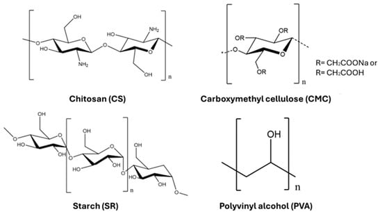
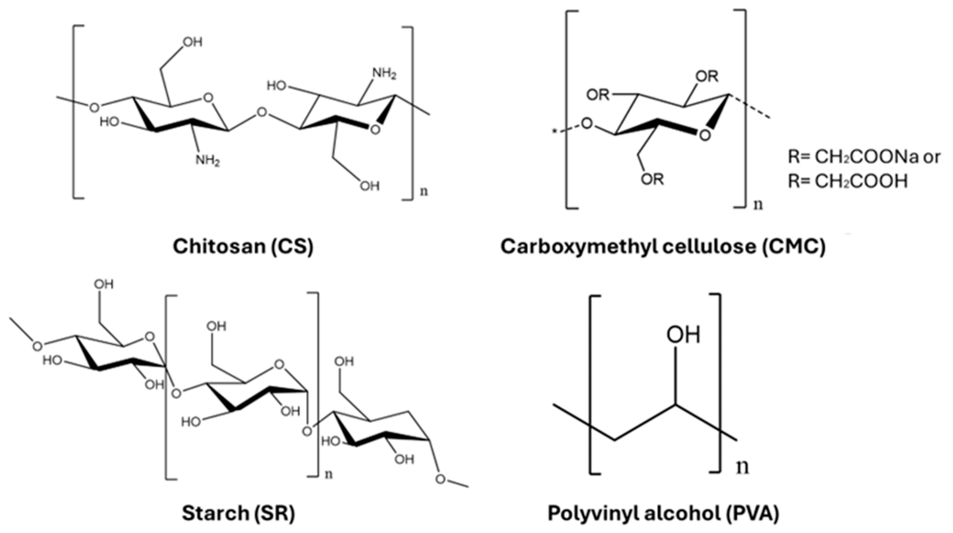
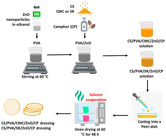
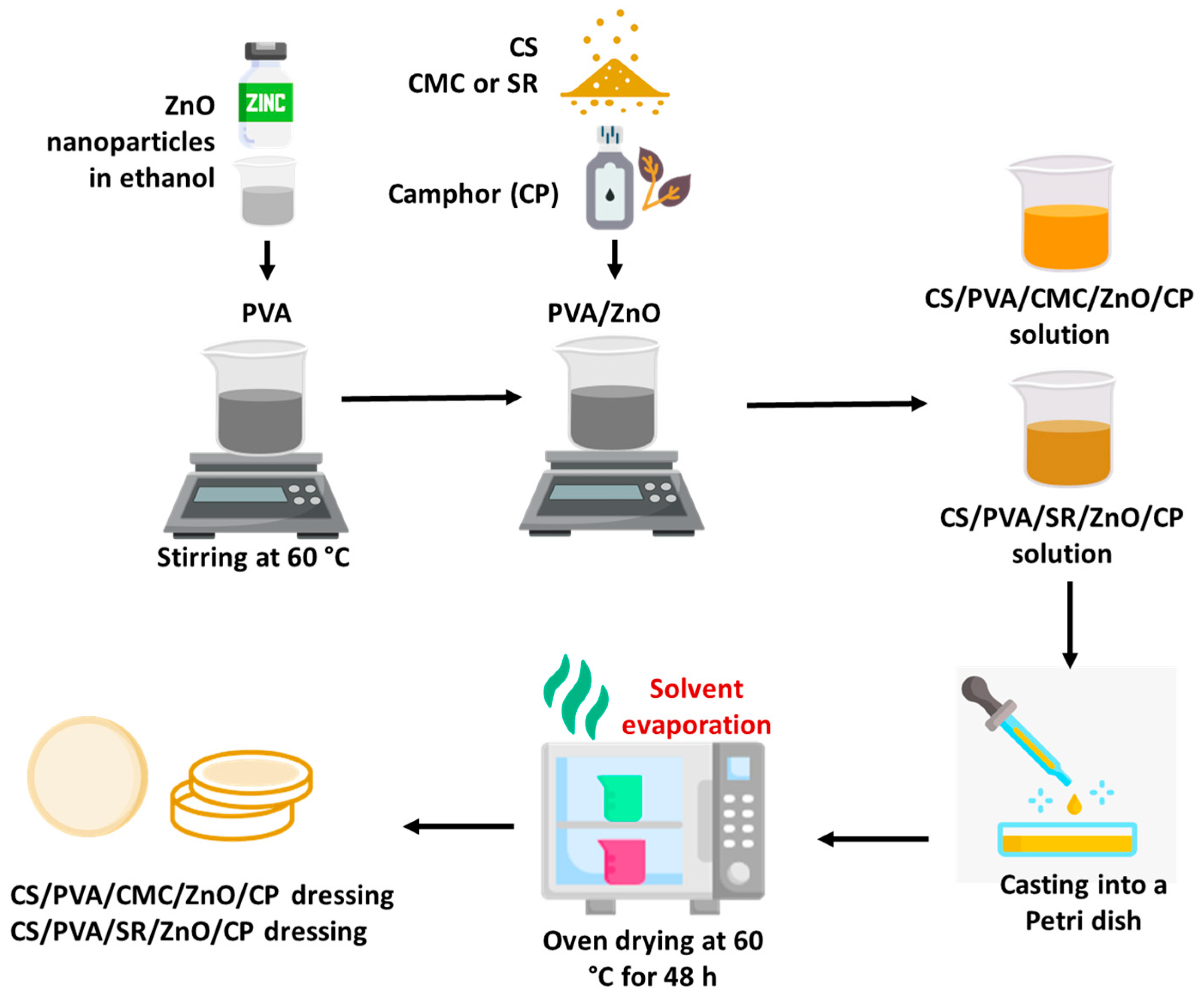
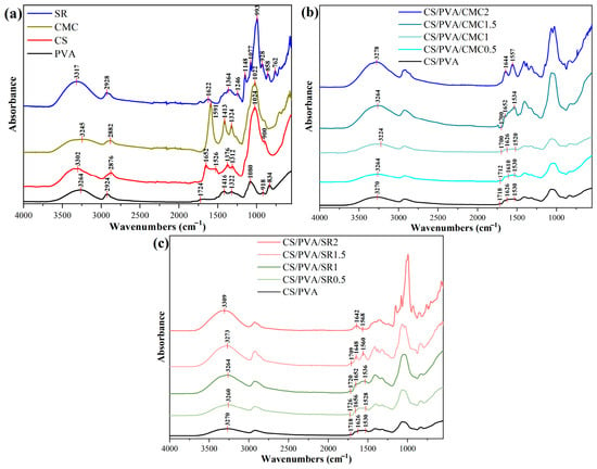
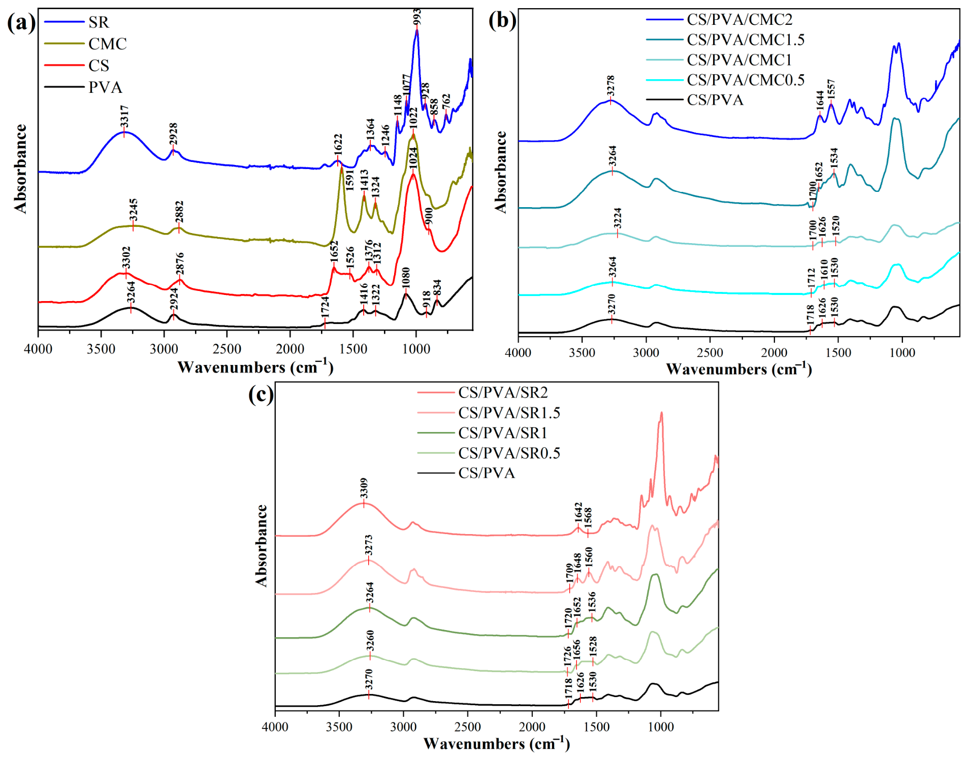
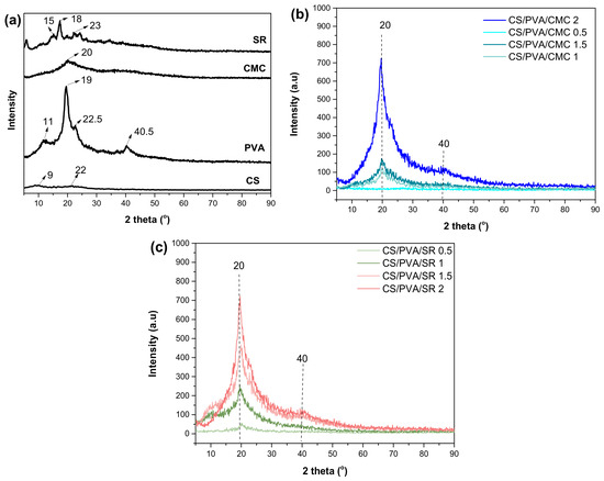
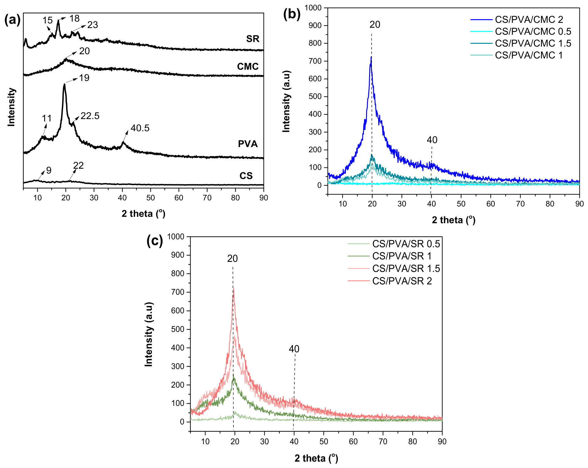
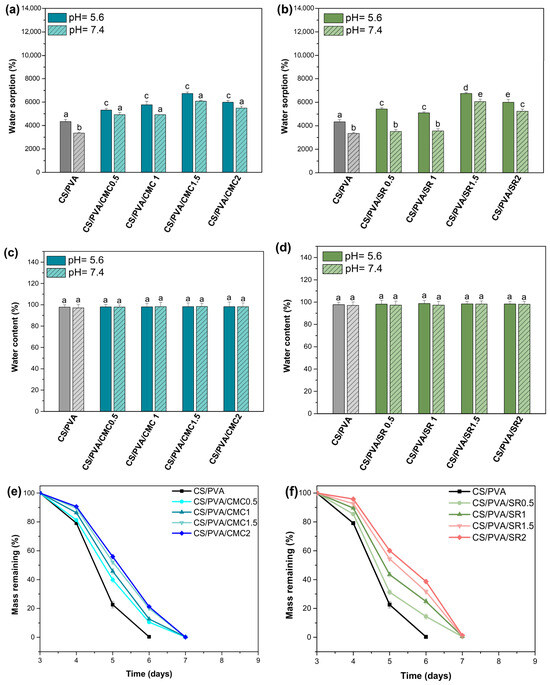
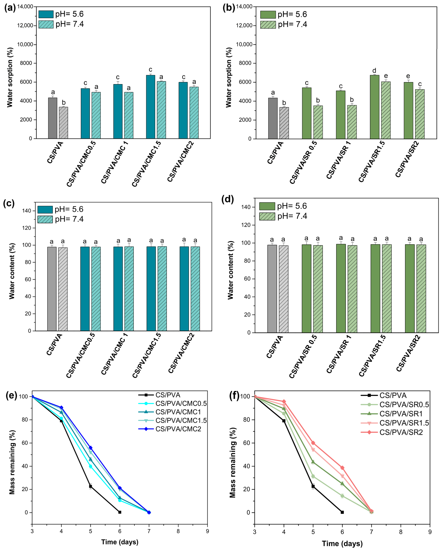
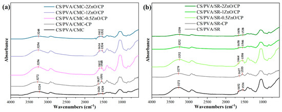
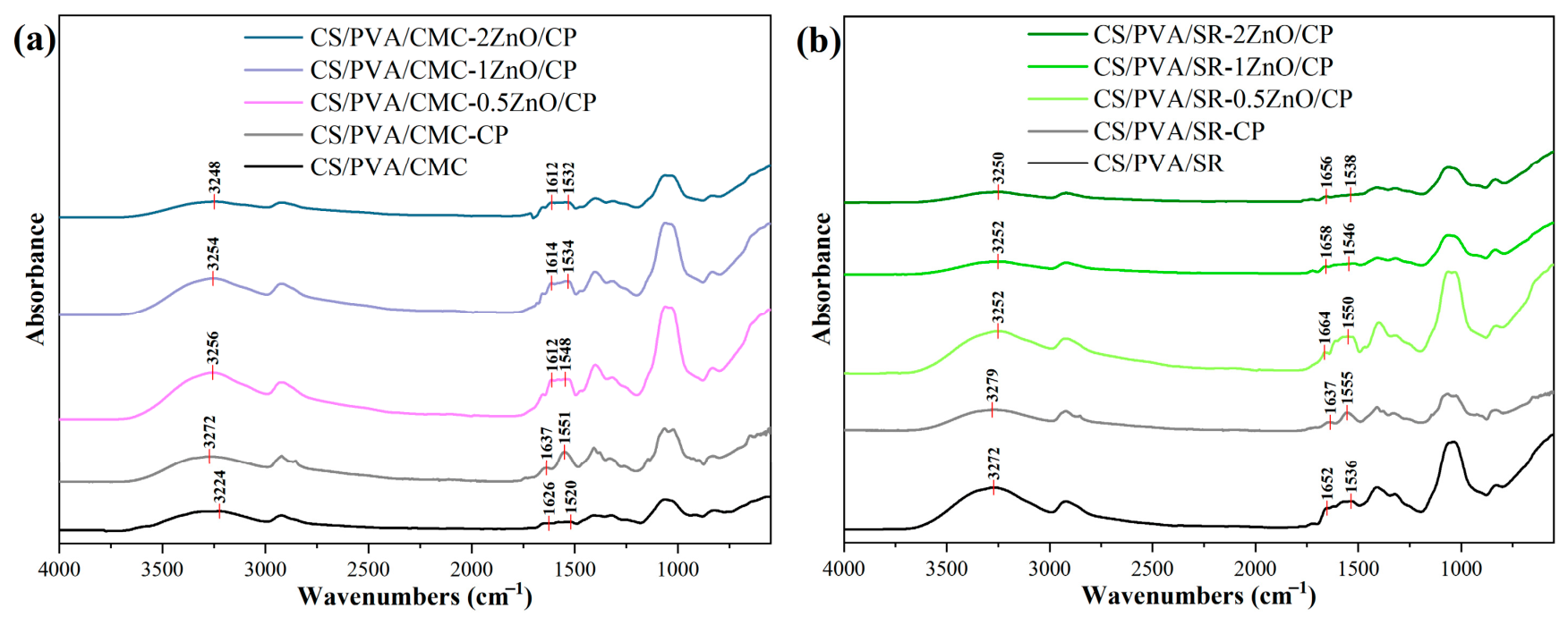
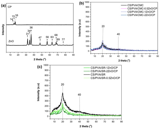
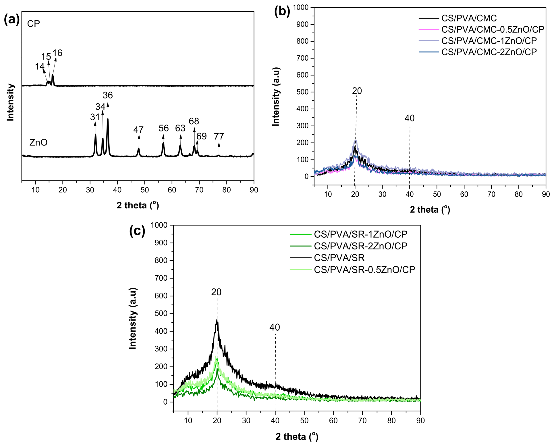
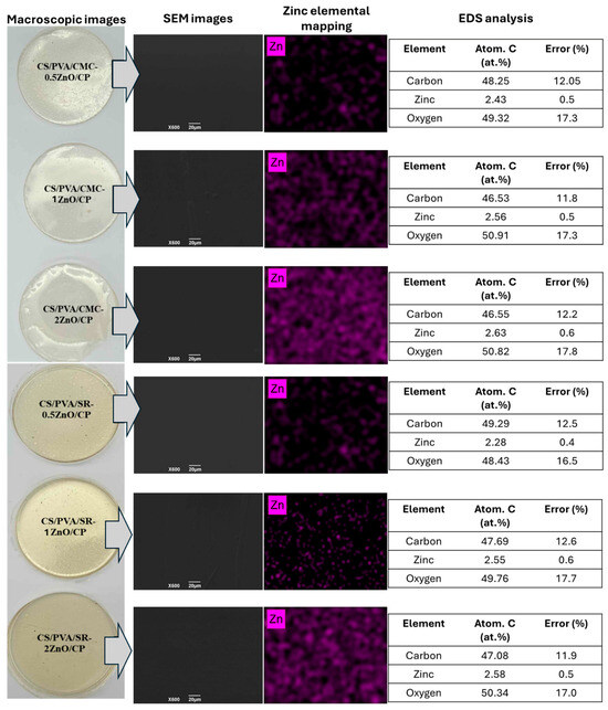
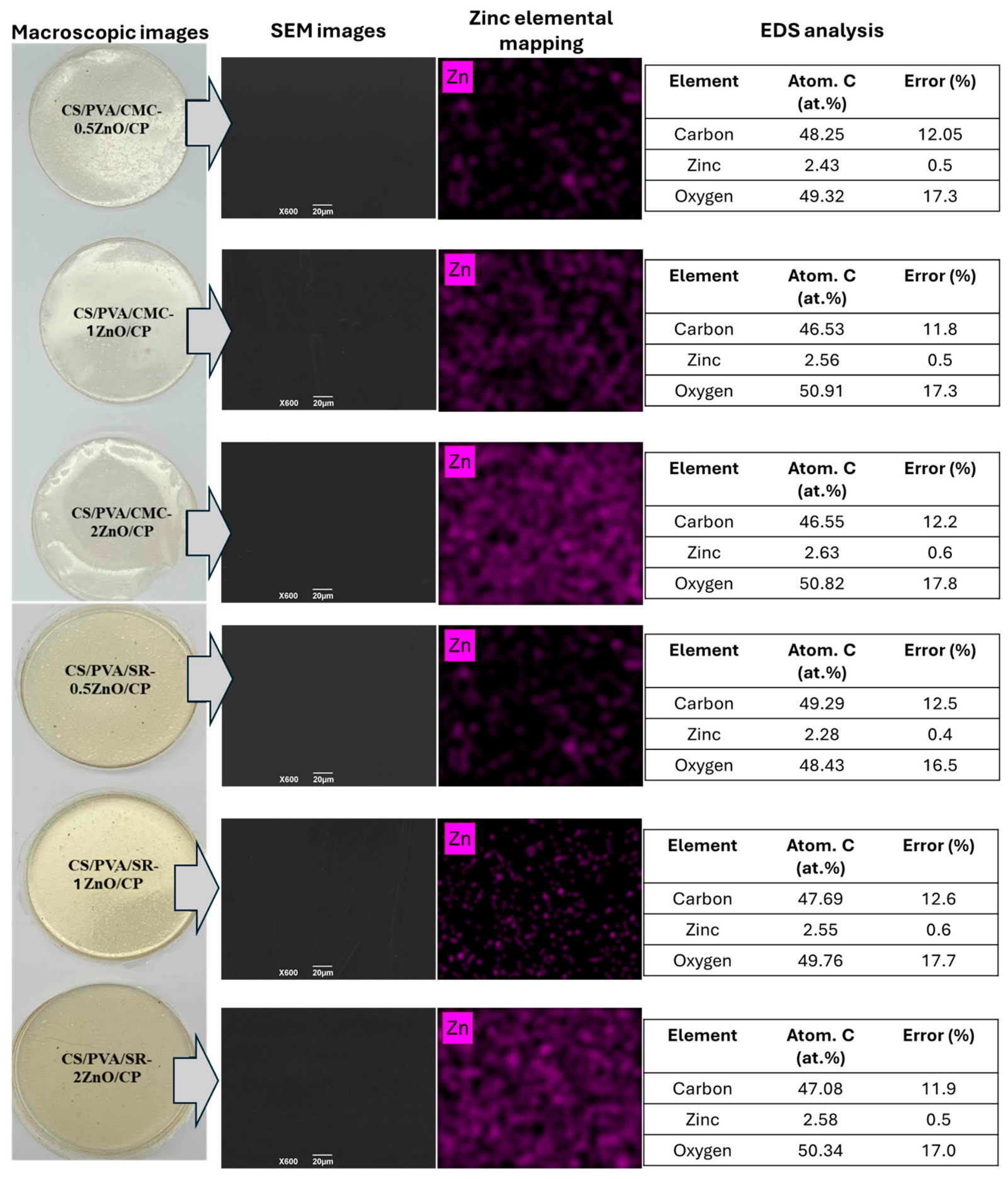
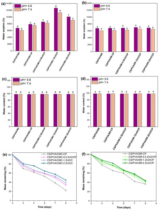
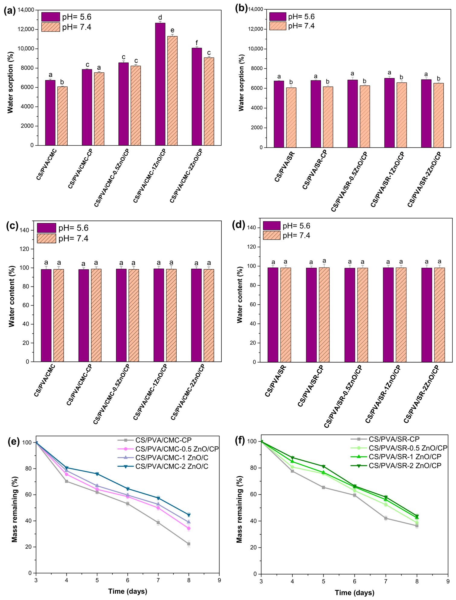
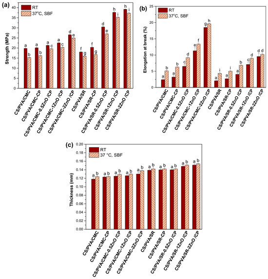
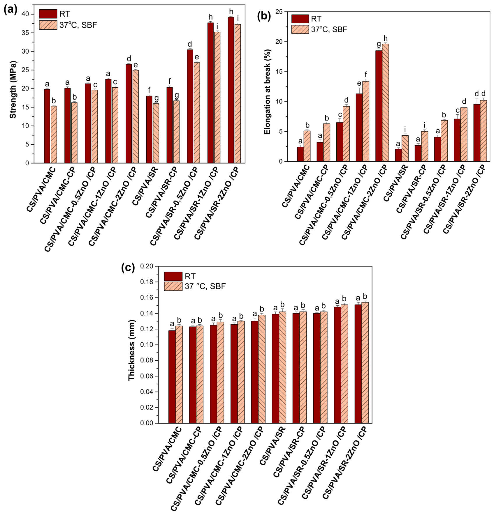
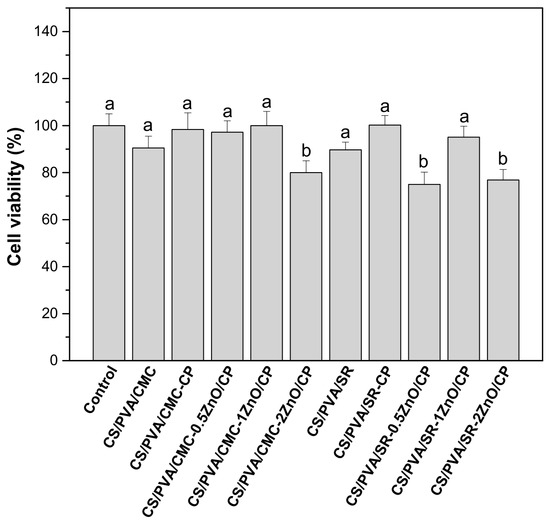
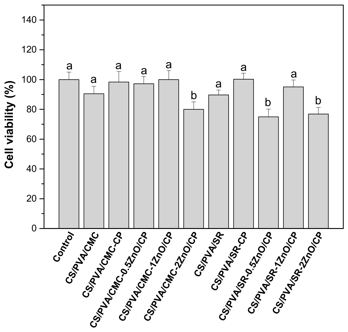
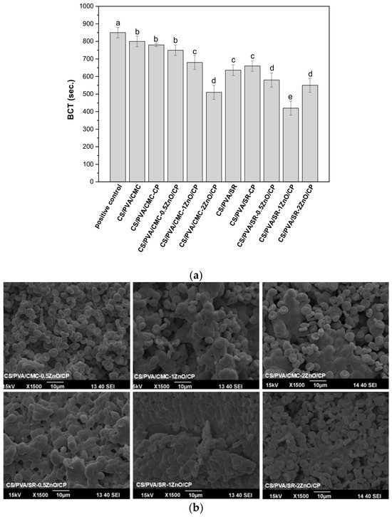
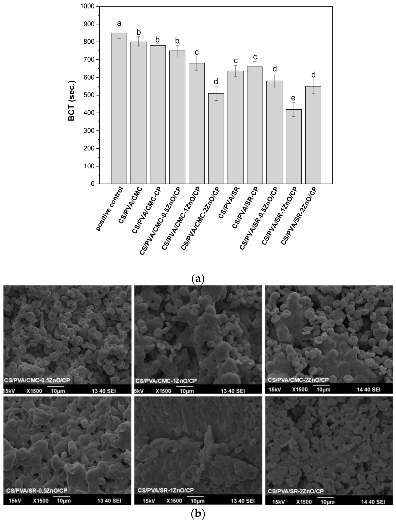
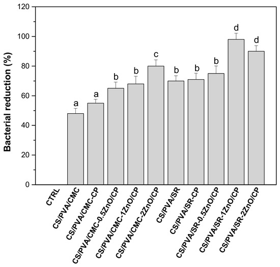
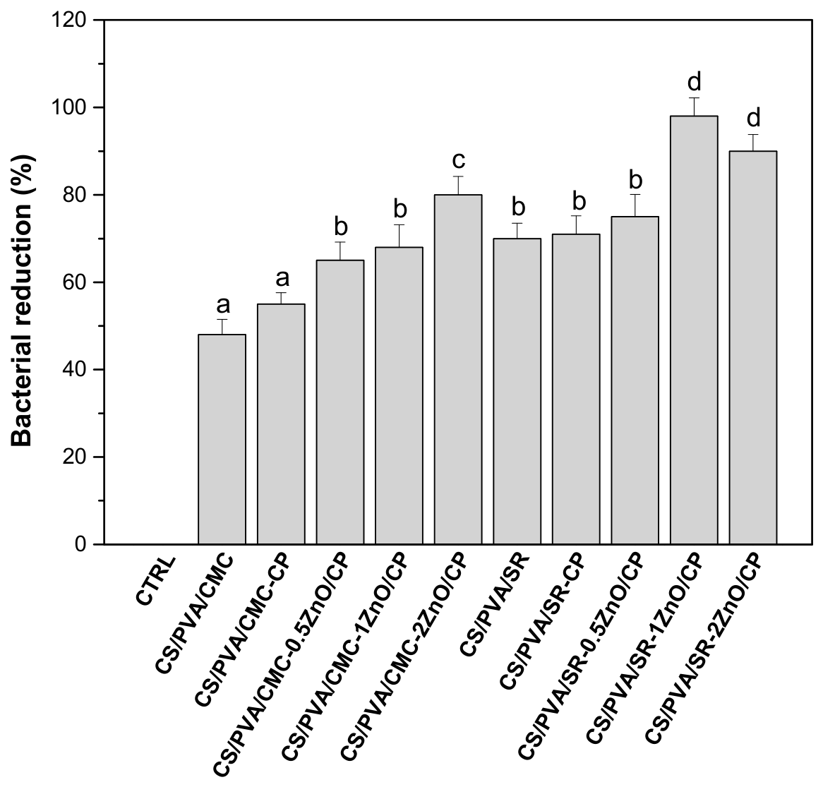

Disclaimer/Publisher’s Note: The statements, opinions and data contained in all publications are solely those of the individual author(s) and contributor(s) and not of MDPI and/or the editor(s). MDPI and/or the editor(s) disclaim responsibility for any injury to people or property resulting from any ideas, methods, instructions or products referred to in the content. |
© 2026 by the authors. Licensee MDPI, Basel, Switzerland. This article is an open access article distributed under the terms and conditions of the Creative Commons Attribution (CC BY) license.