


Abstract
The microencapsulation of plant bioactive compounds by spray drying enhances their stability and controlled delivery in food systems. In this study, flaxseed hydrocolloid (Linum usitatissimum L.) was evaluated as a natural wall material for encapsulating phenolic extracts from mashua (Tropaeolum tuberosum Ruiz & Pav.) and oca (Oxalis tuberosa Molina). Microcapsules were produced using hydrocolloid concentrations of 2.5–10%. The resulting particles showed low moisture content (3.79–5.42%), low water activity (0.31–0.39), and high solubility (90.94–96.45%). Encapsulation efficiency ranged from 78.67 to 62.32% for mashua and from 71.94 to 40.45% for oca, decreasing with increasing wall material concentration. Phenolic content ranged from 14.48 to 11.47 mg GAE/g (mashua) and 8.52 to 4.82 mg GAE/g (oca), with antioxidant capacity between 293.19–143.77 and 84.49–10.33 µmol TE/g, respectively. Particle size ranged from 4.02–10.50 µm (mashua) and 3.93–4.82 µm (oca), and zeta potential values (−37.86 to −27.55 mV) indicated good colloidal stability. Release kinetics showed a biphasic profile and were predominantly diffusion-controlled. The Higuchi model showed significant diffusion (p < 0.05), while the Korsmeyer–Peppas analysis indicated mainly Fickian diffusion (n = 0.234–0.426) with anomalous transport at higher mashua concentrations. These results demonstrate that flaxseed hydrocolloid is an effective and sustainable wall material for controlled release of phenolic compounds from Andean tubers.
1. Introduction
Plant-based phenolic compounds play an important role in preventing chronic diseases associated with oxidative stress, such as cardiovascular disease, diabetes, and certain cancers [1,2]. In this context, Andean tubers, such as oca (Oxalis tuberosa Molina) and mashua (Tropaeolum tuberosum Ruiz & Pav.), have attracted growing scientific interest due to their high content of anthocyanins and polyphenols with high antioxidant capacity, representing an underutilized source of bioactive compounds for the development of functional foods and nutraceuticals [3,4]. In the present study, the phenolic extracts obtained from mashua and oca showed total phenolic contents of 18.26 and 11.84 mg GAE/g (dry basis), respectively, and antioxidant capacities of 301.89 and 99.08 µmol TE/g, confirming their high potential as natural antioxidant sources. However, the industrial application of these compounds is limited due to their chemical and physical instability, as they are highly sensitive to environmental factors such as oxygen, light, temperature, and pH, which accelerate their degradation during processing and storage [1,5]. In response to this problem, microencapsulation has emerged as an effective strategy to protect phenolic compounds and allow their controlled release in food matrices and biological systems [5,6].
Microencapsulation is a key emerging technology for protecting food components and functional constituents from adverse processing conditions by immobilizing sensitive molecules within polymeric or non-polymeric matrices, thereby reducing oxidative and thermal degradation, masking undesirable flavors or odors, and regulating the release of the core [5,7]. Among the available techniques, spray drying stands out for its simplicity, low cost, high speed, and ease of industrial scale-up to produce stable powders [1]. The performance of these systems depends largely on the wall material, which directly influences the encapsulation efficiency, storage stability, and bioavailability of the encapsulated compound [1]. Although conventional materials such as maltodextrin, gum arabic, and modified starches have been widely used for their technological properties, the growing demand for “clean label” products and a sustainable approach has driven the use of plant-based biopolymers. In this context, plant-based hydrocolloids stand out for their renewable, biodegradable, and biocompatible nature, as well as their ability to form efficient functional matrices in food applications [7].
Flaxseed hydrocolloid (Linum usitatissimum L.), also known as flaxseed mucilage, is a natural polysaccharide found in the outer layer of the seed, characterized by rheological, emulsifying, and film-forming properties that allow it to be used as a thickener and stabilizer in food systems [8,9]. These characteristics position it as a promising wall material for nutrient delivery systems, with the potential to replace commercial gums and form complex coacervates with proteins, improving the viscosity and stability of encapsulating matrices [3,8,9]. However, its behavior during spray drying can be affected by reductions in viscosity and water retention capacity, and its application for protecting phenolic compounds in Andean tubers remains poorly explored [3,4,8].
The oca (Oxalis tuberosa Molina) and mashua (Tropaeolum tuberosum Ruiz & Pav.) are notable sources of anthocyanins and polyphenols with high antioxidant capacity; however, there is limited information on their performance in microencapsulation systems, particularly in terms of physicochemical stability and controlled release [3,4,5,6]. In this context, the present study evaluates flaxseed hydrocolloid as a sustainable wall material for spray-dried microencapsulation of phenolic compounds from oca and mashua, analyzing the effect of polymer concentration on stability and release profile, in line with the trend toward the use of “green” biopolymers and the valorization of Andean crops [1,7].
2. Materials and Methods
2.1. Materials
The flaxseeds (Linum usitatissimum) were from the local market of Andahuaylas District, Andahuaylas Province, Apurímac Region, Peru (13°39′14.64″ S, 73°23′06.45″ W; 2915 m above sea level). The seeds were cleaned manually to remove impurities, dust, and foreign material. The Andean tubers mashua negra (Tropaeolum tuberosum) and oca Huasquilla (Oxalis tuberosa) were obtained from local agricultural producers in Huayana District, Andahuaylas Province, Apurímac Region, Peru (14°03′02.26″ S, 73°36′33.76″ W; 3184 m above sea level). The tubers were selected based on quality criteria (absence of mechanical damage, disease, and physiological deterioration). Ultrapure water was used to prepare solutions and perform analyses. The reagents used were purchased from recognized commercial suppliers (Merck, Darmstadt, Germany; Sigma-Aldrich, St. Louis, MI, USA and Spectrum Chemical, New Brunswick, NJ, USA). The vegetable oils used in the emulsifying property tests were food-grade.
2.2. Obtaining Flaxseed Hydrocolloid
Flaxseed hydrocolloid (Linum usitatissimum), located primarily in the outermost or epidermal layer of the seed coat, was obtained through an aqueous extraction process to facilitate the release of mucilage [8]. The seeds were dispersed in distilled water using a ratio of 1:20 w/v and subjected to constant agitation at a temperature of 80 ± 5 °C and 150 rpm for 3 h using a thermostatically controlled water bath with an agitation system (WTB50, Memmert GmbH, Schwabach, Germany) [9,10]. Mechanical separation was then carried out by sieving, allowing the seeds to be effectively isolated from the viscous solution, which was subsequently dehydrated at 60 °C in a forced convection oven (FED 115 model, Binder GmbH, Tuttlingen, Germany) to obtain the polymer powder. Finally, the powder obtained was stored in airtight containers at 25 °C in airtight 50 mL polypropylene centrifuge tubes (Falcon type) to protect it from hygroscopicity and ensure its structural stability.
2.3. Physical, Chemical, and Techno-Functional Characterization of Flaxseed Hydrocolloid
The physical and chemical properties evaluated included moisture content, water activity (aw), hygroscopicity, and bulk density, all of which are critical for ensuring physical and microbiological stability during processing and storage. Moisture content was determined following the AOAC 950.10 methodology by drying in an oven at 105 °C until constant weight [11,12] using a forced convection oven (FED 115 model, Binder GmbH, Tuttlingen, Germany). Water activity was measured with a hygrometer previously calibrated with standard solutions [3], using a HygroPalm23-AW water activity meter (Rotronic AG, Bassersdorf, Switzerland). Hygroscopicity was evaluated by exposing the material to a saturated sodium chloride atmosphere (75% relative humidity) at 25 °C in hermetically sealed 150 mL glass containers, and the result was expressed as the percentage increase in mass after 7 days [11,13]. Apparent density was calculated as the ratio between the total mass and the volume occupied by the powder [3,14].
The optical properties were analyzed by determining the color coordinates L*, a* and b* of the CIE system, using a standardized colorimeter with the D65 light source (CR-5 model, Konica Minolta, Tokyo, Japan) [15]. Colloidal stability was evaluated by determining the ζ potential with a Zetasizer ZSU3100 (Malvern Instruments Ltd., Worcestershire, UK) and using dynamic light scattering to measure repulsive forces between particles and predict their stability in suspension [11,16]. In addition, the particle size distribution and polydispersity index (Span) were determined by laser diffraction with a Mastersizer 3000 (Malvern Instruments Ltd., Worcestershire, UK), parameters that directly influence the material’s ability to form uniform microcapsules during atomization [11,17,18].
The chemical structure of the hydrocolloid was analyzed by Fourier transform infrared spectroscopy (FTIR) in the midrange from 4000 to 400 cm−1 with a Nicolet iS50 FTIR spectrometer (Thermo Fisher Scientific, Waltham, MA, USA). For this purpose, potassium bromide (KBr) pellets were used, and the transmission module was employed [19]. The surface morphology was evaluated using low-vacuum scanning electron microscopy (SEM), which allowed observation of particle shape and porosity. In addition, elemental analysis was performed using energy-dispersive X-ray spectroscopy (EDS) [20].
Additionally, the technofunctional properties of flaxseed hydrocolloid were evaluated, including water retention capacity, emulsification, and foaming, to analyze its interaction with water, oil, and air, respectively, and its performance as a functional material in food and encapsulation systems [9,21]. Water retention capacity indicated the biopolymer’s ability to absorb water and form hydrated structures, a property associated with the abundant hydroxyl groups in its arabinoxylan and rhamnogalacturonan matrix, which is relevant for obtaining stable matrices. Emulsification capacity was evaluated in oil-water systems, reflecting the hydrocolloid’s ability to stabilize liquid–liquid interfaces by increasing the viscosity of the continuous phase, a key characteristic for acting as a wall material [15]. Foaming was determined by the volume generated upon air incorporation, a parameter related to the structural flexibility of the mucilage and the formation of viscoelastic networks that promote interfacial stability [9].
2.4. Automated Extraction of Bioactive Compounds
The tubers of mashua (Tropaeolum tuberosum) and oca (Oxalis tuberosa) were washed with drinking water, disinfected with a sodium hypochlorite solution (100 mg/L) for 5 min, rinsed with distilled water, and mechanically crushed using an agate mortar and pestle to obtain a homogeneous sample. Approximately 5 g of sample was placed into Q-Disc™ C1 extraction vessels and loaded into clear 40 mL collection vials in the automated EDGE® pressurized solvent extraction system (CEM Corporation, Matthews, NC, USA), a technique that enables efficient, reproducible extraction of phenolic compounds from plant matrices [22].
A hydroalcoholic mixture composed of 30% ethanol and 70% water (v/v) was used as the extraction solvent, selected for its high solubilization capacity of phenolic compounds and its compatibility with food applications [23]. The extraction was performed independently for black mashua and oca under controlled temperature and pressure conditions programmed in the EDGE® equipment (CEM Corporation, Matthews, NC, USA), including solvent addition (20 mL top add, 10 mL bottom add), rinsing with 10 mL solvent, and heating at 80 °C with a hold time of 10 min, ensuring reproducibility of the process.
The extracts were concentrated under reduced pressure using a rotary evaporator (HS-2005V-N, HAHNVAPOR, Shanghai, China) at 40 °C until the volume was reduced to 20% of its initial value. The concentrated extracts were then stored at 4 ± 1 °C in 200 mL amber glass bottles with airtight caps until use in the microencapsulation process.
2.5. Spray Drying Microencapsulation
For microencapsulation, flaxseed hydrocolloid was used as a natural wall material. Aqueous solutions of the hydrocolloid were prepared at different concentrations (2.5, 5.0, 7.5 and 10.0% w/v) and hydrated under constant magnetic stirring at room temperature (25 ± 2 °C) until complete dispersion was achieved. Subsequently, the phenolic extracts of black mashua and oca were incorporated into the encapsulating solutions under continuous stirring for 30 min to ensure homogeneous distribution of the bioactive compounds within the hydrocolloid matrix [1,14].
The mixtures were dried in a laboratory-scale spray dryer (Mini Spray Dryer B-290, Büchi Labortechnik AG, Flawil, Switzerland) equipped with a standard two-fluid nozzle with a 0.7 mm diameter. The spray drying process was conducted under the following operating conditions: inlet air temperature of 120 °C, aspirator rate of 100%, feed pump rate of 30%, atomization airflow rate of 650 L/h, and nozzle diameter of 0.7 mm. The outlet temperature was continuously monitored during the spray drying process and remained within the range of 66–68 °C.
The microencapsulated powder obtained was collected from the cyclone separator, packaged in 50 mL amber glass tubes with airtight caps, and stored in a desiccator at 20 °C under controlled temperature and humidity conditions until its subsequent physicochemical and functional characterization.
2.6. Physical Properties of Microcapsules
The moisture content of the microcapsules was determined following the AOAC 950.10 methodology, by drying the samples in a forced-convection oven at 105 °C until a constant weight was reached [11,12], in a forced convection oven (FED 115 model, Binder GmbH, Tuttlingen, Germany). Water activity (aw) was measured at 25 °C [11], with a HygroPalm23-AW water activity meter (Rotronic AG, Bassersdorf, Switzerland), previously calibrated with saturated salt solutions.
Hygroscopicity was evaluated by exposing the microcapsules to a controlled relative humidity atmosphere (75% RH) generated by a saturated sodium chloride solution at 25 °C in hermetically sealed 180 mL glass containers. The results were expressed as the percentage increase in mass after 7 days of equilibrium, allowing the matrix’s affinity for ambient water vapor to be classified [3,24].
Water solubility was determined by dispersing a known amount of microcapsules in distilled water, followed by centrifugation with a TDL-5M centrifuge (Bioridge Instrument Co., Ltd., Shanghai, China) and subsequent drying of the supernatant at 105 °C [25] in a forced convection oven (FED 115 model, Binder GmbH, Tuttlingen, Germany). This parameter, expressed as a percentage of dissolved solids, is essential for evaluating the behavior of the wall material and the ease with which the bioactive core can be released or reconstituted in food matrices [5].
The color properties of the microcapsules were determined by colorimetric analysis in the CIE L*, a*, and b* system using a benchtop colorimeter (CR-5 model, Konica Minolta, Tokyo, Japan), previously calibrated and standardized with white and black reference plates. The standard D65 light source was used for the measurements [19].
2.7. Determination of Content of Bioactive Compounds in Microcapsules
For analysis, the bioactive compounds were released from the microcapsule matrix by dissolving them in 80% methanol. The total phenol content was quantified using the Folin–Ciocalteu colorimetric method, measuring the absorbance at 755 nm in a spectrophotometer (Genesys 150, Thermo Fisher Scientific, Waltham, MA, USA) using a gallic acid calibration curve, after a 24 h incubation period in darkness [3,19,26]. The results were expressed in milligrams of gallic acid equivalents per gram of microcapsule (mg GAE/g).
The total flavonoid content was determined by complexation with aluminum chloride (AlCl3), measuring absorbance at 450 nm with the same UV–Vis spectrophotometer (Genesys 150, Thermo Fisher Scientific, Waltham, MA, USA) and using quercetin as a reference standard (mg QE/g) [19,27]. Finally, antioxidant capacity was evaluated using the DPPH free radical scavenging assay, measuring the decrease in absorbance at 515 nm after 30 min of reaction with the same instrument. The values obtained were expressed as micromoles of Trolox equivalents per gram of sample (µmol TE/g) [3,19,28].
2.8. Encapsulation Efficiency
Encapsulation efficiency (EE%) was determined by measuring the retention of phenolic compounds after spray drying. To do this, the total phenolic content of the feed solution before drying (TPCi) and the total phenolic content of the microcapsules obtained (TPCe) were quantified using the Folin–Ciocalteu colorimetric method [29], as described in Section 2.7. The initial phenolic content corresponded to the extract incorporated into the hydrocolloid solution before atomization, while the phenolic content in the microcapsules was determined after complete dissolution of 1 g of powder in 80% methanol, in order to ensure the total release of the compounds retained in the matrix. The encapsulation efficiency was expressed as the percentage of phenolic compounds retained in the microcapsules relative to the total initial phenolic content in the feed solution, allowing the protective capacity of the flaxseed hydrocolloid during the spray-drying process to be evaluated.
2.9. Determination of Phenolic Compound Release in Aqueous Medium
The release of polyphenols was determined by dispersing the microcapsules (100 mg) in ultrapure water (10 mL) at room temperature, protected from light. Aliquots were removed at defined intervals (0–48 h) with solvent replacement, and the released compounds were quantified using the Folin–Ciocalteu method at 755 nm [5,19]. This assay allows the characterization of sustained release by diffusion or dissolution mechanisms from the hydrocolloid matrix, a critical parameter for ensuring the stability and bioaccessibility of bioactives in functional beverages and processed foods [5,19].
2.10. Modeling of Release Kinetics of Encapsulated Phenolic Compounds
The kinetics of phenolic compound release were determined by fitting the experimental data to the Higuchi and Korsmeyer–Peppas models, which are standard tools for characterizing solute transport in polymer matrix systems. The Higuchi model was applied to evaluate diffusion-controlled processes, in which the fraction of bioactive compound released is proportional to the square root of time [30]. The Korsmeyer–Peppas model was used to elucidate the overall transport mechanism by using the diffusion exponent (n), which distinguishes between Fickian diffusion (n ≤ 0.43 for spheres), anomalous transport, and Case II transport [31].
The kinetic parameters were calculated using linear regression in OriginPro 2025b software (version 10.2.5.234, OriginLab Corporation, Northampton, MA, USA), evaluating the fit quality through the coefficient of determination (R2) and the level of statistical significance (p < 0.05) [31]. Overall, this modeling is essential for understanding how the biopolymer matrix protects the bioactive core and regulates its delivery and bioavailability in food or pharmaceutical applications. The mathematical equations and parameters of the Higuchi and Korsmeyer–Peppas models are presented in Table 1.
Table 1.
Higuchi and Korsmeyer–Peppas models applied for phenolic compound release kinetics.
2.11. Colloidal Stability and Particle Size of Microcapsules
Particle size distribution was determined using a Mastersizer 3000 laser diffraction (Malvern Instruments, Worcestershire, UK), with isopropanol as the dispersing medium to prevent dissolution of the microcapsule matrix [11,33]. The samples were homogenized by ultrasound to determine representative diameters (D10, D50, D90) and calculate the polydispersity index (Span), parameters that characterize the uniformity and homogeneity of the system.
The zeta potential (ζ) was analyzed using a Zetasizer ZSU3100 (Malvern Instruments, Worcestershire, UK) equipped with a He-Ne laser, using ultrapure water at 25 °C, allowing the evaluation of surface charge and electrostatic stability in suspension [11,33]. High absolute values suggest greater colloidal stability, as repulsive forces between particles prevent coalescence and aggregate formation [34].
2.12. Structural Characterization
The chemical structure of the microcapsules was analyzed using a Nicolet IS50 FTIR instrument (ThermoFisher, Waltham, MA, USA) in the midrange of 400–4000 cm−1 using KBr pellets, which allowed the identification of functional groups [11,19,35].
Surface morphology and microstructure were evaluated by scanning electron microscopy (SEM) with a Prisma E microscope (Thermo Fisher Scientific, Brno, Czech Republic), analyzing shape, size, wall continuity, and the presence of pores or cracks [5,19].
2.13. Statistical Analysis
All extractions, microencapsulation experiments, and analytical determinations were performed in triplicate. The results were expressed as mean ± standard deviation. Prior to statistical analysis, the data were evaluated for normality and homogeneity of variance. One-way analysis of variance (ANOVA) was conducted to determine significant differences among treatments. When significant differences were detected, Tukey’s multiple-comparison test was used to identify differences between mean values at the 95% confidence level (p < 0.05). OriginPro 2023 software (OriginLab Corporation, Northampton, MA, USA) was used for statistical analysis and graphical representation of the results.
3. Results and Discussion
3.1. Characterization of Flaxseed Hydrocolloid
3.1.1. Physical Properties of Flaxseed Hydrocolloid
Table 2 presents the physical properties of flaxseed hydrocolloid, including moisture content, water activity (aw), hygroscopicity, bulk density, and color parameters (L*, a*, and b*). The hydrocolloid exhibited low moisture content and water activity values, indicating adequate stability and a reduced risk of microbial growth. The bulk density was consistent with values typically reported for dried polysaccharide powders, reflecting good packing and handling properties. Regarding color, the hydrocolloid showed high lightness and low chromaticity values, indicating a light-colored material with slight yellowish tones, characteristic of flaxseed-derived hydrocolloids.
Table 2.
Physical properties of flaxseed hydrocolloid.
These results suggest that the process of obtaining the hydrocolloid did not cause significant darkening, which is favorable for its application as an encapsulating material in food matrices [15], complying with the standards for wall materials in spray-dried microencapsulation [21,36]. These results are consistent with previous studies that place aw in the range of 0.36 to 0.48, depending on the drying method and formulation, confirming that the material is technologically suitable for preventing biochemical deterioration during storage [15,21,37].
The bulk density is consistent with that reported for polysaccharide powders obtained by atomization, which tend to develop porous internal structures and low compaction [36,38]. Similar values have been described for other plant mucilages processed by spray drying, which favors adequate powder handling and efficient functional performance during atomization [36,37], suggesting a structural organization compatible with high-quality nutrient delivery systems [38].
In relation to optical properties, the high brightness together with the low a* and b* indices confirm that the extraction process prevented significant darkening or thermal browning [15], which is essential for its use in the food industry [11,15].
Finally, the hygroscopicity of the material is consistent with the hydrophilic nature of mucilage, derived from the abundant hydroxyl groups in its arabinoxylan and galactomannan structures, which also enhance the formation of continuous matrices and effective interactions with hydrophilic bioactive compounds [21]. Taken together, the physicochemical results confirm the potential of flaxseed hydrocolloid as a sustainable protective matrix for sensitive bioactives [37].
3.1.2. Techno-Functional Properties
Table 3 shows the techno-functional properties of flaxseed hydrocolloid, evaluated through swelling power, emulsification capacity, and foaming capacity, parameters that allow analysis of its interaction with water, oil, and air, respectively, and its performance as a functional material in food and encapsulation systems.
Table 3.
Techno-functional properties of flaxseed hydrocolloid.
The swelling power of linseed hydrocolloid demonstrates a high water affinity, characteristic of mucilages rich in arabinoxylans and pectic fractions, capable of stabilizing the feed prior to spray drying and promoting the formation of continuous hydrogen matrices [39,40]. This value is considered moderate-to-high among seed hydrocolloids and, although lower than that of chia mucilage, is technologically competitive as a thickening agent [41]. The emulsifying capacity confirms its potential as a stabilizer, attributable to the increased viscosity of the continuous phase and the synergy between polysaccharides and the conlinin protein, allowing its application as a functional alternative to gum arabic in food systems [21,42]. The effectiveness of this property depends on the structural balance between neutral and acidic fractions, which is determined by the extraction method [21,39]. The foaming capacity was moderate, consistent with the flexible biopolymer structure, which reduces interfacial tension and limits liquid drainage. Although inferior to protein systems, this behavior is comparable to that of other seed hydrocolloids, where the viscosity of the medium restricts bubble coalescence, and can be optimized by adjusting the mixing conditions or the experimental protocol [9,39,40].
Overall, the technofunctional profile of flaxseed hydrocolloid, characterized by high hydration, adequate emulsifying capacity, and stable foam formation, confirms its suitability as an encapsulating matrix, promoting the integrity of the continuous phase and the protection of the core during atomization, in accordance with current sustainability and functional efficiency requirements in the food and pharmaceutical industries [21,37,43].
3.1.3. Colloidal Stability and Particle Size
The colloidal stability of the hydrocolloid was evaluated by analyzing zeta potential and particle size distribution; the results are presented in Table 4. The zeta potential showed negative values, suggesting adequate electrostatic repulsion between particles and good suspension stability.
Table 4.
Colloidal stability and particle size of flaxseed hydrocolloid.
Flaxseed hydrocolloid exhibited a negative zeta potential attributable to ionized carboxyl groups that promote electrostatic repulsion and confer colloidal stability to the system [39]. In polysaccharides, absolute values ≥25–30 mV indicate adequate physical stability, consistent with the ranges reported for plant mucilages (−20 to −54 mV), depending on pH, ionic strength, and extraction method [34,44].
The average particle size and its homogeneous distribution are consistent with those of polysaccharide powders obtained by aqueous methods and conventional drying, usually in the range of 100–500 µm [11,38]. This size is well-suited for use as a wall material, as it promotes rehydration and feed stability prior to atomization, key factors for the formation of continuous microcapsules and the protection of the core from degrading agents [6,37].
The particle size of native flaxseed powder is significantly larger than that of microcapsules obtained by spray drying. This difference is attributed to the atomization process, which transforms the feedstock into fine droplets, followed by rapid solvent evaporation that fixes the structure into small microspheres [38,45].
While the native hydrocolloid retains its original aggregate structure through intermolecular associations [8,38], atomization fragments the matrix, generating fine particles [1]. This phenomenon, widely described in the literature, substantially improves the dispersion properties and technological functionality of the final powder [1,45,46].
3.1.4. Structural Characterization of Flaxseed Hydrocolloid
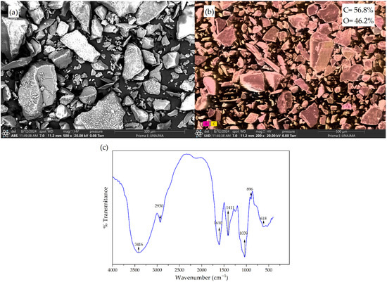
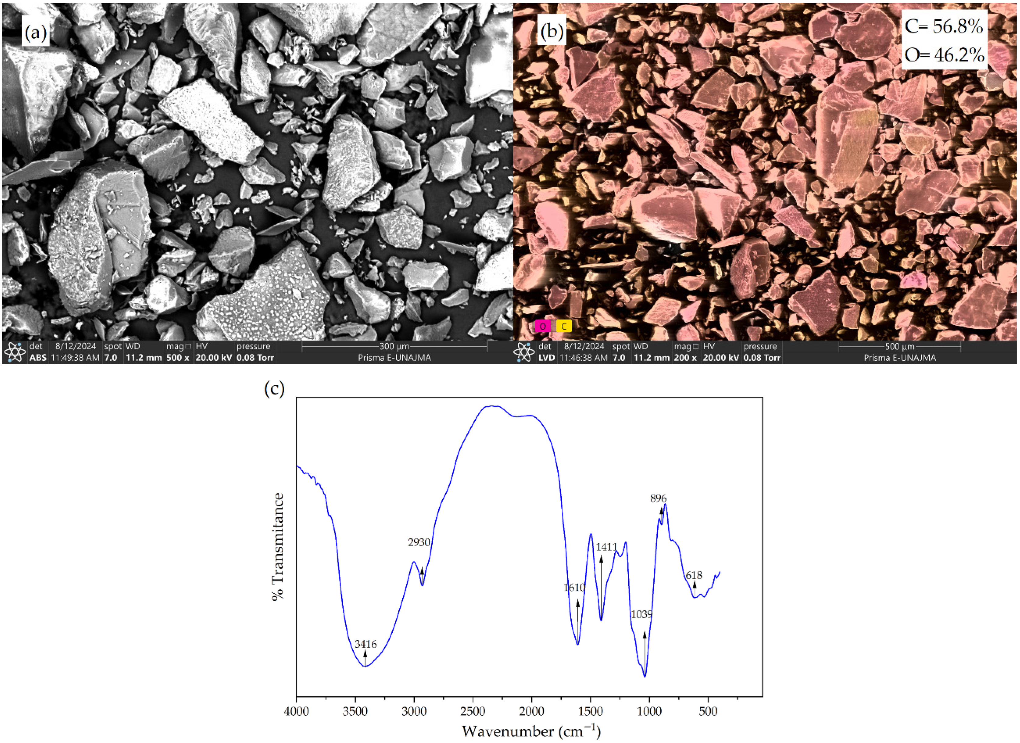
Figure 1 shows the structural characterization of flaxseed hydrocolloid using SEM, EDS, and FTIR. SEM micrographs revealed an irregular, predominantly amorphous morphology with rough, fractured surfaces, typical of polysaccharide hydrocolloids obtained by drying. EDS analysis confirmed a surface composition dominated by carbon and oxygen, consistent with the material’s organic nature. The FTIR spectrum showed characteristic bands of polysaccharides, highlighting signals associated with hydroxyl groups, C–H and C–O–C bonds, confirming the chemical integrity of the hydrocolloid and its suitability as an encapsulating matrix.
Figure 1.
Colloidal stability and structural characterization of flaxseed hydrocolloid. (a) SEM micrograph; (b) EDS analysis of the surface; (c) FTIR spectrum.
The irregular and amorphous morphology observed by SEM (Figure 1a) is characteristic of vegetable mucilage powders (such as flaxseed, chia, or guar) obtained by drying, which usually have rough surfaces and flaky structures [38,44]. This microstructure increases surface area, facilitating interaction with bioactive nuclei and optimizing trapping efficiency during atomization [25]. EDS elemental analysis (Figure 1b) confirmed a surface dominated by carbon (39.0–45.4%) and oxygen (54.6–61.0%), confirming the organic and polysaccharide nature of the hydrocolloid [11,19]. These results show that the material is highly pure, free of significant inorganic contaminants, validating its suitability as an encapsulating matrix [19].
On the other hand, the FTIR spectrum (Figure 1c) identified the typical bands of polysaccharides, identifying hydroxyl groups (–OH) [47]; a broad signal between 3393 and 3442 cm−1 associated with the high water affinity of arabinoxylans and galactomannans [11,48]. Also, the presence of C–H and C–O–C bonds [49]; characteristic vibrations in the carbohydrate “fingerprint” area (800–1200 cm−1) [39].
The preservation of these signals confirms that the extraction process did not chemically degrade the matrix, preserving its functionality for nutrient delivery systems [11]. Taken together, these structural properties position flaxseed hydrocolloid as a sustainable and effective protective matrix, comparable to other commercial polymers used in spray-drying microencapsulation.
3.2. Characterization of Microcapsules
3.2.1. Physical Properties of Microcapsules
Table 5 shows the physical properties of microcapsules made with mashua and oca extracts at different encapsulant concentrations. Variations are observed associated with both the type of raw material and the level of encapsulant, demonstrating the influence of the formulation on the stability and physical behavior of the microcapsules.
Table 5.
Physical properties of mashua and oca microcapsules.
Increasing the encapsulant concentration reduced moisture content in both cores, demonstrating greater spray-drying efficiency with higher solids content in the feed [1]. Likewise, water activity values (aw ≤ 0.39) ensure microbiological and physicochemical stability, consistent with reports for polysaccharide matrices, where the wall material limits water mobility during atomization [25].
Hygroscopicity decreased progressively with increasing encapsulant, attributed to the formation of more compact matrices that restrict the adsorption of ambient moisture, a behavior described for natural hydrocolloids [11,19,21]. In contrast, solubility remained high across all formulations, with slight reductions at higher wall material concentrations, confirming the preservation of the system’s hydrophilic nature and its adequate rehydration, a key attribute for aqueous food applications [50].
The color coordinates showed a clear effect of the encapsulant concentration and core type. In both systems, increasing the wall material increased brightness (L*) and reduced the a* and b* values, reflecting a dilution and masking effect of the pigments by the encapsulating matrix [1]. Core B showed a more pronounced increase in L*, associated with differences in light scattering and pigment–matrix interactions [15].
Comparatively, Core B exhibited higher brightness and lower hygroscopicity at equivalent encapsulant concentrations, suggesting that the core’s chemical composition influences the organization of the matrix and its water affinity, even when using the same wall material [3].
Encapsulation efficiency (EE%) ranged from 62.32 to 78.67% in mashua and 40.45–71.94% in oca, confirming the remarkable ability of flaxseed hydrocolloid to retain phenolic compounds during atomization [21]. The significant reduction in EE observed with increasing biopolymer concentration is attributed to the high viscosity of the feedstock, which hinders optimal droplet formation and the formation of a uniform protective matrix [50].
This behavior is consistent with the limitations in mass transfer and wall solidification reported in polysaccharide-based systems [46]. Overall, the values obtained confirm the viability and technological competitiveness of flaxseed mucilage as a natural and effective alternative to commercial matrices for stabilizing phytonutrients [36].
3.2.2. Content of Bioactive Compounds
Table 6 shows the content of bioactive compounds (total phenolics, flavonoids, and antioxidant capacity) in microcapsules prepared with different concentrations of the encapsulating material for cores A and B. For Core A, the content of total phenolic compounds, flavonoids, and antioxidant capacity decreased progressively as the concentration of the encapsulating material increased. A similar trend was observed for Core B, where higher encapsulant concentrations were associated with lower levels of bioactive compounds and antioxidant activity.
Table 6.
Phenolic compound content, flavonoids, and antioxidant capacity of mashua and oca microcapsules.
The decrease in total phenolic compounds, flavonoids, and antioxidant capacity as the encapsulant concentration increases is mainly due to dilution, without implying inefficient protection of the system [51,52]. In this sense, the hydrocolloid matrix acts as a physical barrier against oxygen, light, and temperature, preserving the stability of bioactive compounds [1,6].
The values obtained are within the ranges reported for microcapsules with polysaccharide matrices (5–20 mg GAE/g) and typical antioxidant capacities of encapsulated polyphenolic extracts, confirming the viability of flaxseed hydrocolloid for the retention and stability of these compounds [1,19,21,40,53]. Mashua microcapsules made from quinoa starch reported significantly lower phenolic levels (1.56–2.02 mg GAE/g) and antioxidant capacities (111.99–134.67 μmol TE/g) [54] than those found in this study. This difference is attributed to the fact that flaxseed hydrocolloid has superior barrier properties and encapsulation efficiency compared to native starch-based matrices [21,40]. Likewise, the automated pressurized extraction system (EDGE) optimized recovery and preservation of phenolic compounds by enabling deeper penetration into the plant matrix than conventional leaching methods [22].
The observed reduction in flavonoids and antioxidant activity follows the trend for phenols, consistent with their thermal susceptibility, although encapsulation in natural biopolymers attenuates their degradation and preserves their functionality during storage [1,6,51].
Finally, the differences between nuclei A and B are due to variations in the phenolic composition and the initial concentration of the raw material [51]. It has been shown that extracts with more complex profiles or higher initial concentrations generate microcapsules with higher residual antioxidant capacity values, even when using the same encapsulating agent, due to the specificity of core–wall interactions [52].
Taken together, the findings confirm that microencapsulation using flaxseed hydrocolloid generates powders with significant retention of bioactive compounds and antioxidant capacity [43]. These values and trends are comparable to those obtained with traditional matrices such as gum arabic, validating its effectiveness as a protective barrier against environmental factors [21]. Therefore, flaxseed mucilage is established as a functional and technologically viable alternative for safeguarding and stabilizing sensitive plant-based phytonutrients.
3.2.3. Release of Phenolic Compounds in Aqueous Medium
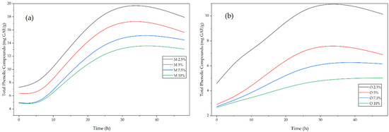
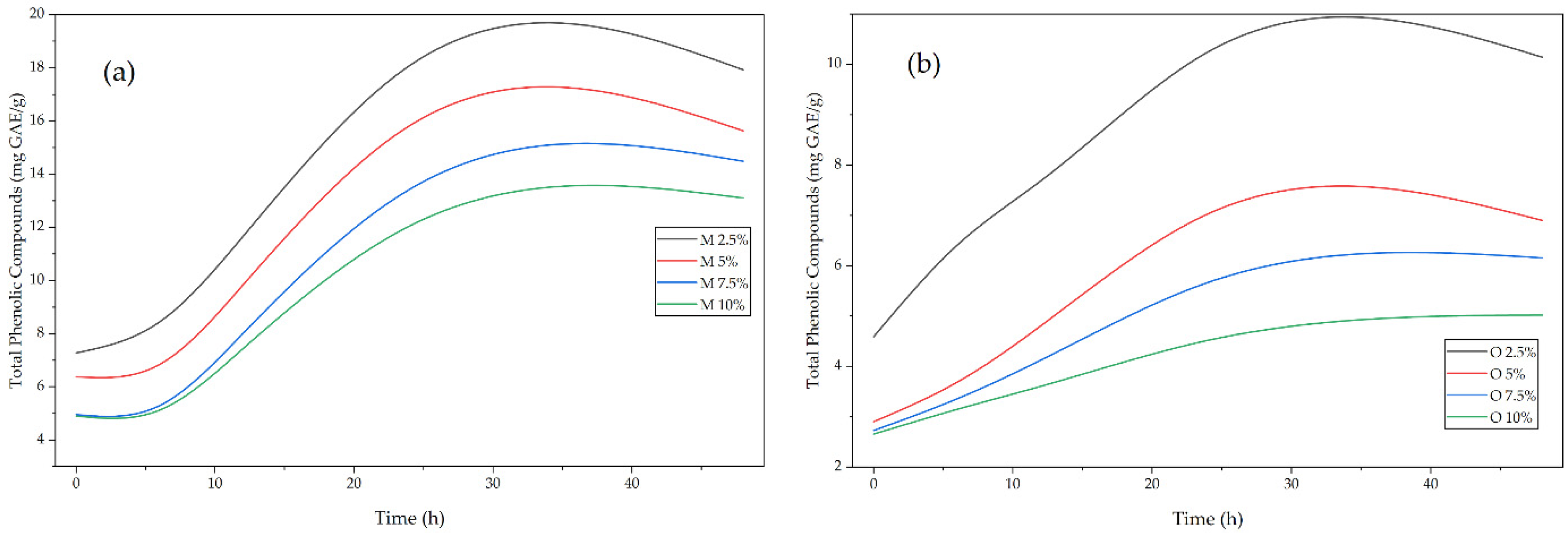
The release behavior of phenolic compounds in aqueous media is a key indicator of the functionality of microcapsules as controlled-release systems. In general, the release kinetics shown in Figure 2 reflect the interaction between the structure of the encapsulating matrix, the hydrophilicity of the wall material, and the distribution of the bioactive extract within the microcapsule.
Figure 2.
Release of phenolic compounds in aqueous medium from mashua and oca microcapsules in a flaxseed hydrocolloid matrix. (a) Release from mashua microcapsules; (b) release from oca microcapsules.
The release profile was biphasic, with a rapid initial stage followed by a sustained phase, controlled by diffusion and relaxation of the polysaccharide matrix [34,39]. The initial release is associated with the burst release of surface compounds, while the later stage occurs by diffusion through the hydrated network of the flaxseed hydrocolloid [5,46].
The concentration of the encapsulant modulated the kinetics: formulations with 2.5–5% facilitated release, while 7.5–10% generated denser matrices that increased diffusional resistance [5,46].The differences between mashua and oca are attributed to their phenolic profile, with faster release in mashua due to its higher content of water-soluble anthocyanins, consistent with studies on encapsulated tubers reporting maximum release between 24 and 48 h [5,34,53].
3.2.4. Release Kinetics Modeling of Encapsulated Phenolic Compounds
The kinetics of phenolic compound release from mashua and oca microcapsules were evaluated using the Higuchi and Korsmeyer–Peppas models. The kinetic parameters, including the Higuchi release constant (kH), the diffusion exponent (n), the kinetic constant (k), the coefficient of determination (R2), and the level of statistical significance (p-value), were obtained using linear regression. These results allow the effect of hydrocolloid concentration on the release behavior and diffusion mechanism of phenolic compounds to be analyzed. The Higuchi model parameters are summarized in Table 7, while the Korsmeyer–Peppas model parameters and corresponding release mechanisms are presented in Table 8.
Table 7.
Higuchi model parameters for phenolic compound release from Mashua and Oca microcapsules.
Table 8.
Korsmeyer–Peppas model parameters for phenolic compound release from Mashua and Oca microcapsules.
Kinetic modeling of the release of phenolic compounds showed that the process is strongly influenced by the concentration of the hydrocolloid and the nature of the plant core [55]. According to the Higuchi model, mashua microcapsules had higher release constants (kH) (1.365–1.809) than oca microcapsules (0.365–0.892), indicating lower diffusive resistance and higher mass transfer rates in the mashua matrix. This model confirms that release occurs predominantly by molecular diffusion proportional to the square root of time (t1/2), validated by statistical significance (p < 0.05) in most formulations [31].
The application of the Korsmeyer–Peppas model enabled discrimination of transport mechanisms based on the diffusion exponent (n). For mashua, the n values (0.389–0.510) showed a transition from Fickian diffusion to anomalous transport at high concentrations (7.5% and 10%), indicating that release depends on both diffusion and relaxation of the polymer chains [30,31]. Regarding oca, it showed lower n values (0.234–0.326), confirming a pure Fickian diffusion mechanism governed solely by concentration gradients [30,31].
The high coefficients of determination (R2 ≥ 0.816) confirm the robustness of these mathematical models for describing the release kinetics in matrix systems [31,32].
3.2.5. Colloidal Stability and Particle Size of Mashua and Oca Microcapsules
Table 9 shows the zeta potential (ζ), particle size (PS), and polydispersity index (Span) for different concentrations of the encapsulating material (2.5, 5.0, 7.5, and 10%). In the case of mashua microcapsules, the zeta potential values were consistently negative, indicating good electrostatic stability, while particle size decreased progressively as the wall material concentration increased. The polydispersity index remained relatively low, suggesting a homogeneous particle size distribution. For oca microcapsules, the zeta potential also showed negative values, though of lower magnitude than for mashua, indicating comparatively lower colloidal stability. Particle size varied with encapsulant concentration, and the polydispersity index indicated a moderately homogeneous distribution, with slight increases at higher wall material concentrations.
Table 9.
Zeta potential, particle size, and polydispersity of mashua and oca microcapsules.
The negative zeta potential observed in all formulations confirms the presence of anionic surface charges that promote electrostatic repulsion and colloidal stability in suspension [34]. The mashua microcapsules showed more negative values (up to −37.86 mV) than those of oca (−30.09 mV), suggesting superior electrostatic stability in the mashua system. In polysaccharide systems, absolute values >25–30 mV indicate robust resistance to aggregation and sedimentation [11,34]. This charge is associated with the ionization of functional groups (such as carboxyls) on the surface of the hydrocolloid matrix [34,56].
Particle size showed an inverse dependence on encapsulant concentration, especially in mashua, where it decreased from 10.50 to 4.02 µm [36]. This behavior indicates that a higher solids content in the feed favors the formation of smaller and more compact particles during atomization. The values obtained are within the typical range for microcapsules produced by spray drying (1–100 µm) [36,46]. The lower variability recorded in the goose suggests more uniform atomization, possibly influenced by the viscosity and rheological properties of the core [36,38].
Polydispersity remained at low levels, indicating a moderately homogeneous size distribution for both cores [34]. Span values close to 1 indicate narrow distributions, which is highly favorable for ensuring industrial reproducibility and uniformity in the final powder [11,34]. The slightly higher variability in mashua microcapsules at low concentrations is consistent with the lower viscosity of the feed system prior to drying [36]. Taken together, these parameters confirm the hydrocolloid’s ability to generate stable, physically uniform systems.
3.2.6. Structural Characterization
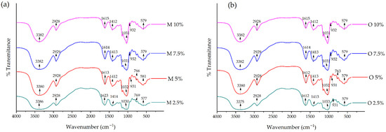
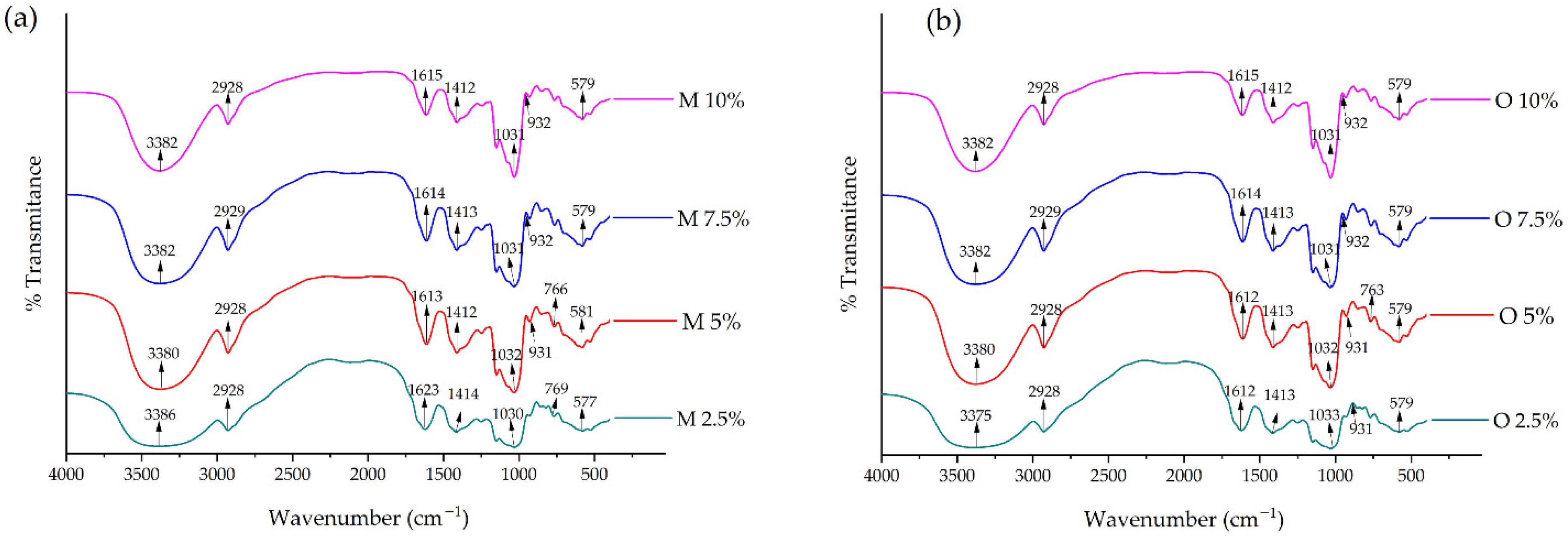
Figure 3 shows the FTIR spectra (% transmittance vs. wave number) of microcapsules prepared with different concentrations of encapsulating material for: (a) mashua microcapsules and (b) oca microcapsules.
Figure 3.
FTIR spectra (a) mashua microcapsules; (b) oca microcapsules.
The broad bands between 3380 and 3375 cm−1 are associated with the O–H stretching of polysaccharides and phenolic compounds, evidencing hydrogen bonds and the hydrophilic nature of the matrix. The slight broadening after microencapsulation indicates physical core–wall interactions without altering the chemical identity of the system [11,39,53].
The signals at 2928–2929 cm−1 correspond to aliphatic C–H bonds, whose stability confirms that spray drying did not induce significant thermal degradation [39,53]. The bands at 1623–1612 cm−1 and 1414–1412 cm−1 are attributed to carbonyl, carboxylate, and aromatic structures of polyphenols, confirming the preservation of phenolic acids and flavonoids within the matrix [19,39,56,57].
The intense bands at 1039–1032 cm−1, characteristic of C–O–C glycosidic bonds, confirm the predominance of polysaccharides and better coating of the core at higher wall concentrations, consistent with more controlled release [19,39]. Finally, signals between 932 and 579 cm−1 reflect structural vibrations of carbohydrates and confirm the integrity of the polysaccharide network after drying [11,39,51,58].
Taken together, the FTIR spectra show the coexistence of signals from the wall material and bioactive extracts without new bands, confirming non-covalent physical entrapment that preserves the structure and functionality of the encapsulated polyphenols [19,51,53,56].
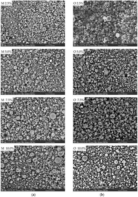
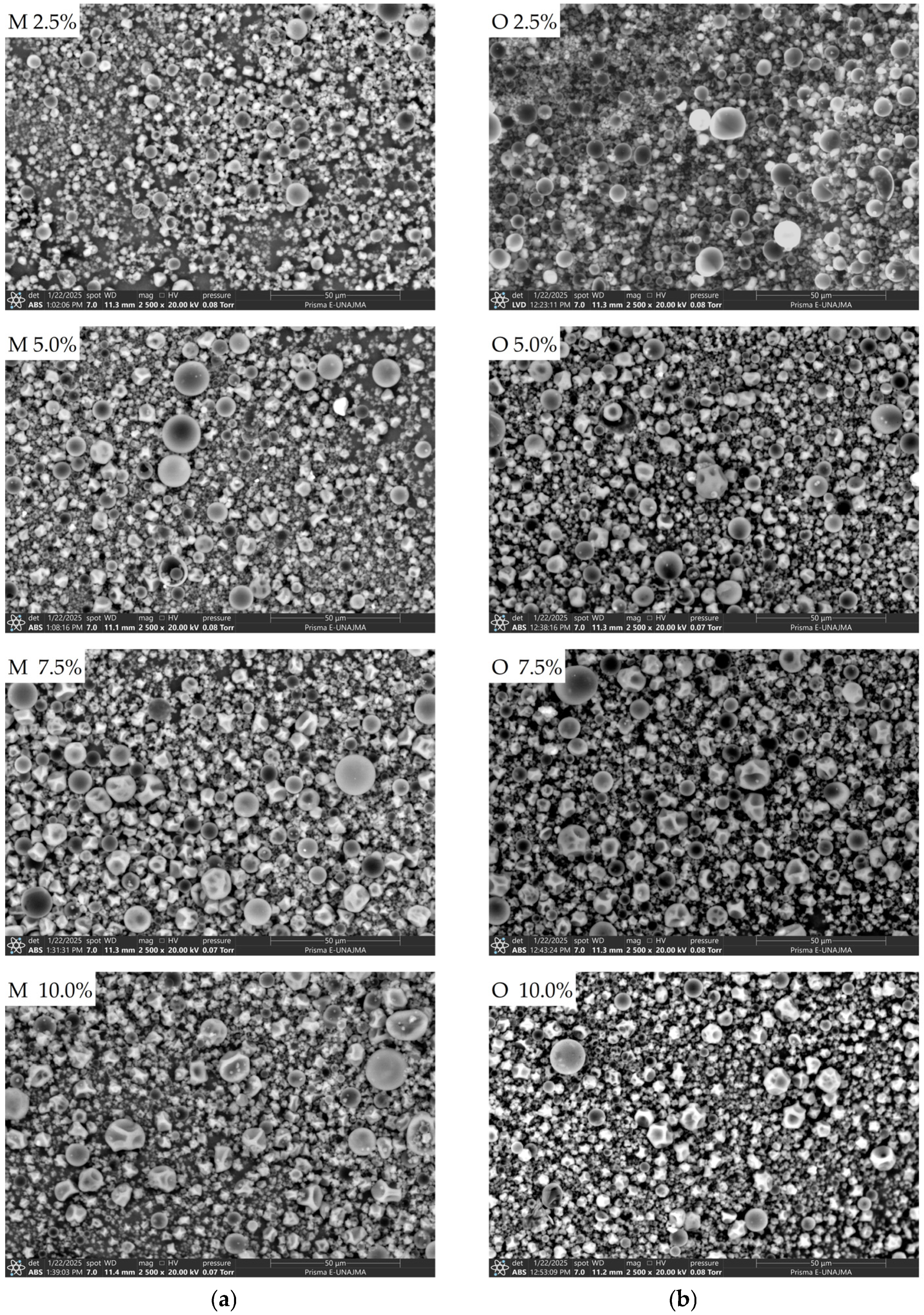
Figure 4 shows that, across all formulations, the morphology is dominated by particles with a predominantly spheroidal geometry, coexisting with smaller particles and agglomerates. In formulations with lower encapsulant concentrations (2.5–5.0%), there is greater size heterogeneity and a higher number of small particles adhering to larger particles. As the percentage of encapsulant increases (7.5–10.0%), there is a tendency toward more defined particles and a more compact distribution, although structures with depressions/dents and some particles with a collapsed appearance, typical of spray drying, persist.
Figure 4.
Surface morphology of mashua and oca microcapsules obtained by spray drying. SEM micrographs of microcapsules formulated with different concentrations of encapsulating material: (a) mashua (M 2.5, 5.0, 7.5, and 10.0%) and (b) oca (O 2.5, 5.0, 7.5, and 10.0%).
The morphology observed, characterized by predominantly spherical particles with depressions, partial collapse, and agglomeration, is consistent with the typical behavior of microencapsulated powders produced by spray drying [36,50]. This phenomenon occurs when rapid surface evaporation forms an outer “shell” or crust; depending on the internal vapor pressure and the elasticity of the matrix, smooth or wrinkled and dented (shrinkage) surfaces are generated during cooling [46,50].
The increase in wall material alters the viscosity of the feed and the formation of droplets during atomization, while also increasing the thickness of the interfacial film that solidifies [50]. The literature reports that higher concentrations of solids favor the formation of a more resistant, compact crust, which improves core retention and reduces porosity [36]. Likewise, if drying is intense and the wall material solidifies quickly, shrinkage dents persist due to the loss of internal pressure during cooling [1,36]. It should be noted that mostly spherical particles with diameters ranging from ~1–100 μm are common in microencapsulation with polysaccharides (such as maltodextrin, gums, or starch), with variations in the degree of agglomeration depending on the nature of the wall material [50].
The observations are consistent with this theoretical framework: the formulations show overall sphericity, but also exhibit dented microspheres and agglomeration of fines, typical of systems that use biopolymers and plant extracts [59]. The formation of bridges between particles is a common behavior in matrices rich in polysaccharides, which establish effective physical interactions [19,59].
The morphological differences observed between mashua and oca cores are due to intrinsic chemical variations that alter feed viscosity, the distribution of extracts within the atomized droplet, and crust formation kinetics [50]. In systems that integrate starches and gums, the profile of polyphenols, sugars, and salts in the extract directly modulates the degree of collapse, porosity, and tendency of particles to agglomerate [50,53].
4. Conclusions
This study demonstrated that flaxseed hydrocolloid (Linum usitatissimum L.) is an effective natural material for the spray-drying microencapsulation of phenolic compounds from mashua (Tropaeolum tuberosum Ruiz & Pav.) and oca (Oxalis tuberosa Molina). The hydrocolloid exhibited suitable physicochemical and technofunctional properties, allowing the formation of stable microcapsules with low moisture content, low water activity, high solubility, and adequate physical stability. The encapsulation efficiency confirms its ability to retain phenolic compounds.
The microcapsules retained levels of phenolic compounds, flavonoids, and antioxidant capacity comparable to those of the original material. Release studies revealed a biphasic release profile, and kinetic modeling using the Higuchi and Korsmeyer-Peppas models indicated that the release of phenolic compounds was mainly diffusion-controlled, with Fickian diffusion and anomalous transport. Particle size, zeta potential, and SEM analysis confirmed the formation of stable spheroidal microstructures, typical of spray drying.
A limitation of this study is that the release behavior was evaluated only under in vitro aqueous conditions, which may not fully represent real food systems or gastrointestinal environments. Future research should focus on evaluating release under simulated gastrointestinal conditions, assessing storage stability, and investigating the performance of these microcapsules when incorporated into functional food matrices.
Author Contributions
Conceptualization, E.M.-M. and D.B.-C.; methodology, D.B.-C., C.V.B.-A. and R.P.-S.; software, E.M.-M. and D.M.-H.; validation, D.B.-C.; formal analysis, E.M.-M. and C.V.B.-A.; investigation, E.M.-M., D.B.-C. and D.M.-H.; data curation, R.P.-S.; writing—original draft preparation, E.M.-M. and D.M.-H.; writing—review and editing, E.M.-M. and D.M.-H.; supervision, E.M.-M.; project administration, R.P.-S. All authors have read and agreed to the published version of the manuscript.
Funding
The Research Group on Nutraceuticals and Biomaterials of the UNAJMA supported the project under Resolution of the University Council No. 230-2024-UNAJMA/CU.
Data Availability Statement
The original contributions presented in this study are included in the article. Further inquiries can be directed to the corresponding author.
Acknowledgments
The authors acknowledge the Food Nanotechnology Research Laboratory of UNAJMA.
Conflicts of Interest
The authors declare no conflicts of interest.
References
- Samborska, K.; Boostani, S.; Geranpour, M.; Hosseini, H.; Dima, C.; Khoshnoudi-Nia, S.; Rostamabadi, H.; Falsafi, S.R.; Shaddel, R.; Akbari-Alavijeh, S.; et al. Green biopolymers from by-products as wall materials for spray drying microencapsulation of phytochemicals. Trends Food Sci. Technol. 2021, 108, 297–325. [Google Scholar] [CrossRef] [Scilit]
- Cicero, A.F.; Fogacci, F.; Colletti, A. Potential role of bioactive peptides in prevention and treatment of chronic diseases: A narrative review. Br. J. Pharmacol. 2017, 174, 1378–1394. [Google Scholar] [CrossRef] [Scilit] [PubMed]
- Acurio, L.; Salazar, D.; Castillo, B.; Santiana, C.; Martínez-Monzó, J.; Igual, M. Characterization of second-generation snacks manufactured from andean tubers and tuberous root flours. Foods 2023, 13, 51. [Google Scholar] [CrossRef] [Scilit]
- Arteaga-Cano, D.; Chacón-Calvo, L.; Samamé-Herrera, V.; Valverde-Cerna, D.; Paucar-Menacho, L.M. Mashua (Tropaeolum tuberosum): Composición nutricional, características químicas, compuestos bioactivos y propiedades beneficiosas para la salud. Agroindustrial Sci. 2022, 12, 95–101. [Google Scholar] [CrossRef] [Scilit]
- Choudhury, N.; Meghwal, M.; Das, K. Microencapsulation: An overview on concepts, methods, properties and applications in foods. Food Front. 2021, 2, 426–442. [Google Scholar] [CrossRef] [Scilit]
- Onwulata, C.I. Microencapsulation and functional bioactive foods. J. Food Process. Preserv. 2013, 37, 510–532. [Google Scholar] [CrossRef] [Scilit]
- Bisht, B.; Lohani, U.; Kumar, V.; Gururani, P.; Sinhmar, R. Edible hydrocolloids as sustainable substitute for non-biodegradable materials. Crit. Rev. Food Sci. Nutr. 2022, 62, 693–725. [Google Scholar] [CrossRef] [Scilit] [PubMed]
- Liu, J.; Shim, Y.Y.; Tse, T.J.; Wang, Y.; Reaney, M.J.T. Flaxseed gum a versatile natural hydrocolloid for food and non-food applications. Trends Food Sci. Technol. 2018, 75, 146–157. [Google Scholar] [CrossRef] [Scilit]
- Lorenc, F.; Jarošová, M.; Bedrníček, J.; Smetana, P.; Bárta, J. Structural Characterization and Functional Properties of Flaxseed Hydrocolloids and Their Application. Foods 2022, 11, 2304. [Google Scholar] [CrossRef] [Scilit]
- Cobbinah-Sam, E.; Ekaette, I.; Yu, Z.; Onyeaka, H.J.F.; Technology, B. Plant Seed Hydrocolloids: Extraction Methods and Techno-Functional Properties—A Review. Food Bioprocess Technol. 2025, 18, 6010–6034. [Google Scholar] [CrossRef] [Scilit]
- Moscoso-Moscoso, E.; Ligarda-Samanez, C.A.; Choque-Quispe, D.; Huamán-Carrión, M.L.; Arévalo-Quijano, J.C.; De la Cruz, G.; Luciano-Alipio, R.; Calsina Ponce, W.C.; Sucari-León, R.; Quispe-Quezada, U.R.; et al. Preliminary Assessment of Tara Gum as a Wall Material: Physicochemical, Structural, Thermal, and Rheological Analyses of Different Drying Methods. Polymers 2024, 16, 838. [Google Scholar] [CrossRef] [Scilit]
- Horwitz, W.J.A.I. Official Methods of Analysis of AOAC International. Volume I, Agricultural Chemicals, Contaminants, Drugs; Horwitz, W., Ed.; Aoac International: Rockville, ML, USA, 2010; Volume 1. [Google Scholar]
- Choque-Quispe, D.; Ligarda-Samanez, C.A.; Huamán-Rosales, E.R.; Aguirre Landa, J.P.; Agreda Cerna, H.W.; Zamalloa-Puma, M.M.; Álvarez-López, G.J.; Barboza-Palomino, G.I.; Alzamora-Flores, H.; Gamarra-Villanueva, W. Bioactive Compounds and Sensory Analysis of Freeze-Dried Prickly Pear Fruits from an Inter-Andean Valley in Peru. Molecules 2023, 28, 3862. [Google Scholar] [CrossRef] [Scilit]
- Ligarda-Samanez, C.A.; Choque-Quispe, D.; Moscoso-Moscoso, E.; Huamán-Carrión, M.L.; Ramos-Pacheco, B.S.; Peralta-Guevara, D.E.; Cruz, G.D.; Martínez-Huamán, E.L.; Arévalo-Quijano, J.C.; Muñoz-Saenz, J.C.; et al. Obtaining and Characterizing Andean Multi-Floral Propolis Nanoencapsulates in Polymeric Matrices. Foods 2022, 11, 3153. [Google Scholar] [CrossRef] [Scilit] [PubMed]
- Hu, Y.; Shim, Y.Y.; Reaney, M.J.T. Flaxseed Gum Solution Functional Properties. Foods 2020, 9, 681. [Google Scholar] [CrossRef] [Scilit] [PubMed]
- Ramos-Pacheco, B.S.; Choque-Quispe, D.; Ligarda-Samanez, C.A.; Solano-Reynoso, A.M.; Palomino-Rincón, H.; Choque-Quispe, Y.; Peralta-Guevara, D.E.; Moscoso-Moscoso, E.; Aiquipa-Pillaca, Á.S. Effect of Germination on the Physicochemical Properties, Functional Groups, Content of Bioactive Compounds, and Antioxidant Capacity of Different Varieties of Quinoa (Chenopodium quinoa Willd.) Grown in the High Andean Zone of Peru. Foods 2024, 13, 417. [Google Scholar] [CrossRef] [Scilit]
- Napiórkowska, A.; Szpicer, A.; Wojtasik-Kalinowska, I.; Perez, M.D.; González, H.D.; Kurek, M.A. Microencapsulation of Juniper and Black Pepper Essential Oil Using the Coacervation Method and Its Properties after Freeze-Drying. Foods 2023, 12, 4345. [Google Scholar] [CrossRef] [Scilit]
- Jedlińska, A.; Samborska, K.; Wieczorek, A.; Wiktor, A.; Ostrowska-Ligęza, E.; Jamróz, W.; Skwarczyńska-Maj, K.; Kiełczewski, D.; Błażowski, Ł.; Tułodziecki, M.; et al. The application of dehumidified air in rapeseed and honeydew honey spray drying—Process performance and powders properties considerations. J. Food Eng. 2019, 245, 80–87. [Google Scholar] [CrossRef] [Scilit]
- Ligarda-Samanez, C.A.; Choque-Quispe, D.; Moscoso-Moscoso, E.; Ramos-Pacheco, B.S.; Palomino-Rincón, H.; Peralta-Guevara, D.E. Microencapsulation of bioactive compounds from Hesperomeles escalloniifolia Schltdl (Capachu) in quinoa starch and Tara Gum. CyTA—J. Food 2025, 23, 2564354. [Google Scholar] [CrossRef] [Scilit]
- Ligarda-Samanez, C.A.; Choque-Quispe, D.; Moscoso-Moscoso, E.; Palomino-Rincón, H.; Taipe-Pardo, F.; Aguirre Landa, J.P.; Arévalo-Quijano, J.C.; Muñoz-Saenz, J.C.; Quispe-Quezada, U.R.; Huamán-Carrión, M.L.; et al. Nanoencapsulation of Phenolic Extracts from Native Potato Clones (Solanum tuberosum spp. andigena) by Spray Drying. Molecules 2023, 28, 4961. [Google Scholar] [CrossRef] [Scilit]
- Puligundla, P.; Lim, S. A Review of Extraction Techniques and Food Applications of Flaxseed Mucilage. Foods 2022, 11, 1677. [Google Scholar] [CrossRef] [Scilit]
- Barp, L.; Višnjevec, A.M.; Moret, S. Pressurized Liquid Extraction: A Powerful Tool to Implement Extraction and Purification of Food Contaminants. Foods 2023, 12, 2017. [Google Scholar] [CrossRef] [Scilit] [PubMed]
- Dias, M.I.; Ferreira, I.C.; Barreiro, M.F. Microencapsulation of bioactives for food applications. Food Funct. 2015, 6, 1035–1052. [Google Scholar] [CrossRef] [Scilit]
- Moreira, G.É.G.; Maia Costa, M.G.; Souza, A.C.R.d.; Brito, E.S.d.; Medeiros, M.d.F.D.d.; Azeredo, H.M.C.d. Physical properties of spray dried acerola pomace extract as affected by temperature and drying aids. LWT—Food Sci. Technol. 2009, 42, 641–645. [Google Scholar] [CrossRef] [Scilit]
- Ubeyitogullari, A.; Ciftci, O.N. Formation of nanoporous aerogels from wheat starch. Carbohydr. Polym. 2016, 147, 125–132. [Google Scholar] [CrossRef] [Scilit] [PubMed]
- Singleton, V.L.; Orthofer, R.; Lamuela-Raventós, R.M. Analysis of total phenols and other oxidation substrates and antioxidants by means of folin-ciocalteu reagent. Methods Enzymol. 1999, 299, 152–178. [Google Scholar]
- Popova, M.; Bankova, V.; Butovska, D.; Petkov, V.; Nikolova-Damyanova, B.; Sabatini, A.G.; Marcazzan, G.L.; Bogdanov, S. Validated methods for the quantification of biologically active constituents of poplar-type propolis. Phytochem. Anal. Int. J. Plant Chem. Biochem. Tech. 2004, 15, 235–240. [Google Scholar] [CrossRef] [Scilit]
- Ibrahim, M.E.E.-D.; Alqurashi, R.M. Anti-fungal and antioxidant properties of propolis (bee glue) extracts. Int. J. Food Microbiol. 2022, 361, 109463. [Google Scholar] [CrossRef] [Scilit]
- Tomé, A.C.; da Silva, F.A. Alginate based encapsulation as a tool for the protection of bioactive compounds from aromatic herbs. Food Hydrocoll. Health 2022, 2, 100051. [Google Scholar] [CrossRef] [Scilit]
- Costa, P.; Sousa Lobo, J.M. Modeling and comparison of dissolution profiles. Eur. J. Pharm. Sci. 2001, 13, 123–133. [Google Scholar] [CrossRef] [Scilit]
- Yeh, Y.-M.; Tsai, T.-Y.; Yang, C.-Y. Encapsulation and release kinetics of polyphenols and p-coumaric acid extracted from Phyllostachys makinoi by ultrasonic-pretreatment autoclaving. LWT 2022, 167, 113838. [Google Scholar] [CrossRef] [Scilit]
- Sadoun, U.A.; Al-Ali, Z.S.; Haddad, A.M. Extraction of phenolic compounds from Iraqi Coriandrum Sativum L. and loaded on copolymeric hydrogels and examine there as drug delivery system and antioxidant. J. Phys. Conf. Ser. 2021, 2063, 012001. [Google Scholar] [CrossRef] [Scilit]
- Schramm, L.L. Emulsions, Foams, Suspensions, and Aerosols: Microscience and Applications; John Wiley & Sons: Hoboken, NJ, USA, 2014. [Google Scholar]
- Nwabor, O.F.; Singh, S.; Marlina, D.; Voravuthikunchai, S.P. Chemical characterization, release, and bioactivity of Eucalyptus camaldulensis polyphenols from freeze-dried sodium alginate and sodium carboxymethyl cellulose matrix. Food Qual. Saf. 2020, 4, 203–212. [Google Scholar] [CrossRef] [Scilit]
- Ligarda-Samanez, C.A.; Ccana-Buleje, T.G.; Choque-Quispe, D.; Palomino-Rincón, H.; Taipe-Pardo, F.; Moscoso-Moscoso, E.; Muñoz-Melgarejo, M.; Luciano-Alipio, R.; Carrión, J.C.; Muñoz-Saenz, J.C.; et al. Spray-Dried Porcine Collagen Microcapsules in Tara Gum–Maltodextrin Matrices: A Sustainable Approach to By-Product Valorization for Functional and Nutraceutical Applications. Appl. Sci. 2025, 15, 12667. [Google Scholar] [CrossRef] [Scilit]
- Domian, E.; Brynda-Kopytowska, A.; Marzec, A. Functional properties and oxidative stability of flaxseed oil microencapsulated by spray drying using legume proteins in combination with soluble fiber or trehalose. Food Bioprocess Technol. 2017, 10, 1374–1386. [Google Scholar] [CrossRef] [Scilit]
- Drozłowska, E.; Bartkowiak, A.; Trocer, P.; Kostek, M.; Tarnowiecka-Kuca, A.; Łopusiewicz, Ł. Formulation and Evaluation of Spray-Dried Reconstituted Flaxseed Oil-in-Water Emulsions Based on Flaxseed Oil Cake Extract as Emulsifying and Stabilizing Agent. Foods 2021, 10, 256. [Google Scholar] [CrossRef] [Scilit]
- León-Martínez, F.M.; Méndez-Lagunas, L.L.; Rodríguez-Ramírez, J. Spray drying of nopal mucilage (Opuntia ficus-indica): Effects on powder properties and characterization. Carbohydr. Polym. 2010, 81, 864–870. [Google Scholar] [CrossRef] [Scilit]
- Tosif, M.M.; Najda, A.; Bains, A.; Kaushik, R.; Dhull, S.B.; Chawla, P.; Walasek-Janusz, M. A Comprehensive Review on Plant-Derived Mucilage: Characterization, Functional Properties, Applications, and Its Utilization for Nanocarrier Fabrication. Polymers 2021, 13, 1066. [Google Scholar] [CrossRef] [Scilit]
- Haseeb, M.T.; Muhammad, G.; Hussain, M.A.; Bukhari, S.N.A.; Sheikh, F.A. Flaxseed (Linum usitatissimum) mucilage: A versatile stimuli–responsive functional biomaterial for pharmaceuticals and healthcare. Int. J. Biol. Macromol. 2024, 278, 134817. [Google Scholar] [CrossRef] [Scilit]
- Hovjecki, M.; Radovanovic, M.; Levic, S.M.; Mirkovic, M.; Peric, I.; Miloradovic, Z.; Jurina, I.B.; Miocinovic, J. Chia Seed Mucilage as a Functional Ingredient to Improve Quality of Goat Milk Yoghurt: Effects on Rheology, Texture, Microstructure and Sensory Properties. Fermentation 2024, 10, 382. [Google Scholar] [CrossRef] [Scilit]
- Gao, X.; Pourramezan, H.; Ramezan, Y.; Roy, S.; Zhang, W.; Assadpour, E.; Zou, J.; Jafari, S.M. Application of gums as techno-functional hydrocolloids in meat processing and preservation: A review. Int. J. Biol. Macromol. 2024, 268, 131614. [Google Scholar] [CrossRef] [Scilit] [PubMed]
- Goksen, G.; Demir, D.; Dhama, K.; Kumar, M.; Shao, P.; Xie, F.; Echegaray, N.; Lorenzo, J.M. Mucilage polysaccharide as a plant secretion: Potential trends in food and biomedical applications. Int. J. Biol. Macromol. 2023, 230, 123146. [Google Scholar] [CrossRef] [Scilit]
- Kaushik, P.; Dowling, K.; Adhikari, R.; Barrow, C.J.; Adhikari, B. Effect of extraction temperature on composition, structure and functional properties of flaxseed gum. Food Chem. 2017, 215, 333–340. [Google Scholar] [CrossRef] [Scilit]
- Rios-Aguirre, S.; Gil-Garzon, M.A. Microencapsulación por secado por aspersión de compuestos bioactivos en diversas matrices: Una revisión. TecnoLógicas 2021, 24, 206–229. [Google Scholar] [CrossRef] [Scilit]
- Pudžiuvelytė, L.; Petrauskaitė, E.; Stabrauskienė, J.; Bernatonienė, J. Spray-Drying Microencapsulation of Natural Bioactives: Advances in Sustainable Wall Materials. Pharmaceuticals 2025, 18, 963. [Google Scholar] [CrossRef] [Scilit] [PubMed]
- Akcicek, A.; Bozkurt, F.; Akgül, C.; Karasu, S. Encapsulation of Olive Pomace Extract in Rocket Seed Gum and Chia Seed Gum Nanoparticles: Characterization, Antioxidant Activity and Oxidative Stability. Foods 2021, 10, 1735. [Google Scholar] [CrossRef] [Scilit] [PubMed]
- Bashir, M.; Haripriya, S. Assessment of physical and structural characteristics of almond gum. Int. J. Biol. Macromol. 2016, 93, 476–482. [Google Scholar] [CrossRef] [Scilit]
- Yahoum, M.M.; Toumi, S.; Tahraoui, H.; Lefnaoui, S.; Kebir, M.; Amrane, A.; Assadi, A.A.; Zhang, J.; Mouni, L. Formulation and Evaluation of Xanthan Gum Microspheres for the Sustained Release of Metformin Hydrochloride. Micromachines 2023, 14, 609. [Google Scholar] [CrossRef] [Scilit]
- Mohammadalinejhad, S.; Kurek, M.A. Microencapsulation of Anthocyanins—Critical Review of Techniques and Wall Materials. Appl. Sci. 2021, 11, 3936. [Google Scholar] [CrossRef] [Scilit]
- de Souza, V.B.; Thomazini, M.; Balieiro, J.C.d.C.; Fávaro-Trindade, C.S. Effect of spray drying on the physicochemical properties and color stability of the powdered pigment obtained from vinification byproducts of the Bordo grape (Vitis labrusca). Food Bioprod. Process. 2015, 93, 39–50. [Google Scholar] [CrossRef] [Scilit]
- Ramakrishnan, Y.; Adzahan, N.M.; Yusof, Y.A.; Muhammad, K. Effect of wall materials on the spray drying efficiency, powder properties and stability of bioactive compounds in tamarillo juice microencapsulation. Powder Technol. 2018, 328, 406–414. [Google Scholar] [CrossRef] [Scilit]
- Legesse, A.B.; Emire, S.A.; Oyinloye, T.M.; Yoon, W.B. Microencapsulation of Phenolic Extracts from Verbascum sinaiticum Leaf Using Maltodextrin and Gum Arabic: Physicochemical Properties, Encapsulation Efficiency, and Storage Stability. Molecules 2026, 31, 471. [Google Scholar] [CrossRef] [Scilit]
- Aiquipa Pillaca, Á.S. Efecto de temperatura de secado y concentración de almidón de quinua (Chenopodium quinoa Willd) en la microencapsulación de antocianinas de mashua negra (Tropaeolum tuberosum). Master’s Thesis, Universidad Nacional José María Arguedas, Andahuaylas, Perú, 2023. [Google Scholar]
- Jokubaite, M.; Marksa, M.; Ramanauskiene, K. Application of Poloxamer for In Situ Eye Drop Modeling by Enrichment with Propolis and Balsam Poplar Buds Phenolic Compounds. Gels 2024, 10, 161. [Google Scholar] [CrossRef] [Scilit]
- Montoya Yepes, D.F.; Murillo Arango, W.; Jiménez Rodríguez, Á.A.; Méndez Arteaga, J.J.; Aldana Porras, Á.E. Encapsulation of phenols of gulupa seed extract using acylated rice starch: Effect on the release and antioxidant activity. J. Funct. Foods 2021, 87, 104788. [Google Scholar] [CrossRef] [Scilit]
- Franca, J.R.; De Luca, M.P.; Ribeiro, T.G.; Castilho, R.O.; Moreira, A.N.; Santos, V.R.; Faraco, A.A. Propolis-based chitosan varnish: Drug delivery, controlled release and antimicrobial activity against oral pathogen bacteria. BMC Complement. Altern. Med. 2014, 14, 478. [Google Scholar] [CrossRef] [Scilit] [PubMed]
- Sousa, C.M.d.M.; Silva, H.R.; Vieira-Jr, G.M.; Ayres, M.C.C.; Costa, C.L.S.d.; Araújo, D.S.; Cavalcante, L.C.D.; Barros, E.D.S.; Araújo, P.B.d.M.; Brandão, M.S. Fenóis totais e atividade antioxidante de cinco plantas medicinais. Química Nova 2007, 30, 351–355. [Google Scholar] [CrossRef] [Scilit]
- Lourenço, S.C.; Moldão-Martins, M.; Alves, V.D. Microencapsulation of Pineapple Peel Extract by Spray Drying Using Maltodextrin, Inulin, and Arabic Gum as Wall Matrices. Foods 2020, 9, 718. [Google Scholar] [CrossRef] [Scilit] [PubMed]

Disclaimer/Publisher’s Note: The statements, opinions and data contained in all publications are solely those of the individual author(s) and contributor(s) and not of MDPI and/or the editor(s). MDPI and/or the editor(s) disclaim responsibility for any injury to people or property resulting from any ideas, methods, instructions or products referred to in the content. |
© 2026 by the authors. Licensee MDPI, Basel, Switzerland. This article is an open access article distributed under the terms and conditions of the Creative Commons Attribution (CC BY) license.