


Abstract
This study compared filling characteristics and apical dye leakage during sealer-only obturation with a bioactive glass-based sealer using three rotary instruments. Straight and 30° curved simulated acrylic canals (apical diameter, 0.25 mm; taper, 0.07) were evaluated using an NT condenser, JIZAI, or a stainless steel (SS) prototype condenser (n = 15 per group per canal model). Sealer condensation was performed at 500 rpm with four up-and-down strokes. Unfilled areas were measured 3, 6, and 9 mm coronal to the working-length endpoint, which was set 2 mm short of the apical end of each model (equivalent to 5, 8, and 11 mm from the apical end). Apical dye penetration was assessed after 48 h in 2% methylene blue. Kruskal–Wallis and Steel–Dwass tests were used. In straight canals, the SS prototype showed the smallest apical unfilled area, whereas JIZAI and the SS prototype showed less dye penetration than NT. In curved canals, JIZAI showed smaller apical and middle unfilled areas and less dye penetration than the other instruments. Filling characteristics and dye penetration differed by instrument and canal geometry; three-dimensional evaluation is needed to establish overall sealing performance.
1. Introduction
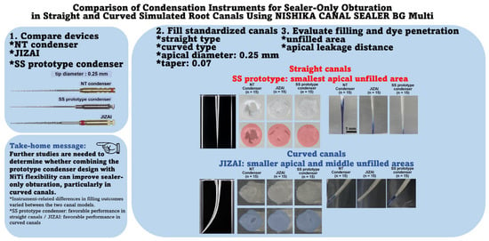
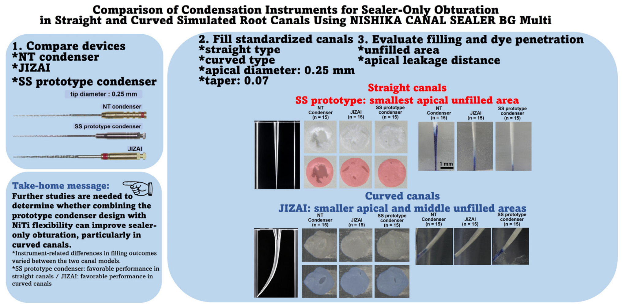
The primary objective of root canal obturation, the final step in endodontic therapy, is to achieve a three-dimensional, hermetic seal of the cleaned and shaped root canal system, thereby preventing bacterial re-entry and the colonization of microorganisms that could compromise periapical tissue health [1]. For decades, the standard clinical protocol based on Grossman’s principles has combined an insoluble solid core material, such as gutta-percha, with a root canal sealer to fill residual space and discrepancies [2]. However, recent advancements in sealer technology have introduced high-performance materials—including bioactive glass-based sealers such as NISHIKA CANAL SEALER BG multi (Nippon Shika Yakuhin Co., Ltd., Shimonoseki, Japan; BG sealer)—which exhibit apatite-forming ability, slight setting expansion, excellent biocompatibility, and strong sealing capabilities, resulting in clinical utility [3,4,5,6]. These developments have facilitated the adoption of single-cone techniques and have prompted investigation of the possibility of sealer-only obturation, in which solid core materials might be eliminated entirely in the future [7,8,9,10,11]. Sealer-only obturation eliminates the need for mechanical or thermal compaction, potentially reducing compaction-related stresses on the root, while simplified obturation techniques may also reduce procedure time [3,9,10,11]. Nevertheless, a major challenge of this approach is the potential for air void entrapment during sealer placement, which can result in apical microleakage [10,11]. Conventional sealer-delivery methods, including syringe-based application and Lentulo spiral placement, may be associated with void formation; syringe-based systems also require disposable applicator tips, generating additional plastic waste [10,11,12]. To address these limitations, our research group designed a stainless steel (SS) prototype condenser featuring specific geometric parameters (11° pitch angle, 22 pitches, 0.02 taper) to effectively apply and condense sealers against canal walls [10,13]. In clinical practice, clinicians frequently encounter curved root canals in addition to straight canals. How instrument material properties (stainless steel vs. nickel–titanium alloy) and blade designs interact with root canal anatomy (straight vs. curved) during BG sealer condensation remains to be fully elucidated. Therefore, the objective of this study was to evaluate and compare the filling characteristics, as assessed by unfilled areas, and apical dye-leakage distances of the SS prototype condenser, NT condenser (NiTi), and JIZAI during sealer-only obturation with BG sealer in standardized straight and curved acrylic root canal models. The null hypothesis was that there would be no significant differences among the three rotary instruments in unfilled areas at the apical, middle, and coronal levels or in apical dye-leakage distances within either the straight or curved canal model. The overall experimental design of the present study is summarized in Figure 1.
Figure 1.
Overview of the experimental design. Three rotary condensation instruments—the NT condenser, JIZAI, and stainless steel (SS) prototype condenser—were evaluated for sealer-only root canal obturation using NISHIKA CANAL SEALER BG multi. Standardized straight and curved simulated root canals with an apical diameter of 0.25 mm and a 0.07 taper were used. Filling and sealing performance was evaluated by measuring unfilled areas and apical dye leakage distances.
2. Materials and Methods
2.1. Sample Size Calculation
Prior to the experiments, sample size estimation was performed using G*Power (version 3.1), informed by our previous investigation [10]. Using a three-group one-way ANOVA framework, α = 0.05, target power = 0.80, and an assumed Cohen’s f = 0.5, the original calculation was reported to require 14 specimens per group. We therefore included 15 specimens per group for each canal model. This original ANOVA-based planning approach did not directly correspond to the nonparametric statistical analysis ultimately used.
The prospectively specified primary outcome was the unfilled area at the apical level, 3 mm coronal to the working-length endpoint (5 mm from the original apical end of the model). Secondary outcomes were unfilled areas at the middle and coronal levels and apical dye-leakage distance.
A supplementary retrospective simulation-based assessment was subsequently conducted using apical unfilled-area measurements from the independent previous study [10] (n = 14 per group). For each assumed distribution scenario, 12,000 empirical resampling replicates were evaluated with the three-group Kruskal–Wallis test (α = 0.05). Sensitivity scenarios progressively mixed group-specific distributions with the pooled distribution. With n = 15 per group, estimated power was 100.0% with the original empirical distributions, 99.3% with a 25% pooled-distribution mixture, and 74.1% with a 50% mixture. This retrospective assessment was not part of the original prospective sample size calculation, and its results were sensitive to the assumed group differences and the use of a different sealer in the previous study. OpenAI ChatGPT was used to assist with the preparation and refinement of computational code for the supplementary simulation-based power analysis. The authors take responsibility for the reported methods and results.
2.2. Root Canal Sealer
A bioactive glass-based root canal sealer, BG sealer, was used in this study. The sealer was manually mixed with a spatula on a mixing pad in accordance with the manufacturer’s instructions before application to the rotary instruments. The mixing duration was not recorded.
2.3. Straight and Curved Simulated Root Canal Models
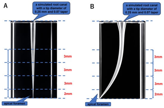
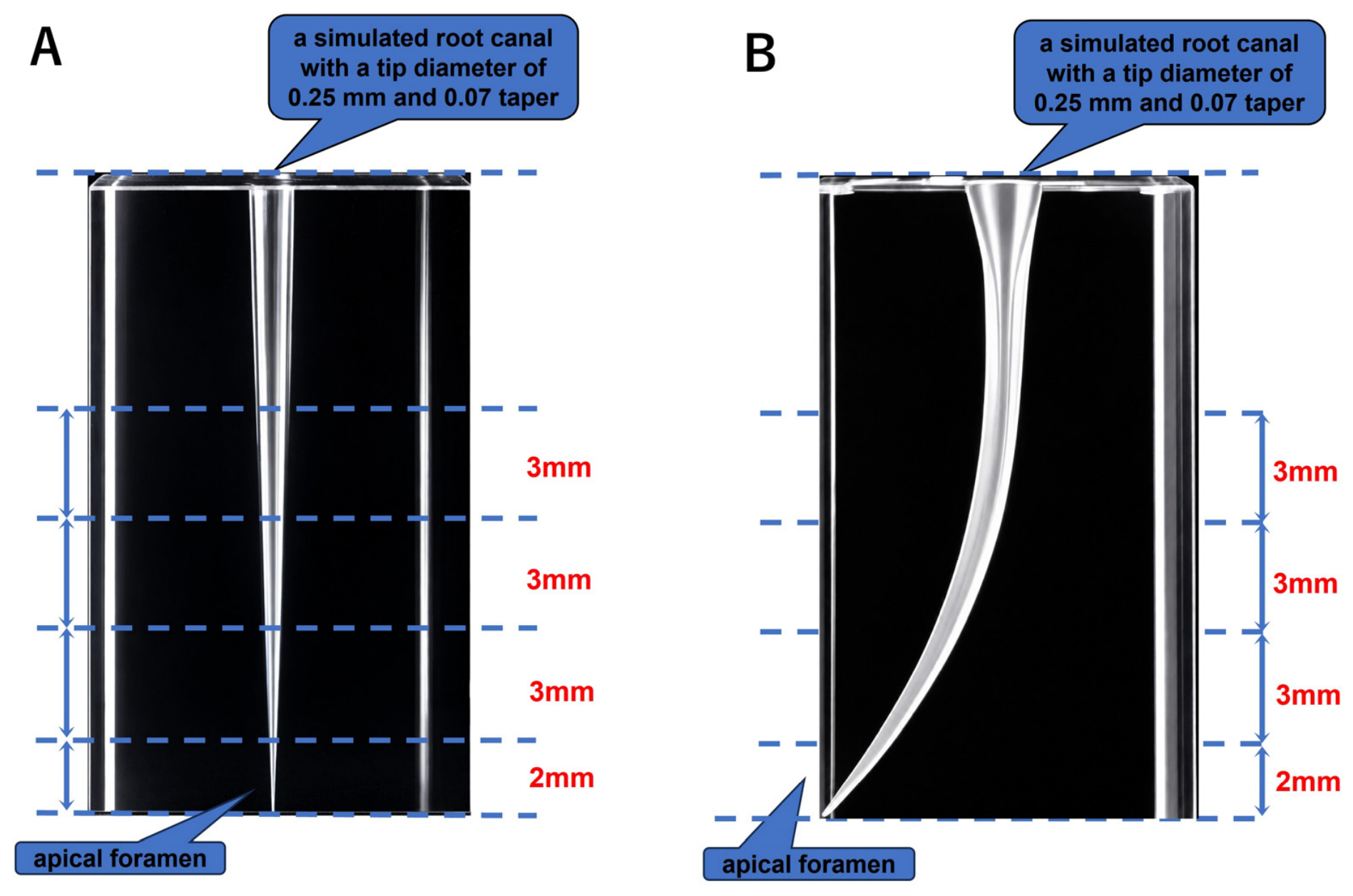
Two types of transparent acrylic simulated root canal models (Nissin Dental Products, Kyoto, Japan) were used: a custom-fabricated straight canal model and a commercially available curved canal model (Figure 2A,B). Because the overall lengths of the straight and curved canal models differed, the working length (WL) was defined relative to the apical end of each model, 2.0 mm short of the original apical end, rather than as a fixed length.
Figure 2.
Schematic illustration of the straight and curved simulated root canal models. (A) Straight simulated root canal model. (B) Curved simulated root canal model. Both models were standardized to an apical diameter of 0.25 mm and a 0.07 taper. The locations corresponding to the apical, middle, and coronal evaluation levels are indicated.
For the straight canal model, a custom-fabricated transparent acrylic model (S1-NC.56; Nissin Dental Products) was used. The simulated canal had an apical diameter of 0.25 mm at the working-length endpoint and a constant taper of 0.07. A simulated apical foramen with a diameter of 0.15 mm was located at the original apical end of the model, and the working-length endpoint was positioned 2.0 mm coronal to it. These dimensions corresponded to those used for the standardized straight canal model in our previous study [10].
For the curved canal model, a commercially available endodontic training block (END1002-30 #20; Nissin Dental Products) containing a size #20 simulated canal with a standardized curvature angle of 30° was used. To standardize the experimental conditions with those of the straight canal model, the working length was established at 2.0 mm short of the original apical end of the model. The curved canal was then enlarged using an RE FILE ONE nickel–titanium rotary instrument (#25/.07; YOSHIDA, Tokyo, Japan) to obtain a final apical preparation size of 0.25 mm with a taper of 0.07. Although the initial canal sizes differed between the straight and curved canal models, both models were prepared to achieve the same final apical preparation size and taper. Therefore, statistical comparisons were performed among the three instruments within each canal model, rather than directly between the straight and curved canal models.
2.4. Rotary Instruments Used for Obturation
Three rotary instruments, each with a tip diameter of 0.25 mm, were evaluated:
- NT condenser: Microseal condenser (Kerr Corp., Orange, CA, USA), manufactured from nickel–titanium (NiTi), with a tip diameter of 0.25 mm;
- JIZAI: A heat-treated nickel–titanium (NiTi) alloy rotary instrument (MANI INC., Utsunomiya, Japan) with a tip diameter of 0.25 mm;
- SS prototype condenser: A prototype stainless steel (SS) rotary condenser with a tip diameter of 0.25 mm, a constant taper of 0.02, a pitch angle of 11°, and a pitch count of 22, co-developed with MANI INC.
The SS prototype condenser was co-developed by our research group and MANI Inc. (Utsunomiya, Japan). MANI Inc. fabricated and supplied the experimental instruments free of charge. The company’s involvement was limited to instrument fabrication and provision; it did not participate in the experimental procedures, data analysis, or manuscript preparation. The dimensions of the prototype condenser were identical to those used in our previous study [10,13].
Although JIZAI is primarily designed for root canal shaping, JIZAI #25/.04 was selected as a comparative NiTi rotary instrument to investigate the potential application of a flexible shaping instrument for sealer transportation. Its off-center modified rectangular cross-section with radial lands and nonuniform flute pitch configuration may influence sealer distribution. The NT condenser (#25/.02) has a reverse-helix design, whereas the SS prototype condenser (#25/.02) has a custom spiral configuration with an 11° pitch angle and 22 pitches. JIZAI was also included in our previous study [10], allowing consistency with the established experimental protocol. Its use for sealer placement in this study was exclusively experimental and should not be interpreted as a recommendation for clinical application. The characteristics of the three instruments are summarized in Table 1.
Table 1.
Specifications and rotational directions of the three rotary instruments used for sealer-only root canal obturation.
2.5. Obturation Procedure
Each simulated root canal model was positioned vertically and securely fixed in a vise. A 16:1 reduction contra-angle handpiece connected to an electric motor (ATR Tecnika Vision Motor, Dentsply Maillefer, Tulsa, OK, USA) was used. The mixed BG sealer was uniformly applied to the entire working portion of each instrument while the instrument was rotated at 200 rpm. However, the amount of sealer applied was not quantitatively standardized by weight or volume.
The instrument was then inserted into the simulated canal to a depth of 1.5 mm short of the WL. Sealer condensation was performed at 500 rpm using four up-and-down strokes, without exceeding this insertion depth. The SS prototype condenser and NT condenser were operated in forward rotation, whereas JIZAI was operated in reverse rotation. The rotational directions were selected to maintain consistency with the experimental conditions employed in our previous study [10]. Therefore, the present experimental design compared instrument–operation combinations rather than isolating the independent effect of rotational direction. This procedure was based on that used in our previous studies [10,13].
All obturation procedures were performed by a single operator (T.S.) to minimize inter-operator variability. For each canal model, specimens were randomly allocated to one of the three instrument groups (NT condenser, JIZAI, or SS prototype condenser), with 15 specimens assigned to each group.
2.6. Measurement of Unfilled Area
Following obturation, the specimens were stored at 37 °C under humid conditions for 1 week to allow the sealer to set. Both straight and curved canal models were horizontally sectioned at three standardized levels measured from the original apical end of the simulated canal: 5.0, 8.0, and 11.0 mm, corresponding to 3.0 mm (apical level), 6.0 mm (middle level), and 9.0 mm (coronal level) from the established working-length endpoint, respectively. Sectioning was performed using a slow-speed precision saw equipped with a diamond cutting disk under continuous water cooling. Each cross-section was examined under a stereomicroscope (SMZ18; Nikon, Tokyo, Japan) equipped with a digital camera. The unfilled area within each canal cross-section was quantified in square millimeters using image-analysis software (SigmaScan Pro 5.0; Hulinks, Tokyo, Japan). The sectioning levels and image-analysis procedure were based on those used in our previous study [10,13]. All unfilled-area measurements were performed by the same examiner (T.S.), who was aware of the instrument group assignments. No formal intra-examiner or inter-examiner reliability assessment was conducted.
2.7. Apical Dye-Leakage Test
Apical sealing was evaluated using a dye-penetration test. The apical portions of the obturated specimens were immersed in a 2% methylene blue aqueous solution for 48 h and subsequently rinsed with distilled water. The specimens were longitudinally sectioned along the canal axis under continuous water cooling. The distance of methylene blue penetration from the original apical end of the model (simulated apical foramen) toward the coronal direction was measured in millimeters under a stereomicroscope using image-analysis software (SigmaScan Pro 5.0; Hulinks, Tokyo, Japan). This procedure was based on that used in our previous study [13]. All apical dye-leakage measurements were performed by the same examiner (T.S.), without blinding to the instrument group assignments. No formal measurement reliability assessment was performed. No positive or negative control groups were included in the dye-leakage experiment.
2.8. Statistical Analysis
The normality of the data distribution was assessed using the Shapiro–Wilk test. Because the data did not satisfy the assumption of normality, nonparametric statistical analyses were performed. Differences among the three instrument groups were analyzed using the Kruskal–Wallis test. When a significant overall difference was detected, the Steel–Dwass post hoc test was performed for pairwise comparisons using KyPlot 6.0 (KyensLab Inc., Tokyo, Japan). Statistical significance was set at p < 0.05.
3. Results
The filling and sealing performance of the three instruments in the straight and curved canal models is summarized in Table 2. For each instrument group, unfilled areas at the apical, middle, and coronal levels and apical dye-leakage distances are presented as medians and interquartile ranges (Q1–Q3). The corresponding p-values from the Kruskal–Wallis test and Steel–Dwass post hoc comparisons are also provided. Statistical comparisons were performed separately within each canal model.
Table 2.
Filling and sealing outcomes by canal model.
3.1. Evaluation in the Straight Canal Model
3.1.1. Unfilled Area
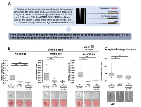
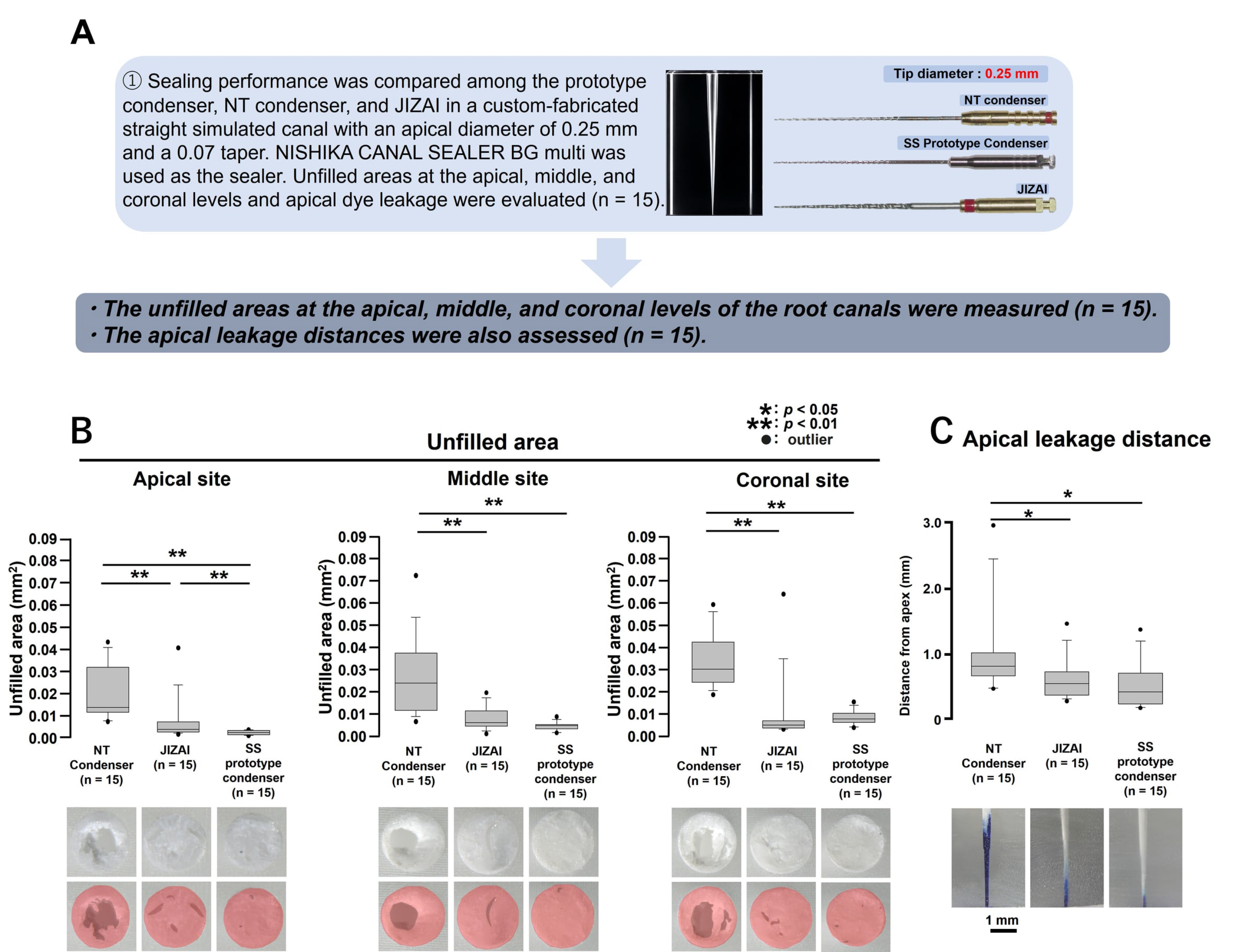
In the straight canal model (Figure 3A), significant differences in unfilled area were observed among all three groups at the apical level (Figure 3B; p < 0.01), with the SS prototype condenser showing the smallest unfilled area. At the middle and coronal levels, both the JIZAI and SS prototype condenser groups showed significantly smaller unfilled areas than the NT condenser group (Figure 3B; p < 0.01), with no significant difference between JIZAI and the SS prototype condenser (p ≥ 0.05).
Figure 3.
Filling and sealing performance of the three rotary condensation instruments in the straight simulated root canal model. (A) Experimental overview and representative images of the NT condenser, JIZAI, and SS prototype condenser used for sealer-only obturation. (B) Unfilled areas measured at the apical, middle, and coronal levels. (C) Apical dye leakage distances. Data were compared among the three instrument groups (n = 15 per group). * p < 0.05; ** p < 0.01. Filled circles indicate outliers.
3.1.2. Apical Dye Leakage
The NT condenser group showed significantly longer apical dye leakage distances than both the JIZAI and SS prototype condenser groups (Figure 3C; p < 0.05). No significant difference was observed between the JIZAI and SS prototype condenser groups (p ≥ 0.05).
3.2. Evaluation in the Curved Canal Model
3.2.1. Unfilled Area
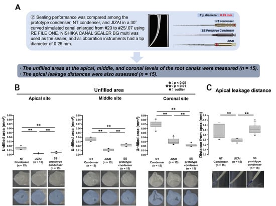
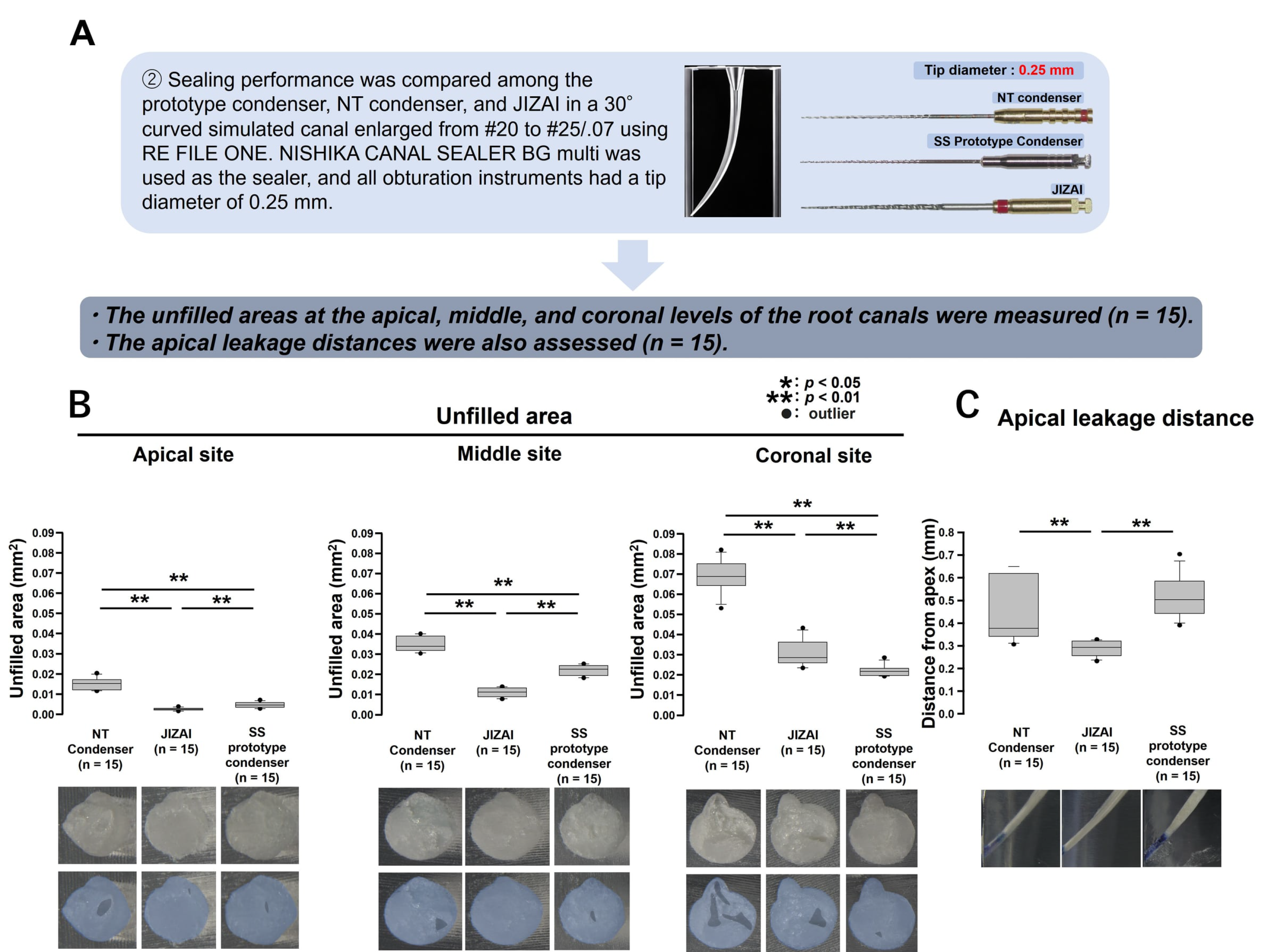
In the curved canal model (Figure 4A), the JIZAI group showed significantly smaller unfilled areas than the NT condenser and SS prototype condenser groups at the apical and middle levels (Figure 4B; p < 0.01). In contrast, at the coronal level, the SS prototype condenser group showed a significantly smaller unfilled area than the NT condenser and JIZAI groups (Figure 4B; p < 0.01).
Figure 4.
Filling and sealing performance of the three rotary condensation instruments in the curved simulated root canal model. (A) Experimental overview and representative images of the NT condenser, JIZAI, and SS prototype condenser used for sealer-only obturation in a 30° curved simulated canal enlarged to #25/.07. (B) Unfilled areas measured at the apical, middle, and coronal levels. (C) Apical dye leakage distances. Data were compared among the three instrument groups (n = 15 per group). * p < 0.05; ** p < 0.01. Filled circles indicate outliers.
3.2.2. Apical Dye Leakage
The JIZAI group showed significantly shorter apical dye leakage distances than both the NT condenser and SS prototype condenser groups (Figure 4C; p < 0.01), whereas no significant difference was observed between the NT condenser and SS prototype condenser groups (p ≥ 0.05).
4. Discussion
The present study evaluated the filling and sealing performance of three rotary instruments during sealer-only obturation with a bioactive glass-based sealer in standardized straight and curved simulated root canals. Although the final apical diameter and taper were standardized between the two models, instrument performance differed according to canal configuration. The SS prototype condenser showed favorable overall performance in the straight canal model, particularly at the apical level and in apical sealing, whereas JIZAI showed favorable performance in the apical and middle regions and in apical sealing in the curved canal model. These findings suggest that the effectiveness of rotary sealer placement may depend not only on instrument design but also on its interaction with root canal anatomy.
4.1. Performance of the SS Prototype Condenser in Straight Canals
In the straight canal model, the SS prototype condenser showed favorable filling performance, particularly at the apical level (Figure 3B). Regarding apical sealing, both the SS prototype condenser and JIZAI showed significantly shorter dye leakage distances than the NT condenser, with no significant difference between the SS prototype condenser and JIZAI (Figure 3C). This finding is consistent with our previous study using the same prototype condenser with a resin-based sealer, in which the prototype reduced unfilled areas and apical dye leakage in standardized straight canal models and also showed favorable apical sealing in bovine dentin models [10]. The present results extend those findings by demonstrating similar favorable performance with a different BG sealer, NISHIKA CANAL SEALER BG multi. Although direct comparison between the two studies is limited by differences in sealer composition and experimental conditions, the consistent performance of the prototype in straight canals suggests that its geometric characteristics may contribute to sealer transportation independently of a specific sealer formulation.
The method used to introduce and distribute sealer within the canal has previously been shown to affect filling quality. Kahn et al. compared several sealer placement methods in simulated curved canals and demonstrated differences in the uniformity of sealer distribution among delivery techniques [14]. Wiemann and Wilcox reported no statistically significant differences among four sealer placement methods; however, the greatest variation in sealer coverage was observed at the apical level [15].
These findings support the concept that the mechanical characteristics and movement of the delivery instrument can influence sealer distribution within the root canal.
The SS prototype condenser used in the present study was designed with a constant 0.02 taper, an 11° pitch angle, and 22 pitches [10,13]. Together with forward rotation, these geometric characteristics may facilitate transportation and redistribution of the sealer along the canal walls. However, the present study did not directly measure intracanal pressure, sealer flow, or contact between the instrument and canal wall. Therefore, the relationship between the prototype geometry and its favorable filling performance remains mechanistic speculation and requires direct experimental verification.
4.2. Instrument Performance in Curved Canals
A different performance pattern was observed in the curved canal model. JIZAI showed favorable filling performance in the apical and middle regions and significantly shorter apical dye leakage, whereas the SS prototype condenser showed favorable filling performance at the coronal level (Figure 4B,C). Thus, the effect of instrument design was not uniform along the curved canal.
Previous studies have indicated that the method of sealer placement may be particularly relevant in curved root canals. Hall et al. compared three sealer placement techniques in curved canals and reported significant differences in the amount of sealer present before obturation, although no significant differences in canal-wall sealer coverage were observed after obturation [16]. In a model simulating curved root canals, differences in sealer distribution resulting from different filling methods were observed [14]. Although those studies used different instruments, materials, and experimental protocols from the present study, they support the general concept that canal curvature and the characteristics of the sealer-delivery method may interact to influence sealer distribution.
The favorable performance of JIZAI in the curved canal model cannot be attributed simply to its NiTi composition because the NT condenser is also manufactured from NiTi. Previous mechanical studies have demonstrated that the bending properties of NiTi rotary instruments vary substantially according to instrument design, including cross-sectional configuration and taper [17]. Therefore, instruments made from the same alloy do not necessarily exhibit equivalent flexibility or mechanical behavior. The difference between JIZAI and the NT condenser observed in the present study may consequently reflect combined differences in flexibility, instrument geometry, and rotational characteristics rather than alloy composition alone.
Rotational direction may also have contributed to the observed differences. JIZAI was operated in reverse rotation, whereas the NT condenser and SS prototype condenser were operated in forward rotation, consistent with the experimental protocol used in our previous study [10]. These rotational directions were maintained to ensure consistency with the previously established experimental conditions. The interaction between blade geometry and rotational direction could alter the direction and magnitude of sealer transportation within the canal. Therefore, the observed differences among the instruments may reflect the combined effects of instrument geometry, material properties, flexibility, and rotational direction rather than any single factor. However, because these factors and canal-wall contact were not independently evaluated, their individual contributions cannot be determined from the present results. Furthermore, the absence of a forward-rotation JIZAI group limits the ability to evaluate the independent effect of rotational direction. Direct measurements of instrument deflection, canal-wall contact, intracanal pressure, and sealer movement, together with comparisons of different rotational directions, would be required to clarify the mechanisms underlying the favorable performance of JIZAI in curved canals.
4.3. Implications for Instrument Design
Taken together, the present findings indicate that the characteristics required for effective sealer-only obturation may differ according to root canal configuration. The favorable performance of the SS prototype condenser in the straight canal model suggests that its specific geometric design may be suitable for efficient sealer transportation when substantial instrument deflection is not required. In contrast, the performance of JIZAI in the curved canal model suggests that adaptability to curvature, together with instrument geometry and rotational characteristics, may become increasingly important as canal curvature increases.
These observations provide a rationale for combining the geometric characteristics of the present prototype condenser with the flexibility of NiTi. A NiTi prototype incorporating the 11° pitch angle and 22-pitch configuration might retain the sealer-transportation characteristics observed with the SS prototype while improving adaptation to curved canals. However, such a benefit cannot be assumed from the present study and should be demonstrated experimentally by comparing stainless-steel and NiTi versions of otherwise geometrically identical prototype instruments.
The present findings also emphasize that the performance of an obturation instrument should be considered according to the canal anatomy. In our previous study, canal size affected the sealing performance of the prototype condenser [10], whereas the present study demonstrates that canal curvature also alters the relative performance of different instruments. Thus, both canal dimensions and canal configuration should be considered when designing instruments specifically for sealer-only obturation.
4.4. Limitations and Future Perspectives
Several limitations of the present study should be considered. First, transparent acrylic canal models were used to standardize canal dimensions and curvature. Although these models allowed controlled comparisons among the instruments, the curved canal model had a regular 30° curvature and did not reproduce the anatomical complexity of natural root canals, including irregular curvatures, variations in canal cross-sectional geometry, and complex canal configurations. Furthermore, acrylic models do not reproduce the dentinal tubules, surface characteristics, or smear layer of natural root canals. Therefore, the present findings should be confirmed in extracted human teeth with more complex root canal anatomies.
In addition, the present study did not include conventional obturation techniques, such as syringe-based sealer delivery, Lentulo spiral placement, or single-cone obturation, as control groups. Therefore, although differences in filling and sealing performance were observed among the three rotary instruments, their superiority over established obturation methods cannot be determined from the present findings. Future comparative studies incorporating conventional obturation techniques are necessary to establish the clinical relevance of these experimental approaches.
Second, although both canal models were designed to have the same final apical diameter (0.25 mm) and taper (0.07), they were prepared using different methods. The straight canal model was custom-fabricated, whereas the curved canal model was enlarged from a commercially available #20 canal to #25/.07 using RE FILE ONE. These differences in fabrication and canal preparation may have influenced the final canal geometry and surface characteristics. Therefore, direct quantitative comparisons between the straight and curved canal models should be avoided, and the findings should be interpreted primarily as comparisons among the three instruments within each model. Furthermore, the final canal dimensions after rotary preparation were not independently verified by direct measurements; therefore, minor differences in the actual canal geometry between the two models cannot be excluded.
Third, unfilled areas were evaluated using two-dimensional horizontal sections at only three selected levels (3, 6, and 9 mm coronal to the working-length endpoint). Although this approach enabled standardized regional comparisons among instruments, it did not provide volumetric information regarding the entire root canal. In particular, the 3-mm region between the working-length endpoint and the first evaluated cross-section, as well as the 2-mm segment apical to the working-length endpoint, was not directly assessed for unfilled areas. Furthermore, voids and incomplete filling between the evaluated sections may have remained undetected. Therefore, the present findings cannot establish the completeness of three-dimensional root canal obturation. Future studies using micro-computed tomography (micro-CT) should evaluate the distribution and volume of voids throughout the entire canal, particularly within the apical 5-mm region [18,19]. Although the coronal level was included in the cross-sectional unfilled-area assessment, coronal sealing performance was not directly evaluated. Future studies should therefore combine three-dimensional micro-CT assessment of void volume and sealer distribution with appropriate coronal leakage tests to provide a more comprehensive evaluation of obturation performance.
Fourth, apical sealing was assessed using methylene blue dye penetration. Although this method provides a comparative in vitro indicator of dye leakage, it does not directly measure bacterial penetration, fluid conductance, or long-term clinical sealing performance. Dye penetration may also be influenced by the physicochemical properties of the dye, specimen preparation, and measurement procedures [20]. Therefore, differences in apical dye-leakage distances should be interpreted as differences in dye penetration under the present experimental conditions, rather than as direct evidence of superior clinical sealing ability. In addition, the amount of sealer applied to each instrument was not quantitatively standardized by weight or volume, which may have introduced variability in the initial sealer amount among specimens and instrument groups. Furthermore, the dye-leakage experiment did not include positive or negative controls, limiting independent validation of the dye-penetration assay. These methodological limitations should be considered when interpreting the observed differences among the instrument groups. In addition, the mixing duration was not recorded, and the uniformity of the manually mixed sealer was not verified using quantitative or microscopic methods. These factors may have introduced variability in the material properties among specimens. Furthermore, long-term sealing stability was not evaluated because dye penetration was assessed only after a one-week setting period followed by 48 h of dye exposure. Future studies incorporating extended aging periods and time-dependent leakage assessments are needed to determine the long-term durability of sealer-only obturation.
Fifth, the mechanical and fluid-dynamic behavior of the instruments was not directly evaluated. Intracanal pressure, sealer flow, instrument deflection, canal-wall contact, and the effects of rotational direction remain unknown. Consequently, the proposed explanations involving instrument geometry and flexibility remain hypotheses. Future investigations should isolate these variables and directly characterize the mechanical behavior of each instrument during sealer placement. In addition, a forward-rotation JIZAI group was not included. Therefore, the influence of rotational direction could not be independently evaluated. Future studies should compare different rotational directions using instruments with identical geometric and material characteristics.
Furthermore, although random allocation of specimens was performed and all procedures were conducted by a single operator to minimize inter-operator variability, the examiner was not blinded to the instrument group assignments. Furthermore, formal intra-examiner and inter-examiner reliability assessments were not conducted. These factors may have introduced observer bias and measurement variability. Future studies should incorporate blinded outcome assessment and quantitative evaluations of measurement reliability to improve methodological rigor and reproducibility.
Finally, the favorable performance of the SS prototype condenser in straight canals and of JIZAI in curved canals provides a basis for developing a NiTi version of the prototype condenser. Future studies should therefore evaluate a NiTi prototype incorporating the geometric features of the present SS instrument and directly compare its mechanical characteristics, sealer-distribution ability, and sealing performance with those of the current SS prototype and commercially available NiTi instruments.
5. Conclusions
The following conclusions were drawn from the present study:
- In sealer-only obturation using BG sealer, the SS prototype condenser showed smaller unfilled areas in straight canals, particularly at the apical level, whereas both the SS prototype condenser and JIZAI showed shorter apical dye-leakage distances than the NT condenser (Figure 3). In curved canals, JIZAI showed smaller unfilled areas at the apical and middle levels and shorter apical dye-leakage distances than the other instruments (Figure 4).
- In the curved canal model, no statistically significant difference in apical dye leakage distance was observed between the NT condenser and SS prototype condenser (Figure 4C).
- The present findings suggest that the filling and sealing performance of the tested instrument–operation combinations varies according to root canal configuration. However, because instrument geometry, material properties, flexibility, and rotational direction were not independently controlled, their individual contributions to the observed differences remain unclear. Further studies are needed to determine whether combining the geometric features of the SS prototype condenser with NiTi flexibility could improve sealer-only obturation in curved canals. These findings are limited to the evaluated cross-sectional levels and the dye-penetration assay and should not be interpreted as evidence of complete three-dimensional obturation or superior long-term clinical sealing performance.
Author Contributions
Conceptualization, K.S., M.H. and O.T.; methodology, T.S., S.K., N.Y., S.N., M.O., T.A., K.I., S.I., K.S., M.H. and O.T.; investigation, T.S., T.Y., S.K., N.Y., S.N., M.O., T.A., K.I., S.I. and K.S.; data curation, T.S., T.Y., S.K., N.Y., S.N., M.O., T.A., K.I., S.I. and K.S.; writing—original draft, K.S. and O.T.; writing—review & editing, T.S., T.Y., S.K., N.Y., S.N., M.O., T.A., K.I., S.I., K.S., M.H. and O.T.; visualization, T.Y. and K.S.; supervision, M.H. and O.T.; funding acquisition, T.Y., M.H. and O.T. All authors have read and agreed to the published version of the manuscript.
Funding
This study was funded by grants from the Dental Research Center (2022, DRC(B)-2024-13) and the Sato Fund (2021, SATO-2023-13, SATO-2024-13, and SATO-2026-13) of the Nihon University School of Dentistry. In-kind material support was provided by MANI Inc. through the fabrication and free provision of the SS prototype condenser.
Data Availability Statement
The raw data supporting the conclusions of this article will be made available by the authors on request.
Acknowledgments
OpenAI ChatGPT was used to assist with English-language editing and the development of computational code for supplementary statistical analyses. The authors take full responsibility for the content and reported results.
Conflicts of Interest
The SS prototype condenser evaluated in this study was co-developed with MANI Inc., which also manufactures JIZAI, one of the comparator instruments. MANI Inc. fabricated and provided the SS prototype condenser free of charge as in-kind material support. The company did not participate in the experimental procedures, data analysis, or manuscript preparation. The authors disclose this relationship as a potential conflict of interest.
Abbreviations
The following abbreviations are used in this manuscript:
| BG sealer | NISHIKA CANAL SEALER BG multi |
| micro-CT | Micro-computed tomography |
| NiTi | Nickel–titanium |
| SS | Stainless steel |
References
- Kratchman, S.I. Obturation of the root canal system. Dent. Clin. N. Am. 2004, 48, 203–215. [Google Scholar] [CrossRef] [Scilit]
- Grossman, L.I. An improved root canal cement. J. Am. Dent. Assoc. 1958, 56, 381–385. [Google Scholar] [CrossRef] [Scilit]
- Washio, A.; Morotomi, T.; Yoshii, S.; Kitamura, C. Bioactive glass-based endodontic sealer as a promising root canal filling material without semisolid core materials. Materials 2019, 12, 3967. [Google Scholar] [CrossRef] [Scilit]
- Washio, A.; Kitamura, C. The next-generation bioactive glass-based root canal sealer inducing the ideal wound healing environment of periapical tissue: Nishika Canal Sealer BG. Dent. Diam. 2017, 42, 178–183. [Google Scholar]
- Chew, S.T.; Eshak, Z.; Al-Haddad, A. Evaluation of interfacial adaptation and penetration of bioceramic-based sealers in oval root canals: A confocal laser scanning microscope study. Microsc. Res. Tech. 2023, 86, 754–761. [Google Scholar] [CrossRef] [Scilit]
- Durgabhavani, G.; Reddy, P.S.; Kakollu, S.; Thummala, L.V. Sealing ability of nano-bioactive glass, Bio-C repair, and EndoSequence Bioceramic-root Repair Material putty as root end restoratives: A confocal laser microscopic study. J. Conserv. Dent. Endod. 2026, 29, 41–46. [Google Scholar] [CrossRef] [Scilit]
- Whitworth, J.M.; Baco, L. Coronal leakage of sealer-only backfill: An in vitro evaluation. J. Endod. 2005, 31, 280–282. [Google Scholar] [CrossRef] [Scilit]
- Retana-Lobo, C.; Tanomaru-Filho, M.; Guerreiro-Tanomaru, J.M.; Benavides-García, M.; Hernández-Meza, E.; Reyes-Carmona, J. Push-out bond strength, characterization, and ion release of premixed and powder-liquid bioceramic sealers with or without gutta-percha. Scanning 2021, 2021, 6617930. [Google Scholar] [CrossRef] [Scilit]
- Ito, S.; Kado, T.; Furuichi, Y. Evaluation of root canal sealing and removal of resin-based sealers. J. Jpn. Endod. Assoc. 2023, 44, 27–35. (In Japanese) [Google Scholar] [CrossRef]
- Shokaku, T.; Shimizu, K.; Arai, T.; Yasukawa, T.; Hayashi, M.; Takeichi, O. Development of novel root canal obturation techniques using prototype obturators in conjunction with a resin-based hybrid Root SEAL (MetaSEAL) Soft Paste sealer. Cureus 2025, 17, e80009. [Google Scholar] [CrossRef] [Scilit]
- Okamoto, M.; Moriyama, K.; Kuriki, N.; Takahashi, Y.; Leprince, J.G.; Suzuki, M.; Iwasaki, K.; Duncan, H.F.; Hayashi, M. A novel root canal filling technique using a resin-based sealer without gutta-percha: A summary of two cases. BMC Oral Health 2025, 25, 1560. [Google Scholar] [CrossRef] [Scilit]
- Carlson, H.; Montebello, J.; Lee, B.; Rendahl, A.; Goldschmidt, S. Evaluation of the efficacy of the Lentulo spiral filler operated at four different speeds and with two different techniques in cadaveric canine teeth of dogs. Front. Vet. Sci. 2023, 10, 1295306. [Google Scholar] [CrossRef] [Scilit]
- Takatsuki-Hira, A.; Kaketani, M.; Shimizu, K.; Tamura, T.; Nakamura, T.; Toyama, Y.; Yoneyama, T.; Takeichi, O. Efficacy of prototype endodontic obturators for novel root canal obturation techniques using a resin-based sealer in various powder-liquid ratios. J. Oral Sci. 2021, 63, 157–162. [Google Scholar] [CrossRef] [Scilit]
- Kahn, F.H.; Rosenberg, P.A.; Schertzer, L.; Korthals, G.; Nguyen, P.N. An in-vitro evaluation of sealer placement methods. Int. Endod. J. 1997, 30, 181–186. [Google Scholar] [CrossRef] [Scilit]
- Wiemann, A.H.; Wilcox, L.R. In vitro evaluation of four methods of sealer placement. J. Endod. 1991, 17, 444–447. [Google Scholar] [CrossRef] [Scilit]
- Hall, M.C.; Clement, D.J.; Dove, S.B.; Walker, W.A., 3rd. A comparison of sealer placement techniques in curved canals. J. Endod. 1996, 22, 638–642. [Google Scholar] [CrossRef] [Scilit]
- Schäfer, E.; Dzepina, A.; Danesh, G. Bending properties of rotary nickel-titanium instruments. Oral Surg. Oral Med. Oral Pathol. Oral Radiol. Endod. 2003, 96, 757–763. [Google Scholar] [CrossRef] [Scilit]
- Keleş, A.; Alcin, H.; Kamalak, A.; Versiani, M.A. Micro-CT evaluation of root filling quality in oval-shaped canals. Int. Endod. J. 2014, 47, 1177–1184. [Google Scholar] [CrossRef] [Scilit]
- Başer Can, E.D.; Keleş, A.; Aslan, B. Micro-CT evaluation of the quality of root fillings when using three root filling systems. Int. Endod. J. 2017, 50, 499–505. [Google Scholar] [CrossRef] [Scilit]
- Mijkovic, M.; Dacic, S.; Popovic, J.; Nikolic, M.; Stosic, N. Review of dye methods for microleakage assessment in restorative dentistry and endodontics. J. Esthet. Restor. Dent. 2026, 38, 1174–1187. [Google Scholar] [CrossRef] [Scilit]

Disclaimer/Publisher’s Note: The statements, opinions and data contained in all publications are solely those of the individual author(s) and contributor(s) and not of MDPI and/or the editor(s). MDPI and/or the editor(s) disclaim responsibility for any injury to people or property resulting from any ideas, methods, instructions or products referred to in the content. |
© 2026 by the authors. Licensee MDPI, Basel, Switzerland. This article is an open access article distributed under the terms and conditions of the Creative Commons Attribution (CC BY) license.