


Abstract
Antimicrobial peptides (AMPs) are molecules that are naturally produced and constitutively expressed in most living organisms. They play an essential role, mainly in the defense against microbial pathogens. Accordingly, these peptides are relevant to public health research and represent a feasible alternative to address persistent microbial resistance. In this study, we present the proteomic analysis of Cryptococcus neoformans treated with RcAlb-PepIII, a synthetic AMP (SAMP) derived from 2S albumin of Ricinus communis seed cake. This research is relevant, since C. neoformans is an emerging fungal pathogen classified as a critical threat by the World Health Organization (WHO). The proteomic profile of C. neoformans revealed that, in cells treated with RcAlb-PepIII, several proteins involved in DNA maintenance and repair systems were detected. In addition, proteins associated with diverse cellular stress responses were detected, including processes not directly related to reactive oxygen species (ROS) metabolism. These findings are important for understanding the mechanism of action of SAMPs and for developing alternative and/or synergistic therapies/treatments against pathogens of such global importance.
1. Introduction
C. neoformans is an encapsulated pathogenic yeast responsible for cryptococcosis, which is potentially severe in immunocompromised individuals [1]. Infection begins with inhalation of airborne yeasts or spores, affecting the respiratory tract and becoming systemic depending on host immune status [1]. If it reaches the brain, it may cause severe manifestations of cryptococcosis, with cryptococcal meningitis representing an important cause of morbidity and mortality. Skin lesions and involvement of other organs may also occur [1,2].
Treatment of invasive fungal infections is challenging and remains complicated because resistance to the available antifungal arsenal restricts therapeutic options [3]. In cryptococcosis, antifungal therapy relies primarily on amphotericin B, flucytosine, and azoles, particularly fluconazole. Despite their clinical importance, limitations associated with current antifungal therapy reinforce the need to identify new molecules and therapeutic strategies against C. neoformans [1,2,3].
In this scenario, synthetic antimicrobial peptides (SAMPs) represent a promising alternative for combating fungal infections caused by C. neoformans. SAMPs can be rationally designed to enhance antimicrobial activity and minimize natural limitations associated with peptides, such as susceptibility to proteolysis, host toxicity, and off-target side effects [4,5]. RcAlb-PepIII was rationally designed from the 2S albumin of Ricinus communis, and previous studies have demonstrated antifungal activity against C. neoformans. Studies revealed multiple cellular effects, including alterations in membrane integrity, pore formation, morphological damage, and interference with processes associated with fungal viability [5,6].
Previous studies from our group demonstrated the antifungal activity of RcAlb-PepIII against C. neoformans and characterized cellular effects associated with peptide exposure [4,6]. Based on these findings and treatment conditions, the present study was designed to further investigate the molecular response of C. neoformans to RcAlb-PepIII. Therefore, we performed a comparative proteomic analysis of C. neoformans cells exposed to RcAlb-PepIII to characterize changes in the fungal proteomic profile and identify cellular processes associated with peptide treatment. By integrating these new proteomic findings with our previously demonstrated cellular effects [4,6], this study provides an additional molecular perspective on the antifungal activity of RcAlb-PepIII.
2. Materials and Methods
2.1. Biological Material and Chemicals
RcAlb-PepIII (AKLIPTIA), with no terminal modifications, was synthesized by ChemPeptide (Shanghai, China) with a purity of >95%, as confirmed by HPLC coupled to ESI-QUAD-TOF mass spectrometry. The fungus C. neoformans (ATCC 32045) was obtained from the Laboratory of Bioinformatics Applied to Human Health at the Federal University of Ceará (UFC). All other chemical reagents were obtained from Sigma-Aldrich (São Paulo, SP, Brazil).
2.2. Antifungal Activity
The antifungal activity of RcAlb-PepIII against C. neoformans was evaluated using a broth microdilution assay based on the Clinical and Laboratory Standards Institute (CLSI) M27 reference method for antifungal susceptibility testing of yeasts, with modifications adapted to the experimental conditions used for the antimicrobial peptide [4]. Briefly, the assay was performed in YPD medium using an inoculum of 2.5 × 10⁶ CFU/mL. Fifty microliters of the fungal suspension were mixed with 50 µL of RcAlb-PepIII solution in a 96-well microplate. The peptide solution was prepared at the appropriate concentration to achieve a final RcAlb-PepIII MIC50 concentration of 0.04 µg/mL in the 100 µL assay volume [4]. RcAlb-PepIII was prepared in 5% (v/v) DMSO, resulting in a final DMSO concentration of 2.5% (v/v) after mixing with the fungal suspension. The solvent control contained the same final DMSO concentration (2.5% v/v) without RcAlb-PepIII. After incubation for 24 h at 37 °C, fungal growth was assessed by measuring absorbance at 600 nm using an Epoch microplate reader (BioTek Instruments Inc., Winooski, VT, USA). Experiments were performed in triplicate.
2.3. Extraction of Proteins from C. neoformans Cells
Protein extraction from C. neoformans cells was performed according to the protocol described by Branco et al. [7]. In summary, after testing with the antifungal agent RcAlb-PepIII, the culture medium was removed, and three washes with 50 mM sodium acetate, pH 5.2 (extraction buffer), were performed, followed by centrifugation at 12,000× g for 15 min at 4 °C. The samples were then resuspended in 200 µL of extraction buffer and frozen at −20 °C for 24 h. Subsequently, the samples were sonicated for 30 min to fracture the cell wall and disrupt the plasma membrane. After this process, the samples were centrifuged again, from which the supernatant was recovered. The protein concentration was determined by the Bradford method [5] using bovine serum albumin (BSA) as a standard, and the extracted proteins were used for proteomic analysis only after this.
2.4. Gel-Free Proteomic Analysis by LC/MS Mass Spectrometry Analysis
The proteins were reduced with 10 mM DTT for 1 h at 37 °C and then alkylated with 15 mM iodoacetamide for an additional 30 min in the dark. Subsequently, the proteins were digested using gold trypsin (Promega, Madison, WI, USA) at a final concentration of 1:20 (w/w), according to the manufacturer’s instructions. Digestion was maintained for 16 h at 37 °C. The remaining samples were then dried in a vacuum concentrator (Eppendorf, Hamburg, Germany) for 3 h, resuspended in 0.1% formic acid, and evaluated by nano-HPLC coupled to an ESI-QUAD-TOF mass spectrometer.
2.5. Protein Identification
Protein identification was based on direct peptide-spectrum matching against the fungal reference sequences available in the MASCOT workflow; no separate orthology analysis or species-specific reassignment to C. neoformans loci was performed. Accordingly, the proteins reported in this study represent the subset that met the predefined identification and reproducibility criteria within the DDA workflow and should not be interpreted as comprehensive coverage of the predicted C. neoformans proteome. The proteomic identification strategy followed the same analytical workflow our group previously used in several proteomic studies. Tandem mass spectra generated by the LC-ESI-QUAD-TOF workflow were exported as .pkl files and analyzed using the MASCOT MS/MS Ion Search platform. The database search used SwissProt, along with the available fungal reference databases. It was selected within the analytical platform, including UP219602_Fusarium oxysporum and UP2311_Saccharomyces cerevisiae, followed by UniProt-based protein characterization and Gene Ontology analysis. Because a C. neoformans-specific reference proteome was not available within the MASCOT database configuration used in this analytical workflow, protein assignments obtained from the available fungal reference databases were interpreted as homology-based fungal protein identifications rather than as direct species-specific C. neoformans accessions. This core analytical methodology has been previously applied in different proteomic studies from our group, including investigations involving C. neoformans and Candida albicans.
During the search, parameters were defined, including fixed modifications for Carbamidomethylation (C) and variable modifications for Oxidation (O), and peptide charges of 2+, 3+, and 4+ were set. The peptide mass tolerance was set to 1.2 Da and the MS/MS tolerance to 0.6 Da, consistent with the previously published ESI-QUAD-TOF/MASCOT workflow used by our group. The instrument was set to ESI-QUAD-TOF, and the following parameters were employed. The FDR was controlled at 1%, and both fixed and variable modifications were considered. Label-Free Quantification (LFQ) was performed using minimum criteria for spectra and peptides, and the principle of maximum parsimony was used to reduce redundancy. Only proteins with at least three peptides during identification were considered. Proteins had to be identified in at least 2 of the 3 biological replicates to be considered.
Proteins detected only in one experimental condition were classified as condition-specific detections. This classification indicates detection within the analytical workflow employed. It does not imply definitive biological absence of the corresponding protein from the other condition. Three groups of proteins were categorized: (1) proteins that were only detected in C. neoformans samples belonging to the control group (treated only with DMSO); (2) proteins that were only detected in the fungus that manifested only in the presence of the studied SAMP (RcAlb-PepIII); and (3) proteins that were identified in both samples (overlapping); in other words, proteins that appeared in the treated group and in the control group.
Protein abundance was determined based on MS1 ion intensity obtained from the LC-MS/MS analysis. For proteins detected in both experimental conditions, the RcAlb-PepIII/control abundance ratio was calculated from the corresponding MS1 ion-intensity values. Proteins with an abundance ratio ≥ 1.5 were classified as increased in abundance. In contrast, proteins with a ratio ≤ 0.5 were classified as decreased in abundance. Proteins with ratios between 0.5 and 1.5 were considered unchanged. Proteins detected only in one experimental condition were not assigned fold-change values and were analyzed separately as condition-specific detections. The same analytical procedure was applied to all biological replicates and experimental conditions. The corresponding FASTA file was downloaded for each protein. The Blast2GO web platform (https://www.blast2go.com/, accessed on 12 December 2025) was used to categorize proteins according to Gene Ontology annotations for molecular function, biological process, and cellular component. The reported GO classifications therefore correspond to the annotations available through the platform at the time of analysis. No missing-value imputation was applied to condition-specific detections because these proteins were analyzed separately and were not included in the quantitative fold-change or statistical analyses.
2.6. Statistical Analysis
All experiments were performed using three independent biological replicates, corresponding to independently prepared C. neoformans cultures for each experimental condition. Quantitative proteomic comparisons were restricted to proteins detected in both control and RcAlb-PepIII-treated conditions. Protein abundance was determined from MS1 ion intensity. Statistical significance was assessed using ANOVA followed by Tukey’s post hoc test, with p < 0.05 considered statistically significant. Fold-change thresholds were applied as an additional classification criterion: proteins with RcAlb-PepIII/control abundance ratios ≥ 1.5 were classified as increased in abundance, whereas ratios ≤ 0.5 were classified as decreased in abundance. Thus, fold-change thresholds were not used as the sole criterion for statistical significance. No separate multiple-testing correction was applied in the original analytical workflow.
3. Results and Discussions
3.1. Analysis of the Proteomic Profile of C. neoformans Treated with the Synthetic Antimicrobial Peptide RcAlb-PepIII
3.1.1. Overview
Proteomic analysis is an important and powerful technique that enables a general and comprehensive view of the molecular/metabolic effects on a given organism under treatment with certain types of molecules, such as SAMPs [6,7]. Proteomic analysis can also provide a better understanding of multidrug-resistance responses in a wide range of pathogenic microorganisms [8].
As previously demonstrated by our research group, the RcAlb-PepIII induced ROS overaccumulation, which can kill C. neoformans. Further experiments demonstrated that ROS accumulation is not the sole component of RcAlb-PepIII antifungal activity. In the presence of 10 mM ascorbic acid (AsA), no detectable ROS accumulation was observed; nevertheless, RcAlb-PepIII retained residual inhibitory activity and continued to induce membrane pore formation, providing experimental evidence for an additional ROS-independent component of its mechanism of action. Therefore, the involvement of ROS previously demonstrated for RcAlb-PepIII and the non-ROS-related cellular processes identified in the present proteomic analysis should not be considered contradictory. Rather, they support a multimodal cellular response in which oxidative stress coexists with alterations involving DNA maintenance, RNA processing, cytoskeletal organization, protein homeostasis, and membrane-associated functions. Moreover, because non-detection in DDA proteomics cannot be interpreted as definitive biological absence, the present dataset does not exclude the participation of ROS-dependent defense pathways under the experimental conditions analyzed [9].
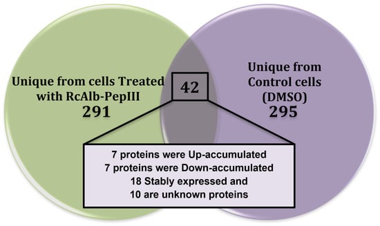
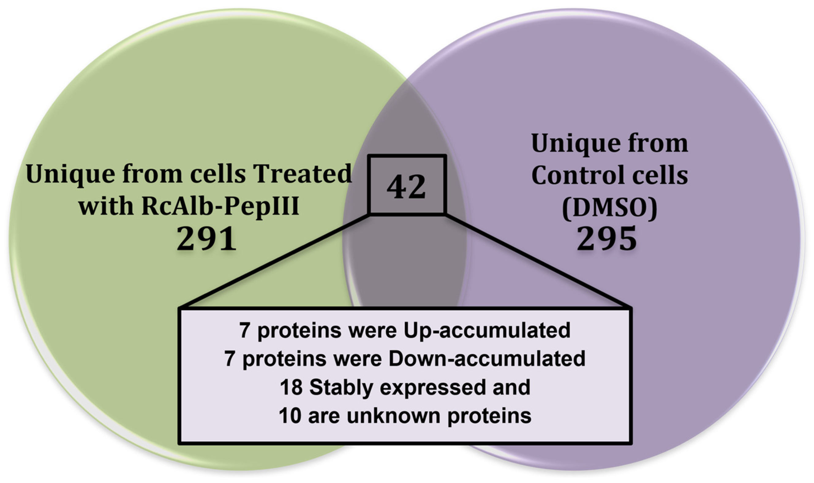
Based on this, we performed a proteomic analysis to shed more light on alternative events underlying RcAlb-PepIII-mediated damage to C. neoformans. In total, 628 proteins were identified (Figure 1); 295 were condition-specific detections in control cells, whereas 291 were condition-specific detections in RcAlb-PepIII-treated cells (Figure 1). Forty-two proteins were shared (overlapping) between the two C. neoformans samples; among these, 7 increased in abundance, 7 decreased in abundance, and 28 did not change (Figure 1 and Table 1).
Figure 1.
The number of proteins observed in C. neoformans using mass spectrometry (ESI-LC-MS/MS), where it is possible to see in the Venn diagram how the groups of proteins treated with RcAlb-PepIII (291), the control treated only with DMSO (295), and the proteins that appeared in both conditions (42 overlapping), and also, among the overlapping ones, those that increased, decreased, remained unchanged, or are unknown.
Table 1.
Identification of overlapping proteins in the group treated with RcAlb-PepIII and the control group treated with DMSO.
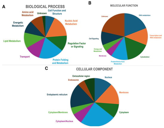
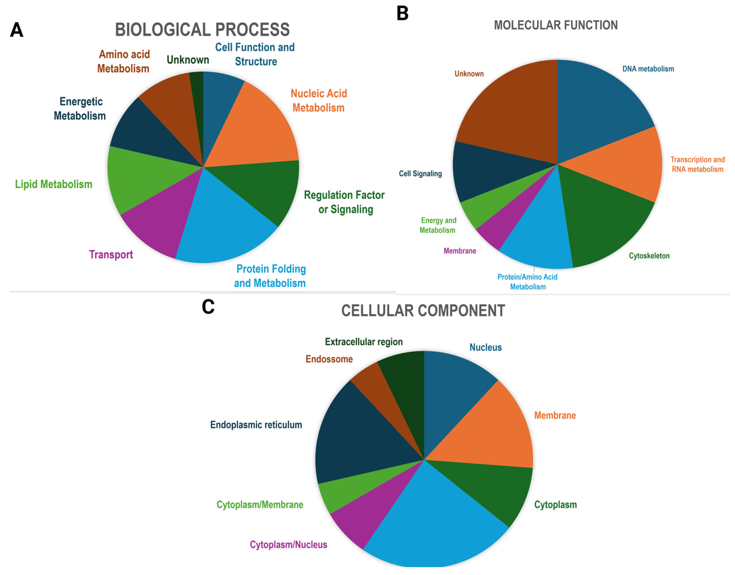
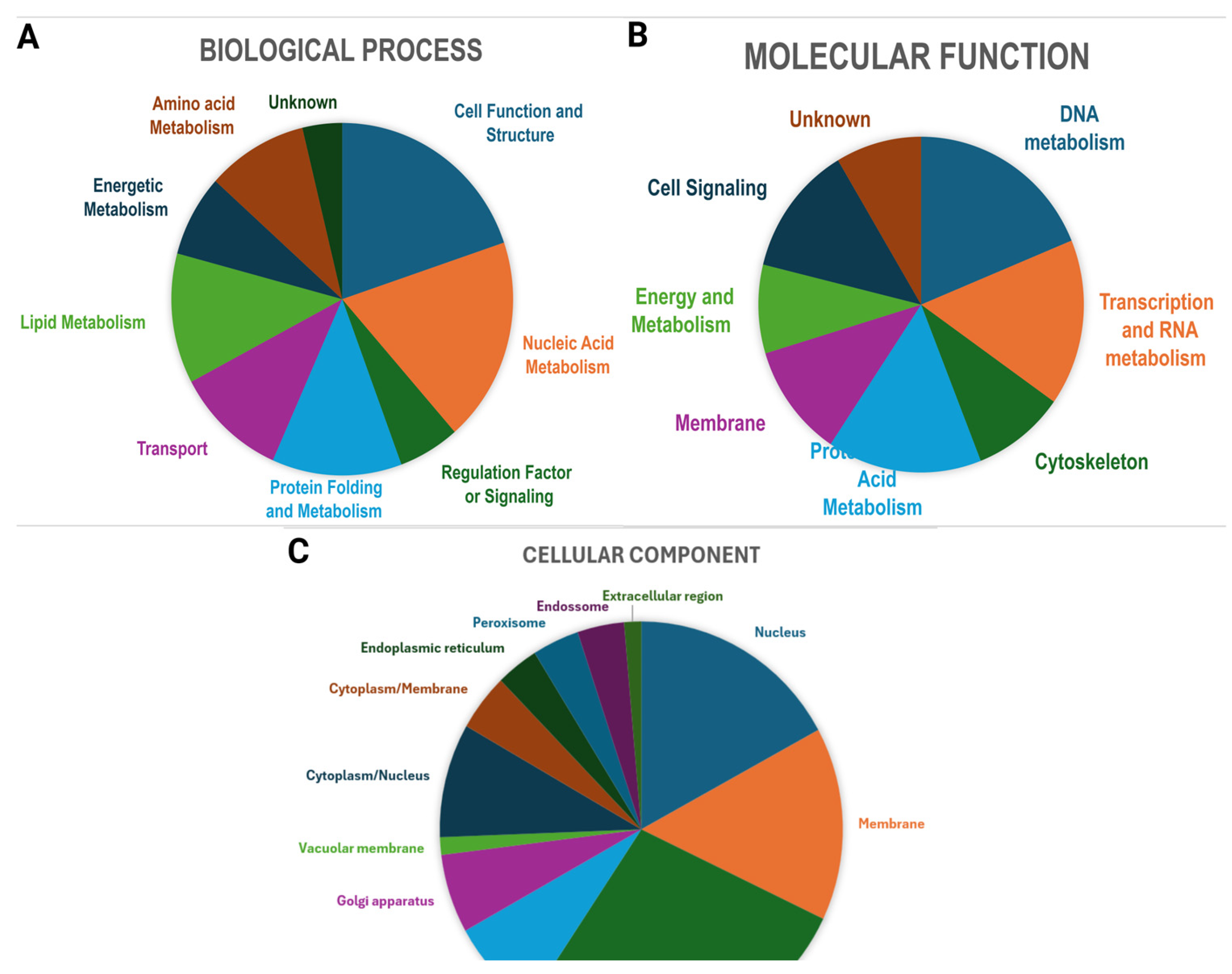
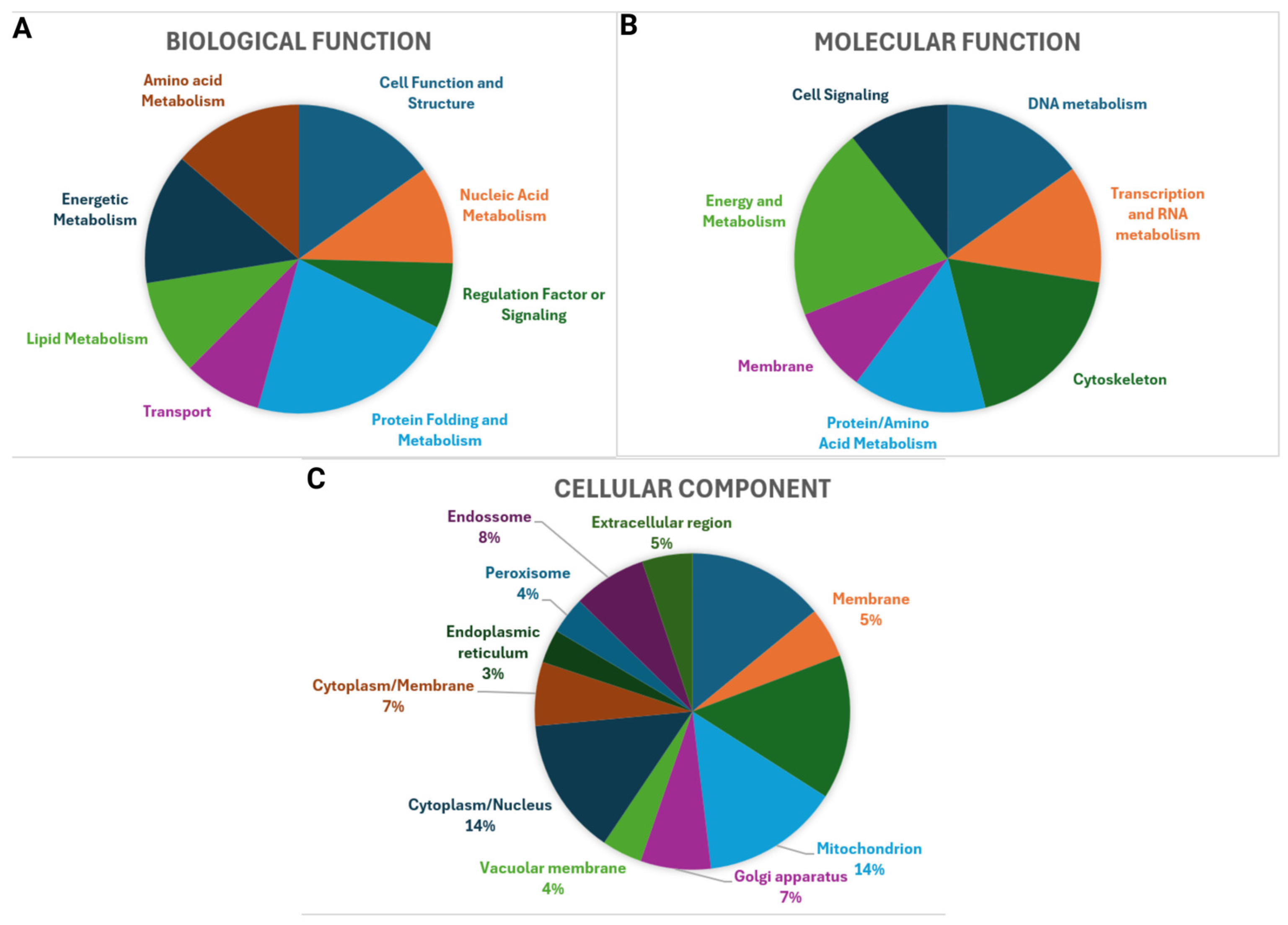
All proteins identified here were classified according to Gene Ontology (Figure 2). The proteins were classified according to biological process, molecular function, and cellular component for overlapping proteins (Figure 2 and Table 1), proteins only identified in the control (Figure 3), and proteins only identified in treated cells (Figure 4).
Figure 2.
Gene Ontology classification of overlapping proteins: (A) biological process, (B) molecular function, and (C) cellular component.
Figure 3.
Gene Ontology classification of proteins identified only in the control group: (A) biological process, (B) molecular function, and (C) cellular component.
Figure 4.
Gene Ontology classification of proteins only identified in the treated group: (A) biological process, (B) molecular function, and (C) cellular component.
3.1.2. DNA Metabolism-Related Proteins
Several proteins associated with DNA repair and genome maintenance, including HOS2, were detected only in RcAlb-PepIII-treated cells. Because condition-specific detection in data-dependent proteomics does not necessarily indicate quantitative upregulation, these proteins should be considered candidate treatment-associated responses. Nevertheless, their detection is particularly relevant in the context of our previous experimental findings demonstrating DNA fragmentation following RcAlb-PepIII exposure [9]. Thus, the present proteomic profile is consistent with the previously observed DNA-associated effects and suggests recruitment or remodeling of cellular processes related to genome maintenance in response to peptide treatment [10].
Although it occasionally deacetylates some non-histone proteins, it primarily targets lysine residues on the tails of histones H3 and H4, removing their acetyl groups. In addition to other functions, such as transcriptional regulation, HOS2 has been reported to co-localize with several proteins involved in response and repair under genotoxic stress, contributing to genome stability [11].
In addition, seven other proteins involved directly in DNA repair and maintenance, namely, DNA repair/transcription protein MET18/MMS19 [12]; DNA mismatch repair protein MLH3 [13]; DNA repair protein RAD5 [14]; DNA repair protein RAD7 [15]; DNA topoisomerase 2 [16]; and maintenance of telomere capping proteins 4 and 5 [17], were found solely among the proteins of C. neoformans cells treated with RcAlb-PepIII. Thus, the antimicrobial peptide may cause DNA damage, leading the cell to devote a significant portion of its resources to defending its genetic material against injury. The proteomic results were consistent with previous findings [4], which reported that RcAlb-PepIII caused DNA fragmentation and apoptosis in C. neoformans.
3.1.3. Transcription and RNA Metabolism Related Proteins
Regarding this group of proteins, two proteins, namely, Pre-mRNA-splicing factor ATP-dependent RNA helicase PRP22 and Type 1 phosphatase regulator YPI1, decreased in abundance in treated cells (0.010 and 0.086), respectively (Table 1). The ATP-dependent RNA helicase PRP22 (also known as Helicase PRP22) is critical for pre-mRNA splicing. It interacts with various spliceosome components and other regulatory factors to promote release of mature mRNA from the spliceosome complex and unwind the RNA duplex. Severe mutations or deletions of the PRP22 gene prevent proper maturation, impair splicing fidelity, and hinder the release of mature mRNA, which may already be severely damaged [18].
Type 1 phosphatase regulator YPI1 (PP1) plays a crucial role in mitotic cell division. Conversely, YPI1 strongly regulates nuclear localization and consequently the function of PP1. Studies have shown that YPI1 depletion disrupts mitosis, leading to aberrant spindle organization and thus cell death [19]. Additionally, it has also been described that PP1 can influence the glycogen content available to the cell, in pathways sensitive to phosphate balance, and also in the processing of the 3′ terminal region of mRNAs [20]. The decreased abundance of PRP22 and YPI1 suggests alterations in RNA-processing and nucleocytoplasmic regulatory processes during the cellular response of C. neoformans to RcAlb-PepIII.
3.1.4. Cytoskeleton-Related Proteins
Among the cytoskeleton-related proteins in this group (Table 1), two showed increased abundance. One was Coronin, which showed a fold change of 1.874. Coronins have previously been described as a highly conserved family of actin-regulating cytoskeletal proteins that assist in cell motility and can modulate several other actin-dependent processes [21].
The molecular interactions of coronins with F-actin and other actin-binding proteins (ABPs) are fundamental for regulating their functions at different points in actin polymerization, including the inhibition of actin nucleation through interactions with the Arp2/3 protein complex [22]. Coronin contributes significantly to increased tolerance to potential stresses in the cell wall, osmotic stress, and oxidative stress through mechanisms of membrane trafficking and the dynamic remodeling of actin filaments [23]. This increase in coronin presence among the overlapping proteins likely reflects the cell’s effort to minimize damage caused by the peptide.
Interestingly, the Dynein heavy chain, a cytoplasmic protein, was the most highly upregulated protein among the overlapping proteins, with a fold change of 15.454. Dynein is a motor protein that, in general, moves toward the minus end and is recognized as directly or indirectly important for a wide variety of cellular functions, such as organelle positioning, mitotic regulation, and intracellular vesicle transport [24].
In filamentous fungi, for example, several proteins associated with dynein pathways have been identified in nuclear distribution and/or spindle orientation [25]. Vesicle trafficking in Fusarium oxysporum, for example, also regulates the fungus’s sensitivity to salt, osmotic stress, and cell wall stress. In addition, they contribute to growth, conidiation, and virulence by forming complexes with other accessory proteins [26].
Under stress (heat, osmotic, oxidative, and cell-wall), cells reprogram secretion, endocytosis, and autophagy. Dynein’s role in retrograde transport and organelle positioning helps move stress-sensing receptors, damaged organelles, autophagosomes, and signaling complexes toward perinuclear regions for processing and degradation. Dynein helps redistribute vesicles and polarity components away from hyphal tips or to repair sites, indirectly affecting stress tolerance and recovery [26].
3.1.5. Protein/Amino Acid Metabolism-Related Proteins
In this category of proteins related to amino acid and protein metabolism, two proteins decreased in abundance after the treatment of C. neoformans cells with RcAlb-PepIII: Peptide hydrolase (fold-change 0.273) and Proteasome subunit alpha type-1 with a fold change of 0.363 (Table 1).
In fungi, Peptide hydrolases (peptidases/proteases) cleave peptide bonds and occur in secreted, vacuolar, cytosolic, and membrane-associated forms. They contribute to nutrient acquisition, protein quality control, morphogenesis, cell wall remodeling, signaling, and interactions with host plants. Several specific peptidases have been associated with invasion and virulence, among them FoAPY. Under stress (abiotic or biotic), their abundance, secretion, and activity change rapidly as part of the fungus’s adaptive response [27].
Alkaline, acidic serine, and metalloproteases are secreted into the extracellular space to degrade host proteins and environmental peptides; they are important for invasion and colonization. Vacuolar and cytosolic peptidases are generally involved in protein turnover, autophagy, and stress-induced proteolysis. In contrast, membrane-associated peptidases modulate signaling by processing peptide hormones or receptors.
These proteases can act under nutritional stress (such as carbon/nitrogen limitation), where upregulation of extracellular proteases releases amino acids from environmental proteins. Intracellular peptidases increase to recycle damaged proteins and provide amino acids. Under oxidative stress, these proteases and peptidases selectively degrade oxidized or misfolded proteins. Some are regulated by oxidative-stress pathways and are also subject to osmotic and/or thermal stress. Proteolytic systems (including proteasome-associated peptidases and vacuolar hydrolases) are activated to remove denatured proteins and support repair pathways [27].
Proteasome subunit alpha type 1, designated SCL1 (also annotated as PSA1), also showed a drastic decrease in abundance in the treated group. The structural alpha subunit of the proteasome’s central particle (PCP) controls substrate access and the assembly of the proteolytic beta rings. The proteasome degrades polyubiquitinated and misfolded proteins to maintain proteostasis [28]. Under stresses that increase the pool of misfolded or damaged proteins (heat shock, oxidative stress, cold/freezing, high salt content, heavy metals), the ubiquitin-proteasome system (UPS) activity and proteasome assembly are regulated or reorganized to eliminate damaged proteins and protect cells. Increased demand for protein degradation often affects the expression, assembly dynamics, and interactions of PCP alpha subunits [29].
Cells under stress can elevate UPS components and modify proteasome assembly pathways to increase degradation capacity. Alpha subunits are critical for the formation of new 20S CPs and for the remodeling of existing proteasomes (e.g., through assembly factors and activators). Chaperones and activators of proteasome assembly associate with alpha rings to influence PCP assembly, control, and activation. Structural studies reveal assembly intermediates involving alpha subunits, illustrating how interaction factors modulate the state of the PCP in response to changes in cellular needs [30].
Impaired SCL1 function decreases the formation of functional 20S cores, impairs proteasomal degradation capacity, and increases sensitivity to proteotoxic stresses (accumulation of ubiquitinated proteins, reduced survival under thermal/oxidative challenges). Proper alpha subunit function is therefore essential for stress tolerance and recovery. Increased proteasome assembly or activity improves the elimination of damaged proteins. It increases tolerance to stresses, such as freezing and oxidative conditions, as observed in studies that modulate UPS activity to improve stress resistance [31].
Interestingly, the decrease in proteasome subunit alpha type 1 (PSA1) is consistent with the concomitant decrease in the peptide hydrolases. The decreased abundance of proteasome-associated proteins suggests a potential impairment of protein homeostasis during the cellular response to RcAlb-PepIII, which may contribute to proteotoxic stress.
3.1.6. Membrane-Related Proteins
Related to membrane proteins, three proteins decrease in abundance in RcAlb-PepII: Rhodopsin domain-containing protein (fold-change 0.008), Rab proteins geranylgeranyltransferase component A (fold-change 0.386), and Ferric reductase transmembrane component 5 (fold-change 0.274).
In fungi, proteins with a rhodopsin domain are identical to opsins, which detect light or function as light-activated ion pumps and are often part of broader signaling networks that ultimately influence physiology [32]. Light is an environmental stress factor (UV, high irradiance) and a signal that indirectly triggers fungal stress pathways (antioxidant defenses, DNA repair, secondary metabolism) [33]. Rhodopsins are important proteins that help cells cope with light stress [33,34]. The reduction in Rhodopsin in treated cells suggests that these cells were more susceptible to light-induced damage.
Geranylgeranyl transferase Rab (RabGGTase) is a heterodimeric enzyme that catalyzes the attachment of geranylgeranyl lipid groups to Rab GTPases, enabling them to bind to membranes and mediate membrane transport. Rab proteins require geranylgeranyl groups to attach to specific organelle membranes and to move between the membrane and the cytosol. These transport processes are fundamental to fungi’s response to environmental stress (cell wall damage, osmotic stress, oxidative stress, nutrient limitation), as they control the delivery/removal of stress response proteins and membrane repair [35].
RcAlb-PepIII damages the cell wall and plasma membrane, as reported in our previous work [9]. Some studies showed that knockouts or hypomorphic alleles of RabGGTase subunits or REP/escort proteins produce mislocalized Rab, secretion defects, and hypersensitivity to osmotic and cell wall stress [36]. Therefore, the decrease in this protein (0.386) is very likely harmful to this organism.
Concerning the transmembrane component 5 of ferric reductase (commonly called FRE5), it belongs to the family of plasma membrane metal reductases that reduce extracellular ferric iron (Fe3+) to ferrous iron (Fe2+) before absorption by the cell. This reduction step is often the rate-limiting step in iron acquisition when soluble Fe3+ is scarce. Thus, FRE activity directly controls the availability of iron for respiration, DNA synthesis, and many other iron-dependent enzymes (metalloenzymes) [37,38].
The loss, decrease, or inhibition of FRE5 reduces the availability of Fe2+, hindering iron-dependent processes and impeding growth under iron limitation or during high metabolic demand (e.g., recovery from oxidative damage and other forms of stress). FRE defects often cause hypersensitivity to oxidative stress, impaired respiration, defective or incomplete cell wall remodeling, and reduced survival capacity during nutrient limitation or in hostile niches. In pathogenic fungi, impaired ferric reductase function severely reduces their ability to survive and thrive, as robust reductase activity is often necessary for colonization and prosperity in harsh environments [38].
A limitation of the present study is that the proteomic dataset was generated using a legacy LC-ESI-QUAD-TOF/MASCOT workflow in which the archived analytical output consisted of processed .pkl files, preventing retrospective deposition of a complete raw dataset in ProteomeXchange/PRIDE.
4. Conclusions
In conclusion, RcAlb-PepIII exposure induces substantial remodeling of the proteomic profile of C. neoformans, involving proteins associated with DNA-related processes, protein homeostasis, membrane-associated functions, cytoskeletal organization, and cellular stress responses. These proteomic alterations are consistent with and extend demonstrated cellular effects of RcAlb-PepIII, including membrane damage, DNA degradation, apoptosis, and oxidative stress. Rather than identifying individual proteins as direct molecular targets, the present findings provide a broader molecular perspective on the cellular response of C. neoformans to RcAlb-PepIII and identify biological processes that may contribute to its previously established antifungal activity.
Author Contributions
Conceptualization: P.F.N.S., M.L.P.B., and J.H.d.A.F.; Data curation: P.F.N.S., M.L.P.B., and J.H.d.A.F.; Formal analysis: P.F.N.S., M.L.P.B., and J.H.d.A.F.; Funding acquisition: P.F.N.S.; Methodology: P.F.N.S., M.L.P.B., and J.H.d.A.F.; Resources: P.F.N.S.; Supervision: P.F.N.S.; Writing—original draft: P.F.N.S., M.L.P.B., and J.H.d.A.F.; Writing—review, and editing: P.F.N.S. All authors have read and agreed to the published version of the manuscript.
Funding
This work was supported by the National Council for Scientific and Technological Development (CNPq) for a research grant to Pedro F. N. Souza (Process number: 307610/2026-8). Pedro F. N. Souza also thanks CAPES (Coordination for the Improvement of Higher Education Personnel) for the senior visiting research grant (process nº 88887.261683/2026-00 do Programa PDPG-FAP). This work was supported by the National Institute of Science and Technology INCT Bioinspir—Bioinspired Molecules as Innovative Solutions for Agricultural Challenges, funded by CNPq (National Council for Scientific and Technological Development), FUNDECT (Foundation for the Support of the Development of Education, Science and Technology of the State of Mato Grosso do Sul), and CAPES (Coordination for the Improvement of Higher Education Personnel), which awarded a grant to Pedro Filho Noronha Souza. We also thank the Conselho Nacional de Desenvolvimento Científico e Tecnológico (CNPq) and Fundação Coordenação de Aperfeiçoamento do Pessoal do Ensino Superior (CAPES) grant number 405934/2022–0 (the National Institute of Science and Technology INCT Funvir), from Brazil, to Pedro F. N. Souza.
Data Availability Statement
The original contributions presented in this study are included in the article. The proteomic data generated and analyzed during the current study are available from the corresponding author upon reasonable request.
Conflicts of Interest
The authors declare no conflicts of interest.
References
- Prasad, R.; Shah, A.H.; Rawal, M.K. Antifungals: Mechanism of Action and Drug Resistance. Adv. Exp. Med. Biol. 2016, 892, 327–349. [Google Scholar] [CrossRef] [Scilit] [PubMed]
- McHale, T.C.; Boulware, D.R.; Kasibante, J.; Ssebambulidde, K.; Skipper, C.P.; Abassi, M. Diagnosis and Management of Cryptococcal Meningitis in HIV-Infected Adults. Clin. Microbiol. Rev. 2023, 36, e00156-22. [Google Scholar] [CrossRef] [Scilit] [PubMed]
- Chen, Y.-C.; Yang, Y.; Zhang, C.; Chen, H.-Y.; Chen, F.; Wang, K.-J. A Novel Antimicrobial Peptide Sparamosin26–54 From the Mud Crab Scylla paramamosain Showing Potent Antifungal Activity Against Cryptococcus neoformans. Front. Microbiol. 2021, 12, 746006. [Google Scholar] [CrossRef] [Scilit] [PubMed]
- Aguiar, T.K.B.; Neto, N.A.S.; Freitas, C.D.T.; Silva, A.F.B.; Bezerra, L.P.; Malveira, E.A.; Branco, L.A.C.; Mesquita, F.P.; Goldman, G.H.; Alencar, L.M.R.; et al. Antifungal Potential of Synthetic Peptides against Cryptococcus neoformans: Mechanism of Action Studies Reveal Synthetic Peptides Induce Membrane–Pore Formation, DNA Degradation, and Apoptosis. Pharmaceutics 2022, 14, 1678. [Google Scholar] [CrossRef] [Scilit] [PubMed]
- Bradford, M.M. A Rapid and Sensitive Method for the Quantitation of Microgram Quantities of Protein Utilizing the Principle of Protein-Dye Binding. Anal. Biochem. 1976, 72, 248–254. [Google Scholar] [CrossRef] [PubMed]
- Aguiar, T.K.B.; Mesquita, F.P.; Neto, N.A.S.; Gomes, F.Í.R.; Freitas, C.D.T.; Carneiro, R.F.; Nagano, C.S.; Alencar, L.M.R.; Santos-Oliveira, R.; Oliveira, J.T.A.; et al. No Chance to Survive: Mo-CBP3-PepII Synthetic Peptide Acts on Cryptococcus neoformans by Multiple Mechanisms of Action. Antibiotics 2023, 12, 378. [Google Scholar] [CrossRef] [Scilit] [PubMed]
- Branco, L.A.C.; Souza, P.F.N.; Neto, N.A.S.; Aguiar, T.K.B.; Silva, A.F.B.; Carneiro, R.F.; Nagano, C.S.; Mesquita, F.P.; Lima, L.B.; Freitas, C.D.T. New Insights into the Mechanism of Antibacterial Action of Synthetic Peptide Mo-CBP3-PepI against Klebsiella pneumoniae. Antibiotics 2022, 11, 1753. [Google Scholar] [CrossRef] [Scilit] [PubMed]
- Tsakou, F.; Jersie-Christensen, R.; Jenssen, H.; Mojsoska, B. The Role of Proteomics in Bacterial Response to Antibiotics. Pharmaceuticals 2020, 13, 214. [Google Scholar] [CrossRef] [Scilit] [PubMed]
- Aguiar, B.; Neto, S.; Freitas, T.; Mesquita, F.P.; Santos-Oliveira, R.; Goldman, G.H. Behind the Curtain: In Silico and In Vitro Experiments Brought to Light New Insights into the Anticryptococcal Action of Synthetic Peptides. Antibiotics 2023, 12, 153. [Google Scholar] [CrossRef] [Scilit] [PubMed]
- Gu, Y.; Gordon, D.M.; Amutha, B.; Pain, D. A GTP:AMP Phosphotransferase, Adk2p, in Saccharomyces cerevisiae. J. Biol. Chem. 2005, 280, 18604–18609. [Google Scholar] [CrossRef] [Scilit] [PubMed]
- Lin, W.-H.; Opoc, F.J.G.; Liao, C.-W.; Roy, K.R.; Steinmetz, L.M.; Leu, J.-Y. Histone Deacetylase Hos2 Regulates Protein Expression Noise by Potentially Modulating the Protein Translation Machinery. Nucleic Acids Res. 2024, 52, 7556–7571. [Google Scholar] [CrossRef] [Scilit] [PubMed]
- Lombaerts, M. Saccharomyces cerevisiae Mms19 Mutants Are Deficient in Transcription- Coupled and Global Nucleotide Excision Repair. Nucleic Acids Res. 1997, 25, 3974–3979. [Google Scholar] [CrossRef] [Scilit] [PubMed]
- Nishant, K.T.; Plys, A.J.; Alani, E. A Mutation in the Putative MLH3 Endonuclease Domain Confers a Defect in Both Mismatch Repair and Meiosis in Saccharomyces cerevisiae. Genetics 2008, 179, 747–755. [Google Scholar] [CrossRef] [Scilit] [PubMed]
- Muellner, J.; Schmidt, K.H. Helicase Activities of Rad5 and Rrm3 Genetically Interact in the Prevention of Recombinogenic DNA Lesions in Saccharomyces cerevisiae. DNA Repair 2023, 126, 103488. [Google Scholar] [CrossRef] [Scilit] [PubMed]
- Lahari, T.; Lazaro, J.; Marcus, J.M.; Schroeder, D.F. RAD7 Homologues Contribute to Arabidopsis UV Tolerance. Plant Sci. 2018, 277, 267–277. [Google Scholar] [CrossRef] [Scilit] [PubMed]
- Pommier, Y.; Sun, Y.; Huang, S.N.; Nitiss, J.L. Roles of Eukaryotic Topoisomerases in Transcription, Replication and Genomic Stability. Nat. Rev. Mol. Cell Biol. 2016, 17, 703–721. [Google Scholar] [CrossRef] [Scilit] [PubMed]
- Stewart, J.A.; Chaiken, M.F.; Wang, F.; Price, C.M. Maintaining the End: Roles of Telomere Proteins in End-Protection, Telomere Replication and Length Regulation. Mutat. Res. Mol. Mech. Mutagen. 2012, 730, 12–19. [Google Scholar] [CrossRef] [Scilit] [PubMed]
- Beusch, I.; Madhani, H.D. Understanding the Dynamic Design of the Spliceosome. Trends Biochem. Sci. 2024, 49, 583–595. [Google Scholar] [CrossRef] [Scilit] [PubMed]
- Pedelini, L.; Marquina, M.; Ariño, J.; Casamayor, A.; Sanz, L.; Bollen, M.; Sanz, P.; Garcia-Gimeno, M.A. YPI1 and SDS22 Proteins Regulate the Nuclear Localization and Function of Yeast Type 1 Phosphatase Glc7. J. Biol. Chem. 2006, 282, 3282–3292. [Google Scholar] [CrossRef] [Scilit] [PubMed]
- Marquina, M.; González, A.; Barreto, L.; Gelis, S.; Muñoz, I.; Ruiz, A.; Álvarez, M.C.; Ramos, J.; Ariño, J. Modulation of Yeast Alkaline Cation Tolerance by Ypi1 Requires Calcineurin. Genetics 2012, 190, 1355–1364. [Google Scholar] [CrossRef] [Scilit] [PubMed]
- Chan, K.T.; Creed, S.J.; Bear, J.E. Unraveling the Enigma: Progress towards Understanding the Coronin Family of Actin Regulators. Trends Cell Biol. 2011, 21, 481–488. [Google Scholar] [CrossRef] [Scilit] [PubMed]
- Han, X.; Hu, Z.; Surya, W.; Ma, Q.; Zhou, F.; Nordenskiöld, L.; Torres, J.; Lu, L.; Miao, Y. The Intrinsically Disordered Region of Coronins Fine-Tunes Oligomerization and Actin Polymerization. Cell Rep. 2023, 42, 112594. [Google Scholar] [CrossRef] [Scilit] [PubMed]
- Koundinya, N.; Aguilar, R.M.; Wetzel, K.; Tomasso, M.R.; Nagarajan, P.; McGuirk, E.R.; Padrick, S.B.; Goode, B.L. Two Ligands of Arp2/3 Complex, Yeast Coronin and GMF, Interact and Synergize in Pruning Branched Actin Networks. J. Biol. Chem. 2025, 301, 108191. [Google Scholar] [CrossRef] [Scilit] [PubMed]
- Qiu, R.; Zhang, J.; Xiang, X. Identification of a Novel Site in the Tail of Dynein Heavy Chain Important for Dynein Function in Vivo. J. Biol. Chem. 2013, 288, 2271–2280. [Google Scholar] [CrossRef] [Scilit] [PubMed]
- Zhang, J.; Li, S.; Musa, S.; Zhou, H.; Xiang, X. Dynein Light Intermediate Chain in Aspergillus Nidulans Is Essential for the Interaction between Heavy and Intermediate Chains. J. Biol. Chem. 2009, 284, 34760–34768. [Google Scholar] [CrossRef] [Scilit] [PubMed]
- Jackson, E.; Li, J.; Weerasinghe, T.; Li, X. The Ubiquitous Wilt-Inducing Pathogen Fusarium Oxysporum—A Review of Genes Studied with Mutant Analysis. Pathogens 2024, 13, 823. [Google Scholar] [CrossRef] [Scilit] [PubMed]
- Qian, H.; Song, L.; Wang, L.; Wang, B.; Liang, W. The Secreted FoAPY1 Peptidase Promotes Fusarium oxysporum Invasion. Front. Microbiol. 2022, 13, 1040302. [Google Scholar] [CrossRef] [Scilit] [PubMed]
- Tanaka, K. The Proteasome: Overview of Structure and Functions. Proc. Jpn. Acad. B Phys. Biol. Sci. 2009, 85, 12. [Google Scholar] [CrossRef] [Scilit] [PubMed]
- Wendler, P.; Enenkel, C. Nuclear Transport of Yeast Proteasomes. Front. Mol. Biosci. 2019, 6, 34. [Google Scholar] [CrossRef] [Scilit] [PubMed]
- Tanahashi, R.; Nishimura, A.; Kan, K.; Ishizaki, N.; Fujishima, S.; Endo, H.; Takagi, H. Enhancing Freezing Stress Tolerance through Regulation of the Ubiquitin–Proteasome System in Saccharomyces cerevisiae. Fermentation 2024, 10, 318. [Google Scholar] [CrossRef] [Scilit]
- Kaur, M.; Chen, X.; Lee, S.Y.; Weaver, T.M.; Freudenthal, B.D.; Walters, K.J.; Roelofs, J. Structure of Blm10:13S Proteasome Intermediate Reveals Parallel Assembly Pathways for the Proteasome Core Particle. bioRxiv 2024. [Google Scholar] [CrossRef] [Scilit] [PubMed]
- Pardo-Medina, J.; Limón, M.C.; Avalos, J. Fusarium Photoreceptors. J. Fungi 2023, 9, 319. [Google Scholar] [CrossRef] [Scilit] [PubMed]
- Kumari, A.; Kumar, A.; Sharma, K.; Pati, S.R.; Mohanty, S.; Kateriya, S. Molecular Characterization of Unique Multi-Domain Harbouring Fungal Rhodopsin for Establishing Their Novel Opto-Synthetic Biological Usages. bioRxiv 2025. [Google Scholar] [CrossRef] [Scilit]
- Lara-Martínez, D.; Tristán-Flores, F.E.; Cervantes-Montelongo, J.A.; Silva-Martínez, G.A. Fungal Stress Responses and the Importance of GPCRs. J. Fungi 2025, 11, 213. [Google Scholar] [CrossRef] [Scilit] [PubMed]
- Guo, Z.; Wu, Y.; Das, D.; Delon, C.; Cramer, J.; Song, Y.; Thuns, S.; Lupilova, N.; Waldmann, H.; Brunsveld, L.; et al. Structures of RabGGTase–Substrate/Product Complexes Provide Insights into the Evolution of Protein Prenylation. EMBO J. 2008, 27, 2444–2456. [Google Scholar] [CrossRef] [Scilit] [PubMed]
- Tian, S.; Zhang, Z.; Qin, G. Function of Rab GTPases in Regulating the Development, Protein Secretion and Virulence of Fungi. Postharvest Pathol. 2014, 7, 3–9. [Google Scholar] [CrossRef] [Scilit]
- Muhammad, I.; Jing, X.-Q.; Shalmani, A.; Ali, M.; Yi, S.; Gan, P.-F.; Li, W.-Q.; Liu, W.-T.; Chen, K.-M. Comparative in Silico Analysis of Ferric Reduction Oxidase (FRO) Genes Expression Patterns in Response to Abiotic Stresses, Metal and Hormone Applications. Molecules 2018, 23, 1163. [Google Scholar] [CrossRef] [Scilit] [PubMed]
- Takemura, K.; Kolasinski, V.; Del Poeta, M.; Vieira, F.; Garg, A.; Ojima, I.; Del Poeta, M.; Pereira, N. Iron Acquisition Strategies in Pathogenic Fungi. MBio 2025, 16, e01211–e01225. [Google Scholar] [CrossRef] [Scilit] [PubMed]

Disclaimer/Publisher’s Note: The statements, opinions and data contained in all publications are solely those of the individual author(s) and contributor(s) and not of MDPI and/or the editor(s). MDPI and/or the editor(s) disclaim responsibility for any injury to people or property resulting from any ideas, methods, instructions or products referred to in the content. |
© 2026 by the authors. Licensee MDPI, Basel, Switzerland. This article is an open access article distributed under the terms and conditions of the Creative Commons Attribution (CC BY) license.