


Abstract
Germination is a biological process that can enhance the nutraceutical value of Andean grains, but its efficiency can be optimized through emerging, sustainable technologies. This study evaluated ultrasound as a pretreatment for the germination of Collana black quinoa. A 23 factorial design was used to assess the effect of ultrasound duration, ultrasound frequency, and germination time on the content of bioactive compounds, antioxidant capacity, color, water activity, amylase, and reducing sugars. Two controls (C48 and C72) were included as additional data points. Subsequently, analysis of variance and multiple-range tests were applied to assess the significance of differences between treatments (Tukey’s test) and between treatments and the control (Dunnett’s test). The content of phenolic compounds and flavonoids, antioxidant capacity, amylose, and reducing sugars were quantified by UV–Visible spectrophotometry. At the same time, FTIR, DSC, and TGA were used to evaluate functional groups and thermal behavior. Ultrasonic treatments resulted in increases of up to 12% in total phenolics, 24.5% in flavonoids, and 15% in antioxidant capacity compared to quinoa germinated for 72 h without ultrasonic treatment, along with reductions in amylose and increases in reducing sugars. Prolonged germination was the most influential factor; however, ultrasound acted as an abiotic stimulus, increasing cell permeability and promoting phenolic biosynthesis. Thermal and FTIR analyses revealed subtle modifications in starch and protein structure, associated with greater thermal stability and molecular reorganization. These results demonstrate that ultrasound-assisted germination is a sustainable and effective strategy for integrating functional quinoa ingredients into modern food systems.
1. Introduction
The growing search for functional and sustainable foods has driven a remarkable interest in Andean grains, particularly quinoa (Chenopodium quinoa), recognized for its excellent nutritional profile, which integrates essential amino acids, minerals, dietary fiber, and a considerable concentration of polyphenols, flavonoids, and other phytochemicals with beneficial effects on health [1,2]. In this global context, characterized by consumers who demand healthy, low-impact foods [3,4,5], quinoa is positioned as a strategic resource to strengthen food security and revalue traditional crops [6,7,8]. Among the different quinoa varieties, Collana black quinoa, a Peruvian variety adapted to high altitudes and valued for its hardiness, high protein content, and small grain size, has sparked increasing scientific interest due to its higher concentration of pigmented compounds, particularly phenolics and flavonoids, which give it a greater antioxidant capacity compared to the white and red varieties [9,10,11]. These characteristics make black quinoa an appropriate model for evaluating technological strategies, such as ultrasound-assisted germination, to improve its functional and physicochemical properties.
Germination, a biological process known to improve the nutritional profile, favorably modifies the content of phenolic compounds, flavonoids, and minerals in various grains by activating secondary metabolic pathways; it improves the biological value of protein and its digestibility; and it reduces the content of antinutritional factors such as phytic acid and tannin [12,13,14,15]. However, the specific effects of germination on the concentration of phenolics, flavonoids, and antioxidant capacity in quinoa are still under investigation, as results can vary depending on germination time, environmental conditions, and grain variety [16,17,18]. In recent years, special attention has been paid to pre-germination treatments to better leverage this technology and enhance the biological activity of grains. Several studies have reported the application of various pre-germination treatments to grains, including ultrasound, high hydrostatic pressure, pulsed electric fields, and low-pressure cold plasma, as well as soaking and enzyme treatment [19,20,21].
Ultrasound, a non-thermal, energy-efficient, sustainable, and environmentally friendly technology, can penetrate the grain matrix and trigger internal metabolic responses with minimal thermal impact. This process regulates physiological metabolism and the production of bioactive compounds [22,23]. Recent research in crops such as wheat, barley, beans, and rice shows that the application of low-frequency ultrasound (20–40 kHz) effectively improves seed hydration, enzyme activation, and mass transfer phenomena without causing irreversible structural damage [23,24,25,26].
Despite advances in ultrasound-assisted germination studies, a knowledge gap remains regarding the combined application of these processes to the physicochemical and thermal properties of germinated quinoa. In particular, there is little information relating these ultrasonic parameters to the structural and functional changes in black quinoa during germination. Some studies have shown that ultrasound enhances the synthesis and release of bioactive compounds, increasing the levels of γ-aminobutyric acid (GABA), phenolics, and flavonoids in white quinoa [27]. Furthermore, ultrasound application improves seed hydration and permeability, suggesting positive effects during imbibition [28]. Similarly, in species such as corn, rice, and capers, ultrasound promoted germination [29,30,31,32]. These studies support the hypothesis that ultrasound used as a preliminary treatment during germination could enhance the biosynthesis and availability of bioactive compounds and modify the physical-chemical and thermal properties of quinoa, thereby justifying the need for the present study.
Therefore, this study aims to evaluate the effects of ultrasound duration and frequency during two germination periods (48 and 72 h) on the bioactive compounds, physicochemical properties, and thermal characteristics of Andean quinoa.
2. Materials and Methods
2.1. Seed Materials
The quinoa seeds used in this study were of the Negra Collana variety, cultivated in the agricultural fields of the Champaccocha community, Andahuaylas, Apurímac, Peru (−13.647205, −73.307359), at an approximate altitude of 3540 m above sea level. The region is characterized by a temperate Andean climate, with average annual temperatures ranging from 8 to 16 °C and a pronounced rainy season from November to March. Homogeneous grains were selected based on their uniform maturity, color, and size. Before soaking, the quinoa samples were visually inspected to remove damaged grains and foreign materials.
2.2. Sample Preparation
Sample preparation was performed according to Zhang, Guo, Dang, Zhang, Zheng and Yang [23] with some modifications.
Germinated sample: Quinoa grains were washed and disinfected in a 1% sodium hypochlorite solution for 20 min; subsequently, they were soaked for 12 h at 20 °C. Germination was carried out at 25 °C for 48 and 72 h in a humidified incubator FOC 200 E (Velp Scientifica™, Usmate Velate, Italy), with distilled water sprayed every 4 h to maintain adequate moisture. After germination, the grains were dried in a FED 115 forced-convection oven (BINDER, Baden-Wurtemberg, Germany) at 40 °C; they were then ground in a Twister cyclone mill (Retsch, Renania del Norte-Westfalia, Germany) and sieved through a 120-mesh screen.
In this study, two control conditions were defined: (i) quinoa grains germinated for 48 h without ultrasonic treatment (C48) and (ii) quinoa grains germinated for 72 h without ultrasonic treatment (C72).
Germinated and sonicated sample: Quinoa was washed and disinfected in a 1% sodium hypochlorite solution for 20 min, then soaked for 12 h at 20 °C. Subsequently, the quinoa was stressed in a DU-220S ultrasonic bath (ARGO LAB, Milan, Italy, manufactured in China) at 28 and 40 kHz, with exposure times of 20 and 30 min, respectively. The temperature was kept constant at 20 °C by circulating the water. After the ultrasonic pretreatment, the seeds were germinated at 25 °C for 48 and 72 h in a humidified FOC 200 E incubator (Velp ScientificaTM, Usmate Velate, Italy). Distilled water was sprayed every 4 h to maintain seed moisture. After germination, the grains were dried in a FED 115 forced convection oven (BINDER, Baden-Wurtemberg, Germany) at 40 °C; they were then crushed in a Twister cyclone mill (Retsch, Renania del Norte-Westfalia, Germany) and sieved through a 120-mesh sieve.
2.3. Bioactive Compounds and Antioxidant Capacity
The bioactive compounds evaluated were total phenolics and flavonoids. Total phenolic compounds in the samples were estimated using the Folin–Ciocalteu (Himedia, Mumbai, India) reagent. Sample extracts were prepared and mixed with 20% sodium carbonate (Spectrum, NB, Canada), 0.25 N Folin–Ciocalteu reagent, and deionized water. The samples were read at 755 nm using a UV-Vis spectrophotometer (Genesys 150, Thermo Fisher Scientific, Waltham, MA, USA). Gallic acid (Sigma-Aldrich, Baviera, Germany) was used as a reference standard, and the results were expressed as mg gallic acid equivalents/100 g of sample [33].
To quantify total flavonoid content, extracts from the samples were mixed with methanol and aluminum chloride (Sigma-Aldrich, Baviera, Germany). The samples were read at 450 nm using a UV–Visible spectrophotometer (Genesys 150, Thermo Fisher Scientific, Waltham, MA, USA). Quercetin (Sigma-Aldrich, Baviera, Germany) was used as the reference standard, and results were expressed as mg of quercetin equivalent/100 g of sample [15].
Antioxidant capacity was measured using the DPPH [2,2-diphenyl-1-picrylhydrazyl] (Himedia, Mumbai, India) assay. Sample extracts were mixed with dilute DPPH solution and measured at 515 nm using a UV-Vis spectrophotometer (Genesys 150, Thermo Fisher Scientific, Waltham, MA, USA). Trolox [6-hydroxy-2,5,7,8-tetramethylchromane-2-carboxylic acid] (Sigma Aldrich, St. Louis, MO, USA) was used as a reference standard, and results were expressed as mg of Trolox equivalents/g of sample [34].
2.4. Physical and Chemical Properties
2.4.1. Color and Water Activity
Color was measured using a CR-5 digital colorimeter (Konica Minolta, Tokyo, Japan), with the measurement scale set to CIE L*, a*, and b*. The instrument was calibrated with a blank calibration plate before each measurement [35].
Water activity was determined using a HygroPalm model 23-AW meter (Rotronic brand, Bassersdorf, Switzerland). It was previously calibrated with calibration standards. Then, 3 g of the sample was taken and transferred to a disposable sample container. Subsequently, the probe was inserted, and the reading was taken [34].
2.4.2. Amylose and Reducing Sugars
For the determination of amylose, 20 mg of the sample was weighed and mixed with 0.2 mL of 95% ethanol (Scharlau, Sentmenat, Spain) and 1.8 mL of 1 M sodium hydroxide (Merck, Darmstadt, Germany). The mixture was allowed to stand for 24 h at room temperature; then, it was diluted to 20 mL with deionized water and shaken vigorously. After standing for 30 min, 1 mL of the solution was taken, and 2 mL of 1 M acetic acid (Sigma-Aldrich, Baviera, Germany) was added with shaking; 0.4 mL of Lugol’s (Merck, Darmstadt, Germany) solution was added, and the volume was brought up to 20 mL with deionized water. The solution was allowed to stand in the dark for 20 min. An unshaken aliquot was taken and transferred to cuvettes for absorbance measurement using a UV-Vis spectrophotometer (Genesys 150, Thermo Fisher Scientific, Waltham, MA, USA) at 620 nm. Previously, a standard curve was prepared with 1 mg/mL of concentrated amylose (Merck, Darmstadt, Germany) in deionized water [36].
Reducing sugars were measured using the DNS [3,5-dinitrosalicylic acid] method. Five grams of DNS (Himedia, Mumbai, India), 150 g of sodium potassium tartrate, and 8 g of sodium hydroxide (NaOH) were weighed. The NaOH was dissolved in 200 mL of distilled water, and the sodium potassium tartrate (Himedia, Mumbai, India) was added slowly. The solution was diluted to 400 mL with distilled water, and the DNS was added slowly. The solution was stirred overnight, brought to a final volume of 500 mL, and filtered. In 10 mL test tubes, 0.5 mL of the sample and 0.5 mL of the DNS reagent were added. The tubes were placed in a water bath at 100 °C for 5 min. After cooling to room temperature, 5 mL of distilled water was added. The mixture was shaken, and the reading was taken at 540 nm using a UV–Visible spectrophotometer (Genesys 150, Thermo Fisher Scientific, Waltham, MA, USA). A glucose standard curve was prepared beforehand [37].
2.4.3. Functional Groups
They were determined by spectrophotometry; the sample was mixed with dried potassium bromide (Thermo Fisher Scientific, Garfield, NJ, USA) in a mortar and pressed into a thin film. The IR spectrum of the sample was obtained using a Nicolet IS50 FTIR spectrophotometer (Thermo Fisher, Waltham, MA, USA) with the transmission module over a wavenumber range of 4000–500 cm−1 [18,38].
2.5. Thermal Properties
2.5.1. Gelatinization Temperature and Enthalpy
The gelatinization temperature and enthalpy were determined using a DSC 2500 differential scanning calorimeter (TA Instruments, Newcastle, DE, USA). 2.0 mg of the sample was weighed into an aluminum tray, and 10 µL of distilled water was added. The container was hermetically sealed and equilibrated at room temperature for 1 h; then, a heating rate of 5 °C/min was programmed from 20 to 100 °C, using an empty sealed container as a reference. The peak gelatinization temperature (Tp) and enthalpy change (ΔH) were automatically calculated [16].
2.5.2. Thermal Stability
Thermal stability was determined by thermogravimetric analysis, using a TGA 550 instrument (TA Instruments, New Castle, DE, USA) with a nitrogen flow of 50 mL/min. To obtain the thermogram, the sample’s weight loss and temperature were recorded at 4-s intervals [39].
2.6. Statistical Analysis
The study was conducted using a completely randomized factorial experimental design. A 23 factorial arrangement was applied to evaluate the effects of ultrasound frequency (20 and 40 kHz), ultrasound treatment time (20 and 30 min), and germination time (48 and 72 h) on the bioactive compound content, antioxidant capacity, color parameters, water activity, amylase, and reducing sugar content of germinated quinoa.
Each experimental treatment was performed in triplicate. A replicate consisted of an independent batch of quinoa grains subjected to the ultrasound treatment and the corresponding germination conditions; therefore, each batch represented an experimental unit.
The factorial design generated eight treatment combinations (2 × 2 × 2). In addition, two control treatments without ultrasound were included at each germination time (48 h and 72 h), designated C48 and C72, respectively, and analyzed under the same experimental conditions.
In total, the experimental design comprised 30 experimental units (8 factorial treatments × 3 replicates + 2 controls × 3 replicates).
Subsequently, an analysis of variance and multiple-range tests were applied to evaluate the significance of differences between treatments (Tukey’s test) and between treatments and the control (Dunnett’s test). The statistical software ORIGIN PRO 2025b was used.
3. Results and Discussion
3.1. Bioactive Compounds and Antioxidant Capacity
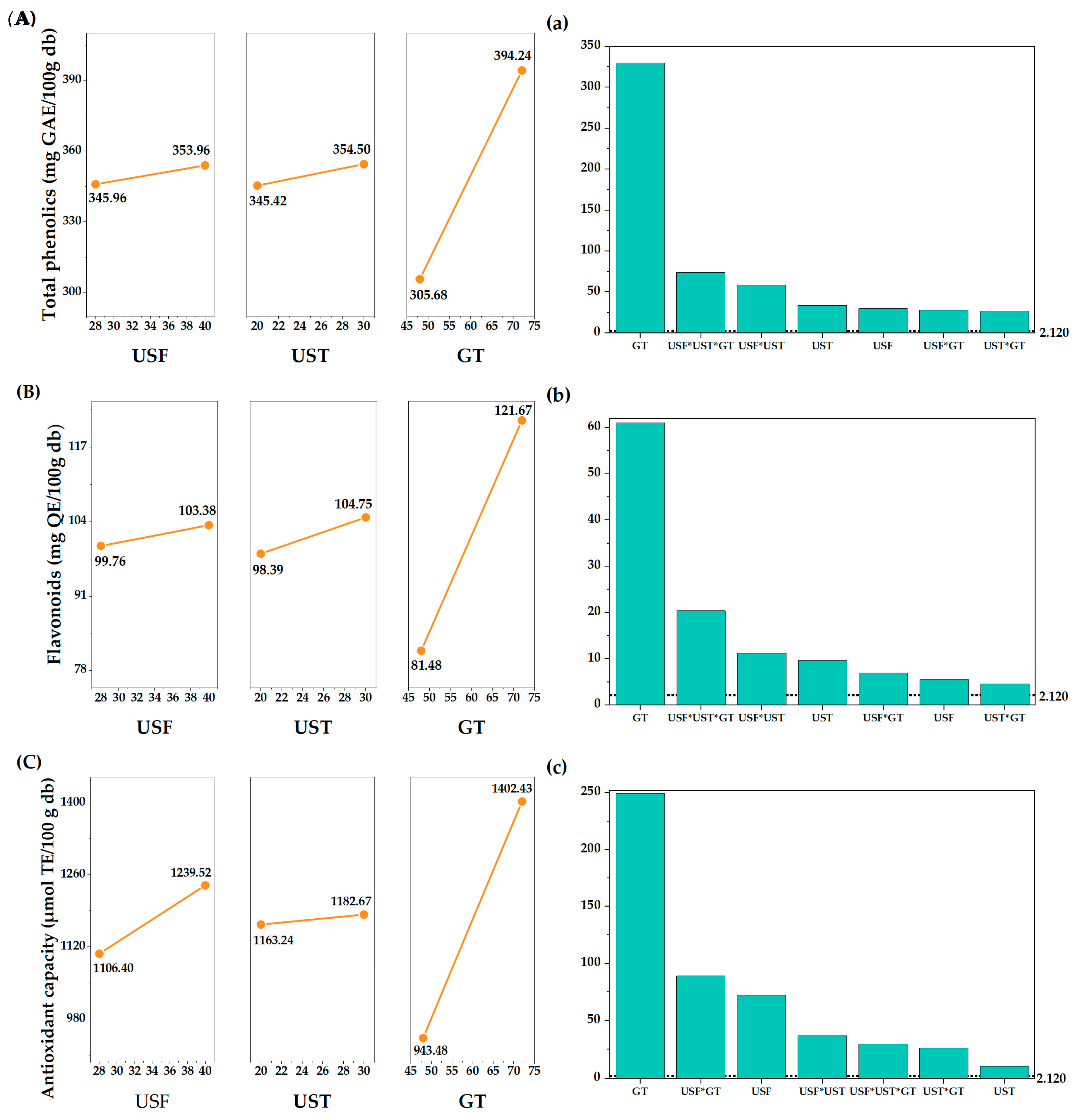
The phenolic and flavonoid content reached values between 295.73–405.26 mg GAE/100 g db and 76.32–132.67 mg QE/100 g db, respectively, for the ultrasound-treated samples (Table 1). In both bioactive compounds, treatment T7 (28 kHz, 30 min) showed the highest concentration (total phenolics: 405.26 ± 0.47 mg GAE/100 g db; flavonoids: 132.67 ± 1.58 mg QE/100 g db), with increases of 12% for total phenolics and 24.5% for flavonoids compared to the control sample germinated for 72 h. Similar increases were reported by Wu, Zhou, Zhou, Wang, Ren, Zheng, Lv and Zhao [27] in white quinoa subjected to ultrasound stress before germination. The observed increase suggests that ultrasonic cavitation induces physical stress at the cellular level, leading to membrane and cell wall permeabilization, facilitating mass transfer and the release of intracellular phenolic compounds. In parallel, ultrasound acts as an abiotic elicitor, activating oxidative signaling pathways and enhancing the activity of phenylalanine ammonia-lyase (PAL), an enzyme in the phenylpropanoid pathway. This combined effect promotes the biosynthesis of phenolic compounds and flavonoids during germination, as evidenced by the significant increases in total phenolics and flavonoids [19,21,40,41].
Table 1.
Phenolic compounds, flavonoids, and antioxidant capacity.
Regarding antioxidant capacity, the values ranged from 900.89 to 1475.61 µmol TE/100 g db (Table 1), with a 15% increase compared to the control after 72 h of germination, indicating a direct increase in the capacity to neutralize free radicals [40,41]. These results confirm that ultrasound, as a pre-germination treatment, increases the enzymatic activity involved in germination-related metabolic pathways [22]. Furthermore, it alters the microstructure of the grain, facilitating water absorption and increasing the availability of the substrate for enzymatic hydrolysis and the release of phenolic compounds [41], including those bound to cell wall components called bound phenolics, which are considered, in some studies, to have a greater antioxidant capacity [40].
Although phenolic compounds are recognized as the main contributors to antioxidant capacity, this study demonstrated that treatment T8 (40 kHz) exhibited the highest antioxidant capacity. In comparison, treatment T7 (28 kHz) showed the highest total phenolic and flavonoid content. This discrepancy indicates that antioxidant capacity depends not only on the total concentration of phenolic compounds but also on their qualitative composition and the contribution of other antioxidant molecules. A higher ultrasound frequency (40 kHz) can induce more intense cavitation, enhancing cell disruption and enzyme activation during germination, which may favor the release or formation of non-phenolic antioxidants such as antioxidant peptides, tocopherols, or other low-molecular-weight metabolites [26].
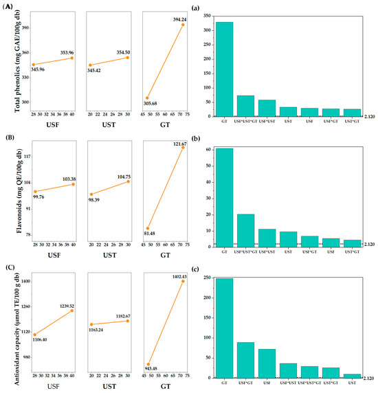
Ultrasound frequency and time, and time germination significantly affected (p < 0.05) phenolic and flavonoid content and antioxidant capacity (Figure 1A–C). In the present study, increasing the ultrasound frequency and duration increased the concentration of bioactive compounds and antioxidant capacity. Previous studies have reported that the optimal ultrasound frequency can vary depending on the specific bioactive compound; for example, 40 kHz has been reported as optimal for increasing gallic acid and quercetin [22]. Regarding ultrasound time, most studies reported a range of 5 to 30 min, as excessive time can be detrimental, potentially damaging the cell membrane [27]. Ultrasound, by generating abiotic stress, activates the enzymes responsible for the biosynthesis of bioactive compounds. Furthermore, a longer germination time is associated with a higher content of phenolics and flavonoids, as well as greater antioxidant capacity. These results are consistent with studies indicating that germination times of 72 to 120 h lead to increased concentrations of bioactive metabolites [20]. The standardized effects analysis (Figure 1a–c) revealed that the factor with the greatest impact was germination time (GT), followed by the USF × UST × GT interaction for phenolics and flavonoids, and the USF × GT interaction for antioxidant capacity. These results indicate that the biosynthesis of bioactive compounds is closely linked to germinative metabolism, while ultrasound acts as an abiotic elicitor through cavitation-induced mechanical stress. This stress promotes transient membrane permeabilization and activates metabolic pathways involved in secondary metabolism, leading to an enhanced synthesis of phenolic compounds and flavonoids, which act synergistically as free radical scavengers [27].
Figure 1.
Main effects plot of ultrasonic frequency (USF), ultrasound time (UST), and germination time (GT) on (A) total phenolic compounds, (B) flavonoids, and (C) antioxidant capacity of germinated black quinoa. Graphs (a–c) show the standardized effects of the factors on the response variables.
3.2. Physical and Chemical Properties
3.2.1. Color and Water Activity
The evaluation of color parameters is essential, as color changes indicate chemical reactions occurring during processing and germination [19,20]. In the present study, lightness (L) values ranged from 63.29 to 73.19 among treatments (Table 2) and decreased with increasing germination time, reflecting a progressive darkening of the samples. This behavior can be associated with the accumulation and redistribution of phenolic pigments, as well as with non-enzymatic browning reactions occurring during germination [35]. Concomitantly, increases in chroma a and chroma b* values were observed, indicating greater saturation of red and yellow hues. These color shifts are consistent with the enhanced presence of bioactive compounds, particularly phenolic compounds and flavonoids, which contribute both to pigmentation and antioxidant functionality. However, the relationship between luminosity and bioactive content was not strictly linear, suggesting that qualitative changes in phenolic composition and matrix structural modifications also influence color perception. Regarding ultrasound effects, presonication at 40 kHz did not induce marked changes in color parameters, despite promoting higher antioxidant capacity. This indicates that high-frequency ultrasound can enhance functional properties without significantly compromising visual quality, which represents an important commercial advantage in terms of consumer acceptance. Similar trends have been reported for pigmented rice subjected to ultrasound-assisted germination [20]. Water activity values ranged from 0.40 to 0.47, ensuring microbiological stability of the product and supporting its potential application in food systems [33,42].
Table 2.
Color and water activity.
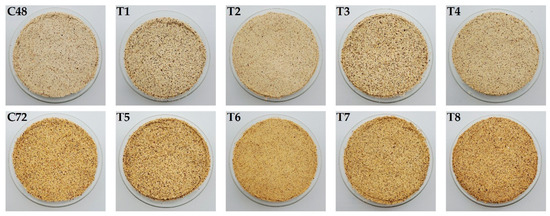
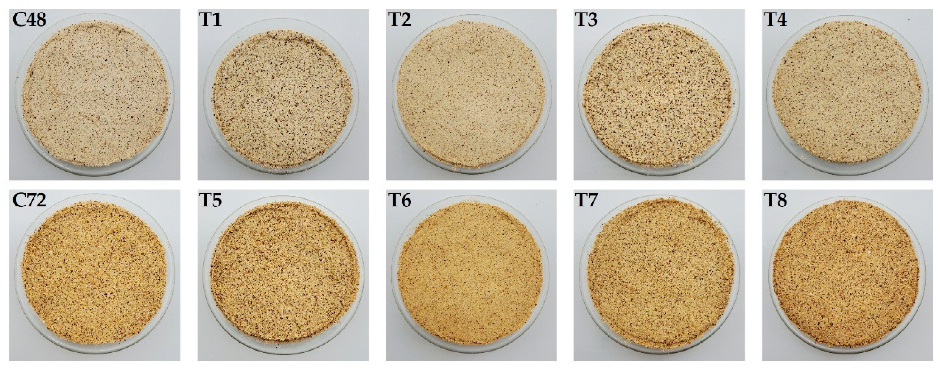
Additionally, the visual differences (Figure 2) showed that the samples germinated for 72 h (C72, T5–T8) had greater color intensity, evidenced by darker, yellower tones, attributable to greater accumulation of phenolic compounds and natural pigments during prolonged germination [41]. Likewise, the treatments with 28 kHz ultrasound (T1, T3, T5, T7) show a slightly more intense coloration, which coincides with the increases in chroma a* and b* values obtained instrumentally. These visual observations support the quantified changes in Table 2 and confirm the synergistic effect of ultrasound and germination time on the flour’s chromatic properties.
Figure 2.
Visual appearance of the samples of germinated Negra Collana quinoa under different ultrasound treatments. C48: germinated for 48 h without ultrasound; C72: germinated for 72 h without ultrasound. T1–T4 correspond to samples germinated for 48 h, treated with ultrasound at 28 kHz–20 min (T1), 40 kHz–20 min (T2), 28 kHz–30 min (T3), and 40 kHz–30 min (T4). T5–T8 correspond to samples germinated for 72 h, treated with ultrasound at 28 kHz–20 min (T5), 40 kHz–20 min (T6), 28 kHz–30 min (T7), and 40 kHz–30 min (T8).
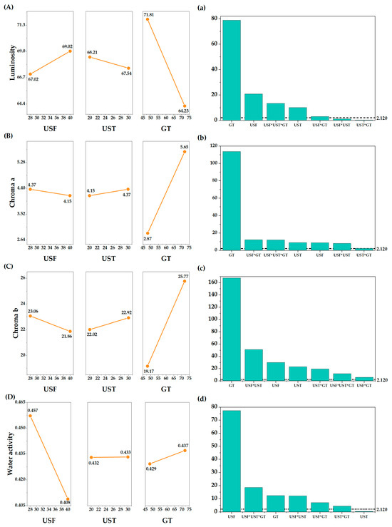
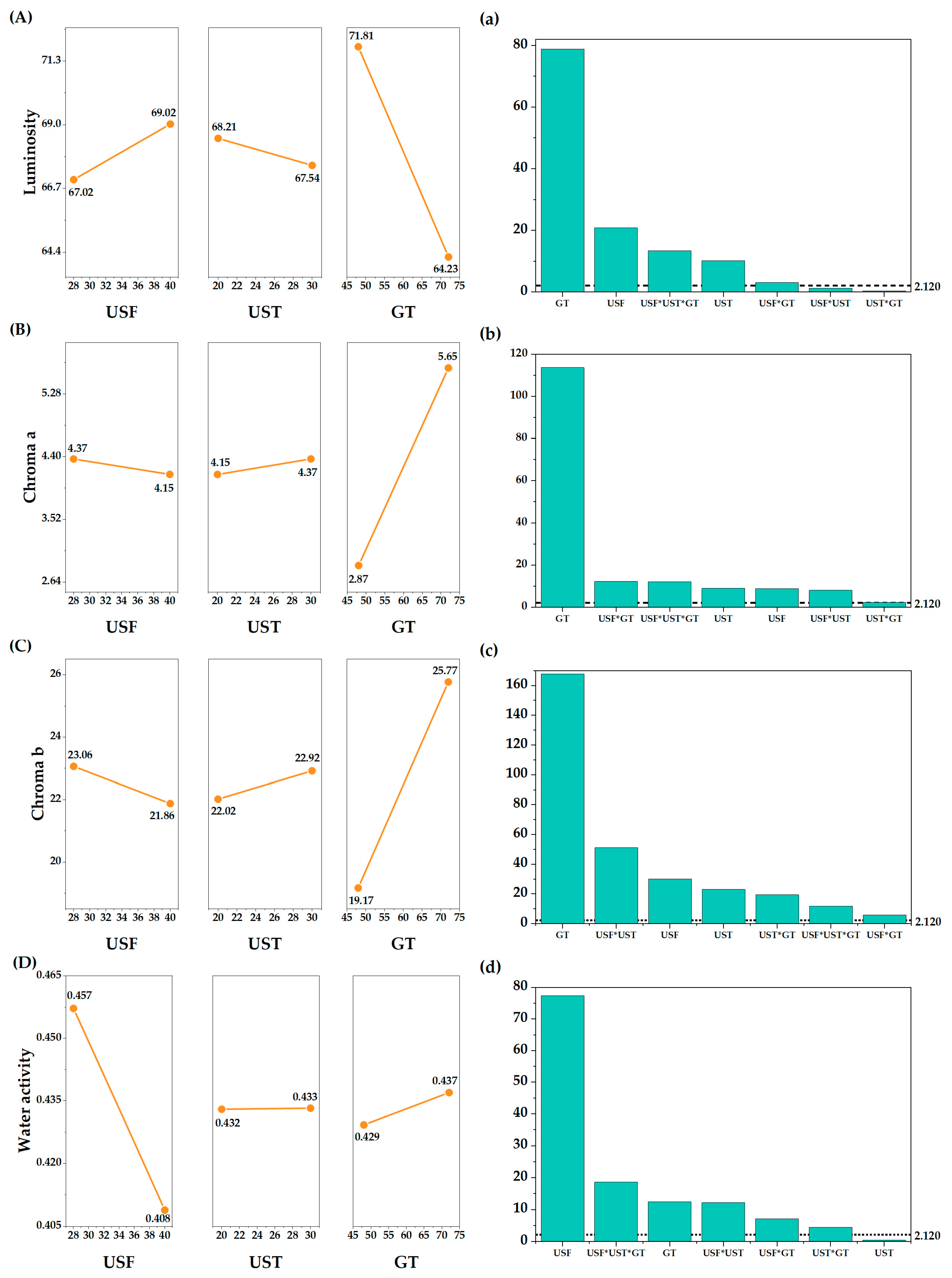
The main standardized effects of the experimental factors indicate that germination time had the highest statistical weight for color (Figure 3a–c) and ultrasound frequency had the highest statistical weight for water activity (Figure 3d). In the effects plots (Figure 3A–C), it can be observed that luminosity decreases, while chroma values a and b increase with germination, reflecting a darkening accompanied by chromatic intensification. This behavior demonstrates a direct relationship between color and the accumulation of phenolic compounds and natural pigments, consistent with observations in Table 1. Water activity increased slightly with low-frequency ultrasound (Figure 3D). The effect of low-frequency ultrasound is mainly due to acoustic cavitation, which creates micropores and microcracks in the grains’ cell walls [20,43], leading to a significant increase in cell membrane permeability and facilitating the diffusion of water and oxygen [40,44].
Figure 3.
Main effects plot of ultrasonic frequency (USF), ultrasound time (UST), and germination time (GT) on (A) luminosity, (B) chroma a, (C) chroma b, and (D) water activity of germinated black quinoa. Graphs (a–d) show the standardized effects of the factors on the response variables.
3.2.2. Amylose and Reducing Sugars
Amylose decreased slightly with increasing germination time (Table 3). This linear polymer is susceptible to germination processes that preferentially affect its molecular structure, while the amylopectin conformation remains relatively stable [41]. Meanwhile, the reducing sugar content increased with germination time, rising from 98.08 mg glucose/g db (C48) to 119.21 mg glucose/g db (C72). This behavior is associated with the activation of hydrolytic enzymes that break down linear amylose chains to generate simple sugars during germination. Sonication creates fissures on the surfaces of quinoa starch particles, increasing the starch’s exposure to enzymes [27].
Table 3.
Amylose and reducing sugar.
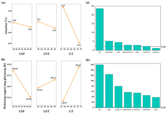
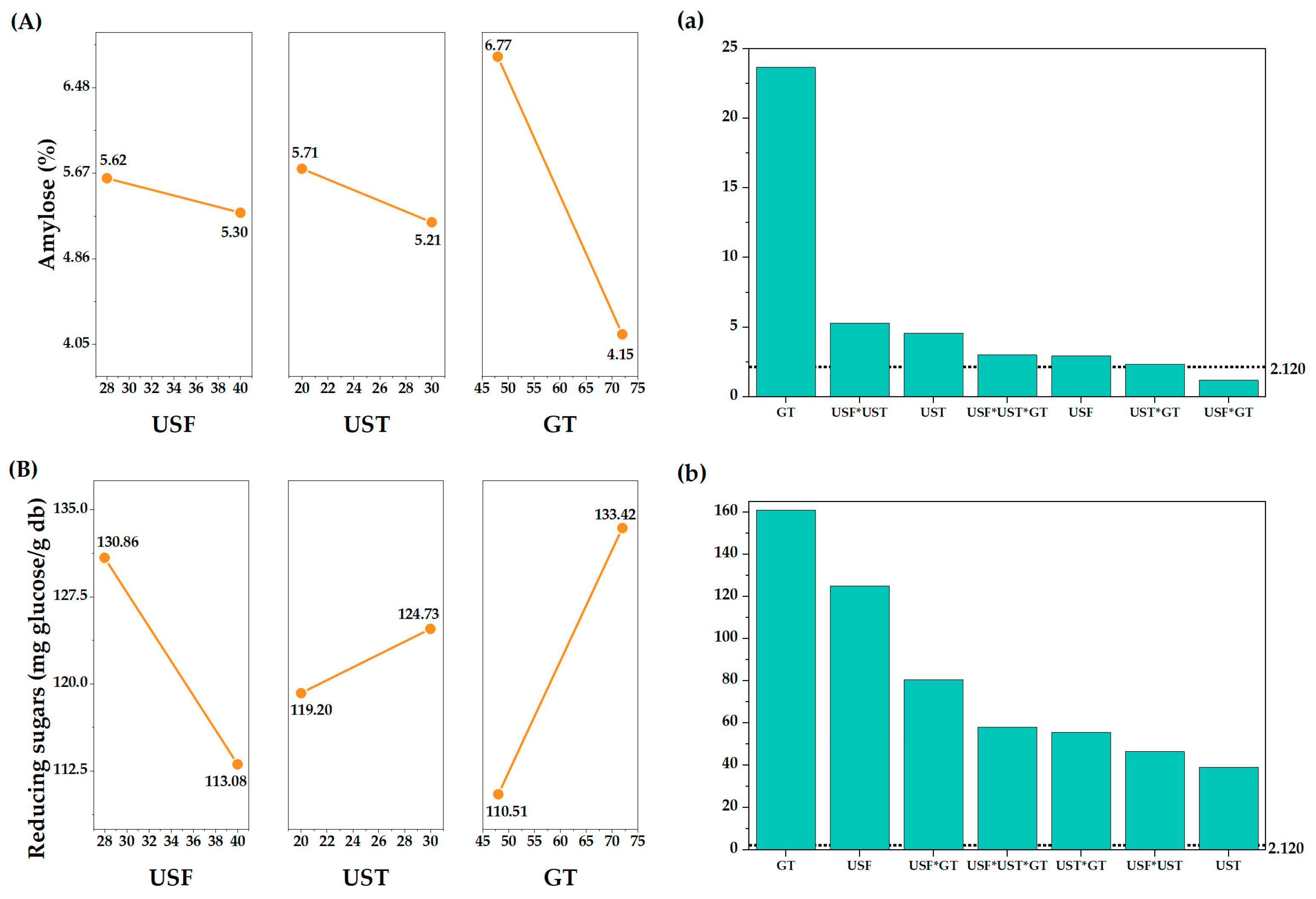
Amylose, a component of starch, and reducing sugars, products of its hydrolysis, are affected by both the germination process and ultrasound pretreatment (Figure 4a,b). In grains such as rice, ultrasound can induce surface cracking, facilitating water penetration and amylose leaching. The absorbed water stimulates the embryo to release gibberellic acid, a chemical promoter of germination [20,27]. In the case of amylose in quinoa flours (Figure 4A), a notable decrease is observed with increasing germination time (GT), confirming greater enzymatic activity over longer periods. Conversely, reducing sugars (Figure 4B) showed an upward trend, with more pronounced increases at 72 h of germination. These structural changes are consistent with the grain’s metabolic transition, in which the breakdown of starch reserves provides energy and soluble compounds for embryo growth. The activation of latent enzymes during germination rapidly degrades carbohydrates, leading to an increase in reducing sugars, while other enzymes break down cell walls and improve the accessibility of internal nutrients [45]. Similar increases were reported for whole-grain brown rice stimulated at 28 kHz, resulting in the degradation of starch particles under enzymatic attack and thus increasing the levels of reducing sugars [40].
Figure 4.
Main effects plot of ultrasonic frequency (USF), ultrasound time (UST), and germination time (GT) on (A) amylose and (B) reducing sugars of germinated black quinoa. Graphs (a,b) show the standardized effects of the factors on the response variables.
3.2.3. Functional Groups
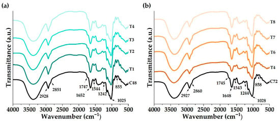
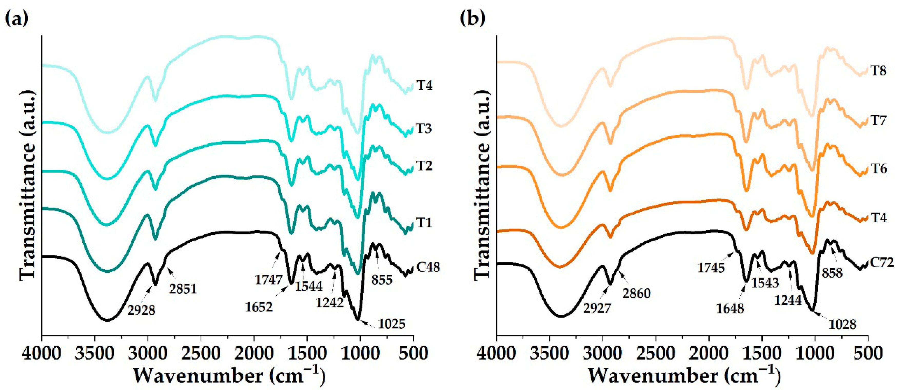
A vibrational analysis was performed using Fourier transform infrared spectrophotometry (FTIR) to study the functional groups (Figure 5) in black quinoa flour samples germinated at 48 h and 72 h, previously treated with ultrasound at different frequencies and exposure times (T1–T8). The spectra showed bands typical of carbohydrate- and protein-rich materials, in the 4000 to 500 cm−1 regions. Characteristic groups of the starch skeleton composed mainly of –CH–, –CH2–, C–OH and –OH were found identified in the bands 2928 cm−1, 2851 cm−1, 1025 cm−1 and 855 cm−1 for samples germinated at 48 h (Figure 5a) and in the bands 2927 cm−1, 2860 cm−1, 1028 cm−1 and 858 cm−1 for samples germinated at 72 h (Figure 5b), the ultrasonic slightly modified the frequencies of the methyl vibration peaks (2928–2927, Figure 5a and Figure 5b, respectively) due to the destruction of hydrogen bonds between the amorphous and crystalline regions [8]. The 1745 –1747 cm−1 bands are associated with ester carbonyl stretching, which is mainly present in lipid components. However, this signal can also include contributions from carbonyl groups attached to the reducing ends of starch polymers, such as amylose and amylopectin. In flour matrices, this spectral region reflects overlapping signals from esterified lipids, carboxylic acids, and carbohydrates [18,46]. As for the bands centered at 1652–1648 cm−1 and 1544–1543 cm−1, they correspond to amide I (C=O) and amide II (N–H) vibrations, respectively, characteristic of proteins, and the 1242 cm−1 (Figure 5a) – 1244 cm−1 (Figure 5b) band corresponds to C–N vibration modes associated with amino acids [18,33,35]. Similarly, the characteristic peaks of C=O, C–N, and N–H shifted slightly towards higher frequencies, indicating that the interactions between proteins and starch were weakened [47].
Figure 5.
Functional groups (a) samples germinated at 48 h and treated with ultrasound, (b) samples germinated at 72 h and treated with ultrasound.
3.3. Thermal Properties
3.3.1. Gelatinization Temperature and Enthalpy
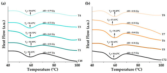
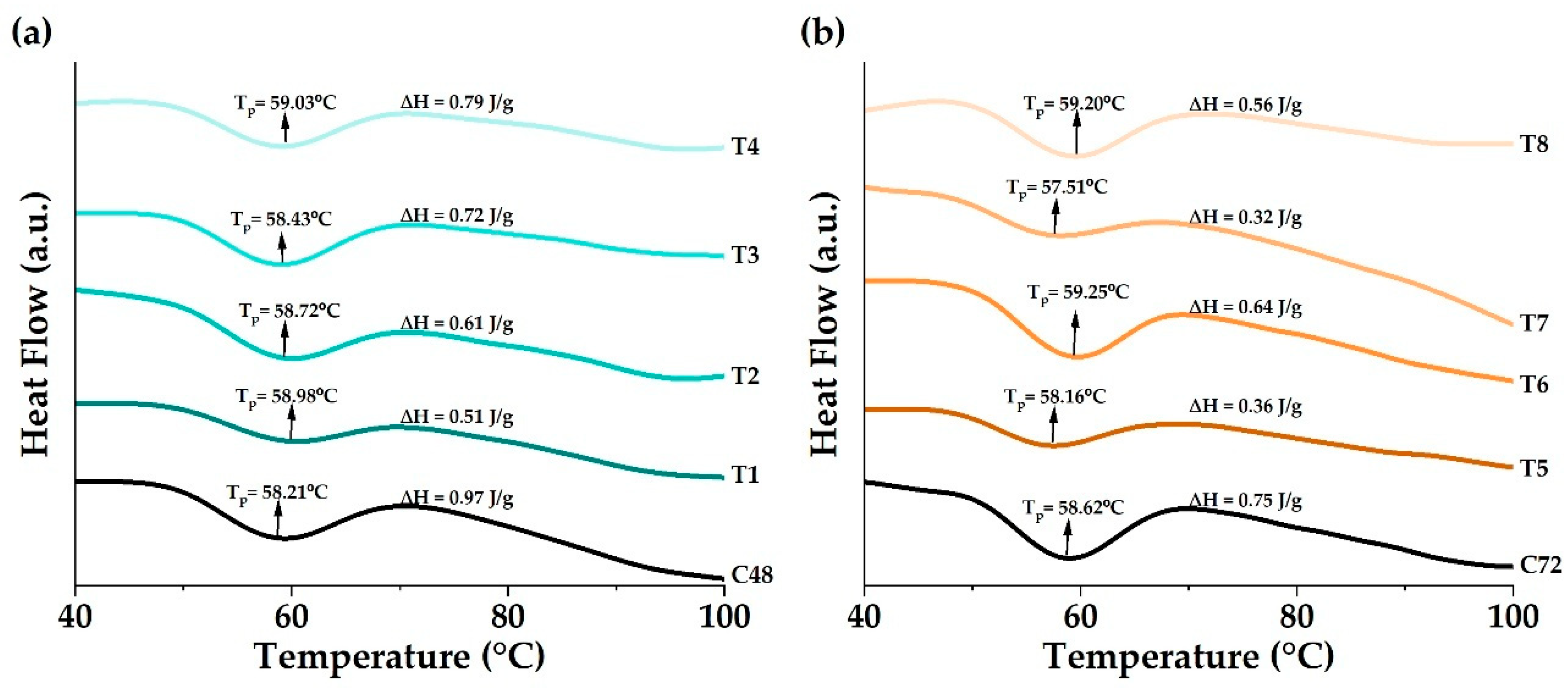
The thermal properties of black quinoa flours were measured using differential scanning calorimetry. The thermograms show the temperature and enthalpy of starch gelatinization (Figure 6a,b). An endothermic transition was observed between 52 and 68 °C, corresponding to starch gelatinization, a process involving the disruption of the amylose and amylopectin crystalline regions during thermal hydration [18]. The thermograms showed differences in thermal behavior between the ultrasound-treated samples and their respective controls (C48: 58.21 °C and C72: 58.62 °C), reflected in shifts in the gelatinization temperatures, especially at T4 (59.03 °C) and T6 (59.25 °C). Ultrasound, especially in combination with germination, disrupts starch granules by promoting the transition from highly ordered (crystalline) structures to disordered amorphous molecules, thereby affecting thermal stability and, consequently, the gelatinization temperature [26,48]. Likewise, at the structural level, amylolytic enzymes partially degrade starch granules, reducing their crystallinity and altering their molecular organization [49]. The slight increase in gelatinization temperature in the germinated samples treated with ultrasound is due to the accumulation of sugars during germination, in addition to the presence of some amino acids and protein hydrolysate [16,33,35,50,51]. This results in a crystalline structure that requires more thermal energy for its decomposition. Conversely, the gelatinization enthalpies decreased, reflecting the loss of molecular structural order, a reduction in crystallinity, and the disruption of double helices in the crystalline and non-crystalline regions of the starch granules [52]. Furthermore, the 72 h samples showed lower ΔH values, confirming that prolonged germination enhances partial starch hydrolysis and acts synergistically with ultrasound. On the other hand, whole-grain quinoa samples are complex systems in which starch molecules can interact with fibers, proteins, and other minor components, thereby generally affecting their heat capacity [53].
Figure 6.
Gelatinization temperature and enthalpy (a) samples germinated at 48 h and treated with ultrasound; (b) samples germinated at 72 h and treated with ultrasound.
3.3.2. Thermal Stability
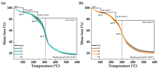
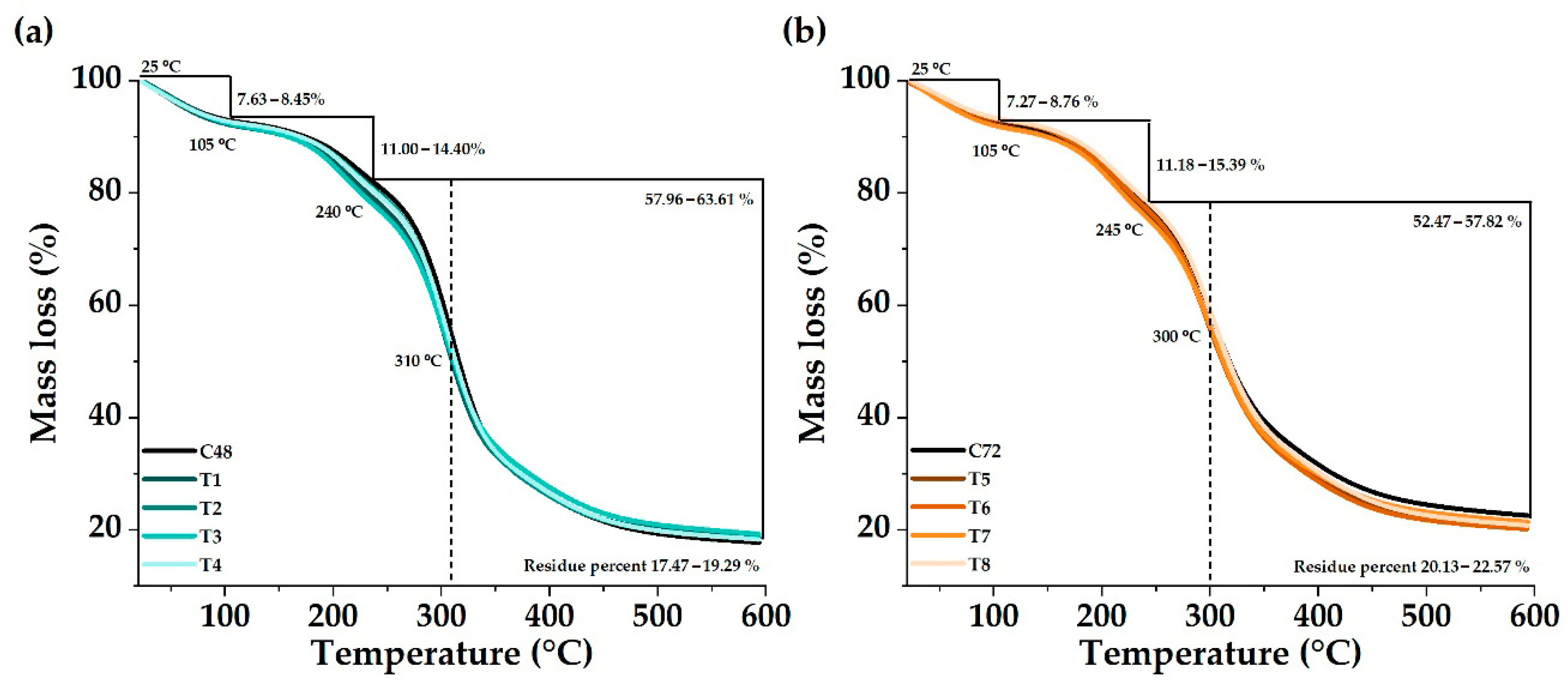
Characterizing a material by its weight loss as a function of temperature is an indicator of its degree of thermal stability and structural compaction, as it reflects the relative proportion of thermally stable fractions compared to those that are more easily degradable [54]. The thermal decomposition of black quinoa samples germinated without ultrasound (C48 and C72) and treated at different frequencies and exposure times (T1–T8) was measured using thermogravimetry (TGA). The TGA thermograms (Figure 7) showed a characteristic behavior of plant matrices composed of carbohydrates, proteins, fibers, lipids, and bioactive compounds, with three stages of mass loss. The first stage corresponds to the loss of moisture and volatile compounds at a temperature below 105 °C. In this region, a mass loss of 7–9% was recorded, associated with the evaporation of physically adsorbed water and the removal of low-molecular-weight volatile components [35,55]. The samples treated with ultrasound showed a slight reduction in the initial mass loss compared to the controls (C48 and C72), suggesting lower moisture retention due to microstructural modifications induced by ultrasonic cavitation, consistent with the lower water activity values observed in Table 2. The second stage corresponds to the degradation of organic biopolymers (up to 240–245 °C). In this stage, thermolabile organic compounds, mainly carbohydrates, proteins, and phenolic acids, decompose. This phase showed losses of 11–15%, with decomposition peaks around 240–310 °C for samples germinated for 48 h (Figure 7a) and 245–300 °C for those germinated for 72 h (Figure 7b). The curves of the treated samples (T1–T8) showed greater thermal stability, that is, a slight shift in degradation towards higher temperatures compared to the controls. This behavior indicates that ultrasound can induce intermolecular interactions (e.g., hydrogen bonds or phenol-protein complexes) that partially stabilize the material’s structure during thermal degradation [56]. The third phase of carbonization and residue formation (320–500 °C) corresponds to the carbonization of recalcitrant compounds, with losses of 52–64% and a final residue between 17.5 and 22.5%. Residues were slightly higher in the samples treated with ultrasound, particularly in T7 and T8, which may be attributed to the formation of thermally more stable structures or to the presence of polymerized phenolic compounds derived from germination and acoustic action. This result aligns with the higher antioxidant capacity reported in Table 1.
Figure 7.
Thermal stability (a) samples germinated at 48 h and treated with ultrasound; (b) samples germinated at 72 h and treated with ultrasound.
3.4. Correlation of Physicochemical Parameters, Bioactive Compounds, and Antioxidant Capacity
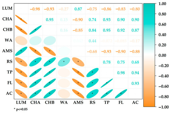
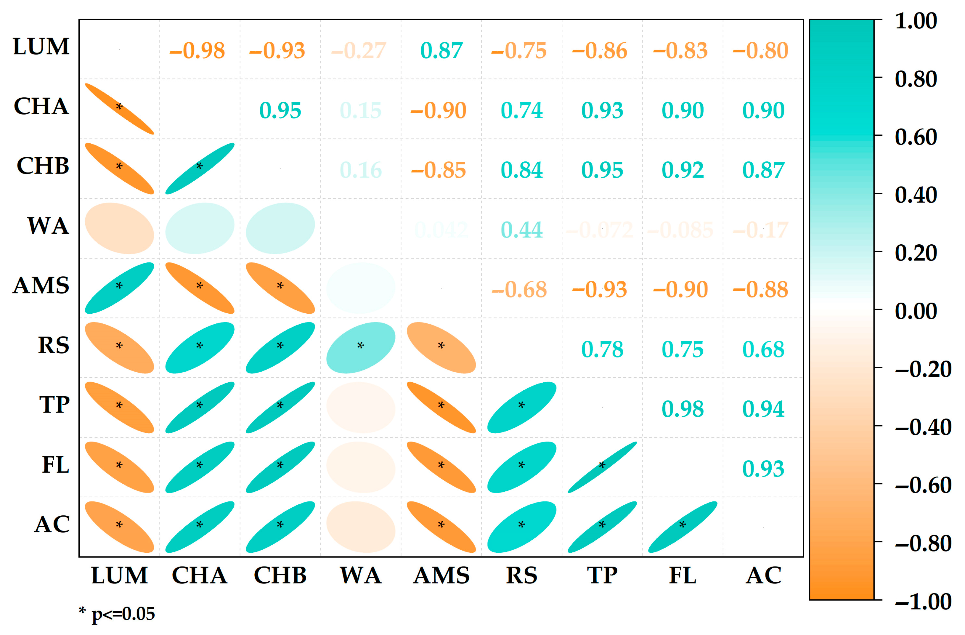
Pearson correlation analysis (Figure 8) revealed significant correlations among physicochemical properties, bioactive compounds, and antioxidant capacity. Very strong positive correlations (r = 0.93–0.94; p < 0.05) were observed between total phenolic compounds (TP), flavonoids (FL), and antioxidant capacity (AC), confirming that the increase in phenolic and flavonoid metabolites directly contributes to a greater capacity for neutralizing free radicals [33,57]. This result supports the evidence that ultrasound-induced synthesis of bioactive compounds is associated with enzyme activation during germination. In contrast, strong negative correlations were observed between lightness (L) and bioactive compounds (r = −0.80 to −0.86), indicating that the darkening of the samples is related to the accumulation of phenolic pigments and flavonoids, as reflected in the instrumental chroma a and b* values. These relationships demonstrate the association between germination and ultrasonic cavitation in modulating the metabolism and nutraceutical functionality of quinoa.
Figure 8.
Pearson correlation matrix between the physicochemical properties, bioactive compounds, and antioxidant capacity.
This study has some limitations, including the evaluation of bioactive compounds based solely on total phenolic and flavonoid contents, without characterization of individual phenolic profiles or non-phenolic antioxidants, and the absence of sensory analysis. In addition, the work was conducted at laboratory scale using a single black quinoa cultivar. Future research should focus on advanced analytical techniques to elucidate qualitative changes in bioactive composition, optimize ultrasound parameters, and assess scale-up feasibility and consumer acceptance for practical application.
4. Conclusions
Ultrasound-assisted germination proved to be an effective strategy to enhance the nutraceutical and functional quality of black quinoa. The combined application of ultrasound and germination promoted the accumulation of bioactive compounds and improved antioxidant capacity, with different ultrasound frequencies favoring distinct responses: treatments at 28 kHz were more effective in enhancing phenolic and flavonoid contents, whereas 40 kHz favored higher antioxidant capacity. Germination time played a key role in these responses, with ultrasound acting as an abiotic stimulus that enhanced the metabolic and structural modifications associated with germination. This approach represents a sustainable and promising technological alternative for adding value to Andean grains and developing functional food ingredients.
Author Contributions
Conceptualization, B.S.R.-P. and D.E.P.-G.; methodology, C.R.Y.-S.; software, B.S.R.-P.; validation, B.S.R.-P. and D.E.P.-G.; formal analysis, C.R.Y.-S.; investigation, B.S.R.-P., D.E.P.-G., C.R.Y.-S., C.A.M.-G. and J.J.L.-B.; data curation, B.S.R.-P. and C.R.Y.-S.; writing—original draft preparation, B.S.R.-P.; writing—review and editing, D.E.P.-G.; visualization, D.E.P.-G.; supervision, B.S.R.-P.; project administration, B.S.R.-P. All authors have read and agreed to the published version of the manuscript.
Funding
This research was funded by the José María Arguedas National University, Peru. Project approved with Resolution No. 230-2024-UNAJMA/CU.
Data Availability Statement
The data are available in the article; further queries can be directed to the corresponding author.
Acknowledgments
The authors thank the Food Nanotechnology Research Laboratory of the Universidad Nacional José María Arguedas.
Conflicts of Interest
The authors declare no conflicts of interest.
References
- Chavan, S.M.; Khadatkar, A.; Hasan, M.; Ahmad, D.; Kumar, V.; Jain, N.K. Quinoa (Chenopodium quinoa Willd.): Paving the way towards nutraceuticals and value-added products for sustainable development and nutritional security. Appl. Food Res. 2025, 5, 100673. [Google Scholar] [CrossRef] [Scilit]
- Agarwal, A.; Rizwana; Tripathi, A.D.; Kumar, T.; Sharma, K.P.; Patel, S.K.S. Nutritional and Functional New Perspectives and Potential Health Benefits of Quinoa and Chia Seeds. Antioxidants 2023, 12, 1413. [Google Scholar] [CrossRef] [Scilit]
- Dixit, V.; Joseph Kamal, S.W.; Bajrang Chole, P.; Dayal, D.; Chaubey, K.K.; Pal, A.K.; Xavier, J.; Manjunath, B.T.; Bachheti, R.K. Functional Foods: Exploring the Health Benefits of Bioactive Compounds from Plant and Animal Sources. J. Food Qual. 2023, 2023, 5546753. [Google Scholar] [CrossRef] [Scilit]
- Ma, Z.F.; Fu, C.; Lee, Y.Y. The Modulatory Role of Bioactive Compounds in Functional Foods on Inflammation and Metabolic Pathways in Chronic Diseases. Foods 2025, 14, 821. [Google Scholar] [CrossRef] [Scilit]
- Vieira, I.R.; Conte-Junior, C.A. Dietary Bioactive Compounds and Human Health: The Role of Bioavailability. Nutrients 2025, 17, 48. [Google Scholar] [CrossRef] [Scilit] [PubMed]
- Flórez-Martínez, D.H.; Rodríguez-Cortina, J.; Chavez-Oliveros, L.F.; Aguilera-Arango, G.A.; Morales-Castañeda, A. Current trends and prospects in quinoa research: An approach for strategic knowledge areas. Food Sci. Nutr. 2024, 12, 1479–1501. [Google Scholar] [CrossRef] [Scilit]
- Gaur, M.; Yadav, S.; Soni, A.; Tomar, D.; Jangra, A.; Joia, S.; Kumar, A.; Mehra, R.; Trajkovska Petkoska, A. Quinoa (Chenopodium quinoa Willd): Nutritional profile, health benefits, and sustainability considerations. Discov. Food 2025, 5, 172. [Google Scholar] [CrossRef] [Scilit]
- Guo, Y.; Qiao, D.; Zhao, S.; Zhang, B.; Xie, F. Starch-based materials encapsulating food ingredients: Recent advances in fabrication methods and applications. Carbohydr. Polym. 2021, 270, 118358. [Google Scholar] [CrossRef] [Scilit]
- Estación Experimental Agraria, Illpa-Puno. Quinua INIA 420-Negra Collana; Instituto Nacional de Innovación Agraria—INIA: Lima, Peru, 2013.
- Apaza Mamani, V.; Cáceres Sanizo, G.; Estrada Zúniga, R.; Pinedo Taco, R.E. Catálogo de Variedades Comerciales de Quinua en el Perú; Instituto Nacional de Innovación Agraria—INIA: Lima, Peru, 2013. [Google Scholar]
- Antezana, R.N.V.; Ticona, G.M.; Yucra, F.E.Z.; Cayllahua, D.Q.; Alejo, R.M.; Churqui, U.J.M. Efecto de la germinación y cocción en las propiedades nutricionales de tres variedades de quinua (Chenopodium quinoa Willd.). Rev. Investig. Altoandin. 2015, 17, 169–172. [Google Scholar] [CrossRef] [Scilit]
- Ding, J.; Feng, H. 5—Controlled germination for enhancing the nutritional value of sprouted grains. In Sprouted Grains; Feng, H., Nemzer, B., DeVries, J.W., Eds.; AACC International Press: St. Paul, MN, USA, 2019; pp. 91–112. [Google Scholar] [CrossRef] [Scilit]
- Dostalíková, L.; Hlásná Čepková, P.; Janovská, D.; Jágr, M.; Svoboda, P.; Dvořáček, V.; Viehmannová, I. The impact of germination and thermal treatments on bioactive compounds of quinoa (Chenopodium quinoa Willd.) seeds. Eur. Food Res. Technol. 2024, 250, 1457–1471. [Google Scholar] [CrossRef] [Scilit]
- Günal-Köroğlu, D.; Esatbeyoglu, T.; Capanoglu, E. Effect of germination on the phenolic compounds: Content, bioavailability, food applications, and health benefits. J. Food Meas. Charact. 2025, 19, 8144–8164. [Google Scholar] [CrossRef] [Scilit]
- Suárez-Estrella, D.; Borgonovo, G.; Buratti, S.; Ferranti, P.; Accardo, F.; Pagani, M.A.; Marti, A. Sprouting of quinoa (Chenopodium quinoa Willd.): Effect on saponin content and relation to the taste and astringency assessed by electronic tongue. LWT 2021, 144, 111234. [Google Scholar] [CrossRef] [Scilit]
- Jimenez, M.D.; Lobo, M.; Sammán, N. 12th IFDC 2017 Special Issue—Influence of germination of quinoa (Chenopodium quinoa) and amaranth (Amaranthus) grains on nutritional and techno-functional properties of their flours. J. Food Compos. Anal. 2019, 84, 103290. [Google Scholar] [CrossRef] [Scilit]
- Guardianelli, L.M.; Salinas, M.V.; Brites, C.; Puppo, M.C. Germination of White and Red Quinoa Seeds: Improvement of Nutritional and Functional Quality of Flours. Foods 2022, 11, 3272. [Google Scholar] [CrossRef] [Scilit] [PubMed]
- Contreras-Jiménez, B.; Torres-Vargas, O.L.; Rodríguez-García, M.E. Physicochemical characterization of quinoa (Chenopodium quinoa) flour and isolated starch. Food Chem. 2019, 298, 124982. [Google Scholar] [CrossRef] [Scilit]
- Altıkardeş, E.; Güzel, N. Impact of germination pre-treatments on buckwheat and Quinoa: Mitigation of anti-nutrient content and enhancement of antioxidant properties. Food Chem. X 2024, 21, 101182. [Google Scholar] [CrossRef] [Scilit]
- Heberle, T.; Rondan, F.S.; Gohlke, G.; Flores, E.M.M.; Ashokkumar, M.; Mesko, M.F. Ultrasound effect on the pre-germination soaking stage of pigmented rice: Impact on nutritional and technological aspects. Ultrason. Sonochem. 2025, 120, 107421. [Google Scholar] [CrossRef] [Scilit]
- Li, R.; Li, Z.-J.; Wu, N.-N.; Tan, B. Effect of pre-treatment on the functional properties of germinated whole grains: A review. Cereal Chem. 2022, 99, 253–269. [Google Scholar] [CrossRef] [Scilit]
- Khan, J.; Li, Y.; Gul, P.; Li, Q.; Liu, K. Drying kinetics and quality characteristics of ultrasound-assisted germination of quinoa grains: A study on functional properties, mineral content and bioactive compounds. Ultrason. Sonochem. 2025, 119, 107393. [Google Scholar] [CrossRef] [Scilit]
- Zhang, J.; Guo, J.; Dang, B.; Zhang, W.; Zheng, W.; Yang, X. Enhancement of Polyphenols and Antioxidant Activity in Germinated Black Highland Barley by Ultrasonication. Molecules 2023, 28, 3679. [Google Scholar] [CrossRef] [Scilit]
- Naumenko, N.; Potoroko, I.; Kalinina, I. Stimulation of antioxidant activity and γ-aminobutyric acid synthesis in germinated wheat grain Triticum aestivum L. by ultrasound: Increasing the nutritional value of the product. Ultrason. Sonochem. 2022, 86, 106000. [Google Scholar] [CrossRef] [Scilit]
- Zhou, Y.; Li, M.; Duan, Y.; Hu, K.; Cai, M.; Zhang, H. Combined frequency ultrasound and controlled germination to improve protein composition, conformation, interactions, and structures in sprouted wheat flour. Int. J. Biol. Macromol. 2025, 308, 142473. [Google Scholar] [CrossRef] [Scilit]
- Tufail, T.; Ul Ain, H.B.; Ashraf, J.; Saeed, F.; Basharat, Z.; Ahmed, Z.; Waseem, M.; Xu, B.; Manzoor, M.F.; Mugabi, R. Effects of germination and ultrasound treatment on the thermodynamics, nutritional and structural quality of highland barley fractions. Ultrason. Sonochem. 2025, 123, 107652. [Google Scholar] [CrossRef] [Scilit]
- Wu, M.; Zhou, Q.; Zhou, L.; Wang, J.; Ren, T.; Zheng, Y.; Lv, W.; Zhao, W. Enhancement of γ-Aminobutyric Acid and the Characteristics of Nutrition and Function in White Quinoa through Ultrasound Stress at the Pre-Germination Stage. Foods 2024, 13, 57. [Google Scholar] [CrossRef] [Scilit]
- Bai, H.; Wu, Y.; Liu, F.; Xuan, D.; Zhao, X.; Dong, W. Enhancing the washing of harvested quinoa seeds with ultrasound-assisted hydration. Ultrason. Sonochem. 2025, 116, 107307. [Google Scholar] [CrossRef] [Scilit]
- Foschi, M.L.; Juan, M.; Pascual, B.; Pascual-Seva, N. Effects of High Intensity Ultrasound Stimulation on the Germination Performance of Caper Seeds. Plants 2023, 12, 2379. [Google Scholar] [CrossRef] [Scilit] [PubMed]
- Gong, M.; Kong, M.; Huo, Q.; He, J.; He, J.; Yan, Z.; Lu, C.; Jiang, Y.; Song, J.; Han, W.; et al. Ultrasonic treatment can improve maize seed germination and abiotic stress resistance. BMC Plant Biol. 2024, 24, 758. [Google Scholar] [CrossRef] [Scilit] [PubMed]
- Huang, S.; Ashraf, U.; Duan, M.; Ren, Y.; Xing, P.; Yan, Z.; Tang, X. Ultrasonic seed treatment improved seed germination, growth, and yield of rice by modulating associated physio-biochemical mechanisms. Ultrason. Sonochem. 2024, 104, 106821. [Google Scholar] [CrossRef] [Scilit] [PubMed]
- Liu, Y.; Wu, Y.; Jia, Y.; Ren, F.; Zhou, S. Effect of GABA combined with ultrasound stress germination treatment on phenolic content and antioxidant activity of highland barley. J. Sci. Food Agric. 2024, 104, 9023–9034. [Google Scholar] [CrossRef] [Scilit]
- Ramos-Pacheco, B.S.; Choque-Quispe, D.; Ligarda-Samanez, C.A.; Solano-Reynoso, A.M.; Palomino-Rincón, H.; Choque-Quispe, Y.; Peralta-Guevara, D.E.; Moscoso-Moscoso, E.; Aiquipa-Pillaca, Á.S. Effect of Germination on the Physicochemical Properties, Functional Groups, Content of Bioactive Compounds, and Antioxidant Capacity of Different Varieties of Quinoa (Chenopodium quinoa Willd.) Grown in the High Andean Zone of Peru. Foods 2024, 13, 417. [Google Scholar] [CrossRef] [Scilit]
- Ligarda-Samanez, C.A.; Choque-Quispe, D.; Moscoso-Moscoso, E.; Pozo, L.M.F.; Ramos-Pacheco, B.S.; Palomino-Rincón, H.; Gutiérrez, R.J.G.; Peralta-Guevara, D.E. Effect of Inlet Air Temperature and Quinoa Starch/Gum Arabic Ratio on Nanoencapsulation of Bioactive Compounds from Andean Potato Cultivars by Spray-Drying. Molecules 2023, 28, 7875. [Google Scholar] [CrossRef] [Scilit]
- Ramos-Pacheco, B.S.; Ligarda-Samanez, C.A.; Choque-Quispe, D.; Choque-Quispe, Y.; Solano-Reynoso, A.M.; Choque-Quispe, K.; Palomino-Rincón, H.; Taipe-Pardo, F.; Peralta-Guevara, D.E.; Moscoso-Moscoso, E.; et al. Study of the Physical–Chemical, Thermal, Structural, and Rheological Properties of Four High Andean Varieties of Germinated Chenopodium quinoa. Polymers 2025, 17, 312. [Google Scholar] [CrossRef] [Scilit] [PubMed]
- Choque-Quispe, D.; Obregón Gonzales, F.H.; Carranza-Oropeza, M.V.; Solano-Reynoso, A.M.; Ligarda-Samanez, C.A.; Palomino-Ríncón, W.; Choque-Quispe, K.; Torres-Calla, M.J. Physicochemical and technofunctional properties of high Andean native potato starch. J. Agric. Food Res. 2024, 15, 100955. [Google Scholar] [CrossRef] [Scilit]
- Gil, D.B.; Bocourt, E.C.; Maqueira, Y.D. Determinación de azúcares reductores totales en jugos mezclados de caña de azúcar utilizando el método del ácido 3, 5 dinitrosalicílico. ICIDCA. Sobre Deriv. Caña Azúcar 2006, 40, 45–50. [Google Scholar]
- Kalita, D.; Bhattacharya, S.; Srivastava, B. Predicting enzymatic starch hydrolysis mechanism during paddy malting by vibrational spectroscopy and multivariate calibration analysis. Food Chem. 2018, 259, 89–98. [Google Scholar] [CrossRef] [Scilit]
- Manals-Cutiño, E.; Penedo-Medina, M.; Giralt-Ortega, G. Análisis termogravimetrico y térmico diferencial de diferentes biomasas vegetales. Tecnol. Química 2011, 31, 180–190. [Google Scholar]
- Liu, H.; Li, Z.; Zhang, X.; Liu, Y.; Hu, J.; Yang, C.; Zhao, X. The effects of ultrasound on the growth, nutritional quality and microbiological quality of sprouts. Trends Food Sci. Technol. 2021, 111, 292–300. [Google Scholar] [CrossRef] [Scilit]
- Minchán-Velayarce, H.H.; Bustos, A.-S.; Paucar-Menacho, L.M.; Vidaurre-Ruiz, J.; Schmiele, M. New Frontiers in Cereal and Pseudocereal Germination: Emerging Inducers for Maximizing Bioactive Compounds. Foods 2025, 14, 3090. [Google Scholar] [CrossRef] [Scilit]
- Fontana, A.J., Jr. Understanding the importance of water activity in food. Cereal Foods World 2000, 45, 7–10. [Google Scholar]
- Mu, H.; Xue, S.; Sun, Q.; Shi, J.; Zhang, D.; Wang, D.; Wei, J. Research Progress of Quinoa Seeds (Chenopodium quinoa Wild.): Nutritional Components, Technological Treatment, and Application. Foods 2023, 12, 2087. [Google Scholar] [CrossRef] [Scilit]
- Lu, C.; Guo, J.; Li, P.; Bai, Z.; Cui, G.; Li, P. Physicochemical properties and in vitro digestion of quinoa starch induced by combination of ultrasound and konjac glucomannan. Food Chem. 2025, 463, 141380. [Google Scholar] [CrossRef] [Scilit]
- Estivi, L.; Brandolini, A.; Condezo-Hoyos, L.; Hidalgo, A. Impact of low-frequency ultrasound technology on physical, chemical and technological properties of cereals and pseudocereals. Ultrason. Sonochem. 2022, 86, 106044. [Google Scholar] [CrossRef] [Scilit]
- Ortiz-Gómez, V.; Fernández-Quintero, A.; Roa-Acosta, D.F.; Bravo-Gómez, J.E.; Solanilla-Duque, J.F. Physicochemical Characterization of Quinoa (Chenopodium quinoa cv. Nariño) Co-products Obtained by Wet Milling. Front. Sustain. Food Syst. 2022, 6, 851433. [Google Scholar] [CrossRef] [Scilit]
- Guo, Y.; Song, R.; Zhu, S.; Liu, X.; Wang, X. Effects of ultrasonic treatment on the texture quality of aged rice flour. J. Cereal Sci. 2024, 117, 103918. [Google Scholar] [CrossRef] [Scilit]
- Pacheco, F.C.; Andressa, I.; Pacheco, A.F.C.; Santos, F.R.d.; Cunha, J.S.; Neves, N.d.A.; Vendruscolo, R.G.; Schmiele, M.; Paiva, P.H.C.; Tribst, A.A.L.; et al. Impact of ultrasound-assisted intermittent hydration during pumpkin seed germination on the structure, nutritional, bioactive, physical and techno-functional properties of flours. LWT 2025, 222, 117654. [Google Scholar] [CrossRef] [Scilit]
- Estevinho, B.N.; Rocha, F.; Santos, L.; Alves, A. Microencapsulation with chitosan by spray drying for industry applications—A review. Trends Food Sci. Technol. 2013, 31, 138–155. [Google Scholar] [CrossRef] [Scilit]
- Gutiérrez-Osnaya, L.J.; Hernández-Uribe, J.P.; Castro-Rosas, J.; Román-Gutiérrez, A.D.; Camacho-Díaz, B.H.; Palma-Rodríguez, H.M.; Rodríguez-Marín, M.L.; Hernández-Ávila, J.; Guzmán-Ortiz, F.A. Influence of germination time on the morphological, morphometric, structural, and physicochemical characteristics of Esmeralda and Perla barley starch. Int. J. Biol. Macromol. 2020, 149, 262–270. [Google Scholar] [CrossRef] [Scilit]
- Li, C.; Jeong, D.; Lee, J.H.; Chung, H.-J. Influence of germination on physicochemical properties of flours from brown rice, oat, sorghum, and millet. Food Sci. Biotechnol. 2020, 29, 1223–1231. [Google Scholar] [CrossRef] [Scilit] [PubMed]
- Sandhiya, R.; Buvaneswaran, M.; Sunil, C.K. Effect of annealing and ultrasound treatments on physicochemical, functional, thermal, and structural properties of proso millet starch. Discov. Food 2025, 5, 49. [Google Scholar] [CrossRef] [Scilit]
- Chávez-Murillo, C.E.; Veyna-Torres, J.I.; Cavazos-Tamez, L.M.; de la Rosa-Millán, J.; Serna-Saldívar, S.O. Physicochemical characteristics, ATR-FTIR molecular interactions and in vitro starch and protein digestion of thermally-treated whole pulse flours. Food Res. Int. 2018, 105, 371–383. [Google Scholar] [CrossRef] [Scilit] [PubMed]
- Consumi, M.; Tamasi, G.; Pepi, S.; Leone, G.; Bonechi, C.; Magnani, A.; Donati, A.; Rossi, C. Analytical composition of flours through thermogravimetric and rheological combined methods. Thermochim. Acta 2022, 711, 179204. [Google Scholar] [CrossRef] [Scilit]
- Sofi, S.A.; Rafiq, S.; Singh, J.; Mir, S.A.; Sharma, S.; Bakshi, P.; McClements, D.J.; Mousavi Khaneghah, A.; Dar, B.N. Impact of germination on structural, physicochemical, techno-functional, and digestion properties of desi chickpea (Cicer arietinum L.) flour. Food Chem. 2023, 405, 135011. [Google Scholar] [CrossRef] [Scilit]
- Yan, S.; Wang, Q.; Yu, J.; Li, Y.; Qi, B. Ultrasound-assisted preparation of protein–polyphenol conjugates and their structural and functional characteristics. Ultrason. Sonochemistry 2023, 100, 106645. [Google Scholar] [CrossRef] [Scilit] [PubMed]
- Sarker, U.; Hossain, M.M.; Oba, S. Nutritional and antioxidant components and antioxidant capacity in green morph Amaranthus leafy vegetable. Sci. Rep. 2020, 10, 1336. [Google Scholar] [CrossRef] [Scilit] [PubMed]

Disclaimer/Publisher’s Note: The statements, opinions and data contained in all publications are solely those of the individual author(s) and contributor(s) and not of MDPI and/or the editor(s). MDPI and/or the editor(s) disclaim responsibility for any injury to people or property resulting from any ideas, methods, instructions or products referred to in the content. |
© 2025 by the authors. Licensee MDPI, Basel, Switzerland. This article is an open access article distributed under the terms and conditions of the Creative Commons Attribution (CC BY) license.