


Highlights
What are the main findings?
- The Paediatric Sleep Questionnaire (PSQ) was not effective in identifying obstructive sleep apnoea (OSA) in children referred for sleep studies.
- Among children with moderate-to-severe OSA, positional OSA was associated with higher odds of PSQ-negative status. This association was not observed in the pooled OSA cohort.
What are the implications of the main findings?
- The PSQ and sleep study provide complementary information. A symptom-based screening alone may miss children with moderate-to-severe OSA.
- Positional sleep characteristics may help explain discordance between caregiver-reported symptoms and sleep study findings. Assessing habitual sleep position or caregiver-reported position-specific symptoms may therefore add value to symptom-based evaluations.
Abstract
Background/Objectives: Paediatric Sleep Questionnaire (PSQ) symptom scores can be discordant with polysomnography (PSG)-defined obstructive sleep apnoea (OSA). We characterised this discordance and examined whether positional OSA (POSA) was associated with PSQ-negative status across OSA severity. Methods: This secondary cross-sectional analysis pooled prospective data from two studies of 2–17-year-olds referred for clinically indicated PSG at two Canadian sites. PSQ sleep-related breathing disorder score < 0.33 defined PSQ-negative status; obstructive apnoea–hypopnoea index (OAHI) ≥ 1.5 and ≥5 events/h defined OSA and moderate-to-severe OSA. POSA required a supine-to-non-supine OAHI ratio ≥ 2 and ≥30 min in supine and non-supine positions. Results: Of 475 participants, 141 were PSQ-negative; 101/141 (71.6%) had OSA and 44/141 (31.2%) had moderate-to-severe OSA. Discrimination was limited (area under the curve [AUC]: OSA, 0.518; moderate-to-severe OSA, 0.569). There was no clear association between POSA and PSQ-negative status (adjusted odds ratio [OR] 1.20, 95% CI 0.71–2.00). A post hoc interaction analysis indicated variation by the presence of moderate-to-severe OSA (ratio of ORs 5.37, 95% CI 1.87–15.4). Within moderate-to-severe OSA, PSQ-negative participants showed no clear evidence of lower PSG-defined severity. POSA occurred in 54.8% of PSQ-negative versus 28.1% of PSQ-positive participants and remained associated in the OAHI-adjusted model (OR 2.94, 95% CI 1.38–6.38). Conclusions: PSQ-negative status did not preclude moderate-to-severe OSA among referred children. Within moderate-to-severe OSA, POSA was associated with PSQ-negative status after accounting for overall OAHI and measured covariates. Prospective studies should determine whether positional characteristics measurable before PSG can help characterise symptom–PSG discordance and complement symptom-based assessment.
1. Introduction
Obstructive sleep apnoea (OSA) affects approximately 1–5% of children and is characterised by recurrent upper-airway obstruction during sleep, resulting in fragmented sleep, increased respiratory and arousal burden, and recurrent intermittent hypoxaemia [1,2]. These physiological disturbances are associated with adverse neurobehavioural, cardiovascular and metabolic outcomes [3,4,5,6], highlighting the importance of early identification of clinically significant OSA. Overnight polysomnography (PSG) objectively measures respiratory events, oxygenation and sleep architecture and remains the reference standard for diagnosis and severity assessment, principally through the obstructive apnoea-hypopnoea index (OAHI) [1]. However, PSG requires specialised equipment, trained personnel, and overnight monitoring, limiting the number of children who can be assessed in many clinical settings. Therefore, symptom-based measures are widely used to support clinical assessment and prioritisation for PSG.
The Paediatric Sleep Questionnaire (PSQ) Sleep-Related Breathing Disorder scale is one such measure, combining caregiver-reported snoring, breathing-related symptoms, daytime sleepiness and behavioural features into a composite score, with 0.33 commonly used as the threshold for a positive result [7]. The PSQ captures caregiver-observed manifestations of sleep-disordered breathing in daily life, whereas PSG quantifies the frequency and physiological consequences of obstructive respiratory events during a single monitored night. Questionnaire score and PSG-defined disease severity therefore need not vary in parallel. This distinction is reflected in many studies, in which the ability of the PSQ to identify PSG-defined OSA has differed substantially across otherwise healthy, obesity-enriched, craniofacial and medically complex paediatric populations [8,9,10,11,12].
From a clinical perspective, PSQ-PSG discordance matters because symptom-based assessment may inform decisions about further evaluation, yet it cannot quantify physiological disease burden or definitively exclude OSA. This discordance is particularly consequential when a child whose PSQ score remains below the conventional threshold nevertheless has moderate-to-severe OSA on PSG, potentially contributing to under-recognition or delayed evaluation of clinically important OSA.
Characterising factors associated with PSQ-PSG discordance may clarify the limitations of symptom-based assessment and guide future research into accessible indicators that could be evaluated before PSG. One potential correlate of such discordance is the intra-night distribution of obstructive events, which is not captured by overall OAHI alone. Positional OSA (POSA), commonly defined by a supine OAHI at least twice the non-supine OAHI, identifies children whose respiratory burden is disproportionately concentrated during supine sleep [13]. Several putative pathways could plausibly link positional phenotype to PSQ–PSG discordance. First, when obstructive events are concentrated in the supine position, the respiratory manifestations experienced or noticed may depend on how often that supine position occurs. Second, caregiver-reported symptoms are influenced by the opportunity to observe the child during sleep, which may vary with sleeping arrangements and the periods during which caregivers are present. Third, age may be relevant both to differences in OSA phenotype and to caregiver observation, as older children may be more likely to sleep independently.
Recent paediatric studies suggest that POSA is accompanied by differences in clinical as well as physiological characteristics, although POSA has also frequently been associated with lower overall OAHI [14,15,16]. Tang et al. [14], for example, found less mouth breathing and dyspnoea during sleep among children with POSA, while broader measures of daytime sleepiness, quality of life and behaviour did not differ clearly; however, the POSA group also had milder overall OSA, and these symptom comparisons did not establish an association independent of OAHI. Prior work thus raises the possibility that positional phenotype may relate to symptom expression but has not established whether this reflects positional characteristics beyond aggregate disease severity, nor has it directly examined POSA as a correlate of PSQ–PSG discordance. This question may be particularly relevant within moderate-to-severe OSA, where a questionnaire-negative result can coexist with clinically important PSG-defined disease.
We therefore characterised PSQ–PSG discordance in children referred for clinically indicated PSG and examined whether positional phenotype was associated with PSQ-negative status. We aimed to (1) evaluate the ability of the PSQ score to discriminate PSG-defined OSA and moderate-to-severe OSA and compare clinical and polysomnographic characteristics according to PSQ status; and (2) examine the association between POSA and PSQ-negative status across children with OSA, test whether it varied according to disease severity, and further characterise the association within moderate-to-severe OSA.
2. Materials and Methods
2.1. Study Design and Participants
This was a secondary cross-sectional analysis of prospectively collected data from children and adolescents aged 2–17 years with symptoms suggestive of OSA who were referred for clinically indicated overnight PSG. Participants were prospectively recruited at two participating paediatric sleep centres, The Hospital for Sick Children (SickKids) and Richmond Hill Sleep and Lung Institute (RHSLI), between December 2024 and July 2026, with questionnaire and anthropometric assessments completed before PSG. Referrals for PSG were made by a paediatric sleep clinician following a clinical interview, where PSQ was not used to determine referral or PSG prioritisation. The analysis used pooled data from two ongoing study protocols that enrolled children using comparable eligibility criteria and study assessments. The administration of the PSQ and the PSG protocol were standardised across the two centres. Both studies excluded children with previous upper airway surgery or adenotonsillectomy, and those with known craniofacial, syndromic or neuromuscular conditions; additional study-specific exclusions concerning previous OSA diagnosis and chronic oral corticosteroid use are summarised in Table S1. No children were enrolled in both studies. Both study protocols were approved by the SickKids Research Ethics Board (REB Nos. 3143 [approved on 15 August 2024] and 3431 [approved on 6 May 2025]). All participants and parents provided written informed consent and/or assent, as appropriate, prior to undergoing any study procedures.
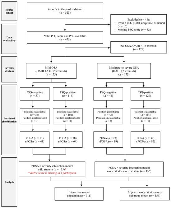
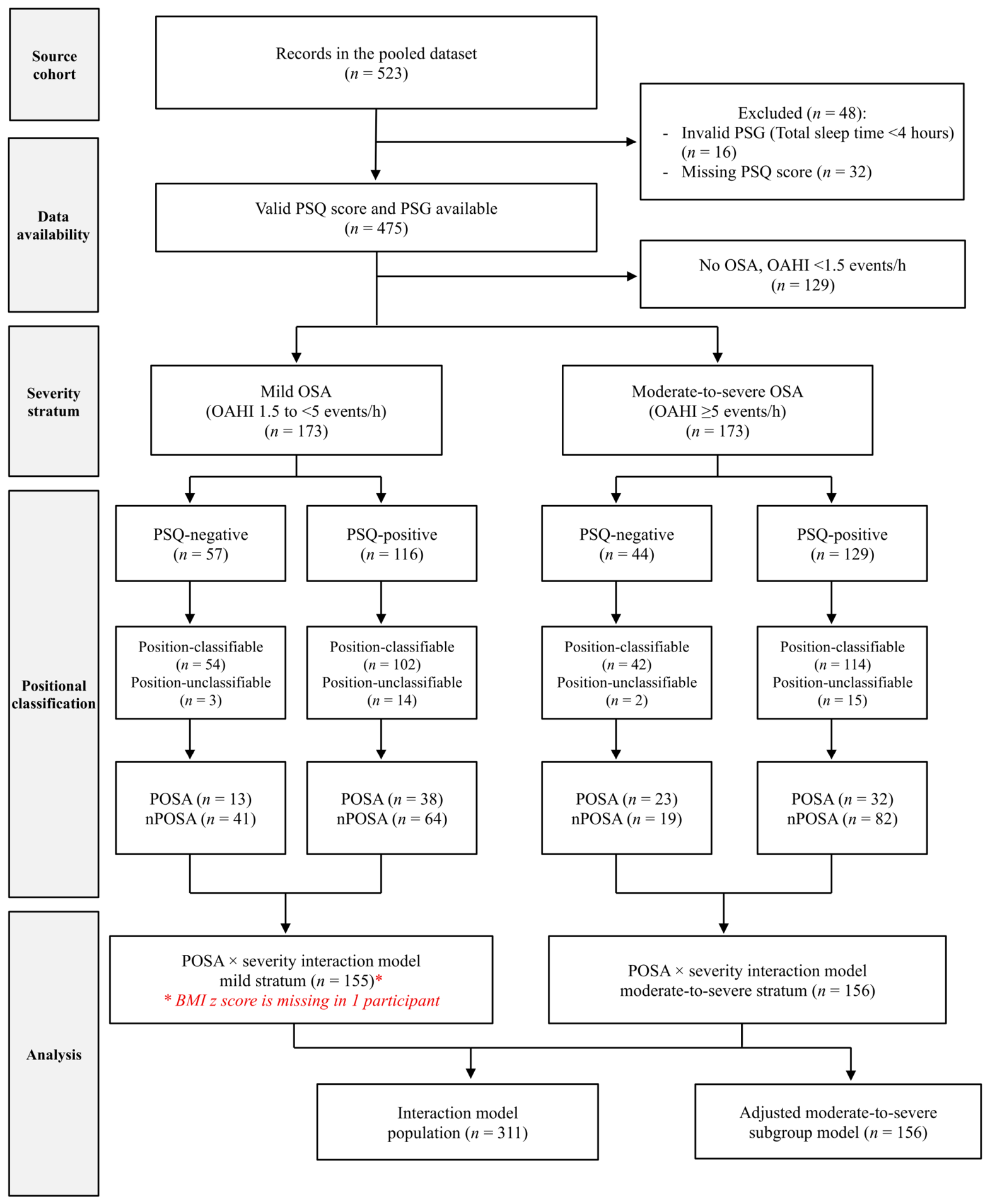
Participant selection and analysis-specific denominators are shown in Figure 1. No a priori sample-size calculation was performed for this analysis, and all available eligible records were considered. Reporting followed the STROBE statement for cross-sectional studies [17].
Figure 1.
Participant selection and analytic populations. Of 523 pooled records, 475 had both a PSQ score and a valid PSG (16 lacked a valid PSG; 32 lacked a PSQ score). Of these, 129 had no OSA, 173 had mild OSA and 173 had moderate-to-severe OSA. In each of the two OSA severity strata, 156/173 participants were position-classifiable (mild: 54/57 PSQ-negative, 102/116 PSQ-positive; moderate-to-severe: 42/44 PSQ-negative, 114/129 PSQ-positive). The POSA × severity interaction model included 311 participants with complete covariate data (155 mild, 156 moderate-to-severe OSA); one position-classifiable participant with mild OSA was excluded because the BMI z-score was unavailable. All 156 position-classifiable participants with moderate-to-severe OSA contributed to the adjusted subgroup model. BMI, body mass index; OAHI, obstructive apnoea–hypopnoea index; OSA, obstructive sleep apnoea; POSA, positional obstructive sleep apnoea; PSQ, Paediatric Sleep Questionnaire; PSG, polysomnography.
2.2. Paediatric Sleep Questionnaire (PSQ)
The PSQ was administered on the night of PSG, where PSQ data were collected solely for research purposes after referral and scheduling. Caregivers completed the 22-item Sleep-Related Breathing Disorder scale of the PSQ, which assesses snoring, breathing-related symptoms, daytime sleepiness and behavioural features [7]. Responses were scored as “yes” = 1 and “no” = 0; “don’t know” and missing responses were excluded from the denominator, and the total score was calculated as the proportion of definitively answered items with an affirmative response. Scores ≥ 0.33 were classified as PSQ-positive and scores < 0.33 as PSQ-negative. No minimum number of affirmative responses was imposed for a valid calculable score. Robustness to questionnaire completeness was examined by repeating analyses among participants with at least 18 of 22 definitively answered items, representing a pragmatic ≥ 80% completeness criterion.
2.3. Polysomnography (PSG) and OSA Severity Classification
Participants underwent standardised overnight PSG, with scoring according to the latest guidelines [18]. For the present analysis, PSG recordings were considered valid when total sleep time (TST) was available and ≥240 min (4 h); recordings not meeting this criterion were excluded from the analyses. Recordings were scored by registered sleep technologists and interpreted by a paediatric sleep physician. The OAHI was defined as the number of obstructive and mixed apnoeas and hypopnoeas per hour of sleep. OSA was defined as OAHI ≥ 1.5 events/h and categorised as mild (1.5 to <5 events/h), moderate (5 to <10 events/h), or severe (≥10 events/h); moderate-to-severe OSA was therefore defined as OAHI ≥ 5 events/h. The cutoff of ≥1.5 events/h was selected based on expert clinical experience [19] and aligned with previously published paediatric PSG studies using this severity classification [20,21,22].
2.4. Positional OSA Definition
Among children with OSA, POSA was defined as a supine-to-non-supine OAHI ratio ≥ 2 with at least 30 min of both supine and non-supine sleep, ensuring a minimum absolute observation period from which each positional event rate could be estimated. This approach is consistent with recent paediatric positional-OSA studies [14,15,16]. Children with adequate sleep time in both supine and non-supine sleep and a supine-to-non-supine OAHI ratio < 2 were classified as non-positional OSA (nPOSA). Children without adequate positional sleep time were classified as position-unclassifiable and were excluded only from analyses requiring POSA/nPOSA classification.
2.5. Covariates
Patients’ demographics and anthropometrics (age, sex, height, weight and body mass index (BMI)) were recorded at the time of the diagnostic PSG. BMI z-scores were derived using the World Health Organization (WHO) growth standards [23,24]. Tonsillar size was graded using the Brodsky scale [25] and examined as an additional sensitivity covariate.
2.6. Statistical Analysis
Continuous variables were summarised as medians and interquartile ranges, and categorical variables as frequencies and percentages. Between-group comparisons used Wilcoxon rank-sum tests for continuous variables and Fisher’s exact tests for categorical variables.
As the present research question was not a pre-specified endpoint of either parent study and no analysis plan for this question was specified before these analyses, all inferential analyses were considered exploratory. Analyses were organised in a clinically motivated sequence: characterisation of PSQ–PSG discordance and diagnostic performance, comparison of clinical and polysomnographic characteristics by PSQ status, assessment of the POSA–PSQ association across OSA severity, and further characterisation within moderate-to-severe OSA. PSQ completeness criteria, REM-related OSA analyses and leave-one-out refitting were used as sensitivity or ancillary analyses to assess the robustness of the main findings.
The ability of the PSQ score to discriminate OSA (OAHI ≥ 1.5 events/h) and moderate-to-severe OSA (OAHI ≥ 5 events/h) was evaluated using receiver operating characteristic (ROC) analysis. Areas under the ROC curve (AUCs) and 95% confidence intervals (CIs) were estimated using DeLong’s method [26]. Sensitivity, specificity, positive predictive value and negative predictive value were calculated for both OAHI cutoffs using PSQ ≥ 0.33 as positive, with exact binomial 95% CIs. Additionally, for moderate-to-severe OSA, an internally derived Youden-optimal PSQ threshold was examined for exploratory purposes only, with stability assessed across 1000 bootstrap resamples.
Among participants with OSA (OAHI ≥ 1.5 events/h), PSQ-negative and PSQ-positive participants were compared across demographic, clinical and polysomnographic characteristics; the same comparisons were repeated within the moderate-to-severe subgroup.
Among children with OSA and a classifiable positional phenotype, the pooled unadjusted association between POSA and PSQ-negative status was estimated using Fisher’s exact test. Logistic regression then estimated the adjusted pooled association among participants with complete covariate data, adjusting for age, sex and BMI z-score. A post hoc POSA × moderate-to-severe OSA interaction adjusted for the same covariates was used to explore whether the association varied according to OSA severity. Severity-specific POSA odds ratios (ORs) and their ratio were obtained using model-based linear contrasts. The interaction analysis was considered hypothesis-generating and served to contextualise the subsequent moderate-to-severe subgroup analysis.
Within the moderate-to-severe OSA stratum, POSA and nPOSA participants were compared across demographic, clinical, and polysomnographic characteristics. The unadjusted association between POSA and PSQ-negative status was estimated using Fisher’s exact test. Adjusted logistic regression first included POSA versus nPOSA, age, sex and BMI z-score. Ln(1 + OAHI) was then added to account for variation in aggregate obstructive burden within the moderate-to-severe stratum. OAHI was log-transformed to reduce the influence of its right-skewed distribution. Additional adjustments for study site and tonsil size were also examined in sensitivity analyses. Unadjusted ORs from Fisher’s exact tests were estimated by conditional maximum likelihood with exact conditional 95% CIs; individual logistic-regression coefficients were reported with profile-likelihood 95% CIs, and model-based contrasts with Wald 95% CIs.
Further sensitivity analyses restricted PSQs to those with at least 18 of 22 definitively answered items. Leave-one-out analyses assessed whether individual observations materially influenced the OAHI-adjusted moderate-to-severe subgroup or interaction estimates. Exploratory ancillary analyses additionally adjusted the moderate-to-severe subgroup model for REM sleep exposure and, separately, REM-related OSA. For each analysis, the subgroup model was refitted in the same complete-case population before the addition of the REM variable to permit like-for-like comparison; harmonisation of REM-related OSA is described in the Supplementary Methods.
Adjusted models used complete observations for included variables; missing values were not imputed. No multiplicity adjustment was applied to the other exploratory comparisons or regression analyses. All tests were two-sided with α = 0.05. Analyses were performed using R Software (version 4.4.1; R Foundation for Statistical Computing, Vienna, Austria).
3. Results
3.1. Participants, PSQ Status and Diagnostic Performance
Participant selection and analysis-specific populations are shown in Figure 1. Distribution of participants according to the research protocol and the study site is summarised in Table S2. Among 475 participants with both a PSQ score and valid PSG, 141 (29.7%) were PSQ-negative and 334 (70.3%) PSQ-positive. The median (IQR) number of affirmative answers on the PSQ was 21 (20−22), ranging from 12 to 22, with 38.5% of participants responding to all 22 items and 92% answering at least 18 items. There were no significant differences in age, sex, BMI z-score, tonsil size or study site (Table 1).
Table 1.
Characteristics of participants with a PSQ score and valid PSG, stratified by PSQ status.
PSQ-negative status occurred across the full spectrum of PSG-defined OSA severity. Of the 141 PSQ-negative participants, 101 (71.6%) had PSG-defined OSA, including 44 (31.2%) with moderate-to-severe OSA. Conversely, 44/173 (25.4%) participants with moderate-to-severe OSA were PSQ-negative.
Discrimination by the PSQ score was limited for both PSG outcomes. For OSA defined as OAHI ≥ 1.5 events/h, the AUC was 0.518 (95% CI 0.460–0.576), with the confidence interval spanning 0.50. Sensitivity and specificity were 70.8 (95% CI 65.7–75.5) and 31.0 (95% CI 23.2–39.7), and positive and negative predictive values were 73.4 (95% CI 68.3–78.0) and 28.4 (95% CI 21.1–36.6), with positive and negative likelihood ratios of 1.03 (95% CI 0.90–1.17) and 0.94 (95% CI 0.69–1.28). For moderate-to-severe OSA defined as OAHI ≥ 5 events/h, the AUC was 0.569 (95% CI 0.516–0.622). Sensitivity and specificity were 74.6 (95% CI 67.4–80.9) and 32.1 (95% CI 26.9–37.7), and positive and negative predictive values were 38.6 (95% CI 33.4–44.1) and 68.8 (95% CI 60.5–76.3), with positive and negative likelihood ratios of 1.09 (95% CI 0.98–1.23) and 0.79 (95% CI 0.58–1.07). (Table S3). The exploratory Youden analysis did not identify a stable alternative threshold.
3.2. Clinical and Polysomnographic Characteristics by PSQ Status Among Children with OSA
Among the 346 participants with OSA, 101 (29.2%) were PSQ-negative and 245 (70.8%) were PSQ-positive (Table 2A). Median OAHI was 4.17 (2.50–8.59) versus 5.22 (3.07–10.27) events/h in PSQ-negative and PSQ-positive participants, respectively (p = 0.056). Mild, moderate and severe OSA accounted for 56.4%, 23.8% and 19.8% of PSQ-negative cases and 47.3%, 26.9% and 25.7% of PSQ-positive cases, respectively; the overall severity distribution did not differ significantly between groups (p = 0.293). There were no significant differences in oxygen desaturation index (p = 0.229), oxygen saturation nadir (p = 0.522), T90 (p = 0.178), arousal index (p = 0.783), or sleep efficiency (p = 0.799). Age, sex, BMI z-score and tonsil size also did not differ significantly between groups. Study site distribution differed, with RHSLI accounting for a higher proportion of PSQ-negative than PSQ-positive participants (82.2% vs. 70.2%; p = 0.023).
Table 2.
Clinical and polysomnographic characteristics by PSQ status among participants with OSA (Panel A) and moderate-to-severe OSA (Panel B).
3.3. Clinical and Polysomnographic Characteristics by PSQ Status in Moderate-to-Severe OSA
Among the 173 participants with moderate-to-severe OSA, 44 (25.4%) were PSQ-negative and 129 (74.6%) were PSQ-positive. Within moderate-to-severe OSA, there were no statistically significant differences in OSA severity between PSQ-positive and PSQ-negative participants as reflected by the PSG parameters examined (Table 2B). Median OAHI was 9.27 (6.95–12.64) events/h in PSQ-negative and 9.70 (6.59–16.20) events/h in PSQ-positive participants (p = 0.739). Severe OSA was present in 20/44 (45.5%) PSQ-negative and 63/129 (48.8%) PSQ-positive participants (p = 0.730). No statistically significant differences were observed in oxygen desaturation index (p = 0.367), oxygen saturation nadir (p = 0.518), T90 (p = 0.570), arousal index (p = 0.264), or sleep efficiency (p = 0.348). Age, sex, study site, BMI z-score and tonsil size also did not differ significantly between groups.
3.4. Association Between POSA and PSQ Status Across OSA Severity
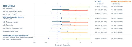
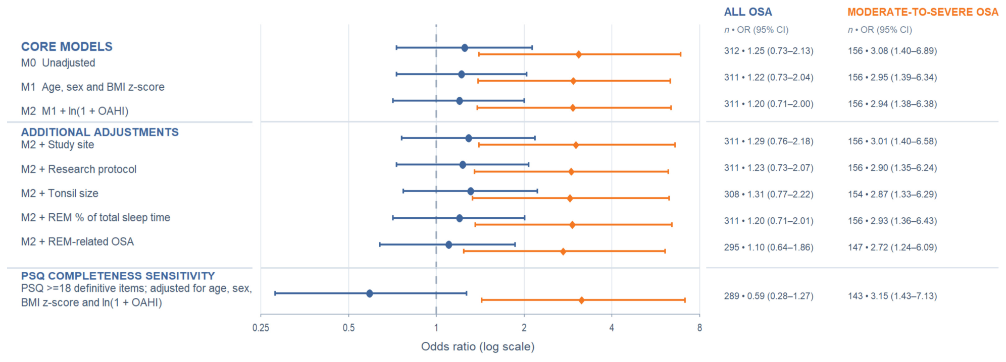
Among the 312 position-classifiable participants with OSA, POSA was present in 36/96 (37.5%) PSQ-negative and 70/216 (32.4%) PSQ-positive participants. In the complete-case pooled model adjusted for age, sex and BMI z-score, there was no significant association between POSA and PSQ-negative status (OR 1.22, 95% CI 0.73–2.04; p = 0.441) (Figure 2 and Table 3A). In the post hoc interaction model comprising 311 participants, the POSA association differed between mild and moderate-to-severe OSA (ratio of ORs 5.37, 95% CI 1.87–15.4; p = 0.002). There was no significant association between POSA and PSQ-negative status within mild OSA (OR 0.54, 95% CI 0.26–1.15; p = 0.110), whereas POSA was associated with higher odds of PSQ-negative status within moderate-to-severe OSA (OR 2.91, 95% CI 1.38–6.17; p = 0.005).
Figure 2.
Association between positional obstructive sleep apnea (POSA) and PSQ-negative status. Forest plot showing the association between POSA and PSQ-negative status across adjusted models and sensitivity analysis. Blue represents all children with OSA; orange represents children with moderate-to-severe OSA. Odds ratios (ORs) and 95% confidence intervals (CIs) are displayed on a logarithmic scale, with the vertical reference line at OR = 1.
Table 3.
Association between positional OSA and PSQ-negative status across OSA severity (Panel A) and within moderate-to-severe OSA (Panel B).
3.5. POSA and PSQ-Negative Status Within Moderate-to-Severe OSA
Following the exploratory interaction analysis, we further characterised positional phenotype within moderate-to-severe OSA and examined whether the POSA–PSQ association persisted after accounting for overall OAHI. Of the 173 participants with moderate-to-severe OSA, 156 (90.2%) were position-classifiable; 55 (35.3%) met criteria for POSA and 101 (64.7%) for nPOSA. Median age was higher among participants with POSA than nPOSA (8.10 versus 5.95 years; p = 0.004), with no significant differences in sex, study site, BMI z-score, tonsil size or overall OAHI (Table 4). Among these 156 participants, POSA was present in 23/42 (54.8%) PSQ-negative participants compared with 32/114 (28.1%) PSQ-positive participants. In the unadjusted analysis, POSA was associated with higher odds of PSQ-negative status (OR 3.08, 95% CI 1.40–6.89; p = 0.003) (Figure 2 and Table 3B). The association remained statistically significant even after adjustment for ln(1 + OAHI), age, sex and BMI z-score (OR 2.94, 95% CI 1.38–6.38; p = 0.005). Additional adjustment for study site produced a materially similar estimate (OR 3.01, 95% CI 1.40–6.58; p = 0.005), as did additional adjustment for tonsil size among participants with available tonsil data (OR 2.87, 95% CI 1.33–6.29; p = 0.008; n = 154) (Figure 2 and Table S4).
Table 4.
Characteristics of position-classifiable participants with moderate-to-severe OSA according to positional phenotype.
3.6. Sensitivity and Ancillary Analyses
Sensitivity analyses were broadly consistent with the main findings (Figure 2 and Table S4). Restricting analyses to PSQs with at least 18 of 22 definitively answered items yielded an adjusted moderate-to-severe subgroup OR of 3.15 (95% CI 1.43–7.13) and a ratio of ORs of 5.09 (95% CI 1.72–15.0). Discrimination of moderate-to-severe OSA remained limited (AUC 0.577, 95% CI 0.521–0.632). Leave-one-out analyses did not identify a single observation that materially determined either model estimate (Supplementary Results). In exploratory ancillary analyses, additional adjustment for REM sleep exposure or REM-related OSA did not materially alter the POSA estimate within moderate-to-severe OSA (Figure 2 and Table S4).
4. Discussion
This study found substantial discordance between PSQ classification and PSG-defined OSA in children referred for PSG. Crucially, PSQ-negative status was not consistently accompanied by lower conventional PSG-defined severity. Among children with OSA, OAHI was numerically lower among PSQ-negative participants, but this difference was not statistically significant and was not accompanied by a corresponding pattern across other OSA severity-related measures such as oxygenation or sleep-fragmentation indices. Among children with moderate-to-severe OSA, there was no clear evidence of lower severity across the PSG parameters examined.
POSA was not associated with PSQ-negative status when OSA was considered across the severity spectrum, but an exploratory severity interaction suggested that the association differed by OSA severity. There was no clear association within mild OSA; within moderate-to-severe OSA, POSA was more common among PSQ-negative participants and remained associated with PSQ-negative status after adjustment for overall OAHI and other clinical characteristics. These findings suggest that symptom–PSG discordance in clinically important paediatric OSA is not readily characterised by lower conventional PSG-defined severity alone and that positional phenotype may help characterise that discordance.
Such discordance is plausible because symptom-based assessment and overnight physiology capture related but non-equivalent constructs. The PSQ reflects caregiver-observed respiratory symptoms, sleepiness and behavioural manifestations in daily life, whereas PSG quantifies respiratory events and their physiological consequences during a single monitored night. Chervin et al. similarly reported only modest correspondence between PSQ scores and individual PSG measures and concluded that the questionnaire was insufficiently reliable to reproduce PSG findings in most individual patients [27]. Similarly, Bseikri et al. found that the PSQ score did not clearly discriminate OSA from non-OSA at various OAHI thresholds (>2, >5, >10, and >25 events/h) among 477 children referred for attended PSG [28]. In contrast, Umano et al. reported substantially higher discrimination (AUC of 0.88) for moderate-to-severe OSA (OAHI >5 events/h) in a small, obesity-enriched cohort. However, the high AUC should not be directly compared with our findings or the wider literature because all participants had PSQ scores ≥ 0.33, and their discrimination analysis was based on a study-specific PSQ threshold of 0.65 [29]. Previous studies have largely examined how well the PSQ identifies PSG-defined OSA; the present analysis instead takes PSQ–PSG discordance as its starting point and examines which clinical and polysomnographic features characterise this discordant subgroup.
Whole-night severity indices summarise obstructive burden across sleep but do not capture the conditions under which events occur. Body position is one such condition: children with comparable overall OAHI may differ in whether obstruction is concentrated during supine sleep or distributed across sleep positions. Recent paediatric studies support POSA as a distinct phenotype. Wang et al. reported a higher prevalence of obesity and allergic rhinitis among children with POSA [16], while Tang et al. described fewer caregiver-observed OSA-related symptoms [14]. However, whether these symptom differences are independent of overall OAHI remains unclear. Body position also influences upper-airway mechanics, and POSA has been associated with greater odds of tongue-base obstruction on paediatric drug-induced sleep endoscopy [30]. One possible explanation for the present finding is that position-dependent respiratory manifestations may be inconsistently captured by caregiver-reported symptoms during habitual sleep, particularly when opportunities to observe sleep are limited. However, habitual sleep position, position-specific symptoms and caregiver opportunity to observe sleep were not measured, and a single monitored night cannot establish habitual positional stability. This remains an untested hypothesis rather than a mechanism demonstrated by the present data.
The absence of an association in the pooled OSA analysis suggests that POSA is not a general correlate of PSQ–PSG discordance. The exploratory interaction instead suggests that the relationship may vary according to OSA severity, although its post hoc nature and the imprecision of the interaction estimate warrant cautious interpretation. The mild-disease estimate provides no clear evidence of an association, whereas an association was observed within moderate-to-severe OSA. Why this relationship should differ by severity is not established; one possibility, consistent with the positional-observability hypothesis above, is that positional concentration may become more relevant to symptom expression as obstructive burden increases.
The association within moderate-to-severe OSA persisted after adjustment for overall OAHI, age, sex and BMI z-score. Adjustment for continuous OAHI helped account for residual variation in obstructive event frequency within the moderate-to-severe OSA stratum. Adjustment for age was important because children with POSA were older than those with nPOSA. Older children may be more likely to sleep independently and therefore be less directly observed by caregivers, potentially reducing caregiver recognition of OSA-related symptoms. Age may also capture developmental differences relevant to the OSA phenotype [13,14,31]. However, neither pathway was directly evaluated in this study. Adjustment for age cannot fully account for age-related behavioural or environmental differences; therefore, residual confounding remains possible. Other unmeasured factors, including upper-airway anatomy and habitual sleep characteristics, may also contribute to the observed association. Although the association persisted after adjustment for measured covariates, these observational findings do not establish a specific mechanism or a causal relationship between POSA and PSQ-negative status.
Estimates were also broadly consistent under additional adjustment for study site, research protocol and tonsil size, and in leave-one-out sensitivity analyses. REM-related sensitivity analyses reduced concern that the association was driven solely by REM-dependent disease, but do not establish a specifically positional mechanism or exclude interactions between sleep stage and body position.
Strengths of this analysis include the use of attended overnight polysomnography, in contrast to the limited-channel testing underlying some previously reported estimates of questionnaire performance [10,29]. Furthermore, the cohort included the full range of PSQ scores rather than restricting inclusion to PSQ-positive children, allowing identification of questionnaire-negative individuals with substantial PSG-defined disease. Analysing the whole OSA severity spectrum before focusing on moderate-to-severe disease also allowed the pooled result to contextualise the subgroup findings rather than evaluating them in isolation.
These strengths should be considered alongside several limitations. As a secondary cross-sectional analysis of a question not pre-specified in either parent study, inference remains exploratory; most analyses were not adjusted for multiplicity, and the post hoc severity interaction was imprecisely estimated and requires prospective confirmation. Generalisability is also limited by the clinically referred population, and the observed discordance frequencies and predictive values should not be extrapolated to community-based screening populations.
Night-to-night variability is another consideration when interpreting PSQ–PSG discordance. Both OSA severity and positional phenotype were assessed on a single monitored night, which cannot capture routine night-to-night variability in respiratory burden or positional distribution [15,32]. In children with POSA, differences in the time spent in supine and non-supine positions across nights could alter the measured OAHI and potentially affect whether the POSA criterion is met. The PSQ, in contrast, captures caregiver-reported symptoms during habitual sleep rather than symptoms specifically observed on the night of PSG. If position-dependent respiratory disturbances vary across nights, or if caregivers observe only some portions of a child’s sleep, reported symptoms may not correspond directly to the respiratory events recorded during a single PSG, potentially contributing to the PSQ–PSG discordance. However, we did not assess habitual sleep position or caregiver observation patterns or perform multi-night PSG; therefore, we could not determine how night-to-night variability influenced the observed association between POSA and PSQ-negative status.
Positional classification required at least 30 min of both supine and non-supine sleep, leaving some participants unclassifiable. There were no significant differences between classifiable and unclassifiable participants across the measured characteristics (Table S5), although the small size of the unclassifiable group means that selection related to measured or unmeasured sleep behaviour cannot be excluded. Some variables, including REM-related measures, were incompletely observed, and adjusted analyses used complete cases without imputation. Residual confounding by incompletely characterised upper-airway features, including adenoidal size and nasal patency, cannot be excluded, while Brodsky grading provides only a coarse measure of tonsillar size. Finally, caregiver opportunity to observe sleep, which may vary with age and sleeping arrangements, was not measured, preventing direct evaluation of the proposed observability pathway. PSQ classification itself relied on caregiver report and permitted variable numbers of definitive responses in the primary scoring, potentially introducing measurement variability around the questionnaire threshold.
5. Conclusions
In this referred cohort, PSQ-negative status did not preclude moderate-to-severe OSA, and within moderate-to-severe disease the discordance was not readily explained by lower conventional PSG-defined severity. POSA was associated with PSQ-negative status in this subgroup, with exploratory evidence that the relationship varied according to OSA severity. These findings support viewing symptom-based assessment and PSG as complementary rather than interchangeable, without establishing a role for POSA in pre-PSG triage or treatment selection. Prospective studies should determine whether positional characteristics obtainable through clinical history or home observation, such as habitual sleep position or caregiver-observed position-specific respiratory symptoms, can be ascertained reproducibly and, if so, whether they relate to symptom–PSG discordance and add value to symptom-based assessment.
Supplementary Materials
The following supporting information can be downloaded at https://www.mdpi.com/article/10.3390/children13111586/s1. The following supplementary materials are published online alongside the manuscript: Supplementary Methods: Additional PSQ and diagnostic-performance analyses. Model-sensitivity analyses. Supplementary Results: Table S1. Summary of the two parent studies. Table S2. Distribution of participants according to the research protocol and the study site. Table S3. Diagnostic performance of the PSQ for PSG-defined OSA. Table S4. Sensitivity and robustness analyses of the POSA–PSQ association. Table S5. Comparison of position-classifiable and position-unclassifiable participants with moderate-to-severe OSA.
Author Contributions
Conceptualization, A.M. and C.T.A.; Methodology, A.M., C.M. and C.T.A.; Formal Analysis, A.M.; Investigation, C.M. and C.T.A.; Resources, G.S., P.P. and I.N.; Data Curation, A.M. and C.T.A.; Writing—Original Draft Preparation, A.M.; Writing—Review and Editing, C.M., G.S., P.P., I.N. and C.T.A.; Visualisation, A.M.; Supervision, I.N. and C.T.A.; Project Administration, C.T.A.; Funding Acquisition, I.N. All authors have read and agreed to the published version of the manuscript.
Funding
This research was funded by the Canadian Institutes of Health Research (CIHR), grant number [195999]. The funding body was not involved in the study design, data collection, analysis, interpretation, writing of the paper, patient recruitment, or any aspect related to the study.
Institutional Review Board Statement
The study was conducted in accordance with the Declaration of Helsinki, and approved by the Research Ethics Board (REB) of The Hospital for Sick Children (REB Nos: 3143 [approved on 15 August 2024] and 3431 [approved on 6 May 2025]).
Informed Consent Statement
Informed consent was obtained from all subjects involved in the study.
Data Availability Statement
The participant-level data underlying this study are not publicly available because of participant privacy and research ethics restrictions. Data may be available from the corresponding author upon reasonable request, subject to institutional and Research Ethics Board approval.
Acknowledgments
We thank all the staff at SickKids Sleep Laboratory and Richmond Hill Sleep and Lung Institute for their administrative and technical support.
Conflicts of Interest
The authors declare no conflicts of interest.
References
- Marcus, C.L.; Brooks, L.J.; Ward, S.D.; Draper, K.A.; Gozal, D.; Halbower, A.C.; Jones, J.; Lehmann, C.; Schechter, M.S.; Sheldon, S.; et al. Diagnosis and Management of Childhood Obstructive Sleep Apnea Syndrome. Pediatrics 2012, 130, e714–e755. [Google Scholar] [CrossRef] [Scilit] [PubMed]
- Lumeng, J.C.; Chervin, R.D. Epidemiology of Pediatric Obstructive Sleep Apnea. Proc. Am. Thorac. Soc. 2008, 5, 242–252. [Google Scholar] [CrossRef] [Scilit] [PubMed]
- Castillo-García, M.; Solano-Pérez, E.; Coso, C.; Romero-Peralta, S.; García-Borreguero, D.; Izquierdo, J.L.; Mediano, O. Impact of Obstructive Sleep Apnea in Cardiovascular Risk in the Pediatric Population: A Systematic Review. Sleep Med. Rev. 2023, 71, 101818. [Google Scholar] [CrossRef] [Scilit] [PubMed]
- Carvalho, D.M.d.; de Almeida, C.M.; Bacelar Ferreira, V.; Batista da Hora, D.A.; Azevedo Soster, L.; Rodrigues Nunes Pinheiro, L.; Macêdo Dantas, J. Behavioral Patterns in Preschool and School-Aged Children with Snoring and Sleep-Disordered Breathing: A Scoping Review. Children 2025, 12, 1614. [Google Scholar] [CrossRef] [Scilit] [PubMed]
- Turgeman, D.; Mazzawi, S. Neurocognitive Deficits in Children with Polysomnographically Confirmed Obstructive Sleep Apnea: A Systematic Review and Meta-Analysis. Int. J. Pediatr. Otorhinolaryngol. 2026, 209, 112989. [Google Scholar] [CrossRef] [Scilit] [PubMed]
- Beeldens, J.; Makhout, S.; Van Hoorenbeeck, K.; Verhulst, S.; Van Eyck, A. The Triadic Relationship between Obesity, Obstructive Sleep Apnea and Metabolic Dysfunction-Associated Steatotic Liver Disease in Children: A Scoping Review. Sleep Breath. 2026, 30, 166. [Google Scholar] [CrossRef] [Scilit] [PubMed]
- Chervin, R.D.; Hedger, K.; Dillon, J.E.; Pituch, K.J. Pediatric Sleep Questionnaire (PSQ): Validity and Reliability of Scales for Sleep-Disordered Breathing, Snoring, Sleepiness, and Behavioral Problems. Sleep Med. 2000, 1, 21–32. [Google Scholar] [CrossRef] [Scilit] [PubMed]
- Lacki, O.A.; Iftikhar, I.H. Meta-Analysis of Diagnostic Test Accuracy of Pediatric Sleep Questionnaire and the OSA-18 Questionnaire in the Screening of Pediatric Obstructive Sleep Apnea. Sleep Med. 2026, 144, 108953. [Google Scholar] [CrossRef] [Scilit] [PubMed]
- Ishman, S.; Heubi, C.; Jenkins, T.; Michalsky, M.; Simakajornboon, N.; Inge, T. OSA Screening with the Pediatric Sleep Questionnaire for Adolescents Undergoing Bariatric Surgery in Teen-LABS. Obesity 2016, 24, 2392–2398. [Google Scholar] [CrossRef] [Scilit] [PubMed]
- Pabary, R.; Goubau, C.; Russo, K.; Laverty, A.; Abel, F.; Samuels, M. Screening for Sleep-Disordered Breathing with Pediatric Sleep Questionnaire in Children with Underlying Conditions. J. Sleep Res. 2019, 28, e12826. [Google Scholar] [CrossRef] [Scilit] [PubMed]
- Kalyoncu, M.; Namlı, N.; Yegit, C.Y.; Yanaz, M.; Gulieva, A.; Ergenekon, A.P.; Selçuk, M.; Atağ, E.; İkizoğlu, N.B.; Sabancı, M.; et al. Is the Pediatric Sleep Questionnaire Sensitive for Sleep-Disordered Breathing in Children with Complex Chronic Disease? Sleep Breath. 2024, 28, 331–337. [Google Scholar] [CrossRef] [Scilit] [PubMed]
- Cielo, C.M.; Silvestre, J.; Paliga, J.T.; Maguire, M.; Gallagher, P.R.; Marcus, C.L.; Taylor, J.A. Utility of Screening for Obstructive Sleep Apnea Syndrome in Children with Craniofacial Disorders. Plast. Reconstr. Surg. 2014, 134, 434e–441e. [Google Scholar] [CrossRef] [Scilit] [PubMed]
- Verhelst, E.; Clinck, I.; Deboutte, I.; Vanderveken, O.; Verhulst, S.; Boudewyns, A. Positional Obstructive Sleep Apnea in Children: Prevalence and Risk Factors. Sleep Breath. 2019, 23, 1323–1330. [Google Scholar] [CrossRef] [Scilit] [PubMed]
- Tang, A.; Dai, S.; Au, C.T.; Yu, M.W.L.; Li, A.M.; Chan, K.C.-C. Prevalence and Risk Factors of Positional Obstructive Sleep Apnea in Chinese Children: A Retrospective Study. Nat. Sci. Sleep 2025, 17, 2933–2946. [Google Scholar] [CrossRef] [Scilit] [PubMed]
- Dai, S.; Yang, M.; Au, C.T.; Yuen, N.T.K.; Zhang, Y.; Tang, A.; Yu, M.W.L.; Li, A.M.; Chan, K.C.C. Supine Position-Related Obstructive Sleep Apnea in Children: Insights from the Childhood Adenotonsillectomy Trial. Sleep Breath. 2025, 29, 230. [Google Scholar] [CrossRef] [Scilit] [PubMed]
- Wang, Q.; Huang, G.; Wang, R.; Cao, Z.; Liang, J.; Li, M.; Gu, Q. Evaluating Positional Obstructive Sleep Apnea in Children: Prevalence, Characteristics, and Risk Factors. Nat. Sci. Sleep 2024, 16, 1569–1581. [Google Scholar] [CrossRef] [Scilit] [PubMed]
- Elm, E.v.; Altman, D.G.; Egger, M.; Pocock, S.J.; Gøtzsche, P.C.; Vandenbroucke, J.P. The Strengthening the Reporting of Observational Studies in Epidemiology (STROBE) Statement: Guidelines for Reporting Observational Studies. Lancet 2007, 370, 1453–1457. [Google Scholar] [CrossRef] [Scilit] [PubMed]
- Troester, M.M.; Quan, S.F.; Berry, R.B.; Plante, D.T.; Abreu, A.R.; Alzoubaidi, M.; Bandyopadhyay, A.; Del Rosso, L.; Ebben, M.; Kwon, Y.; et al. The AASM Manual for the Scoring of Sleep and Associated Events: Rules, Terminology and Technical Specifications, 3rd ed.; American Academy of Sleep Medicine: Darien, IL, USA, 2023. [Google Scholar]
- Stowe, R.C.; Afolabi-Brown, O. Pediatric Polysomnography—A Review of Indications, Technical Aspects, and Interpretation. Paediatr. Respir. Rev. 2020, 34, 9–17. [Google Scholar] [CrossRef] [Scilit] [PubMed]
- Katz, S.L.; Barwell, T.; Bijelić, V.; Barrowman, N.; Blinder, H.; Dussah, N.; Shamsi, R.; Leitman, A.R.; Ersu, R. Preliminary Estimates of the Diagnostic Accuracy of Video Clips for Obstructive Sleep Apnea in Children. Pediatr. Pulmonol. 2025, 60, e71228. [Google Scholar] [CrossRef] [Scilit] [PubMed]
- Ioan, I.; Mulier, G.; Taytard, J.; Césaire, A.; Beydon, N. Evaluation of Obesity and Asthma as Risk Factors for Moderate to Severe Obstructive Sleep Apnea in Children. J. Clin. Sleep Med. 2022, 18, 1639–1648. [Google Scholar] [CrossRef] [Scilit] [PubMed]
- Ward, T.M.; Chen, M.L.; Landis, C.A.; Ringold, S.; Beebe, D.W.; Pike, K.C.; Wallace, C.A. Congruence between Polysomnography Obstructive Sleep Apnea and the Pediatric Sleep Questionnaire: Fatigue and Health-Related Quality of Life in Juvenile Idiopathic Arthritis. Qual. Life Res. 2017, 26, 779–788. [Google Scholar] [CrossRef] [Scilit] [PubMed]
- Group, W.M.G.R.S.; de Onis, M. WHO Child Growth Standards Based on Length/Height, Weight and Age. Acta Paediatr. 2006, 95, 76–85. [Google Scholar] [CrossRef] [Scilit] [PubMed]
- Onis, M.d.; Onyango, A.W.; Borghi, E.; Siyam, A.; Nishida, C.; Siekmann, J. Development of a WHO Growth Reference for School-Aged Children and Adolescents. Bull. World Health Organ. 2007, 85, 660–667. [Google Scholar] [CrossRef] [Scilit] [PubMed]
- Brodsky, L. Modern Assessment of Tonsils and Adenoids. Pediatr. Clin. N. Am. 1989, 36, 1551–1569. [Google Scholar] [CrossRef] [Scilit] [PubMed]
- DeLong, E.R.; DeLong, D.M.; Clarke-Pearson, D.L. Comparing the Areas under Two or More Correlated Receiver Operating Characteristic Curves: A Nonparametric Approach. Biometrics 1988, 44, 837–845. [Google Scholar] [CrossRef] [Scilit] [PubMed]
- Chervin, R.D.; Weatherly, R.A.; Garetz, S.L.; Ruzicka, D.L.; Giordani, B.J.; Hodges, E.K.; Dillon, J.E.; Guire, K.E. Pediatric Sleep Questionnaire: Prediction of Sleep Apnea and Outcomes. Arch. Otolaryngol.–Head Neck Surg. 2007, 133, 216–222. [Google Scholar] [CrossRef] [Scilit] [PubMed]
- Bseikri, M.; Zhang, J.; Kirley, J.; Lee, C.; Castillo, A.; Feliciano, E.M.C. Predicting Obstructive Sleep Apnea Severity in Children Referred for Polysomnography: Use of the Pediatric Sleep Questionnaire and Subscales. Sleep Breath. 2023, 27, 545–552. [Google Scholar] [CrossRef] [Scilit] [PubMed]
- Umano, G.R.; Rondinelli, G.; Luciano, M.; Pennarella, A.; Aiello, F.; Mangoni di Santo Stefano, G.S.R.C.; Di Sessa, A.; Marzuillo, P.; Papparella, A.; Miraglia del Giudice, E. Pediatric Sleep Questionnaire Predicts Moderate-to-Severe Obstructive Sleep Apnea in Children and Adolescents with Obesity. Children 2022, 9, 1303. [Google Scholar] [CrossRef] [Scilit] [PubMed]
- Kirkham, E.M.; Melendez, J.B.; Hoi, K.; Chervin, R.D. Drug-Induced Sleep Endoscopy in Children with Positional Obstructive Sleep Apnea. Otolaryngol. Head Neck Surg. 2021, 164, 191–198. [Google Scholar] [CrossRef] [Scilit] [PubMed]
- Huang, X.; Gong, X.; Gao, X. Age-Related Hypertrophy of Adenoid and Tonsil with Its Relationship with Craniofacial Morphology. BMC Pediatr. 2023, 23, 163. [Google Scholar] [CrossRef] [Scilit] [PubMed]
- Xiao, L.; Terrance, M.; Au, C.T.; Massicotte, C.; Narang, I. High Night-to-Night Variability in Childhood Obstructive Sleep Apnea Severity. Sleep Med. 2026, 140, 108784. [Google Scholar] [CrossRef] [Scilit] [PubMed]

Disclaimer/Publisher’s Note: The statements, opinions and data contained in all publications are solely those of the individual author(s) and contributor(s) and not of MDPI and/or the editor(s). MDPI and/or the editor(s) disclaim responsibility for any injury to people or property resulting from any ideas, methods, instructions or products referred to in the content. |
© 2026 by the authors. Licensee MDPI, Basel, Switzerland. This article is an open access article distributed under the terms and conditions of the Creative Commons Attribution (CC BY) license.