


Highlights
What are the main findings?
- Congenital heart disease was the predominant predisposing condition, and Staphylococcus species were the most frequently identified pathogens.
- Younger age and lower body weight were associated with mortality in this pediatric infective endocarditis cohort.
What are the implications of the main findings?
- Pediatric infective endocarditis continues to carry substantial morbidity and mortality in children with underlying cardiac disease.
- Early recognition and multidisciplinary management, including timely surgical evaluation when indicated, remain essential.
Abstract
Background/Objectives: Infective endocarditis (IE) is a rare but life-threatening infection in children, most often associated with congenital heart disease. This study aimed to evaluate the clinical characteristics, microbiological and echocardiographic findings, management strategies, outcomes, and factors associated with mortality in pediatric IE over a 23-year period at a tertiary care center. Methods: We retrospectively reviewed patients aged 0–18 years diagnosed with definite IE according to the modified Duke criteria between January 2000 and December 2022. Demographic, clinical, microbiological, echocardiographic, therapeutic, and outcome data were analyzed. Results: Seventeen patients were identified (mean age: 9.0 ± 5.9 years; 58.8% male). Congenital heart disease was present in 82%, and 47% had undergone prior cardiac surgery. Blood cultures were positive in 58.8%, with Staphylococcus aureus and Staphylococcus epidermidis being the most frequently identified pathogens. Echocardiography revealed vegetations in 88% of cases. Surgical intervention was performed in 70.5% of patients. Extracardiac complications occurred in 70.5%, most commonly acute kidney injury (35%) and thromboembolic events (23%). Three patients (17.6%) died; younger age and lower body weight were significantly associated with mortality. Conclusions: Pediatric IE continues to carry substantial morbidity and mortality. Younger age and lower body weight were associated with mortality in our cohort; however, the association with lower body weight may be confounded by age. Early recognition and multidisciplinary management, including timely surgical intervention when indicated, remain essential for improving outcomes.
1. Introduction
Infective endocarditis (IE) is a microbial infection of the endocardial surface of the heart and is less frequently encountered in children than in adults [1]. The most important predisposing factor is underlying congenital heart disease (CHD), with complex cyanotic malformations and the presence of intracardiac prosthetic material conferring the greatest risk [2]. The incidence of rheumatic heart disease (RHD) has markedly declined in developed countries and is now primarily a concern in low- and middle-income regions. Less commonly, IE can develop in patients without structural heart disease. Additional risk factors include the increasing use of central venous catheters and underlying immunodeficiencies [3]. The most frequently reported causative microorganisms are viridans group streptococci and Staphylococcus aureus. Diagnosis is established using the modified Duke criteria [4,5]. Despite advances in antimicrobial therapy and surgical techniques, IE in children remains associated with significant morbidity and mortality [6,7]. Outcomes vary according to the underlying cardiac anatomy, the causative pathogen, and the timeliness of diagnosis and intervention. This study aimed to evaluate the clinical characteristics, complications, microbiological profile, management strategies, and outcomes of pediatric IE cases over a 23-year period in a tertiary care center.
2. Materials and Methods
2.1. Study Design and Data Collection
Patients aged 0–18 years who were diagnosed with definite IE according to the modified Duke criteria between January 2000 and December 2022 at Bursa Uludağ University Faculty of Medicine Hospital were included in this study [4]. Currently, our institution is the largest tertiary care center in the Southern Marmara region of Türkiye, with approximately 1000 congenital heart disease diagnoses, 150 congenital cardiac surgical procedures, and 400 catheter-based interventions annually. Demographic data, presenting symptoms, underlying CHD and other predisposing factors, laboratory results, echocardiographic findings, blood culture results, antimicrobial and surgical treatments, and follow-up complications were retrospectively retrieved from electronic medical records. Laboratory parameters were recorded based on admission values. In culture-negative cases, a definite diagnosis of IE was made when one major and at least three minor Duke criteria were fulfilled. Surgical intervention within the first 7 days after diagnosis was defined as early surgery [6]. All patients underwent transthoracic echocardiography performed by two experienced pediatric cardiologists; one patient also underwent transesophageal echocardiography. Roth spots were assessed by an ophthalmologist. The study was conducted in accordance with the Declaration of Helsinki and approved by the Clinical Research Ethics Committee of Bursa Uludağ University Faculty of Medicine (Approval No. 2023-8/5; 12 April 2023).
2.2. Statistical Analysis
Statistical analyses were performed using the Statistical Package for the Social Sciences (SPSS) version 25.0 for Windows (IBM Corp., Armonk, NY, USA). Data were summarized as frequencies, medians with ranges (minimum–maximum), and means with standard deviations, as appropriate. For missing data, the number of available observations was specified. The Shapiro–Wilk test was used to assess normality. The Mann–Whitney U test was applied for comparisons of continuous variables without normal distribution, and Fisher’s exact test was used for categorical variables. A p-value < 0.05 was considered statistically significant.
3. Results
3.1. Patient Characteristics
Over a 23-year period, 17 pediatric patients were diagnosed and treated for definite infective endocarditis. Of these, 58.8% (n = 10) were male and 41.2% (n = 7) were female. The mean age at presentation was 9 ± 5.9 years. Fourteen patients had previously diagnosed CHD, with complex defects in 7 cases. Eight patients had undergone prior cardiac surgery, and prosthetic conduits were present in 5 of them; none had prosthetic heart valves. The mean interval between cardiac surgery and endocarditis diagnosis was 8.6 ± 5.8 years (range: 2.4–17.5 years). Individual patient characteristics, microbiological findings, surgical interventions, and outcomes are summarized in Supplementary Table S1.
No patients had a history of rheumatic heart disease. Two had central venous catheters: one was a renal transplant recipient on immunosuppressive therapy, and the other was a premature infant (29 weeks, birth weight 1270 g) hospitalized in the neonatal intensive care unit with neonatal sepsis. Additional comorbidities included hypogammaglobulinemia (n = 1) and thalassemia (n = 1). Two patients had no identifiable predisposing factors.
Fever was the most common presenting symptom, and 64.7% of patients had received antibiotics prior to admission. Aside from heart murmurs, hepatomegaly was the most frequent physical examination finding. Congestive heart failure was observed in five patients, and no arrhythmias were recorded. Janeway lesions and splinter hemorrhages were identified in one patient; Roth spots and Osler nodes were not detected. The most common laboratory abnormalities at admission were elevated erythrocyte sedimentation rate and C-reactive protein, leukocytosis, and anemia. Low complement levels (C3 and C4) were detected in six patients, and rheumatoid factor positivity in one. Clinical features at presentation are summarized in Table 1, and congenital heart diseases and previous surgical interventions in Table 2.
Table 1.
Characteristics of the patients with infective endocarditis.
Table 2.
Congenital heart diseases and previous cardiac surgeries in patients with IE.
3.2. Echocardiographic Findings
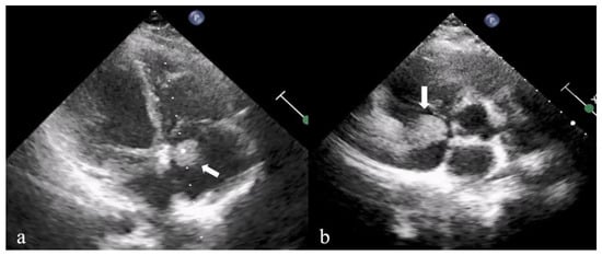
All patients had at least one echocardiographic finding consistent with IE. Transthoracic echocardiography was performed in all cases. In one patient with a ventricular septal defect and repeated positive blood cultures, transesophageal echocardiography was performed despite normal transthoracic findings. Right-sided cardiac involvement was observed in 10 patients, and left-sided involvement in 7. Vegetations were detected in 88.2% of patients (Figure 1). Other findings included new-onset valvular regurgitation, particularly involving prosthetic conduits or valves. The most frequent vegetation sites were the right ventricle, conduits, and the mitral valve, respectively. Echocardiographic findings are summarized in Table 3.
Figure 1.
Transthoracic echocardiography in the apical four-chamber (a) and parasternal short-axis (b) views demonstrate a hyperechoic vegetation (arrow) located within the right atrium.
Table 3.
Echocardiographic findings and blood culture results in patients with IE.
3.3. Microbiology and Causative Agents
A pathogenic microorganism was isolated from blood cultures in 58.8% of patients (n = 10). Two patients had more than one microorganism isolated. The most frequently identified pathogens were Staphylococcus aureus (n = 3), Staphylococcus epidermidis (n = 3), Streptococcus sanguinis (n = 2), Candida albicans (n = 1), Candida lusitaniae (n = 1), Candida parapsilosis (n = 1), and Kytococcus sedentarius (n = 1). Serological tests were not performed in any patient.
3.4. Treatment and Outcomes
3.4.1. Antibiotic and Supportive Treatments
All patients initially received empirical antibiotic therapy, which was subsequently adjusted according to culture and susceptibility results when available. The most commonly used antibiotics were cefotaxime, vancomycin, gentamicin, and meropenem, respectively. The average total duration of treatment was 42.1 ± 14 days (range: 16–81 days). During follow-up, 8 patients required intensive care for mechanical ventilation or inotropic support. A total of 10 patients received low-molecular-weight heparin; 5 of these had previously been on aspirin.
3.4.2. Surgical Interventions
Surgical intervention was required in 12 patients (70.5%). The mean time from diagnosis to surgery was 31.4 ± 28.2 days (range: 3–95 days). Early surgery was performed in two patients: one due to a large vegetation with a cerebral abscess, and another due to recurrent septic pulmonary embolism.
Procedures included conduit replacement (n = 4), vegetation excision (n = 3), vegetation excision with ventricular septal defect repair (n = 2), vegetation excision with aortic valve repair and right ventricular muscle bundle resection (n = 1), vegetation excision with mitral valve replacement (n = 1), and combined aortic and mitral valve replacement (n = 1).
One patient with Candida parapsilosis endocarditis was a 16-year-old male renal transplant recipient with chronic kidney disease and no underlying congenital heart disease. Despite the absence of fever, repeated blood cultures were positive for C. parapsilosis and Staphylococcus epidermidis, and echocardiography revealed a right atrial vegetation. He received liposomal amphotericin B followed by micafungin and underwent vegetation excision 22 days after diagnosis. He was discharged without recurrence during follow-up.
No early or late postoperative mortality was observed among the surgically treated patients.
3.4.3. Extracardiac Complications
Extracardiac complications were observed in 12 patients (70.5%). The most common was acute kidney injury (AKI) (n = 6); of these, 2 had low complement levels and 2 required hemodialysis. Thromboembolic complications occurred in 4 patients. In two, cerebral abscesses and seizures developed due to septic embolism to the central nervous system, resolving without surgical intervention following dexamethasone and anticoagulant therapy; one of these two also had peripheral embolic findings (Janeway lesions and splinter hemorrhages). Another patient developed pulmonary embolism and pleural effusion requiring tube thoracostomy. One patient experienced splenic infarction and required popliteal artery endarterectomy. In one case, gastrointestinal bleeding developed during follow-up and resolved spontaneously.
3.4.4. Mortality and Risk Factors
Three patients (17.6%) died during treatment. One was a premature infant born at 29 weeks with Candida albicans isolated from blood cultures; the patient died on day 41 due to sepsis-related multiorgan failure and necrotizing enterocolitis. Another was a 15-month-old girl with prior atrioventricular septal defect repair and residual moderate left atrioventricular valve regurgitation; Candida lusitaniae was isolated from her blood culture, she had hemodialysis-dependent renal failure, and she died on day 16. The third was a 4-year-old girl with Noonan syndrome and mitral valve prolapse with moderate regurgitation, who presented with a mitral vegetation and severe mitral regurgitation leading to congestive heart failure; blood cultures were negative, and she died on day 40 due to multiorgan failure.
Due to poor clinical status, surgery could not be performed in the last two patients. Analysis of clinical and laboratory parameters showed that younger age and low body weight were associated with mortality (Table 4). Because only three patients died, multivariable analysis could not be performed.
Table 4.
Analysis of factors associated with mortality.
4. Discussion
IE is a systemic infection associated with significant morbidity and mortality. Although rare in children, with an incidence of 0.43–0.84 per 100,000, it poses serious clinical challenges [7,8]. In pediatric IE, CHD remains the predominant predisposing factor, reported in 53.5–87% of cases [7,9]. In a recent study by Demir et al., 74% of patients had underlying structural heart disease [10]. Consistent with these findings, 82.3% of patients in our cohort had CHD, and eight had undergone prior cardiac surgery, with prosthetic conduits used in five.
In a study by Jortveit et al., 75% of children with CHD and IE had a history of cardiac surgery, and 96% of these involved prosthetic material [11]. Most cases developed within the first year postoperatively, reflecting the high-risk period highlighted in the European Society of Cardiology (ESC) guidelines—particularly the first six months after congenital heart surgery [5]. In contrast, the earliest onset in our cohort occurred 29 months post-surgery, possibly reflecting the impact of prosthetic material use and residual shunts on late IE development.
RHD is considered the second most common risk factor for pediatric IE. While RHD-associated IE accounts for 10–23.3% of pediatric cases in low-income countries, studies from developed countries report no such cases [9,12,13,14]. Turkish studies similarly report low rates (4.3–4.4%) [10,15]. Despite Turkey’s high RHD prevalence, none of our patients had a history of RHD, potentially due to improved adherence to prophylaxis among at-risk patients.
In children without structural heart disease, the increasing use of central venous catheters is a major risk factor for Staphylococcus aureus and coagulase-negative staphylococcal bacteremia, as well as right-sided IE [5]. Two of our patients with central venous catheters developed Candida endocarditis; both were high-risk due to prematurity or immunosuppression. Two additional patients lacked any identifiable risk factor—one developed S. aureus-associated mitral valve IE with cerebral abscess, while the other had pulmonary valve vegetations and negative blood cultures. Comparable studies report 18–22% of pediatric IE cases occurring in patients without known predisposing factors, often linked to community-acquired infections [16,17].
In our series, blood cultures identified pathogens in 58.8% of cases. Although culture positivity can reach 92% with optimized collection and the use of serological and molecular diagnostics, culture-negative rates of up to 30% persist, often due to prior antibiotic use or fastidious organisms [9,18]. Indeed, 64.7% of our patients had received antibiotics before admission, and no serological testing was performed. The most frequently isolated organisms were S. aureus and Staphylococcus epidermidis. While some studies report viridans streptococci and S. aureus as co-predominant, global data indicate a predominance of S. aureus, likely reflecting the growing impact of healthcare-associated infections, declining RHD, and increasing prosthetic material use [19,20,21].
Surgical intervention was required in 70.5% of our patients, with two undergoing early surgery. This rate is relatively high compared with the 9.2–78.7% range reported in the literature, possibly due to the high prevalence of complex CHD and prosthetic material in our cohort [7,15]. Indications mirrored those in existing guidelines—decompensated heart failure, uncontrolled infection, and high embolic risk [6,22]. Notably, no surgery-related mortality occurred, consistent with recent reports of operative mortality rates below 10% [22,23,24].
Complications of IE can be classified as cardiac or extracardiac. Cardiac complications—such as valvular regurgitation, patch dehiscence, and heart failure—are less common in children than adults, occurring in 14–42.5% of cases [1,10,12]. Our cohort’s 29% heart failure rate aligns with these data. Extracardiac complications, particularly thromboembolism, are reported in up to 40% of pediatric cases, with cerebrovascular events comprising 8–34% [13,25,26,27]. We observed thromboembolic events in 23% of patients, with two cases of cerebral abscess. Interestingly, AKI was our most frequent extracardiac complication (35%), exceeding the 7.1–16.3% range reported in previous pediatric series [1,8,28]. Possible mechanisms include glomerulonephritis, septic embolism, or immune complex deposition.
The in-hospital mortality rate in our cohort was 17.6%, which is consistent with the previously reported range of 2–19.2% in pediatric IE [7,12,14]. The most common causes of death in children are reported as heart failure, uncontrolled infection, and cerebrovascular complications. In multicenter pediatric and adult studies, Staphylococcus aureus has been identified as the most important risk factor for mortality [8,19,29]. In our study, although S. aureus was among the most frequently isolated pathogens, no S. aureus-associated deaths were recorded. Conversely, Candida species were isolated in two of the three patients who died. Pediatric fungal endocarditis is rare but associated with high mortality, ranging from 38% to 56.2% [30,31], with premature neonates being the highest-risk group.
Apart from the causative pathogen, younger age and low body weight were also observed to be associated with mortality in our series. However, the association between lower body weight and mortality may be confounded by age, as the deceased patients were considerably younger than the survivors. Additionally, none of the patients who died underwent surgical intervention. Early surgical intervention is recommended in selected patients, particularly in the presence of uncontrolled infection, heart failure, or a high risk of embolism [6,32]. These findings highlight the importance of timely surgical evaluation and individualized decision-making in high-risk pediatric IE patients.
Limitations
The principal limitation of this study is its retrospective design, which inevitably resulted in incomplete data for some cases. Moreover, in earlier years, serological testing was not performed for culture-negative endocarditis, thereby reducing diagnostic accuracy. Another limitation is that only patients meeting the modified Duke criteria for definite infective endocarditis were included, which restricted the overall sample size. Consequently, the relatively small cohort limited our ability to conduct a detailed statistical analysis of complications and mortality-associated risk factors. The extended study period may have introduced temporal heterogeneity due to advances in diagnostic modalities, antimicrobial therapy, surgical techniques, and intensive care management. However, the small sample size precluded meaningful comparisons between different treatment eras.
5. Conclusions
Congenital heart disease remains the predominant predisposing condition for pediatric infective endocarditis, while central venous catheters represent an additional risk factor in susceptible patients. Staphylococcus species were the most frequently identified pathogens in our cohort, whereas Candida species were identified in two of the three fatal cases. Pediatric IE continues to carry substantial morbidity and mortality despite advances in diagnosis and treatment. Younger age and lower body weight were associated with mortality in our cohort; however, the association with lower body weight may be confounded by age. Early recognition and multidisciplinary management, including timely surgical evaluation when indicated, remain essential. Larger, multicenter studies are needed to better define risk factors and optimize management strategies for pediatric IE.
Supplementary Materials
The following supporting information can be downloaded at: https://www.mdpi.com/article/10.3390/children13111580/s1, Table S1: Individual clinical characteristics, microbiological findings, surgical interventions, and outcomes of 17 pediatric patients with infective endocarditis.
Author Contributions
Conceptualization, H.T. and F.U.; methodology, H.T., F.U. and A.G.; formal analysis, H.T., F.U., M.T.K., H.K. and Ö.M.B.; investigation, H.T., A.G., M.T.K., H.K., I.Ş.S. and S.Ç.; resources, Ö.M.B., I.Ş.S. and S.Ç.; data curation, H.T., A.G., M.T.K., H.K., I.Ş.S. and S.Ç.; writing—original draft preparation, H.T.; writing—review and editing, H.T., F.U., M.T.K., H.K., Ö.M.B., I.Ş.S. and S.Ç.; supervision, Ö.M.B. and S.Ç. All authors have read and agreed to the published version of the manuscript.
Funding
This research received no external funding.
Institutional Review Board Statement
The study was conducted in accordance with the Declaration of Helsinki and approved by the Institutional Review Board of Bursa Uludağ University (approval No. 2023-8/5; 12 April 2023).
Informed Consent Statement
The requirement for informed consent was waived due to the retrospective nature of the study, as approved by the Institutional Review Board.
Data Availability Statement
The data that support the findings of this study are available from the corresponding author upon reasonable request. The data are not publicly available due to privacy and ethical restrictions.
Conflicts of Interest
The authors declare no conflicts of interest.
References
- Vicent, L.; Goenaga, M.A.; Muñoz, P.; Marín-Arriaza, M.; Valerio, M.; Fariñas, M.C.; Cobo-Belaustegui, M.; de Alarcón, A.; Rodríguez-Esteban, M.Á.; Miró, J.M.; et al. Infective endocarditis in children and adolescents: A different profile with clinical implications. Pediatr. Res. 2022, 92, 1400–1406. [Google Scholar] [CrossRef] [Scilit] [PubMed]
- Eleyan, L.; Khan, A.A.; Musollari, G.; Chandiramani, A.S.; Shaikh, S.; Salha, A.; Tarmahomed, A.; Harky, A. Infective endocarditis in paediatric population. Eur. J. Pediatr. 2021, 180, 3089–3100. [Google Scholar] [CrossRef] [Scilit] [PubMed]
- Georges, H.; Leroy, O.; Airapetian, N.; Lamblin, N.; Zogheib, E.; Devos, P.; Preau, S. Outcome and prognostic factors of patients with right-sided infective endocarditis requiring intensive care unit admission. BMC Infect. Dis. 2018, 18, 85. [Google Scholar] [CrossRef] [Scilit] [PubMed]
- Li, J.S.; Sexton, D.J.; Mick, N.; Nettles, R.; Fowler, V.G., Jr.; Ryan, T.; Bashore, T.; Corey, G.R. Proposed modifications to the Duke criteria for the diagnosis of infective endocarditis. Clin. Infect. Dis. 2000, 30, 633–638. [Google Scholar] [CrossRef] [Scilit] [PubMed]
- Delgado, V.; Ajmone Marsan, N.; de Waha, S.; Bonaros, N.; Brida, M.; Burri, H.; Caselli, S.; Doenst, T.; Ederhy, S.; Erba, P.A.; et al. 2023 ESC Guidelines for the management of endocarditis. Eur. Heart J. 2023, 44, 3948–4042. [Google Scholar] [CrossRef] [Scilit] [PubMed]
- Prendergast, B.D.; Tornos, P. Surgery for infective endocarditis: Who and when? Circulation 2010, 121, 1141–1152. [Google Scholar] [CrossRef] [Scilit] [PubMed]
- Gupta, S.; Sakhuja, A.; McGrath, E.; Asmar, B. Trends, microbiology, and outcomes of infective endocarditis in children during 2000–2010 in the United States. Congenit. Heart Dis. 2017, 12, 196–201. [Google Scholar] [CrossRef] [Scilit] [PubMed]
- Mahony, M.; Lean, D.; Pham, L.; Horvath, R.; Suna, J.; Ward, C.; Veerappan, S.; Versluis, K.; Nourse, C. Infective endocarditis in children in Queensland, Australia: Epidemiology, clinical features and outcome. Pediatr. Infect. Dis. J. 2021, 40, 617–622. [Google Scholar] [CrossRef] [Scilit] [PubMed]
- Kelchtermans, J.; Grossar, L.; Eyskens, B.; Cools, B.; Roggen, M.; Boshoff, D.; Louw, J.; Frerich, S.; Veloso, T.R.; Claes, J.; et al. Clinical characteristics of infective endocarditis in children. Pediatr. Infect. Dis. J. 2019, 38, 453–458. [Google Scholar] [CrossRef] [Scilit] [PubMed]
- Demir, F.; Varan, C.; Erdem, S.; Atmış, A.; Akbaş, T.; Subaşı, B.; Güzel, Y.; Özbarlas, N. Infective endocarditis in childhood: A single-centre experience of 26 years. Cardiol. Young 2023, 33, 1950–1955. [Google Scholar] [CrossRef] [Scilit] [PubMed]
- Jortveit, J.; Klcovansky, J.; Eskedal, L.; Birkeland, S.; Døhlen, G.; Holmstrøm, H. Endocarditis in children and adolescents with congenital heart defects: A Norwegian nationwide register-based cohort study. Arch. Dis. Child. 2018, 103, 670–674. [Google Scholar] [CrossRef] [Scilit] [PubMed]
- Jomaa, W.; Ben Ali, I.; Abid, D.; Hajri Ernez, S.; Abid, L.; Triki, F.; Kammoun, S.; Gharbi, A.; Ben Rejeb, O.; Mahdhaoui, A.; et al. Clinical features and prognosis of infective endocarditis in children: Insights from a Tunisian multicentre registry. Arch. Cardiovasc. Dis. 2017, 110, 676–681. [Google Scholar] [CrossRef] [Scilit] [PubMed]
- Willoughby, M.L.; Basera, W.; Perkins, S.R.; Comitis, G.A.M.; Fourie, B.; Lawrenson, J.B.; Zühlke, L.J. Infective endocarditis in infants and children in the Western Cape, South Africa: A retrospective analysis. Cardiol. Young 2019, 29, 1282–1286. [Google Scholar] [CrossRef] [Scilit] [PubMed]
- Song, S.H.; Bang, J.S.; Han, M.S.; Yun, K.W.; Lee, S.Y.; Song, M.K.; Kim, G.B.; Bae, E.J.; Kim, W.H.; Choi, E.H. Changes in the etiology and clinical characteristics of pediatric infective endocarditis in South Korea. Pediatr. Infect. Dis. J. 2021, 40, 123–127. [Google Scholar] [CrossRef] [Scilit] [PubMed]
- Yakut, K.; Ecevit, Z.; Tokel, N.K.; Varan, B.; Ozkan, M. Infective endocarditis in childhood: A single-center experience of 18 years. Braz. J. Cardiovasc. Surg. 2021, 36, 172–182. [Google Scholar] [CrossRef] [Scilit] [PubMed]
- Marom, D.; Ashkenazi, S.; Samra, Z.; Birk, E. Infective endocarditis in previously healthy children with structurally normal hearts. Pediatr. Cardiol. 2013, 34, 1415–1421. [Google Scholar] [CrossRef] [Scilit] [PubMed]
- Lin, Y.-T.; Hsieh, K.-S.; Chen, Y.-S.; Huang, I.-F.; Cheng, M.-F. Infective endocarditis in children without underlying heart disease. J. Microbiol. Immunol. Infect. 2013, 46, 121–128. [Google Scholar] [CrossRef] [Scilit] [PubMed]
- DeSimone, D.C.; Garrigos, Z.E.; Marx, G.E.; Tattevin, P.; Hasse, B.; McCormick, D.W.; Hannan, M.M.; Zuhlke, L.J.; Radke, C.S.; Baddour, L.M. Blood culture–negative endocarditis: A scientific statement from the American Heart Association. J. Am. Heart Assoc. 2025, 14, e040218. [Google Scholar] [CrossRef] [Scilit] [PubMed]
- Abegaz, T.M.; Bhagavathula, A.S.; Gebreyohannes, E.A.; Mekonnen, A.B.; Abebe, T.B. Short- and long-term outcomes in infective endocarditis patients: A systematic review and meta-analysis. BMC Cardiovasc. Disord. 2017, 17, 291. [Google Scholar] [CrossRef] [Scilit] [PubMed]
- Vogkou, C.T.; Vlachogiannis, N.I.; Palaiodimos, L.; Kousoulis, A.A. The causative agents in infective endocarditis: A systematic review comprising 33,214 cases. Eur. J. Clin. Microbiol. Infect. Dis. 2016, 35, 1227–1245. [Google Scholar] [CrossRef] [Scilit] [PubMed]
- Ambrosioni, J.; Hernandez-Meneses, M.; Téllez, A.; Pericàs, J.; Falces, C.; Tolosana, J.M.; Vidal, B.; Almela, M.; Quintana, E.; Llopis, J.; et al. The changing epidemiology of infective endocarditis in the twenty-first century. Curr. Infect. Dis. Rep. 2017, 19, 21. [Google Scholar] [CrossRef] [Scilit] [PubMed]
- Lee, J.H.; Kwak, J.G.; Cho, S.; Kim, W.-H.; Lee, J.R.; Kwon, H.W.; Song, M.K.; Lee, S.-Y.; Kim, G.B.; Bae, E.J. Surgical outcomes of infective endocarditis in children: Should we delay surgery for infective endocarditis? Eur. J. Cardiothorac. Surg. 2021, 60, 920–927. [Google Scholar] [CrossRef] [Scilit] [PubMed]
- Khoo, B.; Buratto, E.; Fricke, T.A.; Gelbart, B.; Brizard, C.P.; Brink, J.; d’Udekem, Y.; Konstantinov, I.E. Outcomes of surgery for infective endocarditis in children: A 30-year experience. J. Thorac. Cardiovasc. Surg. 2019, 158, 1399–1409. [Google Scholar] [CrossRef] [Scilit] [PubMed]
- Carrillo, S.A.; Duenas, H.; Blaney, C.; Eisner, M.; Nandi, D.; McConnell, P.I. Surgical outcomes of infective endocarditis in pediatrics: Moving the needle to a contemporary, multidisciplinary approach. J. Thorac. Cardiovasc. Surg. 2023, 165, 275–284. [Google Scholar] [CrossRef] [Scilit] [PubMed]
- Thom, K.; Hanslik, A.; Russell, J.L.; Williams, S.; Sivaprakasam, P.; Allen, U.; Male, C.; Brandão, L.R. Incidence of infective endocarditis and its thromboembolic complications in a pediatric population over 30 years. Int. J. Cardiol. 2018, 252, 74–79. [Google Scholar] [CrossRef] [Scilit] [PubMed]
- AlBassri, T.; Sheikho, M.; Chaikhouni, F.; Al Habshan, F.; Kabbani, M.S. Neurological complications in children with infective endocarditis: Incidence, risk factors, and outcome: A 10-year single-center experience. Int. J. Pediatr. Adolesc. Med. 2021, 8, 198–202. [Google Scholar] [CrossRef] [Scilit] [PubMed]
- Jacobwitz, M.; Favilla, E.; Patel, A.; Giglia, T.M.; Taing, K.; Ravishankar, C.; Gaynor, J.W.; Licht, D.J.; McGuire, J.L.; Beslow, L.A. Neurologic complications of infective endocarditis in children. Cardiol. Young 2023, 33, 463–472. [Google Scholar] [CrossRef] [Scilit] [PubMed]
- Takata, T.; Mae, Y.; Sugihara, T.; Isomoto, H. Infective endocarditis-associated glomerulonephritis: A comprehensive review of the clinical presentation, histopathology, and management. Yonago Acta Med. 2022, 65, 1–7. [Google Scholar] [CrossRef] [Scilit] [PubMed]
- Schuler, S.K.; Crisinel, P.A.; Joye, R.; Rohr, M.; Bressieux-Degueldre, S.; Glöckler, M.; Paioni, P.; Agyeman, P.K.A.; Knirsch, W. Swiss Evaluation Registry for Pediatric Infective Endocarditis (SERPIE)—Risk factors for complications in children and adolescents with infective endocarditis. Int. J. Cardiol. 2023, 370, 463–471. [Google Scholar] [CrossRef] [Scilit] [PubMed]
- Martins, N.D.G.; Bezerra, B.D.S.; Esteves, C.A.; Rossato, L. Fungal endocarditis in neonates: Case report and review of 79 cases (1983 to 2024). J. Mycol. Med. 2025, 35, 101542. [Google Scholar] [CrossRef] [Scilit] [PubMed]
- Ganesan, V.; Ponnusamy, S.S.; Sundaramurthy, R. Fungal endocarditis in paediatrics: A review of 192 cases (1971–2016). Cardiol. Young 2017, 27, 1481–1487. [Google Scholar] [CrossRef] [Scilit] [PubMed]
- Marques, A.; Cruz, I.; Caldeira, D.; Alegria, S.; Gomes, A.C.; Broa, A.L.; João, I.; Pereira, H. Risk factors for in-hospital mortality in infective endocarditis. Arq. Bras. Cardiol. 2020, 114, 1–8. [Google Scholar] [CrossRef] [Scilit] [PubMed]

Disclaimer/Publisher’s Note: The statements, opinions and data contained in all publications are solely those of the individual author(s) and contributor(s) and not of MDPI and/or the editor(s). MDPI and/or the editor(s) disclaim responsibility for any injury to people or property resulting from any ideas, methods, instructions or products referred to in the content. |
© 2026 by the authors. Licensee MDPI, Basel, Switzerland. This article is an open access article distributed under the terms and conditions of the Creative Commons Attribution (CC BY) license.