


Highlights
What are the main findings?
- No statistically significant associations between BDNF rs6265, BDNF rs56164415, or NTRK2 rs10868235 and ADHD were identified in 270 children with confirmed high prenatal alcohol exposure.
- No significant genotype-by-FASD phenotype interactions were identified; the exploratory NTRK2 finding in partial fetal alcohol syndrome did not survive correction for multiple comparisons.
What are the implications of the main findings?
- The findings do not establish these variants as markers of ADHD susceptibility in alcohol-exposed children, although limited statistical precision means that modest genetic effects cannot be excluded.
- Larger, independent studies are needed to clarify these associations; assessing genotype-by-alcohol exposure interactions requires exposure contrasts not available in this cohort.
Abstract
Background/Objectives: Brain-derived neurotrophic factor (BDNF) and its receptor TrkB, encoded by NTRK2, are involved in neurodevelopment and may contribute to variability in outcomes following prenatal alcohol exposure. We investigated whether BDNF rs6265, BDNF rs56164415, and NTRK2 rs10868235 were associated with attention-deficit/hyperactivity disorder (ADHD) in children with confirmed high prenatal alcohol exposure (hPAE), and whether these associations differed across fetal alcohol spectrum disorder (FASD) phenotype groups. Methods: The study included 270 children aged 4–17 years with hPAE, comprising 111 with previously established clinical ADHD diagnoses and 159 without ADHD. Participants were classified as having fetal alcohol syndrome (FAS), partial FAS (pFAS), or a non-FAS phenotype. Genotyping was performed using TaqMan real-time PCR assays. Additive genetic associations were evaluated using logistic regression adjusted for sex, FASD phenotype, and age modeled with natural cubic splines. Exploratory genotype-by-FASD phenotype interactions were assessed, with Holm correction for multiple comparisons. Results: No statistically significant associations were identified in the adjusted additive models: NTRK2 rs10868235, per C allele (OR = 0.91, 95% CI: 0.63–1.31; p = 0.619); BDNF rs56164415, per T allele (OR = 1.20, 95% CI: 0.48–2.99; p = 0.701); and BDNF rs6265, per A allele (OR = 1.29, 95% CI: 0.79–2.09; p = 0.308). No statistically significant global genotype-by-FASD phenotype interactions were identified. In the pFAS subgroup (n = 37), the NTRK2 rs10868235 C allele showed a nominal association with lower odds of ADHD (OR = 0.25, 95% CI: 0.07–0.92; p = 0.038), which did not remain significant after Holm correction (adjusted p = 0.114). Conclusions: No statistically significant associations between the investigated variants and ADHD were identified in this hPAE cohort. Limited statistical precision precludes excluding modest genetic effects. The exploratory pFAS finding is compatible with chance and does not establish a phenotype-specific genetic association. Because all participants had hPAE, genotype-by-prenatal alcohol exposure interactions could not be assessed.
1. Introduction
Attention-deficit/hyperactivity disorder (ADHD) is one of the most prevalent neurodevelopmental disorders, affecting approximately 8% of children and adolescents worldwide [1]. It is characterized by persistent symptoms of inattention and/or hyperactivity–impulsivity that interfere with daily functioning [2]. Prenatal alcohol exposure (PAE) is a well-established cause of neurodevelopmental impairment and may result in fetal alcohol spectrum disorders (FASD) [3]. ADHD is particularly common among children with FASD, with a systematic review and meta-analysis reporting a prevalence of 52.9% [4]. ADHD and FASD share several clinical features, including difficulties with attention, impulse control, and executive functioning, which may complicate differential diagnosis and the recognition of co-occurring ADHD [5]. This clinical overlap highlights the importance of investigating factors associated with ADHD among children with PAE.
Neurodevelopmental outcomes are influenced by both genetic susceptibility and environmental exposures during critical periods of brain development [6,7]. Among the molecular pathways involved, brain-derived neurotrophic factor (BDNF) plays a key role in neuronal survival, differentiation, and synaptic plasticity [8]. Its high-affinity receptor, tropomyosin receptor kinase B (TrkB), is encoded by the NTRK2 gene [9]. Experimental studies suggest that PAE may alter BDNF–TrkB signaling and related developmental processes [10]. Although human evidence remains limited, several findings support the relevance of BDNF to alcohol-related neurodevelopmental impairment. Fiore et al. [11] reported lower serum BDNF levels in a small sample of 12 prepubertal children with fetal alcohol syndrome (FAS) or partial fetal alcohol syndrome (pFAS) compared with healthy controls. Darbinian et al. [12] also reported reduced BDNF expression in human fetal brain samples and lower BDNF levels in fetal brain-derived extracellular vesicles associated with prenatal alcohol exposure. Although these studies focused on BDNF concentrations or expression rather than direct measurements of TrkB signaling, they provide a biological rationale for investigating genetic variation in the BDNF–TrkB pathway.
In the present study, we investigated three selected polymorphisms within this pathway using a targeted candidate-gene approach. The functional BDNF rs6265 (Val66Met) polymorphism affects activity-dependent BDNF secretion [13,14] and has been extensively investigated in relation to ADHD [15]. Another BDNF variant, rs56164415 (C270T), has also been examined in association with ADHD [16], although its functional significance remains unclear. The third polymorphism, NTRK2 rs10868235, is an intronic variant selected on an exploratory basis. Previous studies have examined this variant in relation to stress-coping behavior [17], attempted suicide in adolescents [18], and cognitive performance in children in the context of environmental exposures [19]. While these observations suggest possible neurobehavioral relevance, they do not establish a direct association between rs10868235 and ADHD.
Whether these variants are associated with ADHD among children with confirmed high prenatal alcohol exposure (hPAE) remains unclear. We hypothesized that genetic variation in BDNF and NTRK2 may contribute to differences in ADHD susceptibility within this population. Our primary aim was to investigate the associations of BDNF rs6265, BDNF rs56164415, and NTRK2 rs10868235 with ADHD status. As a secondary exploratory aim, we examined whether these associations differed across FAS, pFAS, and non-FAS phenotype groups. Importantly, all participants had confirmed hPAE; therefore, the non-FAS group did not represent an unexposed comparison group, and genotype-by-PAE interactions could not be assessed.
2. Materials and Methods
2.1. Study Population
The original cohort comprised 303 children (FAS, n = 141; pFAS, n = 42; non-FAS, n = 120) [20,21,22]. Following the age restriction, 33 children were excluded leaving 270 participants for analysis (FAS, n = 125; pFAS, n = 37; non-FAS, n = 108). The reduction from the original cohort to the present analytical sample of 270 participants did not reduce the number of represented mothers (n = 251), as the excluded children had siblings who remained in the study. Participants were consecutively recruited between 2015 and 2019 from pediatric neurology and child and adolescent psychiatry outpatient clinics and hospital wards in West Pomerania, Poland. The present analysis was restricted to children aged 4–17 years, with the lower age limit established in accordance with pediatric guidelines recommending ADHD evaluation from this age onwards [23]. The cohort included 185 girls and 85 boys, with a mean age of 9.40 ± 3.53 years.
All participants were of Polish origin, although genetic ancestry was not independently assessed. FASD-related phenotypes were assessed during the original study by a multidisciplinary team of child and adolescent psychiatrists and pediatric neurologists using the Washington 4-Digit Diagnostic Code [24]. This system evaluates four domains: growth deficiency, characteristic facial features, central nervous system (CNS) abnormalities, and prenatal alcohol exposure. Clinical assessments included growth measurements, standardized photographic facial analysis, psychiatric examinations, and neuropsychological evaluation. Participants were classified into three operational phenotype groups: fetal alcohol syndrome (FAS; n = 125), partial fetal alcohol syndrome (pFAS; n = 37), and non-FAS (n = 108). The non-FAS group included children with confirmed hPAE who did not meet the characteristic morphological criteria for FAS or pFAS but could nevertheless present neurodevelopmental or behavioral impairments. These groups did not encompass all diagnostic categories distinguished by the 4-Digit Diagnostic Code.
ADHD status was determined on the basis of previously established clinical diagnoses according to ICD-10 criteria and available specialist medical records. The ADHD group comprised 111 children with a documented diagnosis of ADHD (ICD-10 code F90), established by child and adolescent psychiatrists prior to study enrollment. The remaining 159 children were classified as non-ADHD based on the absence of a documented ADHD diagnosis in their available medical records. No additional standardized diagnostic assessment was conducted specifically for this study to independently confirm or exclude ADHD, and DSM-5 criteria were not applied. All 111 children in the ADHD group were receiving methylphenidate treatment under regular specialist care; however, treatment status was considered a clinical characteristic rather than a diagnostic or classification criterion. The clinicians responsible for the original diagnoses were unaware of the participants’ genotypes.
Prenatal alcohol exposure was documented during the original recruitment through maternal questionnaires, social history interviews, and available medical and institutional records, including alcohol-related admissions to sobering or detoxification facilities during pregnancy. Maternal drinking patterns were classified into four categories: chronic drinking (CHD), alcohol abuse (ALAB), weekend binge drinking (WBD), and early binge drinking (EBD), as described previously [20,25]. All 270 children included in the present study met the original eligibility criteria for high prenatal alcohol exposure (hPAE). The distribution of maternal drinking patterns among the study participants is presented in Table 1. Maternal smoking and other psychoactive substance use were also assessed. Eligibility required confirmed hPAE and no evidence of maternal polysubstance use or substance use disorders involving substances other than alcohol and nicotine. Maternal drinking patterns were reported descriptively and were not included as covariates in the genetic association models.
Table 1.
Maternal drinking patterns and their distribution among children included in the present study [20].
The present sample overlaps with previously published studies investigating different genetic and environmental aspects of FASD and ADHD [20,21,22]. Original recruitment was approved by the Bioethics Committee (KB-0012/95/15), while additional approval (KB-006/44/2024-A-33) was obtained in 2024 for genetic analyses of previously collected biological material. No new participants were recruited under the latter approval.
Exclusion criteria included severe psychiatric disorders (psychotic disorders and severe mood or anxiety disorders requiring pharmacological treatment), current or past substance use disorders, genetic syndromes, severe systemic diseases (endocrine, cardiovascular, renal, neoplastic, or autoimmune), and neurological conditions such as epilepsy. Autism spectrum disorder (ASD) was also excluded to reduce diagnostic and phenotypic heterogeneity arising from overlapping neurodevelopmental symptoms. No additional psychiatric or neurological diagnoses were identified in the available clinical documentation beyond those already described.
The study was conducted in accordance with the Declaration of Helsinki. Written informed consent was obtained from all participants’ legal guardians and additionally from children older than 16 years. Demographic and clinical characteristics are presented in Table 2.
Table 2.
Demographic and clinical characteristics of children stratified by ADHD status.
2.2. Genetic Analysis
Genomic DNA was extracted from buccal swabs using the Genomic Micro AX SWAB Gravity Kit (A&A Biotechnology, Gdynia, Poland), according to the manufacturer’s protocol. Buccal epithelial cells were collected using sterile synthetic swabs and stored in preservative buffer at 4 °C until DNA isolation. DNA concentration and purity were assessed spectrophotometrically using absorbance measurements at 260/280 nm, and samples were standardized to a concentration of 10 ng/μL.
Genotyping focused on three selected single-nucleotide polymorphisms within genes involved in neurotrophic signaling: BDNF rs6265, BDNF rs56164415, and NTRK2 rs10868235. These variants were selected based on the biological relevance of the BDNF–TrkB pathway to neurodevelopment and previous evidence linking this pathway to neurodevelopmental phenotypes, including ADHD. Allelic discrimination was performed using prevalidated TaqMan real-time PCR assays (Assay IDs: C__11592758_10, C__89097201_10, and C_11923615_10, respectively; Applied Biosystems, Waltham, MA, USA). All reactions were conducted in duplicate in a final volume of 12 μL, with 100% concordance between duplicate genotype calls. No-template controls were included on the genotyping plates and showed no amplification. The cycling conditions consisted of initial denaturation at 95 °C for 20 s, followed by 40 cycles at 95 °C for 1 s and 60 °C for 20 s. Fluorescence data were collected after 40 cycles using the ViiA7 Real-Time PCR System (Applied Biosystems, Waltham, MA, USA). Genotypes were assigned using TaqMan Genotyper software (v1.7.1, Thermo Fisher Scientific, Waltham, MA, USA). Genotyping was performed by laboratory personnel blinded to participants’ clinical status.
Alleles and genotypes were reported using conventional gene-oriented nomenclature for BDNF rs6265 (G/A; Val66Met) and rs56164415 (C/T; C270T), and C/T nomenclature for NTRK2 rs10868235. Because BDNF is located on the negative genomic strand, conventional BDNF allele designations are complementary to the corresponding alleles on the genomic forward strand. For rs6265, the TaqMan assay alleles C and T correspond to G (Val) and A (Met), respectively, whereas for rs56164415, assay alleles G and A correspond to C and T, respectively. Allele nomenclature, strand orientation, and correspondence with the genotyping assays are summarized in Table 3. To ensure comprehensive reporting, the STREGA guidelines were followed, and the corresponding checklist is detailed in Supplementary Table S1.
Table 3.
Allele nomenclature, strand orientation, and correspondence with TaqMan genotyping assays for the investigated NTRK2 and BDNF polymorphisms.
2.3. Statistical Analysis
Continuous variables were compared between the ADHD and non-ADHD groups using Welch’s t-test. Categorical variables were compared using Pearson’s chi-square or Fisher’s exact test, as appropriate. Overall genotype distributions were compared using Pearson’s chi-square test for NTRK2 rs10868235 and the Fisher’s exact test for BDNF rs56164415 and rs6265. Hardy–Weinberg equilibrium was assessed using an exact test in the non-ADHD subgroup.
Associations between the investigated polymorphisms and ADHD were evaluated using binary logistic regression under an a priori specified additive genetic model (0, 1, or 2 copies of the effect allele). This one-degree-of-freedom model was selected because no established dominant or recessive effect was available for the investigated variants, allowing consistent per-allele comparisons across SNPs. Multivariable models were adjusted for sex, FASD phenotype, and age, with the latter modeled using a natural cubic spline with three degrees of freedom because preliminary analyses indicated a nonlinear association with ADHD.
Genotype-by-FASD phenotype interactions were assessed using likelihood-ratio tests. Genetic models were selected based on genotype distributions evaluated before interaction testing. An additive model was applied to NTRK2 rs10868235, whereas dominant models were used for BDNF rs56164415 and rs6265 because of the limited numbers of minor-allele homozygotes (n = 1 and n = 7, respectively). Collapsing minor-allele carriers into a single category was intended to mitigate sparse-data problems in phenotype-stratified analyses, particularly in the small pFAS subgroup (n = 37). Model selection was independent of the resulting interaction p-values. Phenotype-specific associations were estimated from interaction models adjusted for sex and nonlinear age.
Firth’s bias-reduced penalized logistic regression was additionally performed for BDNF rs56164415 to address sparse genotype counts and quasi-complete separation, with profile penalized-likelihood 95% confidence intervals and associated p-values. An age-restricted sensitivity analysis repeated the three primary additive logistic regression models in participants aged 6–17 years, retaining adjustment for sex, FASD phenotype, and age modeled using a natural cubic spline with three degrees of freedom. The improvement in model fit associated with nonlinear age adjustment was also reassessed in this subgroup. For both the primary and age-restricted additive models, 95% confidence intervals and two-sided p-values were calculated using the Wald method.
For descriptive comparison, allele frequencies from the European (EUR) population of the 1000 Genomes Project Phase 3 (n = 503) were retrieved using the Ensembl REST API (accessed on 17 October 2026), with allele orientations harmonized before comparison. Holm’s correction was applied separately to the three primary additive association tests and the three global genotype-by-FASD phenotype interaction tests. For exploratory phenotype-specific analyses, Holm correction was performed separately for each polymorphism across the three FASD phenotype groups (non-FAS, pFAS, and FAS). Thus, each polymorphism constituted a separate family of three phenotype-specific comparisons rather than combining all nine comparisons into a single family. Given the nonsignificant global interaction tests, phenotype-specific findings were interpreted as exploratory.
Statistical significance was defined as a two-sided p-value < 0.05. As no a priori sample size calculation was performed, a post hoc sensitivity analysis estimated the minimum detectable per-allele odds ratios using the observed minor allele frequencies, ADHD (n = 111) and non-ADHD (n = 159) group sizes, 80% statistical power, and two-sided significance thresholds of 0.05 and 0.05/3 (Bonferroni-type adjustment for the three primary tests). These estimates were based on asymptotic approximations without covariate adjustment. Two additional sensitivity analyses were performed for each of the three primary additive genetic models. The first excluded FASD phenotype from covariate adjustment while retaining sex and age modeled using a natural cubic spline with three degrees of freedom. The second included maternal smoking during pregnancy as an additional binary covariate, while retaining adjustment for sex, FASD phenotype, and nonlinear age. To assess the potential influence of within-family correlation, the three primary additive logistic regression models were additionally evaluated using cluster-robust standard errors at the maternal level. All 270 participants were retained, with adjustment for sex, FASD phenotype, and nonlinear age. A complementary sensitivity analysis included one child per maternal identifier, selected according to the lowest DNA sample identifier.
Statistical analyses were performed in R version 4.3.2 (2023-10-31 ucrt), using readxl (1.4.5), dplyr (1.1.4), and stringr (1.6.0) for data handling. Natural cubic splines were fitted using the splines package, and likelihood-ratio tests were performed using the anova.glm function (test = “LRT”) from the stats package, both included with R 4.3.2. The Hardy–Weinberg equilibrium was assessed using a custom R implementation of the Wigginton exact-test algorithm, without an external package. Maternal cluster-robust standard errors were calculated using sandwich (3.1-0), and statistical results were exported using writexl (1.5.4). Figures were generated using ggplot2 (4.0.1).
3. Results
3.1. Genotyping Quality Control and Genotype Distributions
Genotyping was successful for all 270 participants across the three investigated polymorphisms. No significant deviations from Hardy–Weinberg equilibrium were observed in the non-ADHD subgroup (n = 159), with exact test p-values of 0.634 for NTRK2 rs10868235, 0.258 for BDNF rs56164415, and 0.768 for BDNF rs6265. Observed allele frequencies were broadly comparable to those in the European (EUR) population of the 1000 Genomes Project Phase 3 [26]: 49.44% vs. 47.02% for the C allele of NTRK2 rs10868235, 4.26% vs. 5.86% for the T allele of BDNF rs56164415, and 16.67% vs. 19.68% for the A (Met) allele of BDNF rs6265.
Children with ADHD were significantly older than those without ADHD (10.07 ± 3.27 vs. 8.92 ± 3.63 years; p = 0.007). No significant differences were observed in sex distribution (female: 67.57% vs. 69.18%; p = 0.779), FASD phenotype distribution (χ2(2) = 4.34; p = 0.114), or genotype distributions for any of the investigated polymorphisms. Demographic and clinical characteristics are presented in Table 2, and genotype distributions in Table 4.
Table 4.
Distribution of NTRK2 and BDNF genotypes and alleles, genotyping quality-control parameters, and unadjusted comparisons between children with and without ADHD.
3.2. Associations Between Polymorphisms and ADHD: Additional Findings on Maternal Smoking
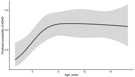
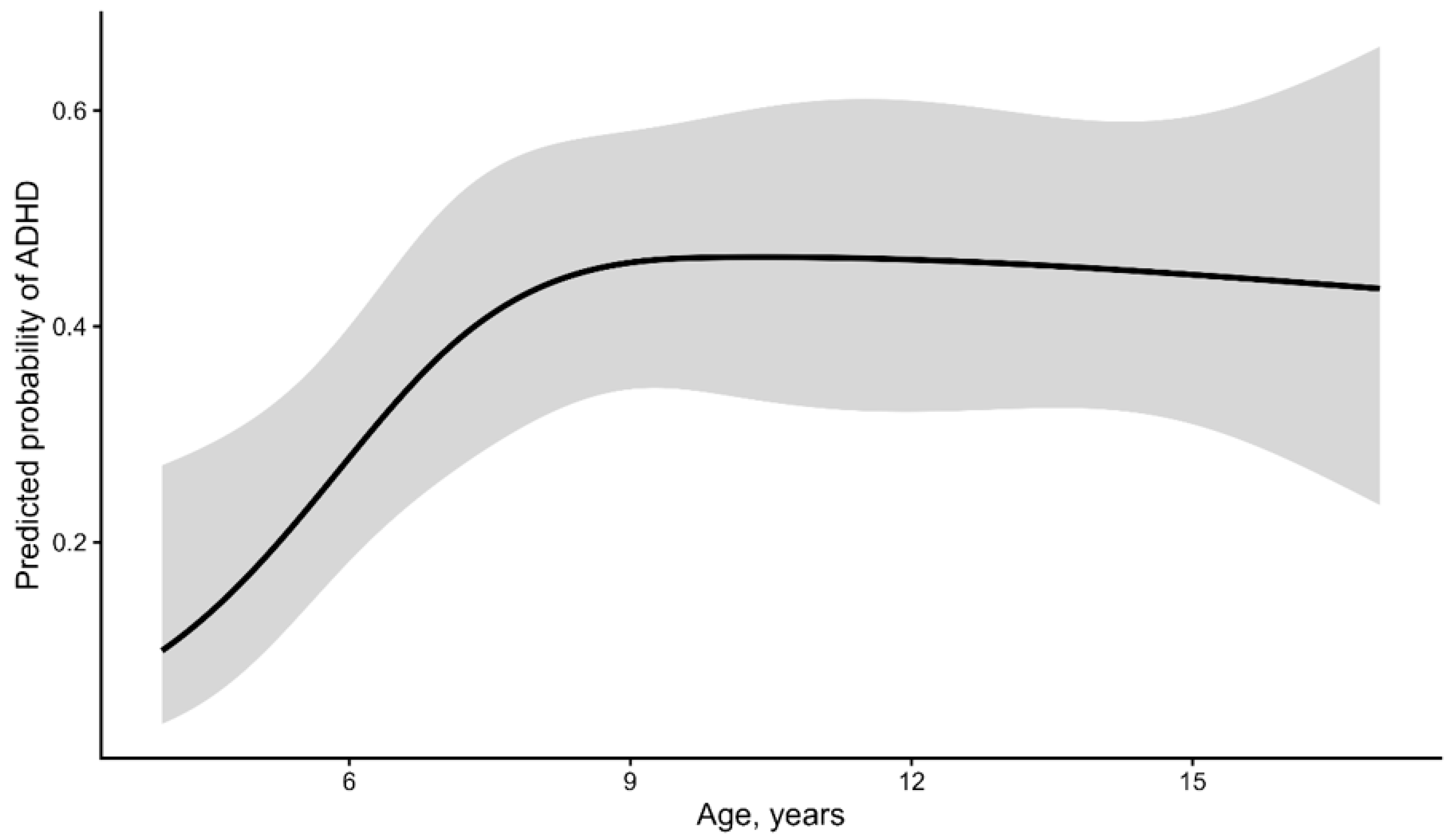
Preliminary model assessment indicated that modeling age using a natural cubic spline with three degrees of freedom provided a significantly better fit than modeling age as a linear term (likelihood-ratio χ2(2) = 8.30, p = 0.016). This supported the use of nonlinear age adjustment in the final multivariable models, with the modeled age–ADHD relationship illustrated in Figure 1.
Figure 1.
Nonlinear association between age and predicted probability of ADHD. Predicted probabilities were derived from a logistic regression model including age as a natural cubic spline with three degrees of freedom, adjusted for sex and FASD phenotype. The shaded area represents the 95% confidence interval.
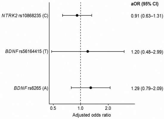
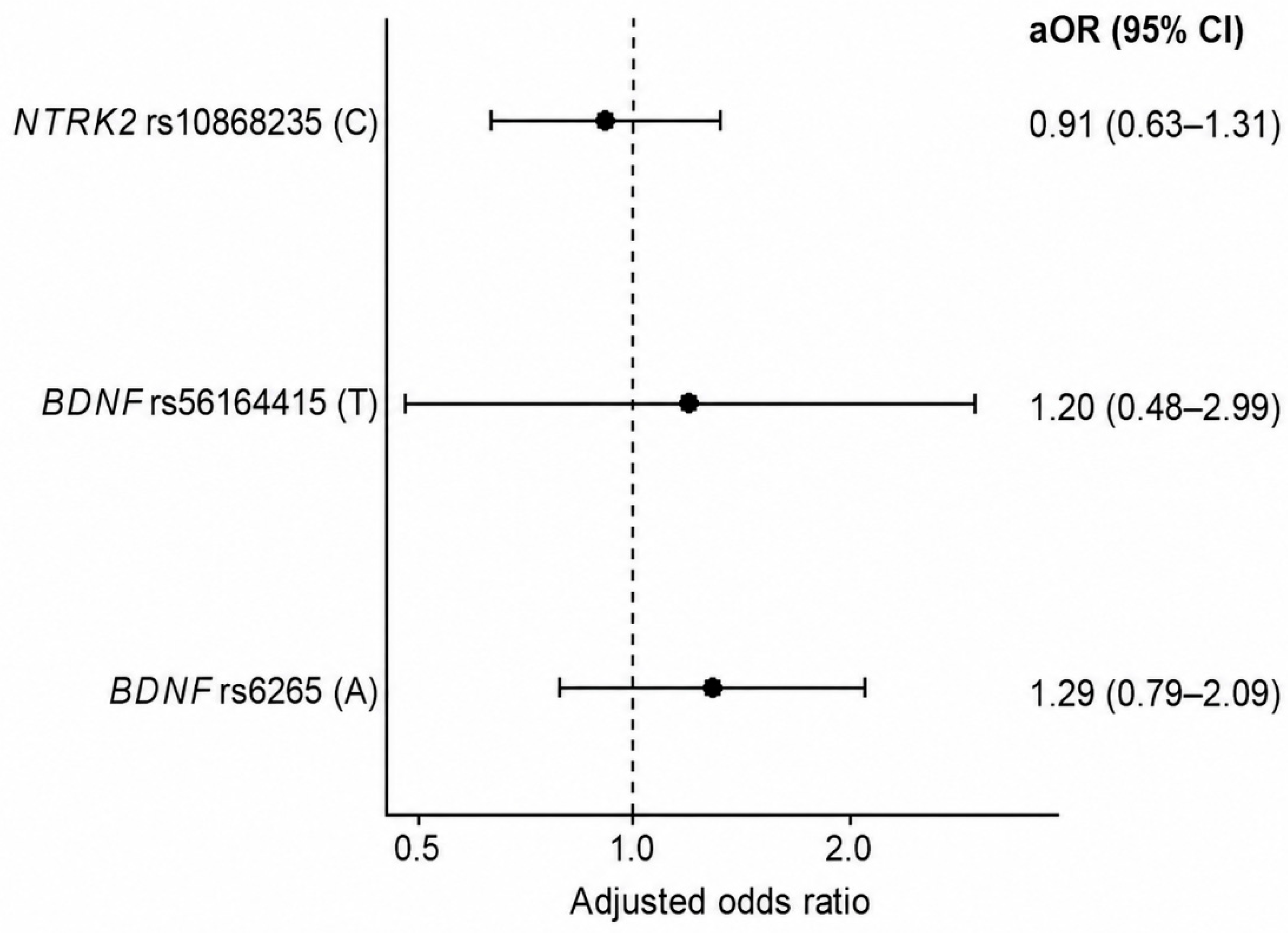
For NTRK2 rs10868235, each additional copy of the C allele was associated with an adjusted odds ratio of 0.91 for ADHD (95% CI: 0.63–1.31; p = 0.619). For BDNF rs56164415, the adjusted odds ratio per T allele was 1.20 (95% CI: 0.48–2.99; p = 0.701). For BDNF rs6265, the adjusted odds ratio per A allele was 1.29 (95% CI: 0.79–2.09; p = 0.308). None of these associations remained statistically significant after correction for multiple comparisons. The Holm-adjusted p values were 1.000 for NTRK2 rs10868235, 1.000 for BDNF rs56164415, and 0.924 for BDNF rs6265. The complete results of the final additive logistic regression models are presented in Table 5, and the corresponding adjusted effect estimates are illustrated in Figure 2.
Table 5.
Multivariable additive associations of NTRK2 and BDNF polymorphisms with ADHD.
Figure 2.
Adjusted associations between BDNF and NTRK2 polymorphisms and ADHD. Adjusted odds ratios and 95% confidence intervals were estimated under additive genetic models adjusted for sex, FASD phenotype, and nonlinear age. The dashed vertical line indicates OR = 1.
The sensitivity analysis indicated approximate minimum detectable per-allele odds ratios of 1.63 for NTRK2 rs10868235, 3.36 for BDNF rs56164415, and 1.93 for BDNF rs6265, assuming 80% statistical power and a two-sided significance level of 0.05. Under a Bonferroni-type threshold of 0.05/3, the corresponding estimates were 1.76, 4.07, and 2.14, respectively. These findings indicate limited statistical power to detect modest genetic effects, particularly for the low-frequency BDNF rs56164415 variant. This limitation was even more pronounced in the small pFAS subgroup (n = 37).
In sensitivity analyses excluding FASD phenotype from the covariate adjustment, the associations remained nonsignificant, with adjusted ORs of 0.90 (95% CI: 0.62–1.28; p = 0.549) for NTRK2 rs10868235, 1.19 (95% CI: 0.48–2.93; p = 0.713) for BDNF rs56164415, and 1.27 (95% CI: 0.79–2.06; p = 0.324) for BDNF rs6265. These results were consistent with the primary models adjusted for FASD phenotype. After excluding 36 children younger than 6 years, the sensitivity analysis included 234 participants (106 with ADHD and 128 without ADHD). None of the three polymorphisms showed a statistically significant association with ADHD, and the adjusted effect estimates remained broadly consistent with those obtained in the full cohort (Supplementary Table S2).
Prenatal maternal smoking was reported in 82 of 111 children with ADHD (73.9%) and 102 of 159 children without ADHD (64.2%). Although maternal smoking was more frequent in the ADHD group, this difference did not reach statistical significance (Pearson’s χ2 test, p = 0.092; Table 2). Additional adjustment for maternal smoking during pregnancy did not materially alter the associations between the investigated polymorphisms and ADHD. The adjusted odds ratios were 0.91 (95% CI: 0.63–1.32; p = 0.629) for NTRK2 rs10868235, 1.17 (95% CI: 0.46–2.95; p = 0.740) for BDNF rs56164415, and 1.27 (95% CI: 0.78–2.07; p = 0.338) for BDNF rs6265. All associations remained statistically nonsignificant (Supplementary Table S3).
Accounting for within-family correlation using maternal cluster-robust standard errors did not materially alter the primary genetic association estimates. The adjusted odds ratios were 0.91 (95% CI: 0.63–1.32; p = 0.621) for NTRK2 rs10868235, 1.20 (95% CI: 0.51–2.81; p = 0.681) for BDNF rs56164415, and 1.29 (95% CI: 0.78–2.12; p = 0.321) for BDNF rs6265. Analyses restricted to one child per maternal identifier (n = 251) also yielded no statistically significant associations (Supplementary Table S4).
3.3. Genotype-by-FASD Phenotype Interactions
No statistically significant global genotype-by-FASD phenotype interactions were observed after adjustment for sex and nonlinear age. For NTRK2 rs10868235 (additive model), the global interaction test was not statistically significant (likelihood-ratio χ2(2) = 5.17; p = 0.075; Holm-adjusted p = 0.226). In exploratory phenotype-specific analyses, each additional C allele was associated with lower odds of ADHD in the pFAS subgroup (adjusted OR = 0.25; 95% CI: 0.07–0.92; p = 0.038). However, this association did not remain significant after Holm correction across the three NTRK2 phenotype-specific comparisons (adjusted p = 0.114). Given the small subgroup size (n = 37), wide confidence interval, and non-significant global interaction, this finding should be interpreted cautiously rather than as evidence of a phenotype-specific genetic effect. Estimates for all three FASD phenotype groups are presented in Table 6.
Table 6.
Exploratory phenotype-specific associations between NTRK2 rs10868235 and ADHD.
Neither BDNF rs6265 nor rs56164415 showed significant genotype-by-FASD phenotype interactions under dominant genetic models. For rs6265, the global interaction test yielded χ2(2) = 0.52 (p = 0.772; Holm-adjusted p = 0.815), while for rs56164415, χ2(2) = 1.80 (p = 0.408; Holm-adjusted p = 0.815). Phenotype-specific estimates are presented in Supplementary Tables S5 and S6, respectively. Due to sparse genotype counts and quasi-complete separation, additional analyses using Firth’s penalized logistic regression were performed for rs56164415. Although finite estimates were obtained across the three FASD phenotype groups, wide confidence intervals, particularly in the pFAS subgroup, indicated substantial statistical uncertainty. The results of all global genotype-by-FASD phenotype interaction tests are summarized in Table 7.
Table 7.
Global genotype-by-FASD phenotype interaction tests in relation to ADHD.
4. Discussion
In this cohort of children with confirmed high prenatal alcohol exposure, none of the investigated BDNF or NTRK2 polymorphisms showed a statistically significant association with ADHD after covariate adjustment and correction for multiple comparisons. No significant global genotype-by-FASD phenotype interactions were identified. Although an exploratory analysis suggested lower odds of ADHD per C allele of NTRK2 rs10868235 in the pFAS subgroup, this association did not remain significant after correction. Importantly, these null findings do not exclude modest genetic effects. The confidence intervals for the primary associations (NTRK2 rs10868235: 0.63–1.31; BDNF rs56164415: 0.48–2.99; BDNF rs6265: 0.79–2.09) remained compatible with potentially meaningful effects, reflecting the limited statistical precision of this study. These findings should be interpreted within the complex polygenic architecture of ADHD. Large-scale genome-wide association studies (GWAS) have identified 27 genome-wide significant ADHD loci [27] and, more recently, 39 independent loci in a meta-analysis combining ADHD symptoms and diagnoses [28]. Neither BDNF nor NTRK2 has emerged as a consistently replicated genome-wide significant ADHD risk locus in these studies. Although this does not exclude smaller or context-dependent effects, it highlights the distinction between biological plausibility and established genetic association. Candidate-gene studies are particularly vulnerable to limited statistical power, restricted genomic coverage, and replication difficulties [29,30]. Accordingly, the present findings should be considered exploratory and interpreted alongside evidence from larger genomic investigations.
Age-related differences also warrant consideration. Modeling age using a natural cubic spline improved model fit compared with a linear age term (p = 0.016), although this improvement was no longer statistically significant after excluding children younger than 6 years (p = 0.501). Importantly, the age-restricted sensitivity analysis yielded genetic effect estimates broadly consistent with those observed in the full cohort, with no statistically significant associations identified. This difference may reflect changes in sample composition or age-related variation in ADHD ascertainment, as suggested by previous research [31]. A further characteristic of the cohort was the predominance of girls (68.5%), particularly within the pFAS subgroup (86.5%). Similar proportions were observed in the original cohort of 303 children (70.3% and 88.1%, respectively) [22], indicating that this imbalance was not introduced by the age restriction applied in the present analysis. Previous studies have described sex-related differences in the presentation of FASD and ADHD [32,33]. May et al. [34] reported greater alcohol-related dysmorphology among girls, suggesting possible sex-specific developmental vulnerability. Nevertheless, these findings cannot establish the cause of the sex imbalance in our cohort. Sex distribution did not differ significantly by ADHD status (p = 0.779), and sex was included in the adjusted models. The small number of boys within the pFAS subgroup (n = 5) nevertheless limits the precision and generalizability of phenotype-specific estimates. Maternal smoking during pregnancy was reported more frequently among children with ADHD than among those without ADHD (73.9% vs. 64.2%), although this difference was not statistically significant (p = 0.092). Gustavson et al. [35] reported an association between maternal prenatal smoking and offspring ADHD (adjusted HR = 1.48; 95% CI: 1.30–1.68). However, their sibling-comparison analyses suggested that shared familial or genetic factors may explain a substantial proportion of this association. Consequently, the difference observed in our cohort should not be interpreted as evidence of a causal effect of maternal smoking on ADHD.
The substantial clinical overlap between FASD and ADHD is another important consideration. A systematic review and meta-analysis estimated ADHD prevalence among children with FASD at 52.9% [4], compared with 41.1% in our selected cohort of children with hPAE. These figures are not directly comparable because our sample included FAS, pFAS, and non-FAS phenotypes and was not designed to estimate population prevalence. Additionally, the use of ICD-10 rather than DSM-5 diagnostic criteria may have contributed to the lower proportion of children diagnosed with ADHD in our cohort, as the ICD-10 criteria for hyperkinetic disorder (F90) are more restrictive. Both conditions may involve inattention, impulsivity, hyperactivity, and executive dysfunction [5,36]. Kingdon et al. [37] identified more pronounced difficulties in planning, cognitive flexibility, and fluency among children with FASD than among those with ADHD, while inhibition and vigilance were less consistently discriminative. Kooistra et al. [38] similarly described overlapping attention difficulties with differences in inhibitory performance. More recently, Capes et al. [5] emphasized that the severity of impairment may be more informative than distinct neuropsychological profiles. Because ADHD status in our study was based on previously established clinical diagnoses without independent reassessment, some degree of diagnostic misclassification arising from overlapping manifestations cannot be excluded.
Among the investigated variants, BDNF rs6265 (Val66Met) has the most extensively characterized functional consequences. This nonsynonymous c.196G>A polymorphism produces a valine-to-methionine substitution at codon 66 of the BDNF prodomain. The Met66 variant affects intracellular trafficking and reduces activity-dependent BDNF secretion, potentially influencing neuronal plasticity and cognitive function [13,14,39,40,41]. However, its phenotypic effects may depend on developmental stage, environmental conditions, and the clinical outcome examined [39]. Previous association studies of rs6265 and ADHD have produced inconsistent findings. Ozturk et al. [42] reported an increased frequency of the Val/Val genotype among children with ADHD, while Kent et al. [43] observed preferential transmission of the Val allele, particularly through paternal transmission. Aureli et al. [16] similarly reported increased frequencies of the Val allele and Val/Met genotype in children with ADHD. Conversely, Xu et al. [44] and Schimmelmann et al. [45] found no significant evidence of association or preferential transmission. A 2022 meta-analysis comprising 15 studies, 4364 individuals with ADHD, 4328 controls, and 1578 families found no overall association between rs6265 and ADHD (OR = 0.95; 95% CI: 0.87–1.04) [15]. A subsequent meta-analysis involving mainland Chinese populations reached a similar conclusion [46]. These findings suggest that rs6265 is unlikely to exert a consistent, substantial effect on ADHD susceptibility across populations. Our results are broadly consistent with this meta-analytic evidence. Neither the adjusted additive association analysis nor the dominant genotype-by-FASD phenotype interaction analysis identified a significant effect of rs6265. Nevertheless, experimental studies provide a rationale for investigating the BDNF–TrkB pathway in the context of developmental alcohol exposure. Using a mouse model carrying Val68Met, the murine equivalent of human Val66Met, Bird et al. [47] reported genotype-dependent effects of developmental ethanol exposure on hippocampal morphology that were no longer apparent later in development. A subsequent investigation found no significant genotype-by-ethanol interaction in adult hippocampal volume [48]. Zeng et al. [49] further demonstrated that prenatal alcohol exposure may disrupt CREB/BDNF/TrkB signaling through oxidative stress, accompanied by abnormalities in GABAergic development and cognitive function. Although these studies support the biological relevance of the pathway, they do not establish an association between human rs6265 and ADHD following prenatal alcohol exposure.
The second investigated BDNF variant, rs56164415 (C270T), is a non-coding polymorphism located within the 5′ untranslated region. It has previously been studied in several neurological and psychiatric conditions, including ADHD [16,50,51]. Unlike rs6265, its functional consequences remain poorly understood. Although its regulatory location raises the possibility of effects on BDNF expression, there is insufficient evidence to establish a consistent functional effect of either allele [52,53]. Evidence concerning rs56164415 and ADHD is limited. Xu et al. [44] reported preferential transmission of the C allele to affected offspring in Taiwanese and pooled UK–Taiwanese family samples, with an estimated odds ratio of 2.29. However, this finding required independent replication and was not corrected for the broader set of genetic comparisons. Aureli et al. [16] subsequently reported overrepresentation of the C/C genotype among children with ADHD and intellectual disability compared with healthy controls. In the present cohort, neither the primary association analysis nor the phenotype interaction analysis identified a statistically significant association after multiple-testing correction. For BDNF rs56164415, the crude allelic OR (0.92) and adjusted additive OR (1.20) differed in direction. These estimates were derived from different analytical approaches, and the low frequency of the T allele (23 copies; 4.26% in the cohort) limited their precision. Xu et al. [44] reported T-allele frequencies of approximately 4–6% in parental samples, while Aureli et al. [16] observed very few T-containing genotypes. The predominance of the C allele does not itself exclude a potential association; rather, the limited number of T-allele carriers reduces the precision with which genotype effects can be estimated. Thus, our findings do not corroborate the previously reported associations, but they cannot definitively exclude an effect of this polymorphism.
NTRK2 encodes tropomyosin receptor kinase B (TrkB), the high-affinity receptor for BDNF and an important regulator of neuronal development and synaptic plasticity [9]. The investigated rs10868235 polymorphism is intronic, and its functional significance remains uncertain. Ribasés et al. [54] reported an association between childhood ADHD and an NTRK2 haplotype comprising rs7816, rs11795386, rs1387926, and rs1586681 (OR = 1.52; p = 0.0084). In contrast, Conner et al. [55] found no significant association between rs1212171 and ADHD symptom scores in adult men. Because neither study examined rs10868235, these findings provide only indirect evidence concerning the involvement of NTRK2 in ADHD. Evidence specific to rs10868235 is particularly limited. Ramírez et al. [19] investigated this variant in 102 Spanish schoolchildren and reported a nominal interaction with estimated dietary bisphenol exposure in relation to verbal comprehension (p = 0.043). However, the environmental exposure and outcome differed from those investigated in our study, preventing direct comparison. In our cohort, rs10868235 was not significantly associated with ADHD overall, and the global genotype-by-FASD phenotype interaction did not reach statistical significance. The exploratory association observed within the pFAS subgroup (adjusted OR = 0.25; 95% CI: 0.07–0.92) was based on only 37 participants and did not remain significant after Holm correction (p = 0.114). Accordingly, this observation should be regarded as hypothesis-generating rather than evidence of a phenotype-specific genetic effect.
Strengths and Limitations of the Study
A major strength of this study is the use of a clinically characterized cohort of children with confirmed high prenatal alcohol exposure, with FASD phenotypes assessed using the Washington 4-Digit Diagnostic Code. The analysis included complete genotyping for the three investigated variants, adjustment for age, sex, and FASD phenotype, nonlinear age modeling, and correction for multiple comparisons. Exploratory genotype-by-FASD phenotype analyses additionally allowed potential differences across clinical phenotype groups to be examined.
Several limitations should be acknowledged. The sample size was relatively modest (n = 270), particularly for phenotype-stratified analyses involving the pFAS subgroup (n = 37; 21 children with ADHD). The approximate descriptive events-per-parameter ratio was 4.2 in this subgroup, although this does not represent the formal ratio for the pooled interaction model. No a priori sample size calculation was performed, and the sensitivity analysis indicated limited power to detect modest genetic associations. This limitation was particularly relevant for BDNF rs56164415, whose low minor allele frequency resulted in sparse genotype categories and substantial uncertainty even when penalized regression was applied.
Although all participants had previously been classified as having hPAE, prenatal alcohol exposure may have differed in intensity, timing, and frequency. Previous research by Pagnin et al. [56] reported increased odds of offspring ADHD associated with binge drinking or regular low-to-moderate alcohol consumption throughout pregnancy, highlighting the potential relevance of different exposure patterns. However, such findings do not imply equivalent developmental consequences across exposure levels. Because reliable individual-level characterization of maternal drinking patterns was insufficient for the present analysis, these patterns were not included in the regression models. Consequently, exposure heterogeneity may have contributed to variability in neurodevelopmental outcomes.
The non-FAS group comprised children with confirmed hPAE who lacked the characteristic morphological criteria for FAS or pFAS but could nevertheless have neurodevelopmental impairments. This heterogeneity, together with the clinical overlap between FASD-related impairment and ADHD, may have reduced phenotypic specificity. Importantly, sensitivity analyses excluding FASD phenotype from the covariate adjustment yielded similar effect estimates and did not alter the interpretation of the primary genetic associations.
ADHD classification was based on previously established clinical diagnoses and available medical records rather than a standardized diagnostic reassessment conducted specifically for this study. Children in the non-ADHD group were classified based on the absence of a documented ADHD diagnosis; therefore, undiagnosed or subthreshold ADHD cannot be definitively excluded. Such potential misclassification may have influenced the estimated genetic associations and, if nondifferential with respect to genotype, could have attenuated the observed associations toward the null, although this assumption could not be verified. All children in the ADHD group were receiving methylphenidate treatment under regular specialist care; however, treatment status was a clinical characteristic rather than a diagnostic criterion. Consequently, the findings may not be fully generalizable to children with untreated or milder forms of ADHD. Furthermore, detailed information on oppositional defiant disorder (ODD) and ADHD presentation subtypes was unavailable, precluding the evaluation of potential differences in psychiatric comorbidity and ADHD clinical presentation.
In addition, information on several potentially relevant factors, including postnatal adversity, placement history, socioeconomic conditions, prematurity, and birth weight, was unavailable. Although additional adjustment for maternal smoking during pregnancy did not materially change the primary genetic association estimates, residual confounding by other unmeasured prenatal and postnatal factors cannot be excluded.
The predominance of girls, particularly within the pFAS subgroup, may limit generalizability despite adjustment for sex. All participants were recruited in Poland, and genetic ancestry was not independently assessed using genome-wide methods; therefore, residual population stratification cannot be excluded. The study cohort comprised 270 children born to 251 mothers, with some mothers contributing more than one child. To account for potential within-family correlation, additional sensitivity analyses were performed using maternal cluster-robust standard errors and a complementary analysis restricted to one child per mother. These analyses yielded results consistent with the primary findings. Nevertheless, the relatively small number of families with multiple participating children may limit the precision with which the influence of familial clustering can be assessed.
Finally, because all participants had confirmed hPAE, the study could not directly compare exposed and unexposed children or assess genotype-by-prenatal alcohol exposure interactions. Larger, independent studies incorporating both alcohol-exposed and unexposed comparison groups, along with broader genomic approaches, are needed to address these questions.
5. Conclusions
The present study contributes new data on the associations between selected BDNF and NTRK2 polymorphisms and ADHD in a clinically characterized cohort of children with confirmed high prenatal alcohol exposure. Although no statistically significant associations or global genotype-by-FASD phenotype interactions were identified after covariate adjustment and correction for multiple testing, these findings expand the limited evidence available for these specific variants in alcohol-exposed populations.
The exploratory association between the C allele of NTRK2 rs10868235 and lower odds of ADHD in the pFAS subgroup did not remain significant after multiple-testing correction. While this observation requires cautious interpretation, it provides a hypothesis for investigation in larger, independent cohorts.
Importantly, the absence of significant associations does not exclude modest genetic effects, particularly given the limited statistical power for low-frequency variants and smaller phenotype groups. Furthermore, the lack of an unexposed comparison group prevented direct assessment of genotype-by-prenatal alcohol exposure interactions.
Future studies incorporating larger cohorts, both exposed and unexposed comparison groups, broader genomic approaches, and functional investigations of BDNF–TrkB signaling may help clarify the contribution of genetic variability to neurodevelopmental outcomes following prenatal alcohol exposure. The present findings provide an initial basis for such investigations and contribute to a more comprehensive understanding of the complex genetic architecture of ADHD in children with prenatal alcohol exposure.
Supplementary Materials
The following supporting information can be downloaded at: https://www.mdpi.com/article/10.3390/children13111528/s1, Table S1: STREGA checklist for the analysis of BDNF and NTRK2 polymorphisms associated with ADHD in children with prenatal alcohol exposure [57]; Table S2: Multivariable additive associations of NTRK2 and BDNF polymorphisms with ADHD after excluding children younger than 6 years; Table S3: Sensitivity analysis of associations between NTRK2 and BDNF polymorphisms and ADHD after additional adjustment for maternal smoking during pregnancy; Table S4: Sensitivity analyses accounting for within-mother clustering in the full cohort of children with high prenatal alcohol exposure; Table S5: Exploratory phenotype-specific associations between BDNF rs6265 and ADHD; Table S6: Phenotype-specific analysis of BDNF rs56164415 and ADHD (using Firth’s penalized logistic regression); Supplementary Code S1: Complete R analysis script.
Author Contributions
Conceptualization, A.K. and M.B.; methodology, D.M. and A.K.; software, D.M. and A.K.; validation, D.M. and M.B.; formal analysis, D.M.; investigation, M.R., K.L. and A.K.; resources, M.R., K.L. and A.K.; data curation, A.K., K.L. and M.R.; writing—original draft preparation, A.K.; writing—review and editing, M.B. and D.M.; visualization, A.K.; supervision, M.B.; project administration, A.K.; funding acquisition, A.K. and M.B. All authors have read and agreed to the published version of the manuscript.
Funding
This research received no external funding.
Institutional Review Board Statement
The study was conducted in accordance with the Declaration of Helsinki and was approved by the local Bioethics Committee at the Pomeranian Medical University in Szczecin (approval no. KB-0012/95/15 and KB-006/44/2024-A-33).
Informed Consent Statement
Written informed consent was obtained from the legal guardians of all participants and, additionally, from participants older than 16 years of age.
Data Availability Statement
The data presented in this study are available on request from the corresponding author. The data are not publicly available due to privacy and legal restrictions.
Acknowledgments
During the preparation of this manuscript, the authors used ChatGPT (OpenAI; GPT-5.6) for language editing, including grammar, spelling, and stylistic corrections. All AI-generated output was reviewed and edited by the authors, who take full responsibility for the content of the publication.
Conflicts of Interest
The authors declare no conflicts of interest.
Abbreviations
The following abbreviations are used in this manuscript:
| Abbreviation | Definition |
| ADHD | Attention-deficit/hyperactivity disorder |
| ALAB | Alcohol abuse |
| ASD | Autism spectrum disorder |
| BDNF | Brain-derived neurotrophic factor |
| CHD | Chronic drinking |
| CI | Confidence interval |
| CREB | cAMP response element-binding protein |
| DNA | Deoxyribonucleic acid |
| DSM-5 | Diagnostic and Statistical Manual of Mental Disorders, Fifth Edition |
| EBD | Early binge drinking |
| FAS | Fetal alcohol syndrome |
| FASD | Fetal alcohol spectrum disorders |
| hPAE | High prenatal alcohol exposure |
| HWE | Hardy–Weinberg equilibrium |
| ICD-10 | International Classification of Diseases, 10th Revision |
| LRT | Likelihood-ratio test |
| MAF | Minor allele frequency |
| NDD | Neurodevelopmental disorders |
| NTRK2 | Neurotrophic receptor tyrosine kinase 2 |
| OR | Odds ratio |
| PAE | Prenatal alcohol exposure |
| PCR | Polymerase chain reaction |
| pFAS | Partial fetal alcohol syndrome |
| SD | Standard deviation |
| SNP | Single-nucleotide polymorphism |
| TrkB | Tropomyosin receptor kinase B |
| WBD | Weekend binge drinking |
References
- Ayano, G.; Demelash, S.; Gizachew, Y.; Tsegay, L.; Alati, R. The global prevalence of attention deficit hyperactivity disorder in children and adolescents: An umbrella review of meta-analyses. J. Affect. Disord. 2023, 339, 860–866. [Google Scholar] [CrossRef] [Scilit] [PubMed]
- American Psychiatric Association. Diagnostic and Statistical Manual of Mental Disorders, 5th ed.; American Psychiatric Association Publishing: Arlington, VA, USA, 2013. [Google Scholar]
- Popova, S.; Charness, M.E.; Burd, L.; Crawford, A.; Hoyme, H.E.; Mukherjee, R.A.S.; Riley, E.P.; Elliott, E.J. Fetal alcohol spectrum disorders. Nat. Rev. Dis. Primers 2023, 9, 11. [Google Scholar] [CrossRef] [Scilit]
- Lange, S.; Rehm, J.; Anagnostou, E.; Popova, S. Prevalence of externalizing disorders and autism spectrum disorders among children with fetal alcohol spectrum disorder: Systematic review and meta-analysis. Biochem. Cell Biol. 2018, 96, 241–251. [Google Scholar] [CrossRef] [Scilit]
- Capes, T.J.; Aldrovandi, S.; Khan, R.; Amanatidou, V.; Tsaprouni, L.; Mavritsaki, E. Differentiating fetal alcohol spectrum disorders from attention-deficit hyperactivity disorder and reviewing diagnostic guidelines. Ann. N. Y. Acad. Sci. 2026, 1562, e70343. [Google Scholar] [CrossRef] [Scilit]
- Parenti, I.; Rabaneda, L.G.; Schoen, H.; Novarino, G. Neurodevelopmental disorders: From genetics to functional pathways. Trends Neurosci. 2020, 43, 608–621. [Google Scholar] [CrossRef] [Scilit]
- De Felice, A.; Ricceri, L.; Venerosi, A.; Chiarotti, F.; Calamandrei, G. Multifactorial origin of neurodevelopmental disorders: Approaches to understanding complex etiologies. Toxics 2015, 3, 89–129. [Google Scholar] [CrossRef] [Scilit]
- Li, Y.; Li, F.; Qin, D.; Chen, H.; Wang, J.; Wang, J.; Song, S.; Wang, C.; Wang, Y.; Liu, S.; et al. The role of brain derived neurotrophic factor in central nervous system. Front. Aging Neurosci. 2022, 14, 986443. [Google Scholar] [CrossRef] [Scilit] [PubMed]
- Teng, K.K.; Hempstead, B.L. Neurotrophins and their receptors: Signaling trios in complex biological systems. Cell. Mol. Life Sci. 2004, 61, 35–48. [Google Scholar] [CrossRef] [Scilit]
- Carito, V.; Ceccanti, M.; Ferraguti, G.; Coccurello, R.; Ciafrè, S.; Tirassa, P.; Fiore, M. NGF and BDNF alterations by prenatal alcohol exposure. Curr. Neuropharmacol. 2019, 17, 308–317. [Google Scholar] [CrossRef] [Scilit]
- Fiore, M.; Petrella, C.; Coriale, G.; Rosso, P.; Fico, E.; Ralli, M.; Greco, A.; De Vincentiis, M.; Minni, A.; Polimeni, A.; et al. Markers of neuroinflammation in the serum of prepubertal children with fetal alcohol spectrum disorders. CNS Neurol. Disord. Drug Targets 2022, 21, 854–868. [Google Scholar] [CrossRef] [Scilit]
- Darbinian, N.; Merabova, N.; Tatevosian, G.; Morrison, M.; Darbinyan, A.; Zhao, H.; Goetzl, L.; Selzer, M.E. Biomarkers of affective dysregulation associated with in utero exposure to EtOH. Cells 2024, 13, 2. [Google Scholar] [CrossRef] [Scilit]
- Chen, Z.Y.; Patel, P.D.; Sant, G.; Meng, C.X.; Teng, K.K.; Hempstead, B.L.; Lee, F.S. Variant brain-derived neurotrophic factor (BDNF) (Met66) alters the intracellular trafficking and activity-dependent secretion of wild-type BDNF in neurosecretory cells and cortical neurons. J. Neurosci. 2004, 24, 4401–4411. [Google Scholar] [CrossRef] [Scilit]
- Egan, M.F.; Kojima, M.; Callicott, J.H.; Goldberg, T.E.; Kolachana, B.S.; Bertolino, A.; Zaitsev, E.; Gold, B.; Goldman, D.; Dean, M.; et al. The BDNF Val66Met polymorphism affects activity-dependent secretion of BDNF and human memory and hippocampal function. Cell 2003, 112, 257–269. [Google Scholar] [CrossRef] [Scilit]
- Mei, S.; Chen, W.; Chen, S.; Hu, Y.; Dai, X.; Liu, X. Evaluation of the relationship between BDNF Val66Met gene polymorphism and attention deficit hyperactivity disorder: A meta-analysis. Front. Psychiatry 2022, 13, 888774. [Google Scholar] [CrossRef] [Scilit]
- Aureli, A.; Del Beato, T.; Sebastiani, P.; Marimpietri, A.; Melillo, C.V.; Sechi, E.; Di Loreto, S. Attention-deficit hyperactivity disorder and intellectual disability: A study of association with brain-derived neurotrophic factor gene polymorphisms. Int. J. Immunopathol. Pharmacol. 2010, 23, 873–880. [Google Scholar] [CrossRef] [Scilit]
- Aizawa, S.; Ishitobi, Y.; Masuda, K.; Inoue, A.; Oshita, H.; Hirakawa, H.; Ninomiya, T.; Maruyama, Y.; Tanaka, Y.; Okamoto, K.; et al. Genetic association of the transcription of neuroplasticity-related genes and variation in stress-coping style. Brain Behav. 2015, 5, e00360. [Google Scholar] [CrossRef] [Scilit]
- Mirkovic, B.; Cohen, D.; Laurent, C.; Lasfar, M.; Marguet, C.; Gerardin, P. A case-control association study of 12 candidate genes and attempted suicide in French adolescents. Int. J. Adolesc. Med. Health 2020, 32, 20170089. [Google Scholar] [CrossRef] [Scilit]
- Ramírez, V.; González-Palacios, P.; González-Domenech, P.J.; Jaimez-Pérez, S.; Baca, M.A.; Rodrigo, L.; Álvarez-Cubero, M.J.; Monteagudo, C.; Martínez-González, L.J.; Rivas, A. Influence of genetic polymorphisms on cognitive function according to dietary exposure to bisphenols in a sample of Spanish schoolchildren. Nutrients 2024, 16, 2639. [Google Scholar] [CrossRef] [Scilit]
- Grzywacz, E.; Brzuchalski, B.; Śmiarowska, M.; Malinowski, D.; Machoy-Mokrzyńska, A.; Białecka, M.A. Significance of Selected Environmental and Biological Factors on the Risk of FASD in Women Who Drink Alcohol during Pregnancy. J. Clin. Med. 2023, 12, 6185. [Google Scholar] [CrossRef] [Scilit]
- Śmiarowska, M.; Brzuchalski, B.; Grzywacz, E.; Malinowski, D.; Machoy-Mokrzyńska, A.; Pierzchlińska, A.; Białecka, M. Influence of COMT (rs4680) and DRD2 (rs1076560, rs1800497) Gene Polymorphisms on Safety and Efficacy of Methylphenidate Treatment in Children with Fetal Alcohol Spectrum Disorders. Int. J. Environ. Res. Public Health 2022, 19, 4479. [Google Scholar] [CrossRef] [Scilit]
- Kukowka, A.; Brzuchalski, B.; Kurzawski, M.; Malinowski, D.; Białecka, M.A. ADH1B, ADH1B/C and CYP2E1 Gene Polymorphism and the Risk of Fetal Alcohol Spectrum Disorder. Genes 2023, 14, 1392. [Google Scholar] [CrossRef] [Scilit]
- Wolraich, M.L.; Hagan, J.F., Jr.; Allan, C.; Chan, E.; Davison, D.; Earls, M.; Evans, S.W.; Flinn, S.K.; Froehlich, T.; Frost, J.; et al. Clinical practice guideline for the diagnosis, evaluation, and treatment of attention-deficit/hyperactivity disorder in children and adolescents. Pediatrics 2019, 144, e20192528. [Google Scholar] [CrossRef] [Scilit] [PubMed]
- Astley, S.J.; Clarren, S.K. Diagnosing the full spectrum of fetal alcohol-exposed individuals: Introducing the 4-digit diagnostic code. Alcohol Alcohol. 2000, 35, 400–410. [Google Scholar] [CrossRef] [Scilit]
- Roozen, S.; Peters, G.J.Y.; Kok, G.; Townend, D.; Nijhuis, J.; Koek, G.; Curfs, L. Systematic Literature Review on Which Maternal Alcohol Behaviours Are Related to Fetal Alcohol Spectrum Disorders (FASD). BMJ Open 2018, 8, e022578. [Google Scholar] [CrossRef] [Scilit]
- The 1000 Genomes Project Consortium. A global reference for human genetic variation. Nature 2015, 526, 68–74. [Google Scholar] [CrossRef] [Scilit] [PubMed]
- Demontis, D.; Walters, G.B.; Athanasiadis, G.; Walters, R.; Therrien, K.; Nielsen, T.T.; Farajzadeh, L.; Voloudakis, G.; Bendl, J.; Zeng, B.; et al. Genome-wide analyses of ADHD identify 27 risk loci, refine the genetic architecture and implicate several cognitive domains. Nat. Genet. 2023, 55, 198–208. [Google Scholar] [CrossRef] [Scilit]
- van der Laan, C.M.; Ip, H.F.; Schipper, M.; Hottenga, J.-J.; St Pourcain, B.; Zayats, T.; Pool, R.; Krapohl, E.M.L.; Brikell, I.; Soler Artigas, M.; et al. Genome-wide association meta-analysis of childhood ADHD symptoms and diagnosis identifies new loci and potential effector genes. Nat. Genet. 2025, 57, 2427–2435. [Google Scholar] [CrossRef] [Scilit]
- MacDonald, H.J.; Kleppe, R.; Szigetvari, P.D.; Haavik, J. The dopamine hypothesis for ADHD: An evaluation of evidence accumulated from human studies and animal models. Front. Psychiatry 2024, 15, 1492126. [Google Scholar] [CrossRef] [Scilit]
- Johnson, E.C.; Border, R.; Melroy-Greif, W.E.; de Leeuw, C.A.; Ehringer, M.A.; Keller, M.C. No Evidence That Schizophrenia Candidate Genes Are More Associated With Schizophrenia Than Noncandidate Genes. Biol. Psychiatry 2017, 82, 702–708. [Google Scholar] [CrossRef] [Scilit]
- Rocco, I.; Corso, B.; Bonati, M.; Minicuci, N. Time of Onset and/or Diagnosis of ADHD in European Children: A Systematic Review. BMC Psychiatry 2021, 21, 575. [Google Scholar] [CrossRef] [Scilit]
- Flannigan, K.; Poole, N.; Cook, J.; Unsworth, K. Sex-related differences among individuals assessed for fetal alcohol spectrum disorder in Canada. Alcohol Clin. Exp. Res. 2023, 47, 613–623. [Google Scholar] [CrossRef] [Scilit]
- Hinshaw, S.P.; Nguyen, P.T.; O’Grady, S.M.; Rosenthal, E.A. Annual research review: Attention-deficit/hyperactivity disorder in girls and women: Underrepresentation, longitudinal processes, and key directions. J. Child Psychol. Psychiatry 2022, 63, 484–496. [Google Scholar] [CrossRef] [Scilit] [PubMed]
- May, P.A.; Tabachnick, B.; Hasken, J.M.; Marais, A.-S.; de Vries, M.M.; Barnard, R.; Joubert, B.; Cloete, M.; Botha, I.; Kalberg, W.O.; et al. Who is most affected by prenatal alcohol exposure: Boys or girls? Drug Alcohol Depend. 2017, 177, 258–267. [Google Scholar] [CrossRef] [Scilit]
- Gustavson, K.; Ystrom, E.; Stoltenberg, C.; Susser, E.; Surén, P.; Magnus, P.; Knudsen, G.P.; Davey Smith, G.; Langley, K.; Rutter, M.; et al. Smoking in pregnancy and child ADHD. Pediatrics 2017, 139, e20162509. [Google Scholar] [CrossRef] [Scilit]
- Young, S.; Absoud, M.; Blackburn, C.; Branney, P.; Colley, B.; Farrag, E.; Fleisher, S.; Gregory, G.; Gudjonsson, G.H.; Kim, K.; et al. Guidelines for identification and treatment of individuals with attention deficit/hyperactivity disorder and associated fetal alcohol spectrum disorders based upon expert consensus. BMC Psychiatry 2016, 16, 324. [Google Scholar] [CrossRef] [Scilit]
- Kingdon, D.; Cardoso, C.; McGrath, J.J. Research Review: Executive function deficits in fetal alcohol spectrum disorders and attention-deficit/hyperactivity disorder—A meta-analysis. J. Child Psychol. Psychiatry 2016, 57, 116–131. [Google Scholar] [CrossRef] [Scilit]
- Kooistra, L.; Crawford, S.; Gibbard, B.; Ramage, B.; Kaplan, B.J. Differentiating attention deficits in children with fetal alcohol spectrum disorder or attention-deficit-hyperactivity disorder. Dev. Med. Child Neurol. 2010, 52, 205–211. [Google Scholar] [CrossRef] [Scilit]
- Nguyen, V.T.; Hill, B.; Sims, N.; Heck, A.; Negron, M.; Lusk, C.; Galindo, C.L. Brain-derived neurotrophic factor rs6265 (Val66Met) single nucleotide polymorphism as a master modifier of human pathophysiology. Neural Regen. Res. 2023, 18, 102–106. [Google Scholar] [CrossRef] [Scilit]
- Peregud, D.; Korolkov, A.; Baronets, V.; Kozlov, K.; Lobacheva, A.; Arkus, M.; Bairamova, S.; Solovieva, M.; Pavlova, O.; Pavlov, K.; et al. Genetic determinants of serum brain-derived neurotrophic factor (BDNF) after alcohol withdrawal. Discov. Med. 2024, 1, 148. [Google Scholar] [CrossRef] [Scilit]
- Tsai, S.-J. Critical Issues in BDNF Val66Met Genetic Studies of Neuropsychiatric Disorders. Front. Mol. Neurosci. 2018, 11, 156. [Google Scholar] [CrossRef] [Scilit]
- Ozturk, O.; Basay, B.K.; Buber, A.; Basay, O.; Alacam, H.; Bacanlı, A.; Yılmaz, Ş.G.; Erdal, M.E.; Herken, H.; Ercan, E.S. Brain-Derived Neurotrophic Factor Gene Val66Met Polymorphism Is a Risk Factor for Attention-Deficit Hyperactivity Disorder in a Turkish Sample. Psychiatry Investig. 2016, 13, 518–525. [Google Scholar] [CrossRef] [Scilit]
- Kent, L.; Green, E.; Hawi, Z.; Kirley, A.; Dudbridge, F.; Lowe, N.; Raybould, R.; Langley, K.; Bray, N.; Fitzgerald, M.; et al. Association of the paternally transmitted copy of common Valine allele of the Val66Met polymorphism of the brain-derived neurotrophic factor (BDNF) gene with susceptibility to ADHD. Mol. Psychiatry 2005, 10, 939–943. [Google Scholar] [CrossRef] [Scilit]
- Xu, X.; Mill, J.; Zhou, K.; Brookes, K.; Chen, C.K.; Asherson, P. Family-based association study between brain-derived neurotrophic factor gene polymorphisms and attention deficit hyperactivity disorder in UK and Taiwanese samples. Am. J. Med. Genet. B Neuropsychiatr. Genet. 2007, 144B, 83–86. [Google Scholar] [CrossRef] [Scilit]
- Schimmelmann, B.G.; Friedel, S.; Dempfle, A.; Warnke, A.; Lesch, K.P.; Walitza, S.; Renner, T.J.; Romanos, M.; Herpertz-Dahlmann, B.; Linder, M.; et al. No evidence for preferential transmission of common valine allele of the Val66Met polymorphism of the brain-derived neurotrophic factor gene (BDNF) in ADHD. J. Neural Transm. 2007, 114, 523–526. [Google Scholar] [CrossRef] [Scilit]
- Lu, Y. Correlation investigation between BDNF (Val66Met/rs6265) polymorphism and attention deficit hyperactivity disorder susceptibility in Chinese mainland population: A meta-analysis. Psychol. Health Med. 2026, 31, 519–530. [Google Scholar] [CrossRef] [Scilit]
- Bird, C.W.; Baculis, B.C.; Mayfield, J.J.; Chavez, G.J.; Ontiveros, T.; Paine, D.J.; Marks, A.J.; Gonzales, A.L.; Ron, D.; Valenzuela, C.F. The brain-derived neurotrophic factor VAL68MET polymorphism modulates how developmental ethanol exposure impacts the hippocampus. Genes Brain Behav. 2019, 18, e12484. [Google Scholar] [CrossRef] [Scilit]
- Bird, C.W.; Barber, M.J.; Martin, J.; Mayfield, J.J.; Valenzuela, C.F. The mouse-equivalent of the human BDNF VAL66MET polymorphism increases dorsal hippocampal volume and does not interact with developmental ethanol exposure. Alcohol 2020, 86, 17–24. [Google Scholar] [CrossRef] [Scilit]
- Zeng, X.; Wu, M.; Cai, Y.; Chen, H.; Sun, M.; Yang, H. Prenatal alcohol exposure impairs offspring cognition through oxidative stress disrupting CREB/BDNF/TrkB signaling and GABAergic neuron deficits. Food Chem. Toxicol. 2026, 207, 115820. [Google Scholar] [CrossRef] [Scilit]
- Kunugi, H.; Ueki, A.; Otsuka, M.; Isse, K.; Hirasawa, H.; Kato, N.; Nabika, T.; Kobayashi, S.; Nanko, S. A novel polymorphism of the brain-derived neurotrophic factor (BDNF) gene associated with late-onset Alzheimer’s disease. Mol. Psychiatry 2001, 6, 83–86. [Google Scholar] [CrossRef] [Scilit]
- Watanabe, Y.; Nunokawa, A.; Someya, T. Association of the BDNF C270T polymorphism with schizophrenia: Updated meta-analysis. Psychiatry Clin. Neurosci. 2013, 67, 123–125. [Google Scholar] [CrossRef] [Scilit]
- Xu, L.; Tian, D.; Li, J.; Chen, L.; Tang, L.; Fan, D. The analysis of two BDNF polymorphisms G196A/C270T in Chinese sporadic amyotrophic lateral sclerosis. Front. Aging Neurosci. 2017, 9, 135. [Google Scholar] [CrossRef] [Scilit]
- Tudor, L.; Konjevod, M.; Nikolac Perkovic, M.; Svob Strac, D.; Nedic Erjavec, G.; Uzun, S.; Kozumplik, O.; Sagud, M.; Kovacic Petrovic, Z.; Pivac, N. Genetic Variants of the Brain-Derived Neurotrophic Factor and Metabolic Indices in Veterans With Posttraumatic Stress Disorder. Front. Psychiatry 2018, 9, 637. [Google Scholar] [CrossRef] [Scilit]
- Ribasés, M.; Hervás, A.; Ramos-Quiroga, J.A.; Bosch, R.; Bielsa, A.; Gastaminza, X.; Fernández-Anguiano, M.; Nogueira, M.; Gómez-Barros, N.; Valero, S.; et al. Association study of 10 genes encoding neurotrophic factors and their receptors in adult and child attention-deficit/hyperactivity disorder. Biol. Psychiatry 2008, 63, 935–945. [Google Scholar] [CrossRef] [Scilit]
- Conner, A.C.; Kissling, C.; Hodges, E.; Hünnerkopf, R.; Clement, R.M.; Dudley, E.; Freitag, C.M.; Rösler, M.; Retz, W.; Thome, J. Neurotrophic factor-related gene polymorphisms and adult attention deficit hyperactivity disorder (ADHD) score in a high-risk male population. Am. J. Med. Genet. B Neuropsychiatr. Genet. 2008, 147B, 1476–1480. [Google Scholar] [CrossRef] [Scilit]
- Pagnin, D.; Zamboni Grecco, M.L.; Furtado, E.F. Prenatal alcohol use as a risk for attention-deficit/hyperactivity disorder. Eur. Arch. Psychiatry Clin. Neurosci. 2019, 269, 681–687. [Google Scholar] [CrossRef] [Scilit]
- Little, J.; Higgins, J.P.; Ioannidis, J.P.; Moher, D.; Gagnon, F.; Von Elm, E.; Khoury, M.J.; Cohen, B.; Davey-Smith, G.; Grimshaw, J.; et al. Strengthening the reporting of genetic association studies (STREGA): An extension of the STROBE statement. Eur. J. Epidemiol. 2009, 24, 37–55. [Google Scholar] [CrossRef] [Scilit]

Disclaimer/Publisher’s Note: The statements, opinions and data contained in all publications are solely those of the individual author(s) and contributor(s) and not of MDPI and/or the editor(s). MDPI and/or the editor(s) disclaim responsibility for any injury to people or property resulting from any ideas, methods, instructions or products referred to in the content. |
© 2026 by the authors. Licensee MDPI, Basel, Switzerland. This article is an open access article distributed under the terms and conditions of the Creative Commons Attribution (CC BY) license.