


Highlights
What are the main findings?
- Among patients who completed 12 months of treatment, lesion size decreased in 17/23 patients and remained stable in 5/23 patients.
- Sirolimus treatment was associated with reduced pain, increased physical activity, and a lower proportion of patients requiring additional treatment, including invasive procedures.
What are the implications of the main findings?
- Sirolimus is an effective and generally safe treatment for pediatric vascular malformations.
- Treatment benefits extend beyond radiological improvement.
Abstract
Background: Vascular anomalies are rare congenital disorders associated with significant morbidity. Local therapies may be insufficient for complex lesions. Sirolimus, an mTOR inhibitor, is a promising systemic treatment option. Methods: We retrospectively analyzed pediatric patients with vascular anomalies treated with sirolimus between October 2020 and June 2025. Of 29 patients who initiated treatment, 23 completed at least 12 months of therapy and had evaluable 12-month outcomes; these patients constituted the primary 12-month efficacy cohort. Treatment discontinuations were analyzed separately, and an all-treated sensitivity analysis was performed. Results: The cohort included 10 patients (43%) with lymphatic malformations (LMs), 9 (39%) with venous malformations (VMs), 2 (9%) with combined malformations, and 2 (9%) with other vascular anomalies. At 12 months, magnetic resonance imaging (MRI) demonstrated lesion size reduction in 17/23 patients (74%), stabilization in 5/23 (22%), and progression in 1/23 patients (4%). In a conservative sensitivity analysis including all 29 treated patients and classifying patients without an evaluable 12-month outcome, documented reduction or stabilization was observed in 22/29 patients (76%). Additional treatment was less frequent during treatment (30% vs. 65%, p = 0.043), although observation periods differed. Pain decreased (p = 0.021), and physical activity increased (p = 0.034). No severe adverse events led to permanent treatment discontinuation. The most common adverse events were laboratory abnormalities (47%) and aphthous stomatitis (32%). Exploratory Multiple Factor Analysis (MFA) demonstrated substantial multidimensional heterogeneity but did not identify stable radiological response phenotypes. Conclusions: Sirolimus was associated with radiological improvement or stabilization, symptom relief, and fewer additional interventions in pediatric vascular anomalies, with generally favorable tolerability.
1. Introduction
Vascular anomalies are a heterogeneous group of congenital disorders resulting from abnormalities in the development of blood and lymphatic vessels [1,2,3,4,5]. The International Society for the Study of Vascular Anomalies (ISSVA) updated its classification of vascular anomalies in 2025, categorizing them into three major groups: vascular tumors, vascular malformations, and potentially unique vascular anomalies (PUVAs) [6]. Vascular malformations are further classified into fast-flow malformations (isolated, multifocal, and syndromic), slow-flow malformations—including capillary (CM), lymphatic (LM), venous (VM), and combined malformations—and developmental anomalies of named vessels [6]. Vascular tumors are further categorized as benign, borderline, or malignant [6].
Slow-flow vascular malformations represent a highly heterogenous group of disorders with diverse clinical presentations, posing significant challenges in both diagnosis and therapeutic management [2,7]. The most common slow-flow malformations are LMs and VMs [8]. These lesions may be localized or extensive, often involving multiple anatomical regions and adjacent structures [8,9]. Fast-flow malformations, particularly arteriovenous malformations (AVMs), are characterized by direct arteriovenous shunting and may lead to severe complications [10]. Tufted angioma (TA) is a rare, benign vascular tumor characterized by abnormal endothelial cell proliferation, typically presenting as an erythematous or violaceous skin lesion. In some cases, TA may be associated with the Kasabach–Merritt phenomenon (KMP), a potentially life-threatening consumptive coagulopathy characterized by severe thrombocytopenia, hypofibrinogenemia, and rapidly enlarging vascular tumor [11,12,13,14].
Depending on their type, location, and extent, vascular anomalies may cause body deformities, chronic pain, recurrent infections, oozing, bleeding, coagulopathies, and functional impairment [1,4,8,15,16]. Some lesions may progressively enlarge and lead to serious complications, including airway obstruction or organ dysfunction, potentially resulting in life-threatening situations [1,17]. Optimal management requires a multidisciplinary approach, especially in patients who are resistant to standard local therapies [2,15]. Current treatment options include surgery, sclerotherapy, endovascular embolization, and laser therapy; however, these modalities may be insufficient for extensive, infiltrative, or anatomically complex vascular anomalies [5,8,9]. Consequently, systemic pharmacological treatment has gained increasing attention.
Molecular studies have demonstrated that activating somatic mutations in the phosphatidylinositol 3-kinase (PI3K)/protein kinase B (AKT)/mammalian target of rapamycin (mTOR) signaling pathway are commonly identified in slow-flow vascular malformations, leading to abnormal localized vascular growth and proliferation [8,9,17]. These discoveries have provided a rationale for the development of targeted therapeutic strategies aimed at pathway inhibition. Sirolimus, an mTOR inhibitor, currently appears to be the most promising systemic therapy for patients with slow-flow vascular malformations [2,15]. Sirolimus exerts potent immunosuppressive and antiangiogenic effects through inhibition of the mTOR pathway, which plays a central role in angiogenesis and vascular endothelial cell proliferation [2,9,15,16,18]. Previous studies have shown that sirolimus therapy in pediatric patients with extensive vascular anomalies can reduce lesion volume and alleviate disease-related symptoms [2,5,9,19,20]. Emerging evidence also suggests that mTOR inhibition may be beneficial in selected patients with fast-flow malformations, such as AVMs, particularly as an adjunct to endovascular embolization; however, data supporting this approach remain limited [7]. Sirolimus has also shown promising therapeutic effects in selected vascular tumors, including TA, particularly in cases complicated by KMP, with reported improvements in tumor size and associated coagulopathy [12,14].
In this study, we present our single-center experience with sirolimus treatment in pediatric patients with various vascular anomalies, focusing on clinical outcomes and treatment-related safety.
2. Materials and Methods
2.1. Participants
A retrospective analysis was conducted to evaluate the efficacy and safety of sirolimus therapy in patients with vascular anomalies. Written informed consent was obtained from legal guardians of all participants and from patients aged >16 years. The study was conducted in accordance with the principles of the Declaration of Helsinki. The study was approved by the Bioethical Committee of the Medical University of Lodz (No. RNN/215/20/KE, 13 October 2020).
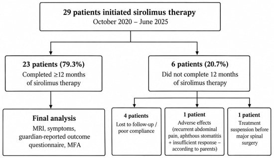
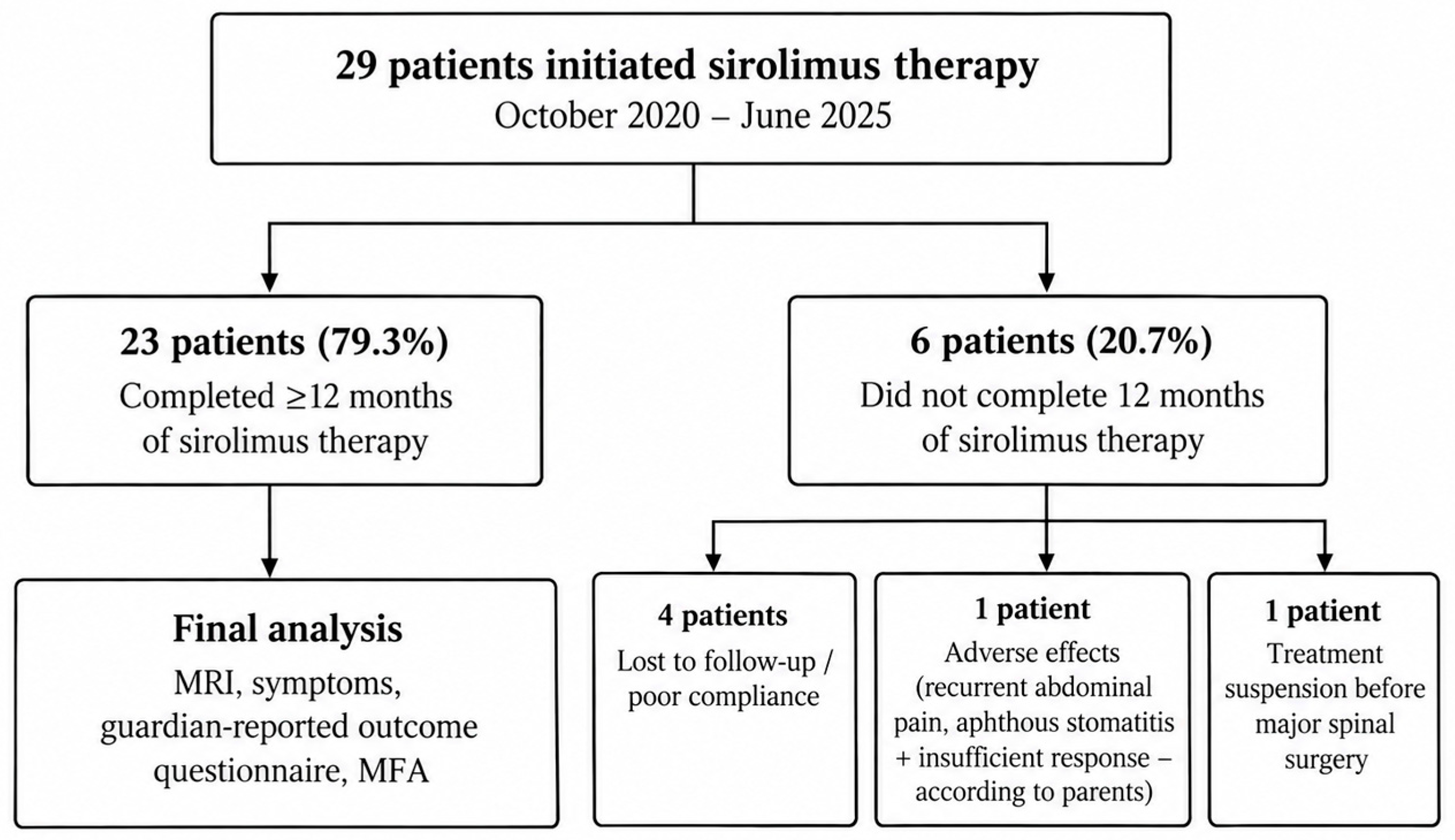
Between October 2020 and June 2025, a total of 29 patients with vascular anomalies initiated sirolimus treatment at the Department of Pediatric Surgery and Oncology and the Department of Pediatrics, Diabetology, Endocrinology and Nephrology at the Central Teaching Hospital of the Medical University of Lodz. Twenty-three patients completed at least 12 months of therapy and had an evaluable 12-month follow-up and therefore constituted the primary 12-month efficacy cohort. Six patients did not complete the predefined 12-month treatment period. These patients were not considered evaluable for the primary 12-month radiological analysis but were retained in the study flow description and in the all-treated sensitivity analysis (Figure 1). Of these six patients, four were lost to regular follow-up because of poor adherence to the treatment and follow-up schedule; in two of these cases, parents also reported dissatisfaction with treatment outcomes. One patient discontinued treatment at our center because of mild adverse events and an insufficient clinical response and subsequently resumed treatment at another center. In one patient, treatment was suspended in preparation for major vertebral surgery. The diagnosis of vascular anomalies was established based on clinical evaluation and confirmed by imaging studies, including magnetic resonance imaging (MRI), ultrasonography, and/or computed tomography (CT) [17]. Patients included in the study were diagnosed with various types of vascular anomalies, including LMs, VMs, combined malformations, AVMs, and TA.
Figure 1.
Study population flow diagram showing patient selection for the final analysis and reasons for exclusion.
2.2. Treatment and Patient Evaluation
Collected data included patient demographics, age at treatment initiation, type and anatomical location of the vascular anomaly, associated comorbidities, treatment duration, drug tolerance, sirolimus trough blood concentrations, radiological response, additional treatments administered before or during sirolimus therapy, adverse effects, current treatment status and patient- or guardian-reported outcomes. All patients were managed by a multidisciplinary vascular anomalies team consisting of pediatric surgeons, pediatric oncologists, radiologists, and other specialists as clinically indicated. Treatment decisions, including qualification for sirolimus therapy and additional interventions, were made jointly during multidisciplinary case discussions. Additional treatment was defined as any therapeutic intervention other than sirolimus administered for the management of vascular anomalies, including surgical procedures, sclerotherapy, embolization, laser therapy, endovascular ablation, and systemic corticosteroid therapy in a patient with KMP. The proportion of patients receiving additional treatment before and during sirolimus therapy was compared.
Before initiation of sirolimus therapy, all patients underwent a comprehensive baseline evaluation, including laboratory tests (complete blood count, liver and renal function tests, coagulation parameters, and lipid profile), viral screening (HBV, HCV, CMV, EBV, HIV), lymphocyte subset analysis, concentrations of immunoglobulins of classes M, A, G, abdominal ultrasonography, contrast-enhanced MRI of the affected area, and cardiology consultation. Additional baseline investigations were performed when clinically indicated.
Sirolimus was administered orally, with liquid formulations used in younger children and tablets in older patients [5]. The initial dose was calculated according to body surface area (BSA) using the Mosteller formula and started at 0.8 mg/m2 every 12 h [1,5,8,9,14,15,16]. Dose adjustments were made during treatment according to sirolimus trough blood concentrations, with a target therapeutic range of 4–10 ng/mL. The treatment strategy aimed to achieve the lowest effective dose while maintaining clinical efficacy.
Patients were closely monitored throughout therapy, with follow-up visits scheduled at 2 weeks, 1 month, 3 months, 6 months, 12 months, and every three months thereafter. Routine assessments at each visit included clinical examination and laboratory testing. Radiological response was assessed using contrast-enhanced MRI or CT examinations performed before treatment initiation and during follow-up at 6 and 12 months, and annually thereafter. Lesion size was evaluated using maximum linear dimensions measured in three orthogonal planes (length × width × depth), expressed in centimeters. Changes in lesion size were assessed by comparing measurements obtained at each follow-up examination. Radiological outcomes were classified as decreased lesion size, stable disease, or increased lesion size based on changes in the recorded lesion dimensions. In cases of extensive, irregular, or highly infiltrative lesions, where reliable linear measurements could not be obtained, radiological response was determined based on the radiologist’s overall assessment of changes in lesion extent. MRI examinations were evaluated by different radiologists as part of routine clinical practice. The radiologists were unaware of the patients’ clinical treatment outcomes at the time of assessment.
Treatment effectiveness was assessed using both objective and subjective criteria. Objective outcomes included reduction in lesion size on imaging studies and improvement in relevant laboratory parameters. In addition, all the guardians were asked to complete a study-specific 20-item questionnaire. Subjective outcomes included changes in clinical symptoms, including pain, bleeding, physical activity, and treatment satisfaction before and after 12 months of sirolimus therapy.
These criteria were used to assess the radiological and clinical efficacy, as well as the safety, of sirolimus therapy.
2.3. Statistical Analyses
Statistical analyses were performed using non-parametric and categorical methods because of the small sample size and non-normal distribution of several variables. Continuous variables are presented as medians with interquartile ranges and/or ranges, and categorical variables as counts and percentages.
Comparisons between two independent groups were performed using the Wilcoxon rank-sum test (Mann–Whitney U test). For paired continuous observations obtained from the same patients at baseline and 12 months, the Wilcoxon signed-rank test was used.
Laboratory measurements obtained at 24 and 36 months were available for progressively smaller subsets of the cohort. Because repeated observations originated from the same patients and the number of evaluable patients decreased substantially at later time points, laboratory results across all four time points were analyzed descriptively rather than using independent-group tests. For each parameter and visit, the number of available observations is explicitly reported. Apparent differences between later follow-up visits were not interpreted as evidence of a longitudinal within-patient trend.
Baseline characteristics of patients with and without an evaluable 12-month follow-up were compared descriptively because of the small number of non-completers.
Associations between categorical variables were assessed using Pearson’s Chi-square test (e.g., changes in abnormal vs. normal parameter frequencies over time) or Fisher’s exact test when expected cell counts were low. Paired categorical data were analyzed using the McNemar test. Caregiver-reported outcomes assessed before and after treatment were evaluated using the Wilcoxon signed-rank test for paired data. Correlations between variables were assessed using Spearman’s rank correlation coefficient.
Baseline characteristics of patients with and without an evaluable 12-month follow-up were compared descriptively. Given the small number of non-completers (n = 6), no formal hypothesis testing was performed for this comparison.
Multiple Factor Analysis (MFA) was performed as an exploratory, hypothesis-generating analysis to examine multidimensional relationships among patient characteristics and treatment-related outcomes. Active variables were organized into four predefined groups: age; demographic and diagnostic characteristics (sex and malformation type); laboratory changes between baseline and 12 months (total cholesterol, LDL cholesterol, and triglycerides); and guardian-reported outcomes (changes in pain, physical activity and bleeding, and treatment satisfaction). Continuous variables were standardized within the MFA framework, whereas sex and diagnosis were treated as nominal variables.
Twelve-month MRI response was included as a supplementary qualitative variable and therefore did not contribute to the construction of the MFA dimensions or to subsequent clustering. Missing data were confined to patient-reported outcomes (2/23 patients, 8.7%) and were handled using regularized MFA-based imputation. A complete-case sensitivity analysis was additionally performed.
Five MFA dimensions, accounting for 77.4% of total variance, were retained for exploratory clustering. Hierarchical clustering of MFA coordinates was performed using Euclidean distance and Ward’s minimum-variance method (Ward.D2). Candidate solutions with two to four clusters were evaluated using average silhouette width, with preference given to solutions without very small clusters. Cluster stability was assessed using 1000 bootstrap resamples and cluster-wise Jaccard similarity. Given the small sample size, all MFA and clustering findings were considered exploratory and were not interpreted as validated treatment-response phenotypes.
All statistical tests were two-tailed, and a p-value <0.05 was considered statistically significant performed in R environment v. 4.5.2.
3. Results
3.1. Patients’ Characteristics
The final analysis cohort comprised 13 females (57%) and 10 males (43%), all of whom received sirolimus therapy for at least 12 months. The diagnoses of vascular anomalies were established based on clinical evaluation and confirmed by imaging studies.
The cohort included 10 patients (43%) with LMs, 9 (39%) with VMs, 2 (9%) with combined malformations (one patient with PIK3CA-related overgrowth spectrum [PROS] and one with Klippel–Trenaunay syndrome [KTS]), and 2 (9%) patients with other vascular anomalies (one with AVM and one with TA). The patient diagnosed with KTS presented with a combined capillary–lymphatic–venous malformation (CLVM) associated with lower limb overgrowth and varicosities. The most common anatomical location was the head and neck region (14 patients, 61%), followed by extremities (6 patients, 26%) and trunk (3 patients, 13%).
The median age at initiation of sirolimus therapy was 2 years. The youngest patient included in study was a 2-week-old boy. Comorbidities (e.g., inhalant allergies, hyperthyroidism, depression) were present in 9 patients (39%). Patients’ characteristics are summarized in Table 1.
Table 1.
Patients’ characteristics.
Baseline characteristics and available clinical trajectories of the six patients without an evaluable 12 month follow-up are summarized alongside those of the 23 patients included in the primary 12 month analysis in Supplementary Table S1.
3.2. Previous and Concomitant Treatments
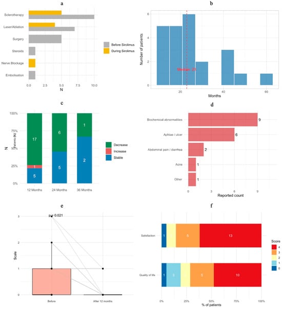
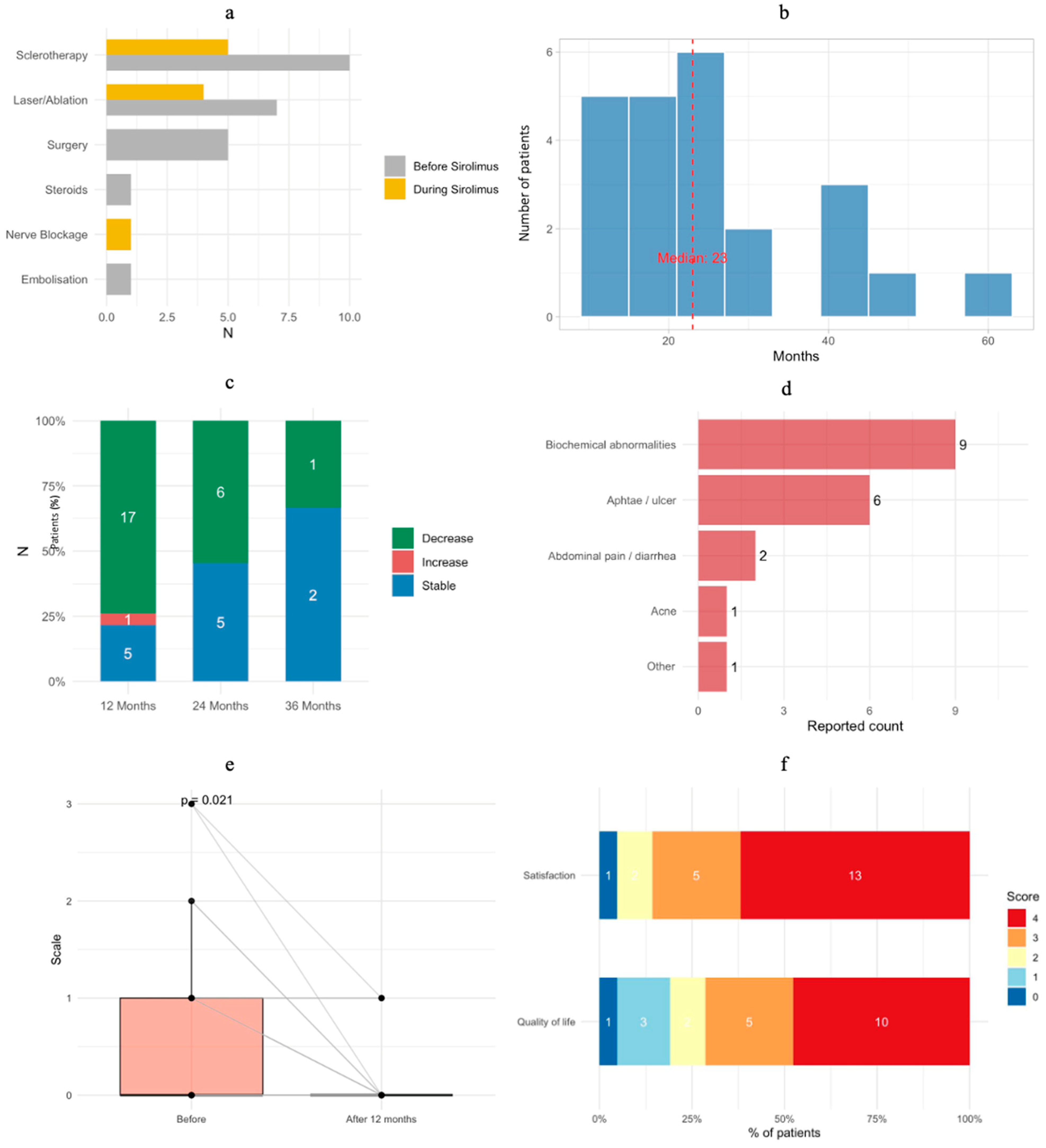
Prior to sirolimus initiation, 15 patients (65%) had received additional treatment including sclerotherapy, surgery, embolization, laser therapy, endovascular ablation, or systemic corticosteroids. During sirolimus therapy, 7 patients (30%) received additional treatment (Figure 2a). The proportion of patients receiving additional treatment was significantly lower during sirolimus therapy than before its initiation (McNemar test, p = 0.0433). However, the duration of the pre-treatment observation period varied considerably between patients, depending on age at sirolimus initiation, disease severity, clinical symptoms, and response to previous treatment, and was not directly comparable with the duration of sirolimus therapy. The median duration of sirolimus treatment was 23 months (range: 12–58 months) (Figure 2b). At the time of data collection, treatment was ongoing in 21 patients (91%), while 2 patients (8.7%) had completed therapy. Malformation recurrence was observed in 1 patient and sirolimus was reinitiated.
Figure 2.
Clinical outcomes and treatment characteristics of patients receiving sirolimus therapy: (a) additional treatment; (b) treatment duration; (c) response to treatment at 12, 24 and 36 months based on radiological assessments; (d) adverse effects reported by guardians in questionnaires; (e) pain severity. Scale: 0—no pain, 1—mild pain, 2—severe pain, 3—very severe pain, 4—excruciating pain; (f) caregiver-reported treatment satisfaction and perceived well-being. Score: 0—dissatisfied, 1—rather dissatisfied, 2—uncertain, 3—rather satisfied, 4—definitely satisfied.
3.3. Radiological Outcomes
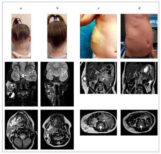
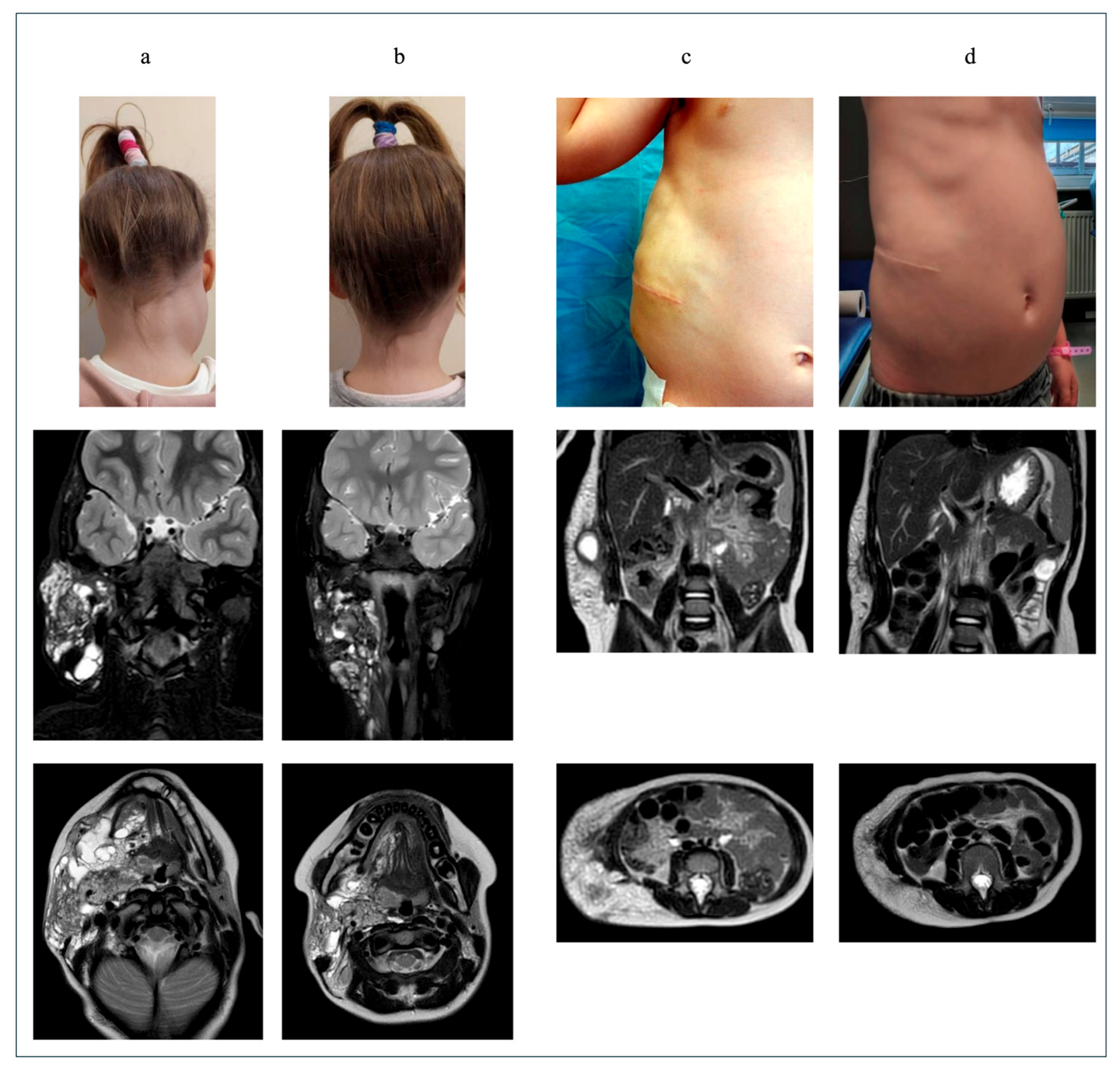
MRI assessment at the 12-month follow-up demonstrated a reduction in lesion size in 17/23 patients (74%), stable disease in 5/23(22%), and an increase in lesion size in 1/23 patient (4%) (Figure 2c and Figure 3a,b). Complete regression of the vascular anomaly was observed after 12 months of treatment in one patient with an LM involving the orbit.
Figure 3.
Radiological outcomes: (a) patient with a lymphatic malformation of the head and neck before starting treatment with sirolimus. Clinical picture and MRI: (b) the same patient as (a), clinical picture after 3 weeks of treatment, and MRI after 12 months of treatment; (c) patient with a lymphatic malformation of the trunk, following previous surgical procedures, before starting treatment with sirolimus; (d) the same patient as (c), after 24 months of sirolimus treatment.
Among the 11 patients treated for more than 24 months, further reduction in lesion size was observed in 6 patients (Figure 3c,d), while the remaining 5 showed stable disease. Of the 3 patients treated for more than 36 months, 2 demonstrated stable disease, whereas 1 showed a continued reduction in lesion size.
3.4. Pharmacokinetics and Laboratory Findings
Sirolimus trough concentrations were regularly monitored. At 12 months, 18 patients (86%) achieved therapeutic levels (4–10 ng/mL), while 3 (14%) had subtherapeutic levels (<4 ng/mL), requiring dose adjustments. Among patients treated for more than 24 months, therapeutic levels were maintained in 10 of 11 cases.
Biochemical parameters remained generally stable throughout treatment. Liver and renal function tests (AST, ALT, creatinine, urea) were within normal ranges at all assessment time points. Lipid profile analysis revealed that total cholesterol and HDL levels were largely within normal limits, whereas an increase in LDL abnormalities and triglyceride levels was observed over time. Triglyceride levels showed a tendency to rise over time, with a higher proportion of abnormal values at later follow-up.
Coagulation parameters demonstrated persistent abnormalities; however, D-dimer levels showed a decreasing trend, while fibrinogen levels remained largely within normal ranges.
The results of laboratory testing are presented in Table 2.
Table 2.
Laboratory test results at different time points during sirolimus treatment.
3.5. Adverse Events and Caregiver-Reported Outcomes
A study-specific questionnaire with evaluable adverse-event information were available for 21 patients. Laboratory/biochemical abnormalities were reported for 9/21 patients (42.9%), and aphthous stomatitis or oral ulceration for 6/21 (28.6%). Individual patients could report more than one adverse-event category. (Figure 2d). Based on the questionnaire responses, pain severity decreased significantly after treatment (p = 0.0206) (Figure 2e).
In addition, caregiver-reported physical activity increased significantly (p = 0.034). Guardians’ assessments of treatment satisfaction and their children’s perceived well-being are presented in Figure 2f.
3.6. Multiple Factor Analysis (MFA)
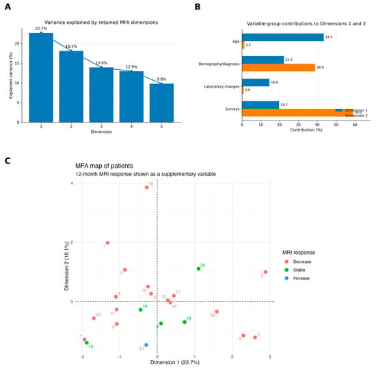
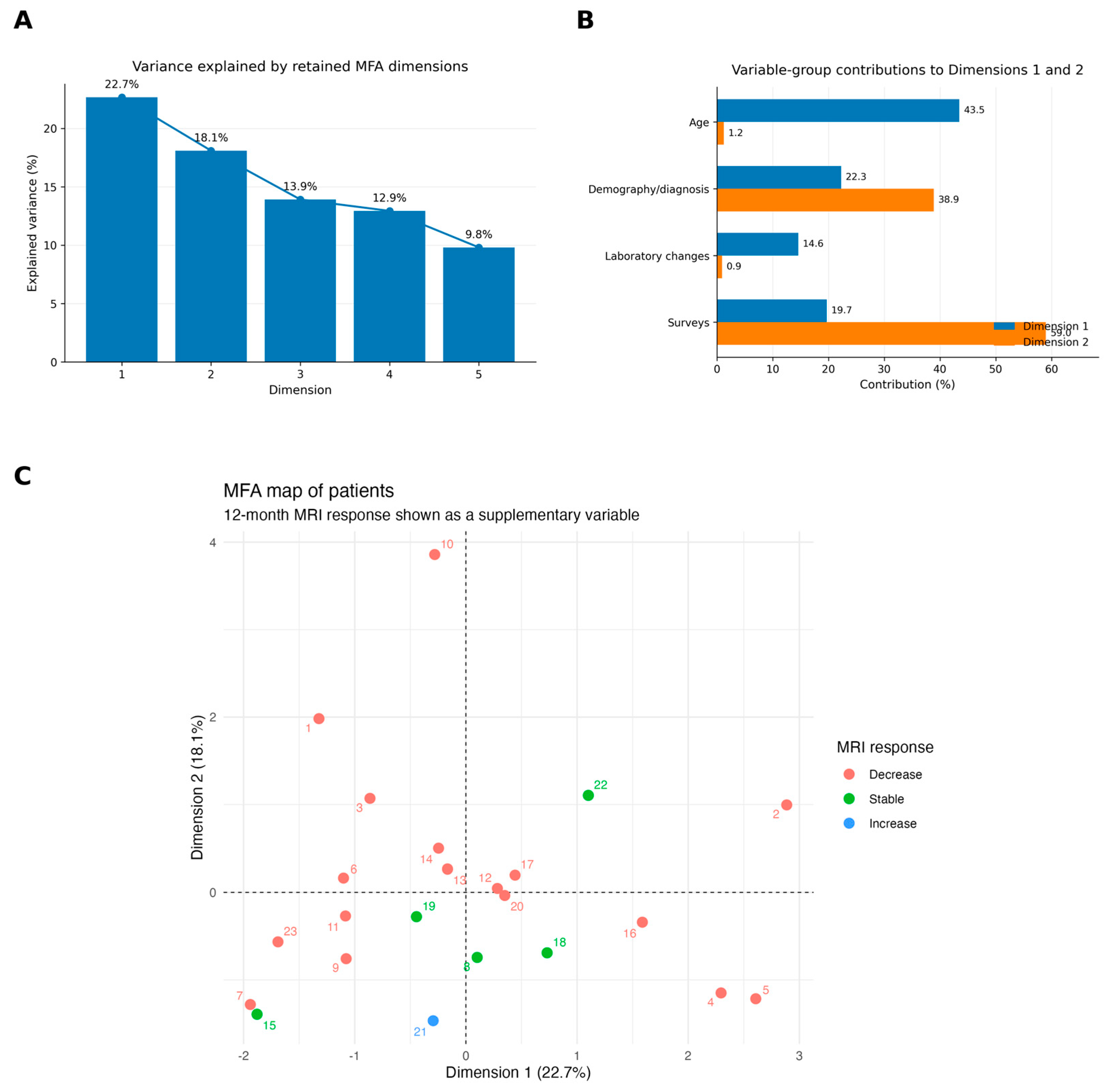
MFA was performed in all 23 patients as an exploratory assessment of multidimensional variation in demographic, diagnostic, laboratory, and guardian-reported characteristics. Twelve-month MRI response was projected as a supplementary variable and did not contribute to the construction of the MFA dimensions.
The first two dimensions explained 22.7% and 18.1% of total variance, respectively, corresponding to 40.8% cumulatively (Figure 4). Dimension 1 was driven primarily by age (43.5% contribution), followed by demographic/diagnostic characteristics (22.3%), patient-reported outcomes (19.7%), and laboratory changes (14.6%). Dimension 2 was predominantly associated with guardian-reported outcomes (59.0%) and demographic/diagnostic characteristics (38.9%). Considerable overlap between the MRI response categories was observed in the MFA space.
Figure 4.
(A) Screen plot showing the proportion of variance explained by the retained MFA dimensions. The first two dimensions accounted for 22.7% and 18.1% of total variance, respectively. (B) Contributions of the predefined active variable groups to Dimensions 1 and 2. Dimension 1 was driven primarily by age, whereas Dimension 2 was predominantly associated with guardian-reported outcomes and demographic/diagnostic characteristics. (C) Individual patient map on the first two MFA dimensions. Twelve-month MRI response is displayed as a supplementary qualitative variable and therefore did not contribute to construction of the MFA dimensions or subsequent clustering.
The exploratory clustering procedure selected a two-cluster solution, with 20 patients in the larger cluster and three patients in the smaller cluster. However, overall cluster separation was limited (average silhouette width, 0.268). Bootstrap analysis demonstrated relatively good stability of the larger cluster (Jaccard similarity, 0.817), whereas the three-patient cluster was poorly reproducible (Jaccard similarity, 0.457) (Supplementary Figure S1). Therefore, the clustering results were not considered evidence of stable clinical response phenotypes.
A complete-case sensitivity analysis yielded a similar proportion of variance explained by the first two dimensions (24.0% and 18.7%, respectively; 42.7% cumulatively), supporting the overall MFA dimensional structure despite the limited missing questionnaire data.
4. Discussion
In this single-center retrospective study, sirolimus therapy was associated with favorable radiological and clinical outcomes in children with vascular anomalies. Most patients experienced lesion reduction or disease stabilization, accompanied by significant improvements in caregiver-reported pain and physical activity, a lower proportion of patients requiring additional treatment, including invasive procedures, and a favorable safety profile [5]. These findings support the growing body of evidence suggesting that mTOR inhibition is an effective treatment strategy for complex vascular anomalies, particularly extensive, symptomatic, or refractory to conventional local therapies [8,9,15].
The radiological outcomes observed in our cohort are consistent with previous studies reporting clinical improvement in most patients with complicated vascular anomalies treated with sirolimus [8,9,15]. In our study, 17/23 (74%) of patients demonstrated lesion reduction after 12 months of treatment, whereas an additional 5/23 (22%) achieved radiological stabilization. Importantly, only one patient experienced progression. These findings suggest that sirolimus may induce lesion regression or halt progression, both representing clinically meaningful outcomes in infiltrative or anatomically challenging malformations.
Another clinically relevant observation was the lower proportion of patients receiving additional treatment during sirolimus therapy compared with the pre-treatment period (30% vs. 65%). Additional treatment included surgery, sclerotherapy, embolization, laser therapy, endovascular ablation, and systemic corticosteroids. Although statistically significant, this difference should be interpreted cautiously because pre-treatment observation periods varied considerably depending on age at treatment initiation, disease severity, symptoms, and previous therapeutic response. Moreover, sirolimus was frequently introduced in patients with complex lesions who had previously undergone other treatment modalities, potentially influencing the observed differences. Therefore, the lower proportion of patients requiring additional treatment cannot be directly attributed to sirolimus alone. Nevertheless, it may reflect a favorable clinical course during systemic treatment and highlight its role within multidisciplinary management. Further prospective studies with standardized observation periods are needed to determine whether sirolimus reduces the requirement for additional therapeutic interventions. In pediatric patients, minimizing repeated procedures is particularly important because of cumulative anesthesia exposure, procedural risks, hospitalization, and psychosocial burden.
The safety profile among patients included in the final analysis was generally favorable and comparable to previous reports [5,8]. However, one of the six patients discontinued sirolimus therapy before completing 12 months because of adverse effects, including recurrent abdominal pain and aphthous stomatitis, combined with insufficient clinical response perceived by the parents. The most commonly reported adverse effects were aphthous stomatitis and biochemical abnormalities, particularly lipid metabolism disturbances [21,22]. Stomatitis was predominantly observed in patients with higher sirolimus trough concentrations and generally resolved following dose reduction. Overall, biochemical alterations were manageable, mainly affecting lipid metabolism, while coagulation abnormalities, likely related to the underlying disease, persisted but partially improved in selected parameters.
Recent studies suggests that lower sirolimus exposure may reduce toxicity while maintaining efficacy. Harbers et al. reported favorable clinical outcomes in patients with low-flow vascular malformations treated with target sirolimus trough concentrations of 4–10 ng/mL, consistent with our study, with a relatively favorable safety profile [23]. Similarly, Zhou et al., in a randomized clinical trial, demonstrated that lower sirolimus trough concentrations (5–8 ng/mL) maintained therapeutic efficacy in patients with kaposiform hemangioendothelioma while reducing certain treatment-related adverse events compared with higher concentrations (10–15 ng/mL) [24]. These findings suggest that minimizing sirolimus exposure may improve treatment tolerability, particularly by reducing mucosal adverse effects, and potentially other dose-related complications, including dyslipidemia. However, adverse effects may still occur at lower concentrations, and the optimal therapeutic range remains uncertain across vascular anomaly subtypes. Individualized dose adjustment based on clinical response, trough concentrations, and adverse effects is essential to achieve an appropriate balance between efficacy and safety, particularly in pediatric patients requiring long-term treatment.
Improvement in caregiver-reported outcomes represents another important aspect of treatment efficacy. While radiological response is often used as a primary endpoint in studies of vascular anomalies, imaging findings do not always correlate with symptom burden. In our cohort, caregiver questionnaires demonstrated significantly reduced pain, increased physical activity, and high treatment satisfaction after 12 months. Similar improvements in quality of life have been described by Cho et al., Kim et al., Wiegand et al., and Hammer et al., emphasizing that symptom control may be equally important as radiological response. Particularly in lesions affecting the head and neck region, functional improvement and reduction of pain can substantially improve daily functioning and psychosocial well-being.
The exploratory MFA illustrated substantial multidimensional heterogeneity within this small cohort rather than clearly separated treatment-response phenotypes. Age contributed most strongly to the first MFA dimension, whereas patient-reported outcomes and demographic/diagnostic characteristics contributed predominantly to the second dimension. Importantly, MRI response categories showed considerable overlap in the MFA space. Although exploratory hierarchical clustering suggested a small subgroup of three patients, bootstrap analysis demonstrated limited stability of this cluster, precluding its interpretation as a reproducible clinical phenotype. These findings therefore should be regarded as hypothesis-generating and require validation in substantially larger, independent cohorts.
Several limitations should be considered. The retrospective design and single-center setting introduce potential selection bias and limit generalizability. The relatively small sample size and heterogeneous spectrum of vascular anomalies restrict subgroup analyses and prevent definitive conclusions regarding specific lesion types. The predominance of lymphatic and venous malformations in our cohort likely contributed to the favorable overall response rates observed. Furthermore, caregiver-reported outcomes were based on questionnaires completed by guardians and may be subject to reporting bias. Another limitation concerns the assessment of radiological response, which was based primarily on the linear measurements obtained from routine MRI examinations rather than volumetric analysis. Formal volumetric measurements were not performed due to technical limitations at our institution. Additionally, MRI examinations were evaluated by different radiologists, which may have introduced interobserver variability. The duration of observation periods before and during sirolimus therapy varied between patients. Previous treatment history and individual indications for sirolimus initiation may also have influenced the observed differences. Therefore, the lower proportion of patients receiving additional treatment during sirolimus therapy should be interpreted with consideration of these factors. An additional limitation is the potential for attrition bias because the primary 12-month efficacy analysis was based on patients who completed the predefined follow-up period. Six of the 29 treated patients did not have an evaluable 12-month outcome, including patients with treatment dissatisfaction and one patient who discontinued treatment at our center because of adverse events and insufficient clinical response. Although an all-treated sensitivity analysis and descriptive comparison of completers and non-completers were added, informative attrition cannot be excluded. Furthermore, the number of patients with laboratory and radiological assessments decreased substantially beyond 12 months. Consequently, later follow-up findings primarily describe the available subset rather than a complete longitudinal cohort. Finally, the MFA and clustering analyses were exploratory. The small cohort size, overlap between patient groups, and very small number of individuals in some clusters limit the stability and generalizability of the identified patterns.
The multidisciplinary approach adopted in our center, integrating pediatric surgery, pediatric oncology, radiology, and other relevant specialties, may have contributed to the favorable clinical outcomes observed in this cohort.
Despite these limitations, the present study contributes additional real-world evidence supporting the use of sirolimus in pediatric vascular anomalies. Strengths of the study include long-term follow-up in a subset of patients, comprehensive assessment of radiological and clinical outcomes, evaluation of procedural burden, and the incorporation of multidimensional analyses exploring treatment-response phenotypes. Future prospective multicenter studies with larger and more homogeneous cohorts are needed to define optimal treatment duration, identify molecular and clinical predictors of response, and further evaluate long-term safety.
Supplementary Materials
The following supporting information can be downloaded at https://www.mdpi.com/article/10.3390/children13111516/s1, Figure S1: Exploratory clustering of MFA coordinates. (A) Hierarchical clustering dendrogram based on the first five MFA dimensions using Euclidean distance and Ward.D2 linkage. (B) Patient distribution according to the selected two-cluster solution; Cluster 1 contained 20 patients and Cluster 2 contained 3 patients. (C) Average silhouette width for candidate solutions with two to four clusters. The two-cluster solution yielded the highest average silhouette width (0.268). (D) Bootstrap assessment of cluster stability based on 1000 resamples. Jaccard similarities were 0.817 for Cluster 1 and 0.457 for Cluster 2, indicating limited reproducibility of the smaller cluster. Accordingly, clustering results were considered exploratory rather than evidence of stable clinical response phenotypes. Table S1: Baseline characteristics and available treatment trajectories of patients with and without an evaluable 12-month follow-up.
Author Contributions
Conceptualization, J.K. (Justyna Kukulska), J.K. (Jakub Kopeć) and P.P.; methodology, J.K. (Justyna Kukulska), M.Z., J.K. (Jakub Kopeć) and P.P.; validation, J.K. (Justyna Kukulska), M.Z., E.S.-Ł., and P.P.; formal analysis, J.K. (Justyna Kukulska), M.Z. and P.P.; investigation, J.K. (Justyna Kukulska), M.Z., J.K. (Jakub Kopeć), J.T., E.S.-Ł., and P.P.; data curation, J.K. (Justyna Kukulska), E.S.-Ł. and J.T.; writing—original draft preparation, J.K. (Justyna Kukulska); writing—review and editing, J.K. (Justyna Kukulska), M.Z., E.S.-Ł., J.K. (Jakub Kopeć), J.T. and P.P.; visualization, J.K. (Justyna Kukulska); supervision, M.Z. and P.P.; project administration, J.K. (Justyna Kukulska), M.Z., E.S.-Ł., J.K. (Jakub Kopeć), J.T. and P.P.; funding acquisition, P.P. All authors have read and agreed to the published version of the manuscript.
Funding
Open access funding provided by the Department of Pediatric Surgery and Oncology, and Department of Pediatrics, Diabetology, Endocrinology and Nephrology, Medical University of Lodz. No other funding for this study was received.
Institutional Review Board Statement
The study was conducted in accordance with the Declaration of Helsinki and was approved by the Ethics Committee of Medical University of Lodz (No. RNN/215/20/KE, 13 October 2020). The ethical approval did not require annual renewal and remained valid throughout the study period.
Informed Consent Statement
Informed consent was obtained from all subjects involved in the study. Written informed consent was obtained from the patients’ legal guardians for the publication of the clinical photographs and MRI images included in this manuscript.
Data Availability Statement
The original contributions presented in this study are included in the article. Further inquiries can be directed to the corresponding author(s).
Acknowledgments
The authors would like to thank Magdalena Krystkiewicz-Orzechowska for her assistance with the statistical analysis. The authors would also like to thank all the healthcare professionals involved in the clinical management and care of the patients included in this study.
Conflicts of Interest
The authors declare no conflicts of interest.
Abbreviations
The following abbreviations are used in this manuscript:
| mTOR | mammalian target of rapamycin |
| LM | Lymphatic malformation |
| VM | Venous malformation |
| MRI | Magnetic resonance imaging |
| MFA | Multiple Factor Analysis |
| ISSVA | International Society for the Study of Vascular Anomalies |
| PUVA | Potentially Unique Vascular Anomalies |
| CM | Capillary malformation |
| AVM | Arteriovenous malformation |
| TA | Tufted angioma |
| KMP | Kasabach–Merritt phenomenon |
| PI3K | Phosphatidylinositol-3-kinase |
| CT | Computed tomography |
| KTS | Klippel–Trenaunay syndrome |
| HDL | High-density lipoprotein |
| LDL | Low-density lipoprotein |
| TG | Triglycerides |
| BSA | Body surface area |
| IQR | Interquartile range |
| F | Female |
| M | Male |
| AST | Aspartate aminotransferase |
| ALT | Alanine aminotransferase |
References
- Cho, Y.J.; Kwon, H.; Kwon, Y.J.; Kim, S.C.; Kim, D.Y.; Namgoong, J.M. Effects of sirolimus in the treatment of unresectable infantile hemangioma and vascular malformations in children: A single-center experience. J. Vasc. Surg. Venous Lymphat. Disord. 2021, 9, 1488–1494. [Google Scholar] [CrossRef] [Scilit] [PubMed]
- Kim, M.; Hong, K.T.; Park, H.J.; Kim, B.K.; Choi, J.Y.; Kim, H.Y.; Kang, H.J. Clinical effectiveness and safety of sirolimus in pediatric patients with complex vascular anomalies: Necessitating personalized and comprehensive approaches. Front. Pediatr. 2023, 11, 1304133. [Google Scholar] [CrossRef] [Scilit] [PubMed]
- Yesil, S.; Tanyildiz, H.G.; Bozkurt, C.; Cakmakci, E.; Sahin, G. Single-center experience with sirolimus therapy for vascular malformations. Pediatr. Hematol. Oncol. 2016, 33, 219–225. [Google Scholar] [CrossRef] [Scilit] [PubMed]
- Lackner, H.; Karastaneva, A.; Schwinger, W.; Benesch, M.; Sovinz, P.; Seidel, M.; Sperl, D.; Lanz, S.; Haxhija, E.; Reiterer, F.; et al. Sirolimus for the treatment of children with various complicated vascular anomalies. Eur. J. Pediatr. 2015, 174, 1579–1584. [Google Scholar] [CrossRef] [Scilit] [PubMed]
- Hammer, J.; Seront, E.; Duez, S.; Dupont, S.; Van Damme, A.; Schmitz, S.; Hoyoux, C.; Chopinet, C.; Clapuyt, P.; Hammer, F.; et al. Sirolimus is efficacious in treatment for extensive and/or complex slow-flow vascular malformations: A monocentric prospective phase II study. Orphanet J. Rare Dis. 2018, 13, 191. [Google Scholar] [CrossRef] [Scilit] [PubMed]
- ISSVA Classification of Vascular Anomalies. International Society for the Study of Vascular Anomalies. 2025. Available online: https://www.issva.org/classification (accessed on 20 October 2026).
- Karastaneva, A.; Gasparella, P.; Tschauner, S.; Crazzolara, R.; Kropshofer, G.; Modl, M.; Pfleger, A.; Burmas, A.; Pocivalnik, M.; Ulreich, R.; et al. Indications and Limitations of Sirolimus in the Treatment of Vascular Anomalies—Insights From a Retrospective Case Series. Front. Pediatr. 2022, 10, 857436. [Google Scholar] [CrossRef] [Scilit] [PubMed]
- Seront, E.; Van Damme, A.; Legrand, C.; Bisdorff-Bresson, A.; Orcel, P.; Funck-Brentano, T.; Sevestre, M.-A.; Dompmartin, A.; Quere, I.; Brouillard, P.; et al. Preliminary results of the European multicentric phase III trial regarding sirolimus in slow-flow vascular malformations. JCI Insight 2023, 8, e173095. [Google Scholar] [CrossRef] [Scilit] [PubMed]
- Tian, R.; Liang, Y.; Zhang, W.; Wang, J.; Shan, Y.; Gao, H.; Xie, C.; Li, J.; Xu, M.; Gu, S. Effectiveness of sirolimus in the treatment of complex lymphatic malformations: Single center report of 56 cases. J. Pediatr. Surg. 2020, 55, 2454–2458. [Google Scholar] [CrossRef] [Scilit] [PubMed]
- Schimmel, K.; Ali, M.K.; Tan, S.Y.; Teng, J.; Do, H.M.; Steinberg, G.K.; Stevenson, D.A.; Spiekerkoetter, E. Arteriovenous malformations—Current understanding of the pathogenesis with implications for treatment. Int. J. Mol. Sci. 2021, 22, 9037. [Google Scholar] [CrossRef] [Scilit] [PubMed]
- Bhari, N.; Jangid, B.; Pahadiya, P.; Singh, S.; Arava, S.; Kumar, A.; Sharma, V.K.; Sethuraman, G. Tufted angioma with recurrent Kasabach-Merritt phenomenon. Indian J. Dermatol. Venereol. Leprol. 2018, 84, 121. [Google Scholar] [CrossRef] [Scilit] [PubMed]
- Wang, H.; Guo, X.; Duan, Y.; Zheng, B.; Gao, Y. Sirolimus as initial therapy for kaposiform hemangioendothelioma and tufted angioma. Pediatr. Dermatol. 2018, 35, 635–638. [Google Scholar] [CrossRef] [Scilit] [PubMed]
- Chiguer, M.; Farid, N.; Amal, S.; Sab, I.A.; Bourahouat, A.; Hocar, O. Tufted angioma with Kasabach-Merritt syndrome: Two cases successfully treated with everolimus. Pediatr. Blood Cancer 2020, 67, e28291. [Google Scholar] [CrossRef] [Scilit] [PubMed]
- Hammill, A.M.; Wentzel, M.; Gupta, A.; Nelson, S.; Lucky, A.; Elluru, R.; Dasgupta, R.; Azizkhan, R.G.; Adams, D.M. Sirolimus for the treatment of complicated vascular anomalies in children. Pediatr. Blood Cancer 2011, 57, 1018–1024. [Google Scholar] [CrossRef] [Scilit] [PubMed]
- Wiegand, S.; Dietz, A.; Wichmann, G. Efficacy of sirolimus in children with lymphatic malformations of the head and neck. Eur. Arch. Oto-Rhino-Laryngol. 2022, 279, 3801–3810. [Google Scholar] [CrossRef] [Scilit] [PubMed]
- Adams, D.M.; Trenor, C.C., III; Hammill, A.M.; Vinks, A.A.; Patel, M.N.; Chaudry, G.; Wentzel, M.S.; Mobberley-Schuman, P.S.; Campbell, L.M.; Brookbank, C.; et al. Efficacy and Safety of Sirolimus in the Treatment of Complicated Vascular Anomalies. Pediatrics 2016, 137, e20153257. [Google Scholar] [CrossRef] [Scilit] [PubMed]
- Wiegand, S.; Wichmann, G.; DIetz, A. Treatment of Lymphatic Malformations with the mTOR Inhibitor Sirolimus: A Systematic Review. Lymphat. Res. Biol. 2018, 16, 330–339. [Google Scholar] [CrossRef] [Scilit] [PubMed]
- Triana, P.; Dore, M.; Cerezo, V.N.; Cervantes, M.; Sánchez, A.V.; Ferrero, M.M.; González, M.D.; Lopez-Gutierrez, J.C. Sirolimus in the Treatment of Vascular Anomalies. Eur. J. Pediatr. Surg. 2017, 27, 086–090. [Google Scholar] [CrossRef] [Scilit] [PubMed]
- Freixo, C.; Ferreira, V.; Martins, J.; Almeida, R.; Caldeira, D.; Rosa, M.; Costa, J.; Ferreira, J. Efficacy and safety of sirolimus in the treatment of vascular anomalies: A systematic review. J. Vasc. Surg. 2020, 71, 318–327. [Google Scholar] [CrossRef] [Scilit] [PubMed]
- Nadal, M.; Giraudeau, B.; Tavernier, E.; Jonville-Bera, A.P.; Lorette, G.; Maruani, A. Efficacy and safety of mammalian target of rapamycin inhibitors in vascular anomalies: A systematic review. Acta Derm.-Venereol. 2016, 96, 448–452. [Google Scholar] [CrossRef] [Scilit] [PubMed]
- Neirotti, A.; Barat, V.; Coppo, P.; La Selva, R.; Manicone, R.; Cotti, R.; Sensini, M.; Mussa, A.; Gatto, M.; Farriet, F.; et al. Therapy with sirolimus in vascular anomalies: The experience of two Italian centers on 14 pediatric patients. Front. Pediatr. 2024, 12, 1434493. [Google Scholar] [CrossRef] [Scilit] [PubMed]
- Zhang, Z.; Li, Y.; Zhang, G.; Yang, K.; Qiu, T.; Zhou, J.; Gong, X.; Ji, Y. Safety Evaluation of Oral Sirolimus in the Treatment of Childhood Diseases: A Systematic Review. Children 2022, 9, 1295. [Google Scholar] [CrossRef] [Scilit] [PubMed]
- Harbers, V.E.M.; Rongen, G.A.P.J.M.; van der Vleuten, C.J.M.; Verhoeven, B.H.; de Laat, P.C.J.; van der Horst, C.M.A.M.; Klein, W.M.; Kool, L.J.S.; te Loo, D.M.W.M. Patients with Congenital Low-Flow Vascular Malformation Treated with Low Dose Sirolimus. Adv. Ther. 2021, 38, 3465–3482. [Google Scholar] [CrossRef] [Scilit] [PubMed]
- Zhou, J.; Lan, Y.; Qiu, T.; Zhang, Z.; Gong, X.; Zhang, X.; Yang, C.; Zhou, Z.; Zhang, Y.; Yang, M.; et al. Efficacy and safety of high-vs low-dose sirolimus in patients with kaposiform hemangioendothelioma: A randomized clinical trial. J. Am. Acad. Dermatol. 2025, 93, 124–131. [Google Scholar] [CrossRef] [Scilit] [PubMed]

Disclaimer/Publisher’s Note: The statements, opinions and data contained in all publications are solely those of the individual author(s) and contributor(s) and not of MDPI and/or the editor(s). MDPI and/or the editor(s) disclaim responsibility for any injury to people or property resulting from any ideas, methods, instructions or products referred to in the content. |
© 2026 by the authors. Licensee MDPI, Basel, Switzerland. This article is an open access article distributed under the terms and conditions of the Creative Commons Attribution (CC BY) license.