


Abstract
Background/Objectives: Long-term immunosuppressive therapy administered to solid organ transplant recipients may induce adverse effects in various tissues, including the gastrointestinal tract. The aim of the present study was to evaluate the influence of prolonged treatment with calcineurin inhibitor (CNI)-based triple immunosuppressive regimens on the morphology of the jejunal mucosa in male rats. Particular attention was paid to fibrotic changes occurring within the mucosal connective tissue, including the intestinal villi and lamina propria. Methods: Animals were treated for six months with regimens containing either cyclosporine A (CyA) or tacrolimus (TAC) in combination with rapamycin and prednisone. To assess the content of collagen fibers, jejunal sections were stained using the Mallory trichrome method and subsequently subjected to quantitative digital image analysis. Results: Histological examination demonstrated increased collagen deposition in the jejunal mucosa of rats receiving immunosuppressive treatment compared with the control group. Quantitative analysis confirmed an increase in collagen fiber content, indicating enhanced extracellular matrix accumulation and changes consistent with fibrotic remodeling of the mucosal connective tissue. The highest collagen fiber content was observed in animals treated with the cyclosporine-based regimen. Conclusions: These findings suggest that long-term administration of CNI-based triple immunosuppressive therapy may be associated with changes consistent with fibrotic remodeling of the jejunal mucosa. The present study provides novel data on the effects of clinically relevant multidrug immunosuppressive protocols, whereas most previous experimental studies investigating the effects of immunosuppressive agents have focused on single-drug exposure in animal models or in vitro systems.
1. Introduction
Immunosuppression plays a key role in transplant medicine, where it prevents the rejection of transplanted organs, as well as in the treatment of autoimmune diseases, in which the immune system mistakenly attacks the body’s own tissues. Immunosuppressive regimens used in parenchymal organ transplantation have evolved considerably in recent years [1].
The type of immunosuppressive therapy required depends on the underlying disease. Monotherapy, involving the use of a single drug, is commonly employed in the treatment of autoimmune diseases, whereas immunosuppressive therapy in organ transplant recipients most often consists of a combination of three drugs. The choice of immunosuppressive agents depends primarily on the type of transplanted organ and the presence of comorbidities. Recipients of parenchymal organ transplants require lifelong suppression of the immune response. In clinical practice, long-term graft survival depends on the careful adjustment and monitoring of immunosuppressive regimens. These drugs play a crucial role in maintaining graft function and preventing transplant rejection [2]; however, they may also cause a variety of adverse effects [3].
Maintenance immunosuppressive therapy commonly includes calcineurin inhibitors (CNIs), antiproliferative agents, mTOR inhibitors, and corticosteroids. In selected cases, calcineurin inhibitors may be replaced with belatacept. Each class of immunosuppressive drugs targets different pathways and exhibits distinct mechanisms of action (Table 1).
Table 1.
Main classes of immunosuppressive drugs used in maintenance therapy after solid organ transplantation and their mechanisms of action.
Calcineurin inhibitors (CNIs), including cyclosporine, voclosporin, tacrolimus, and pimecrolimus, exert potent immunosuppressive effects and are among the most widely used agents in maintenance immunosuppressive regimens [12]. Cyclosporine and tacrolimus are the best-established representatives of this drug class; however, their long-term use is associated with significant nephrotoxicity and may contribute to the development of chronic kidney injury.
The introduction of effective immunosuppressive therapy has markedly improved both graft and patient survival following transplantation. Nevertheless, accumulating evidence indicates that long-term exposure to calcineurin inhibitors (CNIs) may promote fibrotic remodeling in various tissues and organs. In the kidney, CNI-associated lesions include interstitial fibrosis, Bowman’s capsule fibrosis, and glomerulosclerosis, whereas fibrotic changes have also been reported in other organs [13]. They may also contribute to the development of hypertension and dyslipidaemia. Furthermore, calcineurin inhibitors have been implicated in direct cardiovascular toxicity, including the induction of cardiomyocyte apoptosis and the promotion of myocardial fibrosis [14]. In cases of intolerance to calcineurin inhibitors following kidney transplantation, conversion to belatacept-based immunosuppression may be considered. Belatacept is a non-nephrotoxic immunosuppressive agent that has emerged as an alternative to CNI-based therapy. A retrospective study by Divard et al. (2024), including 311 kidney transplant recipients treated between 2007 and 2020, demonstrated that conversion to belatacept was associated with a reduced risk of graft failure and superior long-term graft survival [11].
Mycophenolate mofetil (MMF), an immunosuppressive agent belonging to the class of antiproliferative drugs, is generally well tolerated by solid organ transplant recipients. However, its use may be associated with a range of adverse effects, including gastrointestinal and hematological complications, increased susceptibility to infections and malignancies, as well as potential teratogenicity [15,16].
Similarly to MMF, immunosuppressive agents belonging to the mTOR inhibitor class, such as everolimus, are generally well tolerated by solid organ transplant recipients [17]. However, mTOR inhibitors may also be associated with a variety of adverse effects. These include viral infections [18] and cardiovascular complications [19]. Glucocorticoids are also widely used as immunosuppressive agents in maintenance therapy following solid organ transplantation. However, long-term corticosteroid treatment may increase the risk of bacterial, fungal, and viral infections [20].
Animal models are frequently used to investigate the adverse effects of long-term immunosuppressive therapy on various organs and tissues. Previous studies have demonstrated the effects of prolonged multidrug immunosuppression on the salivary glands [21], kidneys [22], prostate gland [23] and other organs [24,25].
Following organ transplantation, immunosuppressive therapy typically involves the administration of several drugs in specific combinations. The choice of immunosuppressive regimen depends on the type of transplanted organ, the patient’s immunological risk, the severity of metabolic disorders, the presence of comorbidities, and graft function. Most transplant recipients receive a three-drug immunosuppressive regimen designed to balance efficacy in preventing graft rejection with safety by minimizing drug-related toxicity [26].
In the present study, we investigated the effects of long-term treatment with calcineurin inhibitor (CNI)-based triple immunosuppressive regimens containing cyclosporine A or tacrolimus in combination with rapamycin and a glucocorticoid on the development of collagen deposition in the jejunal mucosa of rats.
2. Materials and Methods
2.1. Research Material and Conducting an Experiment on Animals
The study was conducted using archival jejunal tissue specimens obtained from 14-week-old sexually mature male rats and preserved in paraffin blocks. The present study is a continuation of our previous investigation. The experimental protocol was approved by the Local Ethics Committee for Animal Experiments in Szczecin [27].
The animals were treated with the following immunosuppressive drugs at the doses presented in Table 2: (i) cyclosporine A (Sandimmun Neoral; C)—5 mg/kg body weight/day; (ii) tacrolimus (Prograf; T)—4 mg/kg body weight/day; (iii) rapamycin (Rapamune; R)—0.5 mg/kg body weight/day; and (iv) prednisone (Encorton; G)—4 mg/kg body weight/day. The doses of immunosuppressive drugs were selected based on previously established experimental protocols in rats. In particular, tacrolimus at a dose of 4 mg/kg/day has been used in previous studies investigating the effects of chronic immunosuppressive therapy in rat models [28,29]. Due to interspecies differences in pharmacokinetics and drug metabolism, doses expressed as mg/kg in rats should not be directly compared with those used clinically in human transplant recipients. Animals assigned to the experimental groups (CRG and TRG) received immunosuppressive treatment for six months according to calcineurin inhibitor (CNI)-based triple-drug regimens (Table 2).
Table 2.
Triple-drug immunosuppressive regimens administered to rats during the experiment and the number of animals (n) included in each experimental group. During the study, two animals from the CRG group died; therefore, the final number of animals in this group was four.
2.2. Histological Staining
Intestinal tissue sections were stained with hematoxylin and eosin (H&E) for morphological evaluation. Collagen fibers within the jejunal mucosa were visualized using the Mallory trichrome staining method (Bio-Optica, Milan, Italy). In this staining procedure, collagen fibers were stained dark blue. Histological assessment of the examined tissues was performed using a light microscope (Leica DM5000B, Wetzlar, Germany).
2.3. Digital Image Analysis
To assess the content of collagen fibers, jejunal tissue sections stained with the Mallory trichrome method were subjected to quantitative digital image analysis using the PatternQuant 2.4 image analysis software (3DHISTECH Ltd., Budapest, Hungary). The following parameters were evaluated in the jejunal mucosa of both the control and experimental groups: the area, perimeter, and staining intensity of the analyzed collagen fibers. Multiple measurements were obtained from several microscopic fields for each animal. The measurements were expressed in pixels and subsequently used for statistical analysis. For each animal, the median value of each analyzed parameter was calculated from all measurements obtained from that animal. These individual animal-level median values were subsequently used for statistical analysis. The total number of measurements was 345 in the Control group, 176 in the CRG group, and 133 in the TRG group. The results are presented in Table 3.
Table 3.
Statistical analysis of the results, obtained from the quantitative digital evaluation of the studied tissues (* p < 0.05).
2.4. Statistical Analysis
Multiple measurements of collagen fiber parameters were obtained from several microscopic fields for each animal using quantitative digital image analysis. A total of 345 measurements were obtained in the Control group, 176 measurements in the CRG group, and 133 measurements in the TRG group. To account for the repeated measurements obtained from the same animal and to avoid pseudoreplication, the median value for each analyzed parameter was calculated separately for each individual animal. These animal-level median values were subsequently used as the units of observation in the statistical analysis. Accordingly, the effective sample sizes were 6 animals in the Control group, 4 animals in the CRG group, and 6 animals in the TRG group.
For each animal, the median value of each analyzed parameter was first calculated from all measurements obtained from that animal. These individual animal-level median values were subsequently used for statistical analysis. The distribution of the variables was assessed using the Shapiro–Wilk test. Due to the small and unequal sample sizes of the study groups (Control, n = 6; CRG, n = 4; TRG, n = 6), non-parametric statistical methods were applied. Differences among the three groups were assessed using the Kruskal–Wallis test. When a statistically significant difference was detected, post-hoc pairwise comparisons were performed using Dunn’s test with Holm correction for multiple comparisons. A p-value < 0.05 was considered statistically significant.
3. Results
3.1. Morphology
Stained sections of the jejunum obtained from animals in the control group and the two experimental groups were examined using light microscopy and subjected to digital image analysis. As described above, the Mallory trichrome staining method enabled the visualization of collagen fibers (Figure 1).
Figure 1.
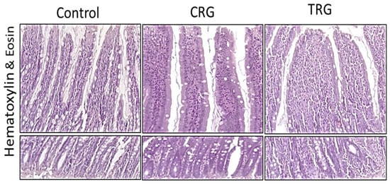
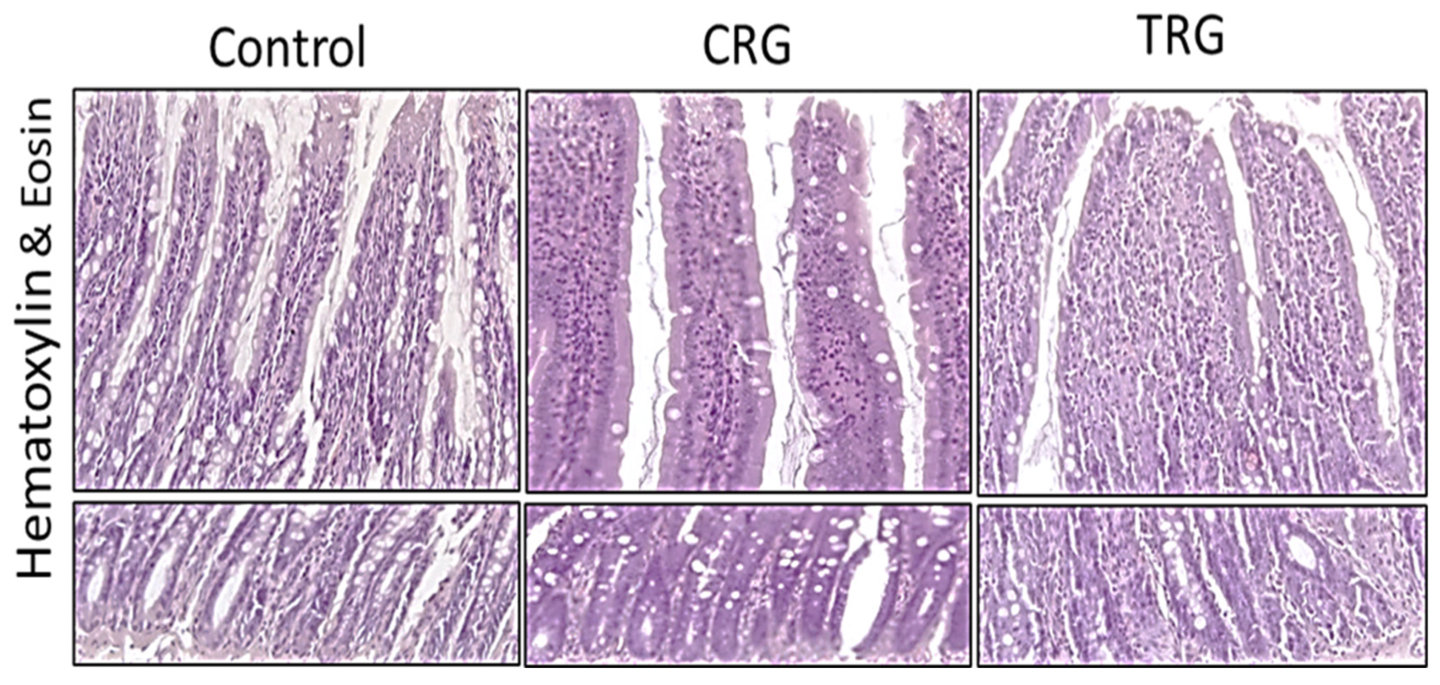
Morphology of the jejunal mucosa in rats from the control group and the two experimental groups receiving calcineurin inhibitor-based immunosuppressive regimens in combination with rapamycin and a glucocorticoid (CRG and TRG). Intestinal villi (upper part) and crypts (lower part) within the lamina propria of the mucosa are shown. Hematoxylin and eosin (H&E) staining; magnification 40×.
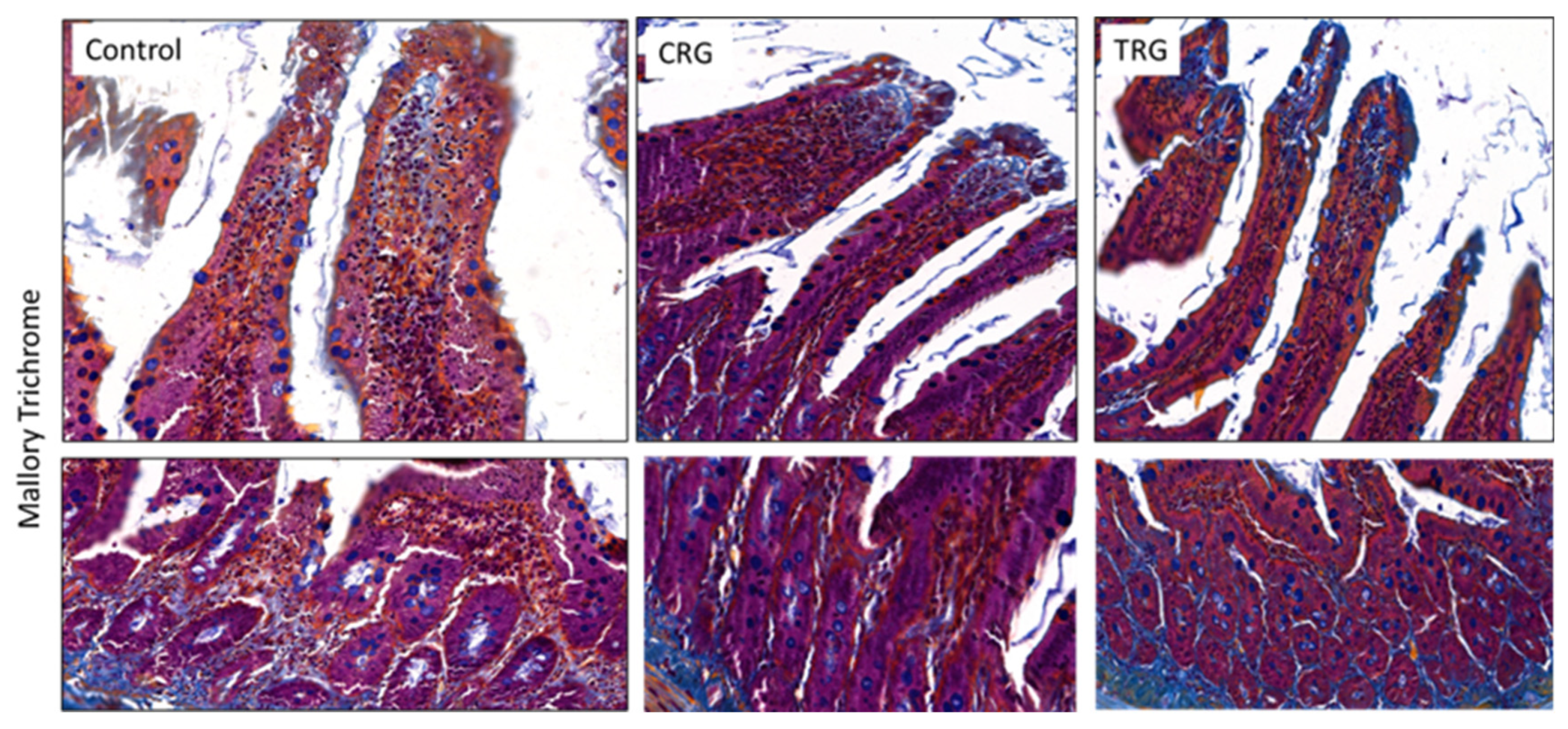
No apparent differences in the morphology of the jejunal mucosa were observed between the control and experimental groups. However, the jejunal mucosa of rats in the CRG group appeared more intensely stained than that of the control and TRG groups (Figure 2).
Figure 2.
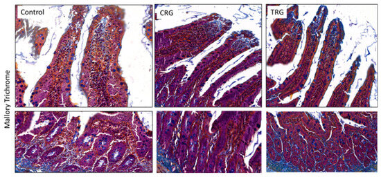
Jejunal mucosa of rats from the control group and the two experimental groups receiving calcineurin inhibitor-based immunosuppressive regimens (CRG and TRG). Intestinal villi and crypts within the lamina propria are shown. Collagen fibers within the mucosa are stained dark blue. Mallory trichrome staining; magnification 40×.
Qualitative histological assessment suggested increased collagen fiber deposition in the jejunal mucosa of both experimental groups compared with the control group.
3.2. Collagen Fiber Content—The Digital Analysis
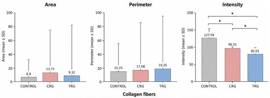
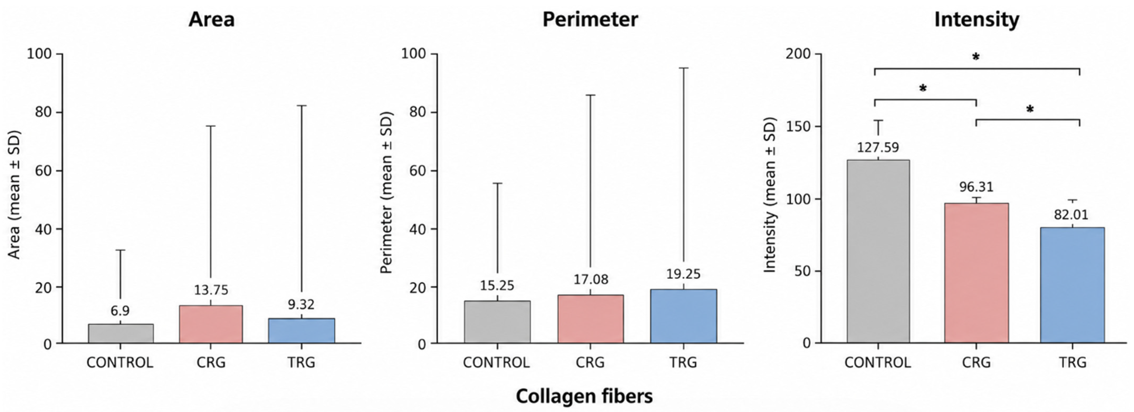
Digital image analysis demonstrated differences in the content of collagen fibers among the examined groups. The highest mean area occupied by collagen fibers was observed in the CRG group (13.75 ± 61.80), followed by the TRG group (9.32 ± 72.88) and the Control group (6.90 ± 26.24) (Table 3, Figure 3).
Figure 3.
Quantitative digital image analysis of collagen fibers in the jejunal mucosa of rats from the control group (C) and the experimental groups receiving calcineurin inhibitor-based immunosuppressive regimens (CRG and TRG). Statistically significant differences between groups are indicated by dashed lines with a star (* p < 0.05).
The highest mean perimeter values of collagen fiber deposits were observed in the TRG group (19.25 ± 76.57), followed by the CRG group (17.08 ± 68.88) and the Control group (15.25 ± 39.33) (Table 3, Figure 3).
Analysis of mean staining intensity revealed the strongest staining reaction in the Control group (127.59 ± 25.62), followed by the CRG group (96.31 ± 6.71) and the TRG group (82.01 ± 13.38) (Table 3, Figure 3).
Statistical analysis using the Kruskal–Wallis test followed by Dunn’s post-hoc test with Holm correction demonstrated statistically significant differences in staining intensity among the examined groups. Pairwise comparisons revealed significant differences between the Control and CRG groups, between the Control and TRG groups, and between the CRG and TRG groups (Figure 3, Table 3).
4. Discussion
In general, the wall structure of the duodenum, jejunum, and ileum is similar throughout the length of the small intestine. It consists of three main layers: the mucosa, submucosa, and muscularis externa. The greatest morphological variation occurs within the mucosa.
The jejunal mucosa is composed of an epithelial layer, the lamina propria containing intestinal glands (crypts of Lieberkühn), and the muscularis mucosae. The lamina propria consists of loose connective tissue rich in fibroblasts and extracellular matrix components, including collagen, elastic, and reticular fibers, as well as ground substance. It also contains numerous elements of the gut-associated lymphoid tissue (GALT) [30]. It forms the core of the intestinal villi and fills the spaces between the crypts.
As mentioned above, the present study investigated the effects of long-term administration of calcineurin inhibitor-based immunosuppressive regimens on fibrotic remodeling of the jejunal mucosa in rats. Fibrosis is defined as a pathological process characterized by excessive accumulation of extracellular matrix components within tissues [31] (Siani et al. 2020). Progressive extracellular matrix deposition may disrupt normal tissue architecture, impair organ function, and ultimately contribute to organ failure [31]. It occurs when there is an excessive accumulation of collagen fibers—particularly types I and III—in the extracellular matrix of tissue [32].
A detailed analysis of our findings demonstrated that the cyclosporine-based triple immunosuppressive regimen was associated with greater collagen deposition in the jejunal mucosa than the tacrolimus-based regimen. However, because both experimental groups also received rapamycin and prednisone, the present experimental design does not allow the observed effects to be attributed exclusively to the individual calcineurin inhibitors. The area occupied by collagen fibers increased in the following order: control group < TRG < CRG, corresponding to values of 16.54%, 32.13%, and 44.71%, respectively. These findings are consistent with previous experimental studies demonstrating that tacrolimus possesses lower fibrogenic potential than cyclosporine under both in vivo and in vitro conditions [33,34,35].
Nevertheless, evidence from the prospective, open-label, 12-month REFINE study, which included 356 recipients of de novo liver transplants performed for hepatitis C virus-related cirrhosis, does not fully support these observations. In that study, no significant differences in fibrosis progression were observed between cyclosporine A- and tacrolimus-based regimens when administered in combination with corticosteroids. Interestingly, patients receiving cyclosporine A without corticosteroids exhibited reduced fibrosis progression during the first year following transplantation [35].
Retrospective studies conducted by Demirci et al. (2025) demonstrated the nephrotoxic effects of calcineurin inhibitors in kidney transplant recipients chronically exposed to cyclosporine A (CsA) and tacrolimus.
One of the adverse consequences of long-term CNI therapy was the development of renal interstitial fibrosis. Myofibroblasts located in the vicinity of injured renal tubules and glomeruli, as well as pericytes surrounding blood vessels, exhibited profibrotic activity and contributed to extracellular matrix accumulation. Notably, these fibrotic processes were more pronounced in animals receiving cyclosporine than in those treated with tacrolimus [36]. These findings are consistent with the results of the present study, which demonstrated more pronounced collagen deposition in the jejunal mucosa of rats receiving the cyclosporine-based regimen.
The connective tissue of the mucosa in the small intestine contains mesenchymal cells that contribute to fibrosis. These include fibroblasts, myofibroblasts, pericytes, stem cells, and smooth muscle cells. All of these cells can produce extracellular matrix (ECM) components and participate in their turnover [37]. Extracellular matrix (ECM) turnover is tightly regulated by matrix metalloproteinases (MMPs) and their endogenous inhibitors, tissue inhibitors of metalloproteinases (TIMPs). The balance between ECM synthesis and degradation is essential for maintaining tissue homeostasis, preserving intestinal architecture, and supporting normal intestinal motility. Disruption of this balance may lead to excessive ECM accumulation and the development of fibrosis [38,39]. The presence of profibrotic cells within the small intestinal mucosa may promote an imbalance between matrix metalloproteinases (MMPs) and their inhibitors (TIMPs) in the connective tissue of organ transplant recipients receiving calcineurin inhibitor-based maintenance immunosuppression. However, these potential mechanisms were not directly investigated in the present study and should therefore be regarded as possible biological explanations for the observed collagen accumulation rather than mechanisms demonstrated by the current findings.
In our previous study, long-term administration of calcineurin inhibitor-based triple-drug regimens (CMG and TMG) disrupted extracellular matrix (ECM) homeostasis in the jejunal wall of rats. Specifically, the MMP-2/TIMP-2 balance was disturbed, with more pronounced alterations observed in the tacrolimus-treated group. These findings suggest that prolonged immunosuppressive therapy may affect extracellular matrix remodeling, thereby contributing to the development of fibrotic changes observed in the present study [40].
The present study has several limitations. First, the relatively small and unequal group sizes, particularly the reduced number of animals in the CRG group, may limit the statistical power of the study. Second, because both experimental groups received multidrug immunosuppressive regimens, the observed effects cannot be attributed exclusively to individual calcineurin inhibitors. Third, the assessment of extracellular matrix remodeling was based primarily on histological visualization and quantitative analysis of collagen fibers. Therefore, although the observed increase in collagen deposition is consistent with fibrotic remodeling, additional molecular and cellular markers would be required to comprehensively characterize the fibrotic process. Finally, potential mechanisms involving fibroblast or myofibroblast activation and regulation of extracellular matrix turnover were not directly investigated in the present study and should be interpreted in the context of previously published evidence.
5. Conclusions
The findings of the present study may differ from those reported by other investigators. However, it should be emphasized that our experimental model was based on long-term administration of clinically relevant triple-drug immunosuppressive regimens containing calcineurin inhibitors, which remain the cornerstone of maintenance therapy following solid organ transplantation. This approach distinguishes our study from many previous experimental investigations, which have predominantly evaluated the effects of single immunosuppressive agents. Therefore, our findings may more closely reflect the conditions encountered in routine clinical practice.
Author Contributions
Conceptualization, K.S. and A.W.; methodology, A.W. and K.K.-K.; software, P.S.; validation, K.S., A.W., P.S., K.K. and K.K.-K.; formal analysis, P.S. and K.K.; investigation, K.S. and A.W.; resources, K.S., A.W. and K.K.-K.; writing—original draft preparation, K.S., A.W., P.S. and K.K.; writing—review and editing, K.S., A.W. and K.K.-K.; visualization, P.S.; supervision, K.K.-K.; project administration, K.S., A.W. and K.K.-K. All authors have read and agreed to the published version of the manuscript.
Funding
This research received no external funding.
Institutional Review Board Statement
The animal study protocol was approved by the Local Ethics Committee for Animal Experiments in Szczecin, Poland (approval codes No. 06/08 and No. 24/08; approval dates 4 February 2008 and 24 November 2008, respectively).
Data Availability Statement
The data presented in this study are available from the corresponding author upon reasonable request.
Conflicts of Interest
The authors declare no conflicts of interest.
Correction Statement
This article has been republished with a minor correction to the Institutional Review Board Statement. This change does not affect the scientific content of the article.
Abbreviations
The following abbreviations are used in this manuscript:
| CNIs | Calcineurin Inhibitors |
| CyA | Cyclosporine A |
| MMF | Mycophenolate Mofetil |
| mTOR inhibitors | Mammalian target of rapamycin inhibitors |
| TAC | Tacrolimus |
References
- Szumilas, K.; Wilk, A.; Wiśniewski, P.; Gimpel, A.; Dziedziejko, V.; Kipp, M.; Pawlik, A. Current Status Regarding Immunosuppressive Treatment in Patients after Renal Transplantation. Int. J. Mol. Sci. 2023, 24, 10301. [Google Scholar] [CrossRef] [Scilit] [PubMed]
- Malhotra, D.; Jethwani, P. Preventing Rejection of the Kidney Transplant. J. Clin. Med. 2023, 12, 5938. [Google Scholar] [CrossRef] [Scilit] [PubMed]
- Azoulay, E. On the Tightrope between Life and Graft: Immunosuppression in Critically Ill Solid Organ Transplant Recipients. Intensive Care Med. 2025, 51, 1699–1702. [Google Scholar] [CrossRef] [Scilit] [PubMed]
- Safarini, O.A.; Keshavamurthy, C.; Patel, P. Calcineurin Inhibitors. In StatPearls; StatPearls Publishing: Treasure Island, FL, USA, 2026. [Google Scholar]
- Bentata, Y. Tacrolimus: 20 Years of Use in Adult Kidney Transplantation. What We Should Know about Its Nephrotoxicity. Artif. Organs 2020, 44, 140–152. [Google Scholar] [CrossRef] [Scilit] [PubMed]
- Tönshoff, B. Immunosuppressants in Organ Transplantation. In Pediatric Pharmacotherapy; Handbook of Experimental Pharmacology; Springer: Cham, Switzerland, 2020; Volume 261, pp. 441–469. [Google Scholar] [CrossRef] [Scilit] [PubMed]
- Mele, T.S.; Halloran, P.F. The Use of Mycophenolate Mofetil in Transplant Recipients. Immunopharmacology 2000, 47, 215–245. [Google Scholar] [CrossRef] [Scilit] [PubMed]
- Wang, J.; Lin, S.; Brown, J.M.; van Wagoner, D.; Fiocchi, C.; Rieder, F. Novel Mechanisms and Clinical Trial Endpoints in Intestinal Fibrosis. Immunol. Rev. 2021, 302, 211–227. [Google Scholar] [CrossRef] [Scilit] [PubMed]
- Allison, A.C. Immunosuppressive Drugs: The First 50 Years and a Glance Forward. Immunopharmacology 2000, 47, 63–83. [Google Scholar] [CrossRef] [Scilit] [PubMed]
- Larsen, C.P.; Pearson, T.C.; Adams, A.B.; Tso, P.; Shirasugi, N.; Strobert, E.; Anderson, D.; Cowan, S.; Price, K.; Naemura, J.; et al. Rational Development of LEA29Y (Belatacept), a High-Affinity Variant of CTLA4-Ig with Potent Immunosuppressive Properties. Am. J. Transplant. 2005, 5, 443–453. [Google Scholar] [CrossRef] [Scilit] [PubMed]
- Divard, G.; Aubert, O.; Debiais-Deschamp, C.; Raynaud, M.; Goutaudier, V.; Sablik, M.; Sayeg, C.; Legendre, C.; Obert, J.; Anglicheau, D.; et al. Long-Term Outcomes after Conversion to a Belatacept-Based Immunosuppression in Kidney Transplant Recipients. Clin. J. Am. Soc. Nephrol. 2024, 19, 628–637. [Google Scholar] [CrossRef] [Scilit] [PubMed]
- Nimbarte, V.D.; Sonak, S.S.; Ishwarkar, S. Understanding the Impact of Calcineurin Inhibitors on T Cell Regulation: Mechanisms and Clinical Implications. Crit. Rev. Immunol. 2025, 45, 43–56. [Google Scholar] [CrossRef] [Scilit] [PubMed]
- Ume, A.C.; Wenegieme, T.-Y.; Williams, C.R. Calcineurin Inhibitors: A Double-Edged Sword. Am. J. Physiol.-Ren. Physiol. 2021, 320, F336–F341. [Google Scholar] [CrossRef] [Scilit] [PubMed]
- Attachaipanich, T.; Chattipakorn, S.C.; Chattipakorn, N. Cardiovascular Toxicities by Calcineurin Inhibitors: Cellular Mechanisms behind Clinical Manifestations. Acta Physiol. 2024, 240, e14199. [Google Scholar] [CrossRef] [Scilit] [PubMed]
- Lin, L.; Hong, M.; Fu, X. Mycophenolate Mofetil Increases the Risk of Diarrhea in Allogeneic Hematopoietic Stem Cell Transplantation Recipients. J. Int. Med. Res. 2023, 51, 3000605231206968. [Google Scholar] [CrossRef] [Scilit] [PubMed]
- Kaye, A.D.; Shah, S.S.; Johnson, C.D.; De Witt, A.S.; Thomassen, A.S.; Daniel, C.P.; Ahmadzadeh, S.; Tirumala, S.; Bembenick, K.N.; Kaye, A.M.; et al. Tacrolimus- and Mycophenolate-Mediated Toxicity: Clinical Considerations and Options in Management of Post-Transplant Patients. Curr. Issues Mol. Biol. 2024, 47, 2. [Google Scholar] [CrossRef] [Scilit] [PubMed]
- Tedesco-Silva, H.; Saliba, F.; Barten, M.J.; De Simone, P.; Potena, L.; Gottlieb, J.; Gawai, A.; Bernhardt, P.; Pascual, J. An Overview of the Efficacy and Safety of Everolimus in Adult Solid Organ Transplant Recipients. Transplant. Rev. 2022, 36, 100655. [Google Scholar] [CrossRef] [Scilit] [PubMed]
- Fishman, J.A. Infection in Solid-Organ Transplant Recipients. N. Engl. J. Med. 2007, 357, 2601–2614. [Google Scholar] [CrossRef] [Scilit] [PubMed]
- Sciarretta, S.; Forte, M.; Frati, G.; Sadoshima, J. The Complex Network of mTOR Signalling in the Heart. Cardiovasc. Res. 2022, 118, 424–439. [Google Scholar] [CrossRef] [Scilit] [PubMed]
- Dashti-Khavidaki, S.; Saidi, R.; Lu, H. Current Status of Glucocorticoid Usage in Solid Organ Transplantation. World J. Transplant. 2021, 11, 443–465. [Google Scholar] [CrossRef] [Scilit] [PubMed]
- Grabowska, M.; Kędzierska-Kapuza, K.; Kram, A.; Gill, K.; Teresiński, L.; Sipak, O.; Piasecka, M. The Long-Term Effects of Multidrug Immunosuppressive Protocols Based on Calcineurin Inhibitors and Conversion to Rapamycin on the Morphology, Apoptosis, and Proliferation of Rat Salivary Glands. Pharmacol. Rep. 2023, 75, 1240–1253. [Google Scholar] [CrossRef] [Scilit] [PubMed]
- Grabowska, M.; Michałek, K.; Kędzierska-Kapuza, K.; Kram, A.; Gill, K.; Piasecka, M. The Long-Term Effects of Rapamycin-Based Immunosuppressive Protocols on the Expression of Renal Aquaporins 1, 2, 3 and 4 Water Channels in Rats. Histol. Histopathol. 2021, 36, 459–474. [Google Scholar] [CrossRef] [Scilit] [PubMed]
- Grabowska, M.; Laszczyńska, M.; Kędzierska-Kapuza, K.; Kram, A.; Gill, K.; Piasecka, M. The Effects of Long-Term Immunosuppressive Therapies on the Structure of the Rat Prostate. Int. J. Environ. Res. Public Health 2020, 17, 4614. [Google Scholar] [CrossRef] [Scilit] [PubMed]
- Suzuki, R.; Hirashima, N.; Tanaka, M. Effects of Calcineurin Inhibitors on Intestinal Barrier in Caco-2 Cells. Biol. Pharm. Bull. 2025, 48, 457–462. [Google Scholar] [CrossRef] [Scilit] [PubMed]
- Rostaing, L.; Jouve, T.; Terrec, F.; Malvezzi, P.; Noble, J. Adverse Drug Events after Kidney Transplantation. J. Pers. Med. 2023, 13, 1706. [Google Scholar] [CrossRef] [Scilit] [PubMed]
- Pilch, N.A.; Bowman, L.J.; Taber, D.J. Immunosuppression Trends in Solid Organ Transplantation: The Future of Individualization, Monitoring, and Management. Pharmacotherapy 2021, 41, 119–131. [Google Scholar] [CrossRef] [Scilit] [PubMed]
- Kedzierska, K.; Sporniak-Tutak, K.; Kolasa, A.; Domański, L.; Domański, M.; Sindrewicz, K.; Smektała, T.; Bober, J.; Safranow, K.; Osekowska, B.; et al. The Effect of Immunosuppressive Therapy on Renal Cell Apoptosis in Native Rat Kidneys. Histol. Histopathol. 2015, 30, 105–116. [Google Scholar] [CrossRef] [Scilit] [PubMed]
- Kędzierska, K.; Sindrewicz, K.; Sporniak-Tutak, K.; Bober, J.; Stańczyk-Dunaj, M.; Dołęgowska, B.; Kaliszczak, R.; Sieńko, J.; Kabat-Koperska, J.; Gołembiewska, E.; et al. Effect of Immunosuppressive Therapy on Proteinogram in Rats. Med. Sci. Monit. 2016, 22, 1987–1998. [Google Scholar] [CrossRef] [Scilit] [PubMed]
- Surówka, A.; Żołnierczuk, M.; Prowans, P.; Grabowska, M.; Kupnicka, P.; Markowska, M.; Szlosser, Z.; Kędzierska-Kapuza, K. The Effect of Chronic Immunosuppressive Regimen Treatment on Apoptosis in the Heart of Rats. Pharmaceuticals 2024, 17, 1188. [Google Scholar] [CrossRef] [Scilit] [PubMed]
- Hassan, Z.A.; Zauszkiewicz-Pawlak, A.; Abdelrahman, S.A.; Algaidi, S.; Desouky, M.; Shalaby, S.M. Morphological Alterations in the Jejunal Mucosa of Aged Rats and the Possible Protective Role of Green Tea. Folia Histochem. Cytobiol. 2017, 55, 124–139. [Google Scholar] [CrossRef] [Scilit] [PubMed]
- Siani, A. Pharmacological Treatment of Fibrosis: A Systematic Review of Clinical Trials. SN Compr. Clin. Med. 2020, 2, 531–550. [Google Scholar] [CrossRef] [Scilit]
- Antar, S.A.; Ashour, N.A.; Marawan, M.E.; Al-Karmalawy, A.A. Fibrosis: Types, Effects, Markers, Mechanisms for Disease Progression, and Its Relation with Oxidative Stress, Immunity, and Inflammation. Int. J. Mol. Sci. 2023, 24, 4004. [Google Scholar] [CrossRef] [Scilit] [PubMed]
- Jain, S.; Bicknell, G.R.; Nicholson, M.L. Tacrolimus Has Less Fibrogenic Potential than Cyclosporin A in a Model of Renal Ischaemia-Reperfusion Injury. Br. J. Surg. 2000, 87, 1563–1568. [Google Scholar] [CrossRef] [Scilit] [PubMed]
- Ume, A.C.; Wenegieme, T.Y.; Shelby, J.N.; Paul-Onyia, C.D.B.; Waite, A.M.J.; Kamau, J.K.; Adams, D.N.; Susuki, K.; Bennett, E.S.; Ren, H.; et al. Tacrolimus Induces Fibroblast-to-Myofibroblast Transition via a TGF-β-Dependent Mechanism to Contribute to Renal Fibrosis. Am. J. Physiol.-Ren. Physiol. 2023, 324, F433–F445. [Google Scholar] [CrossRef] [Scilit] [PubMed]
- Levy, G.; Villamil, F.G.; Nevens, F.; Metselaar, H.J.; Clavien, P.-A.; Klintmalm, G.; Jones, R.; Migliaccio, M.; Prestele, H.; Orsenigo, R.; et al. REFINE: A Randomized Trial Comparing Cyclosporine A and Tacrolimus on Fibrosis after Liver Transplantation for Hepatitis C. Am. J. Transplant. 2014, 14, 635–646. [Google Scholar] [CrossRef] [Scilit] [PubMed]
- Demirci, H.; Popovic, S.; Dittmayer, C.; Yilmaz, D.E.; El-Shimy, I.A.; Mülleder, M.; Hinze, C.; Su, M.; Mertins, P.; Kirchner, M.; et al. Immunosuppression with Cyclosporine versus Tacrolimus Shows Distinctive Nephrotoxicity Profiles within Renal Compartments. Acta Physiol. 2024, 240, e14190. [Google Scholar] [CrossRef] [Scilit] [PubMed]
- Koliaraki, V.; Pallangyo, C.K.; Greten, F.R.; Kollias, G. Mesenchymal Cells in Colon Cancer. Gastroenterology 2017, 152, 964–979. [Google Scholar] [CrossRef] [Scilit] [PubMed]
- Bos, S.; Laukens, D. Metabolic Modulation during Intestinal Fibrosis. J. Dig. Dis. 2020, 21, 319–325. [Google Scholar] [CrossRef] [Scilit] [PubMed]
- Vilardi, A.; Przyborski, S.; Mobbs, C.; Rufini, A.; Tufarelli, C. Current Understanding of the Interplay between Extracellular Matrix Remodelling and Gut Permeability in Health and Disease. Cell Death Discov. 2024, 10, 258. [Google Scholar] [CrossRef] [Scilit] [PubMed]
- Wilk, A.; Szumilas, K.; Gimpel, A.; Pilutin, A.; Rzeszotek, S.; Kędzierska-Kapuza, K. An Artificial Intelligence-Based Digital Analysis of Immunolocalization of MMP2 and TIMP2 in the Jejunum of Rats Treated with Calcineurin Inhibitors. Biomedicines 2024, 12, 1966. [Google Scholar] [CrossRef] [Scilit] [PubMed]


Disclaimer/Publisher’s Note: The statements, opinions and data contained in all publications are solely those of the individual author(s) and contributor(s) and not of MDPI and/or the editor(s). MDPI and/or the editor(s) disclaim responsibility for any injury to people or property resulting from any ideas, methods, instructions or products referred to in the content. |
© 2026 by the authors. Licensee MDPI, Basel, Switzerland. This article is an open access article distributed under the terms and conditions of the Creative Commons Attribution (CC BY) license.