


Abstract
Background: Phospholipid transfer protein (PLTP) facilitates lipid transfer among plasma lipoproteins, but the contribution of this protein to coronary heart disease (CHD) remains controversial. In this context, evaluating single nucleotide polymorphisms (SNPs) in the PLTP gene, specifically rs6065904 A/G and rs378114 T/C, may hold clinical utility for predicting CHD risk. Nevertheless, currently evidence on the association between these variants and cardiovascular outcomes remains scarce. To address this gap, we investigated whether the rs6065904 A/G and rs378114 T/C SNPs of the PLTP gene are associated with the incidence of acute coronary syndrome (ACS) and explored whether this potential relationship is mediated by plasma lipid levels. Methods: This study included 2377 Mexican Mestizos (1378 ACS patients and 999 control individuals). The PLTP SNPs (rs6065904 A/G, and rs378114 T/C) were genotyped using TaqMan assays on Real-Time PCR equipment, following the manufacturer’s cycling protocol. Results: Logistic regression analysis under different inheritance models revealed that homozygosity for the rs6065904 AA and rs378114 TT genotypes was associated with a significant increased risk of ACS. In subgroup analysis restricted to ACS patients, both genotypes were also significantly associated with altered triglyceride and glucose levels. Complementary bioinformatic analysis using the Genotype-Tissue Expression (GTEx) Project suggested that these genotype–phenotype associations might be mechanistically linked to reduced PLTP mRNA expression. Conclusions: In summary, the rs6065904 A/G and rs378114 T/C SNPs in the PLTP gene are associated with ACS risk, and with circulating triglyceride and glucose levels.
1. Introduction
In recent decades, it has been established that genetic background, combined with cardiovascular risk factors such as lifestyle, dyslipidemia, obesity, elevated blood pressure, type 2 diabetes mellitus (T2DM), sex, age, and smoking, plays a major role in the early development of atherosclerotic plaques, which can lead to acute coronary syndrome (ACS) [1,2,3]. Among these risk factors, dyslipidemias represent primary drivers in the development of cardiovascular diseases, including ACS [1,2,3]. Recent evidence indicates that the PLTP gene, which encodes phospholipid transfer protein (PLTP), plays an important role in lipid and lipoprotein metabolism [4,5,6,7]. PLTP participates in high-density lipoprotein (HDL) remodeling through multiple pathways, shaping plasma HDL size distribution and mediating lipid transfer from triglyceride-rich lipoproteins to HDL during lipolysis [6,7]. Under physiological conditions, PLTP contributes to the formation of low-density lipoproteins (LDLs) from very-low-density lipoprotein (VLDL) particles and promotes HDL maturation [4,5,6,7,8].
Genetic analyses have shown that two single nucleotide polymorphisms (SNPs) in the intronic region of the PLTP gene, located on chromosome 20p14 (rs6065904 A/G, and rs378114 T/C; https://www.ncbi.nlm.nih.gov/snp/ (accesed on 3 February 2023)), contribute to susceptibility to hypercholesterolemia [4], coronary artery disease (CAD) [9,10], T2DM [11], and alterations in fatty acids and the plasma lipid profile [12,13,14]. Based on this information, we hypothesized that PLTP gene polymorphisms influence the early development of ACS through the dysregulation of plasma lipid levels. Therefore, this study aimed to determine whether the rs6065904 A/G, and rs378114 T/C polymorphisms of the PLTP gene are associated with alterations in plasma lipid levels and the risk of developing ACS.
2. Materials and Methods
2.1. Population
The sample size was calculated for an unmatched case–control study, using an alpha of 0.05 and a statistical power of 80% (http://www.openepi.com/SampleSize/SSCC.html (accesed on 7 July 2025)). The required minimum sample size was 1200 individuals (600 ACS and 600 controls). In total, 2377 individuals were enrolled: 999 control individuals and 1378 patients diagnosed with ACS. ACS patients were recruited between July 2018 and November 2025 at the Instituto Nacional de Cardiología Ignacio Chávez. The diagnosis of ACS was made following European Society of Cardiology (ESC) guidelines [15,16]; clinical manifestations, electrocardiographic findings, creatine kinase-MB, and cardiac troponin were determined for diagnosis. Exclusion criteria for patients with ACS comprised heart failure, liver disease, thyroid dysfunction, cancer, infectious diseases, or autoimmune disorders. The comparison group included 999 healthy participants of the Genetics of Atherosclerosis Disease (GEA) Mexican study, without personal or family history of cardiovascular diseases (CVDs) or heart failure, recruited between June 2008 and January 2013 [17]. Exclusion criteria for controls comprised renal, hepatic, thyroid, or oncological conditions, based on clinical history and biochemical testing. Both groups were ethnically matched and classified as Mexican Mestizos (defined as individuals of mixed Indigenous Amerindian and European ancestry born in Mexico). The study was conducted in strict compliance with the ethical standards set forth in the Declaration of Helsinki. All participants provided written informed consent. The institutional Ethics and Research Committees approved the study protocol (No. 25-1519).
2.2. Laboratory Analyses
Plasma samples were analyzed in the clinical laboratory on the day of collection for both ACS patients and healthy controls; frozen samples were not used for routine chemical chemistry determinations. Lipid profiles and glucose levels were measured using commercial kits from Randox Laboratories-US Ltd. (Kearneysville, WV, USA). HDL cholesterol (HDL-C) concentrations were measured after selective precipitation of apo B-containing lipoproteins (SPINREACT, Naucalpan de Mexico, Mexico). LDL cholesterol (LDL-C) was calculated using Friedewald’s formula for samples with triglyceride levels below 400 mg/dL [18]. Dyslipidemia was defined as total cholesterol > 200 mg/dL, LDL-C > 130 mg/dL, HDL-C < 40 mg/dL, or triglycerides > 150 mg/dL [19]. Patients were classified as diabetic if their fasting glucose exceeded 125 mg/dL or if they were receiving anti-diabetic medication [20]. Hypertension was defined as systolic blood pressure > 130 mmHg, diastolic blood pressure > 80 mmHg, or current use of oral antihypertensive treatment [21].
2.3. Genetic Analysis
DNA was obtained from peripheral blood cells using the QIAamp DNA Blood Mini Kit (QIAGEN, Hilden, Germany) and preserved at −80 °C until analysis. To minimize the likelihood of genotyping mistakes, DNA integrity was assessed prior to determining the polymorphisms. The PLTP SNPs (rs6065904 A/G, and rs378114 T/C) were genotyped using TaqMan assays on Real-Time PCR equipment, following the manufacturer’s cycling protocol. Thermal cycling consisted of an initial activation step at 95 °C for 10 min, followed by 40 cycles of 94 °C for 15 s and annealing step at 60 °C for 1 min.
2.4. Statistical Analysis
Hardy–Weinberg equilibrium was assessed in both groups using the chi-squared test. Statistical analysis was performed with SPSS version 18.0 (SPSS, Chicago, IL, USA). Data distribution was assessed using the Shapiro–Francia test. Normally distributed continuous variables are reported as mean ± standard deviation (SD) and were compared using Student’s t-test. Non-normally distributed variables are presented as median [interquartile range] and were analyzed using the Mann–Whitney U test. Categorical variables were evaluated using Pearson’s chi-squared test or Fisher’s exact test. To evaluate the association between rs6065904 A/G and rs378114 T/C SNPs of the PLTP gene with the susceptibility to development ACS, we used the following inheritance models: additive (major allele homozygotes versus heterozygotes versus minor allele homozygotes), codominant (major allele homozygotes versus minor allele homozygotes), dominant (major allele homozygotes versus heterozygotes + minor allele homozygotes), over-dominant (heterozygotes versus major allele homozygotes + minor allele homozygotes), and recessive (major allele homozygotes + heterozygotes versus minor allele homozygotes) using logistic regression [22,23]. All models were evaluated using multivariable logistic regression adjusted for sex, age, BMI, hypertension, smoking status, and T2DM. p-values were adjusted using Bonferroni correction (pC). Results are reported as odds ratios (ORs) with 95% confidence intervals (CIs). p-values < 0.05 were considered statistically significant.
2.5. Association of PLTP SNPs with Plasma Lipid and Glucose Levels
To assess the relationship between the rs6065904 A/G and rs378114 T/C SNPs and biochemical phenotypes (plasma lipids and glucose), ACS patients were stratified by genotype. Potential statistical associations between genotypes and metabolic parameters (total cholesterol, HDL-C, LDL-C, triglycerides, and glucose) were determined using the Kruskal–Wallis test, followed by post hoc Mann–Whitney U tests. p-values were corrected by Bonferroni test (pC). Outliers were removed prior to analysis to prevent statistical skewness.
2.6. Expression Quantitative Trait Locus (eQTL) Analysis
Quantitative trait locus (eQTL) analysis was performed using data from the Genotype-Tissue Expression (GTEx) Project (https://gtexportal.org/home/ (accesed on 15 September 2025)). This public resource evaluates human gene expression regulation and its relationship to genetic variation across diverse tissues. The software evaluates effects on SNPs on PLTP mRNA expression in relevant human tissues, including visceral adipose tissue, subcutaneous adipose tissue, coronary artery, left ventricle, and blood [24].
3. Results
3.1. Characteristics of the Study Sample
Table 1 summarizes the demographic and clinical characteristics of the study groups. Significant baseline differences were observed between groups. The ACS group contained a significantly higher proportion of males and had a higher prevalence of hypertension and active smoking (p < 0.001). Fasting glucose, systolic blood pressure, and diastolic blood pressure were significantly higher in ACS patients than in controls (p < 0.001). Conversely, total cholesterol, LDL-C, and HDL-C levels were significantly lower in ACS patients (p < 0.001), a finding likely attributable to acute statin administration.
Table 1.
Clinical and demographic parameters of the study groups.
3.2. Association of PLTP SNPs with ACS
Genotypic distributions for both SNPs agreed with HWE expectations in both groups (p > 0.05). Association analysis for rs6065904 A/G revealed that the AA genotype significantly increased the risk of developing ACS under codominant (OR = 1.39, pC = 0.027), recessive (OR = 1.40, pC = 0.007), and additive (OR = 1.16, pC = 0.039) models (Table 2). For rs378114 T/C, individuals carrying the TT genotype exhibited an elevated risk of ACS under codominant (OR = 4.37, pC = 0.023), recessive (OR = 4.17, pC = 0.013), and additive (OR = 1.39, pC = 0.028) models (Table 2).
Table 2.
Association of PLTP gene polymorphisms with ACS according to inheritance models.
3.3. Relationship of PLTP SNPs with Plasma Lipid Levels
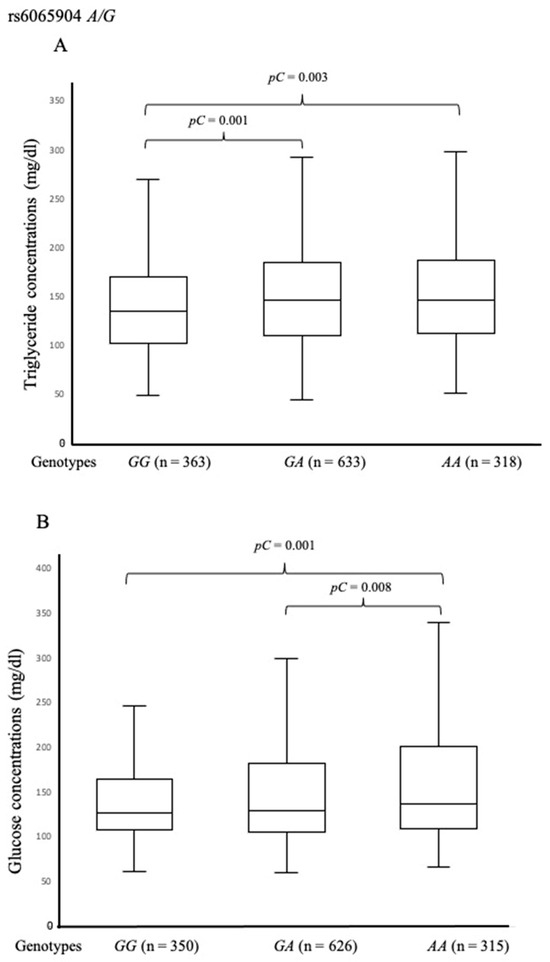
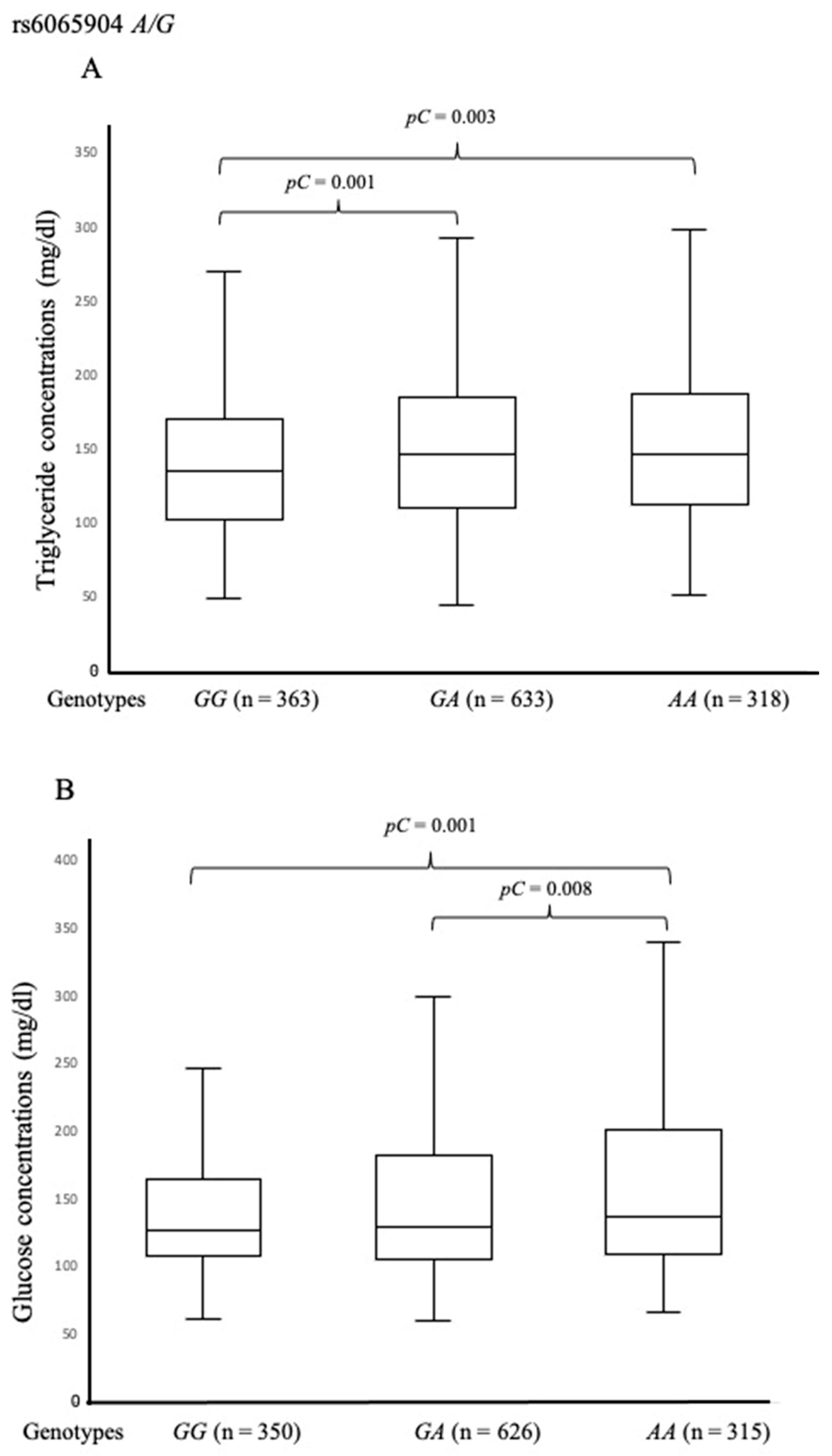
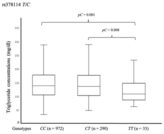
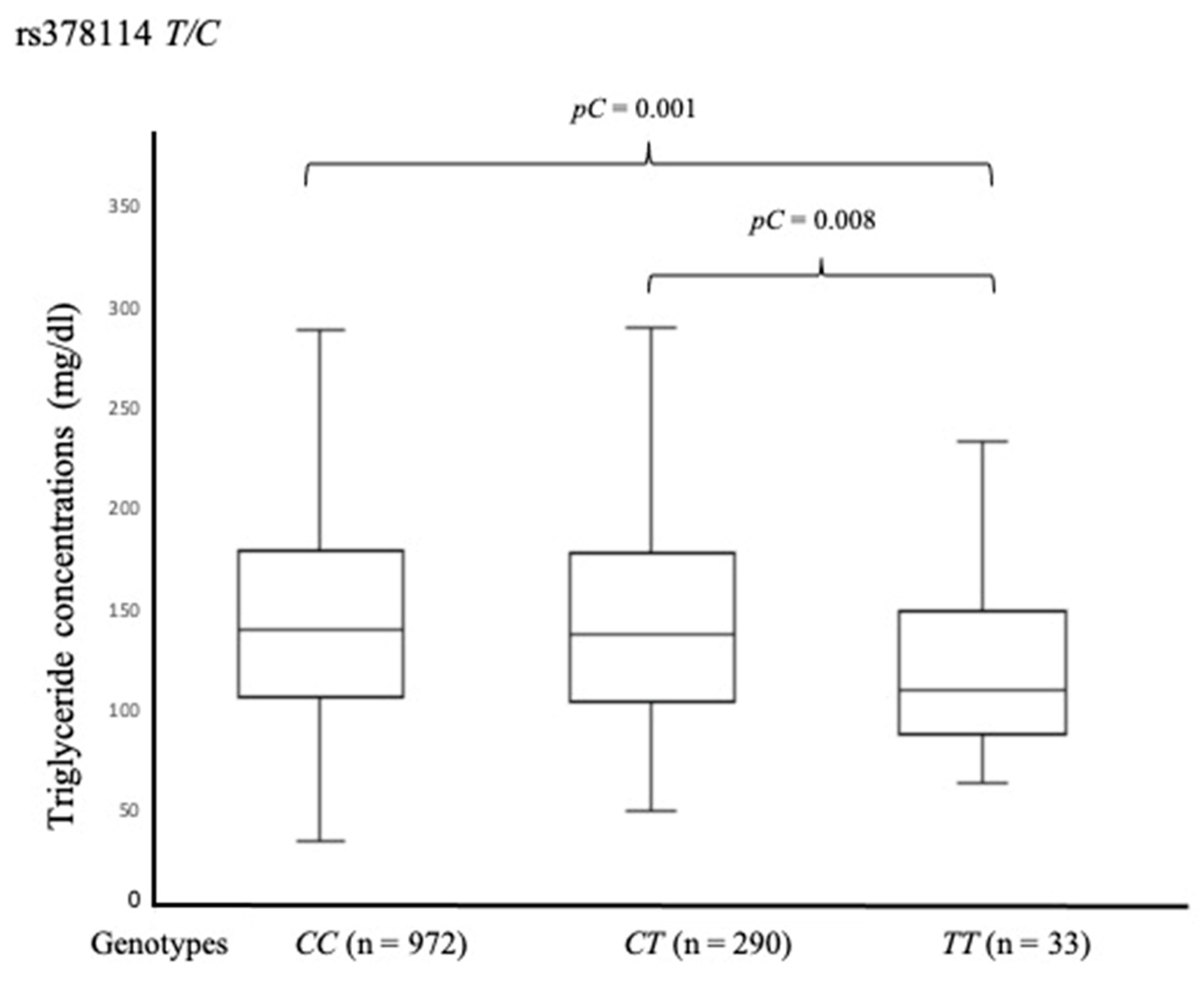
Genotypic stratification in ACS patients demonstrated significant associations between PLTP SNPs and metabolic parameters. Carriers of the rs6065904 GA and AA genotypes displayed significantly higher triglyceride concentrations than GG homozygotes [146 mg/dL (110–185), pC = 0.001 and 146 mg/dL (112–187), pC = 0.003 vs. 135 mg/dL (103–169.5), respectively] (Figure 1A). Furthermore, the AA genotype was associated with higher glucose levels compared to GA and GG genotypes [137 mg/dL (109–201.5) mg/dL vs. 130 mg/dL (105–183) mg/dL, pC = 0.008; and vs. 128 (108–165) mg/dL, pC = 0.001, respectively] (Figure 1B). In contrast, analysis of rs378114 T/C showed that TT homozygotes had significantly lower triglyceride levels [110 mg/dL (88–150)] compared to CT [138 mg/dL (104–179), pC = 0.008] and CC carriers [140 mg/dL (107–180), pC = 0.001] (Figure 2).
Figure 1.
Plasma triglyceride (A) and glucose (B) concentrations in ACS patients stratified by PLTP rs6065904 A/G genotypes. Data are presented as median [interquartile range]. Intergroup differences were assessed using the Kruskal–Wallis test, followed by post hoc Mann–Whitney U tests. p-values were corrected by Bonferroni test (pC).
Figure 2.
Plasma triglyceride concentrations in ACS patients stratified by PLTP rs378114 T/C genotypes. Data are presented as median [interquartile range]. Intergroup differences were assessed using the Kruskal–Wallis test, followed by post hoc Mann–Whitney U tests. p-values were corrected by Bonferroni test (pC).
3.4. Effect of PLTP Polymorphisms on eQTL Analysis
GTEx eQTL analysis confirmed that rs6065904 A/G and rs378114 T/C modulate PLTP mRNA expression. The rs6065904 AA genotype was associated with reduced PLTP mRNA expression in visceral adipose tissue, subcutaneous adipose tissue, and coronary artery tissue (p < 0.001) relative to the GG genotype (Supplementary Figure S1). Conversely, the rs378114 TT genotype was associated with increased PLTP mRNA expression across the same tissues (p < 0.001) relative to CC (Supplementary Figure S2).
4. Discussion
Plasma lipid alterations are central to the pathogenesis of cardiovascular diseases, including ACS. Over the past decade, studies investigating PLTP gene SNPs (rs6065904 A/G and rs378114 T/C) have yielded mixed findings regarding their impact on lipid profiles, cardiometabolic risk, and T2DM [9,10,11,25,26,27,28]. Our results demonstrate that the minor alleles (rs6065904 A and rs378114 T) are associated with an increased risk of ACS. Specifically, rs6065904 AA and GA genotypes correlated with higher plasma glucose and triglyceride levels, as well as reduced PLTP mRNA expression in adipose and coronary tissues, according to GTEx data [24]. Paradoxically, while the rs378114 TT genotype conferred an increased risk of ACS, TT homozygous patients exhibited lower triglyceride levels alongside higher tissue PLTP mRNA expression [24]. This suggests that the contribution of PLTP to ACS risk is not strictly mediated by overt dyslipidemia, as initially hypothesized in this study. Instead, PLTP participates in complex lipoprotein interactions, mediating the transfer of cholesterol and phospholipids from chylomicrons and VLDL to HDL, thereby altering particle size distribution and turnover [29,30,31]. Therefore, rather than simple contribution to plasma lipid levels, PLTP participates in the complex dynamic interactions between lipoproteins, which depends on the quantity and quality of donor and receptor particles [31,32]. To bridge the gap between the observed PLTP gene polymorphisms associations in this study and biological mechanisms, future functional studies are warranted. Given that PLTP plays a pivotal role in phospholipid transfer between lipoproteins [29,30,31], lipid delivery to cells may be altered and membrane lipid raft assembly impaired [33]. Then, macrophage and endothelial cell models could help to elucidate whether the rs6065904 and rs378114 variants alter membrane microdomain structure. Such structural changes could modify inflammation or nitric oxide synthetase signaling pathways [33], accelerating vascular inflammation in acute coronary events.
Moreover, some experimental evidence has demonstrated that A allele of the rs6065904 SNP is associated with low PLTP activity and low mRNA expression [11,26], findings that are in agreement with the GTEx data [24]. Beyond lipid transport, PLTP possesses anti-inflammatory properties through lipopolysaccharide (LPS) neutralization and maintains tissue vitamin E homeostasis [34,35]. Consequently, based on the results of this study and the broad ability of PLTP to interact with hydrophobic molecules, genetic variants of PLTP may serve as valuable markers of cardiovascular risk, independently of measurable lipid activity, which remains difficult to interpret. In this context, previous population studies report divergent outcomes. Jarvik et al. observed that the rs6065904 A and rs378114 T alleles were protective against carotid artery disease in Caucasian populations [25]. Vergeer et al. determined that the C allele of rs378114 and the A allele of rs6065904 were associated with low PLTP activity, and the combination of both polymorphisms determined liver mRNA expression, lower HDL size, and lower risk of cardiovascular disease in Caucasian populations [26]. Conversely, Zhao et al. reported the association of the rs6065904 A allele with increased CAD risk and elevated small HDL particles [9]. Genome-wide association and meta-analysis studies also link the rs6065904 and rs378114 PLTP SNPs with altered lipid profiles [12,13,14] and risk of cardiovascular diseases [10,27,28]. These discrepancies highlight the influence of ethnic variations in allele frequencies. In this context, the frequency of the A rs6065904 allele is substantially higher in Mexican Mestizos (44.7%) and Mexican-Americans in Los Angeles (50.0%) than in Caucasian (24.4%), Asian (33.7%), and African (21.2%) populations [36]. Conversely, the rs378114 T allele frequency is lower in Mexican Mestizos (11.7%) and Mexican-Americans (15.6%) compared to Caucasian (24.4%), Asian (38.3%), and African (42.9%) cohorts. Evaluating these SNPs in ethnically diverse populations is therefore necessary to establish their global role in atherogenesis and ACS [37] (Supplementary Table S1).
We acknowledge that our study has some limitations. First, samples from ACS patients and controls were independently collected in different periods that may introduce temporal bias; laboratory quality controls and DNA integrity analysis helped reduce this temporal factor, but it cannot be presumed that it was completely excluded. Second, the gender distribution was different between groups. However, male gender was considered in the statistical analyses as a covariable to statistically compensate this disparity. Also, PLTP polymorphisms associations with lipid levels have some limitations; lower levels of cholesterol in ACS patients than in controls clearly indicate an effect of the ACS-associated acute state and the use of statins as stated before. For this reason, the analysis of the PLTP gene polymorphisms on plasma lipid levels was performed only in ACS patients, assuming that the effect of statins and acute state is homogeneous in all the included individuals, but such an assumption cannot be verified. Finally, this study only showed a statistical relationship between genetic background and cardiovascular risk of developing ACS but did not establish the molecular mechanisms connecting PLTP variants to lipid and glucose shifts and ACS risk, which remain to be elucidated. Therefore, future experimental studies on mRNA expression such as luciferase assays, RT-qPCR (quantitative reverse transcription-polymerase chain reaction), or RNA sequencing (RNA-Seq) are required to ensure the validity and reliability of these polymorphisms in clinical practice.
5. Conclusions
Our findings showed that minor allele frequencies of the rs6065904 A/G and rs378114 T/C polymorphisms of the PLTP gene are associated with susceptibility to developing ACS and with triglyceride and glucose levels in the Mexican population. Further prospective studies across diverse ethnic groups are recommended to validate these variants as clinical biomarkers for cardiovascular risk.
Supplementary Materials
The following supporting information can be downloaded at: https://www.mdpi.com/article/10.3390/biomedicines14092025/s1, Supplementary Table S1. Allele frequencies of PLTP polymorphisms across reference populations. Supplementary Figure S1. According with the expression quantitative trait locus (eQTL) analysis of Genotype-Tissue Expression (GTEx) consortium data, the rs6065904 AA genotype leads to lower mRNA expression in the visceral adipose tissue (A), subcutaneous adipose tissue (B), and artery coronary (C). Supplementary Figure S2. According with the expression quantitative trait locus (eQTL) analysis of Genotype-Tissue Expression (GTEx), the rs378114 TT genotype showed a higher mRNA expression in the visceral adipose tissue (A), subcutaneous adipose tissue (B), and artery coronary (C).
Author Contributions
Conceptualization, G.V.-A., O.P.-M. and J.M.F.; Methodology, R.P.-S., J.C.D.-M. and M.A.-G.; Software, R.P.-S., M.A.-G. and T.J.-C.; Validation, G.E.; Formal analysis, R.P.-S., M.A.-G., G.E., T.J.-C. and J.M.F.; Investigation, G.V.-A., O.P.-M., G.E. and J.M.F.; Resources, J.C.D.-M. and M.A.-G.; Data curation, R.P.-S., J.C.D.-M. and M.A.-G.; Writing—original draft, G.V.-A., O.P.-M. and J.M.F.; Writing—review and editing, J.M.F.; Project administration, J.M.F.; Funding acquisition, J.M.F. All authors have read and agreed to the published version of the manuscript.
Funding
This study was funded by the Instituto Nacional de Cardiología Ignacio Chávez, Mexico City, México (Project-25-1519).
Institutional Review Board Statement
The study was conducted following the guidelines of the Declaration of Helsinki and approved by the Ethics and Research Committees of Instituto Nacional de Cardiología Ignacio Chávez (approval date: 10 April 2025, protocol number: 25-1519).
Informed Consent Statement
Informed consent for participation was obtained from all subjects involved in the study.
Data Availability Statement
The data presented in this study are available from the corresponding author upon request, due to privacy, legal, or ethical reasons.
Acknowledgments
The authors are grateful to Silvestre Ramirez Fuentes for his technical support, as well as to the CORE-Lab personnel of our institution. Open access funding for this article was supported by the Instituto Nacional de Cardiología Ignacio Chávez.
Conflicts of Interest
The authors declare no conflicts of interest.
References
- Sarma, V.K.; Henry, R.A.; Ahamed, H.; George, S.M. Clinical Utility of Immature Platelet Fraction (IPF) as a Biomarker in the Diagnosis of Acute Coronary Syndrome (ACS). Cureus 2025, 17, e81406. [Google Scholar] [CrossRef] [Scilit] [PubMed]
- Huang, J.X.F.; Yousaf, A.; Moon, J.; Ahmed, R.; Uppal, K.; Pemminati, S. Recent Advances in the Management of Dyslipidemia: A Systematic Review. Cureus 2025, 17, e81034. [Google Scholar] [CrossRef] [Scilit] [PubMed]
- Fularski, P.; Czarnik, W.; Dąbek, B.; Lisińska, W.; Radzioch, E.; Witkowska, A.; Młynarska, E.; Rysz, J.; Franczyk, B. Broader Perspective on Atherosclerosis-Selected Risk Factors, Biomarkers, and Therapeutic Approach. Int. J. Mol. Sci. 2024, 25, 5212. [Google Scholar] [CrossRef] [Scilit] [PubMed]
- Paththinige, C.S.; Sirisena, N.D.; Dissanayake, V. Genetic determinants of inherited susceptibility to hypercholesterolemia—A comprehensive literature review. Lipids Health Dis. 2017, 16, 103. [Google Scholar] [CrossRef] [Scilit] [PubMed]
- Kim, D.S.; Burt, A.A.; Ranchalis, J.E.; Vuletic, S.; Vaisar, T.; Li, W.F.; Rosenthal, E.A.; Dong, W.; Eintracht, J.F.; Motulsky, A.G.; et al. PLTP activity inversely correlates with CAAD: Effects of PON1 enzyme activity and genetic variants on PLTP activity. J. Lipid Res. 2015, 56, 1351–1362. [Google Scholar] [CrossRef] [Scilit] [PubMed]
- Jiang, X.C.; Yu, Y. The Role of Phospholipid Transfer Protein in the Development of Atherosclerosis. Curr. Atheroscler. Rep. 2021, 23, 9. [Google Scholar] [CrossRef] [Scilit] [PubMed]
- Márquez, A.B.; Nazir, S.; van der Vorst, E.P.C. High-Density Lipoprotein Modifications: A Pathological Consequence or Cause of Disease Progression? Biomedicines 2020, 8, 549. [Google Scholar] [CrossRef] [Scilit] [PubMed]
- Albers, J.J.; Vuletic, S.; Cheung, M.C. Role of plasma phospholipid transfer protein in lipid and lipoprotein metabolism. Biochim. Biophys. Acta 2012, 182, 345–357. [Google Scholar] [CrossRef] [Scilit] [PubMed]
- Zhao, Q.; Wang, J.; Miao, Z.; Zhang, N.R.; Hennessy, S.; Small, D.S.; Rader, D.J. A Mendelian randomization study of the role of lipoprotein subfractions in coronary artery disease. Elife 2021, 10, e58361. [Google Scholar] [CrossRef] [Scilit] [PubMed]
- Teslovich, T.M.; Musunuru, K.; Smith, A.V.; Edmondson, A.C.; Stylianou, I.M.; Koseki, M.; Pirruccello, J.P.; Ripatti, S.; Chasman, D.I.; Willer, C.J.; et al. Biological, clinical and population relevance of 95 loci for blood lipids. Nature 2010, 466, 707–713. [Google Scholar] [CrossRef] [Scilit] [PubMed]
- Dullaart, R.P.; Vergeer, M.; de Vries, R.; Kappelle, P.J.; Dallinga-Thie, G.M. Type 2 diabetes mellitus interacts with obesity and common variations in PLTP to affect plasma phospholipid transfer protein activity. J. Intern. Med. 2012, 271, 490–498. [Google Scholar] [CrossRef] [Scilit] [PubMed]
- Kettunen, J.; Tukiainen, T.; Sarin, A.P.; Ortega-Alonso, A.; Tikkanen, E.; Lyytikäinen, L.P.; Kangas, A.J.; Soininen, P.; Würtz, P.; Silander, K.; et al. Genome-wide association study identifies multiple loci influencing human serum metabolite levels. Nat. Genet. 2012, 44, 269–276. [Google Scholar] [CrossRef] [Scilit] [PubMed]
- Karjalainen, M.K.; Karthikeyan, S.; Oliver-Williams, C.; Sliz, E.; Allara, E.; Fung, W.T.; Surendran, P.; Zhang, W.; Jousilahti, P.; Kristiansson, K.; et al. Genome-wide characterization of circulating metabolic biomarkers. Nature 2024, 628, 130–138. [Google Scholar] [CrossRef] [Scilit] [PubMed]
- Sun, Y.; Xu, H.; Ye, K. GWAS and multi-omics integrative analysis reveal novel loci and their molecular mechanisms for circulating fatty acids. HGG Adv. 2025, 6, 100470. [Google Scholar] [CrossRef] [Scilit] [PubMed]
- Knuuti, J.; Wijns, W.; Saraste, A.; Capodanno, D.; Barbato, E.; Funck-Brentano, C.; Prescott, E.; Storey, R.F.; Deaton, C.; Cuisset, T.; et al. 2019 ESC guidelines for the diagnosis and management of chronic coronary syndromes. Eur. Heart J. 2020, 41, 407–477, Erratum in Eur. Heart J. 2020, 4, 4242. [Google Scholar] [CrossRef] [Scilit] [PubMed]
- Winther, S.; Schmidt, S.E.; Rasmussen, L.D.; Juárez Orozco, L.E.; Steffensen, F.H.; Bøtker, H.E.; Knuuti, J.; Bøttcher, M. Validation of the European Society of Cardiology pre-test probability model for obstructive coronary artery disease. Eur. Heart J. 2021, 42, 1401–1411. [Google Scholar] [CrossRef] [Scilit] [PubMed]
- Posadas-Sanchez, R.; Perez-Hernandez, N.; Angeles-Martinez, J.; Lopez-Bautista, F.; Villarreal-Molina, T.; Rodríguez-Perez, J.M.; Fragoso, J.M.; Posadas-Romero, C.; Vargas-Alarcón, G. Interleukin 35 polymorphisms are associated with decreased risk of premature coronary artery disease, metabolic parameters, and IL-35 Levels: The Genetics of Atherosclerotic Disease (GEA) Study. Mediat. Inflamm. 2017, 2017, 6012795. [Google Scholar] [CrossRef] [Scilit] [PubMed]
- DeLong, D.M.; DeLong, E.R.; Wood, P.D.; Lippel, K.; Rifkind, B.M. A comparison of methods for the estimation of plasma low- and very low-density lipoprotein cholesterol. The Lipid Research Clinics Prevalence Study. JAMA 1986, 256, 2372–2377. [Google Scholar] [CrossRef] [Scilit]
- Available online: https://www.nhlbi.nih.gov/resources/third-report-expert-panel-detection-evaluation-and-treatment-high-blood-cholesterol-0 (accessed on 5 December 2024).
- Buse, J.B.; Wexler, D.J.; Tsapas, A.; Rossing, P.; Mingrone, G.; Mathieu, C.; D’aLessio, D.A.; Davies, M.J. 2019 update to: Management of hyperglycaemia in type 2 diabetes, 2018. A consensus report by the American Diabetes Association (ADA) and the European Association for the Study of Diabetes (EASD). Diabetes Care 2020, 43, 487–493, Erratum in Diabetes Care 2020, 43, 1670. [Google Scholar] [CrossRef] [Scilit] [PubMed]
- Wang, B.X. Diagnosis and Management of Hypertensive Heart Disease: Incorporating 2023 European Society of Hypertension and 2024 European Society of Cardiology Guideline Updates. J. Cardiovasc. Dev. Dis. 2025, 12, 46. [Google Scholar] [CrossRef] [Scilit] [PubMed]
- Schaid, D.J. Disease-Marker Association. Biostatistical Genetics and Genetic Epidemiology; Elston, R.C., Olson, J.M., Palmer, L., Eds.; Wiley: Chichester, UK, 2002; pp. 216–217. [Google Scholar]
- Clayton, D. Population Association. Handbook of Statistical Genetics; Balding, D.J., Bishop, M., Cannings, C., Eds.; Wiley: Chichester, UK, 2001; pp. 519–540. [Google Scholar]
- GTEx Consortium. Human genomics. The Genotype-Tissue Expression (GTEx) pilot analysis: Multitissue gene regulation in humans. Science 2015, 348, 648–660. [Google Scholar] [CrossRef] [Scilit] [PubMed]
- Jarvik, G.P.; Rajagopalan, R.; Rosenthal, E.A.; Wolfbauer, G.; McKinstry, L.; Vaze, A.; Brunzell, J.; Motulsky, A.G.; Nickerson, D.A.; Heagerty, P.J.; et al. Genetic and nongenetic sources of variation in phospholipid transfer protein activity. J. Lipid Res. 2010, 51, 983–990. [Google Scholar] [CrossRef] [Scilit] [PubMed]
- Vergeer, M.; Boekholdt, S.M.; Sandhu, M.S.; Ricketts, S.L.; Wareham, N.J.; Brown, M.J.; de Faire, U.; Leander, K.; Gigante, B.; Kavousi, M.; et al. Genetic variation at the phospholipid transfer protein locus affects its activity and high-density lipoprotein size and is a novel marker of cardiovascular disease susceptibility. Circulation 2010, 122, 470–477. [Google Scholar] [CrossRef] [Scilit] [PubMed]
- Edmondson, A.C.; Braund, P.S.; Stylianou, I.M.; Khera, A.V.; Nelson, C.P.; Wolfe, M.L.; DerOhannessian, S.L.; Keating, B.J.; Qu, L.; He, J.; et al. Dense genotyping of candidate gene loci identifies variants associated with high-density lipoprotein cholesterol. Circ. Cardiovasc. Genet. 2011, 4, 145–155. [Google Scholar] [CrossRef] [Scilit] [PubMed]
- Lettre, G.; Palmer, C.D.; Young, T.; Ejebe, K.G.; Allayee, H.; Benjamin, E.J.; Bennett, F.; Bowden, D.W.; Chakravarti, A.; Dreisbach, A.; et al. Genome-wide association study of coronary heart disease and its risk factors in 8,090 African Americans: The NHLBI CARe Project. PLoS Genet. 2011, 7, e1001300. [Google Scholar] [CrossRef] [Scilit] [PubMed]
- Qin, S.; Kawano, K.; Bruce, C.; Lin, M.; Bisgaier, C.; Tall, A.R.; Jiang, X. Phospholipid transfer protein gene knock-out mice have low high density lipoprotein levels, due to hypercatabolism, and accumulate apoA-IV-rich lamellar lipoproteins. J. Lipid Res. 2000, 41, 269–276. [Google Scholar] [CrossRef] [Scilit]
- van Haperen, R.; van Tol, A.; Vermeulen, P.; Jauhiainen, M.; van Gent, T.; van den Berg, P.; Ehnholm, S.; Grosveld, F.; van der Kamp, A.; de Crom, R. Human plasma phospholipid transfer protein increases the antiatherogenic potential of high density lipoproteins in transgenic mice. Arterioscler. Thromb. Vasc. Biol. 2000, 20, 1082–1088. [Google Scholar] [CrossRef] [Scilit] [PubMed]
- Setälä, N.L.; Holopainen, J.M.; Metso, J.; Wiedmer, S.K.; Yohannes, G.; Kinnunen, P.K.; Ehnholm, C.; Jauhiainen, M. Interfacial and lipid transfer properties of human phospholipid transfer protein: Implications for the transfer mechanism of phospholipids. Biochemistry 2007, 46, 1312–1319. [Google Scholar] [CrossRef] [Scilit] [PubMed]
- Soares, A.A.S.; Tavoni, T.M.; de Faria, E.C.; Remalay, A.T.; Maranhão, R.C.; Sposito, A.C.; Brasilia Heart Study Group. HDL acceptor capacities for cholesterol efflux from macrophages and lipid transfer are both acutely reduced after myocardial infarction. Clin. Chim. Acta 2018, 478, 51–56. [Google Scholar] [PubMed]
- Muñoz-Vega, M.; Massó, F.; Páez, A.; Vargas-Alarcón, G.; Coral-Vázquez, R.; Mas-Oliva, J.; Carreón-Torres, E.; Pérez-Méndez, Ó. HDL-Mediated Lipid Influx to Endothelial Cells Contributes to Regulating Intercellular Adhesion Molecule (ICAM)-1 Expression and eNOS Phosphorylation. Int. J. Mol. Sci. 2018, 19, 3394. [Google Scholar] [CrossRef] [Scilit] [PubMed]
- Nguyen, M.; Pallot, G.; Jalil, A.; Tavernier, A.; Dusuel, A.; Le Guern, N.; Lagrost, L.; de Barros, J.-P.P.; Choubley, H.; Bergas, V.; et al. Intra-Abdominal Lipopolysaccharide Clearance and Inactivation in Peritonitis: Key Roles for Lipoproteins and the Phospholipid Transfer Protein. Front. Immunol. 2021, 12, 622935. [Google Scholar] [CrossRef] [Scilit] [PubMed]
- Zhou, T.; He, Q.; Tong, Y.; Zhan, R.; Xu, F.; Fan, D.; Guo, X.; Han, H.; Qin, S.; Chui, D. Phospholipid transfer protein (PLTP) deficiency impaired blood-brain barrier integrity by increasing cerebrovascular oxidative stress. Biochem. Biophys. Res. Commun. 2014, 445, 352–356. [Google Scholar] [CrossRef] [Scilit] [PubMed]
- Available online: https://www.ensembl.org/Homo_sapiens/Variation/Population?db=core;r=20:45905512-45906512;v=rs6065904;vdb=variation;vf=1054672306 (accessed on 20 January 2026).
- Available online: https://www.ensembl.org/Homo_sapiens/Variation/Population?db=core;r=20:45909288-45910288;v=rs378114;vdb=variation;vf=1054554809 (accessed on 20 January 2026).

Disclaimer/Publisher’s Note: The statements, opinions and data contained in all publications are solely those of the individual author(s) and contributor(s) and not of MDPI and/or the editor(s). MDPI and/or the editor(s) disclaim responsibility for any injury to people or property resulting from any ideas, methods, instructions or products referred to in the content. |
© 2026 by the authors. Licensee MDPI, Basel, Switzerland. This article is an open access article distributed under the terms and conditions of the Creative Commons Attribution (CC BY) license.