


Abstract
Background/Objectives: Cardiac allograft fibrosis is an important limiting factor for long-term graft survival. However, the fibrotic process operating in patients with acute cellular rejection (ACR) remains unclear. We aimed to identify altered serum mRNAs related to cardiac fibrosis in patients with ACR and to evaluate their diagnostic accuracy in detecting rejection episodes. Methods: We included 40 serum samples from recipients of transplants undergoing routine endomyocardial biopsies. Results: Several altered mRNAs associated with fibrosis were detected in patients with ACR. Specifically, the activators of fibroblasts and myofibroblasts (TNS1, FAP and ACTA2), TGF-β signaling (TGFBR1 and JAK1) and WNT signaling (WNT7A and WLS) pathways were significantly different when we compared grade ≥ 2R ACR and/or grade 1R ACR groups with the nonrejection group. Furthermore, TNS1 and WLS presented an area under the curve value > 0.90 for identifying patients with moderate and severe grades of cardiac rejection. Conclusions: In conclusion, we found alterations in the relative abundance of circulating activators of fibroblasts and myofibroblasts, such as FAP or ACTA2, as well as in major profibrotic pathways, including TGF-β and WNT signaling, especially in clinically relevant cardiac rejection. These findings may contribute to improving the surveillance of patients with cardiac transplant and provide new therapeutic strategies for targeting fibrosis process activation.
1. Introduction
Heart transplantation remains the most effective therapeutic option for patients with end-stage heart failure (HF) [1]. Despite major advances in immunosuppression, allograft rejection remains a significant problem in the first year following heart transplantation, leading to adverse short- and long-term outcomes [2]. The gold standard method for monitoring and diagnosing cardiac allograft rejection continues to be the evaluation of pathologic cardiac tissue obtained through an endomyocardial biopsy (EMB) [1], although this procedure has potential negative consequences for patients [3] and is subject to sampling error and interobserver variability [4]. Therefore, noninvasive methods and reliable biomarkers are being actively sought to screen for heart transplant rejection and to better understand the pathophysiology of this post-transplant complication [5].
The pathophysiological process involved in cardiac allograft rejection is complex and is characterized by severe cardiac inflammation, cardiomyocyte damage, and progressive ventricular dysfunction in the transplanted heart [6]. In addition to inflammation, cardiac allograft fibrosis has been identified as an important limiting factor for long-term graft survival [6,7]. Fibrosis is one of the major causes of cardiac allograft malfunction and is mainly driven by fibroblasts [8]. Myocardial fibrosis is a described histopathological feature in recipients of heart transplants, and several studies have demonstrated the appearance of this condition during time course analyses in different populations [9,10,11,12]. Whether acute cellular rejection (ACR) in recipients of heart transplants is associated with myocardial fibrosis remains unclear.
Organ fibrosis is characterized by the excessive deposition of extracellular matrix (ECM) proteins within the tissue and around blood vessels [13]. Several effectors and molecular mechanisms are involved in fibrosis. Among them, activated fibroblasts are central cellular effectors that serve as the main ECM-producing cells [14]. Activated fibroblasts and myofibroblasts are well recognized as significant contributors to graft fibrosis [15]. In addition, transforming growth factor beta (TGF-β) is the best characterized fibrogenic growth factor, and its activation has been consistently demonstrated in different models of cardiac fibrosis and in human hearts with fibrotic cardiomyopathic changes [14]. Another pathway involved in fibrosis is WNT signaling, which is activated in many cell types and contributes to reparative, regenerative, and fibrotic responses [14]. Interestingly, it has been proposed that the crosstalk between TGF-β and Wnt/β-catenin pathways regulates cardiac fibrosis [16].
However, the relationship between this process and cardiac allograft rejection remains unknown. For this reason, we used serum samples of patients diagnosed with ACR to analyze mRNA relative abundance of the genes implicated in the fibrotic process, including ECM components, such as collagens, and activators of fibroblasts and myofibroblasts. Furthermore, we examined the relative abundance of genes encoding for proteins involved in the major pathways of the fibrotic process, such as TGF-β and WNT signaling. Finally, we assessed the diagnostic capability of those altered molecules related to the fibrosis process in patients with ACR.
2. Materials and Methods
2.1. Sample Collection
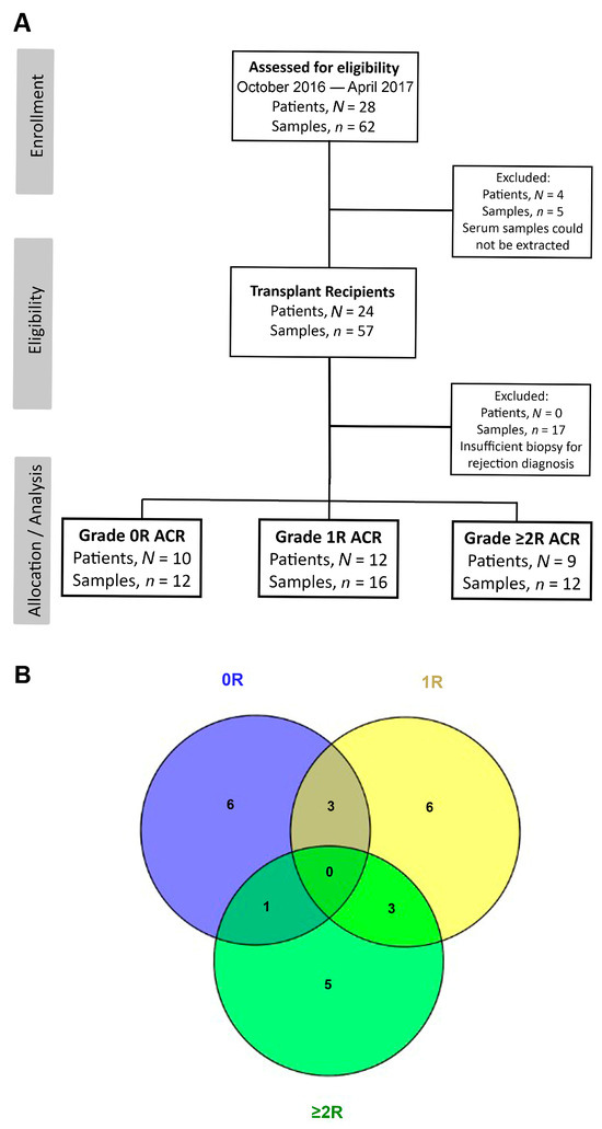
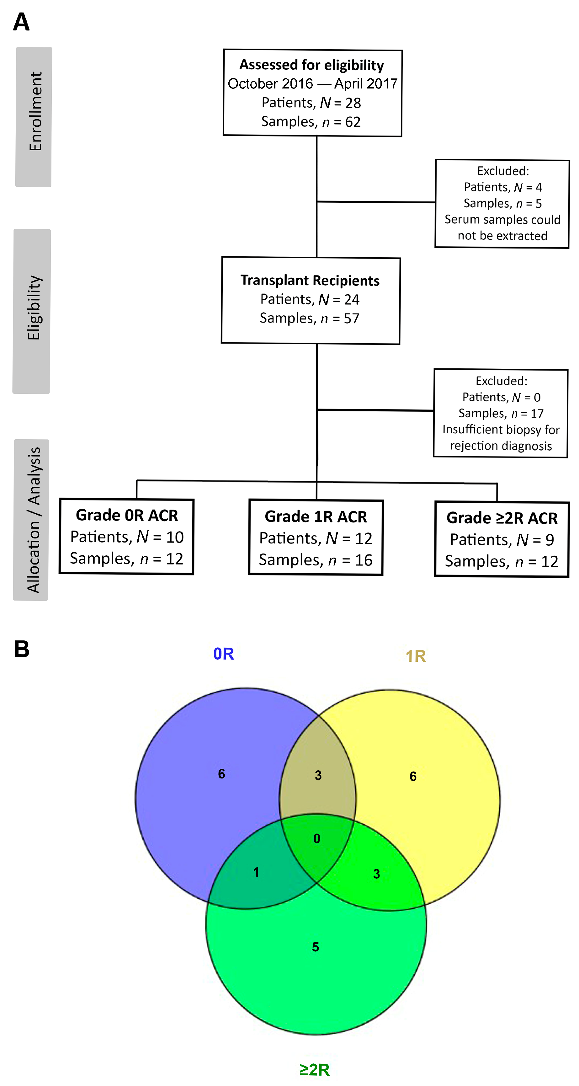
This study included serum samples collected from patients (>18 years old) who received a heart transplant and were referred for EMB as a scheduled routine screening at a single center. All consecutive patients who underwent primary heart transplantation at La Fe University and Polytechnic Hospital between October 2016 and April 2017 were considered eligible for inclusion. No additional clinical selection criteria were applied for study participation beyond those required for heart transplantation according to the institutional transplant program and current international guidelines. Exclusion criteria were death during the initial postoperative period and retransplantation. Samples were collected during follow-up visits of the recipients. The hospital follows an established rejection-monitoring protocol in which the first biopsy is performed 10–12 days after the transplant, the next one 10 days after the first biopsy, then every 6 weeks until the sixth month, and finally every 2 months until 1-year post-transplantation. In this period, 62 consecutive samples from 28 patients were included. Serum samples that could not be extracted from four patients (n = 5) were excluded, as well as samples from 12 patients lacking a histopathological diagnosis (n = 17) due to insufficient or poor-quality tissue samples for analysis. Conclusively, 40 consecutive samples with different rejection grades belonging to 24 patients were included in this study (Figure 1A), so, randomly, there are patients with more than one sample (Figure 1B). Blood samples were collected immediately before EMB for laboratory analysis. Once the samples were collected, they were processed, and the serum was separated through centrifugation (Eppendorf Model 5415R Centrifuge, Eppendorf Ibérica S.L.U., Madrid, Spain) at 1500× g for 15 min at 4 °C, aliquoted, and immediately stored at −80 °C until analysis.
Figure 1.
Samples and patients included in this study. CONSORT flow diagram showing patient and sample enrollment, eligibility and exclusions, as well as the allocation depending on the degree of cardiac rejection for the final analysis (A). Venn diagram showing patients with analyzed samples belonging to more than one study group (B). ACR, acute cellular rejection.
Patients were maintained on a standard immunosuppressive regimen, and rejection episodes were assessed according to the International Society for Heart and Lung Transplantation consensus report [17]. The associated clinical data were collected at the time of each biopsy. For each sample, we recorded the age, sex, body mass, primary heart disease that was the cause of transplantation, time interval between transplantation and inclusion in the study (i.e., obtaining the blood sample), biochemical markers, echocardiographic parameters, and other relevant clinical characteristics at the time of each biopsy (Table 1). The researchers were blind to the group assignments and outcome assessments for all experiments.
Table 1.
Patient characteristics at the time of blood sample extraction.
2.2. RNA Sequencing Analysis
An RNA sequencing analysis was performed on 40 serum samples from patients diagnosed with biopsy-proven allograft rejection (ACR grade 1R, n = 16 and ACR grade ≥ 2R, n = 12) and from patients who did not experience allograft rejection (nonrejection, n = 12). This methodology has been described in extensive detail by Tarazon et al. [18]. Briefly, RNA was extracted using NucleoSpin miRNA Plasma from Macherey-Nagel (Düren, Germany) following the protocol and instructions provided by the manufacturer. cDNA libraries were then pooled and sequenced through two lanes of 100 bp paired-end sequencing using an Illumina HiSeq 2500 sequencer (Illumina, San Diego, CA, USA).
2.3. Statistical Analysis
For continuous variables, data were expressed as median and interquartile range, and as a percentage for discrete variables. Continuous clinical variables which had a non-normal distribution were analyzed using a linear mixed-effects model, while a generalized linear mixed-effects model was used for discrete clinical variables. The DESeq2 method (version 3.4) was used to perform an internal normalization of data [19]. The false discovery rate (FDR) method was used to adjust the original p value using the number of tests to prevent the identification of false positives in the differential expression data. An interpretation of this method was implemented in DESeq2, in which genes were ranked based on the p-value and multiplied by a correction rank value, similar to the original FDR method. In this exploratory study, the relative abundance of the mRNAs in the study groups was compared using a linear mixed-effects model. The study was primarily designed to collect independent observations. The additional observations were not systematically planned. The outcome variable, each of the fibrosis-related molecules examined in this study, was analyzed in its original form without transformation. The model included rejection grade as a fixed effect and patients as a random effect. To account for within-subject variability, a “no structure” covariance option was employed. This flexible strategy allows each variance and covariance between repeated measurements to be freely estimated, which was considered appropriate given that only a small number of cases involved repeated measurements within the same patient. The model employed the identity link function, which is the default for continuous outcomes in linear mixed models. The diagnostic capacity of fibrosis-related serum mRNA molecule-coding proteins for the presence of transplant rejection was evaluated. For this, receiver operating characteristic (ROC) curves were constructed to determine the sensitivity and specificity of the selected mRNA molecules. To calculate specificity and sensitivity, we took as a selection criteria an often-used cut-off point of FC ≥ 1.5. Molecules with significant discrimination capability (area under the curve [AUC] > 0.8) for detecting heart transplant rejection were selected. A p < 0.05 was considered to define a statistically significant difference. All statistical analyses were performed using the SPSS software version 20 for Windows (version 20.0; IBM SPSS Inc., Armonk, NY, USA), R (version R-4.3.1), and GraphPad Prism 8 (Dotmatics, Woburn, MA, USA).
3. Results
3.1. Clinical Characteristics of Patients
The study populations included in this transcriptomics study are presented in Table 1. All groups showed similar clinical characteristics with regard to variables such as age, sex, body mass, and comorbidities such as diabetes mellitus and immunosuppressive therapy. Only patients with ACR grade ≥ 2R displayed significant differences between specific clinical characteristics when we compared them with the nonrejection group. Patients with ACR grade ≥ 2R had the shortest time from the time of transplant until their incorporation into the study when compared with nonrejection patients (2.3 (0.4–6.8) vs. 9.2 (5.8–12) months, respectively; p < 0.01). In relation to the echo–Doppler study, we found, in patients with ACR grade ≥ 2R, higher values of the left ventricular end systolic diameter compared with the nonrejection group (32 (30–35) vs. 25 (23–29) mm, respectively; p < 0.05). Furthermore, these patients showed worse hemodynamic function compared with nonrejection patients since they had a higher right atrial pressure (7.5 (6.3–8.8) vs. 3.0 (2.0–5.5) mm Hg, respectively; p < 0.05), a higher systolic right ventricular pressure (44 (39–48) vs. 33 (29–38) mm Hg, respectively; p < 0.05) and a higher diastolic right ventricular pressure (7.0 (6.3–12) vs. 5.0 (2.5–5.5) mm Hg, respectively; p < 0.05). We also found, in patients with ACR grade ≥ 2R, higher values of neutrophils compared with the nonrejection group (6.3 (2.1–12) vs. 3.7 (2.5–4.4) thousands/mm3, respectively; p < 0.01) and higher values of leukocytes (11 (5.2–15) vs. 6.2 (5.3–6.7) thousands/mm3, respectively; p < 0.01). Moreover, we found an increase in N-terminal pro-B-type natriuretic peptide levels in patients with ACR grade ≥ 2R compared with those in the nonrejection group (1209 (736–2382) vs. 152 (113–467) pg/mL, respectively; p < 0.01).
3.2. Serum Relative Abundance of Genes Related to Fibrotic Process
Transcriptomics differences between the nonrejection and rejection groups were examined to determine whether cardiac allograft rejection was related to fibrosis. We identified alterations in the mRNAs of ECM and in those involved in the activation of fibroblast and myofibroblast, as well as in the TGF-β and WNT signaling pathways (Table 2).
Table 2.
Alterations in the relative abundance of mRNAs encoding proteins related to fibrosis process in nonrejection, grade 1R and grade 2R groups.
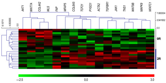
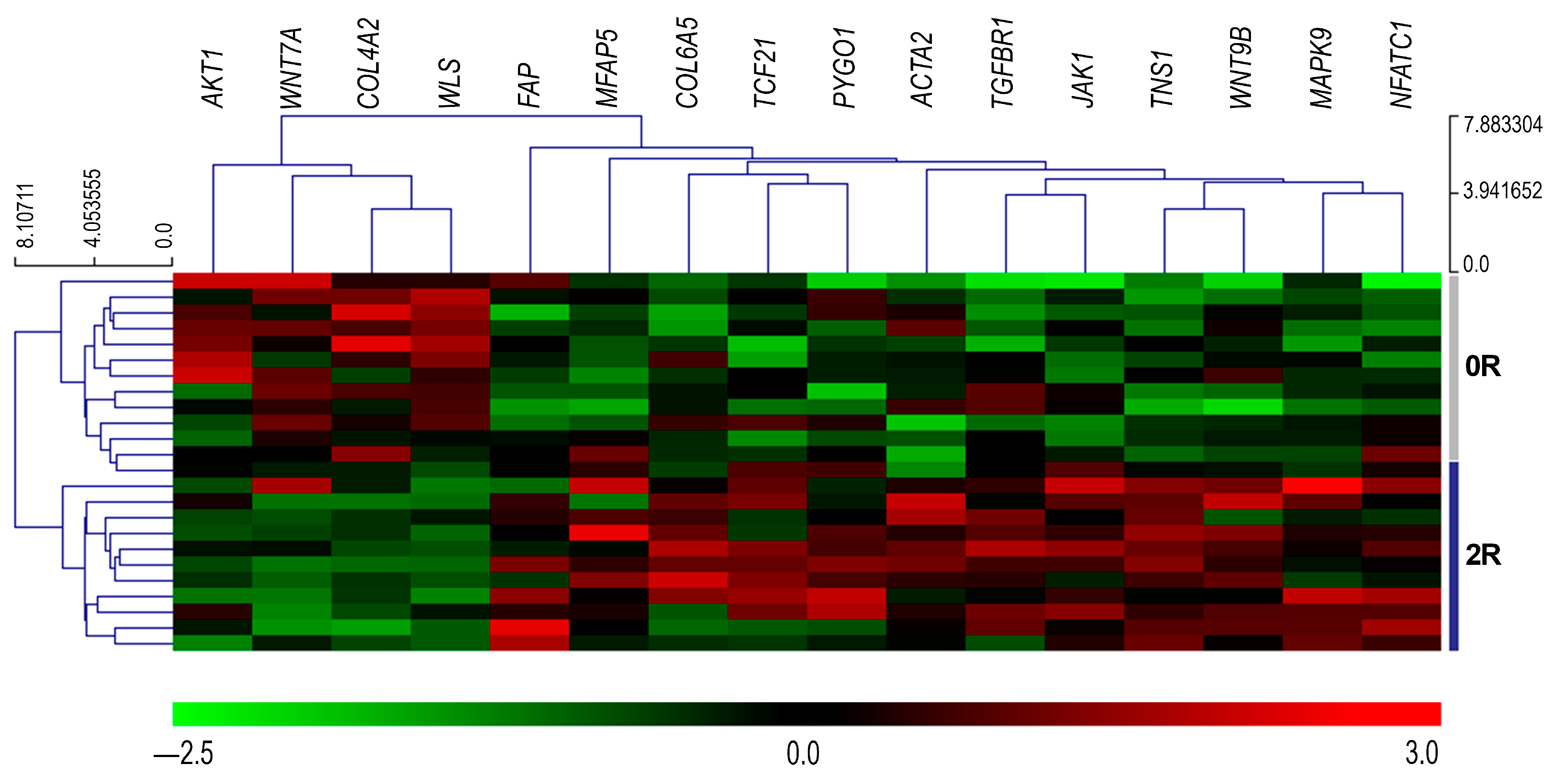
In addition, altered genes were represented in a heat map and hierarchical clustering based on the fold change values using MeV software (version 4.9) [20]. Here, we observed two divergent mRNA profiles, indicating a clear distinction between nonrejection and ACR grade ≥ 2R groups (Figure 2). We have also observed that samples from the same patient are not closely linked in the dendrogram; hence, there is no evidence of dependence on the repeated measurements in this representation.
Figure 2.
Serum relative abundance of genes related to the fibrotic process. Heat map summarizing mRNA relative abundance of genes related to the fibrotic process from the nonrejection (0R) and acute cellular rejection grade ≥ 2R groups. Each row represents a sample, and each column represents an altered mRNA. Colors depict the relative abundance of each molecule, with green being the lowest and red the highest.
3.3. Serum mRNAs Encoding Components of the ECM
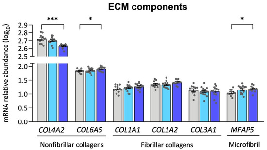
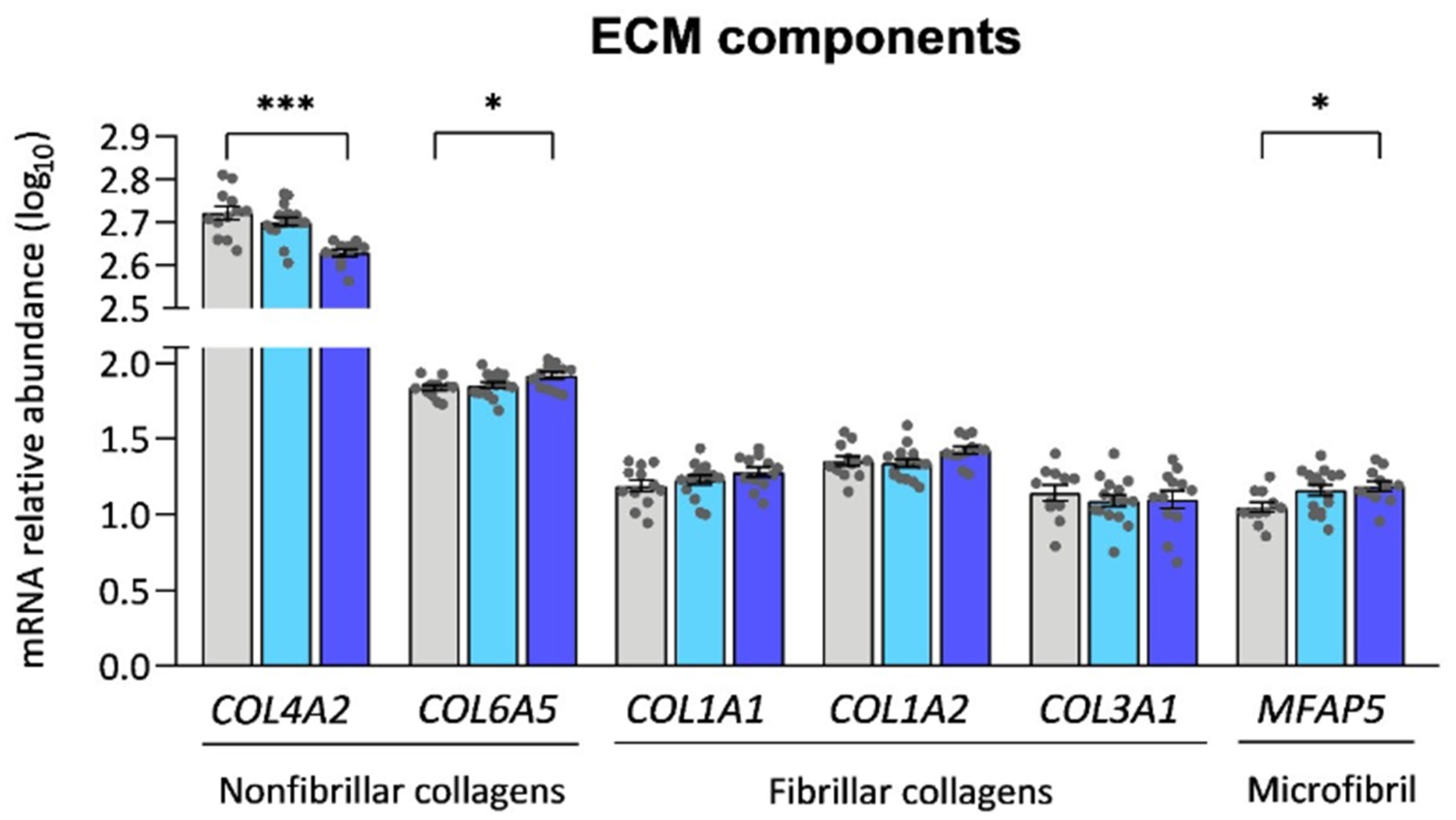
We identified 44 collagen genes (Table S1); however, only two of them (4.5%), compared to the nonrejection group, were differentially expressed in patients with ACR grade ≥ 2R, namely COL4A2 and COL6A5 (Figure 3). However, we did not find significant differences in the serum mRNA levels of fibrillar collagens I and III, which are the main markers of fibrosis (Figure 3). In addition, when we analyzed the relative abundance of different ECM components, we observed in patients with ACR grade ≥ 2R an increase in MFAP5 mRNA relative abundance (Figure 3).
Figure 3.
Circulating relative abundance of mRNAs encoding extracellular matrix proteins. Comparison of the serum mRNA relative abundance of the nonfibrillar collagens, the fibrillar collagens and extracellular matrix components between the nonrejection group and the groups representing various grades of acute rejection of heart allografts. Bars represent the mean values of mRNA relative abundance ± standard error of the mean value in log10 scale. The assigned colors denote the nonrejection group (gray), acute cellular rejection grade 1R group (light blue) and acute cellular rejection grade ≥ 2R group (dark blue). * p < 0.05; *** p < 0.001. COL4A2, collagen type IV alpha 2 chain; COL6A5, collagen type VI alpha 5 chain; COL1A1, collagen type I alpha 1 chain; COL1A2, collagen type I alpha 2 chain; COL3A1, collagen type III alpha 1 chain; MFAP5, microfibril-associated protein 5.
3.4. Serum mRNA Activators of Fibroblasts and Myofibroblasts
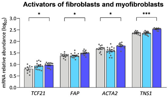
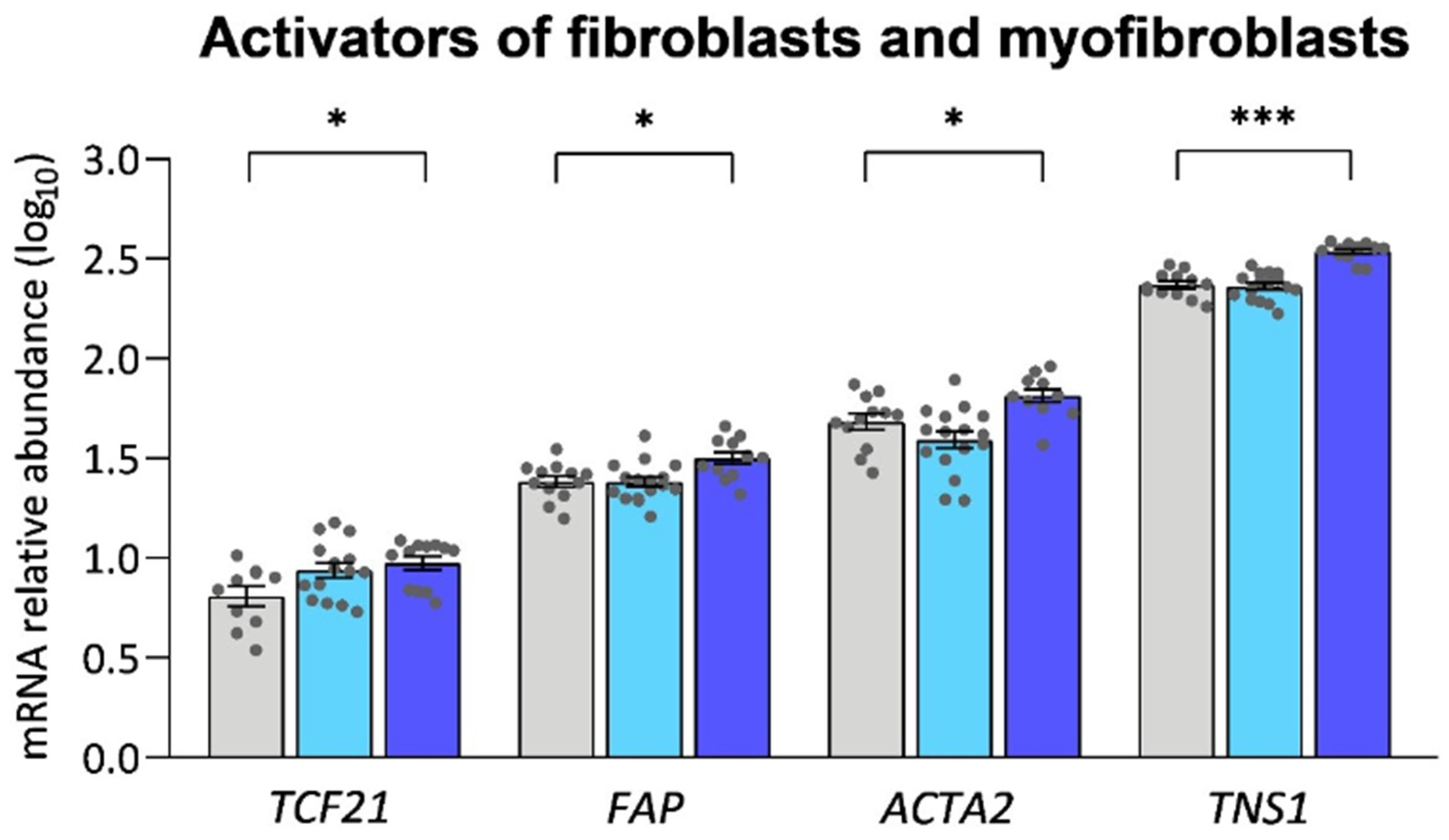
Since myocardial fibrosis is driven by the activation of fibroblasts, we wanted to evaluate whether serum mRNA abundances of the main markers of fibroblasts and myofibroblasts were altered. We observed significant differences in the relative abundance of several activators of fibroblasts and myofibroblasts when we compared the nonrejection group with patients with ACR grade ≥ 2R, such as TCF21, FAP, ACTA2 and TNS1 (Figure 4).
Figure 4.
Circulating relative abundance of mRNAs encoding relevant activators of fibroblasts and myofibroblasts. Comparison of the serum mRNA relative abundance between the nonrejection group and the groups representing various grades of acute rejection of heart allografts. The assigned colors denote the nonrejection group (gray), acute cellular rejection grade 1R group (light blue) and acute cellular rejection grade ≥ 2R group (dark blue). Bars represent the mean values of mRNA relative abundance ± standard error of the mean value in log10 scale. * p < 0.05; *** p < 0.001. TCF21, transcription factor 21; FAP, fibroblast activation protein; ACTA2, alpha-smooth muscle actin; TNS1, tensin-1.
3.5. Serum mRNAs Encoding Proteins Involved in the TGF-β and WNT Signaling Pathways
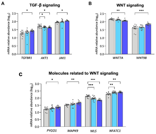
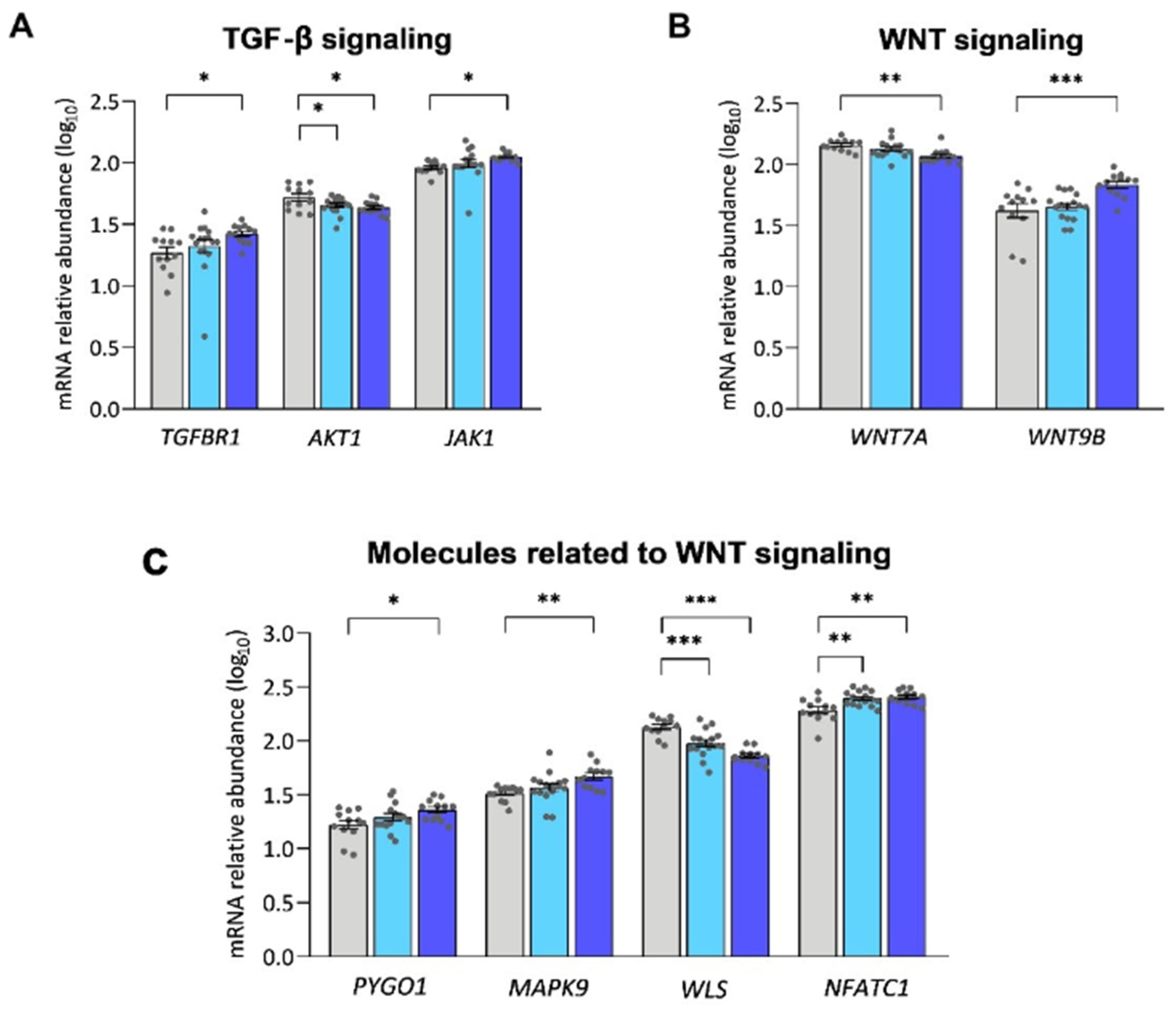
In light of our observation that several mRNAs related to the activation of fibrosis showed altered relative abundance in the serum of patients with ACR grade ≥ 2R, we probed whether the relative abundance of serum mRNAs of proteins involved in the major signaling pathways of the fibrotic process were also altered in these patients. First, we analyzed the TGF-β signaling pathway, which has a key role in the development of fibrosis. We found that, in the comparison between nonrejection and ACR grade ≥ 2R groups, there was an increase in the abundance of the TGF-β type 1 receptor (TGFBR1), which has a crucial role in the activation of canonical and noncanonical pathways of TGF-β signaling. In addition, we observed significant differences in the relative abundance of molecules that participate in noncanonical pathways such as AKT1 and JAK1. AKT1 relative abundance was also altered in patients with ACR grade 1R (Figure 5A).
Figure 5.
Circulating relative abundance of mRNAs encoding proteins involved in the TGF-β and WNT signaling pathways. Comparison of the serum mRNA relative abundance between the nonrejection group and the groups representing various grades of acute rejection of heart allografts of TGF-β signaling (A), WNT signaling (B) and proteins related to WNT signaling (C). Bars represent the mean values of mRNA relative abundance ± standard error of the mean value in log10 scale. The assigned colors denote the nonrejection group (gray), acute cellular rejection grade 1R group (light blue) and acute cellular rejection grade ≥ 2R group (dark blue). * p < 0.05; ** p < 0.01; *** p < 0.001. TGFBR1, transforming growth factor beta receptor 1; AKT1, RAC-alpha serine/threonine protein kinase 1; JAK1, Janus-Kinase 1; WNT7A, Wnt family member 7A; WNT9B, Wnt family member 9B; PYGO1, Pygopus family PHD finger 1; MAPK9, mitogen-activated protein kinase 9; WLS, Wntless; NFATC1, nuclear factor of activated T cells 1.
We also analyzed the WNT signaling pathway because it is known to intervene in the pathological development of fibrosis through both canonical and noncanonical pathways. Therefore, we examined the relative abundance of circulating mRNAs encoding WNT proteins in patients with ACR grade ≥ 2R. We found that, in these patients, the abundance of WNT, which shows an affinity for both pathways, was altered. Specifically, WNT7A abundance was significantly lower in contrast to WNT9B, whose abundance increased in ACR grade ≥ 2R patients (Figure 5B).
Furthermore, we analyzed the relative abundance of serum mRNAs encoding proteins that actively participate in WNT signaling (Figure 5C). Compared with the nonrejection group, we found that patients with ACR grade ≥ 2R showed altered relative abundance in several molecules participating in the WNT pathway. We observed differences in the relative abundance of PYGO1, a mediator of WNT signaling, and MAPK9, which participates in the noncanonical pathway. Further, we observed differences in the relative abundance of WLS, a regulator of WNT protein secretion, and NFATC1, which is related to the noncanonical pathway. Moreover, WLS and NFATC1 relative abundances were altered in patients with ACR grade 1R (Figure 5C).
3.6. Circulating Fibrosis-Related mRNAs Detect Acute Cellular Rejection
Considering our findings, we wanted to investigate the diagnostic capacity of ECM-related mRNAs and those of fibroblast and myofibroblast activators and TGF-β and WNT signaling pathways to detect cardiac allograft rejection. Therefore, ROC curves were obtained, and these data are summarized in Table 3, Figures S1 and S2, and the Supplementary Materials. Compared with the nonrejection group, we observed several molecules that presented an AUC > 0.80 to detect ACR grade ≥ 2R. Here, we also identified molecules that produced an AUC > 0.90 to differentiate patients with ACR grade ≥ 2R from those without rejection, specifically TNS1 (AUC = 0.97, p < 0.001) and WLS (AUC = 0.99, p < 0.001) (Figure S1). Additionally, some molecules presented significant differences in AUC scores when we compared the group of patients without rejection to the group of patients with mild rejection (grade 1R), maintaining a remarkable and significant detection power. The molecules with a good capability to detect ACR grade 1R were WLS (AUC = 0.84, p < 0.01) and NFATC1 (AUC = 0.83, p < 0.01) (Figure S2).
Table 3.
Receiver operating characteristic (ROC) curve of circulating altered genes related to fibrosis process for detecting heart transplant rejection (grade ≥ 2R). Sensitivities and specificities for the diagnosis of cardiac rejection (cut-off point FC ≥ 1.5).
4. Discussion
Cardiac allograft rejection continues to represent a challenge in the heart transplantation field because immune injury contributes to graft failure [21] and cardiac graft vasculopathy [22]. Although the incidence of treated rejection and mortality after transplantation has continued to decrease due to maintenance immunosuppression, 12.6% of patients were treated for rejection between discharge and 1 year after transplantation [23], and an increased incidence was detected in the latest published report [24]. Given the invasive nature and technical limitations of EMBs, many recent studies have focused on the search for circulating biomarkers as a noninvasive alternative [25]. Myocardial fibrosis has been reported in previous studies using serial EMB of cardiac allografts. Gramley et al. observed extensive and progressive tissue remodeling in cardiac allografts, and myocardial fibrosis was central to this process [26]. In addition, Hughes et al. employed magnetic resonance imaging to reveal a close association between the prevalence of myocardial fibrosis and the severity of cardiac allograft vasculopathy and with a higher incidence of all-cause death or major adverse cardiac events [11]. Consistent with these findings, previous cardiovascular magnetic resonance studies have shown that heart transplant recipients exhibit impaired cardiac mechanics compared with controls, accompanied by increased myocardial fibrosis, with no significant differences between donation after circulatory death and donation after brain death transplants, despite the longer warm ischemic time associated with donation after circulatory death [27]. However, whether ACR is related to fibrotic processes in patients with heart transplant remains unclear. This current exploratory study used RNA-seq analysis of serum samples to arrive at new insights into the activation of the fibrotic process in patients with cardiac transplant who were diagnosed with ACR.
Regarding myocardial fibrosis, our results showed that 4.5% of all collagens were significantly altered in patients with ACR grade ≥ 2R compared with nonrejection patients. Among these, we did not find significant differences in the serum mRNA levels of fibrillar collagens I and III, whose secretion is a hallmark of cardiac fibrosis [28]. The increase in relative abundance of several nonfibrillar collagens has also been associated with cardiac fibrosis [14]. In this study, we observed COL6A5 and COL4A2 to be dysregulated in patients with ACR grade ≥ 2R. It has been previously reported that collagen VI promotes myofibroblast conversion and enhances fibrosis in experimental myocardial infarction (MI) [14]. In a study of patients undergoing renal transplant surgery, Stribos et al. observed increased collagen type VI expression in fibrotic lesions. In addition, circulating PRO-C6 levels in plasma reflect the stage of chronic kidney disease, and levels significantly increase with disease severity [29]. This issue requires further investigation given that there are no previous studies on the abundance patterns of nonfibrillar collagens in cardiac allograft rejection.
Considering that the activation of cardiac fibroblasts is one of the main cardiac fibrosis-related signaling pathways, we focused on its analysis in patients with ACR grade ≥ 2R. First, we found that several activated fibroblast markers increased their relative abundance, which may indicate that cardiac fibroblasts had become activated and acquired greater relevance in cardiac pathological conditions as the main producers of type I and III collagens [30]. Among them, we highlighted the detected higher relative abundance of FAP, since we have recently provided new insights into the central role this protein plays in the process of cardiac fibrosis, particularly in the activation of cardiac fibroblasts in patients with HF [31]. Interestingly, in response to injury, fibroblasts differentiate into highly specialized synthetic and contractile myofibroblasts, which have a great capacity to produce ECM proteins and increase fibrillar collagen accumulation, resulting in myocardial fibrosis [32]. In this study, we found that, in patients with ACR grade ≥ 2R, compared with nonrejection patients, there was a higher relative abundance of markers of activated myofibroblasts, such as ACTA2 and TNS1. In addition to synthesizing fibrillar collagens, activated cardiac fibroblasts are responsible for the deposition of new ECM after injury and during the remodeling phases of wound healing [33]. We observed significant differences in the mRNA relative abundance of ECM components related to fibrosis, such as MFAP5 which may promote scar formation and fibrosis [33]. These findings are manifest signs of activation of the fibrotic process in patients with rejection, since we observed activation of fibroblast and myofibroblast markers that could initiate the production of fibrillar collagens and ECM proteins. This topic requires further investigation, given the exploratory nature of the study and that there is no previous evidence of any relationship or regulation between confounding factors related to rejection status, such as response time or immunosuppressive burden and fibrosis activation, in patients diagnosed with ACR.
In line with these results, we analyzed the different repair signaling pathways involved to ensure that the fibrotic process was being activated. First, TGF-β higher relative abundance has been previously linked with chronic rejection and may negatively impact graft survival through chemotactic and profibrotic effects [7]. It has also been observed that TGF-β stimulation potently induces myofibroblast conversion and increases ECM protein synthesis by activated fibroblasts [14]. In a murine model of chronic transplant rejection, Brunner et al. found that TGF-β1 is produced by macrophages after stimulation by IL-13 via IL-13Ra2. They observed that blockage of the IL-13/IL-13Ra2 pathway with an siRNA could prevent cardiac allograft fibrosis, marking IL-13-Ra2 as an exploitable therapeutic target for preventing allograft fibrosis [34]. TGF-β1 and its two receptors, TGFβR1 and TGFβR2, play key roles in epithelial–mesenchymal transition and fibrogenesis [35]. Tang et al. identified JAK1 as a TGFR1-interacting protein and showed that JAK1 activates STAT3 via both SMAD-independent and -dependent mechanisms due to the TGF-induced fibrotic response in hepatic cells [36]. When we analyzed TGF-β signaling, we found alterations in TGFBR1, JAK1 and AKT1 mRNA relative abundance in patients with ACR grade ≥ 2R compared with nonrejection patients. Both JAK/STAT and PI3K/Akt signaling play key roles in the occurrence and development of myocardial fibrosis, which is directly or indirectly related to the modulation of cardiac fibroblasts and/or ECM [37,38]. These results indicate an activation of the TGF-β signaling pathway, which has an important role in the activation of the fibrotic process.
On the other hand, we also found significant differences in the mRNA relative abundance of molecules that participate in the WNT signaling pathway, such as WNT7A and WNT9B, which show a preference for noncanonical and canonical pathways [39]. Ye et al. have previously suggested an association between canonical WNT/β-catenin signaling and epicardial fibrosis in failed pediatric heart allografts [40]. Additionally, we observed ACR grade-dependent differences in WLS mRNA relative abundance, which is involved in the secretion of WNT proteins [41] and whose deletion promotes cardiac fibroblast activation post-MI [42]. Despite the lack of knowledge about the role of WNT signaling in the physiopathology of ACR and the development of fibrosis in this pathology, our results suggest that this pathway could also play an important role in the development of fibrosis associated with cardiac rejection. Recent reports have highlighted the extensive crosstalk between the TGF-β and WNT pathways, which could be responsible for the transcription of profibrotic genes [43]. Blyszczuk et al. proposed that the targeting of WNT proteins or the WNT pathway may represent an attractive alternative for anti-TGF-β treatment by offering a broader range of available pharmacological compounds with well-defined function. They observed that the inhibition of WNT signaling improved cardiac function, indicating that pharmacological targeting of extracellular WNT proteins or the canonical WNT pathway may represent a promising therapeutic option. Antifibrotic therapy in patients with inflammatory cardiomyopathy can potentially prevent disease progression [44]. Further studies are necessary to delve into the relationship between TGF-β and WNT pathways in patients with ACR. Therefore, our analysis of the serum of patients with varying grades of acute cardiac allograft rejection and immunosuppressive load revealed an activation of the fibrotic process, given that we discovered that pertinent genes, like FAP or ACTA2, which are associated with the activation of fibroblasts and myofibroblasts, as well as the major profibrotic pathways, namely TGF-β and WNT signaling, exhibit differential relative abundance. Interestingly, patients with clinically relevant rejection showed most of these alterations in relative abundance compared with nonrejection patients. As mentioned above, it is crucial to provide a noninvasive alternative to EMB to achieve an optimal approach for diagnosing cardiac allograft rejection, especially in the early phase [45]. In the present exploratory study, among the altered molecules related to the fibrotic process found in patients with ACR, TNS1 and WLS had the best diagnostic capabilities for identifying heart transplant rejection grade ≥ 2R. This issue requires further investigation and external validation before their potential implementation in routine clinical surveillance. Collectively, these findings highlight the relevance of these molecules in the context of cardiac allograft rejection and profibrotic signaling and their potential utility as exploratory biomarkers for the detection of ACR in heart transplant recipients.
We acknowledge several limitations of this study, and the results must be interpreted within this context. This investigation involved only a single center, focused on cellular rejection, and did not specifically evaluate antibody-mediated rejection, and, given the sample size, extensive exploration of the effect of clinical variables on mRNA relative abundance is not feasible. Given our limited cohort, future independent external studies are needed to examine our findings using large patient populations, including confounders such as immunosuppressive load or the early post-transplant timing of rejection events. However, we present our findings as the first step in supporting future research focused on the inhibition of fibrotic process activation in patients with ACR. In addition, we provide new insights into the search for biomarkers related to fibrosis process activation, not only with the objective of detecting and targeting fibrosis, but also as early biomarkers of cardiac rejection in patients with cardiac transplant. Hence, future validation studies with larger cohorts and an in-depth analysis of the pathophysiological implications of the fibrotic process are needed.
5. Conclusions
We found alterations in the relative abundance of circulating activators of fibroblasts and myofibroblasts, such as FAP or ACTA2, as well as in major profibrotic pathways, including TGF-β and WNT signaling, especially in clinically relevant cardiac rejection. These findings may contribute to improving the surveillance of patients with cardiac transplant and provide new therapeutic strategies for targeting fibrosis process activation. Future studies in larger cohorts are needed to better understand the potential impact of confounding variables.
Supplementary Materials
The following supporting information can be downloaded at: https://www.mdpi.com/article/10.3390/biomedicines14061371/s1, Figure S1: Receiver operating characteristic (ROC) curve of coding serum mRNA of altered genes related to fibrosis process for the detection of cardiac allograft rejection (ACR grade ≥ 2R); Figure S2: Receiver operating characteristic (ROC) curve of coding serum mRNA of altered genes related to fibrosis process for the detection of cardiac allograft rejection (ACR grade 1R); Table S1: Detected serum collagens in ACR patients.
Author Contributions
Design, supervision and conceptualization, M.P., E.T. and E.R.-L.; method ology, M.D.-A. and P.G.; acquiring data, L.M.-D. and L.P.-C.; formal analysis, I.G.-T., I.G.-E., L.P.-C. and C.B.; review, editing and/or supervision, M.P., E.T., E.R.-L. and L.P.-C.; writing the manuscript, M.D.-A. and P.G.; funding acquisition, L.M.-D., M.P., E.R.-L. and E.T. All authors have read and agreed to the published version of the manuscript.
Funding
This research was funded by the National Institute of Health “Fondo de Investigaciones Sanitarias del Instituto de Salud Carlos III” (Projects: PI24/01310, PI24/00088) co-funded by European Union; Miguel Servet contract: CP21/00041 co-funded by European Union; contracts FI25/00125]; “Consorcio Centro de Investigación Biomédica en Red” [CIBERCV, under Grant CB16/11/00261]; Ministry of Universities and the European Recovery, Transformation and Resilience Plan (PRTR, Next Generation EU) [“Ayuda Margarita Salas para la formación de jóvenes doctores”: contract: MS21-162]; Ministry of Science and Innovation (MCIN, 10.13039/501100011033) and State Investigation Agency (AEI) (Project CNS2024-154396) co-funded by European Union “Next Generation EU” and the European Recovery, Transformation and Resilience Plan (PRTR); Conselleria de educación, universidades y empleo (Project CIAICO/2022/246, contract CIACIF/2022/429).
Institutional Review Board Statement
The study was conducted according to the guidelines of the Declaration of Helsinki [46] and approved by the Ethics Committee (Biomedical Investigation Ethics Committee of La Fe University Hospital of Valencia, Spain; Protocol Code 2016/0320, 15 November 2016).
Informed Consent Statement
Informed consent was obtained from all subjects involved in the study.
Data Availability Statement
The RNA-seq data discussed in this publication have been deposited in NCBI’s Gene Expression Omnibus [47] and are accessible through GEO Series Accession Number GSE315025 (https://www.ncbi.nlm.nih.gov/geo/query/acc.cgi?acc=GSE315025, accessed on 15 March 2023).
Acknowledgments
The authors are grateful to the Hemodynamic Unit (University and Polytechnic La Fe Hospital) for their help with obtaining samples and the Big Data, Artificial Intelligence, Biostatistics and Bioinformatics platform (Health Research Institute Hospital La Fe) for their support in the statistical analysis.
Conflicts of Interest
The authors declare no conflicts of interest.
Abbreviations
The following abbreviations are used in this manuscript:
| ACR | Acute Cellular Rejection |
| AUC | Area Under the Curve |
| ECM | Extracellular Matrix |
| EMB | EndoMyocardial Biopsy |
| FDR | False Discovery Rate |
| MI | Myocardial Infarction |
| ROC | Receiver Operating Characteristic |
| TGF-β | Transforming Growth Factor Beta |
References
- Coutance, G.; Desiré, E.; Duong Van Huyen, J.P. A Review of Biomarkers of Cardiac Allograft Rejection: Toward an Integrated Diagnosis of Rejection. Biomolecules 2022, 12, 1135. [Google Scholar] [CrossRef] [Scilit] [PubMed]
- Kopecky, B.J.; Dun, H.; Amrute, J.M.; Lin, C.-Y.; Bredemeyer, A.L.; Terada, Y.; Bayguinov, P.O.; Koenig, A.L.; Frye, C.C.; Fitzpatrick, J.A.; et al. Donor Macrophages Modulate Rejection After Heart Transplantation. Circulation 2022, 146, 623–638. [Google Scholar] [CrossRef] [Scilit] [PubMed]
- Cooper, L.T.; Baughman, K.L.; Feldman, A.M.; Frustaci, A.; Jessup, M.; Kuhl, U.; Levine, G.N.; Narula, J.; Starling, R.C.; Towbin, J.; et al. The role of endomyocardial biopsy in the management of cardiovascular disease: A scientific statement from the American Heart Association, the American College of Cardiology, and the European Society of Cardiology. Endorsed by the Heart Failure Society of America and the Heart Failure Association of the European Society of Cardiology. J. Am. Coll. Cardiol. 2007, 50, 1914–1931. [Google Scholar] [CrossRef] [Scilit] [PubMed]
- Nielsen, H.; Sørensen, F.B.; Nielsen, B.; Bagger, J.P.; Thayssen, P.; Baandrup, U. Reproducibility of the acute rejection diagnosis in human cardiac allografts. The Stanford Classification and the International Grading System. J. Heart Lung Transplant. 1993, 12, 239–243. [Google Scholar] [PubMed]
- Pérez-Carrillo, L.; Giménez-Escamilla, I.; Sánchez-Lázaro, I.; Triviño, J.C.; Feijóo-Bandín, S.; Lago, F.; González-Juanatey, J.R.; Martínez-Dolz, L.; Portolés, M.; Tarazón, E.; et al. Alpha-cardiac Actin Serum Expression Levels Detect Acute Cellular Rejection in Heart Transplant Patients. Transplantation 2023, 107, 466–474. [Google Scholar] [CrossRef] [Scilit] [PubMed]
- Vanhoutte, D.; van Almen, G.C.; Van Aelst, L.N.; Van Cleemput, J.; Droogné, W.; Jin, Y.; Van de Werf, F.; Carmeliet, P.; Vanhaecke, J.; Papageorgiou, A.-P.; et al. Matricellular proteins and matrix metalloproteinases mark the inflammatory and fibrotic response in human cardiac allograft rejection. Eur. Heart J. 2013, 34, 1930–1941. [Google Scholar] [CrossRef] [Scilit]
- Booth, A.J.; Csencsits-Smith, K.; Wood, S.C.; Lu, G.; Lipson, K.E.; Bishop, D.K. Connective Tissue Growth Factor Promotes Fibrosis Downstream of TGFb and IL-6 in Chronic Cardiac Allograft Rejection. Am. J. Transplant. 2010, 10, 220–230. [Google Scholar] [CrossRef] [Scilit] [PubMed]
- Sun, X.; Wang, T.; Gong, H.; Qiu, Y.; Zhang, Y.; Chen, M.; Xue, J.; Ye, G.; Mou, R.; Teng, P.; et al. Circulating CD34+ Fibroblast Progenitors Engaged in Heart Fibrosis of Allograft. Circ. Res. 2026, 138, e326558. [Google Scholar] [CrossRef] [Scilit] [PubMed]
- Armstrong, A.T.; Binkley, P.F.; Baker, P.B.; Myerowitz, P.D.; Leier, C.V. Quantitative investigation of cardiomyocyte hypertrophy and myocardial fibrosis over 6 years after cardiac transplantation. J. Am. Coll. Cardiol. 1998, 32, 704–710. [Google Scholar] [CrossRef] [Scilit] [PubMed]
- Riesenkampff, E.; Chen, C.K.; Kantor, P.F.; Greenway, S.; Chaturvedi, R.R.; Yoo, S.-J.; Greiser, A.; Dipchand, A.I.; Grosse-Wortmann, L. Diffuse Myocardial Fibrosis in Children After Heart Transplantations. Transplantation 2015, 99, 2656–2662. [Google Scholar] [CrossRef] [Scilit] [PubMed]
- Hughes, A.; Okasha, O.; Farzaneh-Far, A.; Kazmirczak, F.; Nijjar, P.S.; Velangi, P.; Akçakaya, M.; Martin, C.M.; Shenoy, C. Myocardial Fibrosis and Prognosis in Heart Transplant Recipients. Circ. Cardiovasc. Imaging 2019, 12, e009060. [Google Scholar] [CrossRef] [Scilit] [PubMed]
- Watanabe, K.; Husain, N.; Arzu, J.L.; Wechsler, J.B.; Arva, N.C. Increased fibrosis and microvessel disease in allograft endomyocardial biopsies of children with chronic graft failure due to cardiac allograft vasculopathy. Cardiovasc. Pathol. 2023, 63, 107509. [Google Scholar] [CrossRef] [Scilit] [PubMed]
- Schiechl, G.; Hermann, F.J.; Gomez, M.R.; Kutzi, S.; Schmidbauer, K.; Talke, Y.; Neumayer, S.; Goebel, N.; Renner, K.; Brühl, H.; et al. Basophils Trigger Fibroblast Activation in Cardiac Allograft Fibrosis Development. Am. J. Transplant. 2016, 16, 2574–2588. [Google Scholar] [CrossRef] [Scilit] [PubMed]
- Frangogiannis, N.G. Cardiac fibrosis. Cardiovasc. Res. 2021, 117, 1450–1488. [Google Scholar] [PubMed]
- Kong, D.; Huang, S.; Miao, X.; Li, J.; Wu, Z.; Shi, Y.; Liu, H.; Jiang, Y.; Yu, X.; Xie, M.; et al. The dynamic cellular landscape of grafts with acute rejection after heart transplantation. J. Heart Lung Transplant. 2023, 42, 160–172. [Google Scholar] [CrossRef] [Scilit] [PubMed]
- Bertaud, A.; Joshkon, A.; Heim, X.; Bachelier, R.; Bardin, N.; Leroyer, A.S.; Blot-Chabaud, M. Signaling Pathways and Potential Therapeutic Strategies in Cardiac Fibrosis. Int. J. Mol. Sci. 2023, 24, 1756. [Google Scholar] [CrossRef] [Scilit] [PubMed]
- Stewart, S.; Winters, G.L.; Fishbein, M.C.; Tazelaar, H.D.; Kobashigawa, J.; Abrams, J.; Andersen, C.B.; Angelini, A.; Berry, G.J.; Burke, M.M.; et al. Revision of the 1990 working formulation for the standardization of nomenclature in the diagnosis of heart rejection. J. Heart Lung Transplant. 2005, 24, 1710–1720. [Google Scholar] [CrossRef] [Scilit] [PubMed]
- Tarazón, E.; Pérez-Carrillo, L.; García-Bolufer, P.; Triviño, J.C.; Feijóo-Bandín, S.; Lago, F.; González-Juanatey, J.R.; Martínez-Dolz, L.; Portolés, M.; Roselló-Lletí, E. Circulating mitochondrial genes detect acute cardiac allograft rejection: Role of the mitochondrial calcium uniporter complex. Am. J. Transplant. 2021, 21, 2056–2066. [Google Scholar] [CrossRef] [Scilit] [PubMed]
- Love, M.I.; Huber, W.; Anders, S. Moderated estimation of fold change and dispersion for RNA-seq data with DESeq2. Genome Biol. 2014, 15, 550. [Google Scholar] [CrossRef] [Scilit] [PubMed]
- Howe, E.A.; Sinha, R.; Schlauch, D.; Quackenbush, J. RNA-Seq analysis in MeV. Bioinformatics 2011, 27, 3209–3210. [Google Scholar] [CrossRef] [Scilit] [PubMed]
- Lund, L.H.; Edwards, L.B.; Kucheryavaya, A.Y.; Dipchand, A.I.; Benden, C.; Christie, J.D.; Dobbels, F.; Kirk, R.; Rahmel, A.O.; Yusen, R.D.; et al. The registry of the International Society for Heart and Lung Transplantation: Thirtieth official adult report 2013; focus theme: Age. J. Heart Lung Transplant. 2013, 32, 951–964. [Google Scholar] [CrossRef] [Scilit] [PubMed]
- Stoica, S.C.; Cafferty, F.; Pauriah, M.; Taylor, C.J.; Sharples, L.D.; Wallwork, J.; Large, S.R.; Parameshwar, J. The cumulative effect of acute rejection on development of cardiac allograft vasculopathy. J. Heart Lung Transplant. 2006, 25, 420–425. [Google Scholar] [CrossRef] [Scilit] [PubMed]
- Khush, K.K.; Cherikh, W.S.; Chambers, D.C.; Harhay, M.O.; Hayes, D., Jr.; Hsich, E.; Meiser, B.; Potena, L.; Robinson, A.; Rossano, J.W.; et al. The International Thoracic Organ Transplant Registry of the International Society for Heart and Lung Transplantation: Thirty-sixth adult heart transplantation report—2019; focus theme: Donor and recipient size match. J. Heart Lung Transplant. 2020, 39, 91. [Google Scholar] [CrossRef] [Scilit] [PubMed]
- González-Vílchez, F.; Hernández-Pérez, F.; Almenar-Bonet, L.; Crespo-Leiro, M.G.; López-Granados, A.; Ortiz-Bautista, C.; Delgado-Jiménez, J.F.; de Antonio-Ferrer, M.; Sobrino-Márquez, J.M.; García-Romero, E. Spanish heart transplant registry. 34th official report of the Heart Failure Association of the Spanish Society of Cardiology. Rev. Esp. Cardiol. 2023, 76, 901–909. [Google Scholar] [CrossRef] [Scilit] [PubMed]
- Tarazón, E.; Corbacho-Alonso, N.; Barderas, M.G.; Gil-Cayuela, C.; García-Manzanares, M.; Feijóo-Bandín, S.; Lago, F.; González-Juanatey, J.R.; Martínez-Dolz, L.; Portolés, M.; et al. Plasma CD5L and non-invasive diagnosis of acute heart rejection. J. Heart Lung Transplant. 2020, 39, 257–266. [Google Scholar] [CrossRef] [Scilit] [PubMed]
- Gramley, F.; Lorenzen, J.; Pezzella, F.; Kettering, K.; Himmrich, E.; Plumhans, C.; Koellensperger, E.; Munzel, T. Hypoxia and Myocardial Remodeling in Human Cardiac Allografts: A Time-course Study. J. Heart Lung Transplant. 2009, 28, 1119–1126. [Google Scholar] [CrossRef] [Scilit] [PubMed]
- Burrage, M.K.; Cheshire, C.; Hey, C.Y.; Azam, S.; Watson, W.D.; Bhagra, S.; Berman, M.; D’ERrico, L.; Jenkins, D.P.; Kaul, P.; et al. Comparing Cardiac Mechanics and Myocardial Fibrosis in DBD and DCD Heart Transplant Recipients. J. Card. Fail. 2023, 29, 834–840. [Google Scholar] [CrossRef] [Scilit] [PubMed]
- Kong, P.; Christia, P.; Frangogiannis, N.G. The pathogenesis of cardiac fibrosis. Cell. Mol. Life Sci. 2014, 71, 549–574. [Google Scholar] [CrossRef] [Scilit] [PubMed]
- Stribos, E.G.D.; Nielsen, S.H.; Brix, S.; Karsdal, M.A.; Seelen, M.A.; van Goor, H.; Bakker, S.J.L.; Olinga, P.; Mutsaers, H.A.M.; Genovese, F. Non-invasive quantification of collagen turnover in renal transplant recipients. PLoS ONE 2017, 12, e0175898. [Google Scholar] [CrossRef] [Scilit] [PubMed]
- Karsdal, M.A.; Nielsen, S.H.; Leeming, D.J.; Langholm, L.L.; Nielsen, M.J.; Manon-Jensen, T.; Siebuhr, A.; Gudmann, N.S.; Ronnow, S.; Sand, J.M.; et al. The good and the bad collagens of fibrosis—Their role in signaling and organ function. Adv. Drug Deliv. Rev. 2017, 121, 43–56. [Google Scholar] [CrossRef] [Scilit] [PubMed]
- Delgado-Arija, M.; Genovés, P.; Pérez-Carrillo, L.; González-Torrent, I.; Giménez-Escamilla, I.; Martínez-Dolz, L.; Portolés, M.; Tarazón, E.; Roselló-Lletí, E. Plasma fibroblast activation protein is decreased in acute heart failure despite cardiac tissue upregulation. J. Transl. Med. 2024, 22, 124. [Google Scholar] [CrossRef] [Scilit] [PubMed]
- Xing, C.; Bao, L.; Li, W.; Fan, H. Progress on role of ion channels of cardiac fibroblasts in fibrosis. Front. Physiol. 2023, 14, 1138306. [Google Scholar] [CrossRef] [Scilit] [PubMed]
- Han, C.; Leonardo, T.R.; Romana-Souza, B.; Shi, J.; Keiser, S.; Yuan, H.; Altakriti, M.; Ranzer, M.J.; Ferri-Borgogno, S.; Mok, S.C.; et al. Microfibril-associated protein 5 and the regulation of skin scar formation. Sci. Rep. 2023, 13, 8728. [Google Scholar] [CrossRef] [Scilit] [PubMed]
- Brunner, S.M.; Schiechl, G.; Kesselring, R.; Martin, M.; Balam, S.; Schlitt, H.J.; Geissler, E.K.; Fichtner-Feigl, S. IL-13 signaling via IL-13Rα2 triggers TGF-β1-dependent allograft fibrosis. Transplant. Res. 2013, 2, 16. [Google Scholar] [CrossRef] [Scilit] [PubMed]
- Hu, H.H.; Chen, D.-Q.; Wang, Y.-N.; Feng, Y.-L.; Cao, G.; Vaziri, N.D.; Zhao, Y.-Y. New insights into TGF-β/Smad signaling in tissue fibrosis. Chem. Biol. Interact. 2018, 292, 76–83. [Google Scholar] [CrossRef] [Scilit] [PubMed]
- Tang, L.Y.; Heller, M.; Meng, Z.; Yu, L.-R.; Tang, Y.; Zhou, M.; Zhang, Y.E. Transforming Growth Factor-β (TGF-β) Directly Activates the JAK1-STAT3 Axis to Induce Hepatic Fibrosis in Coordination with the SMAD Pathway. J. Biol. Chem. 2017, 292, 4302–4312. [Google Scholar] [CrossRef] [Scilit] [PubMed]
- Liu, M.; Li, Y.; Liang, B.; Li, Z.; Jiang, Z.; Chu, C.; Yang, J. Hydrogen sulfide attenuates myocardial fibrosis in diabetic rats through the JAK/STAT signaling pathway. Int. J. Mol. Med. 2018, 41, 1867–1876. [Google Scholar] [CrossRef] [Scilit] [PubMed]
- Qin, W.; Cao, L.; Massey, I.Y. Role of PI3K/Akt signaling pathway in cardiac fibrosis. Mol. Cell. Biochem. 2021, 476, 4045–4059. [Google Scholar] [CrossRef] [Scilit] [PubMed]
- Akoumianakis, I.; Polkinghorne, M.; Antoniades, C. Non-canonical WNT signalling in cardiovascular disease: Mechanisms and therapeutic implications. Nat. Rev. Cardiol. 2022, 19, 783–797. [Google Scholar] [CrossRef] [Scilit] [PubMed]
- Ye, B.; Ge, Y.; Perens, G.; Hong, L.; Xu, H.; Fishbein, M.C.; Li, F. Canonical Wnt/β-catenin signaling in epicardial fibrosis of failed pediatric heart allografts with diastolic dysfunction. Cardiovasc. Pathol. 2013, 22, 54–57. [Google Scholar] [CrossRef] [Scilit] [PubMed]
- Nygaard, R.; Yu, J.; Kim, J.; Ross, D.R.; Parisi, G.; Clarke, O.B.; Virshup, D.M.; Mancia, F. Structural Basis of WLS/Evi-Mediated Wnt Transport and Secretion. Cell 2021, 184, 194–206. [Google Scholar] [CrossRef] [Scilit] [PubMed]
- Liu, S.; Tang, L.; Zhao, X.; Nguyen, B.; Heallen, T.R.; Li, M.; Wang, J.; Wang, J.; Martin, J.F. Yap Promotes Noncanonical Wnt Signals From Cardiomyocytes for Heart Regeneration. Circ. Res. 2021, 129, 782–797. [Google Scholar] [CrossRef] [Scilit] [PubMed]
- Yousefi, F.; Shabaninejad, Z.; Vakili, S.; Derakhshan, M.; Movahedpour, A.; Dabiri, H.; Ghasemi, Y.; Mahjoubin-Tehran, M.; Nikoozadeh, A.; Savardashtaki, A.; et al. TGF-β and WNT signaling pathways in cardiac fibrosis: Non-coding RNAs come into focus. Cell Commun. Signal. 2020, 18, 87. [Google Scholar] [CrossRef] [Scilit] [PubMed]
- Blyszczuk, P.; Müller-Edenborn, B.; Valenta, T.; Osto, E.; Stellato, M.; Behnke, S.; Glatz, K.; Basler, K.; Lüscher, T.F.; Distler, O.; et al. Transforming growth factor-β-dependent Wnt secretion controls myofibroblast formation and myocardial fibrosis progression in experimental autoimmune myocarditis. Eur. Heart J. 2017, 38, 1413–1425. [Google Scholar] [CrossRef] [Scilit]
- Pérez-Carrillo, L.; Sánchez-Lázaro, I.; Triviño, J.C.; Feijóo-Bandín, S.; Lago, F.; González-Juanatey, J.R.; Martínez-Dolz, L.; Portolés, M.; Tarazón, E.; Roselló-Lletí, E. Diagnostic value of serum miR-144-3p for the detection of acute cellular rejection in heart transplant patients. J. Heart Lung Transplant. 2022, 41, 137–147. [Google Scholar] [CrossRef] [Scilit] [PubMed]
- Macrae, D.J. The Council for International Organizations and Medical Sciences (CIOMS) Guidelines on Ethics of Clinical Trials. Proc. Am. Thorac. Soc. 2007, 4, 176–179. [Google Scholar] [CrossRef] [Scilit] [PubMed]
- Edgar, R.; Domrachev, M.; Lash, A.E. Gene Expression Omnibus: NCBI gene expression and hybridization array data repository. Nucleic Acids Res. 2002, 30, 207–210. [Google Scholar] [CrossRef] [Scilit] [PubMed]

Disclaimer/Publisher’s Note: The statements, opinions and data contained in all publications are solely those of the individual author(s) and contributor(s) and not of MDPI and/or the editor(s). MDPI and/or the editor(s) disclaim responsibility for any injury to people or property resulting from any ideas, methods, instructions or products referred to in the content. |
© 2026 by the authors. Licensee MDPI, Basel, Switzerland. This article is an open access article distributed under the terms and conditions of the Creative Commons Attribution (CC BY) license.