


Abstract
Background: Non-small cell lung cancer (NSCLC) is one of the leading causes of cancer-related deaths. MicroRNAs (miRNAs) are important regulators of tumor biology, and Aurora kinases (AURKA, AURKB) play a critical role in the cell cycle and tumor development. Objective: This study aimed to investigate the association of miR-6757-3p modulation with Aurora kinase expression and apoptosis-related responses under in vitro conditions. Methods: A549 and BEAS-2B cells were treated with miR-6757-3p mimic, inhibitor, and the multi-target kinase inhibitor AT9283. Gene expression levels of BAX, BCL2, CASP3, CASP9, MTOR, AURKA, and AURKB were determined by qRT-PCR. miR-6757-3p expression was analyzed separately using a miRNA-specific qRT-PCR assay. Findings: Changes in the expression of apoptosis-related markers were observed following miR-6757-3p modulation and AT9283 treatment. These findings reflect treatment-associated molecular responses and do not, by themselves, establish the occurrence or extent of apoptosis. Changes in MTOR expression were also observed following treatment. Protein-level analyses showed partially consistent patterns with the corresponding gene expression findings, although not all changes reached statistical significance. Time-dependent wound-healing analyses revealed differences in wound closure following miR-6757-3p inhibition, miR-6757-3p mimic treatment, and AT9283 treatment. Conclusions: miR-6757-3p modulation was associated with changes in AURKA/AURKB expression and apoptosis-related markers in A549 and BEAS-2B cells. These findings provide preliminary in vitro evidence for an association between miR-6757-3p modulation, Aurora kinase expression, and apoptosis-related responses. Further mechanistic and in vivo studies are required.
1. Introduction
Lung cancer remains the leading cause of cancer-related mortality worldwide [1,2]. It is also one of the most commonly diagnosed cancers, with approximately 2 million new cases and around 1.76 million deaths reported annually [3]. NSCLC accounts for approximately 85% of all lung cancer cases [4]. In past decades, advances in understanding disease biology, the use of predictive biomarkers, and improvements in therapeutic approaches have significantly enhanced clinical outcomes and continue to show promise [5]. In this context, investigating the molecular mechanisms of non-coding RNAs has emerged as a promising strategy, as their regulatory functions are believed to facilitate the development of more effective treatment approaches [6].
The Aurora kinase family comprises Aurora-A, Aurora-B, and Aurora-C. Aurora-A and Aurora-B are generally considered the principal regulators of mitotic processes, whereas Aurora-C appears to play a more specialized role during mitosis [7,8]. Alterations in Aurora kinase activity, including overexpression or gene amplification, have been reported across a range of human cancers, suggesting their potential contribution to oncogenic processes [7,8,9,10]. AURKA has been associated with several key mitotic and tumor-related functions, including spindle assembly, centrosome regulation, chromosome alignment and segregation, and cytokinesis [11,12]. In comparison, AURKB, a central component of the chromosomal passenger complex, is involved in chromatin modification and spindle assembly checkpoint control. It also supports proper kinetochore–microtubule interactions and contributes to the completion of cytokinesis [11,12]. The involvement of both kinases in cancer progression has been documented in various contexts, including solid tumors and hematologic malignancies. Among these, AURKA has attracted particular interest as a potential therapeutic target, as reflected by the considerable number of clinical studies evaluating Aurora kinase inhibitors [13].
AT9283 treatment are designed to limit cancer cell proliferation by targeting the enzymes AURKA, AURKB, and AURKC, which play key roles in the regulation of mitosis [14]. These inhibitors are generally thought to exert their effects through two main mechanisms: ATP-competitive inhibition and allosteric inhibition [14]. ATP-competitive inhibitors bind to the ATP-binding pocket within the kinase domain, thereby competing with ATP. For instance, 4-anilinoquinoline derivatives containing a sulfatonamide moiety (5a–d, 9a–d, 11a–d) have been reported to act via this mechanism [15]. These compounds are suggested to interact through the N1 nitrogen of the quinoline ring with Ala213 in AURKA and Ala173 in AURKB, as well as through the SO2 oxygen of the sulfatonamide group with Lys162 in AURKA and Lys122 in AURKB. Such interactions are thought to suppress the catalytic activity of the kinase. Subsequently, this inhibition may promote ubiquitination, lead to mitotic arrest, and ultimately result in cell death [14,15,16,17].
Apoptosis is an important process in maintaining tissue homeostasis. Disruptions in its regulation may lead to uncontrolled cell proliferation and contribute to cancer development. In lung cancer, tumor cells can evade apoptosis, which may support tumor progression and the development of resistance to therapy. Apoptosis can occur through two main signaling pathways [14,15,16]. The mitochondria-mediated apoptotic pathway is typically activated by chemical or stress-related stimuli and is regulated by proteins such as Bax, Bcl-2, and cytochrome c. This process ultimately contributes to programmed cell death through the activation of caspase-3 and poly(ADP-ribose) polymerase (PARP) [14,15,16,18,19,20].
MicroRNAs (miRNAs) are small, non-coding RNA molecules that regulate gene expression at the post-transcriptional level [21,22,23]. They are known to play roles in essential cellular processes such as proliferation, differentiation, development, and programmed cell death [17,18,19,20,24]. It is also suggested that miRNAs may influence key mitotic regulators, such as Aurora kinases, and thereby potentially affect cancer cell proliferation and survival [19,20].
Previous studies suggest that silencing of Aurora A may trigger caspase-3-mediated apoptosis through modulation of NF-κB, Bcl-2, and Bax, and may also have effects on the cell cycle [25]. In addition, miR-885-3p has been suggested to act as a direct regulator of Aurora A, and its reduced expression may be associated with docetaxel resistance in LAD cells. These findings indicate that the miR-885-3p/Aurora A pathway may play a role in regulating cell proliferation, apoptosis, and chemoresistance, and could potentially serve as a predictor of response to docetaxel [25].
Evidence regarding the role of miR-6757-3p in cancer remains limited. Previous studies have investigated different arms of miR-6757-3p in distinct biological contexts. For example, miR-6757-3p has been associated with the suppression of fibrosis-related gene expression, whereas miR-6757-5p has been implicated in the regulation of EPHA2 in breast cancer [26,27]. However, the potential association of miR-6757-3p with Aurora kinase expression in lung cancer has not been established.
In this in vitro study, the effects of the miR-6757-3p mimic, miR-6757-3p inhibitor, and AT9283 treatment on BEAS-2B cells were included as a non-tumorigenic lung epithelial comparator, whereas A549 cells were used as the lung cancer model. The study was primarily designed with a focus on AURKB, while AURKA was also included to allow for a more comprehensive evaluation. The expression levels of apoptosis- and proliferation-related genes, including BAX, BCL2, CASP3, CASP9, MTOR, AURKA, and AURKB, were assessed at both the transcriptional and protein levels using ELISA. Through this approach, the study aimed to characterize the association of miR-6757-3p modulation with AURKA/AURKB expression and apoptosis-related molecular responses in A549 and BEAS-2B cells.
2. Materials and Methods
miRNA inhibitor negative control (lot no. 121-N3950401), miRNA mimic negative control (lot no. 121-M3750401), miR-6757-3p inhibitor (121-IN3950401), miR-6757-3p mimic (lot no. 121-M3950401), and ID3EALmiRNA qPCR assay were purchased from Bioneer Corporation (Daejeon, Republic of Korea). miR-6757-3p (cat. no. 339306; GeneGlobe ID: YP02107562) and RNU6B_13 primers (cat. no. 339306; GeneGlobe ID: YP00203907) were purchased from Qiagen (Hilden, Germany). The present study specifically evaluated the miR-6757-3p arm. AT9283 (Selleck Chemicals, Houston, TX, USA; catalog no. S1134) was used in this study. is a multi-target kinase inhibitor with activity against Aurora A and Aurora B kinases and was used in the present study as a pharmacological comparator.
2.1. Cell Culture
A549 and BEAS-2B cell lines, obtained from the American Type Culture Collection (ATCC, Manassas, VA, USA), were maintained in Dulbecco’s modified Eagle’s medium (DMEM; GIBCO, Grand Island, NY, USA). The culture medium was supplemented with 10% fetal bovine serum (FBS; Invitrogen, Carlsbad, CA, USA) and 1% antibiotic–antimycotic solution (GIBCO, Grand Island NY, USA) to support cell growth and prevent contamination. Cells were maintained under humidified conditions at 37 °C with 5% CO2 and 95% air. For the experimental procedures, 5 × 10⁵ cells were plated in each 100 mm culture dish. When the cultures reached approximately 70% confluency, the cells were lysed using QIAzol Reagent (Qiagen GmbH, Hilden, Germany). The lysates were then stored at −20 °C until microRNA extraction was performed. Cell line authentication by short tandem repeat (STR) profiling and mycoplasma testing were not independently performed in the present study.
A549 and BEAS-2B cells were transfected with miR-6757-3p inhibitor or miR-6757-3p mimic using EcoTRAX transfection reagent in 6-well plates, according to the manufacturer’s instructions. A total of 25 pmol of the respective miRNA inhibitor or mimic was used per well. Corresponding negative-control oligonucleotides were included. At 24 h after transfection, total RNA was isolated, and miR-6757-3p expression levels were measured qRT-PCR.
An independent functional assay to determine transfection efficiency was not performed in the present study. Therefore, the effects of miR-6757-3p inhibitor- and mimic-mediated transfection were evaluated based on miR-6757-3p expression measurements, without independent confirmation of functional transfection efficiency.
2.2. Total RNA Isolation and cDNA Synthesis
Researchers isolated total RNA, including microRNAs, following the protocol provided with the miRNeasy Mini Kit (Qiagen GmbH, Hilden, Germany). For miR-6757-3p expression analysis, Complementary DNA (cDNA) was generated from the extracted RNA using the miScript II RT Kit, following the protocol provided by Qiagen (Hilden, Germany). All reverse transcription reactions were performed using a Veriti Dx 96-well thermal cycler (Applied Biosystems, Foster City, CA, USA).
2.3. Quantitative Real-Time PCR (qRT-PCR)
Quantification of gene expression was carried out using a SYBR Green-based quantitative real-time PCR approach on the 7500 Fast Real-Time PCR System (Applied Biosystems). For the analysis of mRNA expression of BAX, BCL2, CASP3, CASP9, MTOR, AURKA, and AURKB, cDNA synthesis was conducted using the High-Capacity cDNA Reverse Transcription Kit (Solis BioDyne, DS-06-15 v3; Tartu, Estonia), according to the manufacturer’s instructions. Gene expression levels were normalized to GAPDH as the reference gene. The primer sequences are listed in Table 1. All mRNA expression analyses were performed using three independent biological replicates (n = 3).
Table 1.
Primer Sequences Used in qRT-PCR.
The expression level of miR-6757-3p was quantified separately using a miRNA-specific quantitative real-time PCR assay. Specific miScript Primer Assays (QIAGEN, Hilden, Germany) for hsa-miR-6757-3p and the endogenous control RNU6B_13 were utilized. Briefly, total RNA, including small RNAs, was reverse-transcribed using the miScript II RT Kit (QIAGEN, Hilden, Germany), according to the manufacturer’s instructions. Real-time PCR was performed using a LightCycler® 96 system (Roche Diagnostics GmbH, Mannheim, Germany). Relative miR-6757-3p expression levels were calculated using the 2−ΔΔCt method, with RNU6B_13 used as the endogenous control. miR-6757-3p expression analyses were performed using three independent biological replicates (n = 3). Amplification efficiencies were not independently determined in the present study, which represents a methodological limitation.
PCR Conditions:
Initial denaturation at 95 °C for 10 min, followed by 45 cycles of 95 °C for 15 s and 60 °C for 60 s.
2.4. ELISA Assays
Following treatment, cells were lysed and centrifuged at 3000 rpm for 15 min. Supernatants were collected and stored at −80 °C until analysis. ELISA kits (FineTest, Wuhan Fine Biotech Co., Ltd., Wuhan, China) were used to quantify AURKA, AURKB, BAX, BCL-2, and CASPASE-3 protein levels according to the manufacturer’s protocols. Absorbance was measured at the appropriate wavelengths using a MULTISCAN GO microplate reader (Thermo Fisher Scientific, Vantaa, Finland).
2.5. Scratch-Induced Migration Assay
BEAS-2B and A549 cells were plated in 24-well plates to achieve 70–80% confluence after 24 h of incubation. A sterile pipette tip was used to create a linear scratch in the cell monolayer to form a wound area. The cells were then gently rinsed to remove detached and damaged cells and provided with fresh medium. Following scratching, the cells were maintained under their respective treatment conditions. For the AT9283 treatment group, AT9283 was applied at a concentration of 10 µg/mL. Wound closure was monitored using a phase-contrast microscope (Leica, Wetzlar, Germany) at 0, 4, 8, 24, and 48 h. The extent of wound closure was quantitatively measured using ImageJ software (version 1.54; National Institutes of Health, Bethesda, MD, USA).
2.6. Statistical Analysis
All experiments were performed in biological triplicates. Gene expression levels were calculated using the 2−ΔCt method for individual samples. Relative fold changes were determined using the 2−ΔΔCt method to compare experimental groups with controls. Statistical analyses were performed using GraphPad Prism software (version 8.0). Differences between groups were analyzed using one-way or two-way analysis of variance (ANOVA), as appropriate, followed by suitable post hoc tests. All data are presented as mean ± standard deviation (SD) from three independent biological replicates (n = 3). A p-value < 0.05 was considered statistically significant.
3. Results
3.1. Effects of miR-6757-3p Modulation and Aurora Kinase Inhibition on AURKA and AURKB Expression
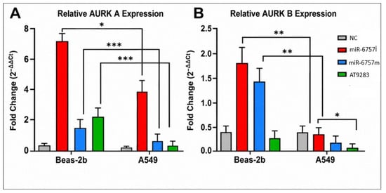
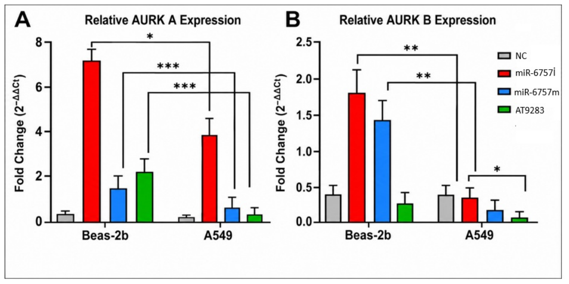
To investigate the potential role of miR-6757-3p in mitotic regulation, BEAS-2B and A549 cells were transfected with a miR-6757-3p inhibitor (miR-6757i) or mimic (miR-6757m), or treated with an AT9283 treatment. As shown in Figure 1, compared to the negative control (NC), an increase in AURKA and AURKB mRNA levels was observed following miR-6757-3p inhibition in both cell lines, with the change reaching statistical significance (* p < 0.05, p < 0.01). In contrast, transfection with the miR-6757-3p mimic tended to reduce AURKA and AURKB expression, while AT9283 treatment also led to a decrease in these levels. AT9283 treatment was associated with decreased AURKA and AURKB expression at the mRNA level. Overall, the results suggest miR-6757-3p inhibition was associated with increased AURKA and AURKB mRNA expression.
Figure 1.
Relative mRNA expression levels of AURKA (A) and AURKB (B) in BEAS-2B and A549 cells following transfection with miR-6757-3p inhibitor (miR-6757i), miR-6757-3p mimic (miR-6757m), or treatment with Aurora kinase inhibitor (AT9283). Negative control (NC) was used as reference. Data are presented as fold change relative to NC. Statistical significance was determined using one-way ANOVA followed by post-hoc tests (* p < 0.05, ** p < 0.01, *** p < 0.001). Data are presented as mean ± standard deviation (SD). Error bars represent SD. three independent biological replicates.
3.2. miR-6757-3p Modulation Alters Apoptosis-Related Gene Expression
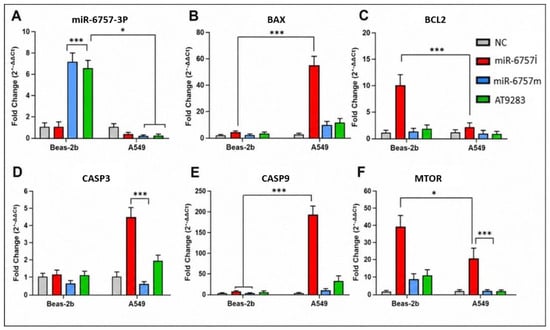
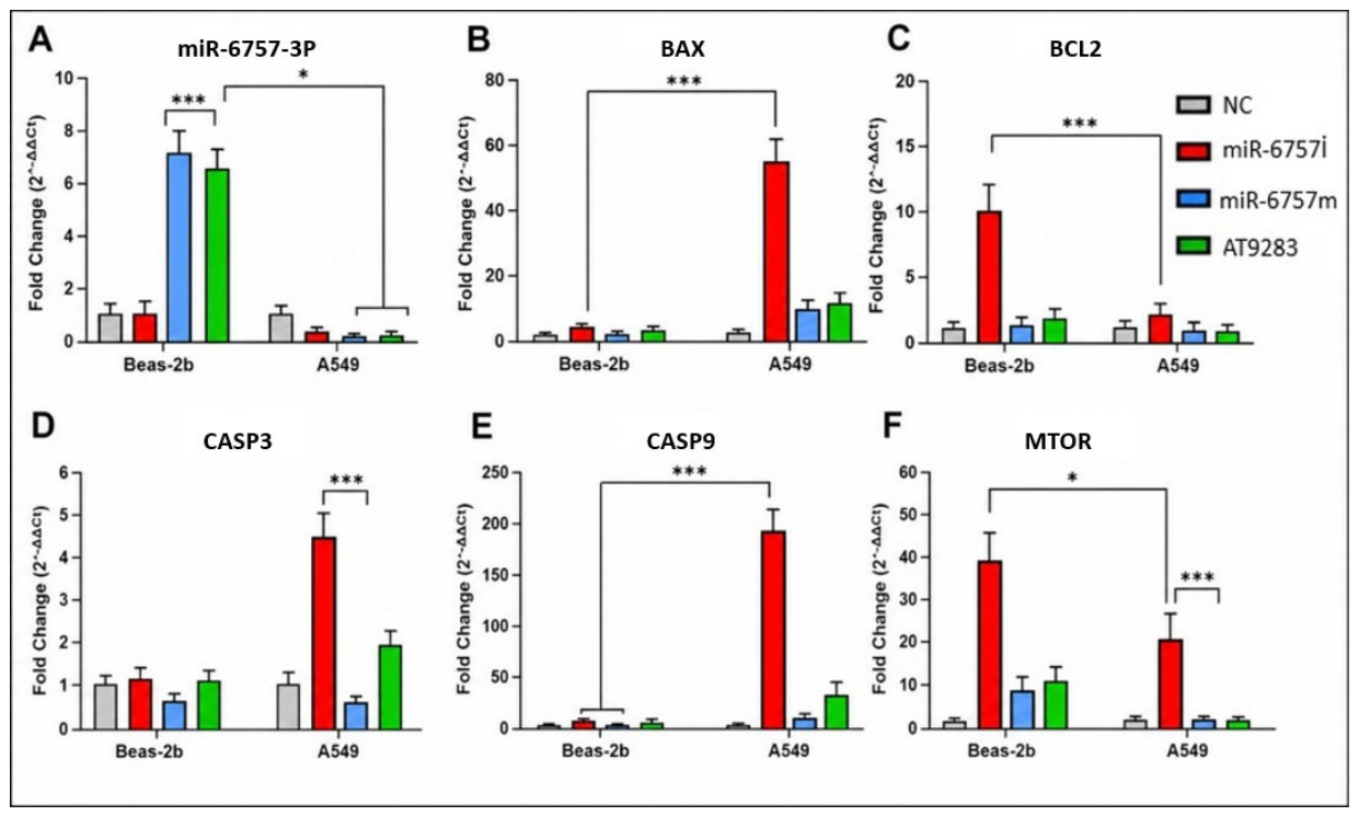
The effects of miR-6757-3p inhibitor and mimic treatment and AT9283 exposure on apoptosis-related gene expression were examined by qRT-PCR. As shown in Figure 2, treatment with the miR-6757-3p inhibitor was associated with increased expression of the pro-apoptotic genes BAX, CASP3, and CASP9 and decreased expression of the anti-apoptotic gene BCL2 in both BEAS-2B and A549 cells (p < 0.05). In contrast, miR-6757-3p mimic treatment was associated with a tendency toward the opposite expression pattern, including lower BAX, CASP3, and CASP9 expression and higher BCL2 expression. Changes in MTOR expression were also observed following the different treatments.
Figure 2.
Relative expression levels of miR-6757-3p (A), BAX (B), BCL2 (C), CASP3 (D), CASP9 (E), and MTOR (F) in BEAS-2B and A549 cells following treatment with miR-6757-3p inhibitor (miR-6757i), miR-6757-3p mimic (miR-6757m), or AT9283. Negative control (NC) cells were used as the calibrator group. Gene expression levels were determined by quantitative real-time PCR (qRT-PCR) and calculated using the 2−ΔΔCt method. Results are presented as fold change relative to NC and expressed as mean ± SD from three independent biological replicates (n = 3). Statistical analyses were performed using one-way ANOVA followed by Tukey’s multiple comparison test. Statistical significance is indicated as * p < 0.05, and *** p < 0.001 compared with the corresponding NC group.
Expression data were normalized to GAPDH (for mRNA) and RNU6B_13 (for miRNA) expression levels. Data are presented as mean ± standard deviation (SD). Error bars represent SD.
Importantly, as shown in Figure 2A, miR-6757-3p expression did not consistently change in the expected direction following inhibitor or mimic treatment. In particular, inhibitor treatment did not result in a clear reduction in miR-6757-3p expression, whereas AT9283 treatment was associated with an unexpected increase in miR-6757-3p expression. Therefore, these expression data do not independently confirm successful miR-6757-3p knockdown or overexpression.
3.3. Protein-Level Analysis by ELISA
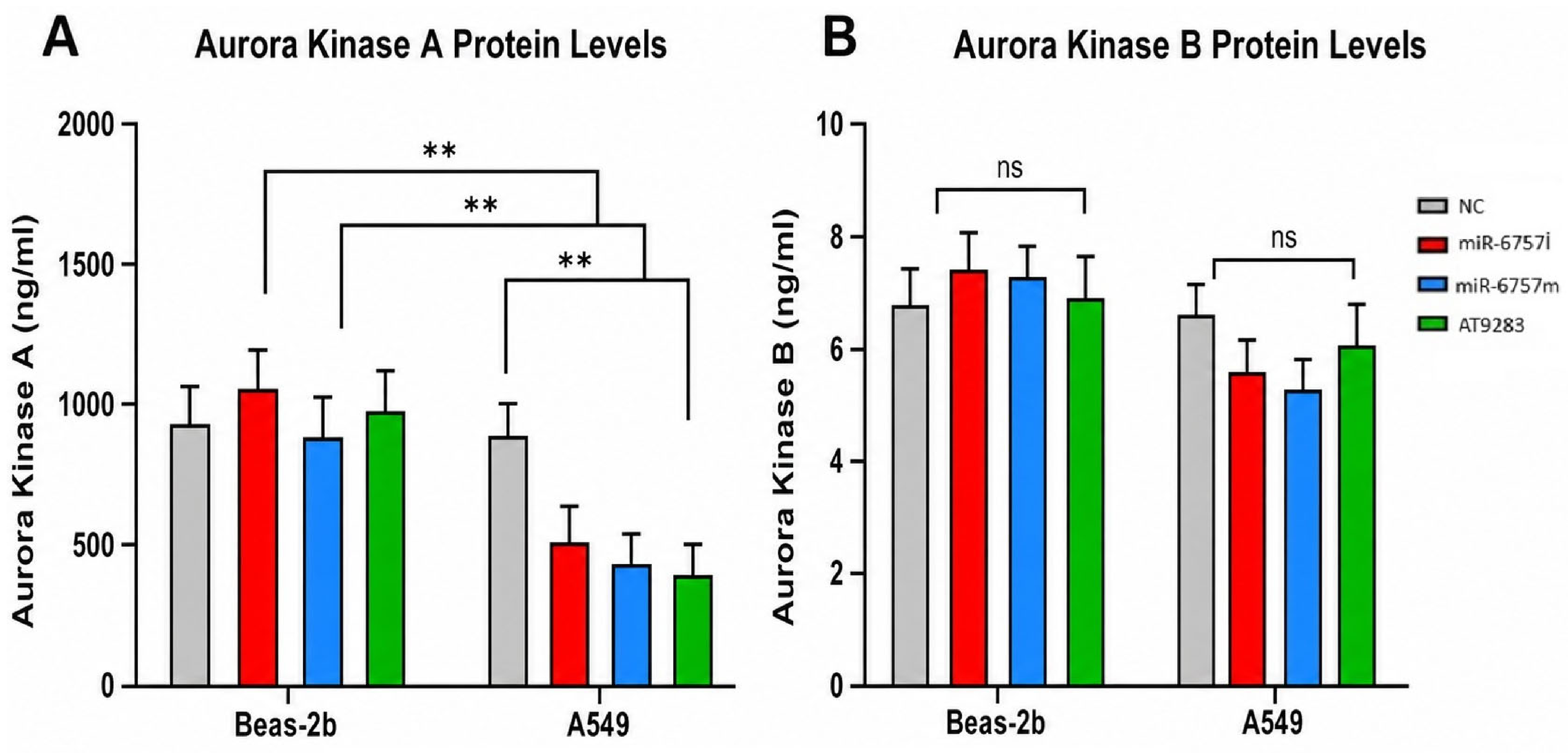
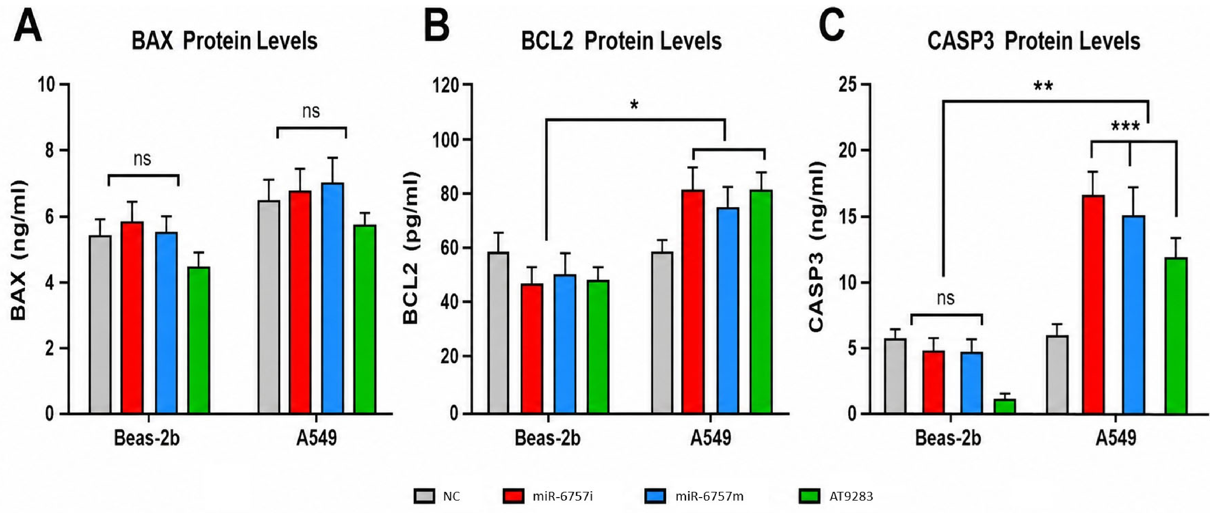
To evaluate whether the transcriptional changes were reflected at the protein level, ELISA analyses were performed. As shown in Figure 3 and Figure 4, at the protein level, some treatment-associated changes in AURKA, AURKB, BAX, CASP3, and BCL2 were observed; however, the magnitude and statistical significance of these changes varied among the treatment groups. In particular, not all AURKB and BAX comparisons reached statistical significance. Therefore, the ELISA findings should be interpreted as partially concordant with the transcriptional data rather than as uniform confirmation of the mRNA results, showing an increasing trend with miR-6757-3p inhibition and a decreasing trend following miR-6757-3p mimic or AT9283 treatment (p < 0.01). Similarly, the apoptosis-related proteins BAX and CASP3 tended to increase after miR-6757-3p inhibition, while a decreasing trend was observed in the anti-apoptotic protein BCL2. miR-6757-3p mimic treatment was associated with lower levels of BAX and CASP3 and higher levels of BCL2 compared with miR-6757-3p inhibition. Overall, these findings indicate that the protein-level changes were partially consistent with the transcriptional findings, although not all observed changes reached statistical significance.
Figure 3.
Protein levels of Aurora Kinase A (A) and Aurora Kinase B (B) in BEAS-2B and A549 cells following treatment with miR-6757-3p inhibitor (miR-6757i), miR-6757-3p mimic (miR-6757m), or Aurora kinase inhibitor (AT9283). Negative control (NC) was used as reference. Protein concentrations were determined by ELISA and expressed as ng/mL. Statistical analysis was performed using one-way ANOVA with post-hoc tests (** p < 0.01; ns: not significant). Data are presented as mean ± standard deviation (SD). Error bars represent SD of three independent biological replicates.
Figure 4.
Protein levels of BAX, BCL-2, and CASP3 in BEAS-2B and A549 cells following miR-6757-3p modulation and AT9283 treatment. (A) BAX protein levels, (B) BCL-2 protein levels, and (C) CASP3 protein levels in BEAS-2B and A549 cells in the negative control (NC), miR-6757-3p inhibitor (miR-6757i), miR-6757-3p mimic (miR-6757m), and AT9283 treatment groups. Protein levels are expressed as ng/mL for BAX and CASP3 and pg/mL for BCL-2. Bars represent the measured protein levels, and error bars indicate variability as presented in the graph. Statistical significance is indicated as follows: ns, not significant; * p < 0.05; ** p < 0.01; *** p < 0.001. Data are presented as mean ± standard deviation (SD). Error bars represent SD.
3.4. Time-Dependent Effects on Cellular Response
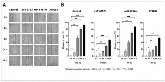
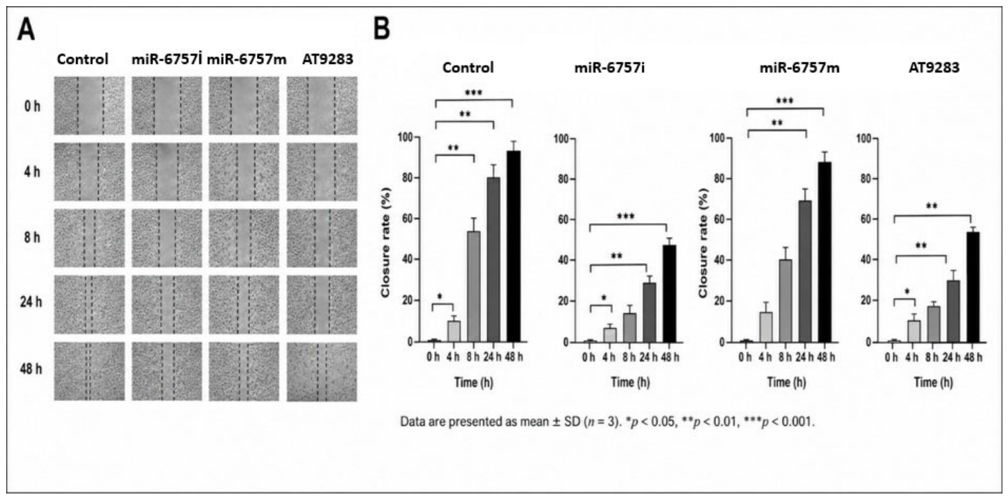
In the time-course analysis performed in A549 cells (Figure 5), wound closure was evaluated at 0, 4, 8, 24, and 48 h following treatment. Differences in wound closure patterns were observed among the control, miR-6757-3p inhibitor, miR-6757-3p mimic, and Aurora kinase inhibitor groups over the observation period. miR-6757-3p inhibition was associated with a distinct wound-closure pattern compared with the control group, whereas miR-6757-3p mimic treatment produced a different temporal response. Similarly, treatment with the Aurora kinase inhibitor resulted in time-dependent changes in wound closure. Overall, these findings suggest that modulation of miR-6757-3p and Aurora kinase inhibition may influence the wound-healing response of A549 cells in a time-dependent manner. The assay was used to assess wound closure over time. Because wound closure can be influenced by both cell migration and cell proliferation, the assay does not independently distinguish these processes.
Figure 5.
Time-dependent effects of miR-6757-3p inhibitor (miR-6757i), miR-6757-3p mimic (miR-6757m), and Aurora kinase inhibitor (AT9283) on wound closure in A549 cells. (A) Representative images and quantitative assessment of wound closure at 0, 4, 8, 24, and 48 h in the control, miR-6757i, and miR-6757m groups. (B) Representative schematic summary of the experimental groups. Data are presented as mean ± standard deviation (SD). Error bars represent SD.
4. Discussion
This study aimed to investigate the association of miR-6757-3p inhibitor or mimic treatment with AURKA and AURKB expression and apoptosis-related molecular responses in BEAS-2B and A549 cells. The findings suggest that miR-6757-3p inhibitor or mimic treatment is associated with changes in cell-cycle- and apoptosis-related molecular responses, although the functional and mechanistic significance of these changes remains to be established [28,29,30].
In the literature, overexpression or dysregulated expression of AURKB has been reported in breast, lung, hematologic, and gastrointestinal cancers, as well as in bladder cancer, uveal melanoma, and neuroblastoma, and may be associated with tumor invasion, metastasis, and drug resistance [10,31]. The efficiency of miR-6757-3p mimic and inhibitor transfection was not independently validated using a dedicated transfection-efficiency assay. Although miR-6757-3p expression was assessed by miRNA-specific qRT-PCR, the observed expression patterns did not consistently demonstrate the expected direction of modulation [31,32]. Therefore, successful functional overexpression or inhibition could not be conclusively established from the available data. In addition, the pronounced changes observed in certain apoptosis-related genes should be interpreted cautiously, as their magnitude cannot be attributed specifically to miR-6757-3p modulation without independent confirmation of transfection efficiency. The observed molecular differences may reflect treatment-associated cellular responses, but their specific relationship to miR-6757-3p remains uncertain. Future studies incorporating validated transfection conditions and independent assessment of miRNA modulation are needed to clarify these relationships. However, the functional significance of this association remains to be established [27]. Notably, the expression profile of miR-6757-3p following inhibitor and mimic treatment did not consistently correspond to the expected direction of change. Therefore, these expression data should be interpreted cautiously and should not be considered definitive evidence of successful functional knockdown or overexpression. The efficiency of miR-6757-3p inhibitor and mimic treatment was not independently verified using a dedicated functional transfection-efficiency assay, which limits the extent to which the observed molecular responses can be specifically attributed to altered miR-6757-3p activity. Additionally, the observed changes in apoptosis-related gene expression indicate that miR-6757-3p inhibitor or mimic treatment was associated with molecular responses involving apoptosis-related markers. However, these findings should not be interpreted as direct evidence of apoptotic cell death in the absence of functional apoptosis assays [27]. These findings indicate that miR-6757-3p inhibitor or mimic treatment is associated with changes in cell-cycle- and cellular-response-related molecular markers; however, the underlying mechanisms remain to be determined. The protein-level results obtained by ELISA were generally consistent with the gene expression findings for AURKA and AURKB, suggesting that some of the observed transcriptional changes were also reflected at the protein-expression level. However, the absence of marked cellular changes may also indicate that the effects of miR-6757-3p are relatively modest or context-dependent.
Secondly, when apoptosis-related genes were evaluated, inhibition of miR-6757-3p was associated with an increase in pro-apoptotic genes (BAX, CASP3, CASP9) and a decrease in BCL2 levels, whereas miR-6757-3p mimic treatment tended to show the opposite pattern. These findings suggest that miR-6757-3p may be associated not only with cell cycle regulation but also with cell death processes. In the literature, activation of BAX and caspases is reported to support cell death, while BCL2 may contribute to cell survival [33,34,35]. Although some molecular responses were directionally similar between A549 and BEAS-2B cells, differences in the magnitude and, in some cases, the direction of treatment-associated changes were also observed. These differences suggest that the molecular responses associated with miR-6757-3p inhibitor or mimic treatment may depend on cellular context. Because some responses were also observed in BEAS-2B cells, the present findings do not establish a cancer-cell-specific effect and may instead indicate cell-context-dependent responses. In this context, the present findings suggest that miR-6757-3p inhibitor or mimic treatment may be associated with changes in molecular markers related to apoptosis and cell-survival processes.
Additionally, the observed alterations in mTOR gene expression suggest a potential association between miR-6757-3p modulation and MTOR expression [36]. This pathway plays a critical role in regulating fundamental cellular processes, including cell growth, energy homeostasis, and cell survival. Previous studies have reported that hyperactivation of the mTOR signaling pathway is associated with increased cell proliferation and resistance to therapy in various cancer types [36,37,38,39]. In this context, the present findings suggest that miR-6757-3p may play a role in modulating the balance between apoptosis and cell survival processes. Although changes in AURKA, AURKB, BAX, BCL2, CASP3, CASP9, and MTOR were observed, these findings do not establish a direct regulatory mechanism. This study does not confirm direct targeting of AURKA/AURKB by miR-6757-3p or the involvement of the PI3K/AKT/mTOR pathway. Furthermore, changes in apoptosis-related gene expression alone do not confirm apoptosis. Further studies involving direct target validation, rescue experiments, and functional apoptosis assays are needed to clarify these relationships.
Third, protein-level analyses performed by ELISA revealed some changes in AURKA, AURKB, BAX, CASP3, and BCL2 levels, which appeared to be partially consistent with the gene expression findings. These observations suggest that the transcriptional alterations may, to some extent, be reflected at the protein level. However, further studies are needed to clarify the potential contribution of miR-6757-3p to cellular responses.
In this study, the potential association of miR-6757-3p treatment with apoptosis-related and cell-survival-related molecular responses, as well as its relationship with Aurora kinase expression, was investigated. The functions of Aurora kinases in mitosis, chromosomal stability, and cell proliferation were considered in relation to the observed changes in BAX, BCL2, CASP3, CASP9, and MTOR expression. Accordingly, BAX, BCL2, CASP3, CASP9, and MTOR were included as exploratory molecular endpoints to characterize apoptosis- and cell-survival-related responses. Changes in AURKA and AURKB mRNA expression suggest an association with miR-6757-3p modulation; however, direct targeting has not been confirmed and requires further validation.
However, although Annexin V/PI analysis and other functional assays would provide more direct evidence of apoptosis, such assays were not included in the experimental design of the present study. Therefore, the analyses were limited to gene- and protein-expression measurements. The inability to perform Western blot analyses at the protein level also represents an important limitation. The lack of luciferase reporter assays to directly evaluate the interaction between miR-6757-3p and the 3′UTRs of AURKA and AURKB limits the ability to determine whether these genes are direct targets of miR-6757-3p.
Accordingly, the observed changes in AURKA and AURKB expression should be interpreted as an association with miR-6757-3p treatment rather than evidence of direct post-transcriptional regulation. Bioinformatic prediction and luciferase reporter assays will be required to determine whether this relationship involves direct post-transcriptional regulation. Furthermore, the relatively low standard deviations observed in some groups may be attributed to the limited number of replicates, and increasing the number of replicates could enhance the robustness of the findings. Additionally, the effects of miR-6757-3p mimic and inhibitor treatments on wound healing and cell migration, as well as cell line-specific responses presented in Figure 5, should be interpreted within the context of these experimental constraints. Overall, these limitations necessitate cautious interpretation of the findings. Time-dependent wound-healing analyses revealed differences in wound closure among the treatment groups over the 48-h observation period. These findings indicate that miR-6757-3p modulation and Aurora kinase inhibition may influence the wound-healing response in A549 cells; however, the scratch assay does not allow direct conclusions regarding cellular damage, protection, or apoptosis. The molecular changes observed following AT9283 treatment were compatible with alterations in apoptosis-related responses; however, given the multi-target nature of AT9283 and the absence of a direct apoptosis assay, these findings should not be interpreted as evidence of a specific Aurora kinase-mediated apoptotic mechanism. These dynamic changes suggest that miR-6757-3p may serve different functions in short-term and long-term cellular responses. AT9283 treatment and miR-6757-3p mimic transfection showed different expression patterns in Aurora kinase and apoptosis-related genes. These differences do not support a single, unified anticancer mechanism. Gene expression changes alone cannot confirm apoptosis or establish a causal relationship. Therefore, the findings are interpreted as treatment-associated molecular changes, and further studies are needed to clarify the underlying mechanisms.
Overall, the findings suggest an association between miR-6757-3p inhibitor or mimic treatment, Aurora kinase expression, and apoptosis-related molecular responses. It has been reported in the literature that increased Aurora kinase activity may be associated with tumor progression, chromosomal instability, and poor prognosis [38,39]. In this context, taken together, the present findings do not allow miR-6757-3p to be classified as either a tumor-suppressive or oncogenic miRNA. Rather, the observed molecular and wound-closure responses suggest that its functional effects may be context-dependent and require further investigation. Furthermore, the relatively low standard deviations observed in some groups may be attributed to limited replicate numbers, and increasing the number of replicates could enhance the robustness of the findings. Another limitation of the present study is that independent STR-based cell line authentication and mycoplasma testing were not performed. Additionally, the effects of miR-6757-3p mimic and inhibitor treatments on wound healing and cell migration, as well as cell line-specific responses presented in Figure 5, should be interpreted within the context of these experimental constraints. The wound closure assay cannot distinguish between cell migration and cell proliferation. This is particularly important for AT9283, which may affect cell division. Therefore, changes in wound closure cannot be attributed solely to altered cell migration. Since cell proliferation was not independently assessed, the results should be interpreted as changes in wound closure rather than direct evidence of migration inhibition.
5. Conclusions
In conclusion, miR-6757-3p modulation and AT9283 treatment were associated with changes in Aurora kinase expression and apoptosis-related molecular markers in A549 cells. These findings provide evidence of treatment-associated molecular responses but do not establish apoptosis induction. Further functional studies are required to clarify the biological significance of these changes. Further functional studies are required to clarify the biological significance of these results. The observed molecular changes indicate treatment-associated differences in AURKA, AURKB, and apoptosis-related markers in A549 and BEAS-2B cells. However, because the efficiency of miR-6757-3p modulation was not independently validated, these findings cannot be conclusively attributed to successful miRNA overexpression or inhibition. Therefore, the results should be interpreted as preliminary observations that require further validation. However, the present study does not establish direct molecular targeting or a causal signaling mechanism. Further studies using direct target-validation approaches, functional apoptosis assays, selective gene-silencing strategies, and in vivo models are required to clarify the biological significance of these findings.
Author Contributions
Conceptualization: H.D.; Methodology: İ.S.Ç.; Investigation: H.D.; Writing—Original Draft: H.D.; Writing—Review & Editing: all authors; Supervision: H.D. All authors have read and agreed to the published version of the manuscript.
Funding
This research received no external funding.
Institutional Review Board Statement
Not applicable.
Informed Consent Statement
Not applicable.
Data Availability Statement
The raw data supporting the conclusions of this article will be made available by the authors on request.
Acknowledgments
We would like to thank the staff of the Biomedical Research and Application Center laboratories for their technical support and contributions to this study. We also express our sincere gratitude to Mustafa Çiçek, and to Mehmet Akif Yılmaz for their valuable contributions.
Conflicts of Interest
The authors declare no conflicts of interest.
Abbreviations
| ANOVA | Analysis of variance |
| AURKA | Aurora kinase A |
| AURKB | Aurora kinase B |
| BAX BCL2 | associated X protein |
| BCL2 B | cell lymphoma 2 |
| CASP3 | Caspase 3 |
| CASP9 | Caspase 9 |
| ELISA | Enzyme-linked immunosorbent assay |
| miRNA | MicroRNA |
| MTOR | Mechanistic target of rapamycin |
| NC | Negative control |
| NSCLC | Non-small cell lung cancer |
| qRT-PCR | Quantitative reverse transcription polymerase chain reaction |
| SD | Standard deviation |
References
- Siegel, R.L.; Miller, K.D.; Jemal, A. Cancer statistics, 2019. CA Cancer J. Clin. 2019, 69, 7–34. [Google Scholar] [CrossRef] [Scilit] [PubMed]
- Gao, J.; Aksoy, B.A.; Dogrusoz, U.; Dresdner, G.; Gross, B.; Sumer, S.O.; Sun, Y.; Jacobsen, A.; Sinha, R.; Larsson, E.; et al. Integrative analysis of complex cancer genomics and clinical profiles using the cBioPortal. Sci. Signal. 2013, 6, pl1. [Google Scholar] [CrossRef] [Scilit] [PubMed]
- Ji, Y.; Zhang, Y.; Liu, S.; Li, J.; Jin, Q.; Wu, J.; Duan, H.; Liu, X.; Yang, L.; Huang, Y. The epidemiological landscape of lung cancer: Current status, temporal trend and future projections based on the latest estimates from GLOBOCAN 2022. J. Natl. Cancer Cent. 2025, 5, 278–286. [Google Scholar] [CrossRef] [Scilit] [PubMed]
- Herbst, R.S.; Morgensztern, D.; Boshoff, C. The biology and management of non-small cell lung cancer. Nature 2018, 553, 446–454. [Google Scholar] [CrossRef] [Scilit] [PubMed]
- Zhou, J.; Xu, Y.; Liu, J.; Feng, L.; Yu, J.; Chen, D. Global burden of lung cancer in 2022 and projections to 2050: Incidence and mortality estimates from GLOBOCAN. Cancer Epidemiol. 2024, 93, 102693. [Google Scholar] [CrossRef] [Scilit] [PubMed]
- Howlader, N.; Forjaz, G.; Mooradian, M.J.; Meza, R.; Kong, C.Y.; Cronin, K.A.; Mariotto, A.B.; Lowy, D.R.; Feuer, E.J. The effect of advances in lung-cancer treatment on population mortality. N. Engl. J. Med. 2020, 383, 640–649. [Google Scholar] [CrossRef] [Scilit] [PubMed]
- Gilyazova, I.; Gimalova, G.; Nizamova, A.; Galimova, E.; Ishbulatova, E.; Pavlov, V.; Khusnutdinova, E. Non-coding RNAs as key regulators in lung cancer. Int. J. Mol. Sci. 2023, 25, 560. [Google Scholar] [CrossRef] [Scilit] [PubMed]
- Yan, M.; Wang, C.; He, B.; Yang, M.; Tong, M.; Long, Z.; Liu, B.; Peng, F.; Xu, L.; Zhang, Y.; et al. Aurora-A kinase: A potent oncogene and target for cancer therapy. Med. Res. Rev. 2016, 36, 1036–1079. [Google Scholar] [CrossRef] [Scilit] [PubMed]
- Zheng, D.; Li, J.; Yan, H.; Zhang, G.; Li, W.; Chu, E.; Wei, N. Emerging roles of Aurora-A kinase in cancer therapy resistance. Acta Pharm. Sin. B 2023, 13, 2826–2843. [Google Scholar] [CrossRef] [Scilit] [PubMed]
- Borah, N.A.; Reddy, M.M. Aurora kinase B inhibition: A potential therapeutic strategy for cancer. Molecules 2021, 26, 1981. [Google Scholar] [CrossRef] [Scilit] [PubMed]
- Mesic, A.; Rogar, M.; Hudler, P.; Bilalovic, N.; Eminovic, I.; Komel, R. Genetic variations in AURORA cell cycle kinases are associated with glioblastoma multiforme. Sci. Rep. 2021, 11, 17444. [Google Scholar] [CrossRef] [Scilit] [PubMed]
- Barton, V.N.; Foreman, N.K.; Donson, A.M.; Birks, D.K.; Handler, M.H.; Vibhakar, R. Aurora kinase A as a rational target for therapy in glioblastoma. J. Neurosurg. Pediatr. 2010, 6, 98–105. [Google Scholar] [CrossRef] [Scilit] [PubMed]
- Du, R.; Huang, C.; Liu, K.; Li, X.; Dong, Z. Targeting AURKA in Cancer: Molecular mechanisms and opportunities for Cancer therapy. Mol. Cancer 2021, 20, 15. [Google Scholar] [CrossRef] [Scilit] [PubMed]
- Al-Sanea, M.M.; Elkamhawy, A.; Paik, S.; Lee, K.; El Kerdawy, A.M.; Abbas, B.S.N.; Roh, E.J.; Eldehna, W.M.; Elshemy, H.A.; Bakr, R.B.; et al. Sulfonamide-based 4-anilinoquinoline derivatives as novel dual Aurora kinase (AURKA/B) inhibitors: Synthesis, biological evaluation and in silico insights. Bioorganic Med. Chem. 2020, 28, 115525. [Google Scholar] [CrossRef] [Scilit] [PubMed]
- Lee, H.; Kim, E.; Hwang, N.; Yoo, J.; Nam, Y.; Hwang, I.; Park, J.-G.; Park, S.-E.; Chung, K.-S.; Chung, H.W.; et al. Discovery of N-benzylbenzamide-based allosteric inhibitors of Aurora kinase A. Bioorganic Med. Chem. 2024, 102, 117658. [Google Scholar] [CrossRef] [Scilit] [PubMed]
- Vats, P.; Saini, C.; Baweja, B.; Srivastava, S.K.; Kumar, A.; Kushwah, A.S.; Nema, R. Aurora kinases signaling in cancer: From molecular perception to targeted therapies. Mol. Cancer 2025, 24, 180. [Google Scholar] [CrossRef] [Scilit] [PubMed]
- De Groot, C.O.; Hsia, J.E.; Anzola, J.V.; Motamedi, A.; Yoon, M.; Wong, Y.L.; Jenkins, D.; Lee, H.J.; Martinez, M.B.; Davis, R.L.; et al. A cell biologist’s field guide to aurora kinase inhibitors. Front. Oncol. 2015, 5, 285. [Google Scholar] [CrossRef] [Scilit] [PubMed]
- Jamshidi, N.; Jamshidi, N.; Zaman, M.; Chehresaz, M.; Roshanfarzad, F.; Chaleshi, V.; Aghdaei, H.A. Medicago sativa extracts enhance the anticancer efficacy of GEM in PANC-1 cells through apoptosis induction and BAX/BCL-2/CASP3 expression modulation. Asian Pac. J. Cancer Prev. APJCP 2025, 26, 1689. [Google Scholar] [CrossRef] [Scilit] [PubMed]
- Li, Y.; Wang, Y.; Yu, X.; Yu, T.; Zheng, X.; Chu, Q. Radix tetrastigma inhibits the non-small cell lung cancer via Bax/Bcl-2/Caspase-9/Caspase-3 pathway. Nutr. Cancer 2022, 74, 320–332. [Google Scholar] [CrossRef] [Scilit] [PubMed]
- Lin, H.-I.; Lee, Y.-J.; Chen, B.-F.; Tsai, M.-C.; Lu, J.-L.; Chou, C.-J.; Jow, G.-M. Involvement of Bcl-2 family, cytochrome c and caspase 3 in induction of apoptosis by beauvericin in human non-small cell lung cancer cells. Cancer Lett. 2005, 230, 248–259. [Google Scholar] [CrossRef] [Scilit] [PubMed]
- Saliminejad, K.; Khorram Khorshid, H.R.; Soleymani Fard, S.; Ghaffari, S.H. An overview of microRNAs: Biology, functions, therapeutics, and analysis methods. J. Cell. Physiol. 2019, 234, 5451–5465. [Google Scholar] [CrossRef] [Scilit] [PubMed]
- Hashemi, A.; Gorji-Bahri, G. MicroRNA: Promising roles in cancer therapy. Curr. Pharm. Biotechnol. 2020, 21, 1186–1203. [Google Scholar] [CrossRef] [Scilit] [PubMed]
- Akkafa, F.; Koyuncu, İ.; Temiz, E.; Dagli, H.; Dïlmec, F.; Akbas, H. miRNA-mediated apoptosis activation through TMEM 48 inhibition in A549 cell line. Biochem. Biophys. Res. Commun. 2018, 503, 323–329. [Google Scholar] [CrossRef] [Scilit] [PubMed]
- Ergun, S.; Tayeb, T.S.; Arslan, A.; Temiz, E.; Arman, K.; Safdar, M.; Dağlı, H.; Korkmaz, M.; Nacarkahya, G.; Kırkbeş, S.; et al. The investigation of miR-221-3p and PAK1 gene expressions in breast cancer cell lines. Gene 2015, 555, 377–381. [Google Scholar] [CrossRef] [Scilit] [PubMed]
- Cao, J.; Geng, J.; Chu, X.; Wang, R.; Huang, G.; Chen, L. miRNA-885-3p inhibits docetaxel chemoresistance in lung adenocarcinoma by downregulating Aurora, A. Oncol. Rep. 2019, 41, 1218–1230. [Google Scholar] [CrossRef] [Scilit] [PubMed]
- Murata, M.; Marugame, Y.; Morozumi, M.; Murata, K.; Kumazoe, M.; Fujimura, Y.; Tachibana, H. (−)-Epigallocatechin-3-O-gallate upregulates the expression levels of miR-6757-3p, a suppressor of fibrosis-related gene expression, in extracellular vesicles derived from human umbilical vein endothelial cells. Biomed. Rep. 2023, 18, 19. [Google Scholar] [CrossRef] [Scilit] [PubMed]
- Han, X.; Shi, F.; Guo, S.; Li, Y.; Wang, H.; Song, C.; Wu, S. LINC02086 promotes cell viability and inhibits cell apoptosis in breast cancer by sponging miR-6757-5p and up-regulating EPHA2. World J. Surg. Oncol. 2023, 21, 371. [Google Scholar] [CrossRef] [Scilit] [PubMed]
- Kumazoe, M.; Tachibana, H. MicroRNA mediates the effects of food factors. Biosci. Biotechnol. Biochem. 2025, 89, 174–178. [Google Scholar] [CrossRef] [Scilit] [PubMed]
- Brouwer, A.; De Laere, B.; Peeters, D.; Peeters, M.; Salgado, R.; Dirix, L.; Van Laere, S. Evaluation and consequences of heterogeneity in the circulating tumor cell compartment. Oncotarget 2016, 7, 48625. [Google Scholar] [CrossRef] [Scilit] [PubMed]
- Ritter, A.; Hirschfeld, M.; Berner, K.; Jaeger, M.; Grundner-Culemann, F.; Schlosser, P.; Asberger, J.; Weiss, D.; Noethling, C.; Mayer, S.; et al. Discovery of potential serum and urine-based microRNA as minimally-invasive biomarkers for breast and gynecological cancer. Cancer Biomark. 2020, 27, 225–242. [Google Scholar] [CrossRef] [Scilit] [PubMed]
- Orengo, J.A.; Rodriguez, W.F.; Velazquez, S.A.; Aquino-Acevedo, A.N.; Ortíz, G.R.; Cruz Robles, M.E.; Irrizary Quintana, E.M.; Saavedra, H.I. Combined targeting of AURKA and AURKB as a potential therapeutic strategy to decrease the expression of epithelial-to-mesenchymal transition drivers in triple-negative breast cancers. Cancer Res. 2025, 85, 6958. [Google Scholar] [CrossRef] [Scilit]
- Wang, C.; Liu, J.; Wu, Y.; Cai, C.; Chai, Z.; Jia, P.; Yuan, Y.; Jiang, Z. AURKB as a Therapeutic Target and Key Driver of Liver Cancer Growth and Metastasis. J. Pathol. Microbiol. Immunol. 2025, 133, e70021. [Google Scholar] [CrossRef] [Scilit] [PubMed]
- Lakkaniga, N.R.; Wang, Z.; Xiao, Y.; Kharbanda, A.; Lan, L.; Li, H.Y. Revisiting Aurora Kinase B: A promising therapeutic target for cancer therapy. Med. Res. Rev. 2024, 44, 686–706. [Google Scholar] [CrossRef] [Scilit] [PubMed]
- Nikonova, A.S.; Astsaturov, I.; Serebriiskii, I.G.; Dunbrack, R.L., Jr.; Golemis, E.A. Aurora A kinase (AURKA) in normal and pathological cell division. Cell. Mol. Life Sci. 2013, 70, 661–687. [Google Scholar] [CrossRef] [Scilit] [PubMed]
- Mou, P.K.; Yang, E.J.; Shi, C.; Ren, G.; Tao, S.; Shim, J.S. Aurora kinase A, a synthetic lethal target for precision cancer medicine. Exp. Mol. Med. 2021, 53, 835–847. [Google Scholar] [CrossRef] [Scilit] [PubMed]
- Glaviano, A.; Foo, A.S.; Lam, H.Y.; Yap, K.C.; Jacot, W.; Jones, R.H.; Eng, H.; Nair, M.G.; Makvandi, P.; Geoerger, B.; et al. PI3K/AKT/mTOR signaling transduction pathway and targeted therapies in cancer. Mol. Cancer 2023, 22, 138. [Google Scholar] [CrossRef] [Scilit] [PubMed]
- Wang, C.; Wang, W.; Han, X.; Du, L.; Li, A.; Huang, G. Methyltransferase-like 1 regulates lung adenocarcinoma A549 cell proliferation and autophagy via the AKT/mTORC1 signaling pathway. Oncol. Lett. 2021, 21, 330. [Google Scholar] [CrossRef] [Scilit] [PubMed]
- Ma, Z.; Zhang, W.; Wu, Y.; Zhang, M.; Wang, L.; Wang, Y.; Wang, Y.; Liu, W. Cyclophilin A inhibits A549 cell oxidative stress and apoptosis by modulating the PI3K/Akt/mTOR signaling pathway. Biosci. Rep. 2021, 41, BSR20203219. [Google Scholar] [CrossRef] [Scilit] [PubMed]
- Baba, Y.; Nosho, K.; Shima, K.; Irahara, N.; Kure, S.; Toyoda, S.; Kirkner, G.J.; Goel, A.; Fuchs, C.S.; Ogino, S. Aurora-A expression is independently associated with chromosomal instability in colorectal cancer. Neoplasia 2009, 11, 418–425. [Google Scholar] [CrossRef] [Scilit] [PubMed]

Disclaimer/Publisher’s Note: The statements, opinions and data contained in all publications are solely those of the individual author(s) and contributor(s) and not of MDPI and/or the editor(s). MDPI and/or the editor(s) disclaim responsibility for any injury to people or property resulting from any ideas, methods, instructions or products referred to in the content. |
© 2026 by the authors. Licensee MDPI, Basel, Switzerland. This article is an open access article distributed under the terms and conditions of the Creative Commons Attribution (CC BY) license.