


Abstract
Background: Maternal diabetes is associated with several adverse perinatal outcomes, but its relationship with hypoxic-ischaemic perinatal injury remains poorly defined. Fetal compromise may reflect an acute intrapartum event, a chronic antepartum process, or both. Objective: To systematically review the evidence linking gestational diabetes mellitus (GDM) and pregestational diabetes to hypoxic-ischaemic perinatal injury, assessing diagnosed or coded neonatal brain injury, acid–base status at birth, and chronic antepartum markers separately. Methods: PubMed, Embase, and Scopus were searched from inception to 16 October 2026. Observational studies comparing pregnancies with and without diabetes and reporting diagnosed or coded hypoxic-ischaemic encephalopathy, neonatal encephalopathy or neonatal seizures, umbilical acid–base status, or a chronic marker of fetal hypoxia were eligible; studies reporting only Apgar-based or other non-specific outcomes were excluded. Risk of bias was assessed with the Newcastle–Ottawa Scale together with a separate judgement of outcome-definition adequacy, certainty of evidence was rated with GRADE, and synthesis followed the SWiM guideline, with vote counting by direction of effect. The protocol was registered in PROSPERO (CRD420261435362). Results: Seventy-four studies were included (30 GDM only, 12 pregestational diabetes only, 32 both or unspecified). Pregestational diabetes was associated with hypoxic-ischaemic encephalopathy, neonatal encephalopathy, or neonatal seizures in all five studies with sufficient events (adjusted odds or risk ratios 1.5–5.1; very low certainty). In GDM, estimates pointed in an adverse direction in six of seven informative comparisons but were mostly small and imprecise; statistically significant associations came only from a large registry (adjusted relative risk 1.24–1.25) and a study restricted to small-for-gestational-age infants (very low certainty). Umbilical acid–base status was less favourable in pregestational diabetes in 9 of 10 studies. In GDM, differences were small (less favourable in 13 of 17 comparisons; pooled mean difference in umbilical arterial pH −0.025, 95% CI −0.041 to −0.010, prediction interval −0.10 to 0.05, three studies; very low certainty for clinically relevant acidaemia). Chronic markers pointed in an adverse direction in 33 of 37 independent evidence sources, most consistently erythropoietin, nucleated red cells, and polycythaemia, but certainty was very low for every marker. No study documented a chronic marker, an identified acute event, and diagnosed or coded brain injury in the same fetus. Conclusions: Pregestational diabetes is associated with diagnosed or coded hypoxic-ischaemic encephalopathy, neonatal encephalopathy, or seizures and with markers of chronic fetal hypoxic stress, but the certainty of this evidence is very low. In GDM, estimates for these outcomes mostly point in an adverse direction but are small and imprecise (very low certainty), and acid–base differences at birth are small. Whether a chronic process and an acute event combine to cause injury in an individual fetus has not been studied.
1. Introduction
Gestational diabetes mellitus (GDM) affects a growing proportion of pregnancies worldwide, and its prevalence continues to rise alongside maternal age and obesity [1,2]. It is associated with fetal macrosomia, hypertensive disorders, and increased rates of operative delivery [1,2]. Its relationship with hypoxic-ischaemic perinatal injury, however, is still less clear, and this relationship carries substantial clinical and medico-legal weight [3,4]. Hypoxic-ischaemic encephalopathy (HIE) is often attributed to a single acute intrapartum event, since intrapartum monitoring naturally focuses attention there [5,6,7]. Yet fetal compromise may instead reflect a chronic, antepartum process. Chronic hyperglycaemia and hyperinsulinaemia can increase fetal oxygen demand, and they can also alter placental structure and function [1,2,8]. The proposed pathway can be summarised in sequence: maternal hyperglycaemia produces fetal hyperglycaemia and hyperinsulinaemia; fetal hyperinsulinaemia raises oxidative metabolism and oxygen consumption; in parallel, placental villous immaturity, a thicker diffusion barrier, and an altered feto-placental weight ratio may limit oxygen transfer; the resulting imbalance between oxygen supply and demand would be signalled by erythropoietin release, nucleated red cells, and polycythaemia; and the reduced fetal reserve would leave the fetus less able to tolerate the transient hypoxaemia of normal labour or an acute intrapartum event. A fetus exposed to this chronic burden may tolerate an acute peripartum event poorly, even when that event would be innocuous in an otherwise healthy pregnancy. Distinguishing an acute cause from a chronic, cumulative one is therefore central to any causal assessment of perinatal brain injury in diabetic pregnancy, and it is rarely straightforward in practice. Existing evidence on this question is fragmented. Studies vary widely in diabetes definition, in comparator selection, and in the hypoxia-related outcome they report. Some measure intrapartum cardiotocography or umbilical cord gases [2,9]. Others measure antepartum markers such as Doppler velocimetry, placental histology, or erythropoietin, but few measure more than one of these domains in the same cohort [10]. Moreover, much of the mechanistic evidence on chronic fetal hypoxia originates from pregestational diabetes rather than from GDM itself. Pregestational diabetes precedes conception, so it can demonstrate genuinely pre-existing vascular pathology, whereas GDM-related changes must, by definition, develop within the index pregnancy. The two conditions differ in at least four respects relevant to this question. First, exposure timing and duration: pregestational diabetes exposes the embryo and fetus from conception, whereas GDM is usually diagnosed in the late second or third trimester. Second, maternal vasculopathy: microvascular disease may pre-date pregnancy in type 1 and type 2 diabetes but is absent, by definition, in GDM. Third, metabolic exposure: hyperglycaemia is typically more severe and more variable in type 1 diabetes and includes periconceptional and first-trimester hyperglycaemia. Fourth, the meaning of a chronic hypoxic marker: in pregestational diabetes, it may reflect pre-existing placental vascular pathology, whereas in GDM, it can only reflect changes acquired within the index pregnancy. Evidence from pregestational diabetes therefore cannot be transferred to GDM without explicit justification, and this review reports the two populations separately throughout. To our knowledge, no previous systematic review has synthesised diagnosed or coded neonatal brain injury, acid–base status at birth, and chronic antepartum markers within a single, methodologically appraised review. In particular, it is unknown whether any study has documented each step of the proposed pathway (a chronic marker, an acute intrapartum event, and diagnosed or coded brain injury) in the same fetus. This gap limits the ability of clinicians and of those assessing causation after the fact to weigh the available evidence properly, and it leaves individual case assessments without a clear evidentiary framework. We therefore conducted a systematic review of the literature on maternal diabetes, both gestational and pregestational, and hypoxic-ischaemic perinatal injury. We assessed acute peripartum biochemical and clinical evidence, diagnosed or coded neurological outcome and follow-up, and chronic antepartum hypoxia markers, alongside the methodological quality of each included study. Our aim was to characterise, systematically and transparently, what the existing evidence does and does not support. To structure this assessment, we defined two working hypotheses. Under Hypothesis A, hypoxic-ischaemic injury results from an acute intrapartum event in a previously uncompromised fetus. Under Hypothesis B, a chronic antepartum hypoxic compromise reduces fetal reserve, so that an acute event, including the physiological stress of labour, causes injury it would not otherwise cause.
2. Materials and Methods
2.1. Study Design and Registration
This systematic review was conducted in accordance with the Preferred Reporting Items for Systematic Reviews and Meta-Analyses (PRISMA) 2020 guidelines [11]. The review protocol was registered in the International Prospective Register of Systematic Reviews (PROSPERO) [CRD420261435362] [12]. The review aimed to systematically evaluate the available evidence on the association between maternal diabetes, GDM and PGDM, and hypoxic-ischaemic perinatal injury, with particular focus on acute intrapartum/peripartum evidence, diagnosed neonatal neurological outcome, and chronic antepartum markers of fetal hypoxic compromise. Several elements were defined after the first analysis and are therefore post hoc: the outcome tiers and the exclusion of studies reporting only Tier 3 outcomes, the requirement for a concurrent non-diabetic comparator, the judgement of outcome-definition adequacy, GRADE and SWiM reporting, the vote-counting rules, the classification of each comparison by the direction of its point estimate, the exploratory pooling of umbilical arterial pH and the sensitivity syntheses. They were applied to all records. The protocol was first registered in PROSPERO on 27 June 2026, and the record was amended on 19 October 2026 to describe these post hoc elements.
2.2. Search Strategy
A systematic search of PubMed (NCBI), Embase (Embase.com), and Scopus was run from inception to 16 October 2026. The strategy combined three concept blocks with the Boolean operator AND: (1) diabetes in pregnancy (gestational diabetes, pregnancy in diabetics, diabetes mellitus, hyperglycaemia); (2) pregnancy, fetus, and newborn; and (3) hypoxia-related outcomes and markers, comprising hypoxic-ischaemic or neonatal encephalopathy, asphyxia, fetal hypoxia or distress, neonatal seizures, therapeutic hypothermia, Apgar score, umbilical cord blood gases, acidaemia, base excess or deficit, lactate, cardiotocography and fetal heart rate, Doppler velocimetry, erythropoietin, nucleated red blood cells, polycythaemia, placental pathology, fetal cardiac function, and autopsy. Limits were English language and human studies. Reference lists of included studies were screened to identify additional studies. The search strategies were developed by D.C. and checked against the PRESS 2015 checklist [13] by M.M. before the search was run; because M.M. is a co-author and not an information specialist, this check was not independent of the review team. The complete strategy for each database, with platform, date, limits, and number of records retrieved, is provided in Supplementary Table S1.
2.3. Eligibility Criteria
Studies were included if they met the following criteria: original observational studies (prospective or retrospective cohort, case-control or cross-sectional) in human pregnancies complicated by GDM and/or pregestational diabetes (type 1 or type 2); with a concurrent comparator group of pregnancies without diabetes; reporting at least one Tier 1, Tier 2, or Tier 4 outcome as defined in Section 2.7 (diagnosed or coded neonatal encephalopathy, hypoxic-ischaemic encephalopathy or neonatal seizures, umbilical acid–base status or fetal oxygenation at birth, or a chronic or antepartum marker of fetal hypoxia); and published in English. Studies were excluded if they enrolled a population not meeting this definition, or in which the diabetic subgroup could not be separated; had no non-diabetic comparator, including studies comparing subgroups of women with diabetes only or using historical or published reference values or reference ranges instead of a concurrent comparator group; reported only Tier 3 outcomes (Apgar scores, resuscitation, neonatal intensive care admission, or undefined “asphyxia”); were case series or case reports; were narrative or systematic reviews, editorials, letters, book chapters, conference abstracts, or animal or in vitro studies; or provided insufficient data for extraction. Studies in which all women with diabetes had a coexisting complication that was also present in the comparator group (for example, diabetes with hypertension compared with hypertension alone) met these criteria and were retained, but they were not counted in the synthesis because they do not estimate the effect of diabetes itself (Section 2.8).
2.4. Study Selection
Study selection followed the PRISMA 2020 two-stage screening process [11], as shown in Supplementary Table S2. Titles and abstracts of all records identified by the search were independently screened for relevance by D.C. and S.I.; records clearly not meeting the eligibility criteria were excluded at this stage. Full-text reports of the remaining records were then retrieved and independently assessed against the inclusion and exclusion criteria described in Section 2.3. Disagreements at either stage were to be resolved by discussion between the two reviewers and, where unresolved, by consultation with a third reviewer (G.M.M.). The two reviewers agreed on all records at both stages, so no disagreement required resolution, and the third reviewer was not consulted. After the first analysis, all reports excluded at full text for lacking a Tier 1, 2, or 4 outcome were re-assessed against the full text; reports whose full text could not be obtained were reclassified as not retrieved.
2.5. Quality Assessment
Methodological quality and risk of bias were assessed using the Newcastle–Ottawa Scale (NOS) [14], applied in its cohort-study form (Selection, Comparability, Outcome; maximum 9 stars) or case-control form (Selection, Comparability, Exposure; maximum 9 stars) according to study design. Studies were classified as low risk of bias (7–9 stars), moderate risk of bias (4–6 stars), or high risk of bias (0–3 stars). Quality assessment was performed independently by D.C. and S.I. with disagreements resolved by discussion between the two reviewers and, where unresolved, by consultation with a third reviewer (G.M.M.). One deliberate modification of the NOS was applied consistently: in case-control studies, controls recruited from routine antenatal clinics were treated as community controls. Because the NOS rewards the method of outcome ascertainment rather than the validity of the outcome definition, we added a separate judgement of the adequacy of each study’s hypoxia-related outcome definition. Adequacy was high for a Tier 1 outcome defined by standard criteria (encephalopathy staging, therapeutic hypothermia, neuroimaging, or neuropathology) or a Tier 2 outcome including base deficit, lactate, or oxygen content; moderate for a Tier 1 diagnosis based on an explicit clinical or coded definition, a Tier 2 outcome based on pH alone, or a Tier 4 biomarker measured in a fetal compartment (erythropoietin, nucleated red cells, blinded placental histology); and low for indirect or non-validated markers (Doppler indices, computerised cardiotocography or fetal cardiac indices, placental morphometry, autopsy findings interpreted as hypoxic) or incompletely reported outcomes. Internal validity (NOS category) and outcome-definition adequacy are reported separately. An overall level of concern for the review question was then assigned as the worse of the two, with high adequacy mapped to low concern, moderate adequacy to moderate concern, and low adequacy to high concern [15]. Study design was classified according to how participants were sampled, not according to the label used in the original publication: studies that selected participants by diabetes status and compared outcomes were classified as cohort studies and appraised with the cohort form, and only studies that selected participants by outcome (neonatal seizures) were classified as case-control studies. Four studies measured exposure and outcome at a single time point (cross-sectional design); they were appraised with the cohort form, in which the item “demonstration that the outcome was not present at the start of the study” was not awarded, and the follow-up items were awarded when the outcome was ascertained in all participants at delivery. Certainty of evidence for each outcome tier, diabetes population, and chronic marker was rated with the GRADE approach for observational evidence [16]. Certainty was not rated up for a large effect when it had been rated down for any domain. Domain-level reasons for every rating are reported in Supplementary Table S5. Selective outcome reporting was assessed by comparing the outcomes listed in each study’s methods with those reported in its results; funnel-plot methods were not applied because no single comparison included 10 or more studies reporting a common effect measure.
2.6. Data Extraction
Data were extracted using a standardised database including study identifier, author and year, country/setting, study design, diabetes type (GDM, PGDM, or mixed population), sample size, diagnostic criteria for diabetes, maternal glycaemic control or treatment, presence and type of comparator group, intrapartum monitoring findings, acute peripartum biochemical and clinical status (umbilical arterial pH, base excess, lactate, Apgar scores, need for resuscitation), diagnosis and staging of hypoxic-ischaemic encephalopathy, use of therapeutic hypothermia, neuroimaging findings, neurodevelopmental follow-up, antepartum/chronic hypoxia markers (Doppler velocimetry, placental histology, hematologic and erythropoietin-based markers), authors’ own interpretation of the relative contribution of acute versus chronic mechanisms, and study limitations. Extraction was performed independently by D.C. and S.I. with discrepancies resolved by discussion. Extracted outcomes were classified into the outcome tiers described in Section 2.7. Each study was also mapped to the working hypothesis it could inform: Hypothesis A (injury caused by an acute intrapartum event in a previously uncompromised fetus) when it reported intrapartum monitoring or acute status at birth and Hypothesis B (chronic antepartum compromise reducing fetal reserve, with a superimposed acute event) when it reported a chronic or antepartum marker; studies reporting both were mapped to both, and studies reporting only a Tier 1 outcome were mapped to neither, because they do not describe the timing of compromise. Diagnostic criteria, setting, glycaemic control, and the mapping of each study are reported in Supplementary Table S4.
2.7. Outcome Classification
Because the included studies reported hypoxia-related outcomes of very different clinical meaning, every outcome was assigned to one of four tiers, and outcomes from different tiers were never combined. Tier 1, diagnosed or coded neonatal encephalopathy, hypoxic-ischaemic encephalopathy or neonatal seizures: clinical encephalopathy with documented staging (Sarnat or Thompson), treatment with therapeutic hypothermia, neuroimaging, neuropathological confirmation, or an explicitly defined or coded diagnosis of hypoxic-ischaemic encephalopathy (HIE), neonatal encephalopathy, or neonatal seizures. Because coded diagnoses and birth-certificate seizure fields do not establish a hypoxic-ischaemic aetiology, and no included study reported encephalopathy staging, Tier 1 outcomes are described as diagnosed or coded rather than confirmed injury. Tier 2, biochemical markers of acute hypoxia at birth: umbilical arterial pH, base excess or lactate, reported as continuous values or at recognised thresholds (e.g., pH < 7.00 or < 7.10; base excess ≤ −12 mmol/L) [17], and umbilical oxygen tension, saturation, or content. Tier 3, non-specific clinical surrogates: Apgar scores, resuscitation or respiratory support, neonatal intensive care admission, and composite “asphyxia” outcomes that were undefined or derived from Apgar thresholds; composite outcomes combining a Tier 2 criterion with an Apgar criterion were classified as Tier 3, and the Tier 2 components were used where they were reported separately. Tier 4, chronic or antepartum markers of fetal hypoxic stress: erythropoietin, nucleated red blood cells, polycythaemia and other indices of increased fetal erythropoiesis (hepatic erythropoiesis, erythrocyte zinc protoporphyrin), placental histology and morphometry, Doppler velocimetry, antepartum fetal heart rate patterns and fetal cardiac function, and stillbirth autopsy findings. Intrapartum fetal heart rate patterns and fetal scalp sampling were not classified as Tier 4, because they describe compromise during labour; they were reported descriptively as intrapartum monitoring and mapped to Hypothesis A. Studies reporting only Tier 3 outcomes were not eligible (Section 2.3), and Tier 3 outcomes reported by included studies were not synthesised. Tier 1 outcomes were interpreted as the closest available proxy for hypoxic-ischaemic brain injury; Tier 2 outcomes were interpreted as evidence of acute compromise at birth, and Tier 4 markers as indicators of chronic fetal hypoxic stress only.
2.8. Data Synthesis
Within each outcome tier, definitions, thresholds, comparators, and diabetes types differed; few studies reported the effect estimates and dispersion statistics required for pooling, and a pooled estimate across different outcome definitions would have combined distinct constructs. Synthesis was therefore conducted without meta-analysis and is reported following the Synthesis Without Meta-analysis (SWiM) guideline [18]. Studies were grouped by outcome tier (Section 2.7) and by diabetes population (GDM only; pregestational diabetes only; both; or unspecified), and studies reporting GDM and pregestational diabetes separately contributed to each population. Findings were summarised by vote counting based on the direction of effect and displayed in an effect-direction plot [19]. The direction of each comparison was classified by its point estimate, irrespective of statistical significance: for Tier 1, the adjusted estimate where reported; for Tier 2, umbilical arterial pH or, if not reported, the main acid–base measure; and for Tier 4, the main marker reported. A comparison was classified as showing no difference (◄►) only when the values in the two groups were identical or the study reported no numerical data; statistical significance is reported alongside direction. The following vote-counting rules were defined post hoc: (i) comparisons with fewer than five events in total were classified as uninformative and did not contribute a vote; (ii) reports from the same research group with documented or possible sample overlap were counted once in the primary synthesis; (iii) studies whose comparator group had another pregnancy complication (pre-eclampsia or hypertension) were described but not counted; and (iv) placental findings that the authors interpreted as compensatory adaptation were coded separately and not counted as adverse. An exploratory random-effects summary of the mean difference in umbilical arterial pH in GDM was restricted to studies reporting the mean and standard deviation against a non-diabetic comparator. Between-study variance was estimated with the DerSimonian–Laird method and, because only three studies were available, the 95% confidence interval was computed with the Hartung–Knapp–Sidik–Jonkman method, and a 95% prediction interval is reported [20,21]. Where a study reported separate GDM subgroups (diet- and insulin-treated), they were combined into a single group with the standard formula for combining means and standard deviations. In a sensitivity analysis, two studies reporting medians and interquartile ranges were added after conversion to means and standard deviations [22]. Post hoc sensitivity syntheses (a) were restricted to studies rated low or moderate overall concern, (b) counted overlapping reports separately, (c) for Tier 2 outcomes in GDM, were restricted to studies of elective caesarean or uncomplicated delivery, and (d) for Tier 4 outcomes, excluded studies whose comparators were autopsied infants who died of other conditions or fetuses selected as non-hypoxic.
3. Results
3.1. Search Results
The database search identified 14,959 records (PubMed 5356; Embase 2734; Scopus 6869; Supplementary Table S1). After removal of 4174 duplicates, 10,785 records were screened by title and abstract, and 10,627 were excluded. Of the 158 reports sought for retrieval, 25 could not be retrieved. Of the 133 reports assessed for eligibility, 59 were excluded (Supplementary Table S3): 36 had no non-diabetic comparator, nine did not report a Tier 1, 2, or 4 outcome, six were case series or case reports, four had an ineligible population or did not report the diabetic group separately, two were duplicate or overlapping reports of included studies, and two were a letter and a book chapter. Seventy-four studies were included (Figure 1).
Figure 1.
Flowchart of the included studies.
3.2. Risk of Bias Assessment
Newcastle–Ottawa Scale scores ranged from 4 to 9 stars (Table 1): 28 studies were rated low risk of bias (7–9 stars) and 46 moderate (4–6 stars); none scored in the high-risk range. Stars were most often withheld for restricted or convenience samples (selection), for the absence of adjustment for confounders, which applied to most biomarker, Doppler, and placental studies (comparability), and for diabetes ascertainment by administrative codes or unstated criteria. Outcome-definition adequacy was high in 17 studies, moderate in 35, and low in 22. Combining the two judgements, overall concern for the review question was low in eight studies [23,24,25,26,27,28,29,30], moderate in 44, and high in 22. High concern was concentrated among Tier 4 studies relying on Doppler indices, computerised cardiotocography or fetal cardiac indices, placental morphometry, or autopsy findings, in which a hypoxic mechanism was inferred rather than measured. Two clusters of overlapping populations were identified: four studies of fetal cardiac function from one South African group [31,32,33,34] and three placental stereology studies from one Nottingham–Aarhus collaboration [35,36,37]; each cluster was counted as a single evidence source in the primary synthesis. Three studies from the Helsinki University Hospital diabetes and pregnancy programme [38,39,40] enrolled women in different periods and reported different outcomes and were counted separately. Regarding selective outcome reporting, one study reported cord blood gases for only 20 of 42 infants, in Supplementary Materials [41]; in the remaining studies, the hypoxia-related outcomes described in the methods were reported in the results.
Table 1.
Risk of bias (Newcastle–Ottawa Scale), adequacy of the hypoxia-related outcome definition, and overall concern for the review question.
Stars were withheld as follows: S1, restricted samples (preterm-only, small-for-gestational-age-only, stillbirth-only, elective caesarean only) or convenience samples; S2, comparator drawn from a different source population; S3, diabetes ascertained by administrative codes or diagnostic criteria not stated; comparability, unadjusted, or unmatched comparison; O1, outcome not defined or not measured in all participants; O3, incomplete outcome data not accounted for.
3.3. Characteristics of Included Studies
The 74 included studies were published between 1977 and 2026 (46 since 2010) and came from 22 countries, most frequently the United States (n = 18), Turkey (n = 8), Finland, Italy, South Africa and the United Kingdom (n = 4 each), and Austria, China, Denmark (three UK–Danish collaborations), Iran, Israel, and Sweden (n = 3 each) (Table 2). Thirty studies enrolled GDM only, 12 pregestational diabetes only, and 32 both types or did not specify the type; type 2 diabetes was reported separately in only three studies [62,63,77]. Sample sizes ranged from 22 pregnancies to approximately 3 million births. Designs, classified by sampling (Section 2.5), comprised prospective and retrospective cohorts, population-based registry cohorts, two case-control studies of neonatal seizures, one case-cohort study, five studies restricted to stillbirths or autopsies, and four cross-sectional studies. Twelve studies reported a Tier 1 outcome, 30 a Tier 2 outcome, and 44 a Tier 4 outcome; 12 reported outcomes in more than one tier. Two further studies reported intrapartum fetal heart rate monitoring, which was not classified as a Tier 4 outcome (Section 2.7). No study reported Sarnat or Thompson staging. Diagnostic criteria, setting, and maternal glycaemic control or treatment are given for each study in Supplementary Table S4; a measure of glycaemic control or treatment was reported in about half of the studies.
Table 2.
Characteristics of included studies.
3.4. Tier 2: Umbilical Acid–Base Status and Oxygenation at Birth
Thirty studies compared umbilical acid–base status, or umbilical oxygenation, between pregnancies with and without diabetes (Table 3). These outcomes describe acute compromise at birth and do not, by themselves, indicate hypoxic-ischaemic brain injury. Pregestational diabetes. In nine of 10 studies with data for pregestational diabetes, acid–base status at birth was less favourable than in controls, statistically significantly in eight. Fetal hypoxia defined by cord pH, lactate, or base deficit occurred in 20.0% of pregnancies with pregestational diabetes versus 4.7% of controls [71]; type 1 diabetes was associated with umbilical arterial pH < 7.10 (adjusted OR 3.35, 95% CI 1.79–6.27) [77]; and pregestational diabetes was associated with cord pH < 7.1 in a registry of 1.7 million deliveries (crude OR 1.69, 1.34–2.13) [76]. Mean pH was 0.03–0.05 units lower in type 1 diabetes in two further studies [39,65] and 0.03 units lower, with higher pCO2, in a third [64]; median pH was 0.06 units lower at elective caesarean section in a fourth [67]; cord pH and base excess were lower in pregestational diabetes in another [62]; and pH was 0.04 units lower, without statistical significance, in one cohort [70]. One study at elective caesarean section under epidural anaesthesia found slightly higher pH and base excess with diabetes [68]. GDM. Of 17 GDM comparisons, 13 showed a less favourable value in GDM, statistically significantly in 9; two showed a more favourable value [39,46], and two identical values or no numerical data [41,77]. Where differences were found, they were small: mean or median pH was 0.02–0.03 units lower, including at elective caesarean section without labour [26,44,47]; base deficit was modestly greater [26,29]; umbilical venous oxygen saturation was lower and lactate higher in well-controlled GDM delivered by elective caesarean section [25] and in obese women with GDM [28]; and a registry found a small excess of cord pH < 7.1 (crude OR 1.11, 1.03–1.20) [76]. In the largest intrapartum cohort, umbilical arterial pH < 7.10 occurred in 5.3% of GDM pregnancies versus 2.6% of those with a normal glucose tolerance test, and base excess < −12 mmol/L in 2.9% versus 0.5% [23]; the composite outcome reported by the authors, which included a 5 min Apgar score < 7, was not used because it combines Tier 2 and Tier 3 criteria. In a matched cohort with tight metabolic control, pH < 7.20 was almost equally frequent (10% vs. 9.4%) [60]. Mixed or unspecified diabetes. Seven of eight studies found less favourable acid–base status, with pH lower by 0.01–0.08 units where reported, statistically significantly in four [81,84,88,95]; the largest difference occurred after spinal anaesthesia with maternal hypotension at elective caesarean section [95]. In a study of 17,358 validated paired cord samples, acidaemia was slightly more frequent with diabetes (22 of 574, 3.8%, versus 496 of 16,784, 3.0%, calculated from the published tables) [30], and pH was lower without statistical significance in two further studies [74,79]; one study found identical fetal scalp pH [90]. Exploratory pooling. Three GDM studies reported umbilical arterial pH as mean and standard deviation against a non-diabetic comparator [26,47,70]; in one, diet- and insulin-treated GDM groups were combined. The random-effects mean difference was −0.025 (95% CI −0.037 to −0.014 with the DerSimonian–Laird method and −0.041 to −0.010 with the Hartung–Knapp method; I2 = 0%), with a 95% prediction interval of −0.10 to 0.05. Two further GDM studies reported medians and interquartile ranges [22,44,46]; adding them after conversion gave −0.014 (Hartung–Knapp 95% CI −0.050 to 0.022; I2 = 75%). Neither estimate approaches a clinically relevant threshold of acidaemia.
Table 3.
Tier 2 outcomes: umbilical acid–base status and oxygenation at birth.
3.5. Tier 1: Diagnosed or Coded Neonatal Encephalopathy, HIE, or Seizures
Twelve studies compared a Tier 1 outcome between pregnancies with and without diabetes (Table 4). Definitions ranged from coded HIE or neonatal seizures in registries and birth certificates to neonatal encephalopathy treated with therapeutic hypothermia [24] and brain injury on term-equivalent magnetic resonance imaging [27]; no study reported encephalopathy staging, so these outcomes are diagnosed or coded rather than confirmed hypoxic-ischaemic injury. Pregestational diabetes. All five studies with sufficient events found an increased risk, statistically significant in each: HIE adjusted OR 5.09 (2.19–11.80) in a UK cohort of 53,649 pregnancies [4]; HIE or neonatal seizures adjusted OR 3.41 (2.58–4.49) for type 1 and 2.54 (1.13–5.69) for type 2 diabetes in 1.34 million Swedish births, after adjustment for maternal body mass index [63]; neonatal encephalopathy adjusted RR 2.42 (2.02–2.91), and 2.11 (1.49–2.98) for cases treated with hypothermia, in approximately 3 million Californian births [24]; neonatal seizures within 72 h adjusted OR 4.32 (1.62–11.51) in a population-based case-control study of Colorado birth certificates, adjusted only for vaginal delivery with herpes infection, post-term delivery, and fetal distress [86]; and neonatal seizures adjusted OR 1.51 (1.15–1.98) in a propensity-matched analysis of US birth certificates [72]. A matched type 1 diabetes cohort found HIE in 1.9% versus 0.6% of controls (OR 3.0), but with only four events, this comparison was classified as uninformative [61]. In the Swedish cohort, the association in type 1 diabetes persisted after restriction to term, normally grown infants of normotensive mothers [63]. GDM. Point estimates were in an adverse direction in six of seven informative comparisons, but most were small and imprecise: HIE 0.3% versus 0.2% [4]; neonatal seizures adjusted OR 1.06 (0.92–1.21) and unadjusted OR 1.53 (0.88–2.65) in two birth-certificate analyses [72,86]; and HIE 9.2% versus 7.8% in a preterm intensive care population with unspecified diagnostic criteria [50]. Statistically significant associations came only from the Californian cohort (adjusted RR 1.24, 1.14–1.34; 1.25, 1.07–1.46 for cases treated with hypothermia), about half that for pre-existing diabetes [24], and from a study restricted to small-for-gestational-age infants (unadjusted RR 5.61, 1.25–25.18, for clinically defined HIE) [53]. Among very preterm infants, brain injury on magnetic resonance imaging was slightly less frequent with GDM (intraventricular haemorrhage 10.6% vs. 13.8%) [27]. Two GDM comparisons were uninformative: no HIE in either group [58] and two cases of neonatal encephalopathy in 5150 births [23]. Unspecified diabetes. In a New Jersey registry, diabetes alone was associated with a non-significant increase in neonatal seizures (adjusted OR 1.37, 0.92–2.05), whereas diabetes with coexisting hypertension was significantly associated (2.30, 1.31–4.04) [82].
Table 4.
Tier 1 outcomes: diagnosed or coded neonatal encephalopathy, hypoxic-ischaemic encephalopathy, or neonatal seizures.
3.6. Tier 4: Chronic and Antepartum Markers of Fetal Hypoxic Stress
Forty-four studies compared a chronic or antepartum marker between pregnancies with and without diabetes (Table 5). After overlapping reports were counted once and two studies with hypertensive comparators were set aside, 33 of 37 independent sources showed an adverse direction in the diabetic group (statistically significant in 26), three no difference, and one changes interpreted as compensatory adaptation. Erythropoietin, nucleated red cells, polycythaemia, and erythropoiesis (15 studies; 15 adverse in direction, 11 statistically significant). In type 1 diabetes, umbilical venous erythropoietin was about 12-fold higher than in controls (294 vs. 23.8 mU/mL) and correlated with maternal HbA1c [40]; amniotic fluid erythropoietin was higher than in controls (median 14.0 vs. 6.3 mU/mL) and correlated with umbilical arterial pH and pO2 [38]; cord erythropoietin exceeded the control range in 36% of infants of diabetic mothers [93]; and cord erythropoietin and erythrocyte zinc protoporphyrin were higher in fetuses of insulin-treated mothers [87]. In three smaller studies, cord erythropoietin was higher with diabetes but not significantly so [65,83,91], and two found significantly higher cord erythropoietin in GDM [54,57]. Nucleated red cells were increased in infants of diabetic mothers in two studies [84,89], in poorly controlled GDM [31] and in two studies of GDM without intrapartum compromise [57,59] and were higher but not significantly so in preterm infants of mothers with GDM [48]. Polycythaemia (venous haematocrit ≥ 65%) occurred in 29.4% of infants of mothers with pregestational diabetes versus 5.9% of pair-matched controls [69], and hepatic erythropoiesis at autopsy was excessive in large infants of diabetic mothers [92]. Placental histology and morphometry (11 studies; 6sixof seven independent sources adverse). Changes interpreted as hypoxic were villous immaturity and placental nucleated red cells in GDM [56], altered villous morphology in well-controlled GDM [25], chorangiosis, villous immaturity and ischaemic changes in GDM [51,52,57], chorangiosis and, after exclusion of hypertensive disorders, chronic hypoxic patterns in a large pathology series [78], and reduced villous membrane diffusing capacity in type 1 diabetes [66]. Three stereology studies from one collaboration described increased oxygen diffusive conductance and thinner fetal plasma layers, which their authors interpreted as compensatory adaptation; they were counted as one source and coded as adaptation rather than as adverse [35,36,37]. Two studies comparing GDM with non-diabetic hypertensive pregnancies found similar vascular lesions; they were not counted, because they do not estimate the effect of diabetes [49,55]. Doppler velocimetry (six studies; four adverse in direction, three statistically significant). Cerebroplacental or cerebroplacental–uterine ratios were lower [44,62], or venous flow was redistributed [71], in three studies; in a fourth, the cerebroplacental ratio was slightly lower in GDM but slightly higher in pregestational diabetes [70]; two found no difference [43,88]. Antepartum fetal heart rate and cardiac function (seven studies; three of four independent sources adverse). The myocardial performance index was impaired in four studies from one group, counted as one source [31,32,33,34]; antepartum computerised cardiotocography showed reduced variability in GDM [42] and pregestational diabetes [67]; and fetal electrocardiographic heart time intervals did not differ in well-controlled GDM [45]. Intrapartum fetal heart rate findings (ZigZag patterns and late decelerations more frequent in GDM [23]; ominous patterns in 17.4% versus 10.9% in a matched cohort [90]) were not classified as Tier 4 and are considered with the evidence for Hypothesis A (Section 3.7). Stillbirth autopsy (four studies; four adverse in direction, three statistically significant). Among stillbirths, fetal cardiomyopathy or cardiomegaly [73,85] and thymic stress involution [80] were more frequent with diabetes, and placental disease was a similarly frequent cause of death (27% vs. 25%) [75]; in these studies, hypoxia was inferred rather than measured. Amniotic fluid pH was slightly lower with diabetes (7.04 vs. 7.07, not significant) [94]. Two studies used comparators that may exaggerate differences, namely autopsied infants who died of other conditions [92] and fetuses selected as non-hypoxic [87]; excluding them left 31 of 35 independent sources adverse. No Tier 4 study also reported a Tier 1 outcome in the same infants.
Table 5.
Tier 4 outcomes: chronic and antepartum markers of fetal hypoxic stress.
3.7. Certainty of Evidence and Direction of Effect
Table 6 and Figure 2 summarise the certainty of evidence and Table 7 the direction of effect for each study; domain-level reasons for each rating are given in Supplementary Table S5. Certainty was very low for an association between pregestational diabetes and Tier 1 outcomes. The evidence is observational and was downgraded for indirectness, because the outcomes were coded or clinical diagnoses that do not distinguish hypoxic-ischaemic from other encephalopathy or seizures; although four of the five informative adjusted estimates exceeded 2, certainty was not rated up, because it had been rated down. Risk of bias was judged not serious: all five informative studies were population-based or large cohort or case-control studies, and none was rated high overall concern, although adjustment ranged from extensive maternal and obstetric covariates in four to three variables (vaginal delivery with herpes infection, post-term delivery, and fetal distress) in one [86]. Certainty was very low for an association between GDM and Tier 1 outcomes, very low for clinically relevant acidaemia in GDM, and low for less favourable acid–base status in pregestational diabetes. For chronic markers, certainty was very low for every marker, including erythropoietin in type 1 diabetes. Of the 74 studies, 19 informed only Hypothesis A (acute compromise at birth), 33 only Hypothesis B (chronic or antepartum markers), 11 both, and 11 reported only a Tier 1 outcome (Supplementary Table S4). Evidence for Hypothesis A came almost entirely from umbilical cord gases; only three studies also reported intrapartum fetal heart rate monitoring or fetal scalp sampling [23,81,90]. No study measured a chronic marker, an acute intrapartum event, and a Tier 1 outcome in the same pregnancies, so the combined chronic-plus-acute model (Hypothesis B) could not be assessed. In sensitivity syntheses, (a) restricting to the 52 studies rated low or moderate concern left the direction of every tier unchanged (Tier 1: seven of eight informative sources adverse in direction, with statistically significant estimates confined to pregestational diabetes, the Californian registry, and the study restricted to small-for-gestational-age infants; Tier 2: 23 of 26; Tier 4: 21 of 22); (b) counting overlapping reports separately did not change the direction of any marker; (c) restricting GDM acid–base evidence to elective caesarean or uncomplicated deliveries [25,26,29] retained small adverse differences in all three studies; and (d) excluding the two Tier 4 studies with atypical comparators left 31 of 35 sources adverse.
Table 6.
Certainty of evidence (GRADE) by outcome tier, population and chronic marker.
Figure 2.
Evidence-synthesis map of hypoxic-ischaemic outcomes in pregestational and gestational diabetes, showing the direction of effect and GRADE certainty for each outcome tier and population.
Table 7.
Effect-direction plot by outcome tier and diabetes population.
4. Discussion
The principal finding of this review is that no included study documented a chronic hypoxic marker, a defined acute intrapartum event, and diagnosed or coded hypoxic-ischaemic brain injury in the same fetus. Every element of the proposed “chronic substrate plus acute trigger” model (Hypothesis B) has been observed separately, mostly in different populations and largely in pregestational rather than gestational diabetes, but the model as a whole has not been tested. Within that limit, the 74 included studies show a gradient between pregestational diabetes and GDM. In pregestational diabetes, diagnosed or coded hypoxic-ischaemic injury was associated with diabetes in all five informative studies, including three large population-based cohorts adjusted for maternal and obstetric factors and a population-based case-control study with limited adjustment [4,24,63,86], with effects mostly of about two- to five-fold; acid–base status at birth was less favourable in most studies; and the most consistent chronic marker, erythropoietin, was elevated mainly in type 1 diabetes and correlated with maternal glycaemic control and umbilical arterial pH [38,40]. In the Swedish cohort, the association in type 1 diabetes persisted after restriction to term, normally grown infants of normotensive mothers [63], which argues against explanation solely by fetal size or hypertensive disease. In GDM, by contrast, estimates were mostly in an adverse direction but small and imprecise, and statistically significant associations came only from a registry based on administrative codes, in which the association was about half that for pre-existing diabetes and residual confounding by fetal size [96] or maternal obesity [63] cannot be excluded [24], and from an unadjusted comparison restricted to small-for-gestational-age infants [53]. Acid–base differences in GDM were small, about 0.02–0.03 pH units, and of uncertain clinical meaning. They were observed even at elective caesarean section without labour [25,26], which suggests a modest antepartum shift rather than intrapartum compromise, and in the largest validated cord-gas dataset, which combined pregestational diabetes and GDM, acidaemia was only slightly more frequent with diabetes (3.8% vs. 3.0%) [30]. Composite “asphyxia” and Apgar-based outcomes cannot serve as proxies for brain injury: in one type 1 diabetes cohort, 26.7% of infants met a composite asphyxia definition, yet none developed HIE [97].
The evidence bearing on the two hypotheses is also unbalanced. With Tier 3 outcomes excluded, Hypothesis A (an acute intrapartum event in a previously uncompromised fetus) rests almost entirely on umbilical cord gases. Cord gases describe the acid–base state at the moment of birth but cannot show when acidaemia developed: a low pH at delivery may follow an acute intrapartum event, a chronic antepartum process with superimposed labour, or an antepartum shift without labour, as suggested by the small differences found at elective caesarean section [25,26]. Only three studies also reported intrapartum fetal heart rate monitoring or fetal scalp sampling, and none linked an intrapartum event to a Tier 1 outcome in the same infants. The available data are therefore compatible with Hypothesis A but cannot distinguish it from Hypothesis B, and the small acid–base differences in GDM, observed even without labour, are at least as consistent with a modest antepartum shift as with intrapartum compromise.
Chronic markers are consistent with, but do not establish, a chronic hypoxic component. Erythropoietin, which is not stored and is synthesised in response to hypoxaemia, offers the most direct physiological evidence. Elevated levels were reported in type 1 or insulin-treated diabetes by different groups over four decades [38,40,87,93], although three smaller studies found smaller, non-significant differences [65,83,91], and GDM-specific data came from two small studies [54,57]. Polycythaemia and nucleated red cells, which also reflect increased erythropoiesis, were more frequent in infants of diabetic mothers [59,69], although polycythaemia did not correlate with maternal HbA1c in the only matched study [69]. Hytinantti et al. [98] proposed that the combination of erythropoietin and leptin might help estimate the duration of fetal hypoxia; this proposal rests on a single study without a concurrent control group and should be regarded as a hypothesis for future research, not as a tool for timing injury. Placental findings were frequent but heterogeneous. Some studies described features read as hypoxic injury, such as villous immaturity, chorangiosis, and nucleated red cells [25,51,52,56,57,78], whereas stereological studies described increased diffusive conductance read as adaptation [35,36,37]; the same tissue changes can therefore be interpreted either as a cause of fetal hypoxia or as compensation for it, and no study linked them to acid–base status or neurological outcome in the same infants. Doppler and fetal cardiac indices are indirect: impaired myocardial performance was reported by only one research group [31,32,33,34], and Doppler redistribution was inconsistent. Fetal hyperinsulinaemia raising oxygen consumption independently of placental perfusion remains a plausible complementary mechanism [31,93]. Plausibility, however, is not evidence of causation, and certainty was very low for every chronic marker (Table 6). Glycaemic control may be a more relevant modifier than the diagnosis itself. Erythropoietin correlated with maternal HbA1c in type 1 diabetes [38,40], cord-gas evidence of fetal hypoxia was concentrated in pregestational rather than gestational diabetes within the same cohort [71], and in a single small study of poorly controlled GDM, nucleated red cells were raised alongside fetal cardiac dysfunction [31]. Whether poorly controlled GDM approaches the hypoxic burden of pregestational diabetes remains an open question, because no study compared the two on the same outcome. Glycaemic control may also not be fully protective: in well-controlled GDM delivered by elective caesarean section, umbilical venous oxygen saturation was lower and lactate higher than in controls [25]. Quality assessment showed that the Newcastle–Ottawa Scale rewards the method of outcome ascertainment rather than the validity of the outcome definition: when the adequacy of each hypoxia-related outcome definition was judged separately, overall concern was low in only eight of 74 studies. Several studies excluded fetal growth restriction or pre-eclampsia by design, which improves internal validity but removes the population in which chronic hypoxic injury is most plausible. Independence was also limited: four fetal cardiac studies came from one group and three placental studies from one collaboration, and each cluster was counted once in the primary synthesis. Three studies from the Helsinki programme, whose GDM criteria (at least two abnormal OGTT values) were stricter than IADPSG criteria [38,39,40], enrolled women in different periods and were counted separately.
5. Strengths and Limitations
This review combined diagnosed or coded neonatal brain injury, acid–base status at birth, and chronic markers within one synthesis, classified outcomes by clinical meaning, required a concurrent non-diabetic comparator, and judged outcome-definition adequacy separately from standard risk-of-bias tools. Its limitations are substantial. First, the eligibility criteria, outcome tiers, appraisal approach, and synthesis methods were defined after the first analysis; these post hoc choices were applied to all records but may have been influenced by knowledge of the included studies. Second, the definitions of HIE and neonatal seizures were heterogeneous and frequently administrative, and no study reported encephalopathy staging. Third, 25 full-text reports, mostly older or regional publications, could not be retrieved, and several concerned erythropoietin, nucleated red cells, or placental findings. Because the retrieved studies of these markers were almost uniformly adverse in direction, the missing reports are unlikely to reverse the direction of the Tier 4 findings, but they could change their precision and the proportion of statistically significant results. In addition, the search strategies were developed by one author and checked against the PRESS checklist by a co-author who is not an information specialist, so peer review of the search was not independent of the review team. Then, most biomarker, Doppler, and placental studies were small, unadjusted, and from high-income settings, and many predate current diagnostic criteria for GDM. Two Tier 4 studies used comparators that may exaggerate differences (autopsied infants who died of other conditions and fetuses selected as non-hypoxic), although excluding them did not change the direction of the findings. Additionally, type 2 diabetes was reported separately in only three studies. Moreover, heterogeneity precluded a full meta-analysis; vote counting by direction of effect does not weigh study size or precision and counts small, non-significant differences in the same way as large ones, which is why statistical significance is reported alongside direction, and the exploratory pooled estimate of umbilical pH rests on three studies, with a wide prediction interval. Finally, only English-language publications were included.
6. Conclusions
Pregestational diabetes, particularly type 1 diabetes, is associated with diagnosed or coded hypoxic-ischaemic encephalopathy, neonatal encephalopathy or seizures, less favourable acid–base status at birth, and markers of chronic fetal hypoxic stress; the certainty of this evidence is low for acid–base status at birth and very low for the other outcomes. In GDM, estimates for these outcomes mostly point in an adverse direction but are small and imprecise (very low certainty), and acid–base differences at birth are small. No study has shown a chronic process and an acute event combining to cause brain injury in the same fetus, so current evidence neither establishes nor excludes a chronic contribution in an individual case. Future prospective studies should measure chronic markers, intrapartum events, and neurological outcomes in the same GDM and pregestational cohorts, with standardised encephalopathy staging, neuroimaging, and follow-up beyond the neonatal period.
Supplementary Materials
The following supporting information can be downloaded at https://www.mdpi.com/article/10.3390/biomedicines14112493/s1: Table S1: Complete search strategy for each database (PubMed, Embase, Scopus); Table S2: PRISMA 2020 study selection flow and screening stages; Table S3: Full-text reports excluded or not retrieved, with reasons; Table S4: Data extraction by study; Table S5: GRADE assessment with domain-level reasons; SWiM reporting checklist; PRISMA 2020 checklist.
Author Contributions
Conceptualisation, D.C. and G.M.M.; methodology, D.C., L.S., and G.M.M.; software, S.I.; validation, L.S., I.M., and G.R.; formal analysis, D.C. and S.I.; investigation, D.C., S.I., M.M., and A.S.; resources, M.G., C.D.C., and G.B.; data curation, S.I., M.M., and A.S.; writing—original draft preparation, D.C.; writing—review and editing, L.S., I.M., G.R., M.G., C.D.C., G.B., and G.M.M.; visualisation, D.C. and M.M.; supervision, G.R., M.G., G.B., and G.M.M.; project administration, G.M.M.; funding acquisition, G.B. All authors have read and agreed to the published version of the manuscript.
Funding
This research received no external funding.
Institutional Review Board Statement
Not applicable.
Informed Consent Statement
Not applicable.
Data Availability Statement
The data supporting the findings of this study are available from the corresponding author upon request.
Conflicts of Interest
The authors declare no conflicts of interest.
Abbreviations
The following abbreviations are used in this manuscript:
| ACOG | American College of Obstetricians and Gynecologists |
| ADA | American Diabetes Association |
| AF | Amniotic fluid |
| AFIP/ARP | Armed Forces Institute of Pathology/American Registry of Pathology |
| AGA | Appropriate for gestational age |
| aOR | Adjusted odds ratio |
| aRR | Adjusted relative risk |
| BE | Base excess |
| BMI | Body mass index |
| CANO | Composite adverse neonatal outcome |
| CI | Confidence interval |
| CPAP | Continuous positive airway pressure |
| CTG | Cardiotocography |
| DV | Ductus venosus |
| EGDM | Early gestational diabetes mellitus |
| EPO | Erythropoietin |
| FGR | Fetal growth restriction |
| GA | Gestational age |
| GDM | Gestational diabetes mellitus |
| Hb | Haemoglobin |
| HbA1c | Glycated haemoglobin |
| Hct | Haematocrit |
| HIE | Hypoxic–ischaemic encephalopathy |
| ICD | International Classification of Diseases |
| IADPSG | International Association of Diabetes and Pregnancy Study Groups |
| IUFD | Intrauterine fetal death |
| IVH | Intraventricular haemorrhage |
| JBI | Joanna Briggs Institute |
| LBW | Low birth weight |
| LGA | Large for gestational age |
| LGDM | Late gestational diabetes mellitus |
| LMIC | Low- and middle-income country |
| MCA | Middle cerebral artery |
| MeSH | Medical Subject Headings |
| Mod-MPI | Modified myocardial performance index |
| N | Number |
| NE | Neonatal encephalopathy |
| NICE | National Institute for Health and Care Excellence |
| NICU | Neonatal intensive care unit |
| nRBC | Nucleated red blood cells |
| NOS | Newcastle–Ottawa Scale |
| NS | Not significant |
| OGTT | Oral glucose tolerance test |
| OR | Odds ratio |
| PET | Pre-eclampsia |
| PGDM | Pregestational diabetes mellitus |
| PlGF | Placental growth factor |
| PRISMA | Preferred Reporting Items for Systematic Reviews and Meta-Analyses |
| PROSPERO | International Prospective Register of Systematic Reviews |
| RDS | Respiratory distress syndrome |
| RoB | Risk of bias |
| ROC | Receiver operating characteristic |
| RR | Relative risk |
| T1D | Type 1 diabetes mellitus |
| UA | Umbilical artery |
| UV | Umbilical vein |
| WBC | White blood cells |
| WHO | World Health Organization |
References
- Caughey, A.B.; Turrentine, M. ACOG Practice Bulletin No. 190: Gestational Diabetes Mellitus. Obstet. Gynecol. 2018, 131, E49–E64. [Google Scholar] [CrossRef] [Scilit] [PubMed]
- Lende, M.; Rijhsinghani, A. Gestational Diabetes: Overview with Emphasis on Medical Management. Int. J. Environ. Res. Public Health 2020, 17, 9573. [Google Scholar] [CrossRef] [Scilit] [PubMed]
- Perna, R.; Loughan, A.R.; Le, J.; Tyson, K. Gestational Diabetes: Long-Term Central Nervous System Developmental and Cognitive Sequelae. Appl. Neuropsychol. Child 2015, 4, 217–220. [Google Scholar] [CrossRef] [Scilit] [PubMed]
- Karkia, R.; Giacchino, T.; Shah, S.; Gough, A.; Ramadan, G.; Akolekar, R. Gestational Diabetes Mellitus: Association with Maternal and Neonatal Complications. Medicina 2023, 59, 2096. [Google Scholar] [CrossRef] [Scilit] [PubMed]
- Misser, S.K.; Archary, M. Mimickers of hypoxic-ischaemic brain injury in term neonates: What the radiologist should know. S. Afr. J. Radiol. 2024, 28, 14. [Google Scholar] [CrossRef] [Scilit] [PubMed]
- Foden, C.J.; Durant, K.; Mellet, J.; Joubert, F.; van Rensburg, J.; Masemola, K.; Velaphi, S.C.; Nakwa, F.L.; Horn, A.R.; Pillay, S.; et al. Genetic Variants Associated with Suspected Neonatal Hypoxic Ischaemic Encephalopathy: A Study in a South African Context. Int. J. Mol. Sci. 2025, 26, 2075. [Google Scholar] [CrossRef] [Scilit] [PubMed]
- Russ, J.B.; Simmons, R.; Glass, H.C. Neonatal Encephalopathy: Beyond Hypoxic-Ischemic Encephalopathy. Neoreviews 2021, 22, e148–e162. [Google Scholar] [CrossRef] [Scilit] [PubMed]
- Sweeting, A.; Wong, J.; Murphy, H.R.; Ross, G.P. A Clinical Update on Gestational Diabetes Mellitus. Endocr. Rev. 2022, 43, 763–793. [Google Scholar] [CrossRef] [Scilit] [PubMed]
- Sarno, L.; Murolo, C.; Giannubilo, S.R.; Ciavattini, A.; Meccariello, M.L.; Mappa, I.; D’ALessandro, P.; Motta, M.; Iannantuoni, S.; D’ANtonio, F.; et al. Fetal heart rate variability and monitoring in gestational diabetes mellitus: A narrative review. J. Matern. Fetal Neonatal Med. 2025, 38, 2576484. [Google Scholar] [CrossRef] [Scilit] [PubMed]
- D’Ambrosi, F.; Rossi, G.; Di Maso, M.; Marino, C.; Soldavini, C.M.; Caneschi, A.; Cetera, G.E.; Erra, R.; Ferrazzi, E. Altered Doppler Velocimetry of Fetal Middle Cerebral Artery in Singleton Pregnancies Complicated by Mild Well-Controlled Gestational Diabetes. Fetal Diagn. Ther. 2022, 49, 77–84. [Google Scholar] [CrossRef] [Scilit] [PubMed]
- Page, M.J.; McKenzie, J.E.; Bossuyt, P.M.; Boutron, I.; Hoffmann, T.C.; Mulrow, C.D.; Shamseer, L.; Tetzlaff, J.M.; Akl, E.A.; Brennan, S.E.; et al. The PRISMA 2020 statement: An updated guideline for reporting systematic reviews. PLoS. Med. 2021, 18, e1003583. [Google Scholar] [CrossRef] [PubMed]
- Booth, A.; Clarke, M.; Dooley, G.; Ghersi, D.; Moher, D.; Petticrew, M.; Stewart, L. The nuts and bolts of PROSPERO: An international prospective register of systematic reviews. Syst. Rev. 2012, 1, 2. [Google Scholar] [CrossRef] [Scilit] [PubMed]
- McGowan, J.; Sampson, M.; Salzwedel, D.M.; Cogo, E.; Foerster, V.; Lefebvre, C. PRESS Peer Review of Electronic Search Strategies: 2015 Guideline Statement. J. Clin. Epidemiol. 2016, 75, 40–46. [Google Scholar] [CrossRef] [Scilit] [PubMed]
- Wells, G.A.; Shea, B.; O’Connell, D.; Peterson, J.; Welch, V.; Losos, M.; Tugwell, P. The Newcastle-Ottawa Scale (NOS) for Assessing the Quality of Nonrandomised Studies in Meta-Analyses. 2000. Available online: https://ohri.ca/en/who-we-are/core-facilities-and-platforms/ottawa-methods-centre/newcastle-ottawa-scale (accessed on 17 January 2026).
- Barker, T.H.; Stone, J.C.; Sears, K.; Klugar, M.; Leonardi-Bee, J.; Tufanaru, C.; Aromataris, E.; Munn, Z. Revising the JBI quantitative critical appraisal tools to improve their applicability: An overview of methods and the development process. JBI Evid. Synth. 2023, 21, 478–493. [Google Scholar] [CrossRef] [Scilit] [PubMed]
- Guyatt, G.; Oxman, A.D.; Akl, E.A.; Kunz, R.; Vist, G.; Brozek, J.; Norris, S.; Falck-Ytter, Y.; Glasziou, P.; DeBeer, H.; et al. GRADE guidelines: 1. Introduction—GRADE evidence profiles and summary of findings tables. J. Clin. Epidemiol. 2011, 64, 383–394. [Google Scholar] [CrossRef] [Scilit] [PubMed]
- Kraus, A.C.; Saucedo, A.M.; Byrne, J.J.; Chalak, L.F.; Pruszynski, J.E.; Spong, C.Y. A comparison of criteria for defining metabolic acidemia in live-born neonates and its effect on predicting serious adverse neonatal outcomes. Am. J. Obstet. Gynecol. 2023, 229, 439.e1–439.e11, Erratum. in Am. J. Obstet. Gynecol. 2024, 230, 108. [Google Scholar] [CrossRef] [Scilit] [PubMed]
- Campbell, M.; McKenzie, J.E.; Sowden, A.; Katikireddi, S.V.; Brennan, S.E.; Ellis, S.; Hartmann-Boyce, J.; Ryan, R.; Shepperd, S.; Thomas, J.; et al. Synthesis without meta-analysis (SWiM) in systematic reviews: Reporting guideline. BMJ 2020, 368, l6890. [Google Scholar] [CrossRef] [Scilit] [PubMed]
- Boon, M.H.; Thomson, H. The effect direction plot revisited: Application of the 2019 Cochrane Handbook guidance on alternative synthesis methods. Res. Synth. Methods 2021, 12, 29–33. [Google Scholar] [CrossRef] [Scilit] [PubMed]
- IntHout, J.; Ioannidis, J.P.; Borm, G.F. The Hartung-Knapp-Sidik-Jonkman method for random effects meta-analysis is straightforward and considerably outperforms the standard DerSimonian-Laird method. BMC. Med. Res. Methodol. 2014, 14, 25. [Google Scholar] [CrossRef] [Scilit] [PubMed]
- Riley, R.D.; Higgins, J.P.T.; Deeks, J.J. Interpretation of random effects meta-analyses. BMJ 2011, 342, d549. [Google Scholar] [CrossRef] [Scilit] [PubMed]
- Wan, X.; Wang, W.; Liu, J.; Tong, T. Estimating the sample mean and standard deviation from the sample size, median, range and/or interquartile range. BMC Med. Res. Methodol. 2014, 14, 135. [Google Scholar] [CrossRef] [Scilit] [PubMed]
- Tarvonen, M.; Hovi, P.; Sainio, S.; Vuorela, P.; Andersson, S.; Teramo, K. Intrapartal cardiotocographic patterns and hypoxia-related perinatal outcomes in pregnancies complicated by gestational diabetes mellitus. Acta. Diabetol. 2021, 58, 1563–1573. [Google Scholar] [CrossRef] [Scilit] [PubMed]
- Bandoli, G.; Suttner, D.; Kiernan, E.; Baer, R.J.; Jelliffe-Pawlowski, L.; Chambers, C.D. Risk factors for neonatal encephalopathy in late preterm and term singleton births in a large California birth cohort. J. Perinatol. 2022, 42, 341–347. [Google Scholar] [CrossRef] [Scilit] [PubMed]
- Taricco, E.; Radaelli, T.; Rossi, G.; de Santis, M.N.; Bulfamante, G.; Avagliano, L.; Cetin, I. Effects of gestational diabetes on fetal oxygen and glucose levels in vivo. BJOG 2009, 116, 1729–1735. [Google Scholar] [CrossRef] [Scilit] [PubMed]
- Golshahi, F.; Iqbal, Z.; Madani, Z.H.; Zamanpour, Z.; Sahebdel, B.; Saedi, N.; Khanjani, S.; Golshahi, J.; Shirazi, M.; Rashidian, P.; et al. Comparing umbilical cord arterial blood gas findings in pregnancies with and without gestational diabetes mellitus following elective cesarean section: A multicenter retrospective cohort study in Iran. BMC Pregnancy Childbirth 2025, 25, 412. [Google Scholar] [CrossRef] [Scilit] [PubMed]
- Sappler, M.; Volleritsch, N.; Hammerl, M.; Pellkofer, Y.; Griesmaier, E.; Gizewski, E.R.; Kaser, S.; Kiechl-Kohlendorfer, U.; Neubauer, V. Microstructural Brain Development and Neurodevelopmental Outcome of Very Preterm Infants of Mothers with Gestational Diabetes Mellitus. Neonatology 2023, 120, 768–775. [Google Scholar] [CrossRef] [Scilit] [PubMed]
- Bianchi, C.; Taricco, E.; Cardellicchio, M.; Mandò, C.; Massari, M.; Savasi, V.; Cetin, I. The role of obesity and gestational diabetes on placental size and fetal oxygenation. Placenta 2021, 103, 59–63. [Google Scholar] [CrossRef] [Scilit] [PubMed]
- Beneventi, F.; Locatelli, E.; Cavagnoli, C.; Simonetta, M.; Lovati, E.; Lucotti, P.; Aresi, P.; Decembrino, L.; Albertini, R.; Negri, B.; et al. Effects of uncomplicated vaginal delivery and epidural analgesia on fetal arterial acid-base parameters at birth in gestational diabetes. Diabetes Res. Clin. Pract. 2014, 103, 444–451. [Google Scholar] [CrossRef] [Scilit] [PubMed]
- Zaigham, M.; Källén, K.; Olofsson, P. Assessment of lactate production as a response to sustained intrapartum hypoxia in large-for-gestational-age newborns. Acta Obstet. Gynecol. Scand. 2018, 97, 1267–1273. [Google Scholar] [CrossRef] [Scilit] [PubMed]
- Bhorat, I.; Foolchand, S.; Reddy, T. Cardiac Doppler in poorly controlled gestational diabetics and its link to markers of intra-uterine hypoxia and adverse outcome. J. Obstet. Gynaecol. 2021, 41, 66–72. [Google Scholar] [CrossRef] [Scilit] [PubMed]
- Bhorat, I.; Pillay, M.; Reddy, T. Assessment of the Fetal Myocardial Performance Index in Well-Controlled Gestational Diabetics and to Determine Whether It Is Predictive of Adverse Perinatal Outcome. Pediatr. Cardiol. 2019, 40, 1460–1467. [Google Scholar] [CrossRef] [Scilit] [PubMed]
- Bhorat, I.; Pillay, M.; Reddy, T. Determination of the fetal myocardial performance index in women with gestational impaired glucose tolerance and to assess whether this parameter is a possible prognostic indicator of adverse fetal outcome. J. Matern. Fetal Neonatal Med. 2018, 31, 2019–2026. [Google Scholar] [CrossRef] [Scilit] [PubMed]
- Bhorat, I.E.; Bagratee, J.S.; Pillay, M.; Reddy, T. Use of the myocardial performance index as a prognostic indicator of adverse fetal outcome in poorly controlled gestational diabetic pregnancies. Prenat. Diagn. 2014, 34, 1301–1306. [Google Scholar] [CrossRef] [Scilit] [PubMed]
- Mayhew, T.M. Thinning of the intervascular tissue layers of the human placenta is an adaptive response to passive diffusion in vivo and may help to predict the origins of fetal hypoxia. Eur. J. Obstet. Gynecol. Reprod. Biol. 1998, 81, 101–109. [Google Scholar] [CrossRef] [Scilit] [PubMed]
- Mayhew, T.M.; Sørensen, F.B.; Klebe, J.G.; Jackson, M.R. The effects of mode of delivery and sex of newborn on placental morphology in control and diabetic pregnancies. J. Anat. 1993, 183, 545–552. [Google Scholar] [PubMed]
- Mayhew, T.M.; Sørensen, F.B.; Klebe, J.G.; Jackson, M.R. Oxygen diffusive conductance in placentae from control and diabetic women. Diabetologia 1993, 36, 955–960. [Google Scholar] [CrossRef] [Scilit] [PubMed]
- Teramo, K.; Kari, M.A.; Eronen, M.; Markkanen, H.; Hiilesmaa, V. High amniotic fluid erythropoietin levels are associated with an increased frequency of fetal and neonatal morbidity in Type 1 diabetic pregnancies. Diabetologia 2004, 47, 1695–1703. [Google Scholar] [CrossRef] [Scilit] [PubMed]
- Loukovaara, M.; Leinonen, P.; Teramo, K.; Andersson, S. Concentration of cord serum placenta growth factor in normal and diabetic pregnancies. BJOG 2005, 112, 75–79. [Google Scholar] [CrossRef] [Scilit] [PubMed]
- Widness, J.A.; Teramo, K.A.; Clemons, G.K.; Voutilainen, P.; Stenman, U.H.; McKinlay, S.M.; Schwartz, R. Direct relationship of antepartum glucose control and fetal erythropoietin in human type 1 (insulin-dependent) diabetic pregnancy. Diabetologia 1990, 33, 378–383. [Google Scholar] [CrossRef] [Scilit] [PubMed]
- Zhang, C.; Huang, Y.; Mai, X.; Liu, L.; Li, Y.; Zheng, F.; Cao, J.; Zou, C.; Wang, C.; Ran, J.; et al. Lipidomic and transcriptomic profiling reveal alterations in the coexistence of gestational diabetes mellitus and preeclampsia impacting maternal and neonatal outcomes. Sci. Rep. 2025, 15, 27102. [Google Scholar] [CrossRef] [Scilit] [PubMed]
- Danciu, B.M.; Simionescu, A.A. Antepartum Computerized Cardiotocography in High-Risk Pregnancies: Comparative Analysis of Fetal Heart Rate Parameters in Hypertensive Disorders of Pregnancy, Diabetes and Intrahepatic Cholestasis. J. Clin. Med. 2026, 15, 720. [Google Scholar] [CrossRef] [Scilit] [PubMed]
- Cakır, Z.E.; Aktaş, H.A.; Arzık, I.G.; Saglam, C.; Cakir, İ.; Toka, İ.; Ceylan, M.; Özer, P.T.; Golbasi, H. Lack of Association Between Middle Cerebral Artery Diastolic Deceleration Area and Gestational Diabetes Mellitus: A Prospective Case-Control Study. Medicina 2026, 62, 957. [Google Scholar] [CrossRef] [Scilit] [PubMed]
- Kayaalp, H.; Tanaçan, A.; Altınboğa, O.; Paksoy, E.; Özdal, B.B.; Bal, A.A.; Öcal, F.D.; Şahin, D. Comparison of cerebro-placental-uterine ratio between patients with gestational diabetes mellitus and healthy pregnant women: A prospective tertiary center observational study. Int. J. Gynecol. Obstet. 2026, 174, 1436–1444. [Google Scholar] [CrossRef] [Scilit] [PubMed]
- Kömmel, L.H.; Ortlam, H.; Loewe, R.S.; Schürer, C.; Heimann, Y.; Schmidt, A.; Fiedler, F.; Weschenfelder, F.; Schleußner, E.; Groten, T.; et al. Advanced fetal cardiac monitoring in gestational diabetes mellitus: HbA1c remains the relevant predictor of perinatal outcome under optimal metabolic control. Arch. Gynecol. Obstet. 2026, 313, 186. [Google Scholar] [CrossRef] [Scilit] [PubMed]
- Szablewska, A.W.; Zając, B. Differences in umbilical cord blood gas parameters between women with and without gestational diabetes: A single-center prospective-retrospective cohort study. Sci. Rep. 2026, 16, 14403. [Google Scholar] [CrossRef] [Scilit] [PubMed]
- Gul Kara, S.M.; Bulbul, G.A.; Kirtis, E.; Kandemir, H.; Kuçukcetin, I.O.; Ozdem, S.; Doğan, N.U.; Sanhal, C.Y. Maternal and cord serum levels of sFlt-1 and PlGF in pregnancies complicated by gestational diabetes mellitus: A prospective cohort study. J. Matern. Fetal Neonatal Med. 2025, 38, 2491454. [Google Scholar] [CrossRef] [Scilit] [PubMed]
- Boskabadi, H.; Behgam, N.; Bagheri, F. Comparison of neonatal prognosis in gestational diabetic and healthy mothers. Ital. J. Gynaecol. Obstet. 2024, 36, 110–116. [Google Scholar] [CrossRef] [Scilit]
- Mor, L.; Tamayev, L.; Laxer, B.; Toledano, E.; Schreiber, L.; Paz, Y.G.; Barda, G.; Levy, M.; Weiner, E. Improved neonatal outcomes in pregnancies with coexisting gestational diabetes and preeclampsia in normal birthweight neonates- insights from a retrospective cohort study. Placenta 2024, 149, 1–6. [Google Scholar] [CrossRef] [Scilit] [PubMed]
- Zhang, C.-H.; Zhang, P.-L. Adverse perinatal outcomes complicated with gestational diabetes mellitus in preterm mothers and preterm infants. Exp. Ther. Med. 2023, 26, 425. [Google Scholar] [CrossRef] [Scilit] [PubMed]
- Dasgupta, S.; Banerjee, U.; Mukhopadhyay, P.; Maity, P.; Saha, S.; Das, B. Clinicopathological study and immunohistochemical analysis of expression of annexin A5 and apelin in human placentae of gestational diabetes mellitus. Diabetes Metab. Syndr. Clin. Res. Rev. 2022, 16, 102435. [Google Scholar] [CrossRef] [Scilit] [PubMed]
- Sutreja, D.; Viswanathan, V.; Bavikar, R.; Dharwadkar, A.; Chandanwale, S.; Odedra, N. Comparing gross morphological and histopatological changes in placenta of mothers with gestational diabetes mellitus with normal pregnancy. J. Pharm. Negat. Results. 2022, 13, 2022. [Google Scholar]
- Chen, J.; Xiao, H.; Yang, Y.; Tang, Y.; Yang, X.; Zhang, Z.; Lu, W.; Yao, J.; Huang, L.; Liu, X.; et al. Demographic and Clinical Features of Small-for-Gestational-Age Infants Born to Mothers with Gestational Diabetes Mellitus. Front. Pediatr. 2021, 9, 741793. [Google Scholar] [CrossRef] [Scilit] [PubMed]
- Idris, M.R.M.; Nordin, F.; Mahdy, Z.A.; Wahid, S.F.A. Gestational Diabetes Mellitus in Pregnancy Increased Erythropoietin Level Affecting Differentiation Potency of Haematopoietic Stem Cell of Umbilical Cord Blood. Front. Med. 2021, 8, 727179. [Google Scholar] [CrossRef] [Scilit] [PubMed]
- Kovo, M.; Granot, Y.; Schreiber, L.; Divon, M.; Ben-Haroush, A.; Bar, J. Pregnancy outcome and placental pathology differences in term gestational diabetes with and without hypertensive disorders. J. Matern. Fetal Neonatal Med. 2016, 29, 1462–1467. [Google Scholar] [CrossRef] [Scilit] [PubMed]
- Daskalakis, G.; Marinopoulos, S.; Krielesi, V.; Papapanagiotou, A.; Papantoniou, N.; Mesogitis, S.; Antsaklis, A. Placental pathology in women with gestational diabetes. Acta Obstet. Gynecol. Scand. 2008, 87, 403–407. [Google Scholar] [CrossRef] [Scilit] [PubMed]
- Madazlı, R.; Tuten, A.; Calay, Z.; Uzun, H.; Uludag, S.; Ocak, V. The Incidence of Placental Abnormalities, Maternal and Cord Plasma Malondialdehyde and Vascular Endothelial Growth Factor Levels in Women with Gestational Diabetes Mellitus and Nondiabetic Controls. Gynecol. Obstet. Investig. 2008, 65, 227–232. [Google Scholar] [CrossRef] [Scilit] [PubMed]
- Sarkar, S.; Watman, J.; Seigel, W.M.; Schaeffer, H.A. A prospective controlled study of neonatal morbidities in infants born at 36 weeks or more gestation to women with diet-controlled gestational diabetes (GDM-class A1). J. Perinatol. 2003, 23, 223–228. [Google Scholar] [CrossRef] [Scilit] [PubMed][Green Version]
- Yeruchimovich, M.; Mimouni, F.B.; Green, D.W.; Dollberg, S. Nucleated red blood cells in healthy infants of women with gestational diabetes. Obstet. Gynecol. 2000, 95, 84–86. [Google Scholar] [CrossRef] [Scilit] [PubMed]
- Drexel, H.; Bichler, A.; Sailer, S.; Breier, C.; Lisch, H.-J.; Braunsteiner, H.; Patsch, J.R. Prevention of Perinatal Morbidity by Tight Metabolic Control in Gestational Diabetes Mellitus. Diabetes Care 1988, 11, 761–768. [Google Scholar] [CrossRef] [Scilit] [PubMed]
- Rizvi, S.R.; Desjardins, D.; Kaur, R.J.; Wood-Wentz, C.M.; Crusan, D.J.; Reid, C.; Trinidad, M.C.; Bailey, K.R.; Kudva, Y.C. Maternal and Perinatal Outcomes in Pregnancies with Type 1 Diabetes and Controls During a 15-Year Period (2006–2020). Mayo Clin. Proc. Innov. Qual. Outcomes 2025, 9, 100664. [Google Scholar] [CrossRef] [Scilit] [PubMed]
- Agaoglu, Z.; Tanacan, A.; Ipek, G.; Peker, A.; Agaoglu, M.O.; Bastemur, A.G.; Kara, O.; Sahin, D. Utility of the cerebro-placental-uterine ratio in predicting composite adverse perinatal outcomes in pregestational diabetes: A prospective cohort study. J. Clin. Ultrasound 2024, 52, 745–752. [Google Scholar] [CrossRef] [Scilit] [PubMed]
- Cnattingius, S.; Lindam, A.; Persson, M. Risks of asphyxia-related neonatal complications in offspring of mothers with type 1 or type 2 diabetes: The impact of maternal overweight and obesity. Diabetologia 2017, 60, 1244. [Google Scholar] [CrossRef] [Scilit] [PubMed]
- Verhaeghe, J.; van Bree, R.; Van Herck, E. Oxidant balance markers at birth in relation to glycemic and acid-base parameters. Metabolism 2011, 60, 71–77. [Google Scholar] [CrossRef] [Scilit] [PubMed]
- Verner, A.M.; Manderson, J.; Lappin, T.R.; McCance, D.R.; Halliday, H.L.; Sweet, D.G. Influence of maternal diabetes mellitus on fetal iron status. Arch. Dis. Child. Fetal Neonatal ED. 2007, 92, F399–F401. [Google Scholar] [CrossRef] [Scilit] [PubMed][Green Version]
- Jauniaux, E.; Burton, G.J. Villous histomorphometry and placental bed biopsy investigation in Type I diabetic pregnancies. Placenta 2006, 27, 468–474. [Google Scholar] [CrossRef] [Scilit] [PubMed]
- Ruozi-Berretta, A.; Piazze, J.J.; Cosmi, E.; Cerekja, A.; Kashami, A.; Anceschi, M.M. Computerized cardiotocography parameters in pregnant women affected by pregestational diabetes mellitus. J. Perinat. Med. 2004, 32, 426–429. [Google Scholar] [CrossRef] [Scilit] [PubMed]
- Ramanathan, S.; Khoo, P.; Arismendy, J. Perioperative Maternal and Neonatal Acid-Base Status and Glucose Metabolism in Patients with Insulin-Dependent Diabetes Mellitus. Anesth. Analg. 1991, 73, 105–111. [Google Scholar] [CrossRef] [Scilit] [PubMed]
- Mimouni, F.; Miodovnik, M.; Siddiqi, T.; Butler, J.; Holroyde, J.; Tsang, R. Neonatal Polycythemia in Infants of InsulinDependent Diabetic Mothers. Obstet. Gynecol. 1986, 68, 370–372. [Google Scholar] [CrossRef] [Scilit] [PubMed]
- Seyhanli, Z.; Karabay, G.; Filiz, A.A.; Bulan, D.D.; Agaoglu, R.T.; Aktemur, G.; Cakir, B.T.; Yucel, K.Y.; Yilmaz, Z.V. Aortic isthmus Doppler hemodynamics and impacts on perinatal outcomes in pregestational and gestational diabetes mellitus. Arch. Gynecol. Obstet. 2026, 313, 133. [Google Scholar] [CrossRef] [Scilit] [PubMed]
- Kopteeva, E.; Shelaeva, E.; Alekseenkova, E.; Korenevsky, A.; Tiselko, A.; Kogan, I.; Kapustin, R. Reduced fetal ductus venosus shunt fraction is associated with adverse perinatal outcomes in pregnancy with pregestational diabetes mellitus. Arch. Gynecol. Obstet. 2025, 311, 621–631. [Google Scholar] [CrossRef] [Scilit] [PubMed]
- Liang, Y.; Liu, J.; Lin, X. The association between maternal diabetes and neonatal seizures: A nested case–control study. Front. Pediatr. 2023, 11, 1145443. [Google Scholar] [CrossRef] [Scilit] [PubMed]
- Lynch, T.A.; Westen, E.; Li, D.; Katzman, P.J.; Malshe, A.; Drennan, K. Stillbirth in women with diabetes: A retrospective analysis of fetal autopsy reports. J. Matern. Fetal Neonatal Med. 2022, 35, 2091–2098. [Google Scholar] [CrossRef] [Scilit] [PubMed]
- Mahon, T.; Chazotte, C.; Cohen, W.R. Attenuation of sympathoadrenal function during delivery in the fetus of a diabetic mother. J. Matern. Fetal Neonatal Med. 2021, 34, 3851–3856. [Google Scholar] [CrossRef] [Scilit] [PubMed]
- Page, J.M.; Allshouse, A.A.; Cassimatis, I.; Smid, M.C.; Arslan, E.; Thorsten, V.; Parker, C.; Varner, M.W.; Dudley, D.J.; Saade, G.R.; et al. Characteristics of Stillbirths Associated with Diabetes in a Diverse US Cohort. Obstet. Gynecol. 2020, 136, 1095–1102. [Google Scholar] [CrossRef] [Scilit] [PubMed]
- Beyerlein, A.; Lack, N.; von Kries, R. No further improvement in pregnancy-related outcomes in the offspring of mothers with pre-gestational diabetes in Bavaria, Germany, between 2001 and 2016. Diabet. Med. 2018, 35, 1420–1424. [Google Scholar] [CrossRef] [Scilit] [PubMed]
- Castelijn, B.; Hollander, K.; Hensbergen, J.F.; IJzerman, R.G.; Valkenburg-van den Berg, A.W.; Twisk, J.; De Groot, C.; Wouters, M. Peripartum fetal distress in diabetic women: A retrospective case-cohort study. BMC Pregnancy Childbirth 2018, 18, 228. [Google Scholar] [CrossRef] [Scilit] [PubMed]
- Stanek, J. Decidual arteriolopathy with or without associated hypertension modifies the underlying histomorphology in placentas from diabetic mothers. J. Obstet. Gynaecol. Res. 2017, 43, 839–847. [Google Scholar] [CrossRef] [Scilit] [PubMed]
- Stanescu, A.; Stoicescu, S.M. Assessment of acid-base balance at birth in newborns from diabetic mothers. J. Med. Life 2014, 7, 95–98. [Google Scholar] [PubMed]
- Edwards, A.; Springett, A.; Padfield, J.; Dorling, J.; Bugg, G.; Mansell, P. Differences in post-mortem findings after stillbirth in women with and without diabetes. Diabet. Med. 2013, 30, 1219–1224. [Google Scholar] [CrossRef] [Scilit] [PubMed]
- Reif, P.; Panzitt, T.; Moser, F.; Resch, B.; Haas, J.; Lang, U. Short-term neonatal outcome in diabetic versus non-diabetic pregnancies complicated by non-reassuring foetal heart rate tracings. J. Matern. Fetal Neonatal Med. 2013, 26, 1500–1505. [Google Scholar] [CrossRef] [Scilit] [PubMed]
- Potti, S.; Jain, N.J.; Mastrogiannis, D.S.; Dandolu, V. Obstetric outcomes in pregnant women with diabetes versus hypertensive disorders versus both. J. Matern. Fetal Neonatal Med. 2012, 25, 385–388. [Google Scholar] [CrossRef] [Scilit] [PubMed]
- Cetin, H.; Yalaz, M.; Akisu, M.; Kultursay, N. Polycythaemia in infants of diabetic mothers: β-hydroxybutyrate stimulates erythropoietic activity. J. Int. Med. Res. 2011, 39, 815–821. [Google Scholar] [CrossRef] [Scilit] [PubMed]
- Jahromi, B.N.; Ahmadi, N.; Cohan, N.; Jahromi, M.R.N. Comparison of the umbilical artery blood gas, nucleated red blood cell, C-reactive protein, and white blood cell differential counts between neonates of diabetic and nondiabetic mothers. Taiwan. J. Obstet. Gynecol. 2011, 50, 301–305. [Google Scholar] [CrossRef] [Scilit] [PubMed][Green Version]
- Russell, N.E.; Holloway, P.; Quinn, S.; Foley, M.; Kelehan, P.; McAuliffe, F.M. Cardiomyopathy and Cardiomegaly in Stillborn Infants of Diabetic Mothers. Pediatr. Dev. Pathol. 2008, 11, 10–14. [Google Scholar] [CrossRef] [Scilit] [PubMed]
- Hall, D.A.; Wadwa, R.P.; Goldenberg, N.A.; Norris, J.M. Maternal Risk Factors for Term Neonatal Seizures: Population-Based Study in Colorado, 1989–2003. J. Child. Neurol. 2006, 21, 795–798. [Google Scholar] [CrossRef] [Scilit] [PubMed]
- Lott, D.G.; Zimmerman, M.B.; Labbé, R.F.; Kling, P.J.; Widness, J.A. Erythrocyte Zinc Protoporphyrin Is Elevated with Prematurity and Fetal Hypoxemia. Pediatrics 2005, 116, 414–422. [Google Scholar] [CrossRef] [Scilit] [PubMed]
- Tan, A.E.; Norizah, W.M.; Rahman, H.A.; Aziz, B.A.; Cheah, F.C. Umbilical artery resistance index in diabetic pregnancies: The associations with fetal outcome and neonatal septal hypertrophic cardiomyopathy. J. Obstet. Gynaecol. Res. 2005, 31, 296–301. [Google Scholar] [CrossRef] [Scilit] [PubMed]
- Vatansever, U.; Acunaş, B.; Demir, M.; Karasalihoglu, S.; Ekuklu, G.; Ener, S.; Pala, Ö. Nucleated red blood cell counts and erythropoietin levels in high-risk neonates. Pediatr. Int. 2002, 44, 590–595. [Google Scholar] [CrossRef] [Scilit] [PubMed]
- Olofsson, P.; Ingemarsson, I.; Solum, T. Fetal distress during labour in diabetic pregnancy. BJOG 1986, 93, 1067–1071. [Google Scholar] [CrossRef] [Scilit] [PubMed]
- Shannon, K.; Davis, J.C.; Kitzmiller, J.L.; Fulcher, S.A.; Koenig, H.M. Erythropoiesis in infants of diabetic mothers. Pediatr. Res. 1986, 20, 161–165. [Google Scholar] [CrossRef] [Scilit] [PubMed]
- Singer, D.B. Hepatic Erythropoiesis in Infants of Diabetic Mothers: A Morphometric Study. Pediatr. Pathol. 1986, 5, 471–479. [Google Scholar] [CrossRef] [Scilit] [PubMed]
- Widness, J.A.; Susa, J.B.; Garcia, J.F.; Singer, D.B.; Sehgal, P.; Oh, W.; Schwartz, R.; Schwartz, H.C. Increased erythropoiesis and elevated erythropoietin in infants born to diabetic mothers and in hyperinsulinemic rhesus fetuses. J. Clin. Investig. 1981, 67, 637–642. [Google Scholar] [CrossRef] [Scilit] [PubMed]
- Fadel, H.E.; Northrop, G.; Misenhimer, H.R.; Harp, R.J. Acid base balance in pre-eclamptic, hypertensive and diabetic pregnancies. Maternal blood and amniotic fluid studies. J. Perinat. Med. 1979, 7, 202–208. [Google Scholar] [CrossRef] [Scilit] [PubMed][Green Version]
- Datta, S.; Brown, W.U. Acid–Base Status in Diabetic Mothers and Their Infants Following General or Spinal Anesthesia for Cesarean Section. Anesthesiology 1977, 47, 272–276. [Google Scholar] [CrossRef] [Scilit] [PubMed]
- Yamada, T.; Akaishi, R.; Yamada, T.; Morikawa, M.; Kaneuchi, M.; Minakami, H. Risk of cerebral palsy associated with neonatal encephalopathy in macrosomic neonates. J. Obstet. Gynaecol. Res. 2014, 40, 1611–1617. [Google Scholar] [CrossRef] [Scilit] [PubMed][Green Version]
- Mimouni, F.; Miodovnik, M.; Siddiqi, T.A.; Khoury, J.; Tsang, R.C. Perinatal asphyxia in infants of insulin-dependent diabetic mothers. J. Pediatr. 1988, 113, 345–353. [Google Scholar] [CrossRef] [Scilit] [PubMed]
- Hytinantti, T.K.; Koistinen, H.A.; Teramo, K.; Karonen, S.-L.; Koivisto, V.A.; Andersson, S. Increased fetal leptin in Type I diabetes mellitus pregnancies complicated by chronic hypoxia. Diabetologia 2000, 43, 709–713. [Google Scholar] [CrossRef] [Scilit] [PubMed][Green Version]
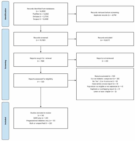
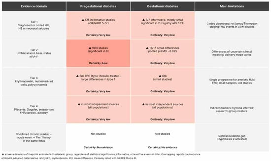
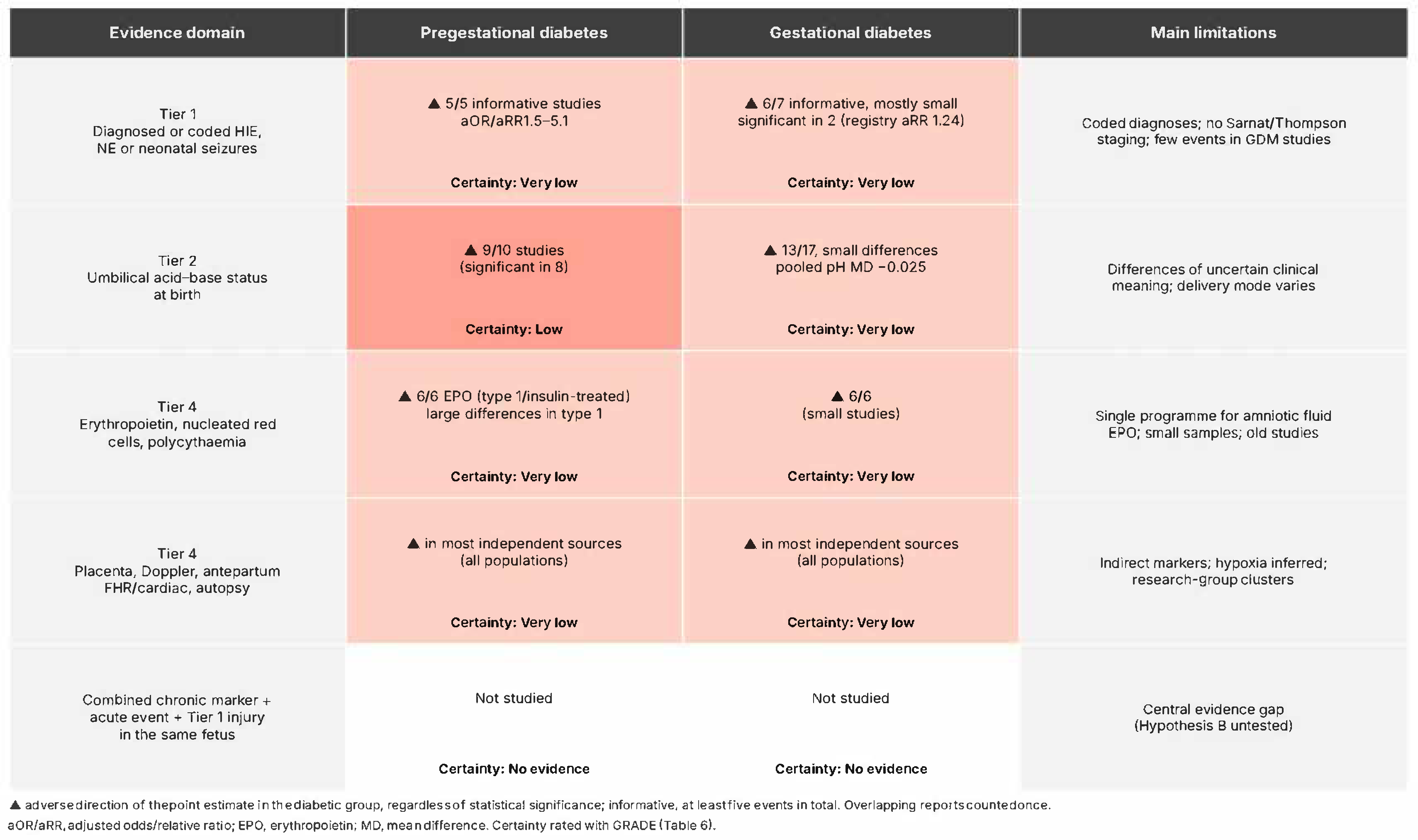

Disclaimer/Publisher’s Note: The statements, opinions and data contained in all publications are solely those of the individual author(s) and contributor(s) and not of MDPI and/or the editor(s). MDPI and/or the editor(s) disclaim responsibility for any injury to people or property resulting from any ideas, methods, instructions or products referred to in the content. |
© 2026 by the authors. Licensee MDPI, Basel, Switzerland. This article is an open access article distributed under the terms and conditions of the Creative Commons Attribution (CC BY) license.