


Abstract
Background: Sepsis-induced systemic inflammation triggers profound metabolic and inflammatory alterations in skeletal muscle, leading to oxidative damage, mitochondrial dysfunction, and muscle wasting. However, the mechanisms linking endotoxemia to skeletal muscle metabolic failure and neuromuscular-associated alterations remain incompletely understood. The present study investigated the effects of bicuculline methiodide (BMI), a GABAA receptor antagonist, on inflammatory responses, oxidative stress, energy metabolism, and selected neural-associated markers in a lipopolysaccharide (LPS)-induced rat model of sepsis. Methods: Adult male Sprague–Dawley rats were allocated into Control, LPS, Bicuculline, and LPS + Bicuculline groups. Twenty-four hours after treatment, gastrocnemius muscle tissues were analyzed for TNF-alpha, IL-10, GABA, and malondialdehyde (MDA) levels, while ATP-related metabolites were quantified by HPLC. NeuN and synaptophysin immunoreactivity were also assessed in muscle sections. Results: LPS administration increased TNF-alpha and MDA levels, altered IL-10 concentrations, and disrupted high-energy phosphate metabolism, accompanied by changes in NeuN and synaptophysin immunoreactivity. Bicuculline co-administration reduced MDA levels, was associated with higher IL-10 concentrations than the LPS group, and markedly increased phosphocreatine levels. Conclusions: These findings indicate that BMI was associated with attenuation of selected inflammatory and oxidative alterations and with changes in skeletal muscle energy metabolites in this experimental model. Because cholinergic markers, alpha7 nicotinic acetylcholine receptor activity, vagal activity, and receptor-specific pharmacological controls were not assessed, the present findings do not establish direct activation of the cholinergic anti-inflammatory pathway or a GABAergic-cholinergic mechanism. The observed effects should therefore be considered exploratory and hypothesis-generating.
1. Introduction
Sepsis is a life-threatening syndrome characterized by organ dysfunction caused by a dysregulated host response to infection, leading to widespread inflammation, multiple organ dysfunction, and, in severe cases, septic shock and death [1,2]. Despite advances in intensive care management, sepsis remains a major global health concern because of its high morbidity, mortality, and long-term functional sequelae [2,3]. The clinical presentation of sepsis is highly heterogeneous and is influenced by the source and severity of infection, the extent of organ dysfunction, the patient’s underlying health status, and the timing of therapeutic intervention [1,3].
Skeletal muscle represents approximately 40–50% of total body mass and is among the tissues most profoundly affected by systemic inflammatory conditions [4,5]. Sepsis-induced skeletal muscle dysfunction is a common and clinically significant complication associated with muscle wasting, weakness, prolonged hospitalization, impaired recovery, and intensive care unit-acquired weakness [4,5,6,7]. Although the pathophysiological mechanisms underlying septic myopathy have not been fully elucidated, accumulating evidence suggests that inflammatory mediators, mitochondrial dysfunction, proteolytic systems, and oxidative stress play central roles in this process [4,5,6,7,8,9]. Pro-inflammatory cytokines, reactive oxygen species (ROS), and cyclooxygenase-derived mediators activate catabolic pathways, including the ubiquitin-proteasome and autophagy-lysosome systems, resulting in accelerated protein degradation and skeletal muscle proteolysis [8,9,10,11]. Furthermore, sepsis-induced muscle injury is characterized by enhanced cytokine signaling, increased oxidative stress, activation of apoptotic pathways, and suppression of protein synthesis, collectively contributing to the loss of muscle mass and function [4,7,8,9,10,11].
Gamma-aminobutyric acid (GABA) is the principal inhibitory neurotransmitter in the central nervous system and exerts its biological effects primarily through GABAA and GABAB receptors [12,13]. Bicuculline methiodide (BMI), a GABAA receptor antagonist, has been widely employed as a pharmacological tool for investigating GABAergic signaling pathways [12,14]. Beyond its classical effects on neurotransmission, emerging evidence indicates that GABAergic signaling can influence peripheral immune-cell activity, cytokine production, and inflammatory responses [13,15,16,17]. These observations provide a biological rationale for examining whether pharmacological blockade of GABAA receptors is associated with inflammatory and metabolic changes during experimental sepsis; however, the downstream pathways responsible for such effects remain uncertain.
The cholinergic anti-inflammatory pathway (CAP) is a neural mechanism through which cholinergic signaling can modulate systemic inflammation [18,19,20]. The cholinergic system also has broader roles in neuronal regulation and neurodegenerative processes [21]. Activation of CAP can suppress production of pro-inflammatory cytokines and promote anti-inflammatory signaling, with the alpha-7 nicotinic acetylcholine receptor (α7nAChR) identified as an important mediator in several experimental models [19,20,22,23,24]. These findings provide a relevant neuroimmune framework for interpreting inflammatory responses in sepsis. Nevertheless, the present study did not directly measure acetylcholine, choline acetyltransferase, α7nAChR expression or activity, or vagal activity. Accordingly, CAP or α7nAChR involvement cannot be established from the present data.
Previous experimental studies have demonstrated that pharmacological or electrical manipulation of cholinergic pathways can attenuate systemic inflammation [18,20,22,23,24,25]. These observations support the broader concept that neural and immune pathways may interact during systemic inflammation, but they do not establish that bicuculline acts through the cholinergic anti-inflammatory pathway. In the present study, the term “neuroimmune modulation” is therefore used to describe a possible conceptual framework rather than a directly demonstrated mechanism. The 1.5 mg/kg bicuculline dose and 24-h endpoint were selected to maintain comparability with our previously published study using the same experimental model [26] and to examine the acute response. This exploratory design was not intended to determine dose–response relationships or temporal dynamics.
Therefore, the present study was designed to investigate the effects of bicuculline administration on inflammatory and oxidative stress responses, high-energy phosphate metabolism, and selected neural-associated immunoreactivity in gastrocnemius muscle tissue of rats subjected to LPS-induced sepsis. We hypothesized that bicuculline administration would be associated with attenuation of inflammatory and oxidative alterations and preservation of muscle energy metabolism. Any involvement of GABAergic-cholinergic or cholinergic anti-inflammatory signaling was considered a hypothesis to be evaluated indirectly rather than a mechanism directly tested in this study.
2. Materials and Methods
Ethical approval number 2017/63 was obtained from the Istanbul Bagcilar Training and Research Hospital Animal Experiments Local Ethics Committee. Adult male Sprague–Dawley rats with an average weight of 230 g were used in the study. The rats were obtained from the Experimental Research and Skill Development Center of Istanbul Bagcilar Training and Research Hospital. Standard rat food and water were provided ad libitum, and no dietary restrictions were imposed.
2.1. Experimental Groups
The rats were divided into 4 groups:
Control Group (C) (n = 8): No substance was administered to this group, and the rats were fed ad libitum.
LPS Group (LPS) (n = 8): Rats in this group were administered 10 mg/kg intraperitoneal LPS (E. coli serotype 055:B5, Sigma-Aldrich, St. Louis, MO, USA), dissolved in 1 mL of 0.9% NaCl solution. The animals were euthanized 24 h after the first injection.
Bicuculline Group (B/Bic) (n = 8): Rats in this group were administered 1.5 mg/kg subcutaneous bicuculline methiodide (Sigma-Aldrich), dissolved in 1 mL of 0.9% NaCl solution. The animals were euthanized 24 h after administration.
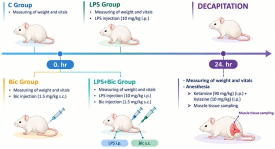
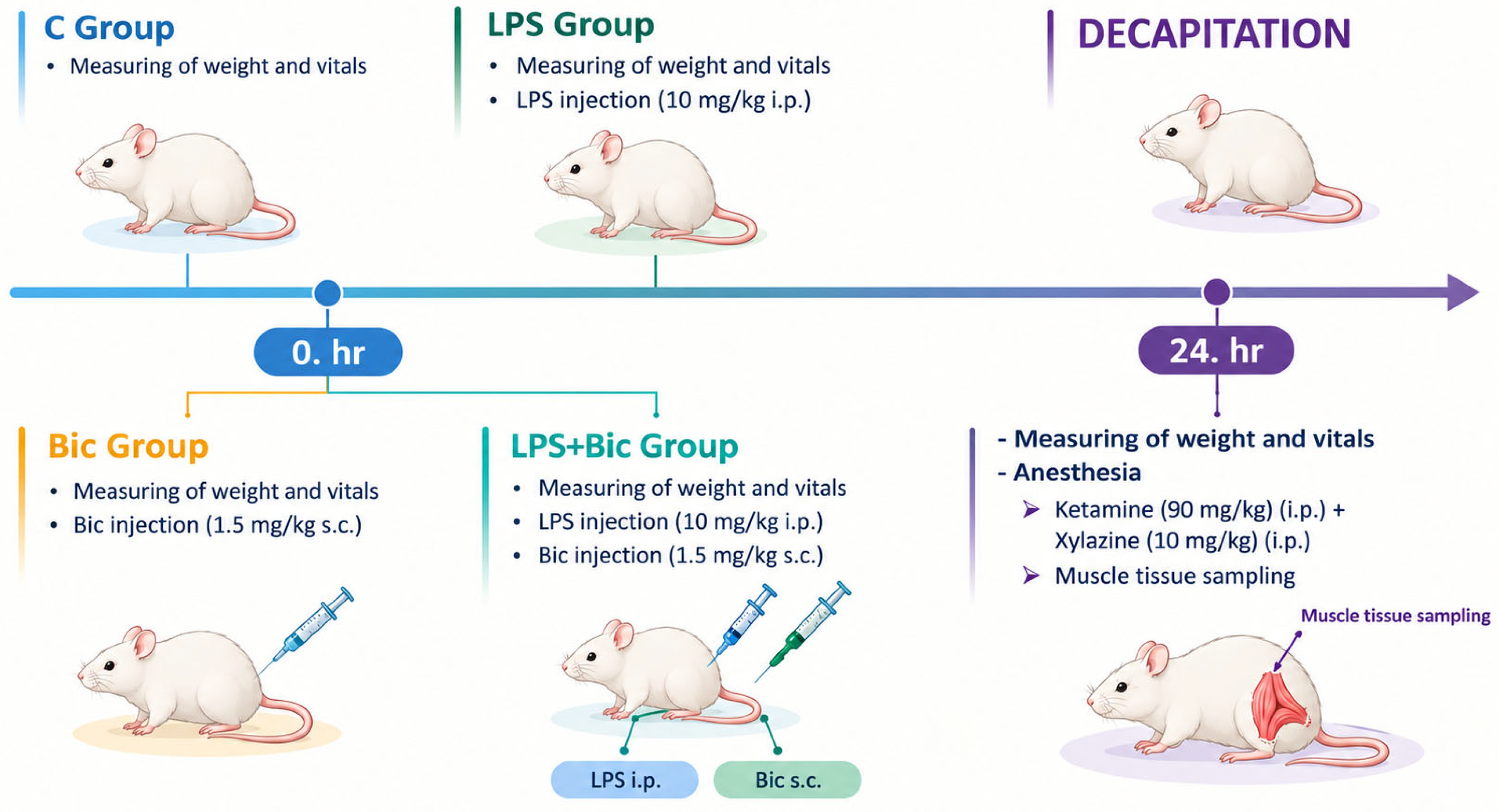
LPS + Bicuculline Group (LPS + B) (n = 8): Rats in this group received 10 mg/kg intraperitoneal LPS immediately followed by 1.5 mg/kg subcutaneous bicuculline methiodide. Figure 1.
Figure 1.
Diagram of experimental procedure.
2.2. Experimental Procedure
Bicuculline methiodide was administered concurrently with LPS at 1.5 mg/kg subcutaneously. Twenty-four hours after the LPS injection, the animals were anesthetized with ketamine (90 mg/kg, i.p.) and xylazine (10 mg/kg, i.p.). Body temperatures were measured rectally before injection and before euthanasia. Blood samples were collected from the heart for biochemical analyses, and gastrocnemius muscle tissue samples were collected for biochemical and histological analyses.
To further characterize the experimental model, rectal body temperature was measured before injections and again before the experimental procedure/euthanasia. Cardiac blood samples were evaluated for leukocyte count and selected biochemical parameters. The blood biochemical panel included AST, ALT, sodium, potassium, chloride, magnesium, calcium, phosphate, lactate dehydrogenase (LDH), creatine kinase (CK), and fibrinogen; the available values are presented in Table 1. Leukocyte-count findings from the same experimental cohort were reported in our previous publication [26].
Table 1.
Blood biochemical parameters in the experimental groups.
Gastrocnemius muscle tissue samples were homogenized to investigate TNF-α, IL-10, and GABA levels using enzyme-linked immunosorbent assay (ELISA), and thiobarbituric acid reactive substances (TBARS) levels were determined using the Buege method [27]. Whole-muscle tissue samples were placed in 10% formaldehyde for histopathological and immunofluorescence evaluations.
2.3. ELISA (Enzyme-Linked Immuno Sorbent Assay) Procedure
ELISA is a quantitative measurement method of an antigen–antibody interaction based on investigating the activity of an enzyme bound to an antibody.
The pro-inflammatory cytokine TNF-α levels were measured in muscle tissue using a rat-specific ELISA kit (Yh-biosearch, Shanghai, China).
The anti-inflammatory cytokine IL-10 levels were measured in muscle tissue using a rat-specific ELISA kit (Yh-biosearch, Shanghai, China).
GABA levels were measured in muscle tissue using a rat-specific ELISA kit (Yh-biosearch, Shanghai, China).
2.4. Malondialdehyde (MDA) Procedure
Lipid peroxidation was assessed by measuring TBARS levels. Tissue homogenates were reacted with thiobarbituric acid reagent (TBA, Sigma-Aldrich) containing trichloroacetic acid (TCA, Sigma, Kawasaki, Japan), concentrated hydrochloric acid (HCl, Merck, Darmstadt, Germany), and butylated hydroxytoluene (Sigma), boiled for 15 min, and then cooled and centrifuged. TBARS levels were measured in the supernatant fraction using spectrophotometry (Shimadzu, Kyoto, Japan) at 532 nm. TBARS concentrations were expressed as nmol/g tissue [27].
2.5. High-Performance Liquid Chromatography (HPLC) Analysis of Creatine, Creatine Phosphate, AMP, ADP, and ATP
Muscle tissue samples (200 mg) were homogenized in 2 mL of 0.42 M perchloric acid (HClO4) using an Ultra-Turrax (IKA, Staufen, Germany) T25 homogenizer for 30 s. The homogenates were centrifuged at 3000 rpm for 5 min, and the resulting supernatants were neutralized with 1.0 M K2HPO4 prior to chromatographic analysis.
The concentrations of creatine, creatine phosphate, AMP, ADP, and ATP were determined by ion-pair reverse-phase HPLC using an Agilent 1100 system equipped with a C18 column (250 × 4.6 mm, 5 μm; Nucleodur (Macherey-Nagel, Düren, Germany). Separation was achieved under isocratic conditions with a mobile phase containing 215 mM KOH/KH2PO4 buffer (pH 6.25), 3 mM tetrabutylammonium phosphate, and 5% acetonitrile. Eluted compounds were monitored at 214 nm, and their concentrations were quantified by comparison with external calibration curves generated from authentic standards, as previously described by Sellevold et al. [28].
2.6. Immunofluorescence Analysis
Muscle tissues were fixed in 10% neutral-buffered formaldehyde overnight, dehydrated through graded ethanol, cleared in xylene, infiltrated with paraffin at 58 °C, embedded, and sectioned at 10 μm using a microtome. Sections were mounted on poly-L-lysine-coated slides. Immunofluorescence staining was performed to assess NeuN and synaptophysin (SYP) immunoreactivity. S100B was included in the original staining panel as an exploratory marker; however, quantitative S100B results were not available for analysis and are therefore not presented or interpreted in this manuscript. NeuN was treated as an exploratory neural-associated marker in muscle sections and was not interpreted as evidence of neuronal cell bodies within skeletal muscle. SYP was assessed as a presynaptic-associated marker. Primary antibodies included NeuN (orb48522, Biorbyt; Durham, NC, USA, Entrez: 146,713; rabbit) and SYP (orb69251, Biorbyt; Entrez: 6855; mouse). DyLight 649-conjugated goat anti-mouse IgG (H&L) (Abbkine, Wuhan, China, #A23610) and FITC-conjugated anti-rabbit secondary antibodies were used for fluorescence detection. Following deparaffinization and rehydration, antigen retrieval was performed in 1× citrate buffer (pH 6.0) by microwave heating for 20 min. Sections were permeabilized with 0.2% Tween-20, washed with PBS (pH 7.6), and incubated overnight at 4 °C with the primary antibodies. NeuN was diluted in PBS containing 2% serum, whereas SYP was diluted in PBS containing 0.3% Triton X-100/0.3% serum and 10% goat serum, respectively. After PBS washes, the appropriate secondary antibodies were applied for 2 h at room temperature. Nuclei were counterstained with DAPI, and the sections were mounted with an antifade mounting medium before fluorescence microscopy.
2.7. Statistical Analysis
Statistical analysis was performed using SPSS 25.0 (SPSS, Inc., Chicago, IL, USA); ANOVA outputs and degrees of freedom were checked in GraphPad Prism v8. Parametric data were evaluated using one-way analysis of variance (ANOVA), followed by Tukey’s multiple-comparison test. Data are presented as mean ± standard deviation (SD). The planned sample size was n = 8 animals per group; however, the number of observations included in individual tissue analyses varied by outcome because potentially unreliable observations identified during median-based review were excluded from the relevant parameter-specific analyses. Consequently, the residual degrees of freedom and total analyzed sample size differed across outcomes and are reported with each analysis. Statistical significance was accepted at p < 0.05.
3. Results
3.1. Findings of Septic Conditions
In the LPS group, animals were either hypothermic (34.7 °C ± 0.4) or hyperthermic (38.8 °C ± 0.7), consistent with an acute systemic response in this model. Body temperatures in the control, bicuculline, and LPS + Bic groups were within standard values. Selected blood biochemical findings are summarized in Table 1. Table S1 shows the biochemical parameter findings for all groups. Compared with the other groups, the LPS group showed higher mean LDH and fibrinogen values, while AST and ALT values are presented descriptively. The leukocyte-count analysis from the same experimental cohort was reported previously; leukocyte counts were significantly lower in the LPS group than in the control, bicuculline, and LPS + bicuculline groups [26].
The study cited as reference [26] used the same animals and experimental model, with the previous publication focusing on brain-related outcomes. The current manuscript presents skeletal-muscle findings together with the available blood biochemical values to provide additional, but not comprehensive, characterization of the model. These measures do not replace a full systemic assessment of sepsis severity, including circulating inflammatory cytokines and lactate.
3.2. Cytokine Findings
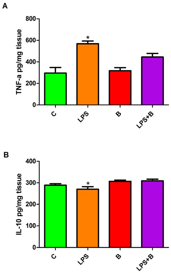
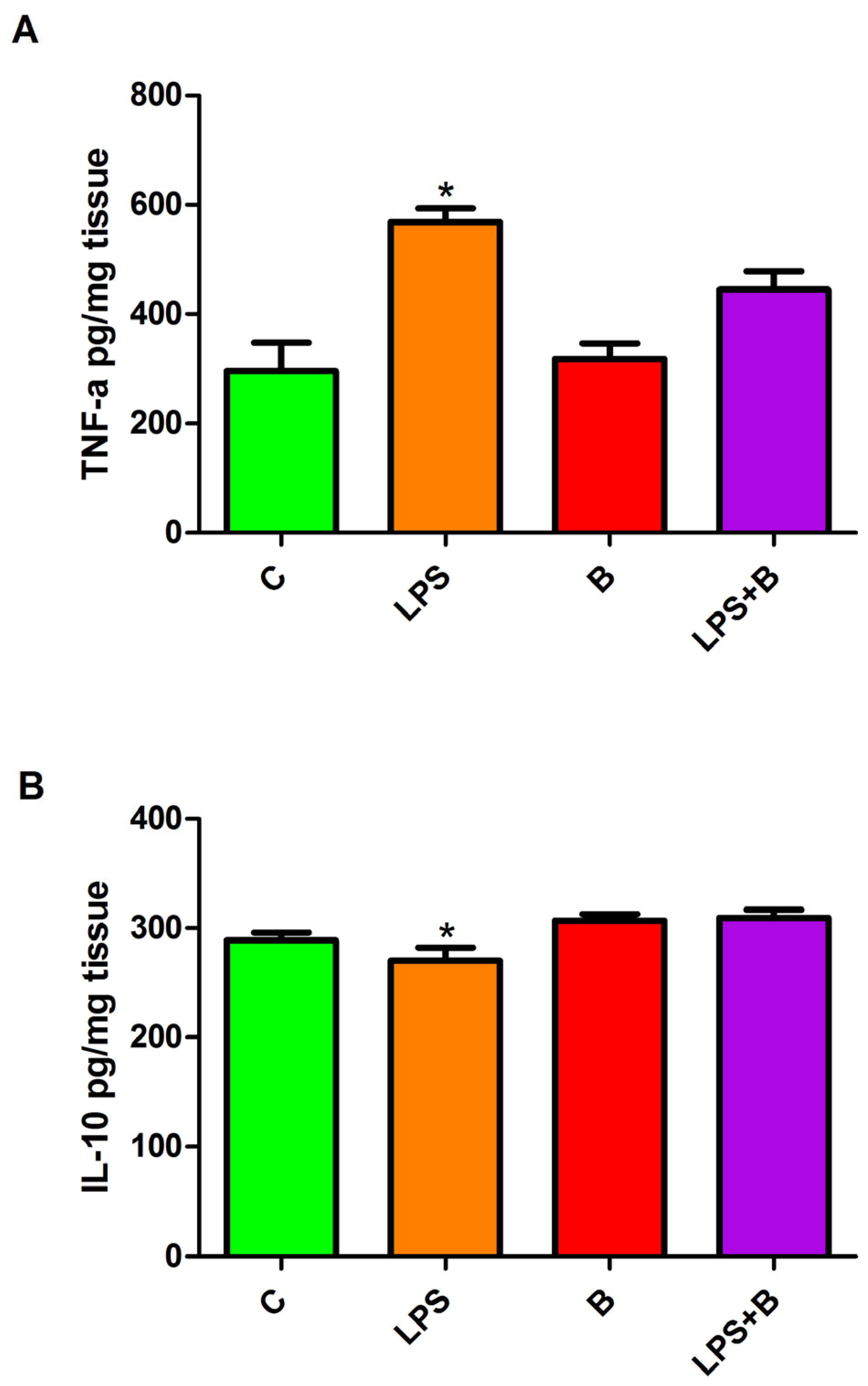
A significant overall difference was detected in tissue TNF-α levels among the experimental groups (F(3,23) = 15.64, p < 0.0001). Tukey’s post hoc analysis demonstrated that the LPS group exhibited significantly higher TNF-α concentrations than the control and bicuculline groups (both p < 0.001). TNF-α levels in the LPS + B group remained significantly elevated compared with the control group (p < 0.05). Importantly, no significant difference was observed between the LPS and LPS + B groups, indicating that bicuculline co-administration did not statistically normalize TNF-α concentrations relative to LPS alone. No significant differences were observed between the bicuculline and LPS + B groups or between the control and bicuculline groups (Figure 2A).
Figure 2.
(A,B): TNF-α and IL-10 levels in gastrocnemius muscle tissue. Data are presented as mean ± SD. The number of observations included varied by outcome (TNF-α: total n = 27; IL-10: total n = 28) after parameter-specific exclusion of potentially unreliable observations. One-way ANOVA followed by Tukey’s multiple-comparison test was used. * p < 0.05; for the indicated comparisons.
Analysis of IL-10 levels also revealed a significant effect of treatment (F(3,24) = 4.701, p = 0.0101). According to Tukey’s multiple-comparison test, IL-10 concentrations in the LPS group were significantly lower than those in both the bicuculline and LPS + B groups (both p < 0.05). No statistically significant differences were identified between the control and LPS groups, control and bicuculline groups, control and LPS + B groups, or bicuculline and LPS + B groups (Figure 2B). Thus, the principal cytokine finding associated with bicuculline was the increase in IL-10 relative to the LPS group rather than a statistically demonstrated normalization of TNF-α.
3.3. GABA ELISA Findings
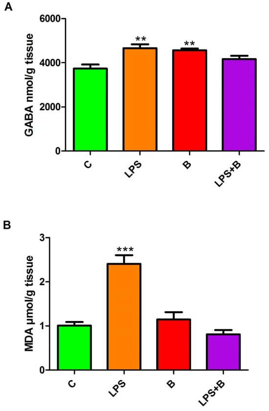
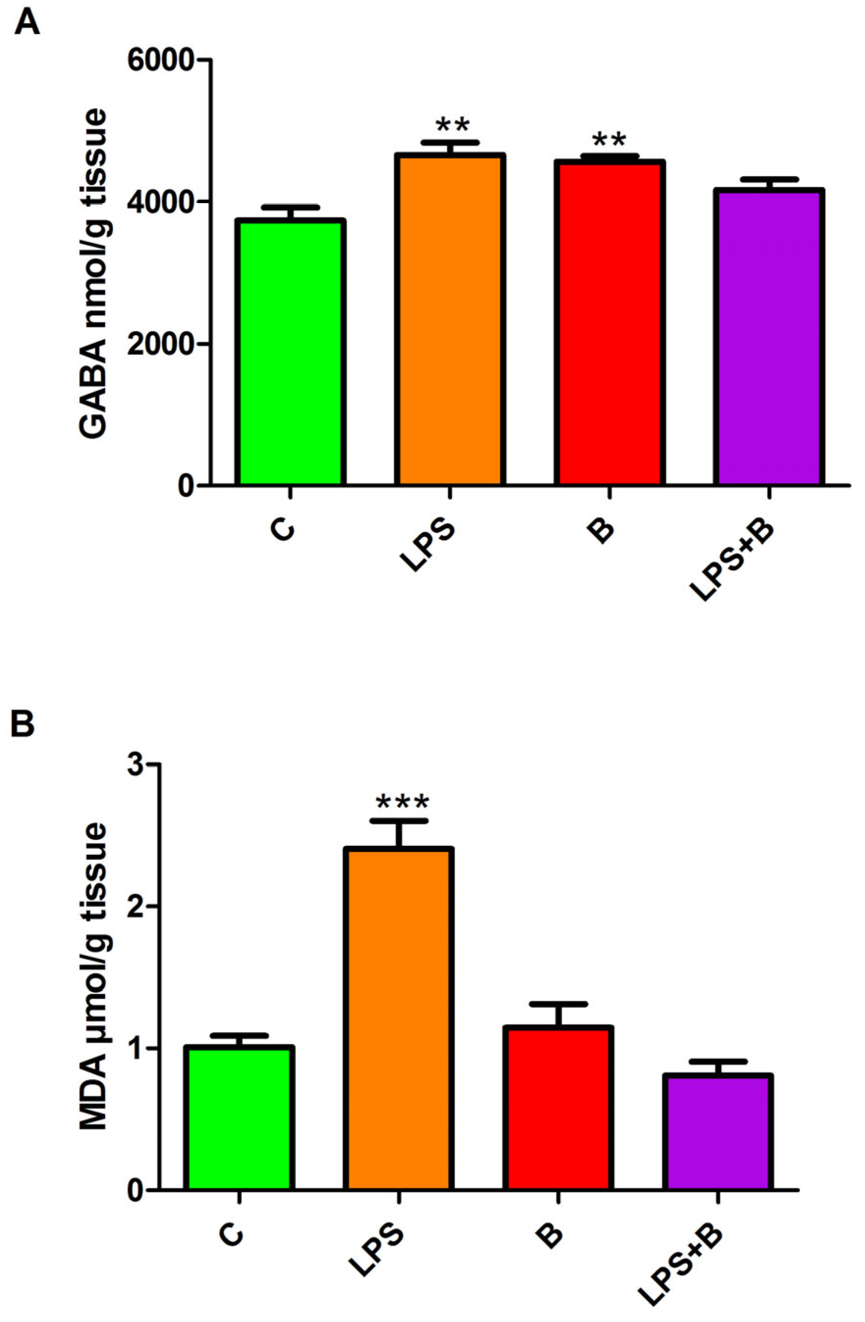
One-way ANOVA demonstrated significant differences in tissue GABA levels among the experimental groups (F(3,16) = 6.103, p = 0.0057). Post hoc comparisons revealed that both the LPS and bicuculline groups displayed significantly higher GABA concentrations than the control group (p < 0.01 and p < 0.05, respectively). In contrast, GABA levels in the LPS + B group did not differ significantly from those in the control, LPS, or bicuculline groups. Likewise, no significant difference was observed between the LPS and bicuculline groups (Figure 3A). The increase in GABA in the bicuculline group was not accompanied by a corresponding reduction in GABA in the LPS + B group. Therefore, the present data do not establish a simple inverse relationship between GABAA receptor blockade and tissue GABA concentration. The observed changes may reflect altered local GABA production, release, uptake, metabolism, or compensatory responses; these possibilities were not directly tested.
Figure 3.
(A,B): GABA and MDA levels in gastrocnemius muscle tissue. Data are presented as mean ± SD; the total analyzed sample size was n = 24 for each outcome after parameter-specific exclusion of potentially unreliable observations. One-way ANOVA followed by Tukey’s multiple-comparison test was used. ** p < 0.01; *** p < 0.001 for the indicated comparisons.
3.4. Lipid Peroxidation Findings
Tissue MDA levels differed significantly among the experimental groups (F(3,16) = 25.18, p < 0.0001). Tukey’s multiple-comparison analysis showed that the LPS group had significantly higher MDA concentrations than both the control and bicuculline groups (both p < 0.001). Moreover, MDA levels were significantly reduced in the LPS + B group compared with the LPS group (p < 0.001). No significant differences were detected between the control and bicuculline groups, the control and LPS + B groups, or the bicuculline and LPS + B groups (Figure 3B).
3.5. Findings of High Energy Compounds, Creatine and Creatine Phosphate
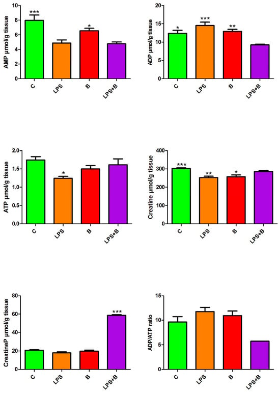
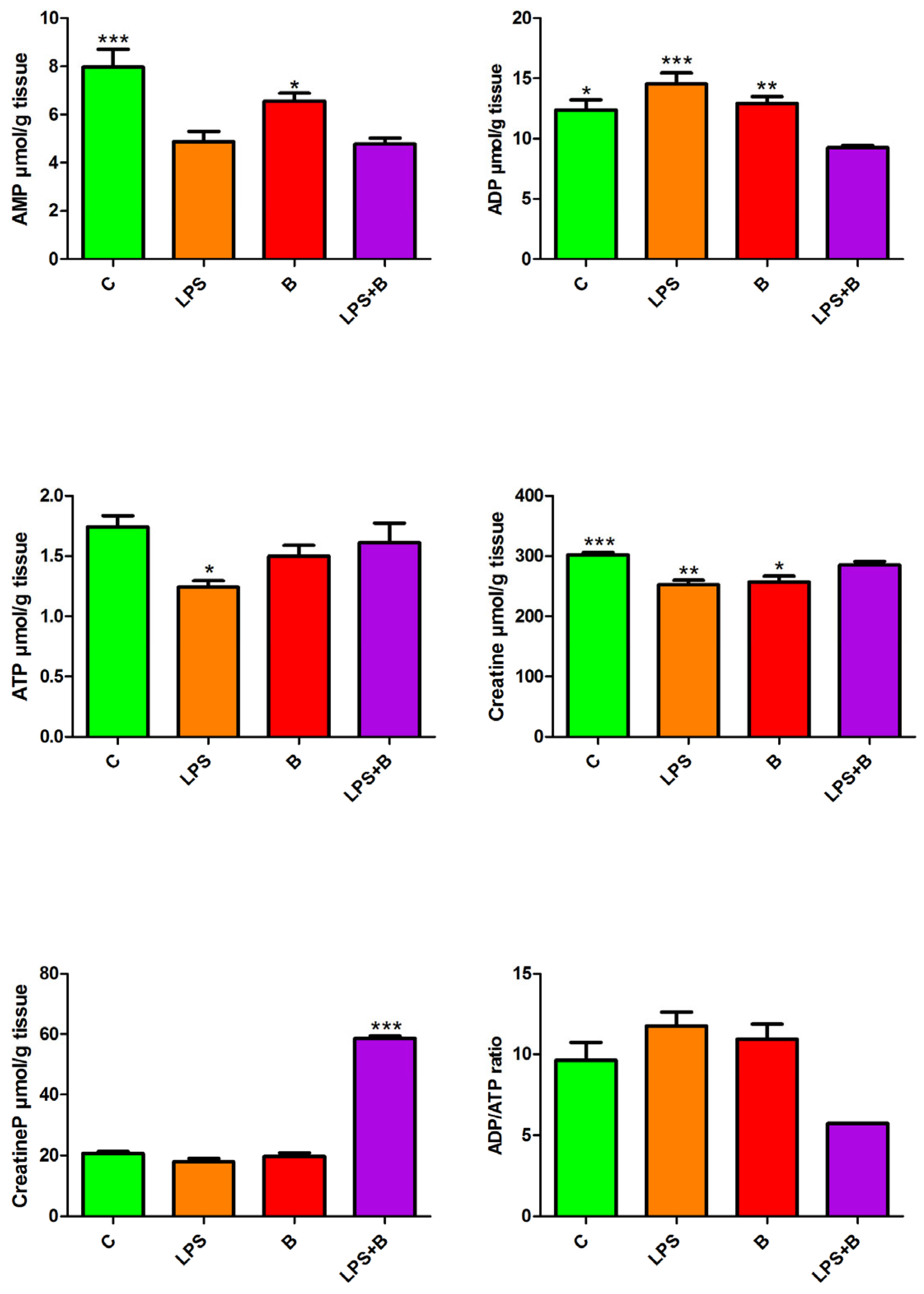
HPLC analysis of muscle tissues revealed significant alterations in energy metabolites among the experimental groups. LPS administration led to a significant decrease in ATP levels compared with the control group (p < 0.05). Conversely, ADP levels were significantly elevated in the LPS group compared with the control (p < 0.05), bicuculline, and LPS + B groups. Creatine levels in the LPS group also differed significantly from the control (p < 0.01) and bicuculline groups. Regarding AMP levels, the control group exhibited the highest concentrations (p < 0.001), followed by a significant increase in the bicuculline-alone group compared with the LPS and LPS + B groups (p < 0.05). Notably, phosphocreatine (CreatineP) levels remained relatively stable across the control, LPS, and bicuculline groups but demonstrated a marked increase in the LPS + B group (p < 0.001) compared with all other groups (Figure 4). The marked increase in phosphocreatine occurred without a statistically demonstrated parallel restoration of ATP. Thus, the phosphocreatine finding should not be interpreted as direct evidence of improved mitochondrial ATP production. An alternative interpretation is that phosphocreatine accumulated because its utilization or transfer of high-energy phosphate to ATP was altered under the combined treatment condition. The present measurements cannot distinguish between increased phosphocreatine synthesis, reduced utilization, or altered creatine kinase flux.
Figure 4.
AMP, ADP, ATP, creatine, phosphocreatine (CreatineP), and ADP/ATP ratio levels in gastrocnemius muscle tissue were measured by HPLC in all experimental groups. Data are presented as mean ± SD (n = 8/group). One-way ANOVA followed by Tukey’s multiple-comparison test was used. * p < 0.05; ** p < 0.01; *** p < 0.001 for the indicated comparisons.
Assessment of Neurosynaptic Integrity via NeuN and Synaptophysin Expression:
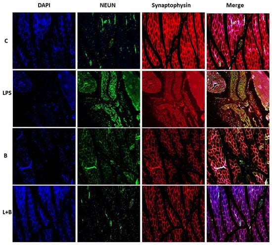
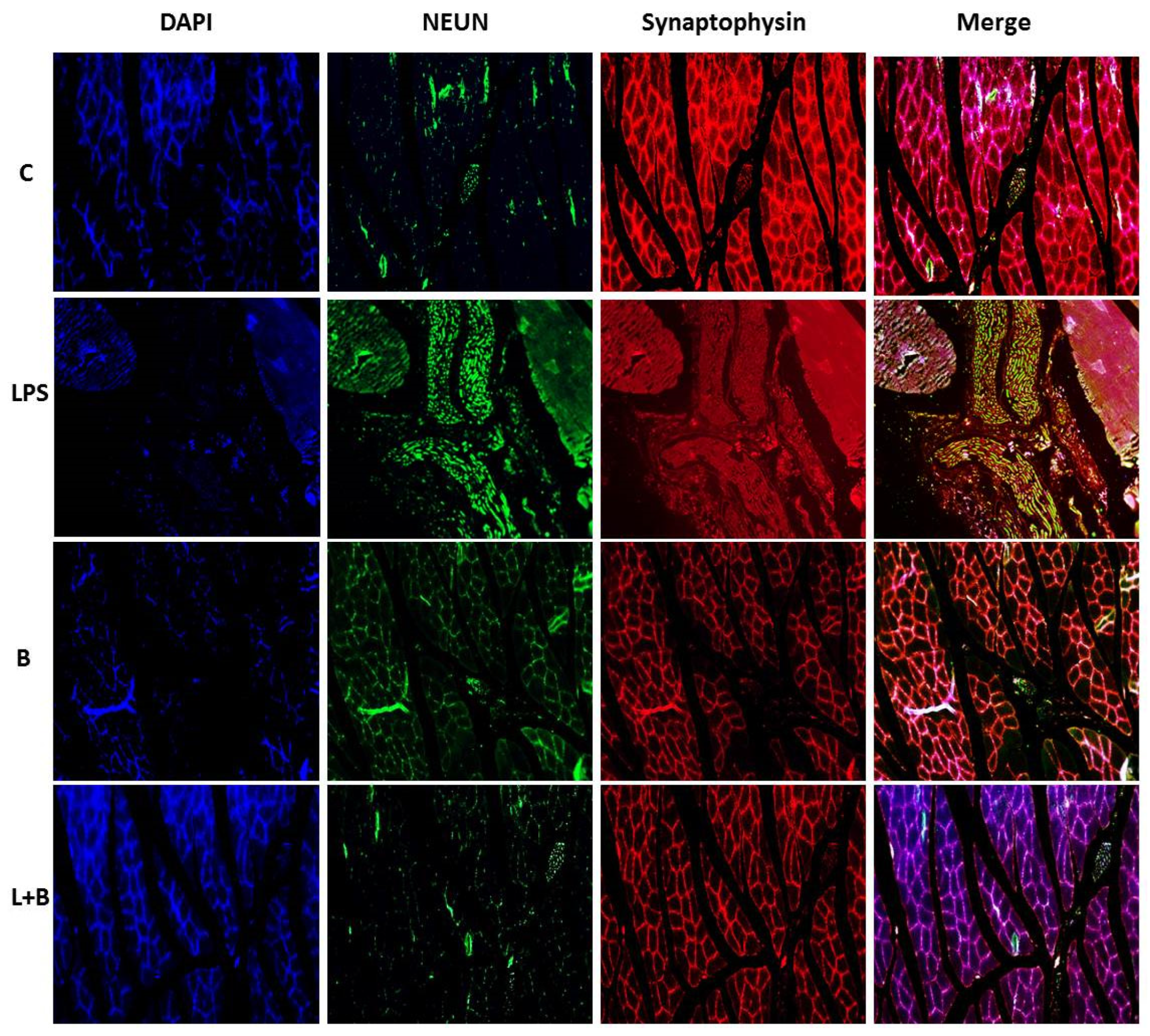
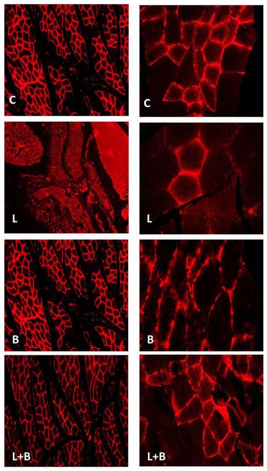
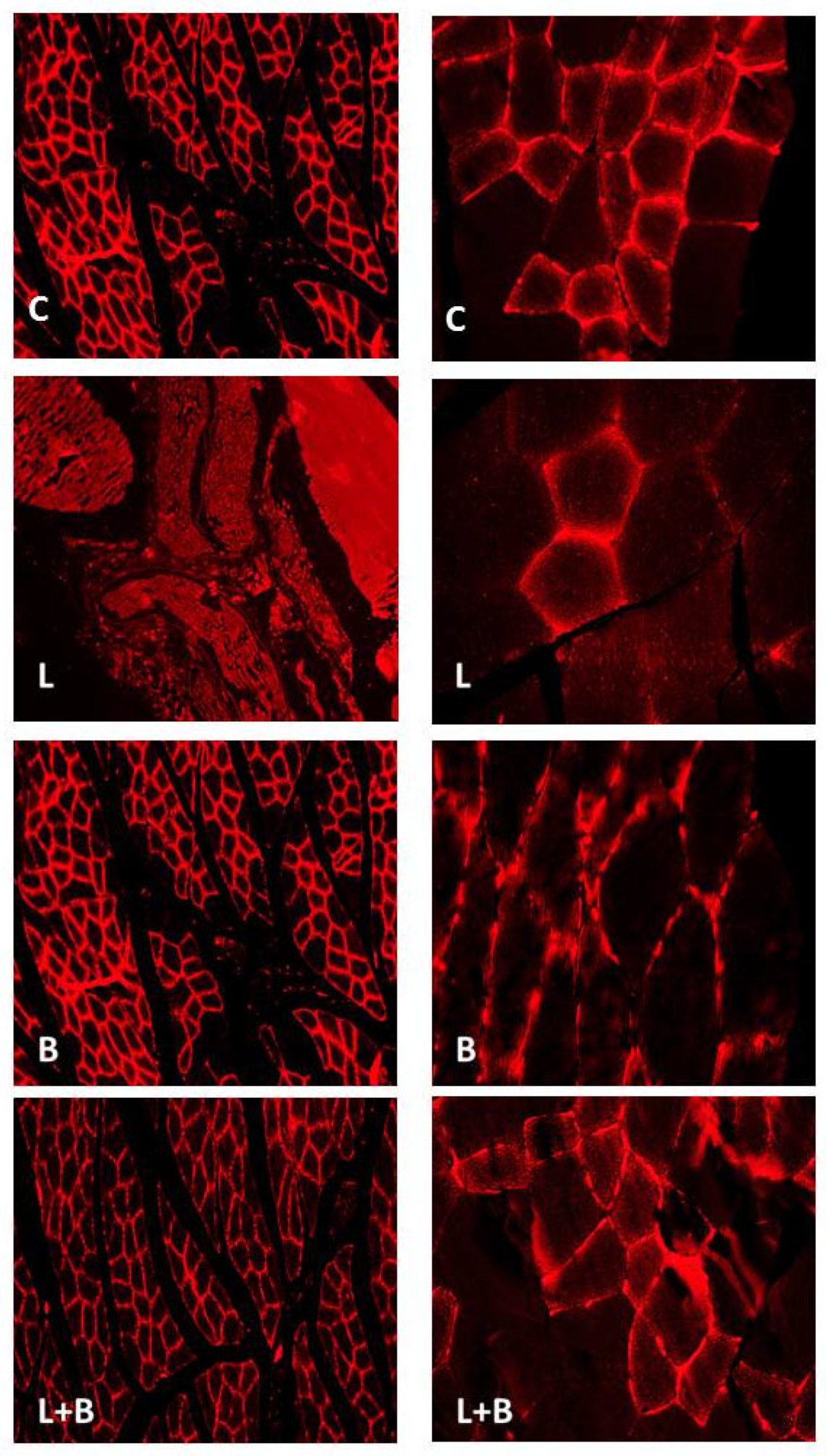
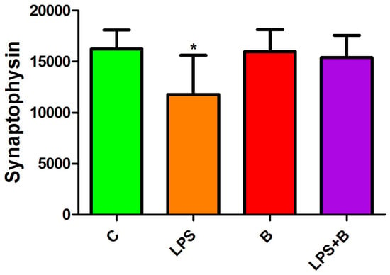
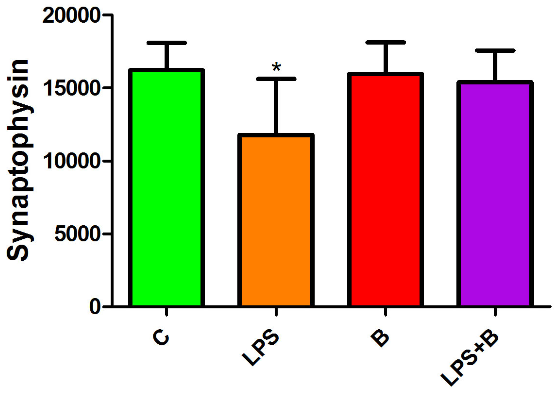
Immunofluorescence imaging and subsequent densitometric analysis demonstrated changes in neural-associated immunoreactivity in the muscle sections. LPS exposure was associated with a significant reduction in NeuN signal and synaptophysin immunoreactivity compared with the control and bicuculline groups (p < 0.05). In the LPS + B group, NeuN immunoreactivity was preserved relative to the LPS group, and synaptophysin immunoreactivity showed an increase relative to the LPS group. These findings indicate alterations in neural-associated markers within the muscle sections. However, because NeuN is primarily a neuronal nuclear marker and skeletal muscle does not contain neuronal cell bodies, NeuN staining in muscle should not be interpreted as direct evidence of neuronal survival or neuromuscular-junction integrity. Similarly, synaptophysin immunoreactivity alone cannot establish functional neuromuscular transmission. These results are therefore considered exploratory structural observations and require confirmation using markers such as neurofilament or neuromuscular-junction-specific markers in future studies. (Figure 5, Figure 6 and Figure 7).
Figure 5.
Muscle tissue sections were stained using DAPI, NeuN, and synaptophysin antibodies at ×10 magnification. (C) Control, LPS, (B) Bicuculline, and (L + B) LPS + Bicuculline groups.
Figure 6.
Muscle tissue sections were stained with synaptophysin antibody at ×40 magnification. (C) Control, (L) LPS, (B) Bicuculline, and (L + B) LPS + Bicuculline groups.
Figure 7.
H-scores of synaptophysin immunoreactivity in gastrocnemius muscle tissue sections from the experimental groups. (C) Control, LPS, (B) Bicuculline, and (LPS + B) LPS + Bicuculline groups. Data are presented as mean ± SD (n = 8/group). One-way ANOVA followed by Tukey’s multiple-comparison test was used. Statistical annotations indicate the pairwise comparisons shown in the figure. * p < 0.05.
4. Discussion
The present study demonstrates that bicuculline methiodide was associated with attenuation of selected inflammatory and oxidative alterations, changes in high-energy phosphate metabolism, and changes in neural-associated immunoreactivity in skeletal muscle during LPS-induced experimental sepsis. LPS exposure increased TNF-α and lipid peroxidation and altered IL-10 and energy-metabolite profiles, while bicuculline co-administration was associated particularly with lower MDA, higher IL-10 relative to the LPS group, and a marked increase in phosphocreatine. Because direct measurements of cholinergic signaling and α7nAChR activity were not performed, these findings should not be interpreted as proof that bicuculline acts through the cholinergic anti-inflammatory pathway.
Sepsis is increasingly recognized as a disorder involving coordinated inflammatory, neuroendocrine, and immunometabolic responses rather than a simple cytokine-mediated syndrome [2,3,18,19,20]. Excessive activation of innate immune pathways can contribute to reactive oxygen species generation, mitochondrial dysfunction, metabolic disturbances, and skeletal muscle wasting [4,5,6,7,8,9,10,11]. In the present model, LPS increased TNF-α and MDA and altered high-energy phosphate metabolites, consistent with a metabolically and oxidatively stressed muscle environment. The observed changes support the presence of muscle-associated metabolic consequences of endotoxemia but do not by themselves establish a causal sequence between inflammation, mitochondrial dysfunction, and muscle weakness.
The inflammatory findings require careful interpretation. Bicuculline co-administration was associated with higher IL-10 concentrations than the LPS group, whereas TNF-α remained significantly higher in the LPS + B group than in the control group and did not differ significantly from the LPS group. Thus, the present data support a selective change in the inflammatory profile rather than complete normalization of inflammation. Although GABAergic signaling can influence immune-cell activity [13,15,16], the present study did not measure acetylcholine, choline acetyltransferase, α7nAChR, vagal activity, or downstream CAP signaling. The possibility that bicuculline-related effects involve neuroimmune pathways therefore remains a hypothesis rather than a demonstrated mechanism.
Oxidative stress represents another important component of sepsis-associated muscle injury [4,7,8,9,10,11]. Increased lipid peroxidation can damage membrane components and impair cellular metabolism. Consistent with this concept, LPS administration significantly elevated MDA levels in skeletal muscle, whereas bicuculline co-administration significantly reduced MDA compared with LPS alone. This finding indicates an association between bicuculline treatment and reduced lipid peroxidation. However, MDA/TBARS measurement alone cannot determine whether bicuculline acts as a direct antioxidant, indirectly reduces oxidative stress through altered inflammatory signaling, or affects another upstream process. A direct antioxidant action is therefore an alternative explanation that should be considered alongside neuroimmune hypotheses.
An important aspect of this study was the evaluation of high-energy phosphate metabolites together with inflammatory biomarkers. Sepsis significantly reduced ATP levels while increasing ADP concentrations, consistent with altered cellular energy balance [7,29,30,31]. Interestingly, phosphocreatine increased markedly in the LPS + Bicuculline group without a statistically demonstrated parallel restoration of ATP. Phosphocreatine functions as an intracellular high-energy phosphate buffer, but its accumulation does not necessarily indicate improved ATP production. One possible explanation is altered utilization or transfer of phosphocreatine through the creatine kinase system, resulting in accumulation despite persistent limitations in ATP availability. Because creatine kinase activity, mitochondrial respiration, and phosphocreatine flux were not measured, the mechanism underlying this accumulation cannot be determined from the present data [32].
Changes in NeuN and synaptophysin immunoreactivity were also observed in the muscle sections. Synaptophysin is a presynaptic vesicle-associated protein, whereas NeuN is primarily a neuronal nuclear marker [33,34]. The use of NeuN in skeletal muscle therefore requires caution because skeletal muscle does not contain neuronal cell bodies. In the present study, NeuN immunoreactivity is reported as an exploratory neural-associated staining pattern rather than evidence of neuronal survival. Likewise, synaptophysin staining alone cannot establish neuromuscular-junction integrity or functional transmission. The apparent preservation of these signals after bicuculline treatment is hypothesis-generating and should be tested in future studies using neurofilament and neuromuscular-junction-specific markers.
Changes in tissue GABA concentrations further indicate that GABAergic signaling is altered in this experimental model. Both LPS and bicuculline alone were associated with higher tissue GABA concentrations than control animals, whereas the LPS + B group did not differ significantly from the other groups. Importantly, these measurements do not demonstrate that bicuculline lowers GABA signaling. Tissue GABA concentration is determined by multiple processes, including synthesis, release, uptake, metabolism, and compartmentalization. The present study did not assess any of these processes directly. Therefore, the GABA findings should be interpreted as evidence of altered tissue GABA homeostasis rather than as proof of a specific receptor-mediated mechanism.
Collectively, the present findings suggest that bicuculline administration is associated with attenuation of selected inflammatory and oxidative changes and with alterations in skeletal muscle energy metabolism during experimental sepsis. The data are compatible with a potential role for GABAergic and broader neuroimmune interactions, but they do not establish direct GABAergic-cholinergic coupling or activation of the cholinergic anti-inflammatory pathway. Alternative mechanisms, including direct or indirect effects on oxidative stress and cellular metabolism, remain possible. The present study should therefore be viewed as an exploratory investigation generating mechanistic hypotheses for future receptor-specific and functional studies.
Several limitations should be acknowledged. First, the study used a relatively small sample size, a single bicuculline dose, and a single 24-h experimental time point. These parameters were selected to align with our previous study using the same model [26], but the present design cannot establish dose–response relationships, toxicity across doses, or temporal dynamics. Second, systemic sepsis characterization remained incomplete: leukocyte-count findings from the same experimental cohort were reported previously [26], and selected blood biochemical values are now presented in Table 1; however, circulating inflammatory cytokines and lactate were not measured, and the available biochemical findings do not provide a comprehensive assessment of sepsis severity. Third, no direct measurements of acetylcholine, choline acetyltransferase, α7nAChR expression or activity, or vagal activity were performed, and no receptor-specific agonist/antagonist experiments were conducted. Consequently, a direct role for the cholinergic anti-inflammatory pathway cannot be established. Fourth, the marked phosphocreatine increase was not accompanied by a demonstrated ATP recovery, and mitochondrial respiration or creatine kinase flux was not measured. Fifth, NeuN is not an ideal marker for intramuscular neural structures, and synaptophysin alone does not establish neuromuscular-junction function. S100B staining was included in the original staining panel but was not quantitatively analyzed in the present dataset and therefore was not interpreted. Finally, an experimental GABA agonist arm would provide an important complementary test of receptor-directionality; this was not included in the present study. Future studies incorporating dose–response and time-course designs, systemic severity markers, receptor-specific pharmacology, cholinergic measurements, mitochondrial functional assays, and validated neuromuscular markers are warranted.
5. Conclusions
The present study demonstrates that bicuculline methiodide administration is associated with attenuation of selected inflammatory and oxidative alterations and with changes in high-energy phosphate metabolism in skeletal muscle during LPS-induced experimental sepsis. Bicuculline co-administration reduced MDA levels, was associated with higher IL-10 concentrations than the LPS group, and markedly increased phosphocreatine levels. Changes in NeuN and synaptophysin immunoreactivity were also observed, although these markers require cautious interpretation in skeletal muscle.
These findings indicate that septic skeletal muscle injury involves concurrent inflammatory, oxidative, and metabolic disturbances. The present results are compatible with the possibility that modulation of GABAergic or other neuroimmune processes contributes to these changes; however, direct involvement of cholinergic signaling, α7nAChR, vagal activity, or the cholinergic anti-inflammatory pathway was not demonstrated.
Although the precise mechanisms require further investigation, the present data provide a basis for future studies examining whether GABAergic manipulation influences skeletal muscle injury during sepsis. Studies using receptor-specific agonists and antagonists, direct measurements of cholinergic signaling, validated neuromuscular-junction markers, systemic sepsis severity measures, and mitochondrial functional assays will be important for determining the mechanisms and translational relevance of the observed associations.
Supplementary Materials
The following supporting information can be downloaded at https://www.mdpi.com/article/10.3390/biomedicines14112490/s1, Table S1: The biochemical parameter findings for all groups.
Author Contributions
Conceptualization, S.T. and. G.A.; methodology, S.T., G.A., E.O. and G.I.G.; investigation, G.A., E.O., G.I.G. and S.T.; writing—original draft preparation, G.A.; writing—review and editing, G.A., S.T., G.I.G. and E.O. All authors have read and agreed to the published version of the manuscript.
Funding
This publication was supported by the Scientific Research Projects Coordination Unit of Istanbul Yeni Yuzyil University.
Institutional Review Board Statement
The animal study protocol was approved by the Local Ethics Committee of Animal Experiments of Istanbul Bagcilar Training and Research Hospital (protocol code 63 and date of approval 25 April 2017).
Informed Consent Statement
Not applicable.
Data Availability Statement
The data supporting the findings of this study are available from the corresponding author upon reasonable request.
Conflicts of Interest
The authors declare no conflicts of interest.
References
- Singer, M.; Deutschman, C.S.; Seymour, C.W.; Shankar-Hari, M.; Annane, D.; Bauer, M.; Bellomo, R.; Bernard, G.R.; Chiche, J.-D.; Coopersmith, C.M.; et al. The Third International Consensus Definitions for Sepsis and Septic Shock (Sepsis-3). JAMA 2016, 315, 801–810. [Google Scholar] [CrossRef] [Scilit] [PubMed]
- Hotchkiss, R.S.; Moldawer, L.L.; Opal, S.M.; Reinhart, K.; Turnbull, I.R.; Vincent, J.L. Sepsis and septic shock. Nat. Rev. Dis. Primers 2016, 2, 16045. [Google Scholar] [CrossRef] [Scilit] [PubMed]
- Cohen, J.; Vincent, J.L.; Adhikari, N.K.J.; Machado, F.R.; Angus, D.C.; Calandra, T.; Jaton, K.; Giulieri, S.; Delaloye, J.; Opal, S.; et al. Sepsis: A roadmap for future research. Lancet Infect. Dis. 2015, 15, 581–614. [Google Scholar] [CrossRef] [Scilit] [PubMed]
- Callahan, L.A.; Supinski, G.S. Sepsis-induced myopathy. Crit. Care Med. 2009, 37, S354–S367. [Google Scholar] [CrossRef] [Scilit] [PubMed]
- Friedrich, O.; Reid, M.B.; Van den Berghe, G.; Vanhorebeek, I.; Hermans, G.; Rich, M.M.; Larsson, L. The sick and the weak: Neuropathies/myopathies in the critically ill. Physiol. Rev. 2015, 95, 1025–1109. [Google Scholar] [CrossRef] [Scilit] [PubMed]
- Schefold, J.C.; Bierbrauer, J.; Weber-Carstens, S. Intensive care unit-acquired weakness (ICUAW) and muscle wasting in critically ill patients with severe sepsis and septic shock. J. Cachexia Sarcopenia Muscle 2010, 1, 147–157. [Google Scholar] [CrossRef] [Scilit] [PubMed]
- Witteveen, E.; Sommers, J.; Wieske, L.; Doorduin, J.; van Alfen, N.; Schultz, M.J.; van Schaik, I.N.; Horn, J.; Verhamme, C. Diagnostic accuracy of quantitative neuromuscular ultrasound for the diagnosis of intensive care unit-acquired weakness: A cross-sectional observational study. Ann. Intensive Care 2017, 7, 40. [Google Scholar] [CrossRef] [Scilit] [PubMed]
- Yoshihara, I.; Kondo, Y.; Ishihara, T.; Hifumi, T. Sepsis-associated muscle wasting: A comprehensive review from bench to bedside. Int. J. Mol. Sci. 2023, 24, 5040. [Google Scholar] [CrossRef] [Scilit] [PubMed]
- Haberecht-Mueller, S.; Krueger, E.; Fielitz, J. Out of control: The role of the ubiquitin proteasome system in skeletal muscle during inflammation. Biomolecules 2021, 11, 1327. [Google Scholar] [CrossRef] [Scilit] [PubMed]
- Supinski, G.S.; Callahan, L.A. Free radical-mediated skeletal muscle dysfunction in inflammatory conditions. J. Appl. Physiol. 2007, 102, 2056–2063. [Google Scholar] [CrossRef] [Scilit] [PubMed]
- Cohen, S.; Nathan, J.A.; Goldberg, A.L. Muscle wasting in disease: Molecular mechanisms and promising therapies. Nat. Rev. Drug Discov. 2015, 14, 58–74. [Google Scholar] [CrossRef] [Scilit] [PubMed]
- Olsen, R.W.; Sieghart, W. GABA(A) receptors: Subtypes provide diversity of function and pharmacology. Neuropharmacology 2009, 56, 141–148. [Google Scholar] [CrossRef] [Scilit] [PubMed]
- Tian, J.; Kaufman, D.L. The GABA and GABA-receptor system in inflammation, anti-tumor immune responses, and COVID-19. Biomedicines 2023, 11, 254. [Google Scholar] [CrossRef] [Scilit] [PubMed]
- Johnston, G.A.R. Advantages of an antagonist: Bicuculline and other GABA antagonists. Br. J. Pharmacol. 2013, 169, 328–336. [Google Scholar] [CrossRef] [Scilit] [PubMed]
- Tian, J.; Chau, C.; Hales, T.G.; Kaufman, D.L. GABA(A) receptors mediate inhibition of T cell responses. J. Neuroimmunol. 1999, 96, 21–28. [Google Scholar] [CrossRef] [Scilit] [PubMed]
- Bhat, R.; Axtell, R.; Mitra, A.; Miranda, M.; Lock, C.; Tsien, R.W.; Steinman, L. Inhibitory role for GABA in autoimmune inflammation. Proc. Natl. Acad. Sci. USA 2010, 107, 2580–2585. [Google Scholar] [CrossRef] [Scilit] [PubMed]
- Sallam, M.Y.; El-Gowilly, S.M.; Abdel-Galil, A.G.; El-Mas, M.M. Central GABAA receptors are involved in inflammatory and cardiovascular consequences of endotoxemia in conscious rats. Naunyn Schmiedebergs Arch. Pharmacol. 2016, 389, 279–288. [Google Scholar] [CrossRef] [Scilit] [PubMed]
- Borovikova, L.V.; Ivanova, S.; Zhang, M.; Yang, H.; Botchkina, G.I.; Watkins, L.R.; Wang, H.; Abumrad, N.; Eaton, J.W.; Tracey, K.J. Vagus nerve stimulation attenuates the systemic inflammatory response to endotoxin. Nature 2000, 405, 458–462. [Google Scholar] [CrossRef] [Scilit] [PubMed]
- Tracey, K.J. The inflammatory reflex. Nature 2002, 420, 853–859. [Google Scholar] [CrossRef] [Scilit] [PubMed]
- Tracey, K.J. Physiology and immunology of the cholinergic antiinflammatory pathway. J. Clin. Investig. 2007, 117, 289–296. [Google Scholar] [CrossRef] [Scilit] [PubMed]
- Schliebs, R.; Arendt, T. The cholinergic system in aging and neuronal degeneration. Behav. Brain Res. 2011, 221, 555–563. [Google Scholar] [CrossRef] [Scilit] [PubMed]
- Pavlov, V.A.; Tracey, K.J. The cholinergic anti-inflammatory pathway. Brain Behav. Immun. 2005, 19, 493–499. [Google Scholar] [CrossRef] [Scilit] [PubMed]
- Wang, H.; Yu, M.; Ochani, M.; Amella, C.A.; Tanovic, M.; Susarla, S.; Li, J.H.; Wang, H.; Yang, N.; Ulloa, L.; et al. Nicotinic acetylcholine receptor alpha7 subunit is an essential regulator of inflammation. Nature 2003, 421, 384–388. [Google Scholar] [CrossRef] [Scilit] [PubMed]
- de Jonge, W.J.; Ulloa, L. The alpha7 nicotinic acetylcholine receptor as a pharmacological target for inflammation. Br. J. Pharmacol. 2007, 151, 915–929. [Google Scholar] [CrossRef] [Scilit] [PubMed]
- Parrish, W.R.; Rosas-Ballina, M.; Gallowitsch-Puerta, M.; Ochani, M.; Ochani, K.; Yang, L.H.; Hudson, L.; Lin, X.; Patel, N.; Johnson, S.M.; et al. Modulation of TNF release by choline requires alpha7 subunit nicotinic acetylcholine receptor-mediated signaling. Mol. Med. 2008, 14, 567–574. [Google Scholar] [CrossRef] [Scilit] [PubMed]
- Ates, G.; Ozkok, E.; Gundogan, G.I.; Tamer, S. The Effects of Ionotropic GABA Receptor Blockage on the Brain in Rats with Induced Sepsis. Mol. Neurobiol. 2025, 62, 3544–3555. [Google Scholar] [CrossRef] [Scilit] [PubMed]
- Buege, J.A.; Aust, S.D. Microsomal lipid peroxidation. Methods Enzymol. 1978, 52, 302–310. [Google Scholar] [CrossRef] [Scilit] [PubMed]
- Sellevold, O.F.; Jynge, P.; Aarstad, K. High performance liquid chromatography: A rapid isocratic method for determination of creatine compounds and adenine nucleotides in the myocardial tissues. J. Mol. Cell. Cardiol. 1986, 18, 517–527. [Google Scholar] [CrossRef] [Scilit] [PubMed]
- Meyer, R.A.; Sweeney, H.L.; Kushmerick, M.J. A simple analysis of the phosphocreatine shuttle. Am. J. Physiol. 1984, 246, C365–C377. [Google Scholar] [CrossRef] [Scilit] [PubMed]
- Wallimann, T.; Tokarska-Schlattner, M.; Schlattner, U. The creatine kinase system and pleiotropic effects of creatine. Amino Acids 2011, 40, 1271–1296. [Google Scholar] [CrossRef] [Scilit] [PubMed]
- Singer, M. The role of mitochondrial dysfunction in sepsis-induced multi-organ failure. Virulence 2014, 5, 66–72. [Google Scholar] [CrossRef] [Scilit] [PubMed]
- Kramer, P.A.; Ravi, S.; Chacko, B.; Johnson, M.S.; Darley-Usmar, V.M. A review of the mitochondrial and glycolytic metabolism in human platelets and leukocytes: Implications for their use as bioenergetic biomarkers. Redox Biol. 2014, 2, 206–210. [Google Scholar] [CrossRef] [Scilit] [PubMed]
- Mullen, R.J.; Buck, C.R.; Smith, A.M. NeuN, a neuronal specific nuclear protein in vertebrates. Development 1992, 116, 201–211. [Google Scholar] [CrossRef] [Scilit] [PubMed]
- Wiedenmann, B.; Franke, W.W. Identification and localization of synaptophysin, an integral membrane glycoprotein of Mr 38,000 characteristic of presynaptic vesicles. Cell 1985, 41, 1017–1028. [Google Scholar] [CrossRef] [Scilit] [PubMed]

Disclaimer/Publisher’s Note: The statements, opinions and data contained in all publications are solely those of the individual author(s) and contributor(s) and not of MDPI and/or the editor(s). MDPI and/or the editor(s) disclaim responsibility for any injury to people or property resulting from any ideas, methods, instructions or products referred to in the content. |
© 2026 by the authors. Licensee MDPI, Basel, Switzerland. This article is an open access article distributed under the terms and conditions of the Creative Commons Attribution (CC BY) license.