


Abstract
Gastric carcinogenesis is traditionally framed by the Correa cascade, progressing from chronic inflammation and atrophy through intestinal metaplasia (IM) and dysplasia to carcinoma. Although this model remains clinically useful, it does not fully capture the cellular heterogeneity, molecular diversity, and variable outcomes of gastric precancerous progression. Advances in lineage tracing, organoid models, single-cell sequencing, and spatial omics have revealed multiple metaplastic and transitional epithelial states after mucosal injury, while IM itself comprises distinct cellular and molecular programs rather than a uniform precursor state. Concurrent remodeling of immune and stromal compartments, together with metabolic and stress-related changes, may influence the establishment, maintenance, and adaptation of abnormal epithelial states. Epigenetic alterations, somatic mutations, and clonal expansion further shape the selection of epithelial populations across disease stages. Collectively, current evidence supports a nonlinear model in which epithelial state transitions, microenvironmental remodeling, and molecular and clonal evolution jointly shape gastric precancerous progression. However, much of the available evidence remains cross-sectional, and pseudotime trajectories, spatial proximity, and molecular similarity cannot establish true longitudinal evolution or causality in humans. Future studies should combine standardized spatial sampling, longitudinal follow-up within individuals, functional perturbation, and cross-model validation. Candidate molecular and digital pathology biomarkers should also demonstrate incremental predictive value beyond established histologic and endoscopic risk-stratification systems before they can inform individualized surveillance.
1. Introduction
Gastric cancer remains one of the most common malignancies worldwide [1]. Intestinal-type gastric cancer may arise through a histologic sequence of chronic gastric mucosal inflammation, atrophy, intestinal metaplasia (IM), dysplasia, and ultimately adenocarcinoma, as described by the classic Correa cascade [2]. With advances in endoscopic diagnosis, histologic staging, and risk assessment, Helicobacter pylori (H. pylori) eradication and surveillance of individuals at increased risk have become important components of gastric cancer prevention [3].
The Correa cascade provides a useful histologic framework for gastric carcinogenesis but does not fully explain the changes in epithelial cell states that occur during progression or their interactions with the local microenvironment. Studies using lineage tracing, organoid models, single-cell approaches, and spatial omics have revealed substantial heterogeneity and plasticity within both the gastric epithelium and its surrounding microenvironment [4]. Patients, anatomical regions of the stomach, and individual cell populations may not necessarily follow the same evolutionary trajectory. Even lesions with the same histologic diagnosis can differ markedly in cellular composition and molecular phenotype. Progression from metaplasia to dysplasia and early neoplasia may therefore involve multiple epithelial states and their interactions with immune, stromal, and metabolic environments rather than a simple unidirectional sequence.
The strength and origin of the available evidence also vary considerably. Some observations are derived directly from human gastric mucosa with atrophy, IM, or dysplasia, whereas mechanistic insights are often based on animal models, organoids, gastric cancer cell lines, or gastric cancer tissues. Findings from these experimental systems cannot be assumed to directly recapitulate human precancerous progression. Against this background, gastric precancerous progression can be viewed as a dynamic process shaped by epithelial state transitions, microenvironmental remodeling, and clonal evolution (Figure 1). This review focuses on epithelial state transitions and microenvironmental remodeling while integrating evidence on epigenetic and clonal evolution. Particular attention is given to the extent of direct human evidence supporting individual findings, the major limitations of the current literature, and the remaining gaps before these observations can be validated longitudinally and incorporated into clinically useful risk stratification.
Figure 1.
Gastric precancerous progression: a nonlinear process shaped by multiple dimensions. Chronic injury can generate heterogeneous epithelial states, including SPEM and distinct forms of IM. Their persistence, regression, or progression toward dysplasia and early neoplasia is influenced by immune, stromal, metabolic, and cellular stress-related factors and further shaped by epigenetic and clonal evolution. The depicted trajectories represent potential state transitions and selective processes rather than a fixed, unidirectional sequence. Abbreviations: CD8, cluster of differentiation 8; ER, endoplasmic reticulum; IM, intestinal metaplasia; PDGFRA, platelet-derived growth factor receptor alpha; ROS, reactive oxygen species; SPEM, spasmolytic polypeptide-expressing metaplasia; SPON2, spondin 2.
Literature Search and Selection
Literature was identified through topic-focused searches of PubMed, Web of Science, and Scopus. The review covered literature published online or in print up to 31 August 2026, including foundational studies and recent advances. Disease- and tissue-related terms, including “gastric,” “stomach,” “atrophic gastritis,” “spasmolytic polypeptide-expressing metaplasia” or “SPEM,” “intestinal metaplasia,” “dysplasia,” and “gastric precancerous lesions,” were combined as appropriate with theme-specific terms such as “epithelial plasticity,” “stem cells,” “immune microenvironment,” “fibroblasts,” “stromal remodeling,” “cellular stress,” “DNA methylation,” “clonal evolution,” “lineage tracing,” “single-cell,” “spatial transcriptomics,” and “organoids”. Reference lists of relevant studies, reviews, and guidelines were also examined to identify additional publications.
Studies were selected according to their relevance to gastric epithelial state transitions, microenvironmental remodeling, epigenetic and clonal evolution, and clinical risk assessment. The evidence considered included human longitudinal and cross-sectional studies, animal models, and organoid and cell-based experiments. Selection emphasized direct evidence from gastric precancerous tissues and complementary experimental approaches addressing the mechanisms discussed. Reviews and clinical guidelines provided background and clinical context. Representative studies were synthesized thematically, with attention to their contribution to each topic.
Reporting and evidence presentation were informed by the SANRA assessment criteria [5]. Critical appraisal considered study design, sampling units, controls, temporal information, functional validation, and applicability. Study characteristics, key findings, quantitative results where available, and critical appraisal of the selected key publications are presented in Supplementary Table S1.
2. Epithelial State Transitions and Heterogeneity During Gastric Precancerous Progression
2.1. Gastric Epithelial Homeostasis and Regenerative Responses to Injury
Homeostatic renewal of the gastric epithelium depends on stem and progenitor cell populations distributed across distinct anatomical regions and specific positions within the gastric glands [6]. In the antral/pyloric glands, Lgr5+ cells contribute to long-term epithelial renewal and maintain glandular homeostasis through clonal competition and gland fission [7,8]. In the gastric corpus, genetic lineage-tracing studies in mice have shown that actively proliferating isthmal cell populations predominantly maintain the upper gland, whereas relatively quiescent cell populations at the gland base also contribute to local epithelial renewal [9,10]. These findings indicate that different regions of the stomach may rely on distinct stem/progenitor cell populations to maintain epithelial homeostasis.
Following mucosal injury, the relative contribution of individual cell populations can change substantially. Early lineage-tracing studies showed that Troy+ or Lgr5+ chief cells can display reserve stem cell-like regenerative capacity under specific injury conditions [11,12]. Caldwell et al. demonstrated that a subset of mature chief cells can undergo reprogramming after acute gastric mucosal injury and contribute to the formation of spasmolytic polypeptide-expressing metaplasia (SPEM) [13]. In contrast, Gpr30-based chief-cell tracing showed that after high-dose tamoxifen, DMP-777 treatment, or H. pylori challenge, most labeled chief-cell lineages were reduced, whereas Kitl-positive neck-cell lineages in the upper gland underwent compensatory expansion [14]. In addition, H. pylori can colonize deep gastric glands and directly affect the Lgr5+ stem/progenitor cell compartment, promoting its proliferation and expansion [15].
The cellular sources identified across these studies are not entirely consistent, and the experimental systems differ in anatomical region, lineage-tracing strategy, and type of injury. Collectively, these findings suggest that regenerative responses after gastric injury are unlikely to arise from a single universal cellular source; rather, the relative contribution of different cell populations appears to depend on the specific biological and experimental context.
2.2. Major Metaplastic States After Gastric Mucosal Injury: SPEM and IM
Persistent injury and glandular atrophy can be accompanied by multiple metaplastic states, of which spasmolytic polypeptide-expressing metaplasia (SPEM) and intestinal metaplasia (IM) have been most extensively studied. SPEM is primarily associated with injury to the oxyntic glands of the gastric corpus and exhibits mucous neck cell-like features. It has been documented in human H. pylori-associated gastritis and in gastric mucosa from patients with gastric cancer [16]. IM, by contrast, reflects activation of an intestinal differentiation program within the gastric epithelium and is conventionally classified into complete and incomplete morphological subtypes. Current evidence suggests a relationship between SPEM and at least some forms of incomplete IM [17].
Halldórsdóttir et al. analyzed resection specimens from 29 patients with early gastric cancer and compared prediagnostic biopsies from 18 patients who subsequently developed gastric cancer with biopsies from 19 gastritis controls who did not. SPEM was detected in 82% of prediagnostic biopsies from patients who later developed gastric cancer, compared with 37% of controls, indicating that SPEM can precede a diagnosis of gastric cancer and may occur in a higher-risk mucosal background [18].
Lee et al. found that, in metaplastic mucosa from the human gastric corpus, AQP5-positive SPEM was observed at the base of TROP2-positive incomplete IM glands, whereas the same structural relationship was not observed in complete IM [17]. This finding supports spatial proximity and phenotypic overlap between SPEM and some forms of incomplete IM, but does not establish direct transdifferentiation of SPEM into IM. Mechanistic studies further showed that SOX9 is highly expressed in both human and murine SPEM. Chief cell-specific deletion of Sox9 suppressed SPEM formation after acute and chronic injury and altered subsequent metaplastic progression and fibroblast recruitment [19]. Although the study also included validation in human tissues and SPEM organoids, the key lineage-based causal evidence was derived primarily from mouse models.
2.3. Cellular and Molecular Heterogeneity of Intestinal Metaplasia
IM is not a uniform epithelial state. Conventional pathology distinguishes complete from incomplete IM primarily according to morphology and mucin phenotype, and these subtypes differ in both histologic features and clinical risk [20]. Jang et al. showed that CEACAM5 and TROP2 expression was increased in incomplete IM within human antral precancerous lesions. Expression of these markers was further elevated in incomplete IM with dysplasia-like morphological features and was accompanied by reduced SOX2 and increased CDX2 expression [21]. These features suggest that different IM states may differ in their potential for further molecular evolution, although these cross-sectional observations do not establish the longitudinal progression of individual lesions.
Single-cell studies have further shown that some forms of IM do not conform to a simple gastric-versus-intestinal dichotomy. Nowicki-Osuch et al. identified human gastric IM cells that simultaneously expressed gastric and intestinal epithelial transcriptional features, consistent with phenotypic mosaicism [22]. Subsequent spatial transcriptomic and single-cell analyses of incomplete IM demonstrated the coexistence of gastric, small-intestinal, and colonic lineage features, with similar hybrid identities also present within stem/progenitor populations [23]. Together, these findings support the presence of transitional and mixed epithelial states within IM.
Human organoid studies have revealed similar heterogeneity. A 2025 study established a cohort of 28 IM organoid lines and 42 normal gastric organoid lines and identified a continuum ranging from mixed gastric–intestinal states to more mature intestinal phenotypes. Distinct proliferative and quiescent stem/progenitor trajectories were also observed, and some mixed IM states exhibited greater lineage plasticity, fetal-like transcriptional programs, and early genomic abnormalities [24]. These features suggest that different IM states may have distinct molecular evolutionary potential, although organoid phenotypes cannot be assumed to reproduce the natural trajectory of disease progression in humans.
Long-term human studies indicate that this heterogeneity may also have clinical relevance. A 10-year prospective study involving 692 participants and 1256 gastric tissue samples, including 1152 IM samples, identified differences among IM lesions in genomic driver events, cellular lineage composition, and transcriptional subtypes. These features were associated with incomplete IM phenotype, inflammatory status, microbiome characteristics, and subsequent gastric cancer risk [25]. Previous cohort studies and systematic reviews have likewise shown that individuals with IM have a higher subsequent risk of gastric cancer than those without IM, with incomplete IM associated with greater risk [26,27,28]. Collectively, these findings support viewing IM as a group of epithelial states with distinct cellular, molecular, and clinical risk profiles rather than as a single pathologic endpoint with uniform biological potential.
2.4. From Metaplasia to Dysplasia: A Single Pathway or Multiple Trajectories?
The Correa cascade describes the overall histologic sequence from atrophy and IM to dysplasia and gastric cancer, but it does not imply that every IM gland follows the same path. Determining whether IM constitutes a direct precursor of dysplasia requires stronger evidence than histologic coexistence alone. Gutierrez-Gonzalez et al. analyzed individual IM and dysplastic glands from human gastric tissues and showed that single IM glands are clonal and can expand through gland fission. Some adjacent IM and dysplastic glands shared genetic markers, while distinct subclones could also arise within dysplastic lesions [29]. These findings provide human evidence for a clonal relationship between at least a subset of IM and dysplasia, but they cannot be generalized to all IM lesions.
Li et al. separately isolated IM, low-grade intraepithelial neoplasia (LGIN), high-grade intraepithelial neoplasia (HGIN), and early gastric cancer regions from endoscopic submucosal dissection specimens obtained from 14 patients with early gastric cancer and performed whole-exome sequencing. LGIN and HGIN showed greater genomic similarity to early gastric cancer than did IM. TP53 alterations were already prominent at the LGIN stage and became more frequent in subsequent stages, leading the authors to propose several possible patterns of molecular progression [30]. However, because the sample size was limited and the different histologic stages were obtained from synchronous lesions, the study is better interpreted as providing candidate molecular trajectories rather than defining their true frequency in the population.
Spatial transcriptomic studies provide complementary evidence at the level of epithelial state. A 2026 study identified OLFM4-positive transitional cell populations across different histopathologic stages in the human stomach and found that proliferative IM crypts showed substantial transcriptional similarity to early gastric cancer, together with enrichment of cell proliferation and DNA repair programs [31]. These observations suggest that IM may contain candidate epithelial states more closely associated with neoplastic progression. However, spatial proximity, transcriptional similarity, and computationally inferred trajectories cannot substitute for direct longitudinal lineage evidence.
Current human evidence supports a clonal relationship between at least a subset of IM lesions and dysplasia, but progression from metaplasia to dysplasia is unlikely to follow a single cellular or molecular trajectory. The Correa cascade remains useful for describing the overall histologic sequence, whereas multiple trajectories and substantial heterogeneity need to be considered at the cellular and molecular levels.
The key features, progression-related associations, and evidence limitations of the major epithelial states are summarized in Table 1.
Table 1.
Major epithelial states during gastric precancerous progression: key features, progression-related associations, and evidence limitations.
3. Microenvironmental Remodeling During Gastric Precancerous Progression
Epithelial states do not evolve in isolation. Immune cells, stromal cells, and metabolic and stress-related conditions collectively shape the local microenvironment and influence the establishment, maintenance, and adaptation of abnormal epithelial states. Advances in single-cell sequencing, spatial omics, and patient-derived models have enabled these interactions to be examined at the levels of cellular composition, spatial organization, and functional regulation. Current evidence suggests that microenvironmental remodeling contributes to the direction of epithelial state evolution by altering tissue repair programs and local selective pressures.
3.1. Remodeling of the Inflammatory and Immune Microenvironment
Chronic H. pylori infection persistently alters the inflammatory and immune landscape of the gastric mucosa and represents an important background condition for gastric precancerous progression [32]. In human gastric IM, Ye et al. analyzed 71,980 cells using single-cell transcriptomic and proteomic approaches and observed an increased proportion of macrophages together with a reduction in selected CD8+ T-cell subsets. Cell–cell communication analysis further indicated substantial reorganization of interaction networks between epithelial and immune cells [33]. Another study found that FOLR2+ macrophages progressively decreased from complete IM through incomplete IM to early gastric cancer and were associated with changes in CD8+ T-cell effector function [34]. These human data indicate that immune alterations during gastric precancerous progression are not simply characterized by increased inflammation, but rather by reorganization of the composition and functional states of distinct immune-cell populations.
Immune remodeling also exhibits marked spatial heterogeneity. Du et al. integrated single-cell and spatial transcriptomic data spanning human precancerous stages to very early gastric cancer and identified localized inflammatory niches in which inferred ligand–receptor communication between epithelial and immune cells was enhanced and associated with increased stemness-related transcriptional programs [35]. These findings suggest that immune effects may be concentrated within specific local niches rather than distributed uniformly across the gastric mucosa. However, the human evidence in this study was based primarily on spatial associations, whereas functional validation relied on gastric cancer cells and xenograft models. These data therefore do not directly establish a causal role for such inflammatory niches in human gastric precancerous tissues.
Animal models provide functional evidence that immune responses can participate in metaplastic remodeling. Following acute parietal cell injury, macrophage depletion reduced SPEM formation, epithelial proliferation, and intestinal-type transcriptional programs; L635-induced SPEM cell numbers increased approximately fivefold in control mice versus twofold in macrophage-depleted mice, relative to their respective untreated groups [36]. Studies of the IL-33–IL-13 axis further showed that disruption of key pathway components suppressed SPEM, whereas exogenous IL-13 partially rescued metaplastic features [37]. Together, these experiments support a role for macrophages and type 2 immune signaling in epithelial remodeling after gastric injury.
Beyond H. pylori, Epstein–Barr virus (EBV) contributes to gastric carcinogenesis through a distinct molecular pathway. EBV-positive gastric cancers are enriched in the fundus and body and exhibit extensive DNA hypermethylation [38]. Pathological studies have documented EBV-positive non-neoplastic epithelial foci in atrophic and metaplastic mucosa, as well as multifocal intramucosal EBV-associated carcinomas. ARID1A loss also increased susceptibility to EBV infection in gastric cancer cell models [39]. These observations support consideration of virus–host interactions alongside epithelial state changes and epigenetic remodeling.
Swallowed oral contents and duodenogastric reflux represent potential routes of microbial exposure to the stomach. Human mucosal profiling has identified enrichment of oral-associated taxa across histologic stages of gastric carcinogenesis [40]. Bile acid–microbial interactions can influence epithelial signaling: experimental studies linked taurodeoxycholic acid and bacterial lipopolysaccharide exposure to IL-6/JAK1/STAT3 activation and gastric precancerous changes [41]. Fu et al. further showed that Streptococcus anginosus induced gastric inflammation, atrophy, mucinous metaplasia, and dysplasia in mice, with its surface protein TMPC interacting with epithelial ANXA2 to activate MAPK signaling [42].
3.2. Fibroblast and Stromal Microenvironment Remodeling
Mouse studies indicate that fibroblasts contribute to the regulation of the gastric epithelial niche. In the antrum, myofibroblasts adjacent to the stem-cell compartment secrete R-spondin 3 to maintain Axin2+/Lgr5+ cell populations, while H. pylori infection further increases stromal R-spondin 3 expression and expands the Axin2+ cell pool [43]. These findings suggest that stromal signals can influence gastric epithelial states before overt tumor formation.
Human single-cell and spatial-omics studies further indicate that stromal remodeling is already evident at the metaplastic stage. Lee et al. combined single-cell transcriptomics, spatial validation, and patient-derived models using non-neoplastic and lesional gastric mucosa from patients with gastric cancer and identified multiple fibroblast subsets. Among them, PDGFRA+ fibroblasts were increased in metaplastic and gastric cancer tissues and were located in closer spatial proximity to abnormal epithelial glands [44]. A large single-cell and spatial transcriptomics atlas comprising approximately 380,000 human gastric cells similarly identified PDGFRA+BMP4+WNT5A+ fibroblasts in spatial proximity to IM-associated epithelium, together with inferred intercellular communication between these compartments [45]. Another study spanning gastritis, IM, and gastric cancer showed that fibroblast composition and communication networks varied according to histopathologic stage and H. pylori status [46].
Patient-derived co-culture models provide functional evidence that fibroblasts from different gastric mucosal environments exert distinct effects on epithelial state. When fibroblasts isolated from different mucosal regions of patients with gastric cancer were co-cultured with SPEM-like metaplastic gastric organoids, fibroblasts derived from metaplastic or gastric cancer tissues promoted abnormal organoid growth more strongly than those from inflamed non-neoplastic mucosa. These changes were accompanied by reduced expression of metaplasia-associated markers such as CD44v9 and AQP5 and increased expression of dysplasia-associated markers including TROP2 and CEACAM5; conditioned medium induced similar effects [44]. These experiments indicate that lesion-associated stroma is not merely spatially adjacent to abnormal epithelium but can influence its phenotype.
Rhodes et al. further linked this stromal effect to the secreted factor SPON2. SPON2 expression was increased in fibroblasts derived from metaplastic and gastric cancer tissues, and PDGFRA/SPON2 co-expressing fibroblasts were found adjacent to metaplastic glands in human specimens. Silencing SPON2 in fibroblasts reduced their ability to induce dysplasia-associated phenotypes in gastric organoids: epithelial area per high-power field decreased from approximately 7% with scrambled-control fibroblasts to approximately 3.5%, while CEACAM5 fluorescence decreased from approximately 1600 to 700 in the plotted fluorescence units (graphical estimates from panels B and E of Figure 4 in the original study by Rhodes et al. [47]). Recombinant SPON2 enhanced dysplasia-associated epithelial changes. After withdrawal of the treatment, some abnormal markers decreased and metaplasia-associated features re-emerged, suggesting that at least some epithelial states in this experimental system may be microenvironment-dependent and partially reversible.
Collectively, these findings support fibroblasts as candidate regulators linking the local microenvironment to abnormal epithelial states. However, most functional evidence has been generated using cells and organoid systems derived from patients who already had gastric cancer, and the generalizability of these findings to individuals without concurrent gastric cancer remains uncertain.
3.3. Metabolic and Cellular Stress Microenvironment
Gastric precancerous progression is also accompanied by alterations in tissue metabolism and cellular stress responses. A metabolomic analysis of 188 precancerous gastric tissues and matched normal samples identified distinct metabolic profiles across normal mucosa, atrophy, and IM. Acyl-related compounds and glycerophospholipid-associated metabolites were relatively enriched in normal tissues, whereas several organic acids and other metabolites were increased in precancerous tissues. Targeted analyses further supported the ability of selected metabolites to distinguish among different histopathologic states [48]. These findings indicate that metabolically distinct phenotypes are already present during the precancerous stages of gastric disease.
Oxidative stress is an important component of H. pylori-associated gastric mucosal injury [49], but redox alterations in precancerous tissues do not appear to increase in a simple unidirectional manner. In a study of 99 patients encompassing 167 gastric biopsies from normal mucosa, gastritis, atrophy with or without IM, and dysplasia, epithelial iNOS expression was more frequent in dysplastic lesions, while the expression of several antioxidant enzymes was altered in more severe atrophic gastritis. Considerable interindividual variability was also observed [50]. These findings suggest that redox states during gastric precancerous progression are shaped by multiple factors, including histopathologic stage, inflammatory burden, and the nature of the underlying injury.
Studies of bile reflux-associated IM provide more specific functional evidence. A 2026 study integrating patient samples, multi-omics analyses, and the INS-GAS mouse model showed that bile acid exposure activated the BiP–IRE1α–XBP1 stress pathway and enhanced HSPA9-mediated calcium transfer between the endoplasmic reticulum and mitochondria. This was associated with mitochondrial calcium overload, mitochondrial dysfunction, and oxidative stress, whereas pharmacologic inhibition of IRE1α attenuated gastric mucosal injury and metaplastic progression in mice. Relative gastric tissue CDX2 expression was approximately 1.0 with deoxycholic acid (DCA) exposure alone versus 0.4 with DCA plus the IRE1α inhibitor 4μ8C (graphical estimates from Figure 4E in the original study by He et al. [51]). However, this mechanism was established primarily in the context of bile acid exposure and should not be generalized to all forms of gastric precancerous progression.
Compared with the immune and fibroblast microenvironments, current evidence on metabolism and cellular stress more often reflects adaptive changes associated with different histopathologic states. These factors may therefore act primarily as selective pressures that influence the survival and adaptation of abnormal epithelial populations rather than as a single, universally applicable driver of gastric precancerous progression.
4. Molecular and Clonal Evolution of High-Risk Epithelial States
4.1. Epigenetic Remodeling and Abnormal Epithelial States
In addition to somatic genetic alterations, precancerous gastric epithelium can undergo extensive epigenetic remodeling, including changes in DNA methylation, chromatin accessibility, and enhancer activity. Early human studies showed markedly increased aberrant CpG island methylation in non-neoplastic gastric mucosa from individuals infected with H. pylori [52]. Animal experiments further demonstrated that suppression of H. pylori-associated inflammation substantially reduced the induction of aberrant DNA methylation, suggesting that chronic inflammation may be an important contributor to these epigenetic changes [53]. These findings indicate that at least some DNA methylation abnormalities can accumulate gradually in the setting of persistent inflammation and tissue injury rather than simply emerging as histologic severity increases.
Huang et al. analyzed 138 IM samples from 148 individuals without gastric cancer in a longitudinal cohort using genomic and epigenomic profiling. Although the overall mutational burden of IM was lower than that of gastric cancer, a subset of IM samples already exhibited marked epigenetic abnormalities. During follow-up, IM regression was observed in 55.6% (10/18) of cases with normal-like DNA methylation profiles versus 9.6% (5/52) with aberrant profiles (P = 0.0003, univariate comparison), whereas telomere shortening and chromosomal abnormalities were associated with subsequent dysplasia or gastric cancer [54]. These findings suggest that different classes of molecular alterations may provide complementary information on disease course and that progression-associated changes cannot be attributed solely to DNA methylation.
A 2024 multi-omics study further showed that epigenetic remodeling in IM cannot be reduced to a simple global increase in DNA methylation. Integrated analyses of genome-wide DNA methylation, H3K27ac, and single-cell chromatin accessibility identified a distinct regulatory landscape in IM compared with normal gastric epithelium, including regional chromatin closure, promoter methylation, and enhancer remodeling [55]. The same study found increased NOS2 expression in IM-derived organoids. Induction of NOS2 in normal cells enhanced DNA methyltransferase activity and promoted aberrant methylation, while IL-17A increased NOS2 expression in IM organoids [55]. These experiments provide more direct functional evidence linking inflammatory signaling to epigenetic remodeling, although the key causal observations were derived primarily from in vitro cell and organoid systems.
Whether epigenetic alterations can predict lesion progression remains uncertain. A 2021 study found that promoter methylation of RPRM and ZNF793 was common in IM and intestinal-type gastric cancer, but neither marker was significantly associated with progression in 264 precancerous biopsies with long-term follow-up [56]. In another longitudinal Chinese study, 21 patients who subsequently developed gastric adenocarcinoma and 48 matched non-progressors were selected from 29,176 individuals who had undergone gastric biopsy. Genome-wide 5-hydroxymethylcytosine (5hmC) profiling identified differential 5hmC signatures between the two groups and enabled construction of an exploratory prediction model [57]. These findings suggest potential value for 5hmC-based risk stratification, although the molecular analysis was based on a relatively small sample. A separate multicenter prospective study showed that methylation levels in non-neoplastic gastric mucosa could predict the risk of metachronous gastric cancer after endoscopic resection of early gastric cancer [58]. However, because these patients already had a history of gastric cancer, their baseline risk differs substantially from that of the general IM population. Thus, although epigenetic features may have potential for progression prediction, robust markers that can be generalized to unselected IM populations remain lacking.
Aberrant methylation in non-neoplastic gastric mucosa also supports the existence of an epigenetic “field effect” under conditions of chronic injury [58]. Epigenetic inactivation of genes such as FAT4 has likewise been observed in non-neoplastic mucosa associated with gastric cancer [59]. However, because most such studies were conducted in patients with existing gastric cancer, they primarily support the presence of an epigenetic field effect rather than demonstrating that the same molecular alterations and progression risks apply to the broader IM population.
Overall, epigenetic remodeling is already detectable during gastric precancerous stages and may contribute to the establishment and maintenance of abnormal epithelial states. However, the biological and clinical significance of individual alterations varies substantially. Epigenetic remodeling is therefore better viewed as one important regulatory layer of gastric precancerous progression, whose risk relevance depends on the specific molecular alteration and the population in which it is observed.
4.2. Genomic Alterations, Clonal Expansion, and Field Cancerization
Somatic mutation accumulation and clonal competition can arise in the gastric mucosa before the development of dysplasia. Using single-gland genome sequencing to compare non-neoplastic gastric glands with gastric cancer glands, Coorens et al. showed that many non-neoplastic gastric glands were already dominated by individual clones and that some harbored mutations in cancer-associated genes, including ARID1A, ARID1B, ARID2, CTNNB1, and KDM6A. The tissue area occupied by these clones increased with age and was associated with severe chronic inflammation [60]. These findings indicate that somatic mutation accumulation and clonal competition are already present in the gastric mucosa before overt neoplasia becomes histologically apparent.
The same study further showed that some IM glands from patients with gastric cancer carried relatively high mutational burdens and were enriched for mutational processes associated with cell proliferation and oxidative damage. Spatially adjacent metaplastic glands were often more closely related phylogenetically, consistent with local clonal expansion, whereas metaplastic regions located farther apart within the same stomach generally lacked a recent common clonal ancestor [60]. Thus, a gastric mucosal “field effect” does not necessarily imply continuous expansion of a single abnormal clone throughout the stomach. Rather, multiple genetically independent clones may arise and expand locally within a shared background of chronic injury.
Single-gland sequencing has also shown that some IM clones acquire larger-scale structural genomic alterations. A 2022 study reported that IM glands accumulated more somatic mutations than non-IM glands and could be grouped into clonal gland clusters on the basis of shared mutations. Some expanded IM clones also exhibited copy-number alterations involving regions of chromosomes 8, 20, and 2, with patterns that partially resembled those observed in intramucosal gastric carcinoma [61]. However, the clustered glands used for deep targeted sequencing and copy-number analysis were obtained from patients with concomitant intramucosal carcinoma, while the IM glands analyzed in the 2025 Nature study were likewise derived from patients with gastric cancer. The prevalence and extent of these alterations in the broader IM population without synchronous gastric cancer therefore remain uncertain.
Taken together, these studies support a multifocal, localized, and genetically heterogeneous model of field cancerization. Genetically distinct abnormal clones may arise independently at different gastric sites, with some expanding locally to occupy larger tissue areas and subsequently accumulating additional mutations or copy-number alterations. A shared inflammatory and injury-related environment may impose similar selective pressures on these clones, whereas the ability of individual clones to persist, expand, and progress through different histopathologic stages may depend on the molecular alterations and selective advantages they acquire over time.
4.3. Molecular Selection During Progression from Metaplasia to Neoplasia
The presence of somatic mutations in precancerous gastric mucosa does not necessarily mean that all such alterations contribute to subsequent tumor formation. Mutation acquisition, clonal expansion, and the persistence of particular molecular alterations across later disease stages represent distinct levels of evolutionary change. Accordingly, the significance of a molecular abnormality detected in IM should be evaluated not only by whether it is present, but also by whether it is retained during clonal expansion and persists across different histopathologic stages.
Xu et al. performed a multiregional paired genomic analysis of 330 IM and matched tumor samples from 93 patients with gastric cancer and classified evolutionary patterns according to the distribution of 20 significantly mutated genes across IM and gastric cancer. Alterations in TP53, APC, and PIK3CA were categorized as “persistently retained,” because they were detected in both IM and matched gastric cancers. In contrast, alterations in MUC6, CFTR, BMP6, and MTRR were more strongly associated with IM, whereas other alterations were predominantly enriched in gastric cancer [62]. These findings suggest that different molecular abnormalities may confer distinct selective advantages in metaplastic versus neoplastic states.
IM-enriched alterations suggest that adaptation to a metaplastic state and acquisition of a selective advantage for neoplastic progression may depend on different molecular conditions. By comparison, persistently retained alterations may be more likely to span multiple histopathologic stages. However, because the study reconstructed evolutionary patterns from synchronous, multiregional paired samples, it cannot establish that a particular IM clone was the direct ancestor of its matched gastric cancer. Likewise, a lower prevalence of a given alteration in gastric cancer cannot by itself be interpreted as evidence of negative selection within the same clone [62]. Interpretation of candidate molecular alterations therefore requires integration of shared mutation patterns, clonal relationships, and functional evidence.
An international multicenter study involving 1582 IM samples from six countries and regions identified 47 significantly mutated genes and found that the frequencies of several mutations differed across populations with different levels of risk. Whole-genome analyses further showed that SBS17 was associated with DNA replication timing, hypomethylated genomic regions, and smoking exposure [63]. These findings indicate that molecular evolution in IM is shaped not only by local epithelial alterations but also by external exposures and host-related factors.
The same study also evaluated clonal hematopoiesis (CH) as a host-level feature. Among 765 participants included in the clinical association analysis, higher CH levels were associated with dysplasia and early gastric neoplastic lesions; this association remained detectable among the 312 participants with long-term follow-up data [63]. Importantly, CH originates from the hematopoietic system and is not part of the gastric epithelial clone itself. It is therefore more appropriately interpreted as a host-related factor associated with gastric precancerous progression rather than as a component of gastric epithelial molecular evolution.
Overall, current evidence favors a selective rather than purely accumulative model of molecular evolution during gastric precancerous progression. Some alterations may primarily support maintenance of the metaplastic state, others may persist across multiple histopathologic stages, and still others may confer advantages predominantly after neoplastic transformation. High-risk states should therefore not be defined solely by mutational burden or the presence of a single molecular abnormality, but should instead be interpreted in the context of epithelial state, clonal extent, persistence across disease stages, and association with subsequent clinical outcomes.
5. Evidence Gaps and Future Research Directions
Single-cell, spatial-omics, and other multi-omics studies have substantially expanded our understanding of gastric precancerous progression, but most current evidence remains cross-sectional and association-based. Three central questions therefore remain unresolved: whether candidate cellular states truly evolve over time, whether the observed molecular and microenvironmental changes have causal roles, and whether these features can improve prediction of progression and clinical decision-making beyond existing risk-stratification systems (Figure 2).
Figure 2.
From descriptive atlases to clinical evidence: a roadmap for future research. The figure highlights four key steps: (1) state discovery using single-cell and spatial omics, (2) longitudinal tracking within the same patient and anatomical region over time, (3) causal validation through functional models and perturbation studies, and (4) clinical translation through risk stratification and real-world application. T0, T1, and T2 denote baseline and successive follow-up sampling time points, respectively, without specifying fixed sampling intervals. Abbreviations: EGGIM, endoscopic grading of gastric intestinal metaplasia; IM, intestinal metaplasia; OLGA, operative link on gastritis assessment; OLGIM, operative link on gastric intestinal metaplasia assessment.
5.1. From Cross-Sectional Atlases to Longitudinal Evolutionary Trajectories
Single-cell transcriptomics and spatial omics can identify distinct cellular states within precancerous gastric mucosa and generate candidate transition trajectories based on transcriptional similarity and pseudotime analysis. However, given the marked heterogeneity of IM, cells positioned along the same pseudotime trajectory may represent true sequential evolution, but they may also reflect convergent states adopted by different cell populations under similar environmental pressures. Pseudotime should therefore be regarded as a tool for proposing candidate trajectories rather than as a substitute for true lineage tracing in humans.
The clinical natural history of IM is likewise dynamic. In the prospective multicenter PROREGAL cohort of 308 patients, with a median follow-up of 48 months, 116 patients (38.0%) showed an increase in OLGIM stage, whereas only 6 (1.9%) developed high-grade dysplasia or gastric cancer [64]. This distinction indicates that expansion of histologic extent and neoplastic progression are not equivalent endpoints. Chou et al. analyzed 3335 endoscopic examinations with EGGIM scoring and, among 486 individuals who underwent repeat examinations, observed changes in the anatomical distribution of IM, including antrum-predominant, transitional, and corpus-involving patterns [65]. However, relatively few cases with higher EGGIM scores showed extension toward the gastric corpus, and the corresponding trend did not reach statistical significance. These anatomical patterns are therefore better interpreted as population-level distributions rather than as a fixed progression pathway from the antrum to the corpus.
Longitudinal human studies should preserve temporal and spatial information while making use of clinically indicated surveillance and follow-up after H. pylori eradication. Serial biopsies from documented, anatomically corresponding gastric regions, supplemented by suitable paired archival specimens, can help distinguish transient, persistent, and regressing epithelial states. Histology and targeted marker assessment can be applied across the available sample series, with more extensive molecular analyses concentrated on selected paired samples. These observations can guide the selection of epithelial states and microenvironmental factors for experimental investigation.
5.2. From Association and Trajectory Inference to Causal Validation
Single-cell, spatial transcriptomic, and multi-omics studies can identify cell populations, signaling pathways, and intercellular communication networks associated with gastric precancerous progression. However, expression correlations, spatial colocalization, and computational inference alone cannot establish a driving role. Candidate mechanisms therefore require further validation through functional perturbation and experimental models.
Organoid and patient-derived models provide experimental systems that more closely approximate human gastric tissue. In human induced pluripotent stem cell (iPSC)-derived gastric organoids, forced CDX2 expression induced intestinal features such as MUC2 expression but was insufficient to fully recapitulate the IM phenotype [66]. This finding indicates that a single transcription factor can promote selected metaplastic features but cannot account for the complexity of human IM.
Patient-derived models further suggest that multiple molecular events may cooperate in shaping abnormal epithelial states. In organoids derived from atrophic gastritis, loss of RUNX3 or activation of BMP signaling promoted expression of CDX2 and MUC2, whereas loss of SMAD4 attenuated this response. Combined loss of TP53 and SMAD4 further enhanced tumorigenic features in xenograft models [67]. These findings support the ability of genetic alterations to influence gastric epithelial state transitions, although their effects need to be interpreted within the relevant histopathologic context.
In a separate study of human IM, a candidate STAT3–CCND2 regulatory relationship identified through multi-omics analysis was subsequently tested functionally. STAT3 knockdown reduced CCND2 expression together with the intestinal metaplasia-associated markers CDX2 and MUC2, while also attenuating SOX9- and LGR5-related progenitor features [68]. These experiments provide functional support for the role of the STAT3–CCND2 axis in maintaining a proliferative metaplastic state. However, such studies are still conducted largely in organoid and other in vitro systems and cannot fully reproduce the complex microenvironment or long-term evolutionary dynamics of human disease.
Candidate mechanisms can be investigated through functional perturbation, rescue experiments, and cross-model validation. Patient-derived gastric organoids and epithelial–fibroblast cocultures can be studied with and without defined inflammatory stimuli or bile acid exposure. For example, comparing control and SPON2-knockdown fibroblasts under matched exposure conditions would allow the contribution of stromal signaling to epithelial responses to be tested. Readouts could include epithelial morphology, proliferation, and state-associated markers such as CEACAM5 and TROP2. Stimulus withdrawal and appropriate rescue experiments could assess the persistence, reversibility, and signal dependence of these responses. Corresponding markers and epithelial–stromal relationships could then be examined in paired human tissues.
5.3. From Molecular High-Risk States to Clinically Validated Risk Stratification
Current clinical risk assessment of gastric precancerous conditions still relies primarily on lesion extent, histologic staging, and established clinical risk factors rather than on any single molecular marker. Previous AGA and British guidelines have incorporated the extent and subtype of IM, family history, and other risk factors into clinical risk assessment [69,70,71]. The 2025 MAPS III guideline further emphasized the importance of high-quality endoscopy and OLGA/OLGIM staging as key components of current surveillance strategies, with follow-up intensity stratified according to lesion extent and histologic stage [72].
For established gastric cancer, molecular characterization complements histologic assessment in multidisciplinary treatment planning. Piccioni et al. examined the association of HER2 and microsatellite instability (MSI) status with pathological response and survival in patients receiving neoadjuvant or conversion therapy followed by surgical resection [73].
OLGA and OLGIM provide standardized histologic frameworks for risk stratification in gastric precancerous conditions. OLGA stages gastric mucosal atrophy according to its severity and distribution, whereas OLGIM uses intestinal metaplasia as the principal histologic basis and improves interobserver reproducibility [74,75]. Long-term follow-up studies have shown that advanced OLGA stages are associated with a higher subsequent risk of gastric cancer [76]. In the prospective Singapore Gastric Cancer Epidemiology Program (GCEP), which included 2980 participants, OLGIM stages III–IV and stage II were associated with increased risk of early gastric neoplasia [77]. Endoscopic grading of gastric intestinal metaplasia (EGGIM), which scores the endoscopic extent of IM within the stomach, has also undergone multicenter validation [78]. Together, these data indicate that lesion extent and histologic background remain the most clinically established sources of risk information.
These established clinical parameters also provide the benchmark against which novel molecular biomarkers should be evaluated. Protein expression patterns, transcriptional states, methylation profiles, and somatic mutations may distinguish normal gastric mucosa, IM, and gastric cancer, but diagnostic discrimination does not necessarily translate into prediction of future progression. Biomarkers intended for clinical risk stratification should be measured before the development of cancer and validated longitudinally for stable associations with subsequent high-grade dysplasia or gastric cancer. They should also demonstrate additional predictive value beyond existing clinical risk factors and histologic staging systems.
Once a new risk model has been developed, its performance should be assessed across independent populations, including calibration and clinical net benefit. One digital pathology study used hematoxylin and eosin (H&E)-stained sections to infer mucin phenotypes and developed a risk model in 80 patients who subsequently developed cancer and 160 matched controls. Incorporation of pathology-derived features increased the area under the receiver operating characteristic curve (AUC) by 0.050 compared with a clinical model alone [79]. This finding suggests that routinely acquired histopathologic images may contain underused prognostic information, but the incremental value of such models over existing risk-stratification systems still requires validation in independent cohorts.
The definition of clinical endpoints also requires careful consideration. Expansion of IM, an increase in OLGIM stage, development of LGIN, and progression to high-grade dysplasia or gastric cancer are not equivalent outcomes. Expansion of IM and an increase in OLGIM stage may reflect changes in histopathologic burden but should not be treated as synonymous with progression to dysplasia or cancer. Candidate biomarkers intended for clinical risk stratification should therefore be validated preferentially against clinically meaningful outcomes rather than relying solely on changes in lesion extent as surrogate endpoints.
Table 2 summarizes established clinical risk-stratification systems and emerging candidate biomarkers for gastric precancerous progression.
Table 2.
Established clinical risk-stratification systems and emerging candidate biomarkers for gastric precancerous progression.
Candidate indicators listed in this table require further validation in longitudinal cohorts for their ability to predict subsequent high-grade dysplasia or gastric cancer and to demonstrate incremental value beyond established clinical risk-stratification systems.
5.4. Spatial Sampling, Regional Heterogeneity, and Study Standardization
Gastric precancerous conditions exhibit substantial spatial heterogeneity within the stomach. An analysis of 78,335 endoscopic cases showed that the detection rate of IM differed across anatomical sites, including the gastric angle, the lesser curvature of the antrum, and the gastric corpus [80]. This indicates that biopsies obtained from different gastric regions may not be equally representative. Whether for histologic staging or molecular analysis, a single-site biopsy may underestimate the extent of disease and distort the assessment of the local lesion state.
Standardized multiregional biopsy is therefore fundamental for reducing spatial sampling bias. The updated Sydney System recommends systematic sampling from the antrum, incisura angularis, and corpus [81]. A 2025 US clinical practice update similarly recommended obtaining biopsies from multiple gastric sites in patients with suspected gastric atrophy or IM, with specimens from the antrum/incisura and corpus submitted separately [82]. Studies using the five-site biopsy protocol of the updated Sydney System have further shown that sampling only the lesser curvature of the antrum may miss patients who warrant follow-up and that some lesions may remain undetected even with multiregional sampling [83]. Thus, determining whether a lesion is present and accurately defining its spatial extent represent two distinct sampling challenges.
Advances in image-enhanced endoscopy may improve the efficiency of biopsy targeting. Systematic reviews and meta-analyses indicate that image-enhanced techniques such as narrow-band imaging can improve detection of gastric precancerous lesions [84]. EGGIM further attempts to quantify the endoscopic extent of IM and relate this distribution to OLGIM stage [78]. In a 2025 prospective study of 648 patients with atrophic gastritis, IM along the lesser curvature of the corpus was associated with advanced OLGIM stages, whereas the absence of IM along the lesser curvature of the antrum was associated with a lower likelihood of advanced OLGIM [85]. These findings suggest that image-enhanced endoscopy and site-specific endoscopic features may help refine risk-directed sampling strategies, but they do not currently replace systematic multiregional biopsy for comprehensive spatial assessment.
Sampling location is particularly important in single-cell, spatial-omics, and clonal-evolution studies. IM with the same histologic diagnosis may differ in epithelial lineage composition, inflammatory environment, and clonal background depending on whether it is sampled from the antrum, incisura, or corpus. Likewise, IM adjacent to a tumor should not be pooled indiscriminately with IM located farther from the tumor. Future studies should therefore record, at a minimum, the precise gastric location, endoscopic appearance, histopathologic subtype, spatial relationship to tumors or other abnormal lesions, H. pylori status, and eradication history. Longitudinal studies should also attempt to resample the same or anatomically corresponding gastric regions whenever possible.
Spatial heterogeneity is both an important biological feature of gastric precancerous progression and a major source of variability across studies. Integrating standardized spatial sampling with cellular states, molecular features, and long-term clinical outcomes will be essential for distinguishing localized abnormalities, field effects, and high-risk states with genuine prognostic relevance.
6. Conclusions
Gastric precancerous progression is not a simple process of linear accumulation, but a dynamic process jointly shaped by epithelial state transitions, local microenvironmental remodeling, and genetic and epigenetic alterations. Persistent injury can generate diverse metaplastic and transitional epithelial states, while immune, stromal, and metabolic environments influence their maintenance and adaptation. Epigenetic remodeling, somatic mutations, and clonal expansion further shape the selective advantages of different cell populations. Consequently, gastric precancerous lesions with the same histologic diagnosis may still differ substantially in cellular composition, molecular features, and subsequent risk.
The focus of current research is gradually shifting from identifying which abnormalities are present to determining which abnormalities have persistent evolutionary and clinical significance. Future studies should combine controlled perturbation experiments in patient-derived models with standardized sampling during clinically indicated endoscopic follow-up, linking epithelial states, microenvironmental changes, and clonal evolution to clinical outcomes. Only after new molecular or digital pathology markers demonstrate a consistent ability to identify individuals at risk of progression beyond existing risk-stratification systems and improve clinical decision-making will such markers be likely to support individualized surveillance strategies.
Supplementary Materials
The following supporting information can be downloaded at: https://www.mdpi.com/article/10.3390/biomedicines14112484/s1, Table S1: Representative studies relevant to epithelial states, microenvironmental remodeling, clonal evolution, and clinical assessment [13,14,17,18,19,23,24,25,26,29,30,31,33,34,35,36,37,39,40,41,42,43,44,45,46,47,51,52,53,54,55,56,57,58,59,60,61,62,63,64,65,66,67,68,76,77,79,80,83,85].
Author Contributions
Conceptualization, X.H. (Xinhui Han), J.L. and G.L.; investigation and literature synthesis, X.H. (Xinhui Han), J.L., Y.W., Q.R. and X.H. (Xiangqi Hui); visualization, X.H. (Xinhui Han) and X.H. (Xiangqi Hui); writing—original draft preparation, X.H. (Xinhui Han) and J.L.; writing—review and editing, Y.W., Q.R., X.H. (Xiangqi Hui) and G.L.; supervision, G.L., X.H. (Xinhui Han) and J.L. contributed equally to this work. All authors have read and agreed to the published version of the manuscript.
Funding
This research was funded by the Heilongjiang Provincial Natural Science Foundation of China Joint Fund Cultivation Project, grant number PL2026H095, and the Heilongjiang Provincial Leading Talent Team Reserve Leader Support Project, grant number 21992230015. The article processing charge will also be covered by these two projects.
Institutional Review Board Statement
Not applicable.
Informed Consent Statement
Not applicable.
Data Availability Statement
No new data were created or analyzed in this study. Data sharing is not applicable to this article.
Acknowledgments
During the preparation of this manuscript, the authors used DeepSeek-V3 for language polishing and assistance with the formatting and layout of figures and tables. The authors reviewed and edited all AI-assisted outputs and take full responsibility for the content of the manuscript.
Conflicts of Interest
The authors declare no conflicts of interest. The funders had no role in the conceptualization of this review; in the selection or interpretation of the literature; in the writing of the manuscript; or in the decision to submit the manuscript for publication.
References
- Bray, F.; Laversanne, M.; Sung, H.; Ferlay, J.; Siegel, R.L.; Soerjomataram, I.; Jemal, A. Global cancer statistics 2022: GLOBOCAN estimates of incidence and mortality worldwide for 36 cancers in 185 countries. CA A Cancer J. Clin. 2024, 74, 229–263. [Google Scholar] [CrossRef] [Scilit] [PubMed]
- Correa, P. Human gastric carcinogenesis: A multistep and multifactorial process—First American Cancer Society Award Lecture on Cancer Epidemiology and Prevention. Cancer Res. 1992, 52, 6735–6740. [Google Scholar] [PubMed]
- Morgan, D.R.; Corral, J.E.; Li, D.; Montgomery, E.A.; Riquelme, A.; Kim, J.J.; Sauer, B.; Shah, S.C. ACG Clinical Guideline: Diagnosis and Management of Gastric Premalignant Conditions. Am. J. Gastroenterol. 2025, 120, 709–737. [Google Scholar] [CrossRef] [Scilit] [PubMed]
- Zhang, T.; Wang, Z.; Zhang, B.; Tang, X. Refining the Correa cascade: Gastric stem cell plasticity, niche remodelling, and parallel pathways to neoplasia. Pharmacol. Res. 2026, 230, 108315. [Google Scholar] [CrossRef] [Scilit] [PubMed]
- Baethge, C.; Goldbeck-Wood, S.; Mertens, S. SANRA—A scale for the quality assessment of narrative review articles. Res. Integr. Peer Rev. 2019, 4, 5. [Google Scholar] [CrossRef] [Scilit] [PubMed]
- Mills, J.C.; Shivdasani, R.A. Gastric Epithelial Stem Cells. Gastroenterology 2011, 140, 412–424. [Google Scholar] [CrossRef] [Scilit] [PubMed]
- Barker, N.; Huch, M.; Kujala, P.; Van De Wetering, M.; Snippert, H.J.; Van Es, J.H.; Sato, T.; Stange, D.E.; Begthel, H.; van den Born, M.; et al. Lgr5+ve Stem Cells Drive Self-Renewal in the Stomach and Build Long-Lived Gastric Units In Vitro. Cell Stem Cell 2010, 6, 25–36. [Google Scholar] [CrossRef] [Scilit] [PubMed]
- Leushacke, M.; Ng, A.; Galle, J.; Loeffler, M.; Barker, N. Lgr5+ gastric stem cells divide symmetrically to effect epithelial homeostasis in the pylorus. Cell Rep. 2013, 5, 349–356. [Google Scholar] [CrossRef] [Scilit] [PubMed]
- Han, S.; Fink, J.; Jörg, D.J.; Lee, E.; Yum, M.K.; Chatzeli, L.; Merker, S.R.; Josserand, M.; Trendafilova, T.; Andersson-Rolf, A.; et al. Defining the Identity and Dynamics of Adult Gastric Isthmus Stem Cells. Cell Stem Cell 2019, 25, 342–356.e7. [Google Scholar] [CrossRef] [Scilit] [PubMed]
- Hayakawa, Y.; Ariyama, H.; Stancikova, J.; Sakitani, K.; Asfaha, S.; Renz, B.W.; Dubeykovskaya, Z.A.; Shibata, W.; Wang, H.; Westphalen, C.B.; et al. Mist1 Expressing Gastric Stem Cells Maintain the Normal and Neoplastic Gastric Epithelium and Are Supported by a Perivascular Stem Cell Niche. Cancer Cell 2015, 28, 800–814. [Google Scholar] [CrossRef] [Scilit] [PubMed]
- Stange, D.E.; Koo, B.-K.; Huch, M.; Sibbel, G.; Basak, O.; Lyubimova, A.; Kujala, P.; Bartfeld, S.; Koster, J.; Geahlen, J.H.; et al. Differentiated Troy+ chief cells act as reserve stem cells to generate all lineages of the stomach epithelium. Cell 2013, 155, 357–368. [Google Scholar] [CrossRef] [Scilit] [PubMed]
- Leushacke, M.; Tan, S.H.; Wong, A.; Swathi, Y.; Hajamohideen, A.; Tan, L.T.; Goh, J.; Wong, E.; Denil, S.L.I.J.; Murakami, K.; et al. Lgr5-expressing chief cells drive epithelial regeneration and cancer in the oxyntic stomach. Nat. Cell Biol. 2017, 19, 774–786. [Google Scholar] [CrossRef] [Scilit] [PubMed]
- Caldwell, B.; Meyer, A.R.; Weis, J.A.; Engevik, A.C.; Choi, E. Chief cell plasticity is the origin of metaplasia following acute injury in the stomach mucosa. Gut 2022, 71, 1068–1077. [Google Scholar] [CrossRef] [Scilit] [PubMed]
- Hata, M.; Kinoshita, H.; Hayakawa, Y.; Konishi, M.; Tsuboi, M.; Oya, Y.; Kurokawa, K.; Hayata, Y.; Nakagawa, H.; Tateishi, K.; et al. GPR30-Expressing Gastric Chief Cells Do Not Dedifferentiate But Are Eliminated via PDK-Dependent Cell Competition During Development of Metaplasia. Gastroenterology 2020, 158, 1650–1666.e15. [Google Scholar] [CrossRef] [Scilit] [PubMed]
- Sigal, M.; Rothenberg, M.E.; Logan, C.Y.; Lee, J.Y.; Honaker, R.W.; Cooper, R.L.; Passarelli, B.; Camorlinga, M.; Bouley, D.M.; Alvarez, G.; et al. Helicobacter pylori Activates and Expands Lgr5+ Stem Cells Through Direct Colonization of the Gastric Glands. Gastroenterology 2015, 148, 1392–1404.e21. [Google Scholar] [CrossRef] [Scilit] [PubMed]
- Schmidt, P.H.; Lee, J.R.; Joshi, V.; Playford, R.; Poulsom, R.; Wright, N.; Goldenring, J. Identification of a metaplastic cell lineage associated with human gastric adenocarcinoma. Lab. Invest. 1999, 79, 639–646. [Google Scholar] [PubMed]
- Lee, S.-H.; Jang, B.; Min, J.; Contreras-Panta, E.W.; Presentation, K.S.; Delgado, A.G.; Piazuelo, M.B.; Choi, E.; Goldenring, J.R. Up-regulation of Aquaporin 5 Defines Spasmolytic Polypeptide-Expressing Metaplasia and Progression to Incomplete Intestinal Metaplasia. Cell. Mol. Gastroenterol. Hepatol. 2022, 13, 199–217. [Google Scholar] [CrossRef] [Scilit] [PubMed]
- Halldórsdóttir, A.M.; Sigurdardóttir, M.; Jónasson, J.G.; Oddsdóttir, M.; Magnússon, J.; Lee, J.R.; Goldenring, J.R. Spasmolytic polypeptide-expressing metaplasia (SPEM) associated with gastric cancer in Iceland. Dig. Dis. Sci. 2003, 48, 431–441. [Google Scholar] [CrossRef] [Scilit] [PubMed]
- Guenther, A.A.; Ruelas, A.; Caldwell, B.; Cho, Y.; Zhang, C.; Jang, B.; Duncan, B.C.; Romero-Gallo, J.; Busada, J.T.; Peek, R.; et al. SRY-Box 9–Driven Spasmolytic Polypeptide-Expressing Metaplasia Cell Lineage Plasticity Is a Potential Therapeutic Target That Mitigates Risk of Metaplastic Progression to Gastric Cancer. Gastroenterology 2026. [Google Scholar] [CrossRef] [Scilit] [PubMed]
- Shah, S.C.; Gawron, A.J.; Mustafa, R.A.; Piazuelo, M.B. Histologic Subtyping of Gastric Intestinal Metaplasia: Overview and Considerations for Clinical Practice. Gastroenterology 2020, 158, 745–750. [Google Scholar] [CrossRef] [Scilit] [PubMed]
- Jang, B.; Lee, S.-H.; Dovirak, I.; Kim, H.; Srivastava, S.; Teh, M.; Yeoh, K.-G.; So, J.B.; Tsao, S.K.K.; Khor, C.J.; et al. CEACAM5 and TROP2 define metaplastic and dysplastic transitions in human antral gastric precancerous lesions and tumors. Gastric Cancer 2024, 27, 263–274. [Google Scholar] [CrossRef] [Scilit] [PubMed]
- Nowicki-Osuch, K.; Zhuang, L.; Cheung, T.S.; Black, E.L.; Masqué-Soler, N.; Devonshire, G.; Redmond, A.M.; Freeman, A.; di Pietro, M.; Pilonis, N.; et al. Single-Cell RNA Sequencing Unifies Developmental Programs of Esophageal and Gastric Intestinal Metaplasia. Cancer Discov. 2023, 13, 1346–1363. [Google Scholar] [CrossRef] [Scilit] [PubMed]
- Kim, H.; Kim, J.; Jeong, I.H.; Park, E.; Yoo, M.; Yoon, S.; Lee, D.; Myung, J.; Choi, E.; Goldenring, J.R.; et al. Hybrid identity and distinct methylation profiles of incomplete intestinal metaplasia in the stomach. Gut 2026, 75, 10–23. [Google Scholar] [CrossRef] [Scilit] [PubMed]
- Yue, S.S.K.; Tong, Y.; Siu, H.C.; Ho, S.L.; Law, S.Y.K.; Tsui, W.Y.; Chan, D.; Huang, Y.; Chan, A.S.Y.; Yun, S.W.; et al. Divergent lineage trajectories and genetic landscapes in human gastric intestinal metaplasia organoids associated with early neoplastic progression. Gut 2025, 74, 522–538. [Google Scholar] [CrossRef] [Scilit] [PubMed]
- Huang, K.K.; Ma, H.; Chong, R.H.H.; Uchihara, T.; Lian, B.S.X.; Zhu, F.; Sheng, T.; Srivastava, S.; Tay, S.T.; Sundar, R.; et al. Spatiotemporal genomic profiling of intestinal metaplasia reveals clonal dynamics of gastric cancer progression. Cancer Cell 2023, 41, 2019–2037.e8. [Google Scholar] [CrossRef] [Scilit] [PubMed]
- Du, S.; Yang, Y.; Fang, S.; Guo, S.; Xu, C.; Zhang, P.; Wei, W. Gastric Cancer Risk of Intestinal Metaplasia Subtypes: A Systematic Review and Meta-Analysis of Cohort Studies. Clin. Transl. Gastroenterol. 2021, 12, e00402, Correction in Clin. Transl. Gastroenterol. 2022, 13, e00432. [Google Scholar] [CrossRef] [Scilit] [PubMed]
- Song, H.; Ekheden, I.G.; Zheng, Z.; Ericsson, J.; Nyrén, O.; Ye, W. Incidence of gastric cancer among patients with gastric precancerous lesions: Observational cohort study in a low risk Western population. BMJ 2015, 351, h3867, Correction in BMJ 2015, 351, h4134. [Google Scholar] [CrossRef] [Scilit] [PubMed]
- Reddy, K.M.; Chang, J.I.; Shi, J.M.; Wu, B.U. Risk of Gastric Cancer Among Patients with Intestinal Metaplasia of the Stomach in a US Integrated Health Care System. Clin. Gastroenterol. Hepatol. 2016, 14, 1420–1425. [Google Scholar] [CrossRef] [Scilit] [PubMed]
- Gutierrez–Gonzalez, L.; Graham, T.A.; Rodriguez–Justo, M.; Leedham, S.J.; Novelli, M.R.; Gay, L.J.; Ventayol–Garcia, T.; Green, A.; Mitchell, I.; Stoker, D.L.; et al. The clonal origins of dysplasia from intestinal metaplasia in the human stomach. Gastroenterology 2011, 140, 1251–1260.e6. [Google Scholar] [CrossRef] [Scilit] [PubMed]
- Li, F.; Wang, Y.; Ping, X.; Yin, J.C.; Wang, F.; Zhang, X.; Li, X.; Zhai, J.; Shen, L. Molecular evolution of intestinal-type early gastric cancer according to Correa cascade. J. Biomed. Res. 2025, 39, 270–285. [Google Scholar] [CrossRef] [Scilit] [PubMed]
- Wen, J.; Gong, Y.; Sun, L.; Lin, Z.; Xu, Q.; Jing, J.; Hu, L.; Duan, H.; Yin, H.; Zhang, H.; et al. Spatial transcriptomics defines IM-crypt as a premalignant niche with bidirectional divergence in early gastric carcinogenesis. Commun. Biol. 2026, 9, 955. [Google Scholar] [CrossRef] [Scilit] [PubMed]
- Amieva, M.; Peek, R.M., Jr. Pathobiology of Helicobacter pylori–Induced Gastric Cancer. Gastroenterology 2016, 150, 64–78. [Google Scholar] [CrossRef] [Scilit] [PubMed]
- Ye, Y.; Gong, X.; Liu, S.; Hu, N.; Zhang, H.; Fei, X.; Huang, A.; Fan, W.; Chen, Z.; Wang, H.; et al. A single-cell multi-omics atlas defines the cellular and molecular landscape of gastric intestinal metaplasia. J. Transl. Med. 2026, 24, 724. [Google Scholar] [CrossRef] [Scilit] [PubMed]
- He, Y.; Wang, J.; Deng, Z.; Feng, H.; Du, M.; Zhang, D.; Zhang, G.; Shi, T.; Chen, W. FOLR2+ macrophage depletion from intestinal metaplasia to early gastric cancer: Single-cell sequencing insight into gastric cancer progression. J. Exp. Clin. Cancer Res. 2024, 43, 326. [Google Scholar] [CrossRef] [Scilit] [PubMed]
- Du, T.; Gao, H.; Gao, J.; Zhang, L.; Li, J.; Du, L.; Li, P.; Wu, H.; Wang, C. Spatial remodeling of inflammatory niches drives stemness acquisition and malignant transformation during early gastric tumorigenesis. Chin. Med. J. 2026. [Google Scholar] [CrossRef] [Scilit] [PubMed]
- Petersen, C.P.; Weis, V.G.; Nam, K.T.; Sousa, J.F.; Fingleton, B.; Goldenring, J.R. Macrophages promote progression of spasmolytic polypeptide-expressing metaplasia after acute loss of parietal cells. Gastroenterology 2014, 146, 1727–1738.e8. [Google Scholar] [CrossRef] [Scilit] [PubMed]
- Petersen, C.P.; Meyer, A.R.; De Salvo, C.; Choi, E.; Schlegel, C.; Petersen, A.; Engevik, A.C.; Prasad, N.; Levy, S.E.; Peebles, R.S.; et al. A signalling cascade of IL-33 to IL-13 regulates metaplasia in the mouse stomach. Gut 2018, 67, 805–817. [Google Scholar] [CrossRef] [Scilit] [PubMed]
- Cancer Genome Atlas Research Network. Comprehensive molecular characterization of gastric adenocarcinoma. Nature 2014, 513, 202–209. [Google Scholar] [CrossRef] [Scilit] [PubMed]
- Abe, H.; Kunita, A.; Otake, Y.; Kanda, T.; Kaneda, A.; Ushiku, T.; Fukayama, M. Virus–host interactions in carcinogenesis of Epstein-Barr virus-associated gastric carcinoma: Potential roles of lost ARID1A expression in its early stage. PLoS ONE 2021, 16, e0256440. [Google Scholar] [CrossRef] [Scilit] [PubMed]
- Coker, O.O.; Dai, Z.; Nie, Y.; Zhao, G.; Cao, L.; Nakatsu, G.; Wu, W.K.; Wong, S.H.; Chen, Z.; Sung, J.J.Y.; et al. Mucosal microbiome dysbiosis in gastric carcinogenesis. Gut 2018, 67, 1024–1032. [Google Scholar] [CrossRef] [Scilit] [PubMed]
- Wang, S.; Kuang, J.; Zhang, H.; Chen, W.; Zheng, X.; Wang, J.; Huang, F.; Ge, K.; Li, M.; Zhao, M.; et al. Bile Acid–Microbiome Interaction Promotes Gastric Carcinogenesis. Adv. Sci. 2022, 9, e2200263. [Google Scholar] [CrossRef] [Scilit] [PubMed]
- Fu, K.; Cheung, A.H.K.; Wong, C.C.; Liu, W.; Zhou, Y.; Wang, F.; Huang, P.; Yuan, K.; Coker, O.O.; Pan, Y.; et al. Streptococcus anginosus promotes gastric inflammation, atrophy, and tumorigenesis in mice. Cell 2024, 187, 882–896.e17, Correction in Cell 2024, 187, 1876783. [Google Scholar] [CrossRef] [Scilit] [PubMed]
- Sigal, M.; Logan, C.Y.; Kapalczynska, M.; Mollenkopf, H.-J.; Berger, H.; Wiedenmann, B.; Nusse, R.; Amieva, M.R.; Meyer, T.F. Stromal R-spondin orchestrates gastric epithelial stem cells and gland homeostasis. Nature 2017, 548, 451–455. [Google Scholar] [CrossRef] [Scilit] [PubMed]
- Lee, S.-H.; Panta, E.W.C.; Gibbs, D.; Won, Y.; Min, J.; Zhang, C.; Roland, J.T.; Hong, S.-H.; Sohn, Y.; Krystofiak, E.; et al. Apposition of Fibroblasts With Metaplastic Gastric Cells Promotes Dysplastic Transition. Gastroenterology 2023, 165, 374–390. [Google Scholar] [CrossRef] [Scilit] [PubMed]
- Tsubosaka, A.; Komura, D.; Kakiuchi, M.; Katoh, H.; Onoyama, T.; Yamamoto, A.; Abe, H.; Seto, Y.; Ushiku, T.; Ishikawa, S. Stomach encyclopedia: Combined single-cell and spatial transcriptomics reveal cell diversity and homeostatic regulation of human stomach. Cell Rep. 2023, 42, 113236, Correction in Cell Rep. 2025, 44, 116503. [Google Scholar] [CrossRef] [Scilit] [PubMed]
- Li, N.; Chen, S.; Xu, X.; Wang, H.; Zheng, P.; Fei, X.; Ke, H.; Lei, Y.; Zhou, Y.; Yang, X.; et al. Single-cell transcriptomic profiling uncovers cellular complexity and microenvironment in gastric tumorigenesis associated with Helicobacter pylori. J. Adv. Res. 2025, 74, 471–491. [Google Scholar] [CrossRef] [Scilit] [PubMed]
- Rhodes, J.D.; Hur, S.; Zhang, C.; Tamayo-Sarver, I.; Sha, E.; Manning, E.H.; Clemenceau, J.R.; Caldwell, B.; Jang, B.; Choi, E.; et al. Fibroblast-Derived Spondin-2 Promotes Dysplastic Transition in Metaplastic Gastric Epithelial Cells. Gastroenterology 2026. [Google Scholar] [CrossRef] [Scilit] [PubMed]
- Zhang, X.; Lin, Z.; Xu, B.; Ma, C.; Jiang, B.; Geng, Y.; Sheng, Y.; Dai, Y.; Xue, Y.; Ren, Y. Unveiling gastric precancerous stages: Metabolomic insights for early detection and intervention. BMC Gastroenterol. 2025, 25, 318. [Google Scholar] [CrossRef] [Scilit] [PubMed]
- Han, L.; Shu, X.; Wang, J. Helicobacter pylori-Mediated Oxidative Stress and Gastric Diseases: A Review. Front. Microbiol. 2022, 13, 811258. [Google Scholar] [CrossRef] [Scilit] [PubMed]
- Pignatelli, B.; Bancel, B.; Estève, J.; Malaveille, C.; Calmels, S.; Correa, P.; Patricot, L.-M.; Laval, M.; Lyandrat, N.; Ohshima, H. Inducible nitric oxide synthase, anti-oxidant enzymes and Helicobacter pylori infection in gastritis and gastric precancerous lesions in humans. Eur. J. Cancer Prev. 1998, 7, 439–447. [Google Scholar] [CrossRef] [Scilit] [PubMed]
- He, Q.; Chen, N.; Gong, S.; Xu, S.; Song, Y.; Li, P.; Yu, H.; Liu, Y.; Jiang, K.; Zhao, J. Mechanisms and therapeutic strategies of BiP-HSPA9 axis-mediated endoplasmic reticulum-mitochondria crosstalk driven by bile acids in gastric intestinal metaplasia. Free Radic. Biol. Med. 2026, 249, 195–210. [Google Scholar] [CrossRef] [Scilit] [PubMed]
- Maekita, T.; Nakazawa, K.; Mihara, M.; Nakajima, T.; Yanaoka, K.; Iguchi, M.; Arii, K.; Kaneda, A.; Tsukamoto, T.; Tatematsu, M.; et al. High levels of aberrant DNA methylation in Helicobacter pylori-infected gastric mucosae and its possible association with gastric cancer risk. Clin. Cancer Res. 2006, 12, 989–995. [Google Scholar] [CrossRef] [Scilit] [PubMed]
- Niwa, T.; Tsukamoto, T.; Toyoda, T.; Mori, A.; Tanaka, H.; Maekita, T.; Ichinose, M.; Tatematsu, M.; Ushijima, T. Inflammatory processes triggered by Helicobacter pylori infection cause aberrant DNA methylation in gastric epithelial cells. Cancer Res. 2010, 70, 1430–1440. [Google Scholar] [CrossRef] [Scilit] [PubMed]
- Huang, K.K.; Ramnarayanan, K.; Zhu, F.; Srivastava, S.; Xu, C.; Tan, A.L.K.; Lee, M.; Tay, S.; Das, K.; Xing, M.; et al. Genomic and Epigenomic Profiling of High-Risk Intestinal Metaplasia Reveals Molecular Determinants of Progression to Gastric Cancer. Cancer Cell 2018, 33, 137–150.e135. [Google Scholar] [CrossRef] [Scilit] [PubMed]
- Takeuchi, C.; Yamashita, S.; Liu, Y.-Y.; Takeshima, H.; Sasaki, A.; Fukuda, M.; Hashimoto, T.; Naka, T.; Ishizu, K.; Sekine, S.; et al. Precancerous nature of intestinal metaplasia with increased chance of conversion and accelerated DNA methylation. Gut 2024, 73, 255–267. [Google Scholar] [CrossRef] [Scilit] [PubMed]
- Gómez, A.; Pato, M.L.; Bujanda, L.; Sala, N.; Companioni, O.; Cosme, Á.; Tufano, M.; Hanly, D.J.; García, N.; Sanz-Anquela, J.M.; et al. Follow-Up Study Confirms the Presence of Gastric Cancer DNA Methylation Hallmarks in High-Risk Precursor Lesions. Cancers 2021, 13, 2760. [Google Scholar] [CrossRef] [Scilit] [PubMed]
- Luo, Z.; Li, W.; Zheng, W.; Shi, Y.; Ye, M.; Guo, X.; Fu, K.; Yan, C.; Wang, B.; Lv, B.; et al. Elucidating epigenetic landscape of gastric premalignant lesions through genome-wide mapping of 5-hydroxymethylcytosines: A 12-year median follow-up study. Clin. Transl. Med. 2024, 14, e70114. [Google Scholar] [CrossRef] [Scilit] [PubMed]
- Asada, K.; Nakajima, T.; Shimazu, T.; Yamamichi, N.; Maekita, T.; Yokoi, C.; Oda, I.; Ando, T.; Yoshida, T.; Nanjo, S.; et al. Demonstration of the usefulness of epigenetic cancer risk prediction by a multicentre prospective cohort study. Gut 2015, 64, 388–396. [Google Scholar] [CrossRef] [Scilit] [PubMed]
- Yoshida, S.; Yamashita, S.; Niwa, T.; Mori, A.; Ito, S.; Ichinose, M.; Ushijima, T. Epigenetic inactivation of FAT4 contributes to gastric field cancerization. Gastric Cancer 2017, 20, 136–145. [Google Scholar] [CrossRef] [Scilit] [PubMed]
- Coorens, T.H.H.; Collord, G.; Jung, H.; Wang, Y.; Moore, L.; Hooks, Y.; Mahbubani, K.; Law, S.Y.K.; Yan, H.H.N.; Yuen, S.T.; et al. The somatic mutation landscape of normal gastric epithelium. Nature 2025, 640, 418–426. [Google Scholar] [CrossRef] [Scilit] [PubMed]
- Kumagai, K.; Shimizu, T.; Takai, A.; Kakiuchi, N.; Takeuchi, Y.; Hirano, T.; Takeda, H.; Mizuguchi, A.; Teramura, M.; Ito, T.; et al. Expansion of Gastric Intestinal Metaplasia with Copy Number Aberrations Contributes to Field Cancerization. Cancer Res. 2022, 82, 1712–1723. [Google Scholar] [CrossRef] [Scilit] [PubMed]
- Xu, X.; Yan, C.; Bian, L.; Li, Z.; Yu, Y.; Zhu, X.; Gao, Y.; Xu, H.; Li, F.; Liu, Y.; et al. Genomic Analyses Reveal the Evolving Characteristics of Intestinal Metaplasia and Gastric Cancer. Cancer Res. 2025, 85, 3123–3138. [Google Scholar] [CrossRef] [Scilit] [PubMed]
- Huang, K.K.; Hagihara, T.; Lian, B.S.X.; Ong, Z.X.; Lim, S.K.; Chong, R.H.H.; Srivastava, S.; Kang, J.X.; Lee, M.Y.; Tan, A.L.-K.; et al. Mutational Signatures and Clonal Hematopoiesis in Intestinal Metaplasia across Countries with Varying Stomach Cancer Incidence. Cancer Discov. 2026, 16, 497–520. [Google Scholar] [CrossRef] [Scilit] [PubMed]
- Nieuwenburg, S.A.V.; Mommersteeg, M.C.; Eikenboom, E.L.; Yu, B.; Hollander, W.J.D.; Holster, I.L.; Hoed, C.M.D.; Capelle, L.G.; Tang, T.J.; Anten, M.-P.; et al. Factors associated with the progression of gastric intestinal metaplasia: A multicenter, prospective cohort study. Endosc. Int. Open 2021, 09, E297–E305. [Google Scholar] [CrossRef] [Scilit] [PubMed]
- Chou, C.; Wu, C.; Chang, Y.; Tsai, F.; Hu, S.; Yu, T.; Lin, C.; Liou, J.; Wu, M.; Hong, T. Reproducible Topographic Phenotypes and Three-Phase Anatomical Evolution of Gastric Intestinal Metaplasia. Dig. Endosc. 2026, 38, e70238. [Google Scholar] [CrossRef] [Scilit] [PubMed]
- Koide, T.; Koyanagi-Aoi, M.; Uehara, K.; Kakeji, Y.; Aoi, T. CDX2-induced intestinal metaplasia in human gastric organoids derived from induced pluripotent stem cells. iScience 2022, 25, 104314. [Google Scholar] [CrossRef] [Scilit] [PubMed]
- Yoshihiro, T.; Yamaguchi, K.; Ariyama, H.; Koreishi, S.; Uehara, K.; Ohmura, H.; Ito, M.; Tsuchihashi, K.; Isobe, T.; Shindo, K.; et al. Elucidation of the mechanism of carcinogenic transformation of human gastric epithelial cells in atrophic gastritis. Biochim. Biophys. Acta (BBA) Mol. Basis Dis. 2025, 1871, 167843. [Google Scholar] [CrossRef] [Scilit] [PubMed]
- Li, F.; Wen, H.; Ren, F.; Zheng, P.; Liu, S. The STAT3-CCND2 Axis Drives a Proliferative Metaplastic Precursor Population in Gastric Intestinal Metaplasia. J. Cell. Mol. Med. 2026, 30, e71283. [Google Scholar] [CrossRef] [Scilit] [PubMed]
- Altayar, O.; Davitkov, P.; Shah, S.C.; Gawron, A.J.; Morgan, D.R.; Turner, K.; Mustafa, R.A. AGA Technical Review on Gastric Intestinal Metaplasia—Epidemiology and Risk Factors. Gastroenterology 2020, 158, 732–744.e16. [Google Scholar] [CrossRef] [Scilit] [PubMed]
- Gupta, S.; Li, D.; El Serag, H.B.; Davitkov, P.; Altayar, O.; Sultan, S.; Falck-Ytter, Y.; Mustafa, R.A. AGA Clinical Practice Guidelines on Management of Gastric Intestinal Metaplasia. Gastroenterology 2020, 158, 693–702. [Google Scholar] [CrossRef] [Scilit] [PubMed]
- Banks, M.; Graham, D.; Jansen, M.; Gotoda, T.; Coda, S.; di Pietro, M.; Uedo, N.; Bhandari, P.; Pritchard, D.M.; Kuipers, E.J.; et al. British Society of Gastroenterology guidelines on the diagnosis and management of patients at risk of gastric adenocarcinoma. Gut 2019, 68, 1545–1575. [Google Scholar] [CrossRef] [Scilit] [PubMed]
- Dinis-Ribeiro, M.; Libânio, D.; Uchima, H.; Spaander, M.C.; Bornschein, J.; Matysiak-Budnik, T.; Tziatzios, G.; Santos-Antunes, J.; Areia, M.; Chapelle, N.; et al. Management of epithelial precancerous conditions and early neoplasia of the stomach (MAPS III): European Society of Gastrointestinal Endoscopy (ESGE), European Helicobacter and Microbiota Study Group (EHMSG) and European Society of Pathology (ESP) Guideline update 2025. Endoscopy 2025, 57, 504–554. [Google Scholar] [CrossRef] [Scilit] [PubMed]
- Piccioni, S.A.; Costantini, M.; Petrioli, R.; Bagnacci, G.; Ferrara, D.; Andreucci, E.; Carbone, L.; Ongaro, A.; Calomino, N.; Sandini, M.; et al. Impact of HER2 and microsatellite instability status on response to neoadjuvant/conversion therapy and survival in patients with gastric cancer. Eur. J. Cancer Prev. 2026, 35, 66–77. [Google Scholar] [CrossRef] [Scilit] [PubMed]
- Rugge, M.; Meggio, A.; Pennelli, G.; Piscioli, F.; Giacomelli, L.; De Pretis, G.; Graham, D.Y. Gastritis staging in clinical practice: The OLGA staging system. Gut 2007, 56, 631–636. [Google Scholar] [CrossRef] [Scilit] [PubMed]
- Capelle, L.G.; de Vries, A.C.; Haringsma, J.; Ter Borg, F.; de Vries, R.A.; Bruno, M.J.; van Dekken, H.; Meijer, J.; van Grieken, N.C.; Kuipers, E.J. The staging of gastritis with the OLGA system by using intestinal metaplasia as an accurate alternative for atrophic gastritis. Gastrointest. Endosc. 2010, 71, 1150–1158. [Google Scholar] [CrossRef] [Scilit] [PubMed]
- Rugge, M.; DE Boni, M.; Pennelli, G.; DE Bona, M.; Giacomelli, L.; Fassan, M.; Basso, D.; Plebani, M.; Graham, D.Y. Gastritis OLGA-staging and gastric cancer risk: A twelve-year clinico-pathological follow-up study. Aliment. Pharmacol. Ther. 2010, 31, 1104–1111. [Google Scholar] [CrossRef] [Scilit] [PubMed]
- Lee, J.W.J.; Zhu, F.; Srivastava, S.; Tsao, S.K.; Khor, C.; Ho, K.Y.; Fock, K.M.; Lim, W.C.; Ang, T.L.; Chow, W.C.; et al. Severity of gastric intestinal metaplasia predicts the risk of gastric cancer: A prospective multicentre cohort study (GCEP). Gut 2022, 71, 854–863. [Google Scholar] [CrossRef] [Scilit] [PubMed]
- Esposito, G.; Pimentel-Nunes, P.; Angeletti, S.; Castro, R.; Libânio, D.; Galli, G.; Lahner, E.; Di Giulio, E.; Annibale, B.; Dinis-Ribeiro, M. Endoscopic grading of gastric intestinal metaplasia (EGGIM): A multicenter validation study. Endoscopy 2019, 51, 515–521. [Google Scholar] [CrossRef] [Scilit] [PubMed]
- Wu, X.; Wang, F.; Dai, W.; Ni, C.; Sun, L.; Gong, Y.; Dong, N.; Wang, Z.; Li, L.; Xu, Q.; et al. Mucin phenotype-based deep learning framework for intestinal metaplasia-carcinogenesis progression prediction. npj Precis. Oncol. 2026, 10, 40. [Google Scholar] [CrossRef] [Scilit] [PubMed]
- Mao, X.-Y.; Xu, S.-F.; Liu, Q.; Jiang, J.-X.; Zhang, H.-H.; Sang, H.-M.; Zhang, G.-X. Anatomical predilection of intestinal metaplasia based on 78,335 endoscopic cases. Saudi J. Gastroenterol. 2016, 22, 154–160. [Google Scholar] [CrossRef] [Scilit] [PubMed]
- Dixon, M.F.; Genta, R.M.; Yardley, J.H.; Correa, P. Classification and Grading of Gastritis. Am. J. Surg. Pathol. 1996, 20, 1161–1181. [Google Scholar] [CrossRef] [Scilit] [PubMed]
- Shah, S.C.; Wang, A.Y.; Wallace, M.B.; Hwang, J.H. AGA Clinical Practice Update on Screening and Surveillance in Individuals at Increased Risk for Gastric Cancer in the United States: Expert Review. Gastroenterology 2025, 168, 405–416.e1. [Google Scholar] [CrossRef] [Scilit] [PubMed]
- Torun, C.; Yavuz, A.; Akan, K.; Seneldir, H.; Toksoz, A.N.; Ulasoglu, H.C.; Tuncer, I. Comparison of the diagnostic accuracy of the updated Sydney system and single biopsy. Saudi J. Gastroenterol. 2022, 28, 441–447. [Google Scholar] [CrossRef] [Scilit] [PubMed]
- Rodríguez-Carrasco, M.; Esposito, G.; Libânio, D.; Pimentel-Nunes, P.; Dinis-Ribeiro, M. Image-enhanced endoscopy for gastric preneoplastic conditions and neoplastic lesions: A systematic review and meta-analysis. Endoscopy 2020, 52, 1048–1065. [Google Scholar] [CrossRef] [Scilit] [PubMed]
- Ma, M.; Liu, M.; Liang, X.; Zuo, X. Assessment of intestinal metaplasia at the lesser curvature guides mapping biopsies strategy. Scand. J. Gastroenterol. 2025, 60, 1053–1060. [Google Scholar] [CrossRef] [Scilit] [PubMed]
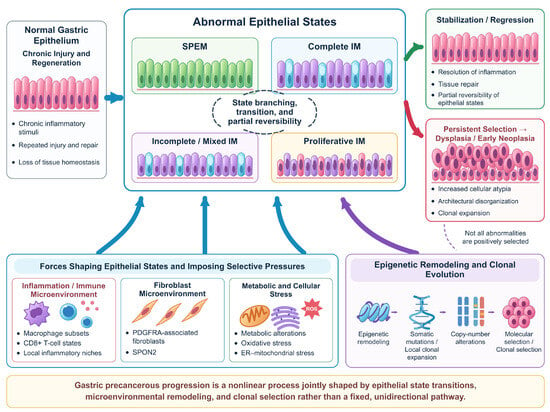
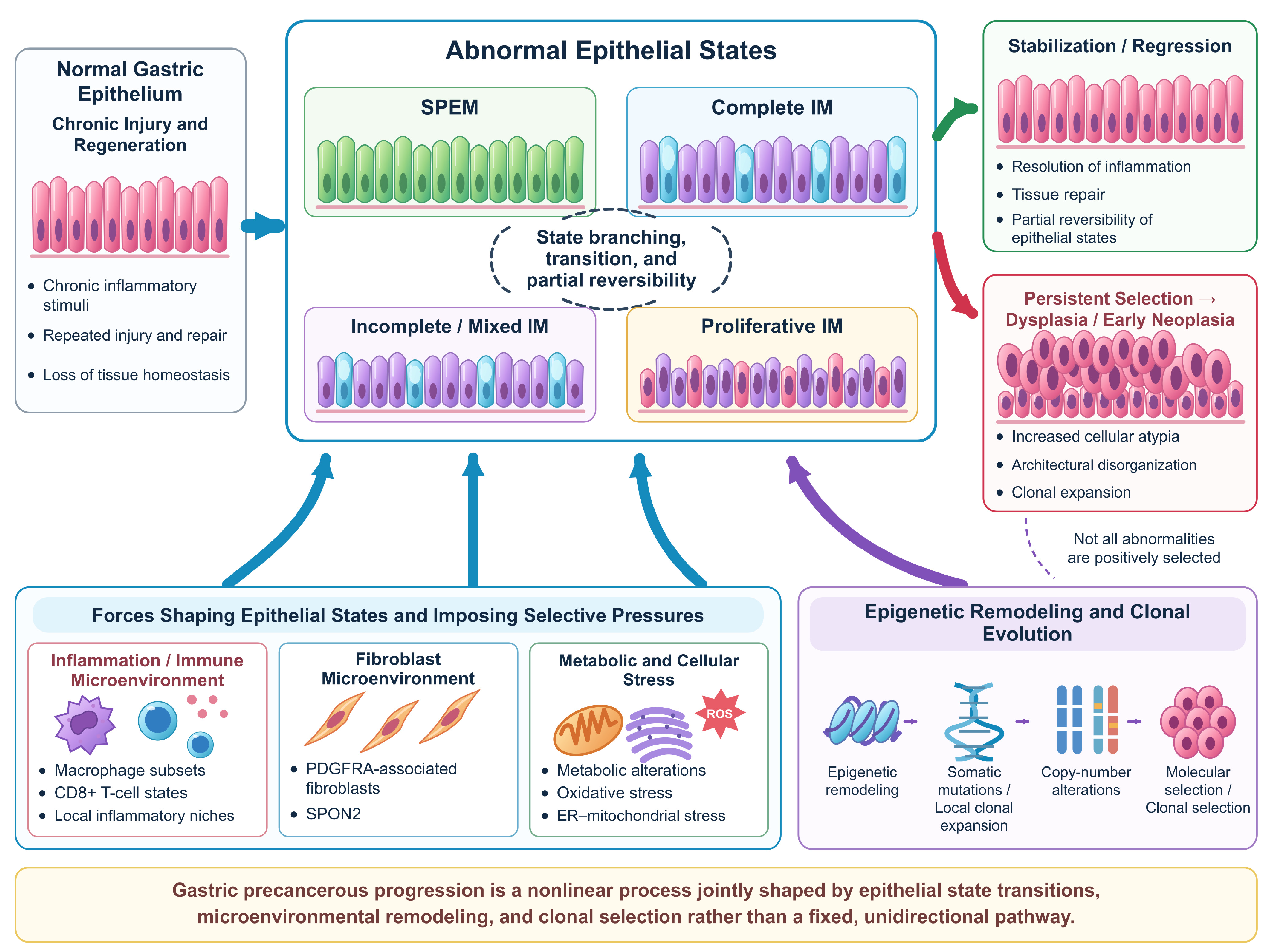
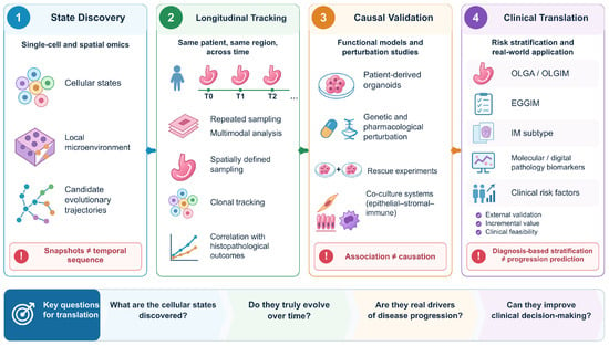
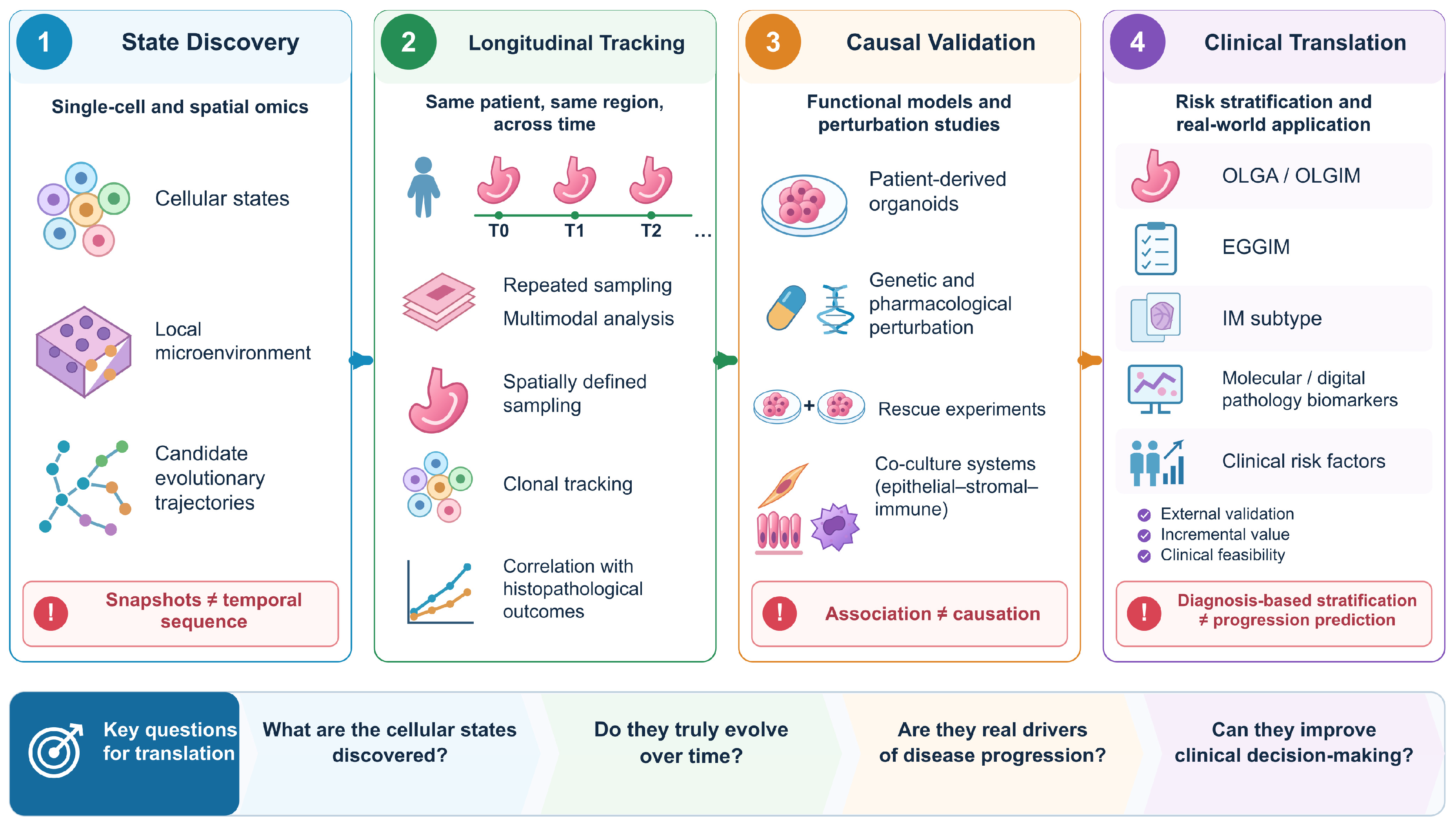

Disclaimer/Publisher’s Note: The statements, opinions and data contained in all publications are solely those of the individual author(s) and contributor(s) and not of MDPI and/or the editor(s). MDPI and/or the editor(s) disclaim responsibility for any injury to people or property resulting from any ideas, methods, instructions or products referred to in the content. |
© 2026 by the authors. Licensee MDPI, Basel, Switzerland. This article is an open access article distributed under the terms and conditions of the Creative Commons Attribution (CC BY) license.