


Abstract
Background/Objectives: Acute kidney injury poses a significant public health concern, contributing to elevated rates of morbidity and mortality. In our study, we sought to explore the involvement of cation channels, known to influence renal physiology, in Rhabdomyolysis-Associated Acute Kidney Injury (RIAKI). Materials and Methods: A total of 32 Sprague–Dawley rats were divided into four groups for our study. All rats underwent 24 h of dehydration prior to experimentation. Subsequently, the control group received physiological saline, while the remaining groups (RIAKI-24, RIAKI-48, and RIAKI-72) were administered intramuscular injections of 5 mL/kg of 50% glycerol solution. The control and RIAKI-24 groups were euthanized 24 h post-injection, the RIAKI-48 group at 48 h, and the RIAKI-72 group at 72 h. Urine, blood, and renal tissue samples were collected for the assessment of renal function and histological damage. Results: Serum urea and creatinine levels exhibited a time-dependent increase, alongside reduced creatinine clearance. Moreover, kidney tissue analysis revealed elevated levels of TRPV1, TRPV4, and TRPC6 in the RIAKI-72 group compared to the control. The RIAKI-24 group displayed the most severe histological damage among the experimental groups. Conclusions: RIAKI was accompanied by changes in renal function and histopathology, as well as altered renal TRPV1, TRPV4, and TRPC6 protein levels. These findings suggest the potential involvement of TRP channels in RIAKI but do not establish their functional activity or whether the observed changes are harmful, protective, or compensatory. Further studies are needed to clarify the functional roles of these channels in renal injury.
1. Introduction
Acute kidney injury (AKI) is a significant global health concern and constitutes a multifaceted clinical entity frequently encountered among hospitalized and critically ill individuals [1]. Current estimates suggest that AKI affects over 13 million people annually and contributes to approximately 1.7 million deaths worldwide [2]. In addition to elevating short-term mortality risk, AKI is a pivotal risk factor for progression to chronic kidney disease (CKD) and is associated with unfavorable long-term outcomes. Although advances in supportive management have been made, timely recognition and the development of efficacious, targeted therapeutic interventions remain suboptimal. Consequently, AKI imposes a considerable burden on both individual patient outcomes and broader healthcare infrastructure [3].
Among the diverse etiologies contributing to acute kidney injury (AKI), rhabdomyolysis (RML) is a clinically important cause, characterized by the breakdown of skeletal muscle fibers and the subsequent release of intracellular constituents, most notably myoglobin. Rhabdomyolysis-Associated Acute Kidney Injury (RIAKI) arises via multifactorial mechanisms, including direct tubular cytotoxicity, intraluminal obstruction by myoglobin casts, renal vasoconstriction, and increased oxidative stress [4]. In those with severe rhabdomyolysis, the incidence of AKI increased to 81%, and 26% required renal replacement therapy. The mortality rate from RML is 10%, which increases in the case of AKI [4]. Conventional biomarkers, such as serum creatinine and blood urea nitrogen (BUN), are insensitive for early detection, as they typically reflect kidney injury at a delayed stage [5]. These limitations have intensified efforts to identify novel molecular biomarkers and therapeutic targets that reflect the early pathogenic events in AKI.
In recent years, transient receptor potential (TRP) channels have garnered attention as key modulators of renal physiology and mediators of injury responses in AKI [6]. These non-selective cation channels participate in the critical aspects of AKI pathogenesis, including mechanotransduction, osmoregulation, inflammation, oxidative stress, and apoptotic signaling [6]. Multiple TRP subtypes have been implicated in the pathogenesis of kidney injury. Elucidating the distinct and potentially synergistic contributions of TRP channel subtypes in AKI may facilitate the development of targeted therapeutic interventions to preserve renal function and mitigate the progression of chronic kidney disease.
Although growing evidence implicates transient receptor potential (TRP) channels in renal pathophysiology, experimental studies elucidating their functional roles in RIAKI remain scarce. Defining the contribution of TRP channels in this context may advance our understanding of the early molecular mechanisms underlying renal injury and support the development of novel therapeutic strategies. In this study, we aimed to quantify renal TRP channel protein levels in a well-established model of rhabdomyolysis-induced acute kidney injury (RIAKI), evaluate renal functional and histopathological changes, and assess changes in calprotectin and endocan levels as biomarkers associated with inflammation and endothelial activation, respectively.
2. Methods
2.1. Animals and Experimental Design
Male Sprague–Dawley rats (2–2.5 months old), bred and housed in the Experimental Animal Unit of Trakya University Faculty of Medicine, were maintained under standard laboratory conditions (22 ± 1 °C; 12-h light/dark cycle) with ad libitum access to standard rat chow and tap water throughout the study. All experimental procedures were approved by the Trakya University Local Ethics Committee for Animal Experiments (TÜHADYEK-2022/25, approval no: 2 July 2022, approval date: 27 February 2022).
The animals were allocated to four experimental groups (n = 8 per group). Random allocation was not performed, and no specific randomization method was used. To minimize procedural variability, all animals were housed and handled under the same experimental conditions, and all procedures and measurements were performed according to a standardized protocol [7].
Control: Subjected to 24 h of dehydration followed by intramuscular administration of physiological saline, and euthanized 24 h post-injection.
RIAKI-24: Subjected to 24 h of dehydration followed by intramuscular administration of 50% hypertonic glycerol (5 mL/kg), and euthanized 24 h post-injection.
RIAKI-48: Subjected to 24 h of dehydration followed by intramuscular administration of 50% hypertonic glycerol (5 mL/kg), and euthanized 48 h post-injection.
RIAKI-72: Subjected to 24 h of dehydration followed by intramuscular administration of 50% hypertonic glycerol (5 mL/kg), and euthanized 72 h post-injection.
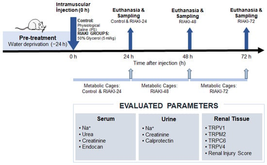
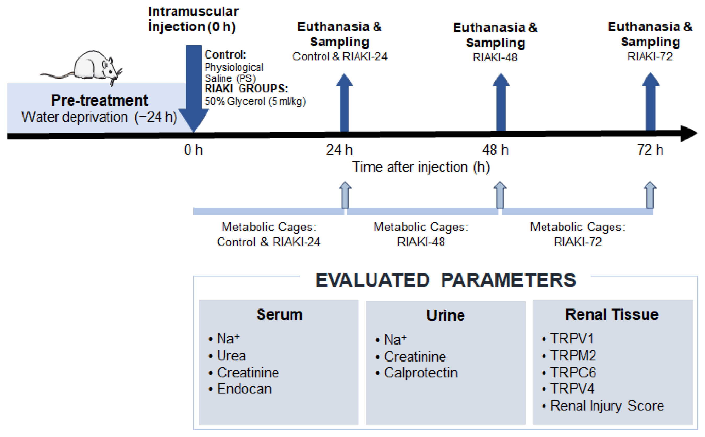
All animals, including those in the control group, were deprived of water for 24 h before the intramuscular injections. A total volume of 5 mL/kg was administered intramuscularly to all animals, divided equally between the muscles of both hindlimbs. The control group received physiological saline (PS), whereas the other groups received 50% glycerol solution. Twenty-four-hour urine samples were collected using metabolic cages. In the control group, urine collection began immediately after physiological saline (PS) injection and continued for the following 24 h (0–24 h). In the RIAKI-24 group, collection began immediately after glycerol injection and continued for 24 h (0–24 h). In the RIAKI-48 and RIAKI-72 groups, urine collection began 24 h and 48 h after glycerol injection, respectively, and continued for the following 24 h (24–48 h and 48–72 h post-injection, respectively). The experimental design is shown in Figure 1.
Figure 1.
Study design.
2.2. Sample Collection
After the 24-h urine collection period, the animals were anesthetized with xylazine (10 mg/kg) and ketamine (50 mg/kg). Under anesthesia, euthanasia was performed by exsanguination, as previously described [7]. Both kidneys were then immediately excised and bisected along the longitudinal axis. One half of each kidney was fixed in 10% neutral-buffered formalin for histopathological evaluation, while the remaining tissue was homogenized in phosphate-buffered saline (PBS) and stored at −80 °C for subsequent biochemical and ELISA-based analyses. Serum samples were separated by centrifugation and stored at −80 °C until further processing.
2.3. Biochemical Analyses
Serum creatinine, urea, and sodium levels, as well as urinary creatinine and sodium concentrations, were quantified using an automated analyzer (AU5800; Beckman Coulter Inc., in Brea, CA, USA) at the Trakya University Health Research and Application Center Laboratory.
Tissue Homogenization and TRP Channel Assessment
Renal tissues were homogenized in ice-cold phosphate-buffered saline (PBS) at a ratio of 1:9 (tissue weight: volume). The homogenates were centrifuged, and the resulting supernatants were collected and stored at −80 °C.
The protein levels of transient receptor potential vanilloid 1 (TRPV1), transient receptor potential vanilloid 4 (TRPV4), transient receptor potential canonical 6 (TRPC6), and transient receptor potential melastatin 2 (TRPM2) in kidney tissue homogenate supernatants were measured using commercially available ELISA kits (BT LAB, Shanghai, China; catalog numbers E1568Ra, E2291Ra, E1350Ra, and E2545Ra, respectively). All assays were performed according to the manufacturers’ instructions.
Kidney tissue homogenate supernatants, stored at −80 °C until analysis, were thawed to room temperature before the assays. Standards were prepared according to the instructions provided with each kit and added to the designated wells (50 μL per well). Tissue homogenate supernatant samples were added to the appropriate wells at the volumes specified in the respective kit protocols. Subsequent assay steps, including the addition of kit-specific detection reagents and streptavidin–horseradish peroxidase (HRP), were performed according to the manufacturers’ instructions. The microplates were incubated at 37 °C for 60 min and then washed five times with 300 μL of wash buffer per well using an automated microplate washer (Biotek Instruments, 100 Tigan Street, Winooski, VT 05404, USA). Subsequently, 50 μL each of substrate solutions A and B was added to each well, followed by incubation at 37 °C for 10 min. The enzymatic reaction was stopped by adding 50 μL of stop solution to each well. Absorbance was measured at 450 nm using a microplate reader (BioTek Instruments, USA).
2.4. Endocan and Calprotectin Measurement
Serum endocan concentrations were determined using a commercially available ELISA kit (BT LAB, Cat. No.: E0961Ra; Shanghai, China), while urinary calprotectin levels were measured using a separate ELISA kit from the same manufacturer (BT LAB, Cat. No.: E1210Ra; Shanghai, China). All assays were conducted according to the manufacturer’s protocols. Sample preparation and assay procedures were performed according to the manufacturers’ instructions, following the general ELISA procedure described in Section 2.3.
2.5. Total Protein Determination
Protein concentrations were quantified using the Lowry method [8], with bovine serum albumin (BSA) as the calibration standard. Absorbance readings were obtained at 660 nm using a spectrophotometer (Helios Alpha, Unicam Instruments, Cheshire, WA14 2DT, United Kingdom).
2.6. Histopathologic Analysis
Kidney tissues fixed in formalin were embedded in paraffin, and 5-μm sections were prepared. The sections were stained with hematoxylin and eosin (H&E) for routine histological assessment. Semi-quantitative histopathological scoring was performed in five randomly selected fields per section at 100× magnification using a 0–5 scale. The evaluation focused on the cortical, outer medullary, inner medullary, and papillary regions, assessing parameters including tubular necrosis, cast formation, and tubular dilatation.
2.7. Statistical Analysis
Data are presented as mean ± standard deviation (SD). The distributional characteristics of the data and the assumptions underlying the statistical tests were evaluated before analysis. Normality was assessed using the Shapiro–Wilk test and Q–Q plots, while homogeneity of variances was assessed using Levene’s test. Because different animals were used at each experimental time point, the groups were treated as independent samples. For variables meeting the assumptions of conventional one-way analysis of variance (ANOVA), differences among groups were assessed using one-way ANOVA followed by Tukey’s post hoc test. When homogeneity of variances was not satisfied, Welch’s ANOVA followed by the Games–Howell post hoc test was used, provided that the data were suitable for parametric analysis. When the distributional characteristics did not support parametric analysis, the Kruskal–Wallis test followed by Dunn’s multiple-comparison test was used, as appropriate. All statistical tests were two-sided, and a p-value < 0.05 was considered statistically significant. Statistical analyses and graphical representations were performed using GraphPad Prism version 9 (GraphPad Software, San Diego, CA, USA).
3. Results
In this study, an experimental RIAKI model induced by intramuscular glycerol administration was investigated in 32 rats divided into four groups. No mortality was observed during the study period. Serum urea, serum creatinine, serum sodium, urine creatinine, urine sodium levels, and TRPV1, TRPV4, TRPC6, and TRPM2 levels of kidney tissues were determined. The findings and statistical evaluations of the serum, urine, and kidney tissues of the groups are presented below.
3.1. Kidney Functions
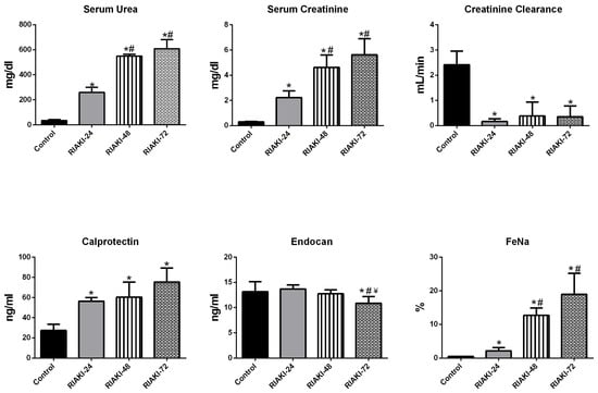
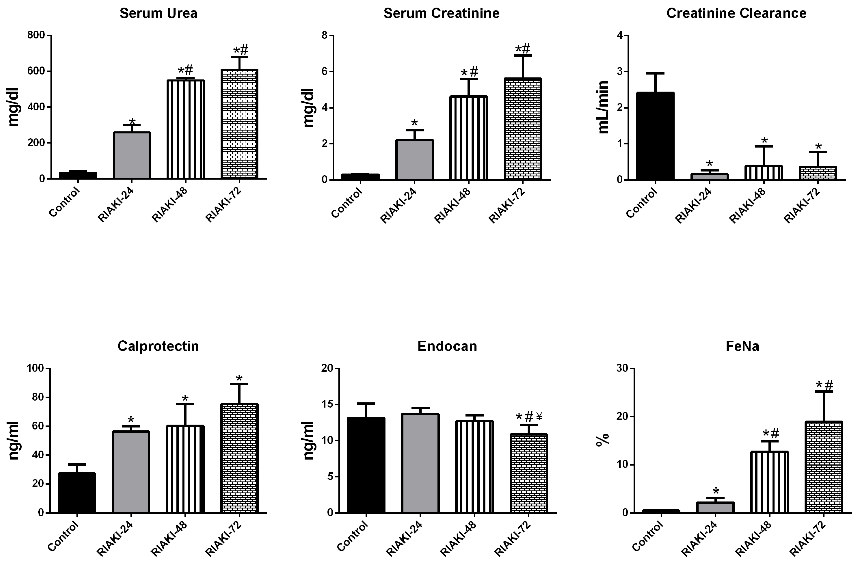
A comparative analysis of the renal function markers across the experimental groups is presented in Figure 2. Serum creatinine levels were significantly higher in the glycerol-treated groups (RIAKI-24, RIAKI-48, and RIAKI-72) than in the control group. In particular, the serum creatinine level was 2.214 ± 0.5459 mg/dL in the RIAKI-24 group, 4.624 ± 0.9786 mg/dL in the RIAKI-48 group, and 5.626 ± 1.273 mg/dL in the RIAKI-72 group. The serum creatinine level in the control group was 0.2950 ± 0.04440 mg/dL. Creatinine clearance decreased in the glycerol-treated groups compared to that in the control group. While it was 2.415 ± 0.5396 in the control group, it was 0.1663 ± 0.1004 in the RIAKI-24 group, 0.3829 ± 0.5513 in the RIAKI-48 group, and 0.3538 ± 0.4237 in the RIAKI-72 group, respectively. Endocan levels were lower in the RIAKI-72 group than in the control group. Calprotectin levels were higher in the acute kidney injury group than in the control group. The highest calprotectin level was detected at 72 h.
Figure 2.
Renal function markers.
Data are presented as mean ± SD (n = 8 per group). Serum urea, serum creatinine, and fractional excretion of sodium (FeNa) were analyzed using Welch’s ANOVA followed by the Games–Howell post hoc test. Serum endocan was analyzed using one-way ANOVA followed by Tukey’s post hoc test. Urinary calprotectin and creatinine clearance were analyzed using the Kruskal–Wallis test followed by Dunn’s post hoc test with Bonferroni correction. * p < 0.05 versus Control; # p < 0.05 versus RIAKI-24; ¥ p < 0.05 versus RIAKI-48.
3.2. TRP Channel Protein Levels
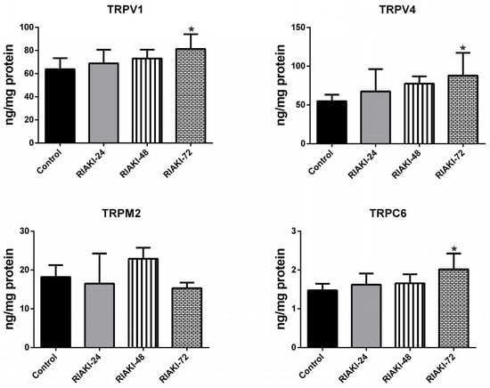
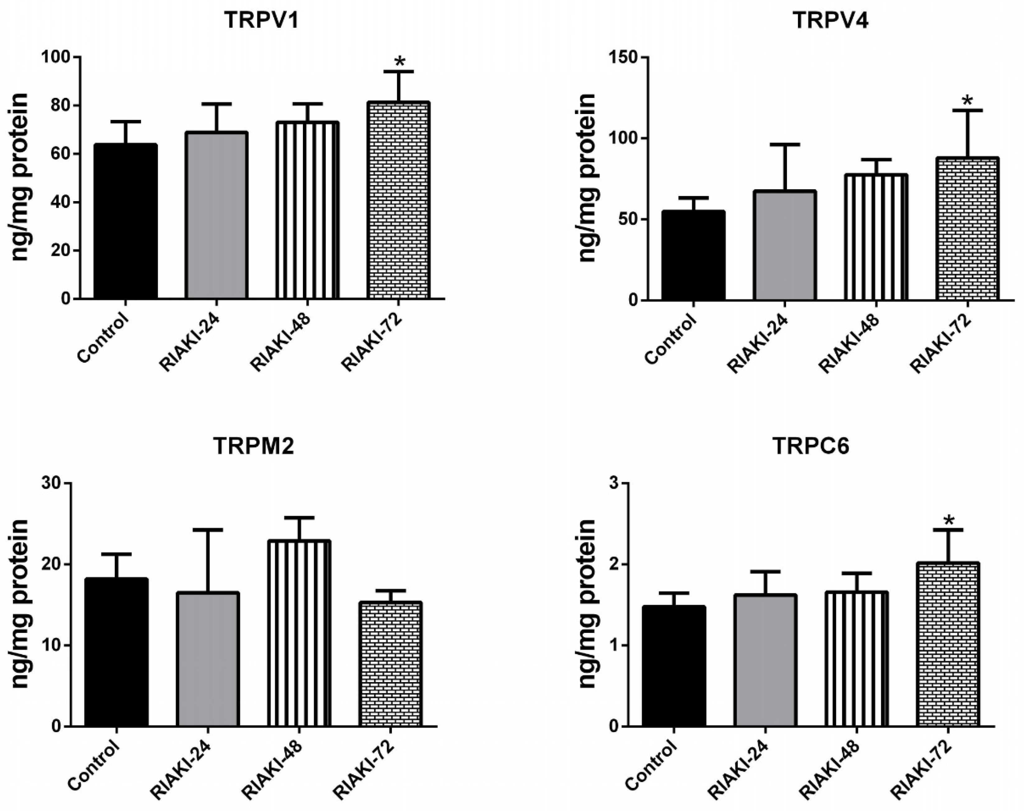
Following glycerol administration, renal TRPV1 protein levels were significantly higher in the RIAKI-72 group than in the control group. No statistically significant differences were detected between the control group and either the RIAKI-24 or RIAKI-48 group, or among the RIAKI time-point groups. Mean TRPV1 protein levels were 63.80 ± 9.54 in the control group, 68.86 ± 11.79 in the RIAKI-24 group, 73.07 ± 7.66 in the RIAKI-48 group, and 81.31 ± 12.70 in the RIAKI-72 group. Similarly, renal TRPV4 protein levels were significantly higher in the RIAKI-72 group than in the control group, whereas no statistically significant differences were detected between the control group and either the RIAKI-24 or RIAKI-48 group, or among the RIAKI time-point groups. Mean TRPV4 protein levels were 54.85 ± 8.34 in the control group, 67.38 ± 28.80 in the RIAKI-24 group, 77.57 ± 9.25 in the RIAKI-48 group, and 87.96 ± 29.30 in the RIAKI-72 group. Renal TRPC6 protein levels were significantly higher in the RIAKI-72 group than in the control group. No statistically significant differences were observed between the control group and the RIAKI-24 or RIAKI-48 groups, or among the RIAKI time-point groups. Mean TRPC6 protein levels were 1.48 ± 0.17 in the control group, 1.622 ± 0.29 in the RIAKI-24 group, 1.66 ± 0.23 in the RIAKI-48 group, and 2.02 ± 0.41 in the RIAKI-72 group. No statistically significant overall difference in renal TRPM2 protein levels was detected among the experimental groups (Kruskal–Wallis test, p = 0.209); therefore, no post hoc analysis was performed. A comparison of renal TRP channel protein levels across the experimental groups is presented in Figure 3.
Figure 3.
TRP channel proteins.
Data are presented as mean ± SD (n = 8 per group). TRPV1, TRPV4, and TRPC6 were analyzed using one-way ANOVA followed by Tukey’s post hoc test. TRPM2 was analyzed using the Kruskal–Wallis test; no post hoc test was performed because the overall group comparison was not statistically significant. No statistically significant differences were detected among the RIAKI time-point groups for TRPV1, TRPV4, or TRPC6. * p < 0.05 versus Control.
3.3. Histopathologic Findings
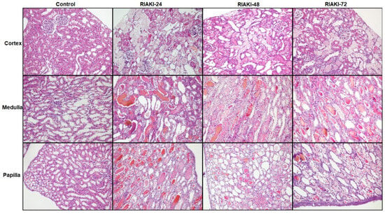
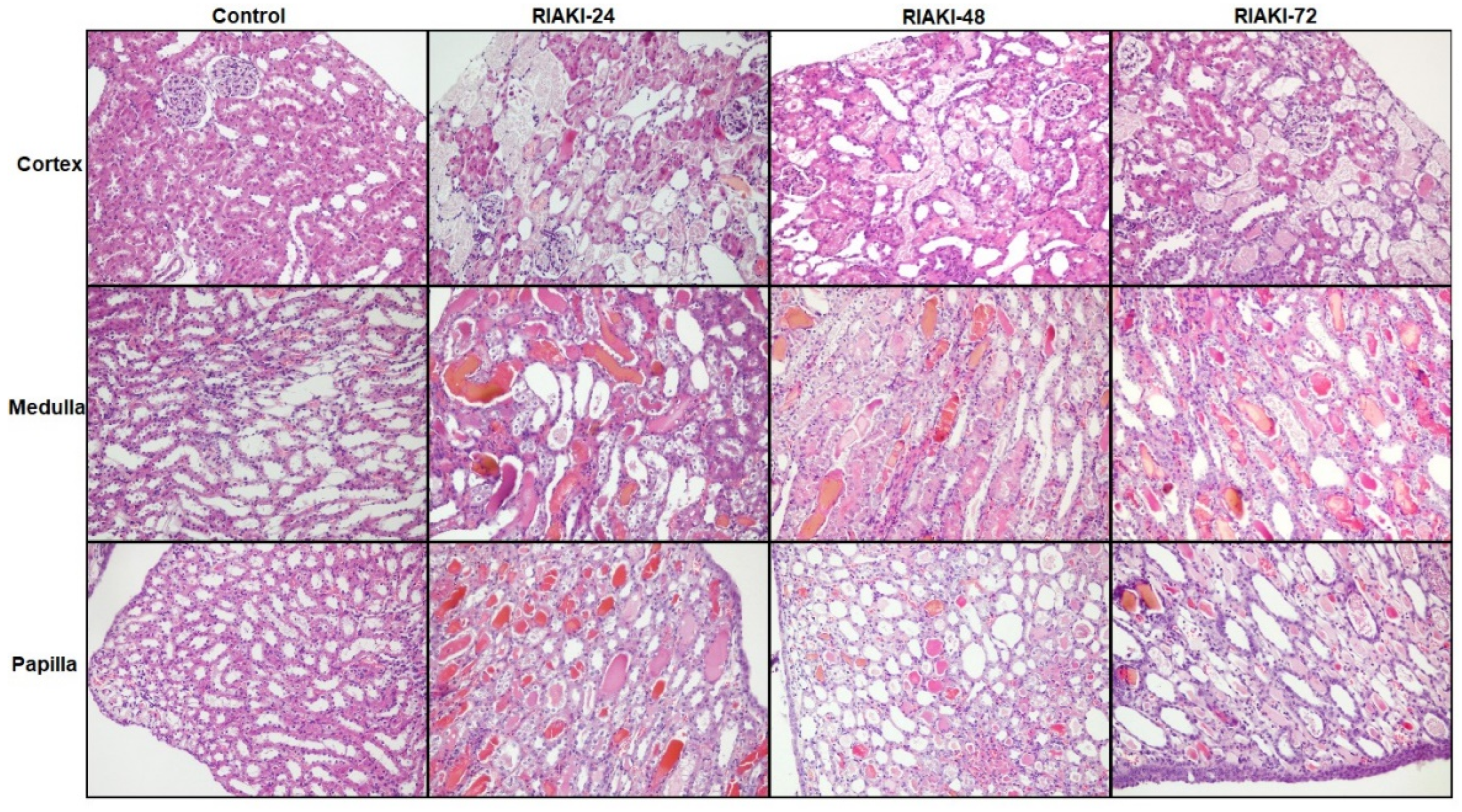
Histological features of the kidney tissues of the control and glycerol groups were examined in hematoxylin and eosin-stained sections. In the kidney tissues of the control group, Malpighi bodies with normal histological appearance, proximal tubules with brush-bordered epithelium, and distal tubules with wide lumina were observed in the cortical regions. The tubule structures had a normal histological appearance in the medulla and papilla regions. In the kidney tissues of the glycerol-administered groups, dilatation–necrosis–desquamation and peritubular edema were observed in the cortex, subcapsular proximal tubules, and collecting tubules of the kidneys. Necrosis of epithelial cells in the proximal tubule wall and dense proteinaceous material accumulation in the distal tubules were also observed. The glomeruli were unchanged, except for mild congestion in some areas. In the RIAKI-24 group, prominent histopathological findings included tubular dilatation and necrosis, along with peritubular edema in the outer medulla, cast accumulation, necrosis, and cytoplasmic vacuolization in the inner medulla. Features of acute tubular necrosis were observed to varying degrees and included altered cytoplasmic eosinophilia (either increased or decreased), cytoplasmic swelling, nuclear pyknosis or fragmentation, and intraluminal cellular debris. In addition, marked tubular dilatation and cast accumulation were observed in the papillary regions of the renal tissue in this group. In the RIAKI-48 group, intensive tubular necrosis and cast accumulation were remarkable, especially from the corticomedullary area to the renal papilla. In the RIAKI-72 group, tubular damage findings were attenuated in the cortical regions of the kidney compared to the other glycerol groups, but there was no significant difference in the medulla and papilla regions compared to the 24- and 48-h groups. The renal injury score showed that histologic deterioration was highest at 24 h, although dysfunction was highest at 72 h. Histological micrographs of kidney tissues are shown in Figure 4, and the renal injury scores between the experimental groups are shown in Table 1.
Figure 4.
Histologic micrographs of renal tissues.
Table 1.
Renal injury scores across experimental groups.
4. Discussion
In this study, we investigated the role of cation channels in the RIAKI model, which have previously been identified to play a role in renal physiology in various models. The progression and severity of kidney injury were evaluated at 24, 48, and 72 h after glycerol administration. Our findings indicate that intramuscular glycerol administration impairs renal function and causes histological damage. Although functional loss intensified over time, histological damage was most severe at 24 h. Another important finding of our study was that renal TRPV1, TRPV4, and TRPC6 protein levels were higher in the RIAKI groups than in the control group, with statistically significant differences observed at 72 h. These changes indicate alterations in renal TRP channel protein abundance during RIAKI; however, they do not establish changes in channel activity or the functional significance of these alterations.
The classic markers used to assess renal function are serum urea and creatinine levels. Consistent with our previous studies using the same model and the existing literature, serum urea and creatinine levels were higher in the glycerol-treated groups than in the control group [9,10]. In our study, creatinine clearance and fractional sodium excretion were also evaluated to assess renal function. In line with the literature, creatinine clearance decreased, whereas fractional sodium excretion increased [10].
Another important finding of our study was the lower serum endocan level in the RIAKI-72 group than in the control group. Endocan is a proteoglycan secreted from the vascular endothelium into the extracellular fluid. It has been suggested that examining serum endocan levels can be used for the differential diagnosis of intrinsic AKI. Endocan, a marker of endothelial dysfunction, is claimed to show an increasing trend in glomerular/vascular pathologies, while decreasing in toxic/ischemic ATN [11,12]. In our glycerol-induced AKI model, serum endocan levels were lower in the 72-h group than in the control group, consistent with this hypothesis.
Calprotectin, a heterodimer composed of S100A8 and S100A9 proteins originating from neutrophils and monocytes, serves a key role in immune regulation and has demonstrated protective effects against oxidative stress. Although it has been shown to increase in unilateral ureteral obstruction, renal ischemia–reperfusion injury, and contrast nephropathy models, its role and mechanisms in RIAKI have not yet been reported [13,14,15]. In a study comparing prerenal AKI induced by volume restriction and renal AKI induced by cisplatin administration, calprotectin levels were higher in the renal AKI group than in the control and prerenal AKI groups [16]. In our study, calprotectin levels were also higher in the acute kidney injury group than in the control group. Furthermore, the highest calprotectin level was detected at 72 h.
The transient receptor potential (TRP) channel family comprises cation-permeable channels capable of integrating a variety of downstream signaling pathways. They are expressed throughout the nephron, are important molecular determinants of kidney function, and play roles in acquired kidney disorders [6,17]. Transient receptor potential vanilloid 1 (TRPV1) is a polymodal sensor that recognizes the presence of irritating chemicals. TRPV1 channels have been shown to act as mechanoreceptors in the visceral sensation of rat kidneys [18]. With its ability to sense harmful compounds, it has been suggested that TRPV1 in renal tubules can detect harmful chemicals after glomerular filtration and coordinate tubular function for the clearance of these compounds. Excessive activity of TRPV1 has been implicated in promoting renal inflammation associated with oxalate nephropathy, while inhibiting TRPV1 has been shown to mitigate inflammation-driven tubular injury in hyperoxaluria models studied both in vitro and in vivo [19]. In our study, renal TRPV1 protein levels were significantly higher in the RIAKI groups than in the control group. However, no statistically significant differences were observed among the RIAKI time-point groups; therefore, the findings do not demonstrate a time-dependent increase. Previous studies have reported anti-inflammatory and protective effects of TRPV1 activation in certain models of kidney injury [20,21]. These findings do not necessarily contradict our results, because protein abundance does not directly reflect channel activity or its downstream functional effects. As TRPV1 activity was not directly assessed in the present study, the functional significance of the observed changes remains unclear. Transient receptor potential vanilloid 4 (TRPV4) channels are activated by mechanical stimuli and heat (28–38 °C), defined as an osmotic sensor, and contribute to the regulation of systemic osmotic pressure. They have also been shown to contribute to vasodilatory effects under hypoxic conditions [6]. In a study where septic acute kidney injury was induced in mice, TRPV4 was reported to be associated with glomerular endothelial inflammation, and TRPV4 blockade could improve glomerular endothelial inflammation and renal dysfunction [22]. A recent investigation found that adult mice lacking TRPV4 exhibited increased vulnerability to renal tubular injury induced by ischemia–reperfusion (IR) of the left kidney after removal of the right kidney, compared to their wild-type counterparts [23]. In a study involving neonatal pigs subjected to 45 min of bilateral renal ischemia followed by a 3-h reperfusion period, an upregulation of TRPV4 channels was observed in the preglomerular microvasculature. Inhibition of TRPV4 was found to mitigate the alterations in kidney function caused by ischemia–reperfusion injury [24]. In our model, renal TRPV4 protein levels were significantly higher in the RIAKI-72 group than in the control group, whereas no statistically significant differences were observed between the control group and the RIAKI-24 or RIAKI-48 groups, or among the RIAKI time-point groups. TRPC6, a member of the transient receptor potential canonical subfamily, is a non-selective and voltage-independent channel that facilitates the entry of calcium and monovalent cations into cells, with widespread expression observed in renal tissues (especially in proximal tubular epithelial cells). In a study on renal ischemia–reperfusion in rats, TRPC6 agonist administration prevented necrosis by accelerating Zn2+ influx and autophagy, whereas TRPC6 inhibition exacerbated ischemia–reperfusion injury [25]. In an experimental study where fibrosis was induced by unilateral renal ischemia–reperfusion in mice, the animals were sacrificed at 7, 14, 21, and 28 days after the operation. A time-dependent increase in TRPC6 expression was observed. In TRPC6 knockout mice, fibrotic damage was milder than that in wild-type mice [26]. In a study where unilateral nephrectomy and ischemia–reperfusion injury were applied to mice and sacrificed at 24 h, TRPC6 inhibition was shown not to affect the short-term outcomes of AKI [27]. In our model, renal TRPC6 protein levels were significantly higher in the RIAKI-72 group than in the control group, whereas no statistically significant differences were observed between the control group and the RIAKI-24 or RIAKI-48 groups, or among the RIAKI time-point groups. The discrepancy in the study findings may be due to differences in the animals and models used, as well as differences in the timing of parameter measurements. Although previous studies have reported functional effects of TRPV4 and TRPC6 modulation in experimental kidney injury, the present study assessed protein levels rather than channel activity. Therefore, the observed changes cannot establish whether these channels contribute to renal injury, exert protective effects, or undergo compensatory regulation in RIAKI. Transient receptor potential melastatin 2 (TRPM2) is a non-selective cation channel permeable to calcium, potassium, and sodium, and is expressed in various organs and tissues, including the kidneys (especially in proximal tubular epithelial cells). It is activated by reactive oxygen species, tumor necrosis factor-α, intracellular calcium, and ADP-ribose (ADPR), contributing to oxidative damage and cell death [28,29]. In a renal ischemia–reperfusion study conducted on mice, TRPM2-deficient mice showed less oxidative stress and lower NADPH oxidase activity levels after ischemia than wild-type mice [30]. In a study where cisplatin-induced kidney injury was induced in mice, TRPM2 deficiency was found to exacerbate cisplatin-induced kidney injury [29]. In our study, TRPM2 levels were similar among the groups with RIAKI. The inconsistency in the study findings may be due to differences in the affected nephron regions (vascular or tubular damage) in different kidney injury models.
When evaluating the histopathological findings of our study, functional impairment was detected at its highest level at 72 h, whereas histological damage was observed to be most severe at 24 h. It is a known fact that in kidney injury, histopathological findings may appear before functional impairment, and the correlation between renal function and renal lesions is weak [31,32]. The compensation of the functions of damaged nephrons by unaffected nephrons in the kidney injury model may explain this.
Several limitations of this study should be acknowledged. First, the relatively small sample size (n = 8 per group) may have limited the statistical power to detect some differences between experimental groups. Second, the animals were not randomly allocated to the experimental groups, which may have introduced selection bias and reduced the comparability of the groups. This should be considered when interpreting the findings. Third, only male rats were included; therefore, potential sex-related differences in the pathophysiology and progression of RIAKI could not be evaluated. Fourth, although renal function was assessed using biochemical and urinary indices and renal tissue injury was evaluated histopathologically, serum cystatin C, urinary protein excretion, and the urinary albumin-to-creatinine ratio were not measured. The inclusion of these biomarkers could have provided complementary information regarding glomerular filtration and renal protein handling. Finally, rats differ from humans in metabolic and regenerative processes, lifespan, and responses to injury. Moreover, unlike the clinical setting, the experimental model was conducted under controlled conditions using a single disease model, without reproducing the full range of comorbidities, environmental influences, and genetic variability encountered in humans. These differences may affect the course of renal injury and recovery and limit the direct translation of our findings to humans. Therefore, the results should be interpreted cautiously, and further studies involving larger sample sizes, randomized group allocation, both sexes, and additional renal biomarkers are warranted.
5. Conclusions
In conclusion, our study demonstrates changes in renal TRPV1, TRPV4, and TRPC6 protein levels in a glycerol-induced RIAKI model, alongside biochemical and histopathological changes. These findings provide further insight into the potential involvement of TRP channels in the pathophysiology of RIAKI and highlight the need for further studies to clarify their functional roles in renal injury.
Author Contributions
Conceptualization, O.K. and S.T.; methodology, O.K. and S.T.; software, O.K. and M.A.A.; validation, O.K. and M.S.M.; formal analysis, O.K., M.S.M. and Ö.G.; investigation, O.K., M.A.A., Ü.E. and D.E.; resources, O.K. and Ü.E.; writing—original draft preparation, O.K. and S.T.; writing—review and editing, O.K., S.T. and M.S.M.; visualization, O.K., M.S.M. and Ö.G.; project administration, O.K. and S.T.; funding acquisition, O.K. and S.T. All authors have read and agreed to the published version of the manuscript.
Funding
This work was supported by the Scientific Research Fund of Trakya University (TÜBAP 2022/212).
Institutional Review Board Statement
The animal study protocol was approved by the Trakya University of the Animal Experiments Local Ethics Committee (TÜHADYEK-2022/25, approval no: 2 July 2022, approval date: 27 February 2022).
Informed Consent Statement
Not applicable.
Data Availability Statement
The original contributions presented in this study are included in the article. Further inquiries can be directed to the corresponding author.
Conflicts of Interest
The authors declare no conflicts of interest.
Abbreviations
The following abbreviations are used in this manuscript:
| RIAKI | Rhabdomyolysis-Associated Acute Kidney Injury |
| AKI | Acute kidney injury |
| CKD | Chronic kidney disease |
| TRP | Transient receptor potential |
| PBS | Phosphate-buffered saline |
References
- Thongprayoon, C.; Wathanavasin, W.; Suppadungsuk, S.; Sheikh, M.S.; Abdelgadir, Y.H.; Miao, J.; Mao, M.A.; Craici, I.M.; Qureshi, F.; Kashani, K.B.; et al. The evolution of public attention in acute kidney injury and continuous renal replacement therapy: Trends analysis from 2004 to 2024. Front. Nephrol. 2024, 4, 1472144. [Google Scholar] [CrossRef] [Scilit] [PubMed]
- Abebe, A.; Kumela, K.; Belay, M.; Kebede, B.; Wobie, Y. Mortality and predictors of acute kidney injury in adults: A hospital-based prospective observational study. Sci. Rep. 2021, 11, 15672. [Google Scholar] [CrossRef] [Scilit] [PubMed]
- Scholz, H.; Boivin, F.J.; Schmidt-Ott, K.M.; Bachmann, S.; Eckardt, K.-U.; Scholl, U.I.; Persson, P.B. Kidney physiology and susceptibility to acute kidney injury: Implications for renoprotection. Nat. Rev. Nephrol. 2021, 17, 335–349. [Google Scholar] [CrossRef] [Scilit] [PubMed]
- Gupta, A.; Thorson, P.; Penmatsa, K.R.; Gupta, P. Rhabdomyolysis: Revisited. Ulst. Med. J. 2021, 90, 61–69. [Google Scholar]
- Kellum, J.A.; Romagnani, P.; Ashuntantang, G.; Ronco, C.; Zarbock, A.; Anders, H.J. Acute kidney injury. Nat. Rev. Dis. Primers 2021, 7, 52. [Google Scholar] [PubMed]
- Markó, L.; Mannaa, M.; Haschler, T.N.; Krämer, S.; Gollasch, M. Renoprotection: Focus on TRPV1, TRPV4, TRPC6 and TRPM2. Acta Physiol. 2017, 219, 589–612. [Google Scholar]
- Kaya, O.; Aydogdu, N.; Tastekin, E.; Karadag, C.H.; Gunduz, O.; Sut, N. Effects of Losartan on Glycerol-induced Myoglobinuric Acute Renal Failure in Rats. Kafkas Univ. Vet. Fak. Derg. 2013, 19, 253–258. [Google Scholar] [CrossRef] [Scilit]
- Lowry, O.H.; Rosebrough, N.J.; Farr, A.L.; Randall, R.J. Protein measurement with the Folin phenol reagent. J. Biol. Chem. 1951, 193, 265–275. [Google Scholar] [CrossRef] [Scilit]
- Hoseini Azad, S.A.; Moshiri, M.; Roohbakhsh, A.; Shakeri, A.; Fatemi Shandiz, A.; Etemad, L. Efficacy of orally administered montmorillonite in myoglobinuric acute renal failure model in male rats. Iran J. Basic. Med. Sci. 2023, 26, 753–759. [Google Scholar] [CrossRef] [Scilit] [PubMed]
- Kaur, T.; Singh, D.; Pathak, D.; Singh, A.P.; Singh, B. Umbelliferone attenuates glycerol-induced myoglobinuric acute kidney injury through peroxisome proliferator-activated receptor-γ agonism in rats. J. Biochem. Mol. Toxicol. 2021, 35, e22892. [Google Scholar] [CrossRef] [Scilit] [PubMed]
- Nalewajska, M.; Gurazda, K.; Marchelek-Myśliwiec, M.; Pawlik, A.; Dziedziejko, V. The role of endocan in selected kidney diseases. Int. J. Mol. Sci. 2020, 21, 6119. [Google Scholar] [CrossRef] [Scilit] [PubMed]
- Azimi, A. Could “calprotectin” and “endocan” serve as “Troponin of Nephrologists”? Med. Hypotheses 2017, 99, 29–34. [Google Scholar] [CrossRef] [Scilit] [PubMed]
- Fujiu, K.; Manabe, I.; Nagai, R. Renal collecting duct epithelial cells regulate inflammation in tubulointerstitial damage in mice. J. Clin. Investig. 2011, 121, 3425–3441. [Google Scholar] [CrossRef] [Scilit] [PubMed]
- Dessing, M.C.; Tammaro, A.; Pulskens, W.P.; Teske, G.J.; Butter, L.M.; Claessen, N.; van Eijk, M.; van der Poll, T.; Vogl, T.; Roth, J.; et al. The calcium-binding protein complex S100A8/A9 has a crucial role in controlling macrophage-mediated renal repair following ischemia/reperfusion. Kidney Int. 2015, 87, 85–94. [Google Scholar] [CrossRef] [Scilit] [PubMed]
- Tan, X.; Zheng, X.; Huang, Z.; Lin, J.; Xie, C.; Lin, Y. Involvement of S100A8/A9-TLR4-NLRP3 inflammasome pathway in contrast-induced acute kidney injury. Cell. Physiol. Biochem. 2017, 43, 209–222. [Google Scholar] [CrossRef] [Scilit] [PubMed]
- Kim, A.J.; Ro, H.; Kim, H.; Chang, J.H.; Lee, H.H.; Chung, W.; Jung, J.Y. Klotho and S100A8/A9 as discriminative markers between pre-renal and intrinsic acute kidney injury. PLoS ONE 2016, 11, e0147255. [Google Scholar] [CrossRef] [Scilit] [PubMed]
- Tomilin, V.; Mamenko, M.; Zaika, O.; Pochynyuk, O. Role of renal TRP channels in physiology and pathology. Semin. Immunopathol. 2016, 38, 371–383. [Google Scholar] [CrossRef] [Scilit] [PubMed]
- Feng, N.H.; Lee, H.H.; Shiang, J.C.; Ma, M.C. Transient receptor potential vanilloid type 1 channels act as mechanoreceptors and cause substance P release and sensory activation in rat kidneys. Am. J. Physiol. Ren. Physiol. 2008, 294, F316–F325. [Google Scholar] [CrossRef] [Scilit] [PubMed]
- Lu, C.L.; Teng, T.Y.; Liao, M.T.; Ma, M.C. TRPV1 hyperfunction contributes to renal inflammation in oxalate nephropathy. Int. J. Mol. Sci. 2021, 22, 6204. [Google Scholar] [CrossRef] [Scilit] [PubMed]
- Yu, S.Q.; Ma, S.; Wang, D.H. Activation of TRPV1 prevents salt-induced kidney damage and hypertension after renal ischemia-reperfusion injury in rats. Kidney Blood Press. Res. 2018, 43, 1285–1296. [Google Scholar] [CrossRef] [Scilit] [PubMed]
- Chen, L.; Markó, L.; Kaßmann, M.; Zhu, Y.; Wu, K.; Gollasch, M. Role of TRPV1 channels in ischemia/reperfusion-induced acute kidney injury. PLoS ONE 2014, 9, e109842. [Google Scholar] [CrossRef] [Scilit] [PubMed]
- Wang, X.; Wang, Y.; Huo, H.; Zhou, G.; Li, Y.; Liang, F.; Xue, J.; Shi, X.; Yin, A.; Xiao, Q.; et al. Transient receptor vanilloid subtype 4-mediated Ca2+ influx promotes glomerular endothelial inflammation in sepsis-associated acute kidney injury. Lab. Investig. 2023, 103, 100126. [Google Scholar] [CrossRef] [Scilit] [PubMed]
- Mannaa, M.; Markó, L.; Balogh, A.; Vigolo, E.; N’dIaye, G.; Kaßmann, M.; Michalick, L.; Weichelt, U.; Schmidt–Ott, K.M.; Liedtke, W.B.; et al. Transient receptor potential vanilloid 4 channel deficiency aggravates tubular damage after acute renal ischaemia reperfusion. Sci. Rep. 2018, 8, 4878. [Google Scholar] [CrossRef] [Scilit] [PubMed]
- Soni, H.; Peixoto-Neves, D.; Olushoga, M.A.; Adebiyi, A. Pharmacological inhibition of TRPV4 channels protects against ischemia-reperfusion-induced renal insufficiency in neonatal pigs. Clin. Sci. 2019, 133, CS20180815. [Google Scholar] [CrossRef] [Scilit] [PubMed]
- Pu, Y.; Zhao, H.; Shen, B.; Zhou, Q.; Xie, P.; Wu, X. TRPC6 ameliorates renal ischemic reperfusion injury by inducing Zn2+ influx and activating autophagy to resist necrosis. Ann. Transl. Med. 2022, 10, 249. [Google Scholar] [CrossRef] [Scilit] [PubMed]
- Sun, A.B.; Li, F.H.; Zhu, L.; Zeng, X.X.; Zhu, M.; Lei, Q.H.; Liao, Y.-H. TRPC6 knockout alleviates renal fibrosis through PI3K/AKT/GSK3B pathway. Curr. Med. Sci. 2024, 44, 589–602. [Google Scholar] [CrossRef] [Scilit] [PubMed]
- Zheng, Z.; Tsvetkov, D.; Bartolomaeus, T.U.P.; Erdogan, C.; Krügel, U.; Schleifenbaum, J.; Schaefer, M.; Nürnberg, B.; Chai, X.; Ludwig, F.-A.; et al. Role of TRPC6 in kidney damage after acute ischemic kidney injury. Sci. Rep. 2022, 12, 3038. [Google Scholar] [CrossRef] [Scilit] [PubMed]
- Khanahmad, H.; Mirbod, S.M.; Karimi, F.; Kharazinejad, E.; Owjfard, M.; Najaflu, M.; Tavangar, M. Pathological mechanisms induced by TRPM2 ion channels activation in renal ischemia-reperfusion injury. Mol. Biol. Rep. 2022, 49, 11071–11079. [Google Scholar] [CrossRef] [Scilit] [PubMed]
- Yu, B.; Jin, L.; Yao, X.; Zhang, Y.; Zhang, G.; Wang, F.; Su, X.; Fang, Q.; Xiao, L.; Yang, Y.; et al. TRPM2 protects against cisplatin-induced acute kidney injury and mitochondrial dysfunction via modulating autophagy. Theranostics 2023, 13, 4356–4375. [Google Scholar] [CrossRef] [Scilit] [PubMed]
- Gao, G.; Wang, W.; Tadagavadi, R.K.; Briley, N.E.; Love, M.I.; Miller, B.A.; Reeves, W.B. TRPM2 mediates ischemic kidney injury and oxidant stress through RAC1. J. Clin. Investig. 2014, 124, 4989–5001. [Google Scholar] [CrossRef] [Scilit] [PubMed]
- Misra, P.S.; Szeto, S.G.; Krizova, A.; Gilbert, R.E.; Yuen, D.A. Renal histology in diabetic nephropathy predicts progression to end-stage kidney disease but not the rate of renal function decline. BMC Nephrol. 2020, 21, 285. [Google Scholar] [CrossRef] [Scilit] [PubMed]
- Trevisani, F.; Di Marco, F.; Capitanio, U.; Dell’aNtonio, G.; Cinque, A.; Larcher, A.; Lucianò, R.; Bettiga, A.; Vago, R.; Briganti, A.; et al. Correction to: Renal histology across the stages of chronic kidney disease. J. Nephrol. 2021, 34, 961. [Google Scholar] [CrossRef] [Scilit] [PubMed]

Disclaimer/Publisher’s Note: The statements, opinions and data contained in all publications are solely those of the individual author(s) and contributor(s) and not of MDPI and/or the editor(s). MDPI and/or the editor(s) disclaim responsibility for any injury to people or property resulting from any ideas, methods, instructions or products referred to in the content. |
© 2026 by the authors. Licensee MDPI, Basel, Switzerland. This article is an open access article distributed under the terms and conditions of the Creative Commons Attribution (CC BY) license.