


Abstract
Although low-density lipoprotein cholesterol (LDL-C) can now be effectively reduced, substantial residual risk of acute ischemic cardiovascular events persists. Lipoprotein(a) [Lp(a)] is an established causal risk factor for atherosclerotic cardiovascular disease (ASCVD), yet its mechanistic links to plaque vulnerability and their clinical implications remain incompletely understood. Clinical imaging studies have associated elevated Lp(a) with plaque progression and features of vulnerability, including lipid-rich necrotic cores, macrophage accumulation, and thin fibrous caps, suggesting that Lp(a)-associated risk may involve destabilization during plaque progression. This review draws on imaging and pathological evidence to examine the potential mechanisms underlying these associations and their implications for clinical risk stratification and preventive decision-making. The apolipoprotein(a) [apo(a)] component unique to Lp(a), together with the oxidized phospholipids (OxPL) it carries, may contribute to plaque vulnerability by sustaining vascular inflammation, promoting necrotic core expansion, and influencing fibrous cap stability and intraplaque neovascularization. Integrating circulating Lp(a) levels with plaque imaging features and baseline cardiovascular risk may help identify individuals at high risk in primary prevention and evaluate residual risk in secondary prevention, informing earlier or more intensive efforts to manage modifiable risk factors. Alongside established preventive measures, therapies aimed at lowering Lp(a) may provide an additional approach to reducing residual risk. In trials conducted to date, substantial reductions in Lp(a) have been observed with antisense oligonucleotides, small interfering RNAs, and oral small-molecule inhibitors; some agents have demonstrated sustained effects and favorable safety profiles. However, whether therapies targeting Lp(a) reduce cardiovascular event risk remains to be established through robust evidence from large cardiovascular outcome trials.
1. Introduction
Over the past three decades, atherosclerotic diseases—including ischemic heart disease, peripheral artery disease, and ischemic stroke—have shown a sustained rise in global age-standardized incidence [1]. The pathological progression of atherosclerosis (AS) is a dynamic and multistep process that leads, through plaque rupture or erosion, to most acute cardiovascular events [2]. Muller et al. [3] first proposed the concept of a “vulnerable plaque” in 1989, equating it with that of a “rupture-prone plaque.” Such plaques typically have a large lipid-rich necrotic core beneath a thin fibrous cap with sparse smooth muscle cells (SMCs) and abundant macrophage infiltration, accompanied by abnormal intraplaque neovascularization. With advances in the understanding of unstable plaque pathophysiology and the development of imaging technologies, in vivo assessment of culprit lesions in acute coronary syndrome (ACS) has revealed that eroded plaques—pathologically defined by a small lipid core, a thick fibrous cap, and a mild inflammatory response—account for more than one-third of ACS cases. The rise in non-ST-segment elevation myocardial infarction incidence has been linked particularly to eroded plaques [4,5]. However, the evidence concerning whether lipoprotein(a) [Lp(a)] is associated with plaque erosion remains limited. The few available studies using optical coherence tomography (OCT) have not found a significant association of circulating Lp(a) with plaque erosion [6], whereas evidence linking higher Lp(a) levels to lipid-rich plaques and thin-cap fibroatheroma (TCFA) has been more consistent [7]. Accordingly, the following discussion focuses primarily on how Lp(a) relates to rupture-prone plaques.
Approximately 20% of people worldwide have elevated Lp(a) levels (>125 nmol/L or >50 mg/dL) [8]. In Lp(a), apolipoprotein(a) [apo(a)] is attached by a disulfide bond to apolipoprotein B-100 (apoB-100) on a low-density lipoprotein (LDL)-like particle [9]. Measurements using quantitative lipoprotein electrophoresis show that cholesterol carried within Lp(a) particles makes a substantial contribution to laboratory-reported low-density lipoprotein cholesterol (LDL-C) values [10]. Unlike LDL, however, Lp(a) has additional proinflammatory, proatherogenic, and prothrombotic activity mediated by apo(a), oxidized phospholipids (OxPL), and other components [11,12,13]. Current evidence indicates that Lp(a)-associated cardiovascular risk is relatively independent of, and additive to, the risk associated with LDL-C. A meta-analysis of six statin trials involving 27,658 participants found that elevated Lp(a) remained associated with atherosclerotic cardiovascular disease (ASCVD) risk even among participants who achieved low LDL-C levels; those with elevations in both Lp(a) and LDL-C had the highest risk [14]. At the plaque level, Shishikura et al. [15] found that patients with coronary artery disease (CAD) who had both LDL-C < 70 mg/dL and Lp(a) < 50 mg/dL showed the lowest near-infrared spectroscopy–derived maximum 4 mm lipid-core burden index across the four lipid groups; a similar pattern was observed in a further analysis using LDL-C < 55 mg/dL. Plasma Lp(a) levels show high heritability, with estimates spanning approximately 51% to 98% across populations and much of the variation between individuals associated with genetic differences at the LPA gene locus [16,17,18,19]. Thus, assessing the actual plaque-related risk linked to elevated Lp(a) may be more relevant to current clinical practice than simply identifying elevated circulating levels.
However, Lp(a) testing remains substantially underutilized worldwide, and well-established strategies for managing Lp(a)-related cardiovascular risk are still lacking [20,21]. Fang et al. [22] have reviewed the association of Lp(a) with high-risk coronary plaques, and Greco et al. [23] have examined Lp(a) as an ASCVD risk factor and therapeutic target. Drawing on mechanistic, pathological, and multimodal imaging evidence, we examine Lp(a) in relation to plaque vulnerability, focusing on the association of elevated Lp(a) with rupture-prone phenotypes and considering whether these phenotypes could refine the clinical interpretation and stratification of Lp(a)-related cardiovascular risk. We also address unresolved inconsistencies in the available evidence and the limitations of its clinical translation.
2. Search Strategy
We searched PubMed, Embase, and Web of Science Core Collection for records published between January 1974 and September 2026, using Medical Subject Headings (MeSH) in PubMed, Emtree terms in Embase, and corresponding free-text terms. Search terms included Lipoprotein(a), Apoprotein(a), Atherosclerosis, Plaque, Atherosclerotic, Inflammation, Neovascularization, Pathologic, Risk Assessment, Primary Prevention, and Secondary Prevention, together with terms related to apo(a)/Lp(a) structure and composition, plaque progression and vulnerability, OxPL, multimodality plaque imaging, and Lp(a)-lowering therapies. Relevant preclinical (in vitro and animal), translational and genetic, human pathological and imaging, and clinical studies (randomized, non-randomized, prospective, and retrospective) were considered eligible. Relevant guidelines, consensus statements, and review articles were also considered, and reference lists were checked to identify additional studies. Studies were selected for discussion according to their relevance to Lp(a)-related plaque vulnerability or its clinical implications and the clarity of their design and outcomes. When multiple studies addressed the same question, we gave priority to original reports with directly relevant outcomes while retaining studies with divergent findings or complementary populations, vascular beds, imaging modalities, or experimental models. For the emerging therapeutics section, ClinicalTrials.gov was additionally searched to identify ongoing registered trials and trials for which peer-reviewed results were not yet available.
3. Elevated Lipoprotein(a): Clinical Imaging Evidence Across Plaque Progression and Vulnerability
Plaque burden and plaque vulnerability are interrelated features of atherosclerosis that may coexist and evolve during plaque progression, disruption, and healing. Kinoshita et al. [24] assessed 990 plaques in 419 patients using coronary computed tomography angiography (CCTA) combined with OCT. Among patients with ACS, greater plaque volume was linked to more frequent macrophage accumulation and a larger lipid arc, whereas among those with chronic coronary syndrome (CCS), it was linked to a higher layered plaque index [24]. Over 12 months of follow-up in patients with ST-segment elevation myocardial infarction, ruptured plaques at the culprit lesion site showed significant healing, with the median lumen area decreasing from 7.5 mm2 to 3.6 mm2 [25], suggesting that plaque healing may contribute to increased plaque burden. Autopsy analyses have likewise linked greater luminal stenosis to a higher number of healed plaque ruptures [26]. Together, these observations suggest that increasing plaque burden and changes in plaque vulnerability are interconnected and that their relationship may vary by clinical presentation [24,26].
Longitudinal findings indicate that Lp(a) remains relatively stable within individuals: 25 years after measurement at ages 9–24 years, 94% of those with Lp(a) ≥ 30 mg/dL still exceeded this threshold, and elevated levels in childhood and young adulthood were associated with a higher risk of ASCVD events in adulthood [27,28]. Together, these findings suggest that sustained Lp(a) elevation may promote plaque formation and progression over time. Its oxidized phospholipid cargo and proinflammatory effects may also be associated with vulnerable plaque phenotypes, with imaging features such as lipid-rich necrotic cores, fibrous caps that are thin or ruptured, and intraplaque hemorrhage [29,30,31,32]. Long-term imaging studies provide supporting evidence. In a 10-year follow-up of 804 participants without cardiovascular disease at baseline, Duan et al. [33] found that Lp(a) ≥ 50 mg/dL was associated with overall carotid plaque progression and, in those initially free of plaque, the development of ultrasound-defined vulnerable plaque. In a separate study, 267 patients with suspected CAD had a second CCTA scan after a median interval of 10.2 years [34]. The study defined elevated Lp(a) as ≥125 nmol/L and found that higher concentrations were significantly associated with greater plaque burden progression, the presence of low-attenuation noncalcified plaque, and higher perivascular adipose tissue attenuation, which may reflect pericoronary inflammation [34]. Taken together, these results support associations of elevated Lp(a) with plaque burden and vulnerability, with a possible contribution from inflammatory pathways. Using OCT in 202 patients with ACS, Di Muro et al. [35] observed more pronounced macrophage infiltration and a higher prevalence of lipid-rich plaques among those with Lp(a) ≥ 300 mg/L than among those with lower levels. Human and ex vivo investigations have associated elevated Lp(a) with greater inflammation of the arterial wall and increased recruitment of peripheral blood mononuclear cells to that site. Monocytes from individuals with elevated Lp(a) levels also showed greater transendothelial migration and more pronounced proinflammatory cytokine responses [11]. These observations further support an association between elevated Lp(a) and inflammatory activity, complementing the imaging evidence linking Lp(a) to vulnerable plaque features. Potential mechanisms that may explain these associations are discussed in subsequent sections.
However, reported associations of Lp(a) with plaque burden and composition have varied across studies. Kaiser et al. [2] studied 191 patients with advanced stable CAD, of whom 160 underwent a second CCTA scan 12 months later. Low-attenuation plaque volume increased more in patients with elevated Lp(a), whereas no statistically significant between-group differences were found in the changes in total, calcified, or noncalcified plaque volumes [2]. In the PROSPECT II substudy of 865 patients with recent myocardial infarction (MI), higher Lp(a) levels were independently associated with focal high-risk lesions combining high local plaque burden with high lipid core burden, while no independent association was found with total coronary plaque volume or the global coronary lipid core burden index [29]. These associations may also vary across patient subgroups. In a sex-stratified analysis of the PARISK cohort with symptomatic carotid stenosis, Lp(a) was associated with intraplaque hemorrhage (IPH) in women and with stenosis severity in men. These observations require validation in larger studies [32]. Overall, the evidence suggests associations of Lp(a) with both plaque burden and plaque vulnerability. However, these should not be regarded as interchangeable outcomes. Comparisons across studies should account for differences in study populations, vascular territories, imaging modalities, and follow-up duration (Table 1). Current evidence does not support delineating distinct stages in which Lp(a) is associated with only plaque burden or only plaque vulnerability.
Table 1.
Clinical imaging studies of lipoprotein(a) in relation to plaque burden and vulnerability.
4. Structural and Compositional Features of Lp(a): Links to Plaque Vulnerability
Lp(a) assembly begins in hepatocytes, where apo(a) interacts transiently with apoB-100 in a lysine-dependent manner; an extracellular covalent disulfide bond then forms between Cys-4057 of apo(a) and Cys-4326 of apoB-100 to yield a stable circulating Lp(a) particle [9]. Circulating Lp(a) concentrations are closely related to apo(a) isoform size, whereas the protein and lipid composition of the particle may influence its accumulation in the vascular wall and its local inflammatory effects [36,37,38]. Considering these two features helps clarify Lp(a)’s relationship to plaque vulnerability.
Variation in apo(a) size is one aspect of Lp(a)’s structural heterogeneity. Apo(a) has 10 distinct kringle IV (KIV) domain types, numbered KIV-1 to KIV-10. Between individuals, KIV-2 repeat number varies substantially and determines apo(a) isoform size; it is generally inversely related to plasma Lp(a) concentrations [36,39,40] (Figure 1). Kinetic analyses in humans have found that Lp(a) production rates are higher with smaller apo(a) isoforms [36], helping to explain the higher circulating concentrations associated with these isoforms. Kamstrup et al. [40] found that fewer KIV-2 repeats were associated with higher plasma Lp(a) concentrations and greater MI risk. Clarke et al. [41] subsequently linked two LPA gene risk alleles, intronic rs10455872 and coding rs3798220, to fewer KIV-2 repeats, smaller apo(a) isoforms, and higher Lp(a) concentrations. Several studies have investigated whether plaque characteristics vary with apo(a) isoform size. In a study of 80 patients undergoing carotid endarterectomy, women with unstable plaques had smaller predominant apo(a) isoforms in serum, higher serum Lp(a) concentrations, and greater plaque Lp(a) content than women with stable plaques. The corresponding differences in these Lp(a)-related measures were not statistically significant among men [42]. In the PARISK study, apo(a) KIV-2 repeat count was significantly inversely related to plasma Lp(a) concentrations, but no significant association emerged with carotid plaque characteristics on MRI or multidetector computed tomography angiography [32]. Taken together, studies consistently show that apo(a) isoform size is closely related to circulating Lp(a) concentrations; whether isoform size affects plaque vulnerability after accounting for Lp(a) concentrations remains unclear.
Figure 1.
Structure and Heterogeneity of Lp(a) Particles. The variable number of KIV2 repeats accounts for the marked size heterogeneity of apo(a); 4KIV2, 8KIV2, and 24KIV2 denote apo(a) isoforms containing 4, 8, and 24 KIV2 repeats, respectively. Abbreviations: Lp(a), lipoprotein(a); OxPL, oxidized phospholipids; ApoB-100, apolipoprotein B-100; apo(a), apolipoprotein(a); KV, kringle V; KIV, kringle IV. Created in BioRender. Sun, Q. (2026) https://biorender.com/6yi72cm (accessed on 5 October 2026).
Beyond differences in isoform size, apo(a) lysine binding is also implicated in the accumulation of Lp(a) within the vascular wall and in effects associated with OxPL. In animal experiments, Lp(a) with a defective lysine-binding site showed less accumulation in the vascular wall than Lp(a) with an intact site, despite comparable circulating levels [37]. Among plasma lipoproteins, Lp(a) is the predominant carrier of OxPL bearing a phosphocholine headgroup [43,44]. Cell-based experiments found that specific mutations of the strong lysine-binding site in apo(a) KIV-10 markedly diminished E06-detectable OxPL on apo(a) and reduced its ability to induce IL-8 expression in macrophages [38]. These findings suggest that this structural feature is linked to OxPL-related inflammatory effects. OxPL can act as damage-associated molecular patterns, engaging surface receptors on immune and non-immune cells. For instance, CD36 recognizes certain OxPL species and can cooperate with innate immune receptors such as Toll-like receptors (TLRs) to initiate intracellular inflammatory signaling [11,45]. Immunostaining shows widespread apo(a) and OxPL in plaques at all stages, with especially marked accumulation in the thin fibrous caps and underlying necrotic cores of vulnerable lesions, such as TCFA and ruptured plaques [46]. The biological effects associated with OxPL also involve their metabolites. Lipoprotein-associated phospholipase A2 (Lp-PLA2), or plasma platelet-activating factor acetylhydrolase, is associated with several lipoprotein classes. A fraction is bound to Lp(a) and participates in the metabolism of OxPL carried by these particles [47,48,49]. When Lp-PLA2 hydrolyzes certain OxPL species, the resulting lysophosphatidylcholine (LPC) and oxidized free fatty acids exert pro-inflammatory and pro-apoptotic effects. In addition, Lp-PLA2 expression is high in the necrotic cores of vulnerable and ruptured plaques and in surrounding macrophages, and the enzyme is also detected in apoptotic macrophages. These findings suggest a possible contribution of Lp-PLA2 to necrotic core–associated inflammation and apoptosis and, consequently, a close link to plaque destabilization and rupture [50,51].
Other lipid components of Lp(a) also have potential biological activity. Lipidomic analyses have shown that, relative to LDL obtained from the same donors, Lp(a) has a lower relative content of polyunsaturated fatty acids, particularly n-3 polyunsaturated fatty acids, and a higher n-6/n-3 fatty acid ratio. The authors proposed that these differences in fatty acid composition could contribute to the stronger inflammatory response elicited by Lp(a) [52]. Dzobo et al. [53] found that Lp(a) contained more diacylglycerols (DGs) than LDL and also detected LPC, a precursor of lysophosphatidic acid, in Lp(a). Cell-based experiments showed that certain DG species promoted inflammatory responses in monocytes and enhanced their transendothelial migration; lysophosphatidic acid also enhanced monocyte transendothelial migration [53]. These findings suggest that these lipids may contribute to plaque pathology through their effects on monocyte recruitment and inflammatory responses, although their link to plaque vulnerability remains largely supported by such indirect evidence.
Lp(a) also contains apoB-100, a protein found in LDL and some triglyceride-rich lipoproteins. An apoB-based genetic analysis of UK Biobank data estimated a roughly 6.6-fold stronger association with CAD risk per Lp(a) particle than per LDL particle [54]. Among patients with stable CAD and elevated Lp(a), Ezhov et al. [55] found that higher total apoB-100 levels correlated with greater necrotic core volume on virtual histology intravascular ultrasound. However, the study did not distinguish the lipoprotein sources of apoB-100. In another study of men undergoing elective PCI, serum IgG against the native apoB-100 p45 peptide correlated inversely with the necrotic plaque component and positively with the fibrous component among those with less than one month of prior statin treatment [56]. These findings raise the possibility that apoB-100 and related immune responses are associated with plaque composition but are insufficient to establish an independent role for apoB-100 within Lp(a) particles in plaque vulnerability.
Taken together, circulating Lp(a) levels influence vascular wall exposure to Lp(a) particles and their lipid cargo; the lysine-binding function of apo(a) is associated with Lp(a) accumulation in the vascular wall; and particle-associated bioactive lipids can contribute to inflammatory responses. These structural and compositional features help explain the relationship between Lp(a) and plaque vulnerability at the molecular level.
5. Mechanisms Linking Lp(a) to Plaque Vulnerability
5.1. Inflammatory Cell Recruitment and Activation
Monocytes, as key drivers of vascular inflammation, play a crucial role in nearly all stages of AS. Monocytes comprise a heterogeneous population whose distinct subsets are defined by CD14 and CD16 expression [57,58]. In Krychtiuk et al.’s study of stable CAD, patients with Lp(a) > 50 mg/dL had a greater proportion of intermediate monocytes (CD14++CD16+) than those with Lp(a) ≤ 50 mg/dL. OxPL/apoB levels were also positively correlated with the proportion of intermediate monocytes [59], suggesting that Lp(a) may be associated with differences in monocyte subset distribution. Intermediate monocytes show increased expression of genes involved in oxygen radical production, decreased expression of antioxidant-related genes, and higher basal reactive oxygen species (ROS) levels, consistent with an inflammatory and activated phenotype [60]. An optical coherence tomography study linked higher circulating intermediate monocyte levels to thinner fibrous caps and a greater frequency of TCFA [61]. OxPL may also contribute to the monocyte activation and vascular wall inflammation associated with Lp(a). Studies in humans and in cell culture indicate that, in individuals with high Lp(a), circulating monocytes show persistent activation, greater endothelial adhesion, and increased transendothelial migration [11,62]. This phenotype is characterized by marked upregulation of markers including C-C chemokine receptor 7, CD11b, CD11c, and CD29. Neutralization of OxPL carried by Lp(a) significantly attenuates this sustained pro-inflammatory activation. Lp(a) can also promote monocyte migration towards the vascular endothelium by modulating endothelial cell metabolism. Specifically, OxPL upregulates 6-phosphofructo-2-kinase/fructose-2,6-bisphosphatase-3, a key enzyme regulating glycolysis, thereby increasing endothelial glycolytic flux to help meet the energy demands of the inflammatory response [12]. Notably, Lp(a) also appears to enhance myelopoiesis, thereby increasing the supply of pro-inflammatory cells. In vitro, exposure of murine bone marrow cells to Lp(a) increased granulocyte–monocyte progenitor numbers and the production of pro-inflammatory Ly6Chigh monocytes and macrophages. With continuous Lp(a) exposure for 7 days, bone marrow cells showed greater proliferation and colony formation, along with increased generation of inflammatory myeloid cells. Blockade of Lp(a)-associated OxPL with E06 abolished the myelopoietic effect of Lp(a), suggesting that OxPL are key mediators of this response [63].
Recently, Rosenson et al. [62] identified a mechanism by which Lp(a) induces monocyte tissue factor (TF) expression via the TLR2/nuclear factor kappa B (NF-κB) signaling axis, thereby linking inflammation and thrombosis. TF not only serves as a key regulator initiating the coagulation protease cascade but also amplifies inflammatory responses [64]. Consistent with findings from Sachetto et al. [64], Sotiriou et al. [65] demonstrated that Lp(a) functions as a ligand for the leukocyte integrin αMβ2 (Mac-1), activating NF-κB, inducing TF expression in monocytes, and promoting prothrombotic factor production. Clinical studies have demonstrated that patients with high Lp(a) exhibit concomitant elevations in plasma TF and pro-inflammatory cytokines, including CCL28 and IL-17D [62], suggesting a vicious cycle of thrombosis and inflammation that may contribute to a prothrombotic milieu. Lipid components carried by Lp(a), including DGs and lysophosphatidic acid, also have pro-inflammatory potential. Studies of patients with elevated Lp(a) have found markedly elevated plasma concentrations of DGs and lysophosphatidic acid, both of which are specifically enriched in Lp(a) particles. Cell culture studies further show that DGs stimulate monocytes to release the pro-inflammatory cytokines IL-6, IL-8, and IL-1β and promote their transendothelial migration, with NLR family pyrin domain containing 3 (NLRP3) inflammasome activation contributing in part to these effects [53]. Taken together, these observations suggest that Lp(a) may facilitate monocyte entry into the arterial wall and sustain a local inflammatory microenvironment through several linked mechanisms involving inflammatory signaling and cell recruitment, thereby contributing to vulnerable plaque development and progression. Beyond TF induction, in vitro studies have shown that Lp(a) and recombinant apo(a) inhibit fibrin-dependent activation of plasminogen (PLG) by tissue-type plasminogen activator and enhance platelet aggregation elicited by a low concentration of the thrombin receptor–activating peptide SFLLRN [66,67]. These effects may compound the consequences of plaque vulnerability by favoring thrombosis after plaque rupture.
Macrophages are highly heterogeneous and plastic cells. Lp(a) can modulate macrophage phenotype and function, thereby participating in both the initiation and maintenance of inflammatory responses. A significant colocalization of Lp(a) with M1-type macrophages has been observed within AS plaques [53]. Carotid plaque tissues from patients with high Lp(a) are specifically enriched with resident macrophages and inflammatory macrophages. Resident macrophages highly express TLR4 and triggering receptor expressed on myeloid cells, contributing to pathogen recognition and inflammation initiation, whereas inflammatory macrophages highly express P-selectin and genes associated with the NLRP3 inflammasome pathway, driving robust inflammatory responses [53]. The dynamic evolution of macrophages is central to AS plaque progression and vulnerability, and their spatial aggregation patterns may contain important pathological information. A recent retrospective OCT study of 404 patients showed that larger macrophage clusters within coronary plaques (angular extent > 72°) were significantly associated with features of plaque vulnerability [68]. Research indicates that as plaques progress, macrophages at the lesion site are replaced by newly recruited macrophages derived from circulating monocytes [69]. Despite their distinct origins, these macrophages converge in their transcriptional profiles after entering the plaque microenvironment and becoming foam cells, exhibiting shared gene expression programs driven in part by local microenvironmental cues [69]. Macrophage foam cell formation has traditionally been attributed largely to the uptake of oxidized LDL through scavenger receptors [70]. However, macrophages within human AS plaques express the very low-density lipoprotein receptor (VLDL-R), which can directly mediate Lp(a) uptake, leading to foam cell formation independently of classical scavenger receptors [71,72]. In cultured cells, VLDL-R expression was not suppressed by sterol-mediated feedback, while VLDL-R overexpression promoted lipid accumulation and foam cell formation after exposure to β-VLDL [71,73,74]. Together, these findings raise the possibility that VLDL-R-mediated Lp(a) uptake could continue despite cellular lipid accumulation and contribute to foam cell formation during sustained exposure to elevated Lp(a).
In addition to macrophages arising from monocytes, SMCs can also undergo phenotypic switching towards a macrophage-like state. Fasolo et al. [75] confirmed that Lp(a) can upregulate myocardial infarction-associated transcript (MIAT) expression, which promotes the transdifferentiation of SMCs into inflammatory macrophage-like cells, a process dependent on Krüppel-like factor 4 (KLF4) activation. Interestingly, MIAT can also promote the proliferation of SMCs through ERK/ELK1/EGR1 signaling, resulting in fibrous cap thickening and enhanced plaque stability [75]. Thus, Lp(a)-induced MIAT may contribute to the progression of plaques while also influencing their stability through distinct effects on SMCs (Figure 2).
Figure 2.
Mechanisms Linking Lipoprotein(a) to Plaque Vulnerability. (a) Lp(a) promotes inflammatory myelopoiesis, monocyte activation, inflammatory-cell recruitment, and pro-inflammatory and prothrombotic signaling. (b) Lp(a) may impair cholesterol efflux and enhance lipoprotein uptake, promoting foam-cell formation, macrophage apoptosis, secondary necrosis, and necrotic-core expansion. (c) Lp(a) may influence fibrous-cap stability through VSMC proliferation, apoptosis and phenotypic modulation, together with MMP-9–mediated ECM degradation. (d) Lp(a) may bidirectionally regulate intraplaque neovascularization through pro-angiogenic effects on macrophages and endothelial cells and anti-angiogenic effects involving M1 macrophages and EPC dysfunction. Solid arrows indicate experimentally supported mechanisms, whereas dashed arrows indicate proposed or indirect mechanistic relationships for which direct Lp(a)-specific evidence remains limited; blunt-ended lines indicate inhibition. Abbreviations: ABCA1, ATP-binding cassette transporter A1; ACTA2, actin alpha 2, smooth muscle; AP-1, activator protein 1; apo(a), apolipoprotein(a); apoA-I, apolipoprotein A-I; CCR7, C-C chemokine receptor type 7; CD, cluster of differentiation; CNN1, calponin 1; CXCL10, C-X-C motif chemokine ligand 10; ECM, extracellular matrix; EGR1, early growth response 1; ELK1, ETS transcription factor ELK1; EPCs, endothelial progenitor cells; ER, endoplasmic reticulum; ERK, extracellular signal-regulated kinase; FADD, Fas-associated death domain protein; FAK, focal adhesion kinase; Fas, Fas cell surface death receptor; HDL, high-density lipoprotein; IL, interleukin; JNK, c-Jun N-terminal kinase; KLF4, Krüppel-like factor 4; Lp(a), lipoprotein(a); MD, mitochondrial dysfunction; MEK, mitogen-activated protein kinase/ERK kinase; MIAT, myocardial infarction associated transcript; MMP-9, matrix metalloproteinase-9; MYH11, myosin heavy chain 11; NF-κB, nuclear factor kappa B; NLR, NOD-like receptor; NLRP3, NLR family pyrin domain containing 3; NOX2, NADPH oxidase 2; p38, p38 mitogen-activated protein kinase; p53, tumor protein p53; PLG, plasminogen; RAF, rapidly accelerated fibrosarcoma kinase; ROS, reactive oxygen species; SMC, smooth muscle cell; TAGLN, transgelin; TF, tissue factor; TLR, Toll-like receptor; TNF, tumor necrosis factor; VLDL-R, very-low-density lipoprotein receptor; VSMC, vascular smooth muscle cell. Created in BioRender. Sun, Q. (2026) https://biorender.com/kgazhmq (accessed on 5 October 2026).
5.2. Necrotic Core Expansion
The necrotic core is an amorphous, soft atheromatous region composed of cellular debris, cholesterol crystals (CCs), and inflammatory mediators. Its lack of collagen matrix support renders plaques highly prone to rupture. Among statin-treated patients with angina, up to 62% still exhibit progression of the necrotic core in non-culprit lesions, with serum Lp(a) level serving as a strong independent predictor of such progression [76]. Using OCT, Xue et al. [77] found that CCs were present in the culprit lesions of 39% of ACS patients, with the only difference in lipid profiles between patients with and without CCs being Lp(a) levels. However, the mechanism by which Lp(a) contributes to CC formation has not yet been experimentally demonstrated. Beyond physical factors influencing cholesterol crystallization kinetics and volume, we hypothesize that Lp(a) may disrupt cholesterol homeostasis and promote the deposition of free CCs.
One key mechanism of necrotic core enlargement is diminished cholesterol efflux capacity (CEC). Under cholesterol-loading conditions, one major pathway for macrophage cholesterol efflux involves ATP-binding cassette transporter A1 (ABCA1), which facilitates the transfer of cholesterol to lipid-poor apolipoprotein A-I (apoA-I), the principal apolipoprotein of high-density lipoprotein (HDL); this transfer represents an early step in reverse cholesterol transport [78]. Although plasminogen (PLG) is not an apolipoprotein, Pamir et al. [79] found that it can also facilitate cholesterol efflux through ABCA1. Subsequently, Tavori et al. [80] reported that total serum CEC was 24.1% to 30.6% lower in patients with Lp(a) levels > 50 mg/dL than in those with low Lp(a) levels. Structural homology with PLG offers a possible explanation for this reduction: Lp(a) may compete with PLG for binding to ABCA1 and thus significantly lower CEC [79,80]. Impaired CEC can promote intracellular cholesterol accumulation and foam cell formation. Excess free cholesterol can trigger endoplasmic reticulum stress and mitochondrial dysfunction, thereby favoring macrophage apoptosis [81,82,83]. In vitro evidence suggests that Lp(a)-associated OxPL can induce apoptosis in macrophages undergoing endoplasmic reticulum stress through a CD36–TLR2/6–ROS-dependent pathway. The involvement of CD36/TLR signaling in macrophage apoptosis and plaque necrosis is further supported by animal studies [84]. Inadequate clearance of apoptotic cells can lead to secondary necrosis, which allows lipid-rich cellular contents to accumulate and exacerbates local inflammation, thereby further promoting necrotic core formation [85]. An enlarged necrotic core directly reduces the plaque’s mechanical stability and may also increase protease release, including the release of matrix metalloproteinases (MMPs), which degrade collagen in the fibrous cap and lead to cap thinning and a higher risk of plaque rupture [76,86] (Figure 2).
5.3. Fibrous Cap Remodeling and Weakening
The marked plasticity of vascular SMCs during AS development permits a shift from a quiescent, contractile state to a synthetic phenotype capable of proliferating, migrating, and synthesizing matrix [87]. This phenotypic switch may serve a compensatory role. On the one hand, SMCs may contribute to positive vascular remodeling by migrating and remodeling the extracellular matrix (ECM), allowing the vessel wall to expand outwards and thereby limiting luminal narrowing during certain stages of plaque growth [88,89]. On the other hand, some SMCs that undergo phenotypic switching can migrate into the fibrous cap region, re-express differentiation-related genes, and deposit ECM components, including collagen, thereby forming a dense fibrous cap that helps maintain plaque stability [90]. Komai et al. [91] demonstrated in vitro that Lp(a) and its oxidized form stimulate vascular SMC proliferation via ERK/MAPK pathway activation. However, this in vitro study could not reproduce the specific stages of AS progression.
Current evidence suggests that Lp(a) may indirectly modulate vascular SMC apoptosis, phenotypic switching towards a macrophage-like state, and intracellular lipid accumulation or foam cell formation [75,92,93], potentially increasing plaque instability. Iwabayashi et al. [94] found that Lp(a) induced ROS generation and premature endothelial cell senescence and increased the expression of p53 and p21, a cyclin-dependent kinase inhibitor downstream of p53. These findings suggest that Lp(a) may promote premature cellular senescence through DNA damage related to oxidative stress, together with cell-cycle arrest. Notably, p53 is also an important regulator of vascular SMC apoptosis. p53 has been shown to promote Fas (CD95) trafficking from the Golgi complex to the cell surface and activate apoptotic signaling dependent on Fas and Fas-associated death domain protein (FADD), inducing SMC apoptosis [95]. Plaque-derived vascular SMCs are particularly susceptible to p53-mediated apoptosis, and this heightened susceptibility may be related to their proliferative state [96]. As previously mentioned, Lp(a) upregulates MIAT expression, indirectly activating KLF4 transcription [75]. Consistent with findings by Shankman et al. [97], vascular SMC-specific knockout of KLF4 markedly suppressed their transdifferentiation into macrophage-like and mesenchymal stem cells, resulting in reduced plaque volume, thickened fibrous caps, increased α-smooth muscle actin-positive cells, and decreased apoptosis. Further investigations revealed that KLF4 directly binds to G/C repressor elements in the promoter regions of SMC marker genes (e.g., Acta2, Tagln), inhibiting their transcription while simultaneously activating genes involved in inflammation, phagocytosis, and migration [97]. In addition, impaired autophagy in SMCs can promote their apoptosis and is an important contributor to fibrous cap thinning [98,99]. However, whether this process is linked to Lp(a) remains unconfirmed and warrants further investigation.
Simultaneously, synthetic SMCs secrete ECM, while MMPs mediate its degradation, thereby reducing fibrous cap thickness and making plaque rupture more likely. Sabbah et al. [100] demonstrated that small apo(a) isoforms, in synergy with type I collagen, significantly stimulate monocytes to secrete ROS and MMP-9, an effect that diminishes as the number of KIV-2 repeats increases. Elevated MMP-9 expression has been confirmed to promote ECM degradation, enhance inflammatory cell infiltration, and increase intraplaque neovascularization (IPN), collectively contributing to plaque destabilization [101,102].
Notably, the effects of Lp(a) on MMP-9 may vary according to the pathological context. Clinical studies have documented substantially elevated circulating MMP-9 concentrations among individuals with ACS. On the day of symptom onset, plasma MMP-9 levels in the unstable angina and acute MI groups were approximately three and two times the levels in healthy controls, respectively [103]. Huang et al. [104] found that, under acute inflammatory conditions, Lp(a) could competitively inhibit PLG activation and reduce the formation of active MMP-9, thereby limiting acute ECM degradation. However, neither pro-MMP-9 nor total MMP-9 levels changed significantly, suggesting that this effect primarily involves MMP-9 activation rather than expression. By contrast, among individuals with stable CAD and Lp(a) concentrations of at least 50 mg/dL, MMP-9 levels varied inversely with the proportions of fibrous and fibrofatty tissue and were significantly related to plaque vulnerability [55]. These observations raise the possibility that Lp(a) upregulates MMP-9 biosynthesis in the setting of chronic inflammation, although the causal relationship remains to be clarified. Conversely, improvements in systemic metabolic and inflammatory status, such as those observed during glucagon-like peptide-1 receptor agonist therapy, have been accompanied by parallel declines in circulating concentrations of Lp(a) and MMP-9 [105].
5.4. Intraplaque Neovascularization
A retrospective analysis involving individuals with carotid stenosis identified an independent positive association between Lp(a) concentration and IPN presence. After multivariable adjustment, the odds ratio for IPN was 1.305 per 10 mg/dL increment in Lp(a) (95% CI, 1.045–1.628; p = 0.019) [106]. IPN is a pathological process driven by intraplaque hypoxia and chronic inflammation; neovessels arising from the lumen and extending into the plaque interior are particularly destructive [107,108]. These neovessels are structurally abnormal, with loose endothelial junctions, making them highly prone to leakage or rupture, inducing recurrent IPH. Following vessel rupture, free cholesterol from erythrocyte membranes promotes necrotic core expansion and the formation of pro-inflammatory CCs, while oxidative stress mediated by hemoglobin and iron ions further induces macrophage recruitment and activation, stimulating MMP secretion. Consequently, the fibrous cap thins and the plaque’s mechanical strength decreases [109,110,111,112]. At the molecular mechanism level, OxPL carried by apo(a) is recognized by macrophage surface receptors (e.g., CD36/TLR2). This activates the ERK1/2 and c-Jun N-terminal kinase (JNK) signaling pathways, synergistically upregulating the activity of transcription factors NF-κB and activator protein 1 (AP-1), and driving IL-8 transcription [38]. In human coronary atherosclerotic plaques, IL-8 expression is significantly increased at both the protein and mRNA levels; both signals colocalize with the endothelial cell marker factor VIII–related antigen and with macrophages [113]. Animal experiments further confirmed that implanting human coronary plaque tissue into the rat cornea induced a strong angiogenic response, which was completely inhibited by IL-8 neutralizing antibodies [113], suggesting IL-8 may mediate a potential link between Lp(a)-related inflammatory signaling and IPN. Furthermore, Liu et al. [114] found that the interaction of apo(a) with integrin αVβ3 activates focal adhesion kinase (FAK) and downstream MAPK signaling involving ERK1/2, p38, and JNK, while reducing transforming growth factor-β (TGF-β) expression and stimulating endothelial cell proliferation and migration. Further in vitro evidence indicates that FAK/ERK1/2 signaling is involved in promoting endothelial cell migration and tube-like structure formation [115], suggesting that this pathway may contribute to neovascularization. Although the direct involvement of TGF-β in IPN remains unexplored, Mallat et al. [116] found that inhibition of TGF-β signaling increased plaque inflammation and reduced collagen content, favoring a vulnerable plaque phenotype.
Previous studies have shown that proteolytic cleavage of PLG generates angiostatin, which exhibits antiangiogenic activity [117]. Given the high structural homology of apo(a) with PLG, some studies have suggested a possible antiangiogenic effect of Lp(a). Currently, evidence supporting this view mainly stems from observations that Lp(a) impairs endothelial cell function as well as the function of endothelial progenitor cells (EPCs). For instance, Zhang and colleagues [118] reported that Lp(a) upregulates miR-221-3p expression in EPCs; miR-221-3p targets and inhibits Sirtuin 1, impeding RAF/MEK/ERK pathway activation and consequently suppressing EPC proliferation, migration, adhesion, and angiogenic capacity. Furthermore, Lp(a) can promote macrophage polarization toward the M1 phenotype, while CXCL10 secreted by M1 macrophages can exert antiangiogenic effects by inhibiting IL-8 activity [119,120].
Taken together, Lp(a) may exert complex, bidirectional effects on IPN, promoting it through inflammatory and oxidative signaling while exerting inhibitory effects by impairing endothelial repair and EPC function (Figure 2).
6. Lp(a) and Cardiovascular Disease Prevention
6.1. Lp(a) Testing and Risk Stratification: From Plaque Evidence to Preventive Decisions
Lp(a) elevation is established as an independent ASCVD risk factor; genetic evidence supports its causal role [121]. As mechanistic and imaging evidence linking Lp(a) to plaque vulnerability accumulates, interest has grown in combining Lp(a) measurement with plaque imaging to refine risk assessment, although the incremental value of this approach for clinical decision-making remains to be established. Within current guideline-based practice, overall risk assessment should account for Lp(a); the magnitude of Lp(a) elevation together with the individual’s baseline risk should inform how intensively LDL-C is lowered and how other modifiable risk factors are managed. The ESC/EAS reiterated in its 2025 focused dyslipidemia update the recommendation to consider at least one Lp(a) measurement over the course of every adult’s life [122]. The 2026 ACC/AHA multisociety dyslipidemia guideline assigned Class I recommendations (Level of Evidence B-NR) both to this testing approach and to cascade Lp(a) testing among first-degree relatives of people with elevated Lp(a), premature ASCVD, or familial hypercholesterolemia [123].
Measuring Lp(a) can help evaluate cardiovascular event risk among patients with ASCVD and should also aid early identification of risk before clinical events occur. In their large study with extended follow-up, Berman and colleagues [124] observed that the likelihood of major adverse cardiovascular events (MACE) rose as Lp(a) levels increased in the secondary prevention cohort, then plateaued at approximately 112 nmol/L. In the primary prevention group, risk continued to increase as Lp(a) rose, and the highest risk was observed at levels ≥ 216 nmol/L. In its 2025 focused update, the ESC/EAS recommended using Lp(a) to modify risk assessment, particularly when reclassifying individuals whose risk is moderate or close to a threshold for treatment decisions [122]. The 2026 ACC/AHA guideline also emphasized that an elevated Lp(a) concentration calls for more intensive efforts to lower LDL-C, together with efforts to control other modifiable risk factors [123]. Thus, within this guideline-based framework, a prudent clinical approach is to consider the additional risk associated with Lp(a) beyond that captured by traditional risk factors, taking the primary or secondary prevention setting into account when determining whether modifiable cardiovascular risk factors warrant earlier or more intensive management, rather than basing treatment decisions solely on a given Lp(a) concentration. Combining Lp(a) measurement with plaque imaging is a promising approach to further characterizing risk, but its incremental value for selecting or intensifying preventive treatment beyond guideline-based assessment remains to be established.
6.2. Primary Prevention
In individuals without diagnosed ASCVD, assessment of Lp(a)-associated risk should consider baseline risk, long-term prognosis, and subclinical atherosclerosis. A study following 1215 individuals without cardiovascular disease at baseline for 25 years found that, after stratification using the CUORE risk charts, the predictive value of Lp(a) for long-term cardiovascular mortality was mainly evident in high-risk individuals and women at intermediate risk [125]. The findings suggest that the prognostic relevance of Lp(a) in primary prevention may vary depending on baseline risk and sex. A large study covering participants from primary as well as secondary prevention cohorts found that each further 50 nmol/L rise in Lp(a) corresponded to significantly greater risks of both MACE and MI, independent of the baseline concentration of high-sensitivity C-reactive protein (hs-CRP) [126]. Among primary prevention participants in EPIC-Norfolk, Lp(a), LDL-C, and hs-CRP were each independent predictors of MACE, and event risk increased additively when all three were elevated [127]. Thus, in primary prevention, systemic inflammation reflected by hs-CRP may contribute to higher absolute risk alongside elevated Lp(a), but low hs-CRP levels do not eliminate the long-term risk associated with Lp(a). Because circulating hs-CRP does not fully capture vascular wall inflammation, this association does not preclude local proinflammatory effects of Lp(a)/OxPL. Future studies should incorporate repeated hs-CRP measurements and upstream inflammatory markers such as IL-6 in ethnically diverse cohorts with sufficient numbers of events to clarify the interaction between inflammation and Lp(a) and determine whether baseline inflammatory status predicts benefit from Lp(a)-lowering therapy. Beyond circulating biomarkers, imaging-based assessment of subclinical atherosclerosis may further refine risk stratification for those whose Lp(a) concentration is elevated. For participants with coronary artery calcium (CAC) scores ≥ 100 and Lp(a) concentrations ≥ 50 mg/dL, Mehta and colleagues [128] reported a 10-year cumulative ASCVD incidence of 23.2%; among those with CAC scores < 100, the association between elevated Lp(a) and greater risk was not statistically significant. These findings suggest that Lp(a) and CAC provide complementary prognostic information in primary prevention. However, a low 10-year risk should not be taken to rule out longer-term risk associated with Lp(a). Whether the combined assessment of Lp(a) and CAC improves preventive treatment decisions beyond guideline-based risk assessment remains to be established.
The possibility that Lp(a) may inhibit fibrinolysis and interact with platelet function has heightened interest in whether aspirin use could improve outcomes in the primary prevention setting for people whose Lp(a) concentrations are elevated. Genetic subgroup analyses and evidence from observational studies suggest that aspirin could reduce cardiovascular event risk among individuals whose Lp(a) is elevated [129,130]. However, a net clinical benefit remains unproven, and current evidence is inadequate to recommend aspirin routinely for primary prevention in people with elevated Lp(a). Decisions about whether to use aspirin should therefore consider each person’s baseline risk of ASCVD, age, and bleeding risk. In a recent expert opinion, the Polish Lipid Association (PoLA) suggested that, after a thorough evaluation of bleeding risk, aspirin at a low dose could be considered as a primary prevention option for individuals at high or very high cardiovascular risk whose Lp(a) concentration exceeds 50 mg/dL [131].
6.3. Secondary Prevention
Among patients with established ASCVD, the clinical focus shifts from identifying the risk of a first event to assessing residual risk despite standard therapy. In light of the imaging evidence discussed above, Lp(a)-associated residual risk may reflect not only plaque burden but also vulnerable plaque phenotypes. However, whether tailoring Lp(a)-related treatment to these imaging phenotypes improves clinical outcomes remains unproven. In secondary prevention populations, higher Lp(a) levels show a graded association with recurrent MACE risk. Madsen et al. [132] estimated that achieving 20% and 40% reductions in MACE risk over five years would require respective Lp(a) decreases of 50 mg/dL and 99 mg/dL, suggesting that clinically meaningful risk reduction may require a substantial decrease in Lp(a). A prospective study of 12,064 patients undergoing percutaneous coronary intervention found that a baseline Lp(a) level above 30 mg/dL was independently associated with recurrent ischemic events; the association persisted among patients receiving statins [133]. Current therapies primarily targeting LDL-C generally have a limited impact on Lp(a) concentrations. However, without definitive cardiovascular outcome evidence for Lp(a)-specific therapies, intensive reduction of LDL-C and comprehensive efforts to manage additional modifiable risk factors, including blood pressure, glycemia, and smoking, remain the mainstays of secondary prevention. For individuals with elevated Lp(a), the 2026 ACC/AHA multisociety guideline recommends that modifiable risk factors be optimized early. For patients with clinical ASCVD, a monoclonal antibody directed against proprotein convertase subtilisin/kexin type 9 (PCSK9) with demonstrated cardiovascular benefit is recommended when either non–high-density lipoprotein cholesterol or LDL-C remains above target despite maximally tolerated statin therapy. A retrospective study of patients with CAD who had elevated Lp(a) and an inadequate response to pharmacotherapy found that regular long-term lipoprotein apheresis significantly reduced Lp(a) concentrations and was linked to a 71.43% reduction in the revascularization rate [134]. However, the clinical use of lipoprotein apheresis remains limited by its strict eligibility criteria, invasiveness, and high cost, as well as the predominantly observational nature of the available evidence.
6.4. Emerging Pharmacotherapies Targeting Lp(a)
Kinetic studies indicate that plasma Lp(a) concentrations depend mainly on apo(a) production rates, with clearance contributing relatively little, although the low-density lipoprotein receptor may participate in the removal of Lp(a) under certain conditions [36,135,136]. Accordingly, two strategies are central to current Lp(a)-specific drug development: targeting LPA mRNA with antisense oligonucleotides (ASOs) or small interfering RNAs (siRNAs) to suppress hepatic apo(a) synthesis and directly interfering with the binding of apo(a) to apoB-100, thereby disrupting the assembly of Lp(a) particles.
Pelacarsen is an ASO targeting apo(a). Its 5′ end is covalently conjugated to N-acetylgalactosamine (GalNAc), which is efficiently recognized by asialoglycoprotein receptors on the hepatocyte surface, enabling selective uptake into hepatocytes. After entering the nucleus, pelacarsen base-pairs with apo(a) mRNA and recruits ribonuclease H1 to degrade the target mRNA, thereby inhibiting apo(a) synthesis [137,138]. A phase II study enrolled 286 individuals with established cardiovascular disease and baseline Lp(a) ≥ 60 mg/dL; the magnitude of Lp(a) lowering increased with pelacarsen dose, and the largest mean decline was 80%. Injection-site reactions were the principal safety signal, while liver and kidney function and platelet counts were generally comparable to those in the placebo group [139]. The phase III Lp(a)HORIZON study (NCT04023552) enrolled 8323 individuals with established cardiovascular disease and baseline Lp(a) ≥ 70 mg/dL; they were assigned at random to pelacarsen (80 mg subcutaneously each month) or matching placebo, with MACE as the primary endpoint [140]. In a topline company announcement dated 4 September 2026, Novartis reported that Lp(a)HORIZON did not meet its primary cardiovascular endpoint [141]. Detailed peer-reviewed trial results were not available at the time of writing.
Lepodisiran consists of an siRNA linked to GalNAc for uptake by hepatocytes, where it targets LPA mRNA and reduces apo(a) synthesis, thereby limiting the supply of apo(a) for Lp(a) assembly. In the phase I dose-ascending study, Lp(a) fell more at higher doses; following a single subcutaneous 608 mg dose, the median level remained 94% below baseline on day 337, and lepodisiran was generally well tolerated in this small cohort. Plasma lepodisiran became undetectable within 48 h, yet Lp(a) lowering persisted; its Dicer-substrate siRNA structure and GalNAc-mediated delivery to hepatocytes are established features, but their contribution to this prolonged effect remains uncertain [142]. The phase II ALPACA study randomized 320 adults with baseline Lp(a) of at least 175 nmol/L. Pooling the two regimens that began with 400 mg yielded a placebo-adjusted, time-averaged change in Lp(a) of −93.9 percentage points over days 60–180. None of the serious adverse events was attributed by the investigators to the assigned treatment. Injection-site reactions were generally mild and became more frequent with increasing dose, affecting 12% of those given 400 mg at both scheduled visits [143]. The phase III ACCLAIM-Lp(a) study (NCT06292013) is testing whether subcutaneous lepodisiran reduces MACE in adults with Lp(a) ≥ 175 nmol/L who have either established ASCVD or a high risk of their first ASCVD event; cardiovascular outcome results have not yet been reported [144].
Olpasiran is a standard siRNA modified with 2′-fluoro/methoxy groups, sharing a similar mechanism of action with lepodisiran. A phase I dose-escalation trial included 64 participants with elevated Lp(a). A single subcutaneous dose of olpasiran lowered Lp(a) levels by 71–97%; at doses ≥ 9 mg, the reductions persisted for several months, and olpasiran was generally well tolerated [145]. Among 281 patients with established ASCVD and baseline Lp(a) > 150 nmol/L, the phase II OCEAN(a)-DOSE trial found that higher olpasiran doses produced greater reductions in Lp(a). At 36 weeks, the placebo-adjusted mean changes in Lp(a) were −97.4% for the 75 mg group and −101.1% for the 225 mg group, with both regimens administered at 12-week intervals. Adverse-event frequencies were similar across trial groups, while drug-related adverse reactions were predominantly mild injection-site reactions [146]. Moreover, olpasiran markedly reduced OxPL bound to apoB, and the magnitude of this reduction was strongly correlated with the decline in Lp(a). However, the study found no significant effect of olpasiran on systemic inflammatory markers, including hs-CRP and high-sensitivity IL-6 [147]. In OCEAN(a)-Outcomes (NCT05581303), an ongoing phase III trial, investigators are assessing whether subcutaneous olpasiran administered every 12 weeks reduces major cardiovascular events among patients with prior ASCVD and Lp(a) ≥ 200 nmol/L [148].
Zerlasiran, another siRNA conjugated to GalNAc, targets the mRNA encoding apo(a) in hepatocytes. In the phase I study, a single subcutaneous injection of zerlasiran yielded maximum median Lp(a) reductions of 96% with 300 mg and 98% with 600 mg. Following multiple doses, Lp(a) concentrations remained 60–90% below baseline on day 201, suggesting prolonged Lp(a) lowering [149]. In ALPACAR-360 (phase II), 178 participants with stable ASCVD and baseline Lp(a) ≥ 125 nmol/L were enrolled; those assigned to zerlasiran received it subcutaneously every 24 weeks at 300 mg or 450 mg, or every 16 weeks at 300 mg. Across the first 36 weeks, the placebo-adjusted time-averaged decrease in Lp(a) exceeded 80% for each regimen; the largest median reduction reached 97.2%, and the effects persisted through week 60. No serious adverse event reported during the study was considered related to zerlasiran [150].
Beyond injectable nucleic acid therapies, preventing Lp(a) particle formation with an oral agent offers an alternative therapeutic strategy. Muvalaplin is an orally administered small molecule that prevents Lp(a) particle formation by interfering with the initial noncovalent association of apo(a) with apoB-100. Among healthy participants with Lp(a) ≥ 30 mg/dL in the phase I randomized, placebo-controlled trial, 14 days of once-daily oral muvalaplin at doses of at least 100 mg lowered Lp(a) by 63–65% compared with placebo. Mild reductions in PLG activity were observed in the two highest-dose groups, peaking at approximately 14% with the 500 mg dose before returning to baseline after treatment discontinuation. PLG concentrations and other fibrinolytic parameters showed no clear dose- or time-dependent changes [151]. A subsequent phase II trial further evaluated its Lp(a)-lowering efficacy and safety in 233 participants whose Lp(a) was ≥175 nmol/L and who had ASCVD, diabetes, or familial hypercholesterolemia. After 12 weeks, once-daily muvalaplin led to placebo-adjusted reductions in intact Lp(a) particle concentrations of 47.6%, 81.7%, and 85.8% at doses of 10, 60, and 240 mg, respectively, without affecting PLG activity [152]. Currently, the phase III MOVE-Lp(a) trial (NCT07157774) is evaluating cardiovascular outcomes; its results are pending [153]. Key clinical trials of these emerging Lp(a)-lowering therapies are summarized in Table 2.
Table 2.
Clinical trials of emerging lipoprotein(a)-lowering therapies.
Overall, published phase I and II trials have reported Lp(a) reductions ranging from approximately 80% to over 95% with selected regimens. However, differences in dosing, follow-up duration, and outcome measures preclude direct comparisons across studies. More importantly, Lp(a) lowering is a pharmacodynamic endpoint and does not, by itself, establish cardiovascular benefit; whether these therapies reduce cardiovascular events requires confirmation in adequately powered randomized cardiovascular outcome trials.
7. Conclusions and Future Perspectives
Lp(a) is an established causal risk factor for ASCVD, but its specific relationship with plaque vulnerability remains incompletely understood. Plaque burden and vulnerability are interrelated manifestations of the dynamic course of AS and may coexist and change during plaque progression, disruption, and healing. Human imaging and pathological studies suggest that elevated Lp(a) is associated with plaque progression and features of vulnerability linked to rupture, but these associations do not establish a direct causal role in increasing plaque vulnerability. Mechanistic studies suggest that Lp(a), apo(a), and their associated OxPL may contribute to plaque vulnerability by influencing inflammatory cell recruitment, lipid accumulation, necrotic core formation, fibrous cap remodeling, and intraplaque neovascularization. However, the strength of evidence varies across mechanisms, and support for the mechanistic links between Lp(a) and plaque vulnerability in humans still comes largely from experimental studies and indirect human evidence. These findings are insufficient to establish Lp(a) as a direct driver of plaque vulnerability.
In clinical practice, current guidelines increasingly emphasize the additional information provided by Lp(a) measurement for cardiovascular risk assessment. In those with elevated Lp(a), modifiable cardiovascular risk factors should be addressed early, and consideration should be given to more intensive LDL-C lowering in light of overall cardiovascular risk. Combining Lp(a) concentrations with plaque imaging features may help identify individuals at high risk. However, it remains to be established whether this combined assessment improves risk prediction and whether treatment adjustments based on it reduce cardiovascular events.
Existing therapies primarily aimed at lowering LDL-C typically have little effect on Lp(a) concentrations, prompting efforts to develop agents directed specifically at Lp(a). Clinical trials have shown substantial Lp(a) lowering with ASOs, siRNAs, and small-molecule inhibitors. However, the observed lowering of Lp(a) does not, by itself, establish a reduction in cardiovascular events. Whether these targeted therapies provide clinical benefit, which populations may benefit, and their long-term safety will need to be assessed in light of the full results of cardiovascular outcomes trials. Whether they improve plaque stability and how such changes relate to clinical outcomes requires further investigation.
Author Contributions
Conceptualization, Q.S., Y.W., X.L. and L.L.; formal analysis, C.S.; data curation, C.S.; writing—original draft preparation, Q.S. and Y.W.; writing—review and editing, Q.S., Y.W., C.S., X.B., X.L. and L.L.; supervision, X.L. and L.L.; funding acquisition, L.L. All authors have read and agreed to the published version of the manuscript.
Funding
This research was funded by Xiyuan Hospital’s Support Program for Leading Talents in Traditional Chinese Medicine (XYZXRC02-04); the Scientific and Technological Innovation Project of China Academy of Chinese Medical Sciences (CI2026A00910); the National Natural Science Foundation of China (No. 82575045); and the High Level Chinese Medical Hospital Promotion Project (No. HLCMHPP20230150).
Institutional Review Board Statement
Not applicable.
Informed Consent Statement
Not applicable.
Data Availability Statement
No new data were created or analyzed in this study. Data sharing is not applicable to this article.
Acknowledgments
During the preparation of this manuscript, the authors used ChatGPT (GPT-5.6 Sol; OpenAI, San Francisco, CA, USA) for English language refinement and translation between Chinese and English, including checking grammatical errors and improving overall readability. The authors reviewed and edited the output and take full responsibility for the content of this publication. The authors also used BioRender to prepare the figures.
Conflicts of Interest
The authors declare no conflicts of interest.
Abbreviations
The following abbreviations are used in this manuscript:
| ABCA1 | ATP-binding cassette transporter A1 |
| ACC | American College of Cardiology |
| AHA | American Heart Association |
| ACS | acute coronary syndrome |
| ACTA2 | actin alpha 2, smooth muscle |
| AE | adverse event |
| AI | artificial intelligence |
| AP-1 | activator protein 1 |
| apo(a) | apolipoprotein(a) |
| apoA-I | apolipoprotein A-I |
| apoB | apolipoprotein B |
| apoB-100 | apolipoprotein B-100 |
| AS | atherosclerosis |
| ASCVD | atherosclerotic cardiovascular disease |
| ASO | antisense oligonucleotide |
| CAC | coronary artery calcium |
| CAD | coronary artery disease |
| CCA | common carotid artery |
| CCL28 | C-C motif chemokine ligand 28 |
| CCR7 | C-C chemokine receptor type 7 |
| CCS | chronic coronary syndrome |
| CCTA | coronary computed tomography angiography |
| CC | cholesterol crystal |
| CD | cluster of differentiation |
| CEC | cholesterol efflux capacity |
| CI | confidence interval |
| CMCS | Chinese Multi-Provincial Cohort Study |
| CNN1 | calponin 1 |
| CRP | C-reactive protein |
| CV | cardiovascular |
| CVD | cardiovascular disease |
| CXCL10 | C-X-C motif chemokine ligand 10 |
| DG | diacylglycerol |
| ECM | extracellular matrix |
| EGR1 | early growth response 1 |
| ELK1 | ETS transcription factor ELK1 |
| EPC | endothelial progenitor cell |
| ER | endoplasmic reticulum |
| ERK | extracellular signal-regulated kinase |
| ESC | European Society of Cardiology |
| EAS | European Atherosclerosis Society |
| FADD | Fas-associated death domain protein |
| FAK | focal adhesion kinase |
| Fas | Fas cell surface death receptor |
| FH | familial hypercholesterolemia |
| GalNAc | N-acetylgalactosamine |
| HDL | high-density lipoprotein |
| hs-CRP | high-sensitivity C-reactive protein |
| ICA | internal carotid artery |
| IL | interleukin |
| IPH | intraplaque hemorrhage |
| IPN | intraplaque neovascularization |
| IVUS | intravascular ultrasound |
| JNK | c-Jun N-terminal kinase |
| KIV | kringle IV |
| KIV-2 | kringle IV type 2 |
| KLF4 | Kruppel-like factor 4 |
| KV | kringle V |
| LCBI | lipid core burden index |
| LDL | low-density lipoprotein |
| LDL-C | low-density lipoprotein cholesterol |
| Lp(a) | lipoprotein(a) |
| LPA | lipoprotein(a) gene |
| Lp-PLA2 | lipoprotein-associated phospholipase A2 |
| LPC | lysophosphatidylcholine |
| LRNC | lipid-rich necrotic core |
| Ly6C | lymphocyte antigen 6 complex locus C |
| Mac-1 | leukocyte integrin αMβ2 |
| M1 | classically activated macrophage |
| MACE | major adverse cardiovascular events |
| MAPK | mitogen-activated protein kinase |
| maxLCBI4mm | maximum LCBI over a 4 mm segment |
| MD | mitochondrial dysfunction |
| MDCTA | multidetector computed tomography angiography |
| MEK | mitogen-activated protein kinase/ERK kinase |
| MeSH | Medical Subject Headings |
| MI | myocardial infarction |
| MIAT | myocardial infarction associated transcript |
| MMP | matrix metalloproteinase |
| MMP-9 | matrix metalloproteinase-9 |
| MRI | magnetic resonance imaging |
| mRNA | messenger RNA |
| MYH11 | myosin heavy chain 11 |
| NF-κB | nuclear factor kappa B |
| NIRS | near-infrared spectroscopy |
| NIRS-IVUS | near-infrared spectroscopy-intravascular ultrasound |
| NLR | NOD-like receptor |
| NLRP3 | NLR family pyrin domain containing 3 |
| NOX2 | NADPH oxidase 2 |
| OCT | optical coherence tomography |
| OR | odds ratio |
| OxPL | oxidized phospholipids |
| p38 | p38 mitogen-activated protein kinase |
| p53 | tumor protein p53 |
| PAV | percent atheroma volume |
| PB | plaque burden |
| PCAT | pericoronary adipose tissue |
| PCI | percutaneous coronary intervention |
| PCSK9 | proprotein convertase subtilisin/kexin type 9 |
| PLG | plasminogen |
| PoLA | Polish Lipid Association |
| RAF | rapidly accelerated fibrosarcoma kinase |
| ROS | reactive oxygen species |
| RR | relative risk |
| SAE | serious adverse event |
| SC | subcutaneous |
| siRNA | small interfering RNA |
| SMC | smooth muscle cell |
| TAGLN | transgelin |
| TCFA | thin-cap fibroatheroma |
| TF | tissue factor |
| TGF-β | transforming growth factor-β |
| TIA | transient ischemic attack |
| TLR | Toll-like receptor |
| TNF | tumor necrosis factor |
| TREM2 | triggering receptor expressed on myeloid cells 2 |
| ULN | upper limit of normal |
| VLDL | very-low-density lipoprotein |
| VLDL-R | very-low-density lipoprotein receptor |
| VSMC | vascular smooth muscle cell |
| wk | week |
References
- Chen, W.; Li, Z.; Zhao, Y.; Chen, Y.; Huang, R. Global and National Burden of Atherosclerosis from 1990 to 2019: Trend Analysis Based on the Global Burden of Disease Study 2019. Chin. Med. J. 2023, 136, 2442–2450. [Google Scholar] [CrossRef] [Scilit] [PubMed]
- Kaiser, Y.; Daghem, M.; Tzolos, E.; Meah, M.N.; Doris, M.K.; Moss, A.J.; Kwiecinski, J.; Kroon, J.; Nurmohamed, N.S.; van der Harst, P.; et al. Association of Lipoprotein(a) with Atherosclerotic Plaque Progression. J. Am. Coll. Cardiol. 2022, 79, 223–233. [Google Scholar] [CrossRef] [Scilit] [PubMed]
- Muller, J.E.; Tofler, G.H.; Stone, P.H. Circadian Variation and Triggers of Onset of Acute Cardiovascular Disease. Circulation 1989, 79, 733–743. [Google Scholar] [CrossRef] [Scilit] [PubMed]
- Gerhardt, T.; Seppelt, C.; Abdelwahed, Y.S.; Meteva, D.; Wolfram, C.; Stapmanns, P.; Erbay, A.; Zanders, L.; Nelles, G.; Musfeld, J.; et al. Culprit Plaque Morphology Determines Inflammatory Risk and Clinical Outcomes in Acute Coronary Syndrome. Eur. Heart J. 2023, 44, 3911–3925. [Google Scholar] [CrossRef] [Scilit] [PubMed]
- Vergallo, R.; Park, S.-J.; Stone, G.W.; Erlinge, D.; Porto, I.; Waksman, R.; Mintz, G.S.; D’Ascenzo, F.; Seitun, S.; Saba, L.; et al. Vulnerable or High-Risk Plaque: A JACC: Cardiovascular Imaging Position Statement. JACC Cardiovasc. Imaging 2025, 18, 709–740. [Google Scholar] [CrossRef] [Scilit] [PubMed]
- Chandra, S.; Nagar, S.; Shukla, A.; Chaudhary, G.; Sharma, A.; Pradhan, A.; Bhandari, M.; Vishwakarma, P.; Sethi, R.; Narain, V.S.; et al. Correlation of Lipoprotein (a) Levels and Plaque Morphology in Very Young Acute Coronary Syndrome Patients Using Optical Coherence Tomography. Indian Heart J. 2022, 74, 357–362. [Google Scholar] [CrossRef] [Scilit] [PubMed]
- Kato, A.; Kinoshita, D.; Nagata, T.; Asakura, K.; Katamine, M.; Katsura, A.; Hashimoto, T.; Minami, Y.; Ako, J. Lipoprotein (a) Levels and Vulnerable Characteristics in Nonculprit Plaque in Patients with Acute Coronary Syndrome. Int. J. Cardiol. Heart Vasc. 2022, 43, 101120. [Google Scholar] [CrossRef] [Scilit] [PubMed]
- Razavi, A.C.; Hong, J.; Bhatia, H.S. Lp(a): Global Public Health Concern: Emerging Knowledge and Therapeutic Approaches. Curr. Cardiol. Rep. 2025, 27, 104. [Google Scholar] [CrossRef] [Scilit] [PubMed]
- Boffa, M.B.; Koschinsky, M.L. Understanding the Ins and Outs of Lipoprotein (a) Metabolism. Curr. Opin. Lipidol. 2022, 33, 185–192. [Google Scholar] [CrossRef] [Scilit] [PubMed]
- Fatica, E.M.; Meeusen, J.W.; Vasile, V.C.; Jaffe, A.S.; Donato, L.J. Measuring the Contribution of Lp(a) Cholesterol towards LDL-C Interpretation. Clin. Biochem. 2020, 86, 45–51, Correction in Clin. Biochem. 2021, 88, 56–57. https://doi.org/10.1016/j.clinbiochem.2020.12.008. [Google Scholar] [CrossRef] [Scilit] [PubMed]
- van der Valk, F.M.; Bekkering, S.; Kroon, J.; Yeang, C.; Van den Bossche, J.; van Buul, J.D.; Ravandi, A.; Nederveen, A.J.; Verberne, H.J.; Scipione, C.; et al. Oxidized Phospholipids on Lipoprotein(a) Elicit Arterial Wall Inflammation and an Inflammatory Monocyte Response in Humans. Circulation 2016, 134, 611–624. [Google Scholar] [CrossRef] [Scilit] [PubMed]
- Schnitzler, J.G.; Hoogeveen, R.M.; Ali, L.; Prange, K.H.M.; Waissi, F.; van Weeghel, M.; Bachmann, J.C.; Versloot, M.; Borrelli, M.J.; Yeang, C.; et al. Atherogenic Lipoprotein(a) Increases Vascular Glycolysis, Thereby Facilitating Inflammation and Leukcyte Extravasation. Circ. Res. 2020, 126, 1346–1359. [Google Scholar] [CrossRef] [Scilit] [PubMed]
- Sha, J.; McCullough, B.; Hart, E.; Nassir, F.; Davidson, N.O.; Hoover-Plow, J. Apo(a) Promotes Thrombosis in a Vascular Injury Model by a Mechanism Independent of Plasminogen. J. Thromb. Haemost. 2005, 3, 2281–2289. [Google Scholar] [CrossRef] [Scilit] [PubMed]
- Bhatia, H.S.; Wandel, S.; Willeit, P.; Lesogor, A.; Bailey, K.; Ridker, P.M.; Nestel, P.; Simes, J.; Tonkin, A.; Schwartz, G.G.; et al. Independence of Lipoprotein(a) and Low-Density Lipoprotein Cholesterol-Mediated Cardiovascular Risk: A Participant-Level Meta-Analysis. Circulation 2025, 151, 312–321. [Google Scholar] [CrossRef] [Scilit] [PubMed]
- Shishikura, D.; Kataoka, Y.; Nicholls, S.J.; Ray, K.K.; Puri, R.; Kusumoto, H.; Yamauchi, Y.; Sakane, K.; Fujisaka, T.; Morita, H.; et al. Characterization of Lipidic Plaque Features in Association with LDL-C<70 Mg/dL and Lipoprotein(a) <50 Mg/dL. J. Clin. Lipidol. 2025, 19, 509–520. [Google Scholar] [CrossRef] [Scilit] [PubMed]
- Scholz, M.; Kraft, H.G.; Lingenhel, A.; Delport, R.; Vorster, E.H.; Bickeböller, H.; Utermann, G. Genetic Control of Lipoprotein(a) Concentrations Is Different in Africans and Caucasians. Eur. J. Hum. Genet. 1999, 7, 169–178. [Google Scholar] [CrossRef] [Scilit] [PubMed]
- Boomsma, D.I.; Kaptein, A.; Kempen, H.J.; Gevers Leuven, J.A.; Princen, H.M. Lipoprotein(a): Relation to Other Risk Factors and Genetic Heritability. Results from a Dutch Parent-Twin Study. Atherosclerosis 1993, 99, 23–33. [Google Scholar] [CrossRef] [Scilit] [PubMed]
- Enkhmaa, B.; Anuurad, E.; Zhang, W.; Kim, K.; Berglund, L. Heritability of Apolipoprotein (a) Traits in Two-Generational African-American and Caucasian Families. J. Lipid Res. 2019, 60, 1603–1609. [Google Scholar] [CrossRef] [Scilit] [PubMed]
- Mukamel, R.E.; Handsaker, R.E.; Sherman, M.A.; Barton, A.R.; Zheng, Y.; McCarroll, S.A.; Loh, P.-R. Protein-Coding Repeat Polymorphisms Strongly Shape Diverse Human Phenotypes. Science 2021, 373, 1499–1505. [Google Scholar] [CrossRef] [Scilit] [PubMed]
- Hanif, M.; Khan, L.A.; Kulkarni, A.; Bhatia, H.S.; Wilkinson, M.J.; Rashid, A.M.; Chew, N.W.S.; Banerjee, S.; Butler, J.; Shapiro, M.D.; et al. Global Lipoprotein (a) Testing Trends from 2015 to 2023. Am. Heart J. 2025, 289, 171–179. [Google Scholar] [CrossRef] [Scilit] [PubMed]
- Shah, N.P.; Mulder, H.; Lydon, E.; Chiswell, K.; Hu, X.; Lampron, Z.; Cohen, L.; Patel, M.R.; Taubes, S.; Song, W.; et al. Lipoprotein (a) Testing in Patients with Atherosclerotic Cardiovascular Disease in 5 Large US Health Systems. J. Am. Heart Assoc. 2024, 13, e035610. [Google Scholar] [CrossRef] [Scilit] [PubMed]
- Fang, S.; Deng, C.; Zhao, R. Lipoprotein(a) and High-Risk Coronary Plaques: Mechanisms, Characteristics, and Emerging Therapeutic Strategies. Rev. Cardiovasc. Med. 2025, 26, 44003. [Google Scholar] [CrossRef] [Scilit] [PubMed]
- Greco, A.; Finocchiaro, S.; Spagnolo, M.; Faro, D.C.; Mauro, M.S.; Raffo, C.; Sangiorgio, G.; Imbesi, A.; Laudani, C.; Mazzone, P.M.; et al. Lipoprotein(a) as a Pharmacological Target: Premises, Promises, and Prospects. Circulation 2025, 151, 400–415. [Google Scholar] [CrossRef] [Scilit] [PubMed]
- Kinoshita, D.; Suzuki, K.; Fujimoto, D.; Niida, T.; Usui, E.; Minami, Y.; Dey, D.; Lee, H.; McNulty, I.; Ako, J.; et al. Relationship between Plaque Burden and Plaque Vulnerability: Acute Coronary Syndromes versus Chronic Coronary Syndrome. J. Cardiovasc. Comput. Tomogr. 2024, 18, 559–566. [Google Scholar] [CrossRef] [Scilit] [PubMed]
- Hougaard, M.; Hansen, H.S.; Thayssen, P.; Antonsen, L.; Jensen, L.O. Uncovered Culprit Plaque Ruptures in Patients with ST-Segment Elevation Myocardial Infarction Assessed by Optical Coherence Tomography and Intravascular Ultrasound with iMap. JACC Cardiovasc. Imaging 2018, 11, 859–867. [Google Scholar] [CrossRef] [Scilit] [PubMed]
- Burke, A.P.; Kolodgie, F.D.; Farb, A.; Weber, D.K.; Malcom, G.T.; Smialek, J.; Virmani, R. Healed Plaque Ruptures and Sudden Coronary Death: Evidence That Subclinical Rupture Has a Role in Plaque Progression. Circulation 2001, 103, 934–940. [Google Scholar] [CrossRef] [Scilit] [PubMed]
- Raitakari, O.; Kartiosuo, N.; Pahkala, K.; Hutri-Kähönen, N.; Bazzano, L.A.; Chen, W.; Urbina, E.M.; Jacobs, D.R.; Sinaiko, A.; Steinberger, J.; et al. Lipoprotein(a) in Youth and Prediction of Major Cardiovascular Outcomes in Adulthood. Circulation 2023, 147, 23–31. [Google Scholar] [CrossRef] [Scilit] [PubMed]
- Raitakari, O.; Kivelä, A.; Pahkala, K.; Rovio, S.; Mykkänen, J.; Ahola-Olli, A.; Loo, B.-M.; Lyytikäinen, L.-P.; Lehtimäki, T.; Kähönen, M.; et al. Long-Term Tracking and Population Characteristics of Lipoprotein (a) in the Cardiovascular Risk in Young Finns Study. Atherosclerosis 2022, 356, 18–27. [Google Scholar] [CrossRef] [Scilit] [PubMed]
- Erlinge, D.; Tsimikas, S.; Maeng, M.; Maehara, A.; Larsen, A.I.; Engstrøm, T.; Kjøller-Hansen, L.; Matsumura, M.; Ben-Yehuda, O.; Bøtker, H.E.; et al. Lipoprotein(a), Cholesterol, Triglyceride Levels, and Vulnerable Coronary Plaques: A PROSPECT II Substudy. J. Am. Coll. Cardiol. 2025, 85, 2011–2024. [Google Scholar] [CrossRef] [Scilit] [PubMed]
- Sun, J.; Zhao, X.-Q.; Balu, N.; Neradilek, M.B.; Isquith, D.A.; Yamada, K.; Cantón, G.; Crouse, J.R.; Anderson, T.J.; Huston, J.; et al. Carotid Plaque Lipid Content and Fibrous Cap Status Predict Systemic CV Outcomes: The MRI Substudy in AIM-HIGH. JACC Cardiovasc. Imaging 2017, 10, 241–249. [Google Scholar] [CrossRef] [Scilit] [PubMed]
- Schindler, A.; Schinner, R.; Altaf, N.; Hosseini, A.A.; Simpson, R.J.; Esposito-Bauer, L.; Singh, N.; Kwee, R.M.; Kurosaki, Y.; Yamagata, S.; et al. Prediction of Stroke Risk by Detection of Hemorrhage in Carotid Plaques: Meta-Analysis of Individual Patient Data. JACC Cardiovasc. Imaging 2020, 13, 395–406. [Google Scholar] [CrossRef] [Scilit] [PubMed]
- van Dam-Nolen, D.H.K.; van Dijk, A.C.; Crombag, G.A.J.C.; Lucci, C.; Kooi, M.E.; Hendrikse, J.; Nederkoorn, P.J.; Daemen, M.J.A.P.; van der Steen, A.F.W.; Koudstaal, P.J.; et al. Lipoprotein(a) Levels and Atherosclerotic Plaque Characteristics in the Carotid Artery: The Plaque at RISK (PARISK) Study. Atherosclerosis 2021, 329, 22–29. [Google Scholar] [CrossRef] [Scilit] [PubMed]
- Duan, Y.; Zhao, D.; Sun, J.; Liu, J.; Wang, M.; Hao, Y.; Li, J.; Liu, T.; Xiao, L.; Hao, Y.; et al. Lipoprotein(a) Is Associated with the Progression and Vulnerability of New-Onset Carotid Atherosclerotic Plaque. Stroke 2023, 54, 1312–1319. [Google Scholar] [CrossRef] [Scilit] [PubMed]
- Nurmohamed, N.S.; Gaillard, E.L.; Malkasian, S.; de Groot, R.J.; Ibrahim, S.; Bom, M.J.; Kaiser, Y.; Earls, J.P.; Min, J.K.; Kroon, J.; et al. Lipoprotein(a) and Long-Term Plaque Progression, Low-Density Plaque, and Pericoronary Inflammation. JAMA Cardiol. 2024, 9, 826–834. [Google Scholar] [CrossRef] [Scilit] [PubMed]
- Di Muro, F.M.; Crociani, M.F.; Nardi, G.; Ciardetti, N.; Biagiotti, L.; Bigi, E.; Meucci, F.; Stolcova, M.; Ristalli, F.; Cecchi, E.; et al. Coronary Plaque Characteristics Assessed by Optical Coherence Tomography and Plasma Lipoprotein(a) Levels in Patients with Acute Coronary Syndrome. Catheter. Cardiovasc. Interv. 2025, 106, 64–72. [Google Scholar] [CrossRef] [Scilit] [PubMed]
- Chan, D.C.; Watts, G.F.; Coll, B.; Wasserman, S.M.; Marcovina, S.M.; Barrett, P.H.R. Lipoprotein(a) Particle Production as a Determinant of Plasma Lipoprotein(a) Concentration across Varying Apolipoprotein(a) Isoform Sizes and Background Cholesterol-Lowering Therapy. J. Am. Heart Assoc. 2019, 8, e011781. [Google Scholar] [CrossRef] [Scilit] [PubMed]
- Hughes, S.D.; Lou, X.J.; Ighani, S.; Verstuyft, J.; Grainger, D.J.; Lawn, R.M.; Rubin, E.M. Lipoprotein(a) Vascular Accumulation in Mice. In Vivo Analysis of the Role of Lysine Binding Sites Using Recombinant Adenovirus. J. Clin. Investig. 1997, 100, 1493–1500. [Google Scholar] [CrossRef] [Scilit] [PubMed]
- Scipione, C.A.; Sayegh, S.E.; Romagnuolo, R.; Tsimikas, S.; Marcovina, S.M.; Boffa, M.B.; Koschinsky, M.L. Mechanistic Insights into Lp(a)-Induced IL-8 Expression: A Role for Oxidized Phospholipid Modification of Apo(a). J. Lipid Res. 2015, 56, 2273–2285. [Google Scholar] [CrossRef] [Scilit] [PubMed]
- Coassin, S.; Kronenberg, F. Lipoprotein(a) beyond the Kringle IV Repeat Polymorphism: The Complexity of Genetic Variation in the LPA Gene. Atherosclerosis 2022, 349, 17–35. [Google Scholar] [CrossRef] [Scilit] [PubMed]
- Kamstrup, P.R.; Tybjaerg-Hansen, A.; Steffensen, R.; Nordestgaard, B.G. Genetically Elevated Lipoprotein(a) and Increased Risk of Myocardial Infarction. JAMA 2009, 301, 2331–2339. [Google Scholar] [CrossRef] [Scilit] [PubMed]
- Clarke, R.; Peden, J.F.; Hopewell, J.C.; Kyriakou, T.; Goel, A.; Heath, S.C.; Parish, S.; Barlera, S.; Franzosi, M.G.; Rust, S.; et al. Genetic Variants Associated with Lp(a) Lipoprotein Level and Coronary Disease. N. Engl. J. Med. 2009, 361, 2518–2528. [Google Scholar] [CrossRef] [Scilit] [PubMed]
- Baldo, G.; Giunco, S.; Kontothanassis, D.; Baiocchi, M.R.; Valerio, A.; Frego, M. Different Apoprotein(a) Isoform Proportions in Serum and Carotid Plaque. Atherosclerosis 2007, 193, 177–185. [Google Scholar] [CrossRef] [Scilit] [PubMed]
- Bergmark, C.; Dewan, A.; Orsoni, A.; Merki, E.; Miller, E.R.; Shin, M.-J.; Binder, C.J.; Hörkkö, S.; Krauss, R.M.; Chapman, M.J.; et al. A Novel Function of Lipoprotein [a] as a Preferential Carrier of Oxidized Phospholipids in Human Plasma. J. Lipid Res. 2008, 49, 2230–2239. [Google Scholar] [CrossRef] [Scilit] [PubMed]
- Leibundgut, G.; Scipione, C.; Yin, H.; Schneider, M.; Boffa, M.B.; Green, S.; Yang, X.; Dennis, E.; Witztum, J.L.; Koschinsky, M.L.; et al. Determinants of Binding of Oxidized Phospholipids on Apolipoprotein (a) and Lipoprotein (a). J. Lipid Res. 2013, 54, 2815–2830. [Google Scholar] [CrossRef] [Scilit] [PubMed]
- Podrez, E.A.; Poliakov, E.; Shen, Z.; Zhang, R.; Deng, Y.; Sun, M.; Finton, P.J.; Shan, L.; Gugiu, B.; Fox, P.L.; et al. Identification of a Novel Family of Oxidized Phospholipids That Serve as Ligands for the Macrophage Scavenger Receptor CD36. J. Biol. Chem. 2002, 277, 38503–38516. [Google Scholar] [CrossRef] [Scilit] [PubMed]
- van Dijk, R.A.; Kolodgie, F.; Ravandi, A.; Leibundgut, G.; Hu, P.P.; Prasad, A.; Mahmud, E.; Dennis, E.; Curtiss, L.K.; Witztum, J.L.; et al. Differential Expression of Oxidation-Specific Epitopes and Apolipoprotein(a) in Progressing and Ruptured Human Coronary and Carotid Atherosclerotic Lesions. J. Lipid Res. 2012, 53, 2773–2790. [Google Scholar] [CrossRef] [Scilit] [PubMed]
- Karabina, S.A.; Elisaf, M.C.; Goudevenos, J.; Siamopoulos, K.C.; Sideris, D.; Tselepis, A.D. PAF-Acetylhydrolase Activity of Lp(a) before and during Cu(2+)-Induced Oxidative Modification in Vitro. Atherosclerosis 1996, 125, 121–134. [Google Scholar] [CrossRef] [Scilit]
- Blencowe, C.; Hermetter, A.; Kostner, G.M.; Deigner, H.P. Enhanced Association of Platelet-Activating Factor Acetylhydrolase with Lipoprotein (a) in Comparison with Low Density Lipoprotein. J. Biol. Chem. 1995, 270, 31151–31157. [Google Scholar] [CrossRef] [Scilit]
- Stremler, K.E.; Stafforini, D.M.; Prescott, S.M.; McIntyre, T.M. Human Plasma Platelet-Activating Factor Acetylhydrolase. Oxidatively Fragmented Phospholipids as Substrates. J. Biol. Chem. 1991, 266, 11095–11103. [Google Scholar] [CrossRef] [Scilit]
- Pantazi, D.; Tellis, C.; Tselepis, A.D. Oxidized Phospholipids and Lipoprotein-Associated Phospholipase A2 (Lp-PLA2) in Atherosclerotic Cardiovascular Disease: An Update. BioFactors 2022, 48, 1257–1270. [Google Scholar] [CrossRef] [Scilit]
- Kolodgie, F.D.; Burke, A.P.; Skorija, K.S.; Ladich, E.; Kutys, R.; Makuria, A.T.; Virmani, R. Lipoprotein-Associated Phospholipase A2 Protein Expression in the Natural Progression of Human Coronary Atherosclerosis. Arterioscler. Thromb. Vasc. Biol. 2006, 26, 2523–2529. [Google Scholar] [CrossRef] [Scilit]
- Lorey, M.B.; Youssef, A.; Äikäs, L.; Borrelli, M.; Hermansson, M.; Assini, J.M.; Kemppainen, A.; Ruhanen, H.; Ruuth, M.; Matikainen, S.; et al. Lipoprotein(a) Induces Caspase-1 Activation and IL-1 Signaling in Human Macrophages. Front. Cardiovasc. Med. 2023, 10, 1130162. [Google Scholar] [CrossRef] [Scilit] [PubMed]
- Dzobo, K.E.; Cupido, A.J.; Mol, B.M.; Stiekema, L.C.A.; Versloot, M.; Winkelmeijer, M.; Peter, J.; Pennekamp, A.-M.; Havik, S.R.; Vaz, F.M.; et al. Diacylglycerols and Lysophosphatidic Acid, Enriched on Lipoprotein(a), Contribute to Monocyte Inflammation. Arterioscler. Thromb. Vasc. Biol. 2024, 44, 720–740. [Google Scholar] [CrossRef] [Scilit] [PubMed]
- Björnson, E.; Adiels, M.; Taskinen, M.-R.; Burgess, S.; Chapman, M.J.; Packard, C.J.; Borén, J. Lipoprotein(a) Is Markedly More Atherogenic than LDL: An Apolipoprotein B-Based Genetic Analysis. J. Am. Coll. Cardiol. 2024, 83, 385–395. [Google Scholar] [CrossRef] [Scilit] [PubMed]
- Ezhov, M.; Safarova, M.; Afanasieva, O.; Mitroshkin, M.; Matchin, Y.; Pokrovsky, S. Matrix Metalloproteinase 9 as a Predictor of Coronary Atherosclerotic Plaque Instability in Stable Coronary Heart Disease Patients with Elevated Lipoprotein(a) Levels. Biomolecules 2019, 9, 129. [Google Scholar] [CrossRef] [Scilit] [PubMed]
- Imai, M.; Kawamura, M.; Kochi, I.; Matsuoka, T.; Kihara, S.; Yamamoto, H. Anti-Apo B-100 Autoantibody Is a Marker of Unstable Coronary Plaque. J. Atheroscler. Thromb. 2021, 28, 1025–1034. [Google Scholar] [CrossRef] [Scilit] [PubMed]
- Ghattas, A.; Griffiths, H.R.; Devitt, A.; Lip, G.Y.H.; Shantsila, E. Monocytes in Coronary Artery Disease and Atherosclerosis: Where Are We Now? J. Am. Coll. Cardiol. 2013, 62, 1541–1551. [Google Scholar] [CrossRef] [Scilit] [PubMed]
- Ziegler-Heitbrock, L.; Ancuta, P.; Crowe, S.; Dalod, M.; Grau, V.; Hart, D.N.; Leenen, P.J.M.; Liu, Y.-J.; MacPherson, G.; Randolph, G.J.; et al. Nomenclature of Monocytes and Dendritic Cells in Blood. Blood 2010, 116, e74–e80. [Google Scholar] [CrossRef] [Scilit] [PubMed]
- Krychtiuk, K.A.; Kastl, S.P.; Hofbauer, S.L.; Wonnerth, A.; Goliasch, G.; Ozsvar-Kozma, M.; Katsaros, K.M.; Maurer, G.; Huber, K.; Dostal, E.; et al. Monocyte Subset Distribution in Patients with Stable Atherosclerosis and Elevated Levels of Lipoprotein(a). J. Clin. Lipidol. 2015, 9, 533–541. [Google Scholar] [CrossRef] [Scilit] [PubMed]
- Zawada, A.M.; Rogacev, K.S.; Rotter, B.; Winter, P.; Marell, R.-R.; Fliser, D.; Heine, G.H. SuperSAGE Evidence for CD14++CD16+ Monocytes as a Third Monocyte Subset. Blood 2011, 118, e50–e61. [Google Scholar] [CrossRef] [Scilit] [PubMed]
- Yamamoto, H.; Yoshida, N.; Shinke, T.; Otake, H.; Kuroda, M.; Sakaguchi, K.; Hirota, Y.; Toba, T.; Takahashi, H.; Terashita, D.; et al. Impact of CD14++CD16+ Monocytes on Coronary Plaque Vulnerability Assessed by Optical Coherence Tomography in Coronary Artery Disease Patients. Atherosclerosis 2018, 269, 245–251. [Google Scholar] [CrossRef] [Scilit] [PubMed]
- Rosenson, R.S.; Tate, A.M.; Grushko, O.G.; Damodaran, D.; Chen, Q.; Boffa, M.; Koschinsky, M.; Narula, J.; Goonewardena, S.N. Lipoprotein (a) Integrates Monocyte-Mediated Thrombosis and Inflammation in Atherosclerotic Cardiovascular Disease. J. Lipid Res. 2025, 66, 100820. [Google Scholar] [CrossRef] [Scilit] [PubMed]
- Schnitzler, J.G.; Poels, K.; Stiekema, L.C.A.; Yeang, C.; Tsimikas, S.; Kroon, J.; Stroes, E.S.G.; Lutgens, E.; Seijkens, T.T.P. Short-Term Regulation of Hematopoiesis by Lipoprotein(a) Results in the Production of pro-Inflammatory Monocytes. Int. J. Cardiol. 2020, 315, 81–85. [Google Scholar] [CrossRef] [Scilit] [PubMed]
- Sachetto, A.T.A.; Mackman, N. Monocyte Tissue Factor Expression: Lipopolysaccharide Induction and Roles in Pathological Activation of Coagulation. Thromb. Haemost. 2023, 123, 1017–1033. [Google Scholar] [CrossRef] [Scilit] [PubMed]
- Sotiriou, S.N.; Orlova, V.V.; Al-Fakhri, N.; Ihanus, E.; Economopoulou, M.; Isermann, B.; Bdeir, K.; Nawroth, P.P.; Preissner, K.T.; Gahmberg, C.G.; et al. Lipoprotein(a) in Atherosclerotic Plaques Recruits Inflammatory Cells through Interaction with Mac-1 Integrin. FASEB J. 2006, 20, 559–561. [Google Scholar] [CrossRef] [Scilit] [PubMed]
- Hancock, M.A.; Boffa, M.B.; Marcovina, S.M.; Nesheim, M.E.; Koschinsky, M.L. Inhibition of Plasminogen Activation by Lipoprotein(a): Critical Domains in Apolipoprotein(a) and Mechanism of Inhibition on Fibrin and Degraded Fibrin Surfaces. J. Biol. Chem. 2003, 278, 23260–23269. [Google Scholar] [CrossRef] [Scilit] [PubMed]
- Rand, M.L.; Sangrar, W.; Hancock, M.A.; Taylor, D.M.; Marcovina, S.M.; Packham, M.A.; Koschinsky, M.L. Apolipoprotein(a) Enhances Platelet Responses to the Thrombin Receptor-Activating Peptide SFLLRN. Arterioscler. Thromb. Vasc. Biol. 1998, 18, 1393–1399. [Google Scholar] [CrossRef] [Scilit] [PubMed]
- Liu, C.; Yang, F.; Hu, Y.; Wang, L.; Li, X.; Cong, H.; Zhang, J. The Relationships between Inflammatory Biomarkers, Plaque Characteristics, and Macrophage Clusters in Coronary Plaque: A Quantitative Assessment of Macrophages Based on Optical Coherence Tomography. Front. Cardiovasc. Med. 2025, 12, 1625239. [Google Scholar] [CrossRef] [Scilit] [PubMed]
- Williams, J.W.; Zaitsev, K.; Kim, K.-W.; Ivanov, S.; Saunders, B.T.; Schrank, P.R.; Kim, K.; Elvington, A.; Kim, S.H.; Tucker, C.G.; et al. Limited Proliferation Capacity of Aortic Intima Resident Macrophages Requires Monocyte Recruitment for Atherosclerotic Plaque Progression. Nat. Immunol. 2020, 21, 1194–1204. [Google Scholar] [CrossRef] [Scilit] [PubMed]
- Ding, Z.; Liu, S.; Wang, X.; Dai, Y.; Khaidakov, M.; Deng, X.; Fan, Y.; Xiang, D.; Mehta, J.L. LOX-1, mtDNA Damage, and NLRP3 Inflammasome Activation in Macrophages: Implications in Atherogenesis. Cardiovasc. Res. 2014, 103, 619–628. [Google Scholar] [CrossRef] [Scilit] [PubMed]
- Takahashi, S. Role of VLDL Receptor in Atherogenesis. Curr. Opin. Lipidol. 2021, 32, 219–225. [Google Scholar] [CrossRef] [Scilit] [PubMed]
- Argraves, K.M.; Kozarsky, K.F.; Fallon, J.T.; Harpel, P.C.; Strickland, D.K. The Atherogenic Lipoprotein Lp(a) Is Internalized and Degraded in a Process Mediated by the VLDL Receptor. J. Clin. Investig. 1997, 100, 2170–2181. [Google Scholar] [CrossRef] [Scilit] [PubMed]
- Wittmaack, F.M.; Gåfvels, M.E.; Bronner, M.; Matsuo, H.; McCrae, K.R.; Tomaszewski, J.E.; Robinson, S.L.; Strickland, D.K.; Strauss, J.F. Localization and Regulation of the Human Very Low Density Lipoprotein/Apolipoprotein-E Receptor: Trophoblast Expression Predicts a Role for the Receptor in Placental Lipid Transport. Endocrinology 1995, 136, 340–348. [Google Scholar] [CrossRef] [Scilit] [PubMed]
- Suzuki, J.; Takahashi, S.; Oida, K.; Shimada, A.; Kohno, M.; Tamai, T.; Miyabo, S.; Yamamoto, T.; Nakai, T. Lipid Accumulation and Foam Cell Formation in Chinese Hamster Ovary Cells Overexpressing Very Low Density Lipoprotein Receptor. Biochem. Biophys. Res. Commun. 1995, 206, 835–842. [Google Scholar] [CrossRef] [Scilit] [PubMed]
- Fasolo, F.; Jin, H.; Winski, G.; Chernogubova, E.; Pauli, J.; Winter, H.; Li, D.Y.; Glukha, N.; Bauer, S.; Metschl, S.; et al. Long Noncoding RNA MIAT Controls Advanced Atherosclerotic Lesion Formation and Plaque Destabilization. Circulation 2021, 144, 1567–1583. [Google Scholar] [CrossRef] [Scilit] [PubMed]
- Nozue, T.; Yamamoto, S.; Tohyama, S.; Fukui, K.; Umezawa, S.; Onishi, Y.; Kunishima, T.; Sato, A.; Nozato, T.; Miyake, S.; et al. Lipoprotein(a) Is Associated with Necrotic Core Progression of Non-Culprit Coronary Lesions in Statin-Treated Patients with Angina Pectoris. Lipids Health Dis. 2014, 13, 59. [Google Scholar] [CrossRef] [Scilit] [PubMed]
- Xue, C.; Chen, Q.; Bian, L.; Yin, Z.; Xu, Z.; Zhang, H.; Zhang, Q.; Zhang, J.; Wang, C.; Du, R.; et al. The Relationships between Cholesterol Crystals, NLRP3 Inflammasome, and Coronary Atherosclerotic Plaque Vulnerability in Acute Coronary Syndrome: An Optical Coherence Tomography Study. Front. Cardiovasc. Med. 2022, 9, 905363. [Google Scholar] [CrossRef] [Scilit] [PubMed]
- Kingwell, B.A.; Chapman, M.J. Future of High-Density Lipoprotein Infusion Therapies: Potential for Clinical Management of Vascular Disease. Circulation 2013, 128, 1112–1121. [Google Scholar] [CrossRef] [Scilit]
- Pamir, N.; Hutchins, P.M.; Ronsein, G.E.; Wei, H.; Tang, C.; Das, R.; Vaisar, T.; Plow, E.; Schuster, V.; Koschinsky, M.L.; et al. Plasminogen Promotes Cholesterol Efflux by the ABCA1 Pathway. JCI Insight 2017, 2, e92176. [Google Scholar] [CrossRef] [Scilit] [PubMed]
- Tavori, H.; Fenton, A.M.; Plubell, D.L.; Rosario, S.; Yerkes, E.; Gasik, R.; Miles, J.; Bergstrom, P.; Minnier, J.; Fazio, S.; et al. Elevated Lipoprotein(a) Levels Lower ABCA1 Cholesterol Efflux Capacity. J. Clin. Endocrinol. Metab. 2019, 104, 4793–4803. [Google Scholar] [CrossRef] [Scilit] [PubMed]
- Frisdal, E.; Lesnik, P.; Olivier, M.; Robillard, P.; Chapman, M.J.; Huby, T.; Guerin, M.; Le Goff, W. Interleukin-6 Protects Human Macrophages from Cellular Cholesterol Accumulation and Attenuates the Proinflammatory Response. J. Biol. Chem. 2011, 286, 30926–30936. [Google Scholar] [CrossRef] [Scilit] [PubMed]
- Devries-Seimon, T.; Li, Y.; Yao, P.M.; Stone, E.; Wang, Y.; Davis, R.J.; Flavell, R.; Tabas, I. Cholesterol-Induced Macrophage Apoptosis Requires ER Stress Pathways and Engagement of the Type a Scavenger Receptor. J. Cell Biol. 2005, 171, 61–73. [Google Scholar] [CrossRef] [Scilit] [PubMed]
- Yao, P.M.; Tabas, I. Free Cholesterol Loading of Macrophages Is Associated with Widespread Mitochondrial Dysfunction and Activation of the Mitochondrial Apoptosis Pathway. J. Biol. Chem. 2001, 276, 42468–42476. [Google Scholar] [CrossRef] [Scilit] [PubMed]
- Seimon, T.A.; Nadolski, M.J.; Liao, X.; Magallon, J.; Nguyen, M.; Feric, N.T.; Koschinsky, M.L.; Harkewicz, R.; Witztum, J.L.; Tsimikas, S.; et al. Atherogenic Lipids and Lipoproteins Trigger CD36-TLR2-Dependent Apoptosis in Macrophages Undergoing Endoplasmic Reticulum Stress. Cell Metab. 2010, 12, 467–482. [Google Scholar] [CrossRef] [Scilit] [PubMed]
- Bentzon, J.F.; Otsuka, F.; Virmani, R.; Falk, E. Mechanisms of Plaque Formation and Rupture. Circ. Res. 2014, 114, 1852–1866. [Google Scholar] [CrossRef] [Scilit] [PubMed]
- Shah, P.K.; Falk, E.; Badimon, J.J.; Fernandez-Ortiz, A.; Mailhac, A.; Villareal-Levy, G.; Fallon, J.T.; Regnstrom, J.; Fuster, V. Human Monocyte-Derived Macrophages Induce Collagen Breakdown in Fibrous Caps of Atherosclerotic Plaques. Potential Role of Matrix-Degrading Metalloproteinases and Implications for Plaque Rupture. Circulation 1995, 92, 1565–1569. [Google Scholar]
- Bennett, M.R.; Sinha, S.; Owens, G.K. Vascular Smooth Muscle Cells in Atherosclerosis. Circ. Res. 2016, 118, 692–702. [Google Scholar] [CrossRef] [Scilit] [PubMed]
- Glagov, S.; Weisenberg, E.; Zarins, C.K.; Stankunavicius, R.; Kolettis, G.J. Compensatory Enlargement of Human Atherosclerotic Coronary Arteries. N. Engl. J. Med. 1987, 316, 1371–1375. [Google Scholar] [CrossRef] [Scilit] [PubMed]
- Alexander, M.R.; Moehle, C.W.; Johnson, J.L.; Yang, Z.; Lee, J.K.; Jackson, C.L.; Owens, G.K. Genetic Inactivation of IL-1 Signaling Enhances Atherosclerotic Plaque Instability and Reduces Outward Vessel Remodeling in Advanced Atherosclerosis in Mice. J. Clin. Investig. 2012, 122, 70–79. [Google Scholar] [CrossRef] [Scilit] [PubMed]
- Yurdagul, A. Crosstalk between Macrophages and Vascular Smooth Muscle Cells in Atherosclerotic Plaque Stability. Arterioscler. Thromb. Vasc. Biol. 2022, 42, 372–380. [Google Scholar] [CrossRef] [Scilit] [PubMed]
- Komai, N.; Morishita, R.; Yamada, S.; Oishi, M.; Iguchi, S.; Aoki, M.; Sasaki, M.; Sakurabayashi, I.; Higaki, J.; Ogihara, T. Mitogenic Activity of Oxidized Lipoprotein (a) on Human Vascular Smooth Muscle Cells. Hypertension 2002, 40, 310–314. [Google Scholar] [CrossRef] [Scilit] [PubMed]
- Ho-Tin-Noé, B.; Meilhac, O.; Rossignol, P.; Lijnen, H.R.; Anglés-Cano, E. Dual Effect of Apolipoprotein(a) on Plasmin(Ogen)-Induced Apoptosis through Modulation of Cell Detachment of Adherent Cells. Thromb. Haemost. 2006, 95, 142–150. [Google Scholar] [CrossRef] [Scilit]
- Tertov, V.V.; Sobenin, I.A.; Gabbasov, Z.A.; Popov, E.G.; Yaroslavov, A.A.; Jauhiainen, M.; Ehnholm, C.; Smirnov, V.N.; Orekhov, A.N. Three Types of Naturally Occurring Modified Lipoproteins Induce Intracellular Lipid Accumulation in Human Aortic Intimal Cells--the Role of Lipoprotein Aggregation. Eur. J. Clin. Chem. Clin. Biochem. 1992, 30, 171–178. [Google Scholar] [CrossRef] [Scilit] [PubMed]
- Iwabayashi, M.; Taniyama, Y.; Sanada, F.; Azuma, J.; Iekushi, K.; Okayama, K.; Chatterjee, A.; Rakugi, H.; Morishita, R. Inhibition of Lp(a)-Induced Functional Impairment of Endothelial Cells and Endothelial Progenitor Cells by Hepatocyte Growth Factor. Biochem. Biophys. Res. Commun. 2012, 423, 79–84. [Google Scholar] [CrossRef] [Scilit] [PubMed]
- Bennett, M.; Macdonald, K.; Chan, S.W.; Luzio, J.P.; Simari, R.; Weissberg, P. Cell Surface Trafficking of Fas: A Rapid Mechanism of P53-Mediated Apoptosis. Science 1998, 282, 290–293. [Google Scholar] [CrossRef] [Scilit] [PubMed]
- Bennett, M.R.; Littlewood, T.D.; Schwartz, S.M.; Weissberg, P.L. Increased Sensitivity of Human Vascular Smooth Muscle Cells from Atherosclerotic Plaques to P53-Mediated Apoptosis. Circ. Res. 1997, 81, 591–599. [Google Scholar] [CrossRef] [Scilit] [PubMed]
- Shankman, L.S.; Gomez, D.; Cherepanova, O.A.; Salmon, M.; Alencar, G.F.; Haskins, R.M.; Swiatlowska, P.; Newman, A.A.C.; Greene, E.S.; Straub, A.C.; et al. KLF4-Dependent Phenotypic Modulation of Smooth Muscle Cells Has a Key Role in Atherosclerotic Plaque Pathogenesis. Nat. Med. 2015, 21, 628–637, Correction in Nat. Med. 2016, 22, 217. [Google Scholar] [CrossRef] [Scilit] [PubMed]
- Chen, Z.; Ouyang, C.; Zhang, H.; Gu, Y.; Deng, Y.; Du, C.; Cui, C.; Li, S.; Wang, W.; Kong, W.; et al. Vascular Smooth Muscle Cell-Derived Hydrogen Sulfide Promotes Atherosclerotic Plaque Stability via TFEB (Transcription Factor EB)-Mediated Autophagy. Autophagy 2022, 18, 2270–2287. [Google Scholar] [CrossRef] [Scilit] [PubMed]
- Nahapetyan, H.; Moulis, M.; Grousset, E.; Faccini, J.; Grazide, M.-H.; Mucher, E.; Elbaz, M.; Martinet, W.; Vindis, C. Altered Mitochondrial Quality Control in Atg7-Deficient VSMCs Promotes Enhanced Apoptosis and Is Linked to Unstable Atherosclerotic Plaque Phenotype. Cell Death Dis. 2019, 10, 119. [Google Scholar] [CrossRef] [Scilit] [PubMed]
- Sabbah, N.; Jaisson, S.; Garnotel, R.; Anglés-Cano, E.; Gillery, P. Small Size Apolipoprotein(a) Isoforms Enhance Inflammatory and Proteolytic Potential of Collagen-Primed Monocytes. Lipids Health Dis. 2019, 18, 166. [Google Scholar] [CrossRef] [Scilit] [PubMed]
- Papalambros, E.; Sigala, F.; Georgopoulos, S.; Panou, N.; Kavatzas, N.; Agapitos, M.; Bastounis, E. Vascular Endothelial Growth Factor and Matrix Metalloproteinase 9 Expression in Human Carotid Atherosclerotic Plaques: Relationship with Plaque Destabilization via Neovascularization. Cerebrovasc. Dis. 2004, 18, 160–165. [Google Scholar] [CrossRef] [Scilit] [PubMed]
- Li, T.; Li, X.; Feng, Y.; Dong, G.; Wang, Y.; Yang, J. The Role of Matrix Metalloproteinase-9 in Atherosclerotic Plaque Instability. Mediat. Inflamm. 2020, 2020, 3872367. [Google Scholar] [CrossRef] [Scilit] [PubMed]
- Kai, H.; Ikeda, H.; Yasukawa, H.; Kai, M.; Seki, Y.; Kuwahara, F.; Ueno, T.; Sugi, K.; Imaizumi, T. Peripheral Blood Levels of Matrix Metalloproteases-2 and -9 Are Elevated in Patients with Acute Coronary Syndromes. J. Am. Coll. Cardiol. 1998, 32, 368–372. [Google Scholar] [CrossRef] [Scilit] [PubMed]
- Huang, M.; Gong, Y.; Grondolsky, J.; Hoover-Plow, J. Lp(a)/Apo(a) Modulate MMP-9 Activation and Neutrophil Cytokines in Vivo in Inflammation to Regulate Leukocyte Recruitment. Am. J. Pathol. 2014, 184, 1503–1517. [Google Scholar] [CrossRef] [Scilit] [PubMed]
- Hachuła, M.; Kosowski, M.; Basiak, M.; Okopień, B. Influence of Dulaglutide on Serum Biomarkers of Atherosclerotic Plaque Instability: An Interventional Analysis of Cytokine Profiles in Diabetic Subjects-a Pilot Study. Medicina 2024, 60, 908. [Google Scholar] [CrossRef] [Scilit] [PubMed]
- Xia, S.; Qiu, W.; Cai, A.; Kong, B.; Xu, L.; Wu, Z.; Li, L. The Association of Lipoprotein(a) and Intraplaque Neovascularization in Patients with Carotid Stenosis: A Retrospective Study. BMC Cardiovasc. Disord. 2021, 21, 285. [Google Scholar] [CrossRef] [Scilit] [PubMed]
- Kashiwazaki, D.; Koh, M.; Uchino, H.; Akioka, N.; Kuwayama, N.; Noguchi, K.; Kuroda, S. Hypoxia Accelerates Intraplaque Neovascularization Derived from Endothelial Progenitor Cells in Carotid Stenosis. J. Neurosurg. 2019, 131, 884–891. [Google Scholar] [CrossRef] [Scilit] [PubMed]
- Horie, N.; Morofuji, Y.; Morikawa, M.; Tateishi, Y.; Izumo, T.; Hayashi, K.; Tsujino, A.; Nagata, I. Communication of Inwardly Projecting Neovessels with the Lumen Contributes to Symptomatic Intraplaque Hemorrhage in Carotid Artery Stenosis. J. Neurosurg. 2015, 123, 1125–1132. [Google Scholar] [CrossRef] [Scilit] [PubMed]
- Virmani, R.; Kolodgie, F.D.; Burke, A.P.; Finn, A.V.; Gold, H.K.; Tulenko, T.N.; Wrenn, S.P.; Narula, J. Atherosclerotic Plaque Progression and Vulnerability to Rupture: Angiogenesis as a Source of Intraplaque Hemorrhage. Arterioscler. Thromb. Vasc. Biol. 2005, 25, 2054–2061. [Google Scholar] [CrossRef] [Scilit] [PubMed]
- Levy, A.P.; Levy, J.E.; Kalet-Litman, S.; Miller-Lotan, R.; Levy, N.S.; Asaf, R.; Guetta, J.; Yang, C.; Purushothaman, K.R.; Fuster, V.; et al. Haptoglobin Genotype Is a Determinant of Iron, Lipid Peroxidation, and Macrophage Accumulation in the Atherosclerotic Plaque. Arterioscler. Thromb. Vasc. Biol. 2007, 27, 134–140. [Google Scholar] [CrossRef] [Scilit] [PubMed]
- Leclercq, A.; Houard, X.; Philippe, M.; Ollivier, V.; Sebbag, U.; Meilhac, O.; Michel, J.-B. Involvement of Intraplaque Hemorrhage in Atherothrombosis Evolution via Neutrophil Protease Enrichment. J. Leukoc. Biol. 2007, 82, 1420–1429. [Google Scholar] [CrossRef] [Scilit] [PubMed]
- Van der Veken, B.; De Meyer, G.R.; Martinet, W. Intraplaque Neovascularization as a Novel Therapeutic Target in Advanced Atherosclerosis. Expert Opin. Ther. Targets 2016, 20, 1247–1257. [Google Scholar] [CrossRef] [Scilit] [PubMed]
- Simonini, A.; Moscucci, M.; Muller, D.W.; Bates, E.R.; Pagani, F.D.; Burdick, M.D.; Strieter, R.M. IL-8 Is an Angiogenic Factor in Human Coronary Atherectomy Tissue. Circulation 2000, 101, 1519–1526. [Google Scholar] [CrossRef] [Scilit] [PubMed]
- Liu, L.; Craig, A.W.; Meldrum, H.D.; Marcovina, S.M.; Elliott, B.E.; Koschinsky, M.L. Apolipoprotein(a) Stimulates Vascular Endothelial Cell Growth and Migration and Signals through Integrin alphaVbeta3. Biochem. J. 2009, 418, 325–336. [Google Scholar] [CrossRef] [Scilit] [PubMed]
- Hu, S.; Liu, Y.; You, T.; Zhu, L. Semaphorin 7A Promotes VEGFA/VEGFR2-Mediated Angiogenesis and Intraplaque Neovascularization in ApoE-/- Mice. Front. Physiol. 2018, 9, 1718. [Google Scholar] [CrossRef] [Scilit] [PubMed]
- Mallat, Z.; Gojova, A.; Marchiol-Fournigault, C.; Esposito, B.; Kamaté, C.; Merval, R.; Fradelizi, D.; Tedgui, A. Inhibition of Transforming Growth Factor-Beta Signaling Accelerates Atherosclerosis and Induces an Unstable Plaque Phenotype in Mice. Circ. Res. 2001, 89, 930–934. [Google Scholar] [CrossRef] [Scilit] [PubMed]
- O’Reilly, M.S.; Holmgren, L.; Shing, Y.; Chen, C.; Rosenthal, R.A.; Moses, M.; Lane, W.S.; Cao, Y.; Sage, E.H.; Folkman, J. Angiostatin: A Novel Angiogenesis Inhibitor That Mediates the Suppression of Metastases by a Lewis Lung Carcinoma. Cell 1994, 79, 315–328. [Google Scholar] [CrossRef] [Scilit] [PubMed]
- Zhang, X.; Wang, S.; Qin, Y.; Guo, H. Downregulation of microRNA-221-3p Promotes Angiogenesis of Lipoprotein(a)-Injured Endothelial Progenitor Cells by Targeting Silent Information Regulator 1 to Activate the RAF/MEK/ERK Signaling Pathway. Mol. Med. Rep. 2024, 30, 223. [Google Scholar] [CrossRef] [Scilit] [PubMed]
- Lin, Y.-K.; Yeh, C.-T.; Kuo, K.-T.; Fong, I.-H.; Yadav, V.K.; Kounis, N.G.; Hu, P.; Hung, M.-Y. Apolipoprotein (a)/Lipoprotein(a)-Induced Oxidative-Inflammatory A7-nAChR/P38 MAPK/IL-6/RhoA-GTP Signaling Axis and M1 Macrophage Polarization Modulate Inflammation-Associated Development of Coronary Artery Spasm. Oxid. Med. Cell. Longev. 2022, 2022, 9964689. [Google Scholar] [CrossRef] [Scilit] [PubMed]
- Strieter, R.M.; Kunkel, S.L.; Arenberg, D.A.; Burdick, M.D.; Polverini, P.J. Interferon Gamma-Inducible Protein 10 (IP-10), a Member of the C-X-C Chemokine Family, Is an Inhibitor of Angiogenesis. Biochem. Biophys. Res. Commun. 1995, 210, 51–57. [Google Scholar] [CrossRef] [Scilit] [PubMed]
- Kronenberg, F.; Mora, S.; Stroes, E.S.G.; Ference, B.A.; Arsenault, B.J.; Berglund, L.; Dweck, M.R.; Koschinsky, M.; Lambert, G.; Mach, F.; et al. Lipoprotein(a) in Atherosclerotic Cardiovascular Disease and Aortic Stenosis: A European Atherosclerosis Society Consensus Statement. Eur. Heart J. 2022, 43, 3925–3946. [Google Scholar] [CrossRef] [Scilit] [PubMed]
- Mach, F.; Koskinas, K.C.; Roeters van Lennep, J.E.; Tokgözoğlu, L.; Badimon, L.; Baigent, C.; Benn, M.; Binder, C.J.; Catapano, A.L.; De Backer, G.G.; et al. 2025 Focused Update of the 2019 ESC/EAS Guidelines for the Management of Dyslipidaemias. Eur. Heart J. 2025, 46, 4359–4378. [Google Scholar] [CrossRef] [Scilit] [PubMed]
- Blumenthal, R.S.; Morris, P.B.; Gaudino, M.; Johnson, H.M.; Anderson, T.S.; Bittner, V.A.; Blankstein, R.; Brewer, L.C.; Cho, L.; de Ferranti, S.D.; et al. 2026 ACC/AHA/AACVPR/ABC/ACPM/ADA/AGS/APhA/ASPC/NLA/PCNA Guideline on the Management of Dyslipidemia: A Report of the American College of Cardiology/American Heart Association Joint Committee on Clinical Practice Guidelines. J. Am. Coll. Cardiol. 2026, 87, 2624–2757, Correction in J. Am. Coll. Cardiol. 2026, 153, 25. https://doi.org/10.1161/CIR.0000000000001457. [Google Scholar] [CrossRef] [Scilit] [PubMed]
- Berman, A.N.; Biery, D.W.; Besser, S.A.; Singh, A.; Shiyovich, A.; Weber, B.N.; Huck, D.M.; Divakaran, S.; Hainer, J.; Kaur, G.; et al. Lipoprotein(a) and Major Adverse Cardiovascular Events in Patients with or without Baseline Atherosclerotic Cardiovascular Disease. J. Am. Coll. Cardiol. 2024, 83, 873–886. [Google Scholar] [CrossRef] [Scilit] [PubMed]
- Fogacci, F.; Cicero, A.F.G.; D’Addato, S.; D’Agostini, L.; Rosticci, M.; Giovannini, M.; Bertagnin, E.; Borghi, C.; Brisighella Heart Study Group. Serum Lipoprotein(a) Level as Long-Term Predictor of Cardiovascular Mortality in a Large Sample of Subjects in Primary Cardiovascular Prevention: Data from the Brisighella Heart Study. Eur. J. Intern. Med. 2017, 37, 49–55. [Google Scholar] [CrossRef] [Scilit] [PubMed]
- Small, A.M.; Pournamdari, A.; Melloni, G.E.M.; Scirica, B.M.; Bhatt, D.L.; Raz, I.; Braunwald, E.; Giugliano, R.P.; Sabatine, M.S.; Peloso, G.M.; et al. Lipoprotein(a), C-Reactive Protein, and Cardiovascular Risk in Primary and Secondary Prevention Populations. JAMA Cardiol. 2024, 9, 385–391. [Google Scholar] [CrossRef] [Scilit] [PubMed]
- Kraaijenhof, J.M.; Nurmohamed, N.S.; Nordestgaard, A.T.; Reeskamp, L.F.; Stroes, E.S.G.; Hovingh, G.K.; Boekholdt, S.M.; Ridker, P.M. Low-Density Lipoprotein Cholesterol, C-Reactive Protein, and Lipoprotein(a) Universal One-Time Screening in Primary Prevention: The EPIC-Norfolk Study. Eur. Heart J. 2025, 46, 3875–3884. [Google Scholar] [CrossRef] [Scilit] [PubMed]
- Mehta, A.; Vasquez, N.; Ayers, C.R.; Patel, J.; Hooda, A.; Khera, A.; Blumenthal, R.S.; Shapiro, M.D.; Rodriguez, C.J.; Tsai, M.Y.; et al. Independent Association of Lipoprotein(a) and Coronary Artery Calcification with Atherosclerotic Cardiovascular Risk. J. Am. Coll. Cardiol. 2022, 79, 757–768. [Google Scholar] [CrossRef] [Scilit] [PubMed]
- Razavi, A.C.; Richardson, L.C.; Coronado, F.; Dzaye, O.; Bhatia, H.S.; Mehta, A.; Quyyumi, A.A.; Vaccarino, V.; Budoff, M.J.; Nasir, K.; et al. Aspirin Use for Primary Prevention among US Adults with and without Elevated Lipoprotein(a). Am. J. Prev. Cardiol. 2024, 18, 100674. [Google Scholar] [CrossRef] [Scilit] [PubMed]
- Bhatia, H.S. Aspirin and Lipoprotein(a) in Primary Prevention. Curr. Opin. Lipidol. 2023, 34, 214–220. [Google Scholar] [CrossRef] [Scilit] [PubMed]
- Banach, M.; Surma, S.; Bielecka-Dąbrowa, A.; Gierlotka, M.; Główczynska, R.; Jankowski, P.; Jóźwiak, J.; Kubica, J.; Streb, W.; Szymanski, F.M.; et al. Rosuvastatin-Based Combination Treatment with Acetylsalicylic Acid or Ezetimibe in the Management of Patients at High and Very High Cardiovascular Risk. Expert Opinion Paper of the Polish Lipid Association 2025. Arch. Med. Sci. AMS 2025, 21, 1–15. [Google Scholar] [CrossRef] [Scilit] [PubMed]
- Madsen, C.M.; Kamstrup, P.R.; Langsted, A.; Varbo, A.; Nordestgaard, B.G. Lipoprotein(a)-Lowering by 50 Mg/dL (105 Nmol/L) May Be Needed to Reduce Cardiovascular Disease 20% in Secondary Prevention: A Population-Based Study. Arterioscler. Thromb. Vasc. Biol. 2020, 40, 255–266. [Google Scholar] [CrossRef] [Scilit] [PubMed]
- Yoon, Y.-H.; Ahn, J.-M.; Kang, D.-Y.; Lee, P.H.; Kang, S.-J.; Park, D.-W.; Lee, S.-W.; Kim, Y.-H.; Han, K.H.; Lee, C.W.; et al. Association of Lipoprotein(a) with Recurrent Ischemic Events Following Percutaneous Coronary Intervention. JACC Cardiovasc. Interv. 2021, 14, 2059–2068. [Google Scholar] [CrossRef] [Scilit] [PubMed]
- Mellwig, K.-P.; Horstkotte, D.; van Buuren, F. Lipoprotein (a) and Coronary Heart Disease—Is There an Efficient Secondary Prevention? Clin. Res. Cardiol. Suppl. 2017, 12, 18–21. [Google Scholar] [CrossRef] [Scilit] [PubMed][Green Version]
- Romagnuolo, R.; Scipione, C.A.; Boffa, M.B.; Marcovina, S.M.; Seidah, N.G.; Koschinsky, M.L. Lipoprotein(a) Catabolism Is Regulated by Proprotein Convertase Subtilisin/Kexin Type 9 through the Low Density Lipoprotein Receptor. J. Biol. Chem. 2015, 290, 11649–11662. [Google Scholar] [CrossRef] [Scilit] [PubMed]
- Ying, Q.; Chan, D.C.; Pang, J.; Marcovina, S.M.; Barrett, P.H.R.; Watts, G.F. PCSK9 Inhibition with Alirocumab Decreases Plasma Lipoprotein(a) Concentration by a Dual Mechanism of Action in Statin-Treated Patients with Very High Apolipoprotein(a) Concentration. J. Intern. Med. 2022, 291, 870–876. [Google Scholar] [CrossRef] [Scilit] [PubMed]
- Tsimikas, S.; Moriarty, P.M.; Stroes, E.S. Emerging RNA Therapeutics to Lower Blood Levels of Lp(a): JACC Focus Seminar 2/4. J. Am. Coll. Cardiol. 2021, 77, 1576–1589. [Google Scholar] [CrossRef] [Scilit] [PubMed]
- Hardy, J.; Niman, S.; Goldfaden, R.F.; Ashchi, M.; Bisharat, M.; Huston, J.; Hartmann, H.; Choksi, R. A Review of the Clinical Pharmacology of Pelacarsen: A Lipoprotein(a)-Lowering Agent. Am. J. Cardiovasc. Drugs 2022, 22, 47–54, Correction in Am. J. Cardiovasc. Drugs 2022, 22, 107. [Google Scholar] [CrossRef] [Scilit] [PubMed]
- Tsimikas, S.; Karwatowska-Prokopczuk, E.; Gouni-Berthold, I.; Tardif, J.-C.; Baum, S.J.; Steinhagen-Thiessen, E.; Shapiro, M.D.; Stroes, E.S.; Moriarty, P.M.; Nordestgaard, B.G.; et al. Lipoprotein(a) Reduction in Persons with Cardiovascular Disease. N. Engl. J. Med. 2020, 382, 244–255. [Google Scholar] [CrossRef] [Scilit] [PubMed]
- Cho, L.; Nicholls, S.J.; Nordestgaard, B.G.; Landmesser, U.; Tsimikas, S.; Blaha, M.J.; Leitersdorf, E.; Lincoff, A.M.; Lesogor, A.; Manning, B.; et al. Design and Rationale of Lp(a)HORIZON Trial: Assessing the Effect of Lipoprotein(a) Lowering with Pelacarsen on Major Cardiovascular Events in Patients with CVD and Elevated Lp(a). Am. Heart J. 2025, 287, 1–9. [Google Scholar] [CrossRef] [Scilit] [PubMed]
- Novartis. Novartis Announces Lp(a)HORIZON Phase III Topline Results for Pelacarsen in Patients with Elevated Lp(a) and Established Cardiovascular Disease (CVD). Available online: https://www.novartis.com/news/media-releases/novartis-announces-lpahorizon-phase-iii-topline-results-pelacarsen-patients-elevated-lpa-and-established-cardiovascular-disease-cvd (accessed on 5 October 2026).
- Nissen, S.E.; Linnebjerg, H.; Shen, X.; Wolski, K.; Ma, X.; Lim, S.; Michael, L.F.; Ruotolo, G.; Gribble, G.; Navar, A.M.; et al. Lepodisiran, an Extended-Duration Short Interfering RNA Targeting Lipoprotein(a): A Randomized Dose-Ascending Clinical Trial. JAMA 2023, 330, 2075–2083. [Google Scholar] [CrossRef] [Scilit] [PubMed]
- Nissen, S.E.; Ni, W.; Shen, X.; Wang, Q.; Navar, A.M.; Nicholls, S.J.; Wolski, K.; Michael, L.; Haupt, A.; Krege, J.H.; et al. Lepodisiran—A Long-Duration Small Interfering RNA Targeting Lipoprotein(a). N. Engl. J. Med. 2025, 392, 1673–1683. [Google Scholar] [CrossRef] [Scilit] [PubMed]
- ClinicalTrials.gov. A Study to Investigate the Effect of Lepodisiran on the Reduction of Major Adverse Cardiovascular Events in Adults with Elevated Lipoprotein(a)—ACCLAIM-Lp(a) (NCT06292013). Available online: https://clinicaltrials.gov/study/NCT06292013 (accessed on 5 October 2026).
- Koren, M.J.; Moriarty, P.M.; Baum, S.J.; Neutel, J.; Hernandez-Illas, M.; Weintraub, H.S.; Florio, M.; Kassahun, H.; Melquist, S.; Varrieur, T.; et al. Preclinical Development and Phase 1 Trial of a Novel siRNA Targeting Lipoprotein(a). Nat. Med. 2022, 28, 96–103. [Google Scholar] [CrossRef] [Scilit] [PubMed]
- O’Donoghue, M.L.; Rosenson, R.S.; Gencer, B.; López, J.A.G.; Lepor, N.E.; Baum, S.J.; Stout, E.; Gaudet, D.; Knusel, B.; Kuder, J.F.; et al. Small Interfering RNA to Reduce Lipoprotein(a) in Cardiovascular Disease. N. Engl. J. Med. 2022, 387, 1855–1864. [Google Scholar] [CrossRef] [Scilit] [PubMed]
- Rosenson, R.S.; López, J.A.G.; Gaudet, D.; Baum, S.J.; Stout, E.; Lepor, N.E.; Park, J.-G.; Murphy, S.A.; Knusel, B.; Wang, J.; et al. Olpasiran, Oxidized Phospholipids, and Systemic Inflammatory Biomarkers: Results from the OCEAN(a)-DOSE Trial. JAMA Cardiol. 2025, 10, 482–486. [Google Scholar] [CrossRef] [Scilit] [PubMed]
- ClinicalTrials.gov. Olpasiran Trials of Cardiovascular Events and Lipoprotein(a) Reduction (OCEAN(a))—Outcomes Trial (NCT05581303). Available online: https://clinicaltrials.gov/study/NCT05581303 (accessed on 5 October 2026).
- Nissen, S.E.; Wolski, K.; Watts, G.F.; Koren, M.J.; Fok, H.; Nicholls, S.J.; Rider, D.A.; Cho, L.; Romano, S.; Melgaard, C.; et al. Single Ascending and Multiple-Dose Trial of Zerlasiran, a Short Interfering RNA Targeting Lipoprotein(a): A Randomized Clinical Trial. JAMA 2024, 331, 1534–1543. [Google Scholar] [CrossRef] [Scilit] [PubMed]
- Nissen, S.E.; Wang, Q.; Nicholls, S.J.; Navar, A.M.; Ray, K.K.; Schwartz, G.G.; Szarek, M.; Stroes, E.S.G.; Troquay, R.; Dorresteijn, J.A.N.; et al. Zerlasiran-a Small-Interfering RNA Targeting Lipoprotein(a): A Phase 2 Randomized Clinical Trial. JAMA 2024, 332, 1992–2002. [Google Scholar] [CrossRef] [Scilit] [PubMed]
- Nicholls, S.J.; Nissen, S.E.; Fleming, C.; Urva, S.; Suico, J.; Berg, P.H.; Linnebjerg, H.; Ruotolo, G.; Turner, P.K.; Michael, L.F. Muvalaplin, an Oral Small Molecule Inhibitor of Lipoprotein(a) Formation: A Randomized Clinical Trial. JAMA 2023, 330, 1042–1053. [Google Scholar] [CrossRef] [Scilit] [PubMed]
- Nicholls, S.J.; Ni, W.; Rhodes, G.M.; Nissen, S.E.; Navar, A.M.; Michael, L.F.; Haupt, A.; Krege, J.H. Oral Muvalaplin for Lowering of Lipoprotein(a): A Randomized Clinical Trial. JAMA 2025, 333, 222–231. [Google Scholar] [CrossRef] [Scilit] [PubMed]
- ClinicalTrials.gov. Assessing the Impact of Muvalaplin on Major Cardiovascular Events in Adults with Elevated Lipoprotein(a) (NCT07157774). Available online: https://clinicaltrials.gov/study/NCT07157774 (accessed on 5 October 2026).
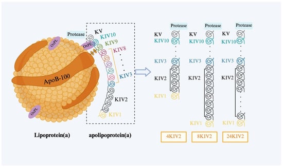
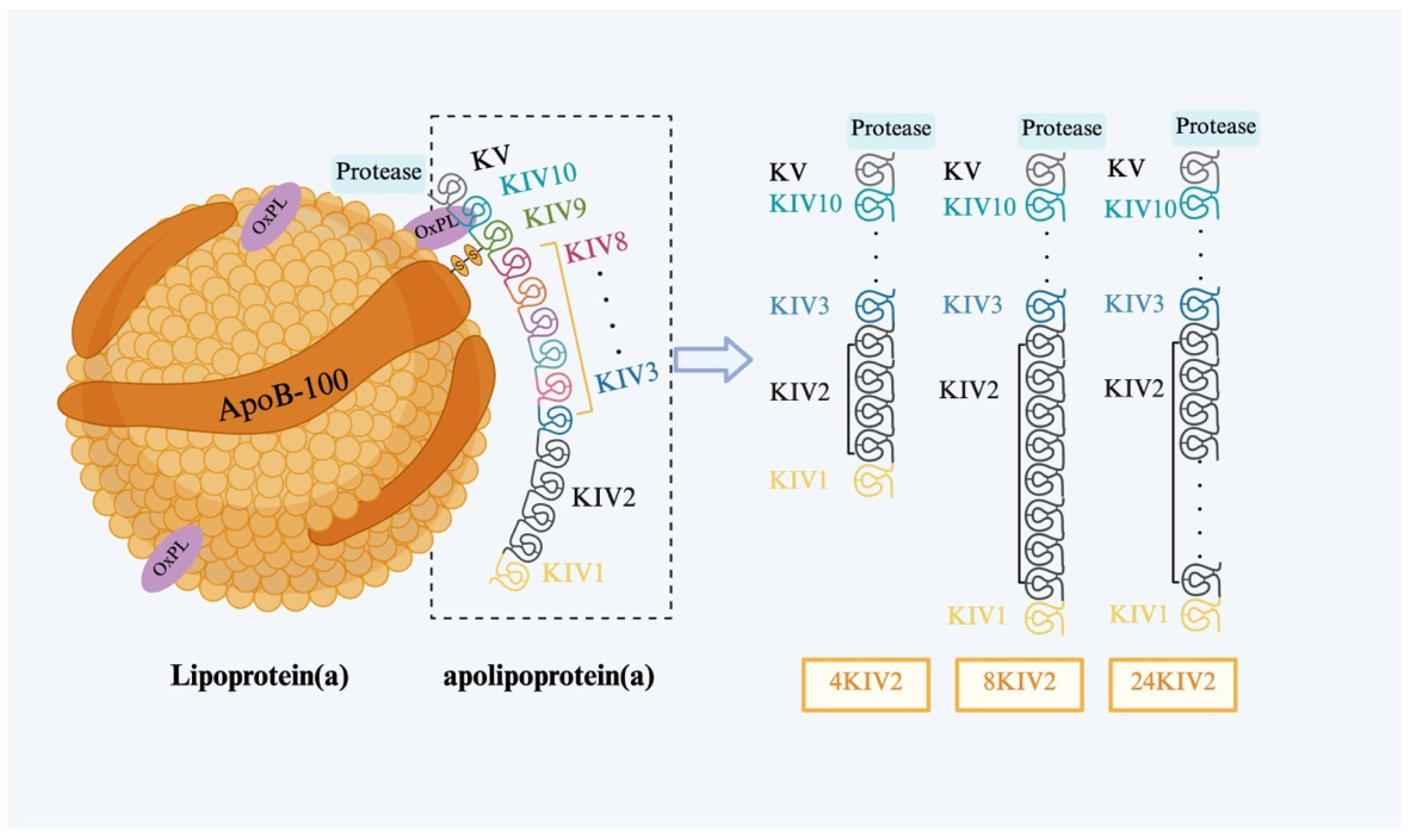
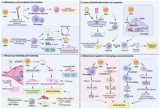
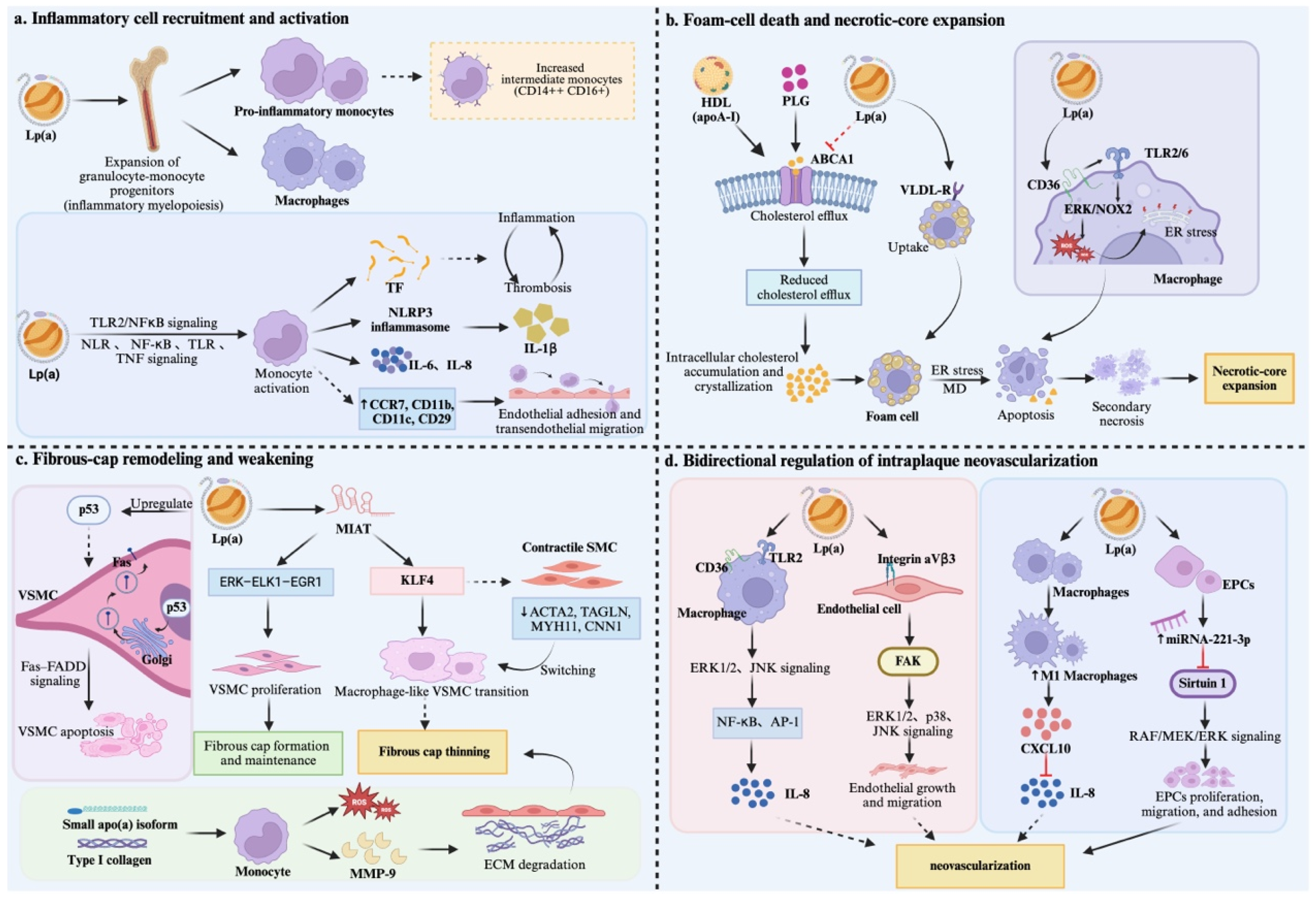

Disclaimer/Publisher’s Note: The statements, opinions and data contained in all publications are solely those of the individual author(s) and contributor(s) and not of MDPI and/or the editor(s). MDPI and/or the editor(s) disclaim responsibility for any injury to people or property resulting from any ideas, methods, instructions or products referred to in the content. |
© 2026 by the authors. Licensee MDPI, Basel, Switzerland. This article is an open access article distributed under the terms and conditions of the Creative Commons Attribution (CC BY) license.