


Abstract
Although assisted reproductive technology (ART) has been widely applied worldwide, thin endometrium (TE), diminished ovarian reserve (DOR), and other conditions remain the core bottlenecks of refractory infertility, and existing therapies fail to effectively preserve tissue and prevent functional damage. Platelet-rich plasma (PRP), as an autologous regenerative agent, has demonstrated promising application potential in reproductive medicine due to its multi-targeted regulatory capacity. Therefore, this review is intended as a narrative overview that aims to systematically elucidate the possible mechanisms by which PRP improves the endometrial microenvironment and ovarian function, integrate the latest clinical evidence to evaluate its therapeutic efficacy, analyze translational challenges such as the lack of standardization, and provide an evidence-based basis for its standardized clinical application.
1. Introduction
In recent years, assisted reproductive technology (ART) has achieved technological iteration and clinical popularization globally, providing core fertility solutions for infertile families. However, thin endometrium (TE), recurrent implantation failure (RIF), diminished ovarian reserve (DOR), and intrauterine adhesion (IUA) remain the core clinical bottlenecks that urgently need to be broken through in the field of reproductive medicine. They are also the key factors in ART cycle cancellation, embryo implantation failures, and pregnancy losses. Epidemiological data show that the incidence of TE (midluteal endometrial thickness ≤ 7 mm) is approximately 2–3% in IVF cycles [1], and insufficient endometrial thickness can lead to adverse perinatal outcomes such as reduced pregnancy rate and live birth rate [2]. RIF, defined as a clinical phenomenon characterized by a lack of implantation after the transfer of several embryos, affects approximately 10% of the ART population, and there is currently no standard diagnostic and treatment protocol [3]. The incidence of DOR is increasing with the delay in female childbearing age and iatrogenic injuries, and due to its unclear mechanism and complex clinical features, there is still a lack of effective means to reverse follicular depletion [4], which significantly restricts pregnancy outcomes. In addition, chronic endometritis (CE), premature ovarian insufficiency (POI), and severe oligoasthenoteratozoospermia in males further complicate the treatment of infertility. The common pathological basis of these challenges lies in the damage to local tissue microvascular networks, decreased stem cell activity, and imbalance of extracellular matrix remodeling, which urgently require novel intervention strategies with multi-targeted regenerative potential.
In the 1990s, Marx et al. first proposed the concept of platelet-rich plasma (PRP) in the field of regenerative medicine, aiming to explore the therapeutic potential of platelet-derived growth factors in tissue healing [5]. With its characteristics of being enriched by centrifugation of autologous peripheral blood, low immunogenicity, and high concentrations of growth factors and cytokines, it has been successfully translated into clinical practice in orthopedics, dermatology, and wound regeneration [6]. In the past five years, the regenerative potential of PRP has been innovatively introduced into the field of reproductive medicine. After activation, it releases active molecules such as VEGF, PDGF, TGF-β, IGF-1, and IL-10, which can synergistically regulate angiogenesis, establish an anti-inflammatory microenvironment, and recruit tissue stem cells through paracrine pathways [7]. Multiple clinical observational studies and early randomized controlled trials (RCTs) have preliminarily provided evidence suggesting that intrauterine PRP infusion can significantly improve endometrial thickness and effectively enhance pregnancy outcomes in patients with thin endometrium [8]. Local ovarian injection of PRP has shown the potential to increase antral follicle count (AFC) and the number of retrieved oocytes in some patients with DOR [9]. Local application may also be associated with exploratory value in improving semen parameters, cryopreservation effects, reducing oxidative stress, and improving the testicular microenvironment in males [10].
Despite the promising regenerative potential of PRP in reproductive medicine, its clinical translation is still limited by insufficient standardization of preparation and weak mechanistic evidence. Currently, there are significant differences among studies in terms of blood collection volume, centrifugation gradient, leukocyte retention strategy (P-PRP/L-PRP), and activator selection, leading to high heterogeneity in platelet concentration multiples and growth factor release profiles [11], which severely impairs the comparability between studies. Furthermore, the molecular mechanisms underlying PRP-mediated reproductive regulation remain incompletely elucidated, particularly the action targets, signaling networks, and time-dependent characteristics in human ovarian tissues, which warrant further investigation via multi-omics and single-cell sequencing technologies. Additionally, the absence of standardized guidelines for clinical operational parameters (including indications, administration routes, dosages, intervention timings, and treatment cycles) severely restricts its precise clinical application and safe dissemination.
Based on this, this article reviews the latest research progress of PRP therapy in reproductive medicine, elaborates on its molecular mechanisms and cellular biological effects in endometrial receptivity reconstruction and ovarian microenvironment improvement, integrates and analyzes the efficacy data of high-quality clinical studies from this year, and discusses the influencing factors, limitations, and core controversies of current PRP clinical application. This study aims to provide a theoretical reference for deepening basic research on PRP in reproductive medicine, offer an evidence-based basis for its standardized and precise clinical application, and ultimately promote the effective and safe translation of PRP technology in reproductive medicine, providing a novel therapeutic option for patients with refractory infertility. Given the broad scope and heterogeneous nature of the available evidence, this article is designed as a narrative rather than a systematic review.
2. Overview and Preparation Technology of PRP
2.1. Definition and Classification System
Platelet-rich plasma refers to a plasma derivative obtained by centrifugation of autologous peripheral venous blood with a platelet concentration significantly higher than the baseline level (usually 2–7 times) [12]. According to leukocyte content and fibrin network structure, PRP and its derivative products can be systematically divided into four categories: pure platelet-rich plasma (P-PRP), leukocyte- and platelet-rich plasma (L-PRP), pure platelet-rich fibrin (P-PRF), and leukocyte and platelet-rich fibrin (L-PRF) [13]. The core biological activity of PRP originates from the abundant bioactive molecules stored in platelet α-granules, mainly including transforming growth factor beta (TGF-β1), platelet-derived growth factor (PDGF), vascular endothelial growth factor (VEGF), and insulin-like growth factor (IGF), which can synergistically drive tissue regeneration, angiogenesis, immune regulation, and extracellular matrix remodeling [14]. These biological characteristics make PRP a promising regenerative intervention in reproductive medicine, which can specifically address the core pathological bases of refractory infertility caused by endometrial and ovarian lesions, including damage to microvascular networks, decreased stem cell activity, and imbalance of extracellular matrix remodeling.
2.2. Standardized Preparation: A Critical Appraisal of Current Heterogeneity
Although the two-step gradient centrifugation method is the mainstream approach for PRP preparation in reproductive medicine [15], a critical examination of the literature reveals that there is no universally accepted “gold standard” [16]. A scoping review reported marked heterogeneity across all steps of PRP preparation, encompassing anticoagulant selection, centrifugation protocols, activation methods, and other critical parameters [17]. This heterogeneity severely compromises the comparability of clinical studies and impedes the establishment of evidence-based guidelines.
The variability in PRP preparation encompasses multiple interrelated domains.
- Variations in centrifugation parameters (relative centrifugal force, duration, temperature) can lead to differences in the platelet concentration and growth factor content of the product. When comparing the first centrifugation parameters (RCF 200–400 g, temperature 5–19 °C, duration 5–19 min) to the second centrifugation parameters (RCF 200–800 g, duration 5–19 min, temperature fixed at 12 °C), Amable et al. found that these different parameters resulted in a fluctuation of platelet recovery rates ranging from 46.9% to 69.5%, with the final platelet concentration varying from 1.4 × 10⁶ to 1.9 × 10⁶ platelets/μL (an increase of 5.4 to 7.3 times) [18]. Importantly, a systematic review found that centrifugation RCF was reported in only 6.7% of included studies, highlighting a critical reporting deficit [6].
- Activation methods also vary considerably (calcium alone, calcium/thrombin, or thermal activation), with significant effects on cytokine release kinetics. Calcium/thrombin activation increases overall cytokine release over seven days, while calcium-only activation sustains VEGF and IGF concentrations in double-spin preparations [19].
- Although there has been ongoing debate over the pros and cons of including white blood cells in PRP [20,21], studies by Kobayashi et al. have found that the level of growth factors is significantly influenced by the concentration of white blood cells: leukocyte concentration positively correlates with PDGF-BB and VEGF but negatively with FGF-β, and MMP-9 (a catabolic factor) correlates only with leukocyte concentration, not platelet concentration [22]. This finding underscores the need for method selection according to target pathology.
- Anticoagulant choice (EDTA, ACD-A, sodium citrate) and commercial preparation systems further contribute to product heterogeneity. Citrate-based anticoagulants are preferred over EDTA (which may damage platelet membranes) [23], and a comparison of seven commercial systems found that platelet and leukocyte concentrations differed largely between systems, with large differences observed for all growth factors both between and within systems [24]. European regulations, governed by directives such as 2002/98/EC and 2005/62/EC, require adherence to Good Manufacturing Practices for blood and blood components, including PRP [25]. Failure to strictly adhere to manufacturing specifications may result in suboptimal platelet concentrations within PRP, thereby affecting its therapeutic efficacy.
In summary, variability in centrifugation parameters, activation methods, leukocyte content, anticoagulants, and commercial systems yields PRP products with divergent biological compositions across studies. This heterogeneity represents a major barrier to evidence-based translation of PRP therapy in reproductive medicine. To ensure safety, efficacy, and reproducibility, unified quality control and reporting standards are urgently needed.
3. Improving the Microenvironment of the Endometrium Through PRP
3.1. Mechanism of Action (Figure 1)
The core of PRP improving the endometrial microenvironment lies in the release of bioactive substances such as growth factors, anti-inflammatory cytokines, and matrix metalloproteinases after enrichment by autologous blood centrifugation and in vitro activation, completing structural repair and functional remodeling of the damaged endometrium through multi-targeted and multi-pathway synergistic cascade effects [26]. High levels of VEGF and PDGF-BB in PRP bind to VEGFR2 and PDGFRβ receptors on endothelial cells, activating the PI3K/Akt and ERK1/2 signaling pathways, which significantly enhances endometrial spiral artery branching and capillary network density, thereby reversing insufficient blood perfusion in the thin endometrium [27,28]. While promoting angiogenesis, PRP releases abundant growth factors, including VEGF, PDGF and TGF-β, and upregulates the expression of multiple proliferation markers in endometrial stromal cells, namely cytokeratin, Ki-67 and HOXA10 [29], stimulating endothelial cell proliferation and inducing endometrial regeneration. Building on cellular proliferation and angiogenesis, PRP releases chemokines, including CCL5, CCL7, CXCL13 and lipoxin A4, and simultaneously inhibits the phosphorylation of NF-κB [30,31]. This effectively suppresses excessive inflammatory responses associated with chronic endometritis, establishes the immune-tolerant microenvironment required for embryo implantation, and prevents fetal rejection. The establishment of immune homeostasis further regulates the dynamic homeostasis of the ECM: the expression levels of COL1A1, TIMP-1, TGF-β1 and TNF-α are significantly downregulated, while the expression of MMP1, MMP3, MMP7 and MMP26 is upregulated [28]. By modulating the expression ratio of these factors, PRP promotes functional collagen deposition while inhibiting pathological fibrosis, providing structural support for tissue integrity reconstruction after hysteroscopic adhesiolysis. Additional studies have demonstrated that PRP can also inactivate the TGF-β1-SMAD2/3 signaling pathway and downregulate the expression of fibrosis-related genes [32]. It should be noted that the mechanistic insights presented in this section are derived from a combination of preclinical studies (including animal models and in vitro experiments using human-derived cell lines) and clinical observational data. Unless otherwise specified, findings related to specific signaling pathways (e.g., PI3K/Akt, TGF-β1-SMAD2/3) are primarily based on preclinical evidence, whereas endometrial receptivity markers (e.g., HOXA10, LIF) and clinical outcomes have been validated in human studies.
Figure 1.
Mechanism by which PRP remodels the endometrial microenvironment and promotes embryo implantation. This figure depicts the preparation of PRP from autologous peripheral blood through collection and centrifugation: (1) angiogenesis and improved blood perfusion; (2) cell proliferation and endometrial regeneration; (3) anti-inflammatory and immunomodulatory effects; (4) ECM remodelling and antifibrotic effects; (5) endometrial receptivity restoration. These regulatory actions ultimately produce tissue-level improvements: increased endometrial thickness, elevated vascular density, enhanced epithelial and stromal proliferation, attenuated inflammatory response, reduced fibrosis and improved endometrial receptivity. Consequently, PRP treatment significantly elevates embryo implantation rate and clinical pregnancy rate, while decreasing the risks of recurrent implantation failure, intrauterine adhesion and potential miscarriage.
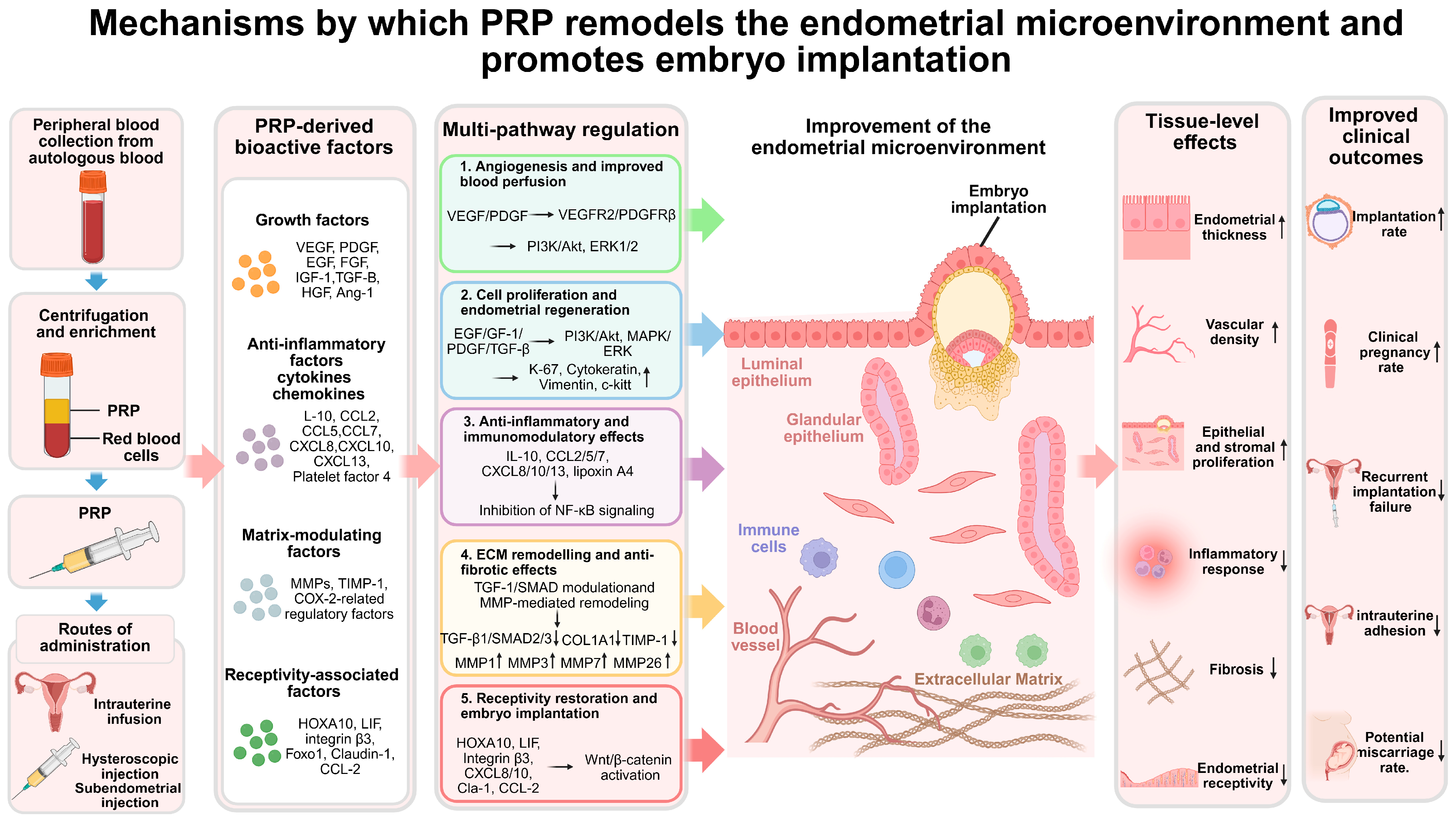
Ultimately, A10. LIF and integrin β3 during the window of implantation, but also releases chemokines including CCL2, CXCL8, CXCL10 and claudin-1 [33], optimizing the embryo-endometrium molecular compatibility and thereby establishing a stable pregnancy microenvironment for RIF patients.
3.2. Clinical Efficacy (Table 1)
Intrauterine application of PRP has become an important regenerative treatment for endometrial-derived infertility, such as TE, RIF, and IUA. Its core clinical benefits are concentrated in the significant increase in endometrial thickness and the substantial improvement in clinical pregnancy rate. Multiple recently published high-quality randomized controlled trials (RCTs) and meta-analyses have provided data supporting its clinical application value in assisted reproductive technology [33,34].
Table 1.
The preceding data in the table are from the control group (patients received routine basic care without PRP treatment), while the subsequent data are from the experimental group (patients received PRP intrauterine perfusion or subendometrial injection). Both groups include subjects’ basic information (age, infertility duration), core uterine assessment indicators (endometrial thickness, endometrial pattern, blood flow resistance index before and after treatment), and pregnancy outcome data (embryo implantation rate, clinical pregnancy rate, miscarriage rate), providing direct evidence for quantifying and comparing the clinical efficacy of PRP in improving endometrial receptivity and enhancing pregnancy success rate.
- (1)
- Improvement effect of PRP on endometrial thickness
TE with endometrial thickness ≤ 7 mm in the mid-luteal phase is an important clinical indicator leading to ART cycle cancellation and embryo implantation failure [27]. Its core pathological feature is impaired endometrial proliferation capacity and insufficient blood perfusion, and PRP can effectively stimulate endometrial growth and achieve an increase in endometrial thickness by releasing a variety of growth factors and bioactive substances [52]. A systematic review and meta-analysis including 2152 patients with thin endometrium showed that among six intervention measures compared with the control group, intrauterine PRP infusion was associated with a statistically significant positive effect in improving endometrial thickness (MD = 0.989, 95% CrI = [0.612, 1.36]), and the PRP group showed the most favorable outcomes in terms of both endometrial thickness improvement and clinical pregnancy rate increase [53]. A retrospective cohort study further suggested that a single PRP infusion was sufficient to increase the median endometrial thickness from 6.5 (IQR 1.0) to 7.3 mm (IQR 1.3) (p < 0.0001) overall [38].
Subgroup analysis of different administration routes showed that subendometrial PRP injection was associated with better pregnancy outcomes. Clinical data showed that the average endometrial thickness with this administration method was 8.31 mm, significantly higher than 7.42 mm in the intrauterine PRP infusion group [41]. The superior efficacy of subendometrial injection is biologically plausible, as direct delivery of platelets to the endometrial stem/progenitor cell niche at the basalis-functional interface may enhance growth factor bioavailability and tissue penetration compared to passive diffusion from the uterine cavity. In addition, dosing frequency represents an additional variable that may influence PRP efficacy. Feng et al. [35] conducted a prospective randomized trial comparing single versus double intrauterine PRP infusions in thin endometrium patients, demonstrating that the double-infusion regimen (1.0 mL on days 11 and 13 of the HRT cycle) yielded a significantly greater increase in endometrial thickness compared to a single infusion on day 11 alone (an additional 0.46 mm, p < 0.01), along with improved uterine hemodynamic parameters. This suggests a cumulative dose–response effect for endometrial applications.
Regarding timing, the interval between PRP administration and embryo transfer varies considerably across studies: Liu et al. administered PRP 24–72 h before FET [50], while Nazari et al. used a precise 48-h interval [48]. Given that activated PRP releases the majority of its growth factors within the first 24 h, with sustained release over 7 days [19], this 24- to 72-h variability may significantly impact the synchronization between peak growth factor concentrations and the endometrial receptivity window. However, no study has systematically compared different timing protocols, representing a critical knowledge gap.
For women with intrauterine adhesions (IUA), intrauterine PRP infusion can effectively inhibit endometrial fibrosis and promote epithelial regeneration. RCT data suggested that the increase in endometrial thickness in patients using PRP was significantly higher than that in patients not using PRP (p = 0.03 and 0.04) [54], laying the necessary structural foundation for embryo adhesion and invasion.
- (2)
- Improvement effect of PRP on clinical pregnancy rate
The improvement in endometrial thickness and upregulation of receptivity ultimately translate into significant benefits in ART cycle pregnancy outcomes. A meta-analysis of 861 patients with thin endometrium, implantation, or pregnancy failure undergoing assisted reproductive technology showed that the clinical pregnancy rate in the PRP group was 2.51 times higher than that in the control group (RR: 2.51; 95% CI: 2.0–3.13; p < 0.00001), with no significant heterogeneity among studies (I2 = 0.0%, p < 0.00001). Moreover, the live birth rate in the PRP group was 7.03 times that in the control group (RR: 7.03; 95% CI: 3.91–12.6; p < 0.00001), suggesting a potential benefit of this therapeutic approach.
Data from a single-center self-controlled before-and-after study showed that the clinical pregnancy rate of patients before LP-PRP treatment was only 24.5%, which significantly increased to 45.3% after treatment (p = 0.022). Meanwhile, the embryo live birth rate per cycle was significantly higher than that in the control group (42.2% vs. 18.5%, p = 0.009), indicating clear clinical translation value [51]. It is worth noting that this study did not compare the efficacy of LP-PRP and LR-PRP at the same time, and many studies lacked a direct comparison of different PRP types in terms of reproductive indications, so it was unable to draw a clear conclusion on the optimal composition of white blood cells.
For patients with RIF, an RCT [55] including 40 subjects found that the clinical pregnancy rate in the intrauterine PRP infusion group reached 55.0%, significantly higher than 20.0% in the control group (p ≤ 0.05). At the same time, the intrauterine adhesion score decreased compared with that before PRP treatment, and no serious treatment-related adverse events were observed.
A systematic review and meta-analysis [33] published in 2024 further indicated that for patients receiving ART treatment, intrauterine PRP infusion not only significantly increased endometrial thickness but also was associated with the clinical pregnancy rate by 2.04 times (RR: 2.04, 95% CI: 1.52 to 2.76, p = 0.000). Moreover, the cycle cancellation rate in the PRP group was significantly lower than that in the control group (RR: 0.46, 95% CI: 0.23 to 0.93, p = 0.000), suggesting enhanced clinical pregnancy benefits.
4. Improving Ovarian Function Through PRP
4.1. Mechanism of Action (Figure 2)
PRP-mediated ovarian function restoration follows a cascade regulatory network of “microenvironmental remodeling → cell survival → metabolic reprogramming → follicle activation” [56]. Studies have indicated that PRP treatment upregulates HIF-1α expression, thereby promoting VEGF release from activated platelet α-granules, and VEGF binds to VEGFR2 on endothelial cells, which undergoes autophosphorylation to activate the VEGFA/VEGFR2/ERK pathway and trigger the PI3K/AKT and Ras/MAPK pathways, rapidly reconstructing the ovarian cortical microvascular network and alleviating tissue hypoxia [57,58,59]. At the granulosa cell level, IGF-1, PDGF and EGF synergistically activate the PI3K/Akt/mTOR survival pathway, upregulate Bcl-2 expression and significantly promote FOXO3a transcription [60,61,62], thereby facilitating follicle activation, granulosa cell proliferation, and inhibiting cellular apoptosis. Targeting the core pathology of oocyte senescence, the active components of PRP induce the upregulation of antioxidant enzyme systems, including Cox7a2 and Mtch2, and downregulate the expression of Prdx2 and Numa1 proteins to scavenge mitochondrial ROS, which also activates PGC-1α to drive mtDNA replication and transcription, thereby maintaining ATP synthesis and meiotic spindle integrity [63,64].
Figure 2.
Mechanism of PRP in improving ovarian function and reproductive outcomes. This figure demonstrates that following intraovarian injection, the active components released by PRP—including growth factors, cytokines, chemokines/adhesion factors, and other bioactive molecules—exert synergistic effects via five core pathways: (1) angiogenesis and hypoxia relief; (2) primordial follicle activation; (3) granulosa cell proliferation and anti-apoptosis; (4) oocyte mitochondrial function improvement; (5) anti-inflammatory and antioxidant microenvironment optimization. These regulatory actions reverse pathological conditions such as ovarian vascular atrophy, reactive oxygen species (ROS) accumulation, granulosa cell apoptosis, and mitochondrial damage, thereby comprehensively improving the ovarian microenvironment. Ultimately, PRP treatment leads to improvements in ovarian reserve markers (anti-Müllerian hormone [AMH], follicle-stimulating hormone [FSH], antral follicle count [AFC], and ovarian blood flow), enhanced efficiency at each stage of assisted reproductive technology (ART), and significantly elevated pregnancy rates and potential live birth rates in patients with diminished ovarian reserve (DOR), poor ovarian response (POR), and premature ovarian insufficiency (POI).
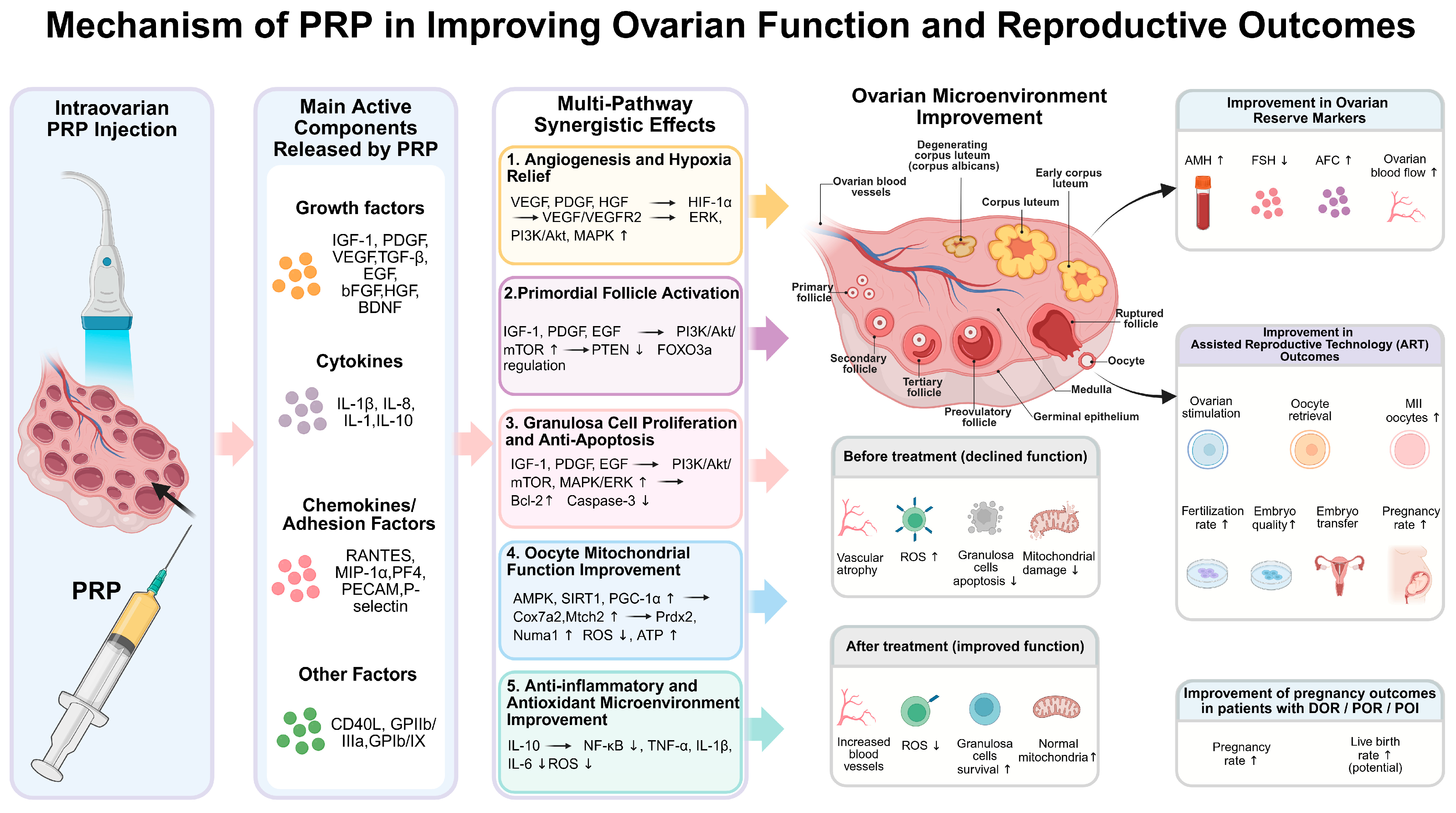
These metabolic reprogramming effects ultimately converge on the precise activation of the primordial follicle pool: PRP reversibly inhibits PTEN to relieve excessive silencing of the PI3K/AKT pathway, promoting dormant follicle entry into the growth phase, which is clinically manifested as a synchronous increase in AMH and AFC [56]. Furthermore, PRP treatment significantly reduces TNF-α levels, and PRP-released cytokines further inhibit the NF-κB pathway [65], collectively establishing a hypoinflammatory, highly vascularized ovarian regenerative microecology. It should also be noted that the following mechanistic framework is primarily derived from preclinical studies, including rodent models of premature ovarian insufficiency and in vitro experiments using human ovarian tissue or granulosa cells. Where human clinical data support these mechanisms—such as changes in AMH, FSH, and AFC following intraovarian PRP injection—these are clinical correlations rather than direct mechanistic evidence.
4.2. Clinical Efficacy (Table 2)
Intraovarian injection of PRP is a promising regenerative intervention for refractory ovarian-derived infertility, such as DOR, poor ovarian response (POR), and POI in the field of assisted reproductive technology, significantly improving the core ovarian reserve via elevated AMH, reduced FSH and increased AFC.
Table 2.
The preceding data in the table are from the control group, and the subsequent data are from the experimental group. Both groups include the basic information of subjects (age, infertility duration), core ovarian reserve indicators (anti-Müllerian hormone [AMH], follicle-stimulating hormone [FSH], antral follicle count [AFC] before and after treatment), and assisted reproductive technology (ART)-related outcome data, providing direct evidence for quantifying and comparing the clinical efficacy of PRP in improving ovarian function.
- (1)
- Improvement effect of PRP on core endocrine markers of ovarian reserve
Serum AMH and FSH are the core endocrine markers for evaluating ovarian reserve function, which can objectively reflect ovarian function status from two dimensions: follicle pool reserve level and ovarian endocrine feedback regulation ability [71]. Intraovarian injection of PRP can simultaneously achieve an increase in serum AMH level and a decrease in FSH level in patients with diminished ovarian function by activating primordial follicles, improving granulosa cell function, and reconstructing the ovarian cortical microenvironment [72], providing direct endocrinological evidence for the improvement of ovarian reserve function.
A systematic review and meta-analysis [9] including 2256 patients with diminished ovarian reserve showed that after intraovarian PRP injection, the level of AMH increased (3 months MD 0.36, p = 0.002, 95% CI: [0.20; 0.52]) and the level of FSH decreased significantly (3 months MD −8.87, p = 0.010, 95% CI: [−14.19; −3.55]). In this patient population, the reported rates were 18% for clinical pregnancy and 11% for live birth.
A retrospective study of 60 patients with poor ovarian response undergoing ART showed that after ultrasound-guided intraovarian PRP injection, the baseline mean AMH level of patients significantly increased from 0.8 ng/mL to 2.7 ng/mL, the baseline mean FSH level decreased from 12.9 IU/mL to 11.1 IU/mL (p = 0.002), and endometrial thickness also significantly increased (from 4.4 mm to 9.6 mm) [69].
A prospective controlled, non-randomized comparative study [66] published in 2020 showed that women treated with PRP experienced a significant improvement in FSH, AMH, and AFC, whereas there was no change in the control group. Furthermore, overall rates of biochemical (26.1% versus 5.4%, p = 0.02) and clinical pregnancy (23.9% versus 5.4%, p = 0.03) were higher in the PRP group, suggesting additional benefits beyond conventional treatment.
- (2)
- Improvement effect of PRP on antral follicle count and key clinical indicators of ART cycles
AFC is an imaging indicator that intuitively reflects the number of recruitable follicles in the ovary, is directly related to the number of retrieved oocytes and embryo quality in ART cycles, and is a core indicator for predicting ovarian responsiveness [73]. Multiple clinical studies have suggested that intraovarian PRP injection can significantly increase the AFC level in patients with diminished ovarian function: an RCT including 83 subjects showed that the average AFC of patients increased by 3.0 after PRP treatment (p < 0.0001) [74]. An experimental study published in 2022 further suggested that PRP injection significantly increased the mean AFC of patients (from 1.60 ± 0. 84 to 2.50 ± 0.90, p = 0.043) [70].
On this basis, the improvement of ovarian reserve function by PRP can be further transformed into comprehensive benefits of key clinical indicators in ART cycles: data from a prospective study showed that after PRP treatment, the number of antral follicles in patients increased from 2.6 to 4.2 (p < 0.001), the number of mature oocytes increased from 1.7 to 2.7 (p < 0.001), while the average number of IVF attempts per patient decreased from 2.8 to 1.6 (p < 0.001), the fertilization rate significantly increased from 57.6% to 66.9%, and the blastocyst formation rate also showed a clinically significant increase, which may improve the probability of patients entering the embryo transfer stage [67].
Similarly, the ovarian response to PRP also exhibits dose-related variability. Guo and Kawamura, in a comprehensive review of ovarian function restoration using PRP, noted that while 2 mL per ovary is a commonly used dose in clinical practice, a dose of ≥4 mL per ovary may be associated with better follicle recruitment effects in patients with severe DOR, whereas excessive volume may cause high-pressure tissue damage [75]. This suggests a potential therapeutic window rather than a simple linear dose–response relationship for ovarian PRP.
5. Discussion
As an autologous regenerative agent, PRP has emerged as a research hotspot in reproductive medicine for refractory endometrial lesions and diminished ovarian function, owing to its multi-targeted tissue repair and microenvironment modulation capacities. While numerous high-quality studies have provided evidence supporting its efficacy in improving endometrial receptivity, enhancing ovarian reserve, and optimizing ART outcomes, substantial research heterogeneity, incomplete mechanistic elucidation, and the absence of standardized systems still impede its clinical translation.
5.1. Synergy and Specificity of PRP Mechanisms in Endometrial and Ovarian Repair
This review demonstrates that the core value of PRP in reproductive medicine lies in its multi-targeted regenerative potential, which can simultaneously address the two major problems of poor endometrial receptivity and diminished ovarian reserve function. Although the target organs of action are different, there is significant synergy in their core molecular mechanisms. The aforementioned mechanisms demonstrate that PRP exerts its effects in both the endometrium and ovarian tissue by releasing high concentrations of growth factors (e.g., VEGF, PDGF, IGF-1), which drive angiogenesis and inhibit apoptosis, thereby reversing tissue hypoxia and degeneration [28]. However, there are specific differences in the primary focus of its actions between these two tissues. In the endometrial microenvironment, the primary role of PRP is to create a “favorable niche” for embryo implantation by upregulating receptivity markers (e.g., HOXA10, LIF) and modulating the immune microenvironment via inhibition of the NF-κB pathway. In contrast, in ovarian function restoration, PRP predominantly focuses on achieving metabolic reprogramming from a “quiescent” to a “proliferative” state by scavenging mitochondrial reactive oxygen species (ROS) and activating the primordial follicle pool through inhibition of the PTEN pathway. This mechanistic difference explains why intrauterine infusion of PRP yields significant improvements in pregnancy rates, whereas intraovarian injection demonstrates greater potential in enhancing endocrine parameters (AMH/FSH).
5.2. Limitations of Current Mechanism Evidence
A notable limitation of the current mechanistic evidence is that the majority of signaling pathway data, including PI3K/Akt, TGF-β1-SMAD2/3, PTEN, and ROS scavenging pathways, are derived from preclinical models (rodent studies and in vitro experiments). While these pathways are biologically plausible and consistent with the known actions of platelet-derived growth factors, direct evidence in human endometrial and ovarian tissues remains limited. So, we believe future studies employing single-cell sequencing and spatial transcriptomics on human biopsy samples are needed to validate these mechanisms in the human reproductive tract.
5.3. Controversies over Standardization, Deficiency, and Dose Effects
The biggest challenge facing the current clinical translation of PRP is the lack of preparation and application standards. As shown in the literature review, there are huge differences among studies in terms of blood collection volume, centrifugation speed, activator use, and other aspects. This “black box” operation leads to high heterogeneity in platelet concentration multiples (usually 3–5 times [76]) and growth factor release profiles of PRP in different studies, severely impairing the comparability between studies.
Regarding the optimal dose, current evidence is contradictory. On the one hand, some studies suggest that 2 mL is a safe and effective dose, and excessive injection may cause ovarian tissue damage due to high pressure; on the other hand, data from patients with severe DOR show that a dose of ≥4 mL per ovary may be associated with better follicle recruitment effects [75]. This contradiction suggests that PRP may not have a simple linear dose–effect relationship but rather a “therapeutic window”. Future research urgently needs to establish an individualized dose calculation model based on ovarian tissue (assessed by 3D ultrasound) to avoid drug waste or tissue damage.
5.4. Safety Considerations and Future Prospects
Although PRP is derived from autologous blood and generally considered safe, its regenerative and mitogenic nature—characterized by high concentrations of VEGF, PDGF, IGF, and TGF-β—warrants a more comprehensive safety analysis beyond the current assertion that no serious adverse reactions have been observed. Severe infectious complications, including tubo-ovarian abscesses and Actinomyces abscess, have been reported following intraovarian PRP injection, although the true incidence remains unknown due to lack of systematic surveillance [77,78]. Beyond infection, intraovarian injection may also carry risks of ovarian tissue damage, bleeding, hematoma, and potential adhesion formation [79,80]. The mitogenic and angiogenic profile of PRP raises theoretical concerns about neoplastic cell proliferation, particularly in patients with hormone-dependent tumors: while no evidence currently links PRP to neoplastic transformation, and a 45-month follow-up of 89 breast cancer patients found no recurrence, the GRIIP 2025 formal consensus identifies certain types of oncologic diseases as a contraindication [80,81]. For patients at risk of hormone-dependent tumors, the long-term stimulatory effects of multiple angiogenic stimuli in PRP that promote tumor progression require longer follow-up data [82]. Given the lack of long-term safety data, PRP should be used with strict aseptic precautions and avoided in patients with active malignancy or hormone-dependent tumors. Future studies must systematically report adverse events, and large RCTs are urgently needed to confirm safety before routine clinical adoption.
Based on the above discussion, future research directions should focus on the following three points:
- Establishing a standardized system: Promote multi-center collaboration to formulate relatively unified PRP preparations (such as using closed automatic separation systems) and quality control standards;
- Precision stratified treatment: Clarify the optimal benefit thresholds for patients with different subtypes (such as different ages and different baseline FSH levels) through larger-scale RCTs;
- Exploration of combination therapies: Combine single-cell sequencing technology to deeply analyze the specific action network of PRP in the reproductive microenvironment, and explore its synergistic effects with antioxidants, growth hormone, or stem cell therapies [83].
6. Conclusions
In conclusion, Platelet-Rich Plasma (PRP) has emerged as a promising regenerative modality in reproductive medicine, offering a novel therapeutic avenue for refractory infertility conditions. This review synthesizes evidence suggesting that PRP effectively addresses the core pathophysiological bases of thin endometrium and diminished ovarian reserve through multi-targeted mechanisms, including angiogenesis promotion, anti-inflammatory immunomodulation, and metabolic reprogramming. However, PRP’s clinical application remains investigational due to non-standardized preparation protocols, heterogeneous dosing and the predominantly preclinical nature of current mechanistic evidence. Establishing unified quality control standards and conducting large-scale multi-center randomized controlled trials are essential to define the therapeutic window, ensure long-term safety, and advance PRP from empirical black-box therapy to precision medicine in reproductive practice.
Author Contributions
Conceptualization, X.Q. and Y.Y.; validation, X.Q., T.Y. and Y.X.; formal analysis, X.Q., T.Y. and X.L.; investigation, L.B. and Y.X.; resources, L.B.; data curation, T.Y.; writing—original draft preparation, X.Q.; writing—review and editing, L.B. and H.S.; supervision, H.S.; project administration, Y.X. and X.L.; funding acquisition, Y.Y. All authors have read and agreed to the published version of the manuscript.
Funding
This research was funded by the Fund of the Gansu Youth Science and Technology Fund (25JRRA1015), Lanzhou University First Hospital Internal Fund (ldyyyn2025-58), Innovation, Entrepreneurship Project of Lanzhou University (20260060102) and Gansu Province Traditional Chinese Medicine Research Project (GZKG-2025-85).
Institutional Review Board Statement
Not applicable.
Informed Consent Statement
Not applicable.
Data Availability Statement
No new data were created or analyzed in this study. Data sharing is not applicable to this article.
Acknowledgments
We thank all the authors for their participation and the personnel for their contribution to the study. The authors have reviewed and edited the output and take full responsibility for the content of this publication.
Conflicts of Interest
The authors declare no conflict of interest.
Abbreviations
The following abbreviations are used in this manuscript:
| ART | Assisted reproductive technology |
| TE | Thin endometrium |
| DOR | Diminished ovarian reserve |
| PRP | Platelet-rich plasma |
| IUA | Intrauterine adhesion |
| RIF | Recurrent implantation failure |
| CE | Chronic endometritis |
| POI | Premature ovarian insufficiency |
| VEGF | Vascular Endothelial Growth Factor |
| PDGF | Platelet-Derived Growth Factor |
| TGF-β | Transforming Growth Factor-beta |
| IGF | Insulin-like Growth Factor |
| IL | Interleukin |
| AFC | Antral follicle count |
| Ki-67 | Antigen Ki-67 |
| HOX | Homeobox |
| CCL | Chemokine (C-C Motif) Ligand |
| CXCL | Chemokine (C-X-C Motif) Ligand |
| COL1A1 | Collagen Type I Alpha 1 Chain |
| TIMP | Tissue Inhibitor of Metalloproteinases |
| TNF-α | Tumor Necrosis Factor-alpha |
| MMP | Matrix Metalloproteinase |
| AMH | Anti-Müllerian Hormone |
| FSH | Follicle-Stimulating Hormone |
| ROS | Reactive Oxygen Species |
| PTEN | Phosphatase and Tensin Homolog |
| RCF | Relative Centrifugal Force |
| EDTA | Ethylenediaminetetraacetic acid |
| ACD | Acid-citrate Dextrose |
| GRIIP | International Research Group on Platelet Injections |
References
- Wang, Y.; Tang, Z.; Teng, X. New advances in the treatment of thin endometrium. Front. Endocrinol. 2024, 15, 1269382. [Google Scholar] [CrossRef] [Scilit] [PubMed]
- Zheng, Y.; Chen, B.; Dai, J.; Xu, B.; Ai, J.; Jin, L.; Dong, X. Thin endometrium is associated with higher risks of preterm birth and low birth weight after frozen single blastocyst transfer. Front. Endocrinol. 2022, 13, 1040140. [Google Scholar] [CrossRef] [Scilit] [PubMed]
- Ma, J.; Gao, W.; Li, D. Recurrent implantation failure: A comprehensive summary from etiology to treatment. Front. Endocrinol. 2023, 13, 1061766. [Google Scholar] [CrossRef] [Scilit] [PubMed]
- Zhao, G.; Hu, Y. Mechanistic insights into intrauterine adhesions. Semin. Immunopathol. 2025, 47, 3. [Google Scholar] [CrossRef] [Scilit] [PubMed]
- Everts, P.A.; Lana, J.F.; Alexander, R.W.; Dallo, I.; Kon, E.; Ambach, M.A.; van Zundert, A.; Podesta, L. Profound Properties of Protein-Rich, Platelet-Rich Plasma Matrices as Novel, Multi-Purpose Biological Platforms in Tissue Repair, Regeneration, and Wound Healing. Int. J. Mol. Sci. 2024, 25, 7914. [Google Scholar] [CrossRef] [Scilit] [PubMed]
- Rahman, E.; Rao, P.; Abu-Farsakh, H.N.; Thonse, C.; Ali, I.; Upton, A.E.; Baratikkae, S.Y.; Carruthers, J.D.A.; Mosahebi, A.; Heidari, N.; et al. Systematic Review of Platelet-Rich Plasma in Medical and Surgical Specialties: Quality, Evaluation, Evidence, and Enforcement. J. Clin. Med. 2024, 13, 4571. [Google Scholar] [CrossRef] [Scilit] [PubMed]
- Moradian, S.A.; Amirkhani, Z.; Movahedin, M.; Gharib, S.N.; Varghaiyan, Y.; Niknafs, B.; Hamdi, K.; Hajipour, H.; Fattahi, A. Platelet-rich plasma (PRP) and the future of male fertility: A path forward for personalized and regenerative therapies. Stem Cell Res. Ther. 2025, 16, 486. [Google Scholar] [CrossRef] [Scilit] [PubMed]
- Maleki-Hajiagha, A.; Razavi, M.; Rouholamin, S.; Rezaeinejad, M.; Maroufizadeh, S.; Sepidarkish, M. Intrauterine infusion of autologous platelet-rich plasma in women undergoing assisted reproduction: A systematic review and meta-analysis. J. Reprod. Immunol. 2020, 137, 103078. [Google Scholar] [CrossRef] [Scilit] [PubMed]
- Éliás, M.; Kónya, M.; Kekk, Z.; Turan, C.; das Virgens, I.P.A.; Tóth, R.; Keszthelyi, M.; Hegyi, P.; Várbíró, S.; Sipos, M. Platelet-rich plasma (PRP) treatment of the ovaries significantly improves fertility parameters and reproductive outcomes in diminished ovarian reserve patients: A systematic review and meta-analysis. J. Ovarian Res. 2024, 17, 104. [Google Scholar] [CrossRef] [Scilit] [PubMed]
- Pang, K.H. The role and implication of platelet-rich plasma in male factor infertility: A systematic review of human studies. Andrology 2026, 14, 284–293. [Google Scholar] [CrossRef] [Scilit] [PubMed]
- Petrović, D.; Dinić, M.; Švraka, D.; Pantović, V.; Kostova, E.P.; Malenković, G.; Ljubić, A. Platelet-Rich Plasma in Sperm Processing for Assisted Reproductive Technology: Molecular Mechanisms, Clinical Applications, and Future Directions. Int. J. Mol. Sci. 2026, 27, 2177. [Google Scholar] [CrossRef] [Scilit] [PubMed]
- Alexander, S.; Horo, I.; Johnson, S.; Daniel, S. Platelet-rich plasma in hair loss—Mechanism, preparation, and classification. J. Cosmet. Dermatol. 2022, 21, 970–978. [Google Scholar] [CrossRef] [Scilit] [PubMed]
- Bos-Mikich, A.; de Oliveira, R.; Frantz, N. Platelet-rich plasma therapy and reproductive medicine. J. Assist. Reprod. Genet. 2018, 35, 753–756. [Google Scholar] [CrossRef] [Scilit] [PubMed]
- Verma, R.; Kumar, S.; Garg, P.; Verma, Y.K. Platelet-rich plasma: A comparative and economical therapy for wound healing and tissue regeneration. Cell Tissue Bank. 2022, 24, 285–306. [Google Scholar] [CrossRef] [Scilit] [PubMed]
- Eftekhar, M.; Neghab, N.; Naghshineh, E.; Khani, P. Can autologous platelet rich plasma expand endometrial thickness and improve pregnancy rate during frozen-thawed embryo transfer cycle? A randomized clinical trial. Taiwan. J. Obstet. Gynecol. 2018, 57, 810–813, Corrigendum in Taiwan. J. Obstet. Gynecol. 2021, 60, 973. [Google Scholar] [CrossRef] [Scilit] [PubMed]
- Tey, R.V.; Haldankar, P.; Joshi, V.R.; Raj, R.; Maradi, R.; Ballini, A. Variability in Platelet-Rich Plasma Preparations Used in Regenerative Medicine: A Comparative Analysis. Stem Cells Int. 2022, 2022, 3852898. [Google Scholar] [CrossRef] [Scilit] [PubMed]
- Pachito, D.V.; Bagattini, Â.M.; de Almeida, A.M.; Mendrone-Júnior, A.; Riera, R. Technical Procedures for Preparation and Administration of Platelet-Rich Plasma and Related Products: A Scoping Review. Front. Cell Dev. Biol. 2020, 8, 598816. [Google Scholar] [CrossRef] [Scilit] [PubMed]
- Amable, P.R.; Carias, R.B.V.; Teixeira, M.V.T.; Pacheco, Í.d.C.; do Amaral, R.J.F.C.; Granjeiro, J.M.; Borojevic, R. Platelet-rich plasma preparation for regenerative medicine: Optimization and quantification of cytokines and growth factors. Stem Cell Res. Ther. 2013, 4, 67. [Google Scholar] [CrossRef] [Scilit] [PubMed]
- Roh, Y.H.; Kim, W.; Park, K.U.; Oh, J.H. Cytokine-release kinetics of platelet-rich plasma according to various activation protocols. Bone Jt. Res. 2016, 5, 37–45. [Google Scholar] [CrossRef] [Scilit] [PubMed]
- Wasterlain, A.S.; Braun, H.J.; Dragoo, J.L. Contents and Formulations of Platelet-Rich Plasma. Oper. Tech. Orthop. 2012, 22, 33–42. [Google Scholar] [CrossRef] [Scilit]
- Anitua, E.; Zalduendo, M.M.; Alkhraisat, M.H.; Orive, G. Release kinetics of platelet-derived and plasma-derived growth factors from autologous plasma rich in growth factors. Ann. Anat. 2013, 195, 461–466. [Google Scholar] [CrossRef] [Scilit] [PubMed]
- Kobayashi, Y.; Saita, Y.; Nishio, H.; Ikeda, H.; Takazawa, Y.; Nagao, M.; Takaku, T.; Komatsu, N.; Kaneko, K. Leukocyte concentration and composition in platelet-rich plasma (PRP) influences the growth factor and protease concentrations. J. Orthop. Sci. 2016, 21, 683–689. [Google Scholar] [CrossRef] [Scilit] [PubMed]
- García-Bordes, L.; Escutia-Marí, L.; Vizcaíno-Navarro, S.; Laiz-Boada, P.; Seijas-Vázquez, R.; Álvarez-Díaz, P.; Cuscó-Segarra, X.; Barastegui-Fernández, D.; Vázquez-Gómez, M.; Ayestaran-Calero, I.; et al. Anticoagulant-Dependent Platelet Morphological Artefacts in Platelet-Rich Plasma Preparation: A Prospective Paired Study Comparing EDTA and Sodium Citrate with Implications for Orthobiologic Therapy. Biomedicines 2026, 14, 1578. [Google Scholar] [CrossRef] [Scilit] [PubMed]
- Kushida, S.; Kakudo, N.; Morimoto, N.; Hara, T.; Ogawa, T.; Mitsui, T.; Kusumoto, K. Platelet and growth factor concentrations in activated platelet-rich plasma: A comparison of seven commercial separation systems. J. Artif. Organs 2014, 17, 186–192. [Google Scholar] [CrossRef] [Scilit] [PubMed]
- European Commission. Commission Directive 2005/62/EC of 30 September 2005 implementing Directive 2002/98/EC of the European Parliament and of the Council as regards Community standards and specifications relating to a quality system for blood establishments. Off. J. Eur. Union 2005, L256, 41–48. [Google Scholar]
- Karadbhajne, P.; Dzoagbe, H.Y.; More, A. Platelet-Rich Plasma (PRP) for Endometrial Treatment Efficacy and Safety in Assisted Reproductive Technology: A Comprehensive Review. Cureus 2024, 16, e59728. [Google Scholar] [CrossRef] [Scilit] [PubMed]
- Efendieva, Z.; Vishnyakova, P.; Apolikhina, I.; Artemova, D.; Butov, K.; Kalinina, E.; Fedorova, T.; Tregubova, A.; Asaturova, A.; Fatkhudinov, T.; et al. Hysteroscopic injections of autologous endometrial cells and platelet-rich plasma in patients with thin endometrium: A pilot randomized study. Sci. Rep. 2023, 13, 945. [Google Scholar] [CrossRef] [Scilit] [PubMed]
- Nagy, B.; Kovács, K.; Sulyok, E.; Várnagy, Á.; Bódis, J. Thrombocytes and Platelet-Rich Plasma as Modulators of Reproduction and Fertility. Int. J. Mol. Sci. 2023, 24, 17336. [Google Scholar] [CrossRef] [Scilit] [PubMed]
- Stefanović, M.; Vukomanović, P.; Kutlesic, R.; Trenkić, M.; Dimitrov, V.; Stefanović, A.; Cvetanović, V. The Effect of Autologous Platelet Rich Plasma on Endometrial Receptivity: A Narrative Review. Medicina 2025, 61, 134. [Google Scholar] [CrossRef] [Scilit] [PubMed]
- Lin, Y.; Qi, J.; Sun, Y. Platelet-Rich Plasma as a Potential New Strategy in the Endometrium Treatment in Assisted Reproductive Technology. Front. Endocrinol. 2021, 12, 707584. [Google Scholar] [CrossRef] [Scilit] [PubMed]
- Hajipour, H.; Farzadi, L.; Latifi, Z.; Keyhanvar, N.; Navali, N.; Fattahi, A.; Nouri, M.; Dittrich, R. An update on platelet-rich plasma (PRP) therapy in endometrium and ovary related infertilities: Clinical and molecular aspects. Syst. Biol. Reprod. Med. 2021, 67, 177–188. [Google Scholar] [CrossRef] [Scilit] [PubMed]
- Qi, J.; Li, X.; Cao, Y.; Long, Y.; Lai, J.; Yao, Y.; Meng, Y.; Wang, Y.; Chen, X.D.; Vankelecom, H.; et al. Locationally activated PRP via an injectable dual-network hydrogel for endometrial regeneration. Biomaterials 2024, 309, 122615. [Google Scholar] [CrossRef] [Scilit] [PubMed]
- Liu, X.; Qian, C.; Jiang, X.; Zhou, Y.; Feng, X.; Ding, Y.; Jin, J.; Hu, M.; Zhou, W.; Liu, B.; et al. Efficacy of platelet-rich plasma in the treatment of thin endometrium: A meta-analysis of randomized controlled trials. BMC Pregnancy Childbirth 2024, 24, 567. [Google Scholar] [CrossRef] [Scilit] [PubMed]
- Sharara, F.I.; Lelea, L.-L.; Rahman, S.; Klebanoff, J.S.; Moawad, G.N. A narrative review of platelet-rich plasma (PRP) in reproductive medicine. J. Assist. Reprod. Genet. 2021, 38, 1003–1012. [Google Scholar] [CrossRef] [Scilit] [PubMed]
- Feng, K.; Zhu, L.; Luo, Y.; Ruan, L.; Li, D.; Li, X.; Li, L.; Wu, L. Comparison of the efficacy of single vs. double autologous platelet-rich plasma intrauterine infusion on endometrial receptivity in thin endometrium patients: A prospective randomized controlled trial. Front. Endocrinol. 2025, 16, 1609556. [Google Scholar] [CrossRef] [Scilit] [PubMed]
- Yu, T.-N.; Lee, T.-H.; Lee, M.-S.; Chen, Y.-C.; Chen, C.-I.; Cheng, E.-H.; Lin, P.-Y.; Huang, C.-C.; Lee, C.-I. Intrauterine Infusion and Hysteroscopic Injection of Autologous Platelet-Rich Plasma for Patients with a Persistent Thin Endometrium: A Prospective Case–Control Study. J. Clin. Med. 2024, 13, 2838. [Google Scholar] [CrossRef] [Scilit] [PubMed]
- Shin, S.Y.; Chung, N.; Shin, J.E.; Kim, J.H.; Park, C.; Kwon, H.; Choi, D.H.; Lee, J.R.; Kim, J.H. Angiogenic factor-driven improvement of refractory thin endometrium with autologous platelet-rich plasma intrauterine infusion in frozen embryo transfer cycles. Front. Endocrinol. 2024, 15, 1431453. [Google Scholar] [CrossRef] [Scilit] [PubMed]
- Russell, S.J.; Kwok, Y.S.S.; Nguyen, T.T.-T.N.; Librach, C. Autologous platelet-rich plasma improves the endometrial thickness and live birth rate in patients with recurrent implantation failure and thin endometrium. J. Assist. Reprod. Genet. 2022, 39, 1305–1312. [Google Scholar] [CrossRef] [Scilit] [PubMed]
- Kim, H.; Shin, J.E.; Koo, H.S.; Kwon, H.; Choi, D.H.; Kim, J.H. Effect of Autologous Platelet-Rich Plasma Treatment on Refractory Thin Endometrium During the Frozen Embryo Transfer Cycle: A Pilot Study. Front. Endocrinol. 2019, 10, 61. [Google Scholar] [CrossRef] [Scilit] [PubMed]
- Wang, X.; Wang, Y.; Li, J.; Han, Y.; Wei, M.; Xu, A.; Xin, L. Effects of intrauterine infusion of autologous platelet-rich plasma gel on endometrial thickness and outcomes of frozen-thawed embryo transfer in women with thin endometrium. J. Assist. Reprod. Genet. 2024, 41, 2635–2647. [Google Scholar] [CrossRef] [Scilit] [PubMed]
- Zaha, I.A.; Huniadi, A.; Bodog, F.; Seles, L.; Toma, M.C.; Maghiar, L.; Szulay-Bimbo, E.; Bodog, A.; Sachelarie, L.; Florea, M.; et al. Autologous Platelet-Rich Plasma (PRP) in Infertility—Infusion versus Injectable PRP. J. Pers. Med. 2023, 13, 1676. [Google Scholar] [CrossRef] [Scilit] [PubMed]
- Yuan, B.; Luo, S.; Mao, J.; Luo, B.; Wang, J. Effects of intrauterine infusion of platelet-rich plasma on hormone levels and endometrial receptivity in patients with repeated embryo implantation failure. Am. J. Transl. Res. 2022, 14, 5651–5659. [Google Scholar] [PubMed]
- Pandey, D.; Bajaj, B.; Kapoor, G.; Bharti, R. Intrauterine instillation of autologous platelet-rich plasma in infertile females with thin endometrium undergoing intrauterine insemination: An open-label randomized controlled trial. Am. J. Obstet. Gynecol. Glob. Rep. 2023, 3, 100172. [Google Scholar] [CrossRef] [Scilit] [PubMed]
- Lin, P.-Y.; Lee, C.-I.; Chen, Y.-C.; Cheng, E.-H.; Huang, C.-C.; Chen, C.-I.; Lee, T.-H.; Lee, Y.-J.; Lee, M.-S. Factors Affecting the Potential Efficacy of Intrauterine Platelet-Rich Plasma Infusion on Thin Endometrium in Women with Recurrent Implantation Failure. J. Pers. Med. 2023, 13, 1419. [Google Scholar] [CrossRef] [Scilit] [PubMed]
- Kusumi, M.; Ihana, T.; Kurosawa, T.; Ohashi, Y.; Tsutsumi, O. Intrauterine administration of platelet-rich plasma improves embryo implantation by increasing the endometrial thickness in women with repeated implantation failure: A single-arm self-controlled trial. Reprod. Med. Biol. 2020, 19, 350–356. [Google Scholar] [CrossRef] [Scilit] [PubMed]
- Abbassi, R.; Haddad, S.; Haneyah, F.; Nakawa, W.; Murad, M.S.; Issa, A.M.; Alkheder, A.; Azar, A.; Bani Hani, M.; Abbassi, H. Treating refractory thin endometrium through a novel way of activation and administration of Platelet-rich plasma in sexually active women: An interventional prospective cohort clinical study. Medicine 2024, 103, e38554. [Google Scholar] [CrossRef] [Scilit] [PubMed]
- Bakhsh, A.S.; Maleki, N.; Sadeghi, M.R.; SadeghiTabar, A.; Tavakoli, M.; Zafardoust, S.; Karimi, A.; Askari, S.; Jouhari, S.; Mohammadzadeh, A. Effects of Autologous Platelet-Rich Plasma in Women with Repeated Implantation Failure Undergoing Assisted Reproduction. JBRA Assist. Reprod. 2022, 26, 84–87. [Google Scholar] [CrossRef] [Scilit] [PubMed]
- Nazari, L.; Salehpour, S.; Hosseini, S.; Sheibani, S.; Hosseinirad, H. The Effects of Autologous Platelet-Rich Plasma on Pregnancy Outcomes in Repeated Implantation Failure Patients Undergoing Frozen Embryo Transfer: A Randomized Controlled Trial. Reprod. Sci. 2022, 29, 993–1000. [Google Scholar] [CrossRef] [Scilit] [PubMed]
- Mehrafza, M.; Pourseify, G.; Zare Yousefi, T.; Raoufi, A.; Saghati Jalali, A.; Hosseinzadeh, E.; Samadnia, S.; Habibdoost, M.; Tamimi, A.; Hosseini, A. The Efficiency of Introducing Intrauterine Infusion of Autologous Platelet-Rich Plasma versus Granulocyte Colony-Stimulating Factor in Repeated Implantation Failure Patients: An Unblinded Randomised Clinical Trial. Int. J. Fertil. Steril. 2024, 18, 30–34. [Google Scholar] [CrossRef] [Scilit] [PubMed]
- Liu, X.-H.; He, L.-X.; Li, M.; Tian, Z.-P.; Qin, A.-Q.; Yao, J. Effects of autologous platelet-rich plasma intrauterine perfusion on clinical outcomes in recurrent implantation failure patients with non-thin endometrium undergoing frozen-thawed embryo transfer. Arch. Gynecol. Obstet. 2025, 311, 1423–1433. [Google Scholar] [CrossRef] [Scilit] [PubMed]
- Ban, Y.; Yang, X.; Xing, Y.; Que, W.; Yu, Z.; Gui, W.; Chen, Y.; Liu, X. Intrauterine Infusion of Leukocyte-Poor Platelet-Rich Plasma Is an Effective Therapeutic Protocol for Patients with Recurrent Implantation Failure: A Retrospective Cohort Study. J. Clin. Med. 2023, 12, 2823. [Google Scholar] [CrossRef] [Scilit] [PubMed]
- Yang, Y.; Zhang, X.; Zhang, Y. Platelet-rich plasma therapy for thin endometrium: A comprehensive review. Arch. Gynecol. Obstet. 2025, 312, 745–753. [Google Scholar] [CrossRef] [Scilit] [PubMed]
- Keng, F.; Ling, W.; Zhao, Z.; Luo, Y.; Li, X.; Li, D.; Zhong, J.; Lan, L.; Zhu, L. Network meta-analysis on the efficacy of different interventions for treating thin endometrium. Front. Endocrinol. 2025, 16, 1575248. [Google Scholar] [CrossRef] [Scilit] [PubMed]
- Martynov, S.A.; Adamyan, L.V.; Arakelyan, A.S.; Fyodorova, T.A.; Bystrykh, O.A. Possibilities of using platelet-rich autoplasm in the complex treatment of patients with intrauterine synechiae. Gynecology 2021, 23, 250–254. [Google Scholar] [CrossRef] [Scilit]
- Wang, Z.; Yang, M.; Mao, L.; Wang, X.; Wang, S.; Cui, G.; Hou, Z.; Yang, M.; Cui, L.; Bai, W. Efficacy and safety of autologous platelet-rich fibrin for the treatment of infertility with intrauterine adhesions. J. Obstet. Gynaecol. Res. 2021, 47, 3883–3894. [Google Scholar] [CrossRef] [Scilit] [PubMed]
- Ahmadi, F.; Gharaei, R.; Shirali, E.; Adabi, K.; Alishahi, V.; Tajaldini, M. The impact of platelet-rich plasma (PRP) on ovarian function and oocyte quality: A comprehensive review. J. Reprod. Immunol. 2025, 172, 104649. [Google Scholar] [CrossRef] [Scilit] [PubMed]
- Demyashkin, G.; Vadyukhin, M.; Murtazalieva, Z.; Pugacheva, E.; Schekin, V.; Bimurzaeva, M.; Pesegova, S.; Shegay, P.; Kaprin, A. Novel Molecular Mechanisms Underlying the Ameliorative Effect of Platelet-Rich Plasma against Electron Radiation-Induced Premature Ovarian Failure. Int. J. Mol. Sci. 2024, 25, 10115. [Google Scholar] [CrossRef] [Scilit] [PubMed]
- Su, L.; Xie, S.; Li, T.; Jia, Y.; Wang, Y. Pretreatment with platelet-rich plasma protects against ischemia–reperfusion induced flap injury by deactivating the JAK/STAT pathway in mice. Mol. Med. 2024, 30, 18. [Google Scholar] [CrossRef] [Scilit] [PubMed]
- Wei, W.; Xu, D.; Hu, F.; Jiang, T.; Liu, H. Platelet-rich plasma promotes wound repair in diabetic foot ulcer mice via the VEGFA/VEGFR2/ERK pathway. Growth Factors 2024, 42, 161–170. [Google Scholar] [CrossRef] [Scilit] [PubMed]
- Chen, C.; Jiang, X.; Ding, C.; Sun, X.; Wan, L.; Wang, C. Downregulated lncRNA HOTAIR ameliorates polycystic ovaries syndrome via IGF-1 mediated PI3K/Akt pathway. Gynecol. Endocrinol. 2023, 39, 2227280. [Google Scholar] [CrossRef] [Scilit] [PubMed]
- Weng, Y.; Zhang, W.; Qu, F.; Deng, Z.; Zhang, X.; Liu, S.; Wei, H.; Hao, T.; Gao, L.; Zhang, M.; et al. Human platelet-rich plasma promotes primordial follicle activation via the PI3K/Akt signaling pathway. Mol. Hum. Reprod. 2025, 31, gaaf007. [Google Scholar] [CrossRef] [Scilit] [PubMed]
- Su, Y.-Q.; Yin, Y.; Guo, J.; Gong, X.; Tian, Y.; Shi, L. MTOR-mediated interaction between the oocyte and granulosa cells regulates the development and function of both compartments in mice. Biol. Reprod. 2022, 107, 76–84. [Google Scholar] [CrossRef] [Scilit] [PubMed]
- Ju, W.; Zhao, Y.; Yu, Y.; Zhao, S.; Xiang, S.; Lian, F. Mechanisms of mitochondrial dysfunction in ovarian aging and potential interventions. Front. Endocrinol. 2024, 15, 1361289. [Google Scholar] [CrossRef] [Scilit] [PubMed]
- Marchante, M.; Buigues, A.; Ramirez-Martin, N.; Martinez, J.; Pellicer, N.; Pellicer, A.; Herraiz, S. Single intraovarian dose of stem cell– and platelet-secreted factors mitigates age-related ovarian infertility in a murine model. Am. J. Obstet. Gynecol. 2023, 228, 561.e1–561.e17. [Google Scholar] [CrossRef] [Scilit] [PubMed]
- Allam, E.A.; Abdel Moniem, R.A.; Soliman, G.Y. Functional and Structural Assessment of the Possible Protective Effect of Platelet-Rich Plasma against Ischemia/Reperfusion-Induced Ovarian Injury in Adult Rats. Chin. J. Physiol. 2022, 65, 64–71. [Google Scholar] [CrossRef] [Scilit] [PubMed]
- Melo, P.; Navarro, C.; Jones, C.; Coward, K.; Coleman, L. The use of autologous platelet-rich plasma (PRP) versus no intervention in women with low ovarian reserve undergoing fertility treatment: A non-randomized interventional study. J. Assist. Reprod. Genet. 2020, 37, 855–863. [Google Scholar] [CrossRef] [Scilit] [PubMed]
- Cakiroglu, Y.; Yuceturk, A.; Karaosmanoglu, O.; Kopuk, S.Y.; Korun, Z.E.U.; Herlihy, N.; Scott, R.T., Jr.; Tiras, B.; Seli, E. Ovarian reserve parameters and IVF outcomes in 510 women with poor ovarian response (POR) treated with intraovarian injection of autologous platelet rich plasma (PRP). Aging 2022, 14, 2513–2523. [Google Scholar] [CrossRef] [Scilit] [PubMed]
- Aflatoonian, A.; Lotfi, M.; Saeed, L.; Tabibnejad, N. Effects of Intraovarian Injection of Autologous Platelet-Rich Plasma on Ovarian Rejuvenation in Poor Responders and Women with Primary Ovarian Insufficiency. Reprod. Sci. 2021, 28, 2050–2059. [Google Scholar] [CrossRef] [Scilit] [PubMed]
- Safarova, S.; Cevher Akdulum, M.F.; Guler, I.; Bozkurt, N.; Erdem, A.; Karabacak, R.O. Does Platelet-Rich Plasma Treatment Increase In Vitro Fertilization (IVF) Success in the Infertile Population? Cureus 2023, 15, e47239. [Google Scholar] [CrossRef] [Scilit] [PubMed]
- Keikha, F.; Shahsavari, S.; Salari, Y.; Roozbeh, N.; Haghollahi, F.; Dehghan Tarazjani, M.; Razavi, M.; Shariat, M.; Bagheri, M. One Side Ovarian Rejuvenation: A Quasi-Experimental Study of the Effect of the Autologous Platelet Rich Plasma in Poor Ovarian Responders in IVF. Ethiop. J. Health Sci. 2022, 32, 1133–1140. [Google Scholar] [CrossRef] [Scilit] [PubMed]
- Gözükara, İ.; Yılmaz, N.; Ceran, M.U.; Atalay, E.; Kahyaoğlu, İ.; Gülerman, H.C.; Engin-Üstün, Y. The role of FSH to AMH ratio in poor prognosis patients undergoing ICSI cycle. J. Turk. Ger. Gynecol. Assoc. 2022, 23, 184–189. [Google Scholar] [CrossRef] [Scilit]
- Merhi, Z.; Wiltshire McLeod, C.; Shamim, F. Platelet-Rich Plasma in Reproductive Endocrinology: Mechanisms and Clinical Applications for Ovarian Reserve, PCOS, and Endometrial Receptivity. Biomedicines 2025, 13, 2488. [Google Scholar] [CrossRef] [Scilit] [PubMed]
- U-krit, W.; Wadsungnoen, S.; Yama, P.; Jitjumnong, J.; Sangkate, M.; Promsao, N.; Montha, N.; Sudwan, P.; Mektrirat, R.; Panatuk, J.; et al. Understanding the Ovarian Interrelationship with Low Antral Follicle Counts (AFC) in the In Vivo Bos indicus Cow Model: Unilateral and Bilateral Main AFC as Possible Biomarkers of Ovarian Response to Hormonal Synchronisation. Biology 2022, 11, 523. [Google Scholar] [CrossRef] [Scilit] [PubMed]
- Herlihy, N.S.; Cakiroglu, Y.; Whitehead, C.; Reig, A.; Tiras, B.; Scott, R.T., Jr.; Seli, E. Effect of intraovarian platelet-rich plasma injection on IVF outcomes in women with poor ovarian response: The PROVA randomized controlled trial. Hum. Reprod. 2024, 39, 1495–1503. [Google Scholar] [CrossRef] [Scilit] [PubMed]
- Guo, Z.; Kawamura, K. Ovarian Function Restoration Using Autologous Platelet-Rich Plasma. Reprod. Med. Biol. 2025, 24, e12666. [Google Scholar] [CrossRef] [Scilit] [PubMed]
- Wu, W.-S.; Chen, L.-R.; Chen, K.-H. Platelet-Rich Plasma (PRP): Molecular Mechanisms, Actions and Clinical Applications in Human Body. Int. J. Mol. Sci. 2025, 26, 10804. [Google Scholar] [CrossRef] [Scilit] [PubMed]
- Kalinowska, V.; Mobin, H.; Babayev, E. Bilateral ovarian abscesses following platelet-rich plasma injection for “ovarian rejuvenation”: A case report. F S Rep. 2026, 7, 112–115. [Google Scholar] [CrossRef] [Scilit] [PubMed]
- Mejia-Gomez, J.; Shaltout, O.; Alkhanbouli, M.; Shaltout, N.; Philippopoulos, E.; Wolfman, W.; Greenblatt, E.M.; Kumar, K.; Danesh, G.; Moore, S. First Case Report of Bacteremia and Bilateral Ovarian Abscesses Post Ovarian Platelet Rich Plasma Injection. Gynecol. Reprod. Endocrinol. Metab. 2023, 4, 97–101. [Google Scholar]
- Moustakli, E.; Potiris, A.; Zikopoulos, A.; Zachariou, A.; Topis, S.; Panagopoulos, P.; Domali, E.; Drakakis, P.; Stavros, S. Platelet-Rich Plasma (PRP) in Reproductive Medicine: A Critical Review of PRP Therapy in Low-Reserve and Premature Ovarian Insufficiency. Biomedicines 2025, 13, 1257. [Google Scholar] [CrossRef] [Scilit] [PubMed]
- Eichler, C.; Üner, J.; Thangarajah, F.; Radosa, J.; Zinser, M.; Fischer, L.A.; Puppe, J.; Warm, M.; Malter, W.; Lenz, C. Platelet-rich plasma (PRP) in oncological patients: Long-term oncological outcome analysis of the treatment of subcutaneous venous access device scars in 89 breast cancer patients. Arch. Gynecol. Obstet. 2022, 306, 1171–1176. [Google Scholar] [CrossRef] [Scilit] [PubMed]
- Eymard, F.; Louati, K.; Noel, É.; Abouqal, R.; Adam, P.; Allali, F.; Antherieu, G.; Caers, J.; Cognasse, F.; Collado, H.; et al. Indications and contraindications to platelet-rich plasma injections in musculoskeletal diseases in case of infectious, oncological and haematological comorbidities: A 2025 formal consensus from the GRIIP (International Research Group on Platelet Injections). Knee Surg. Sports Traumatol. Arthrosc. 2025, 33, 2293–2306. [Google Scholar] [CrossRef] [Scilit] [PubMed]
- Wang, X.; Li, J.; Lu, W.; Gao, F.; Zhang, S.; Li, J. Therapeutic roles of platelet-rich plasma to restore female reproductive and endocrine dysfunction. Front. Endocrinol. 2024, 15, 1374382. [Google Scholar] [CrossRef] [Scilit] [PubMed]
- Zeng, J.; Quan, J.; Liu, H.; Geng, W.; Qiu, F.; Liu, J. Comprehensive single-cell transcriptome analysis of autologous platelet-rich plasma therapy on human thin endometrium. Sci. Rep. 2025, 15, 14637. [Google Scholar] [CrossRef] [Scilit] [PubMed]
Disclaimer/Publisher’s Note: The statements, opinions and data contained in all publications are solely those of the individual author(s) and contributor(s) and not of MDPI and/or the editor(s). MDPI and/or the editor(s) disclaim responsibility for any injury to people or property resulting from any ideas, methods, instructions or products referred to in the content. |
© 2026 by the authors. Licensee MDPI, Basel, Switzerland. This article is an open access article distributed under the terms and conditions of the Creative Commons Attribution (CC BY) license.