


Abstract
Disulfidptosis is a recently identified form of regulatory cell death (RCD), Which has emerged as a research hotspot due to its distinctive feature of accumulating protein disulfide bonds, setting it apart from other RCD mechanisms. This discovery may offer new therapeutic strategies for cancer and various chronic diseases. This review aims to summarize the molecular mechanisms, inhibitors, regulatory networks, distinctions and connections between disulfidptosis and other regulatory death pathways, and the application of disulfidptosis in tumors and other chronic diseases. It also identifies unresolved issues and provides an outlook on future prospects.
Keywords:
disulfidptosis; cysteine; cystine; SLC7A11; NADPH; protein disulfide bonds; regulatory death; tumors; chronic diseases 1. Introduction
In the past, cell death was thought to be an uncontrolled and unregulated process. However, since Kerr et al. coined the term “apoptosis” in 1972, RCD emerged as a focal point in current research as a distinct category of cell death relative to ACD (accidental cell death) [1]. To date, many RCDs have been discovered, such as apoptosis, lysosome cell death, ferroptosis, cupric death, disulfidptosis, among others [2]. In their latest study, Liu et al. discovered that under glucose deprivation conditions, cancer cells that overexpress the cystine transporter SLC7A11, also called xCT, tend to gather excessive disulfide bonds, which triggers a unique type of cell death unlike traditional programmed cell death mechanisms like apoptosis or ferroptosis. This newly identified process has been dubbed disulfidptosis [3]. Regulatory cell death (RCD) often occurs due to complex signal transduction pathways, but the initial factors of different types of RCD vary, resulting in RCD with its own characteristics [4]. In Liu’s research, disulfidptosis was named for its characteristics of high disulfide bonds, such as the collapse of the actin cytoskeletal protein network disulfide bond stress and cystine overload [3].
In the literature on disulfidptosis, the collapse of F-actin is related to high expression of SLC7A11, indicating that SLC7A11 plays a central role in disulfidptosis [3]. SLC7A11 is a cysteine transporter. For every 1 mol glutamate released into extracellular space, 1 mol cysteine enters intracellular space [5]. SLC7A11 provides cysteine, an important raw material for the synthesis of intracellular GSH (glutathione). It has the capacity for intracellular cysteine synthesis, this being the rate-limiting factor for glutathione production [6]. Therefore, SLC7A11 enhances the survival ability of tumor cells in an oxidative stress environment [7]. As a result, SLC7A11 is often highly expressed in many tumor cells [8,9]. In addition, nicotinamide adenine dinucleotide phosphate (NADPH) is also an important participant in the formation of GSH. NADPH mainly originates from pentose phosphate pathway (PPP) and can reduce one molecule of cysteine transported into the cell into two molecules of cysteine for the synthesis of GSH [10]. Furthermore, tumor cells with high expression of SLC7A11 can increase PPP flux and upregulate PPP genes and GLUT1 (glucose transporter) expression, which may be a synergistic adaptation to the complex tumor microenvironment [11]. Therefore, when this synergistic relationship is broken and high SLC7A11-expressing tumor cells are exposed to glucose deprivation or GLUT1 inhibitor therapy, the tumor cells will rapidly undergo disulfidptosis [3].
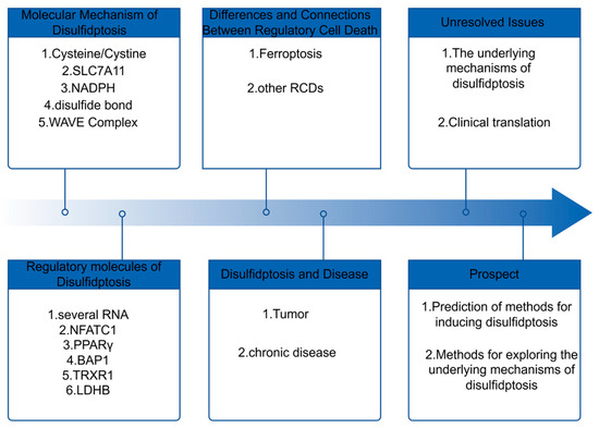
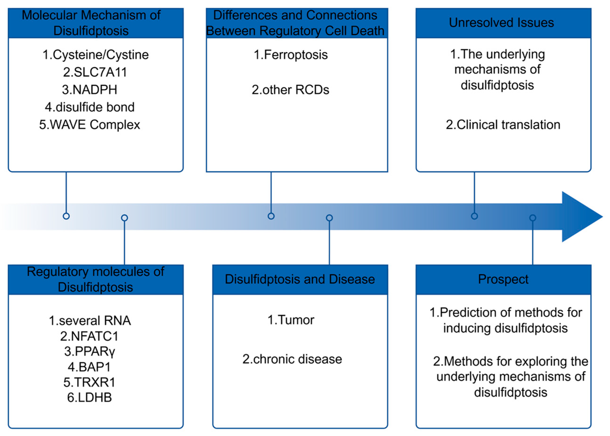
Although the disulfidptosis mechanism remains unclear, recent research indicates its involvement in regulating and treating chronic illnesses such as renal cancer [3], pancreatic cancer [12], liver cancer [13], bladder cancer [14], non-alcoholic fatty liver disease [15], ulcerative colitis [16], hypertrophic cardiomyopathy [17] and other chronic diseases. Therefore, research on disulfidptosis is exciting for the future treatment of many diseases.To facilitate a clearer reading of the full text, we have drawn a flowchart of the entire narrative, as shown in Figure 1.
Figure 1.
Overview of this Article.
2. Molecular Mechanism of Disulfidptosis
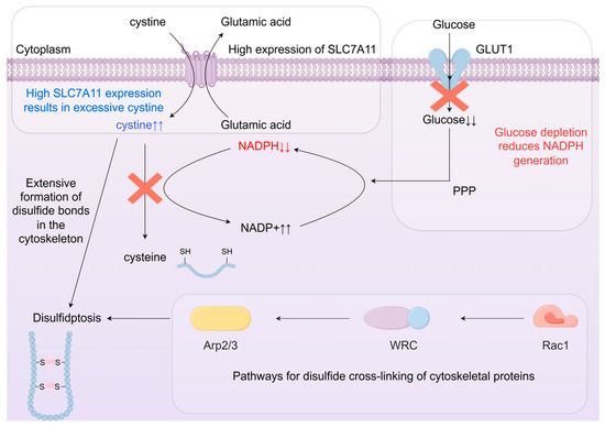
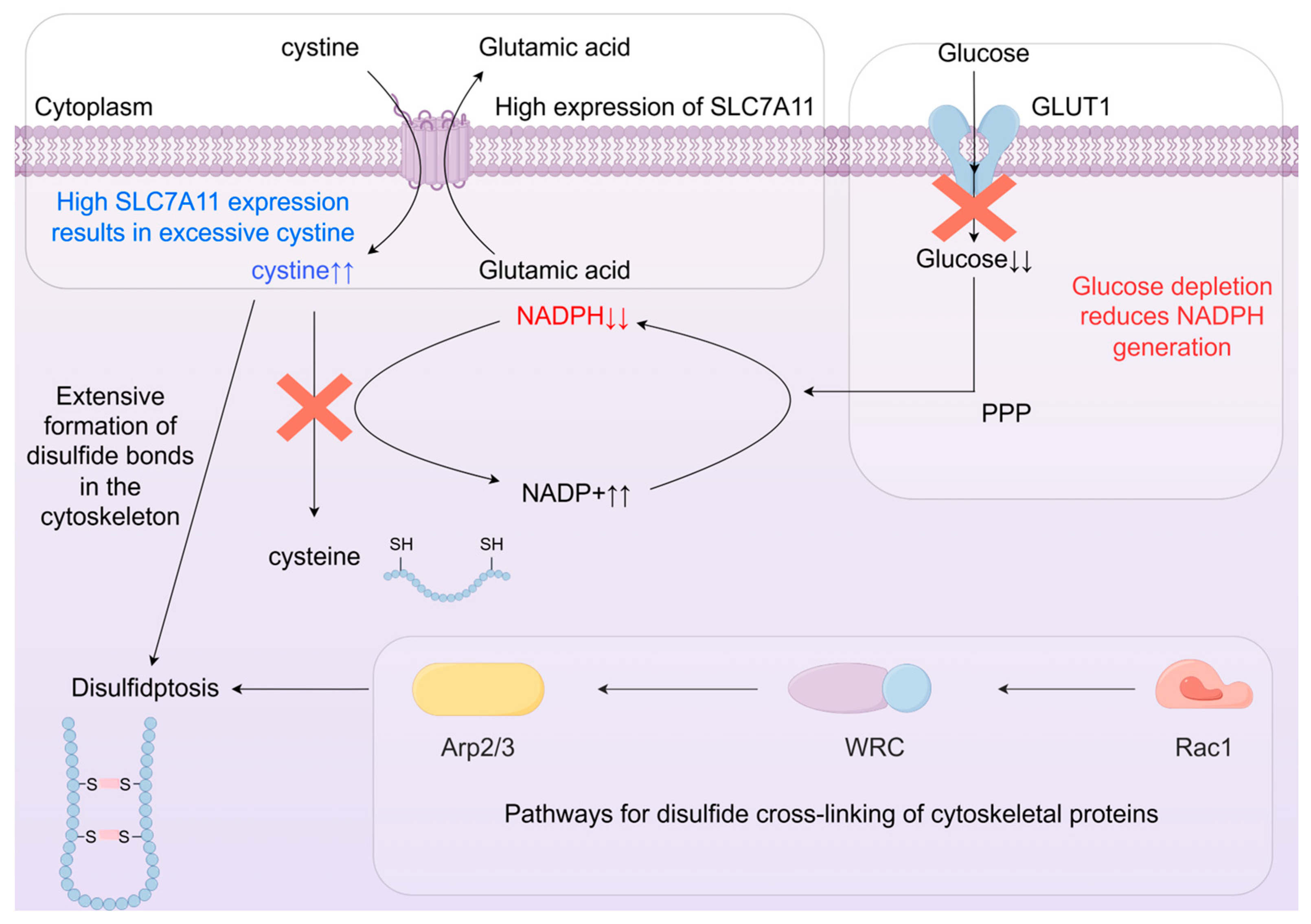
In SLC7A11-overexpressing cells subjected to glucose scarcity, augmented cystine uptake exacerbates the NADPH deficit, resulting in reduced NADPH levels, GSH decline, cystine overload, abnormal disulfide bonds in actin cytoskeletal proteins, actin network collapse triggering cell contraction, morphological changes such as membrane blistering, and subsequent cell death. This entire process is called disulfidptosis [3].
2.1. Metabolism of Cystine/Cysteine
It is known that cysteine starvation induces ferroptosis [18]. In Liu’s study, even in low SLC7A11-expressing cells, disulfidptosis still occurred when excess cystine was added to the culture medium [3]. It can be seen that in ferroptosis and disulfidptosis, the content of cysteine plays an important role in both types of RCD. However, cystine possesses extremely low solubility, and its intracellular accumulation to high levels can exert significant toxicity [11], Cystine accumulation in cells and tissues results in multi-organ toxicity. For instance, cystine buildup in lysosomes of renal tissues gives rise to cystinosis, whereas its accumulation in other tissues can induce complications including portal hypertension, diabetes mellitus, photophobia, and thyroid dysfunction [19]. Furthermore, the transport of cystine via SLC7A11 is accompanied by excessive extracellular glutamate accumulation, which elicits neurotoxicity and causes peripheral neuronal damage [20]. Therefore, cells actually need an important non-essential amino acid—cysteine. Cysteine has numerous functions, such as participating in oxidative stress, metal binding, and transport [21], Moreover, this demand for cysteine is particularly strong in cancer cells. When cysteine cannot be synthesized, GSH is also depleted, results in lipid peroxide buildup, triggering ferroptosis [18], It also prevents disulfide bonds from being reduced, causing them to accumulate in large quantities and leading to disulfidptosis [3]. Thus, a more profound grasp of cysteine’s distinct metabolic processes will facilitate a more thorough examination into the roots of disulfidptosis.
Within cells, cysteine primarily has three destinations. The first destination is the synthesis of proteins and peptides, with protein synthesis being the primary destination for cysteine, accounting for a significant proportion of its utilization [21]. The second pathway involves taurine acting as a precursor in lipid metabolism, participating in the synthesis of cholesterol through a conjugation reaction to produce bile acids, and contributing to cellular energy metabolism. The third pathway involves cysteine synthesizing hydrogen sulfide (H2S), which is associated with certain cancers such as lung cancer [22].
Intracellular cysteine is primarily derived from three sources: SLC7A11-mediated uptake of extracellular cystine, the transsulfuration pathway (TSS), or glutathione degradation [23]. Since many cancers exhibit compromised TSS activity, they become dependent on SLC7A11 upregulation to satisfy cysteine demands. Consequently, SLC7A11 has emerged as a promising therapeutic target for inducing regulated cell death in cancer [24,25].
2.2. SLC7A11’s Regulation Is Important
In numerous studies on ferroptosis, SLC7A11 has been identified as an inhibitor of ferroptosis. Numerous studies have examined SLC7A11 cellular expression to determine whether tumor cells undergo ferroptosis, with the aim of treating tumors [26]. Enhanced SLC7A11 levels mitigate cellular vulnerability to ferroptosis, consequently safeguarding against cell death [27]. Research by Jiang, Zhang, Dixon, and colleagues demonstrates that SLC7A11 overexpression confers ferroptosis resistance in cancer cells [28,29,30]. Therefore, when drug resistance develops, relying solely on ferroptosis limits treatment options. It is known that glucose starvation induces apoptosis in cancer cells [31], In this case, disulfidptosis can be exploited to eliminate ferroptosis-resistant cancer cells. Furthermore, upregulating the level of SLC7A11 can enhance cellular sensitivity to glucose deprivation [32], Once glucose makes its way into the cell through specialized transporters, it quickly hops into the glycolytic pathway to form G6P. From there, G6P can keep progressing through glycolysis or, alternatively, be diverted into the pentose phosphate pathway under the influence of G6PD. This route is responsible for producing approximately 60% of the cell’s NADPH [33], thereby establishing a connection between SLC7A11 and the PPP and NADPH pathways. Therefore, similar to the regulation of SLC7A11 by ferroptosis, altering the expression level of SLC7A11 in a glucose-starved environment is a practical way to induce disulfidptosis.
2.3. NADP+/NADPH Ratio and Thiol-Dependent Antioxidant System
In the reduction of disulfide bonds, NADPH acts through the thiol-dependent antioxidant system, namely the thioredoxin system TrXRs and the glutathione-disulfide bond reduction system GSH/Grxs [34]. Therefore, changes in NADPH content are also important indicators in the occurrence of disulfidptosis.
The thioredoxin system (TrXRs) comprises NADPH, thioredoxin reductase (TrxR/TXNRD), and thioredoxin (Trx) [35]. The TrxR system can utilize the electrons donated by NADPH to reduce the disulfide bonds of substrates, and it can also cleave cystine into cysteine [36]. Furthermore, Trx is a repair protein that prevents abnormal disulfide bond formation in cells [37], Studies have found that inhibiting TrxR1 can increase the likelihood of disulfidptosis [38]. The electrons in the thioredoxin system can originate not only from NADPH but also from the GSH system, and they can work simultaneously and complement each other.
The glutathione-disulfide reduction system GSH/Grxs requires glutathione reductase Grxs, GSH, GSSG, and NADPH. Glutathione reductase uses the cysteine residue at its active site to bind with disulfide substrates to form mixed disulfides, then nucleophilically attacks the disulfide bonds of the substrates, reducing them to their thiol forms, while forming disulfide bonds itself. After the reaction, the N-terminus of glutathione peroxidase can bind with one molecule of GSH to form a mixed disulfide. The mixture is further reduced by GSH, reactivating the system while converting GSH to GSSG. Glutathione reductase then regenerates GSH from GSSG using NADPH [39]. The glutathione disulfide reduction system not only protects proteins from inactivation but also has a profound impact in the redox system [40].
Currently, reducing NADPH production or increasing NADPH consumption can affect the occurrence of disulfidptosis. Therefore, GLUT inhibitors, like glucose starvation, induce disulfidptosis by reducing NADPH production in the PPP, leading the accumulation of disulfide bonds. Xiang’s research team developed an innovative disulfidptosis-inducing artificial membrane system. This system simultaneously upregulates SLC7A11 protein expression in malignant cells, accelerating disulfide bond aggregation [41]. At the same time, intracellular reducing agents such as GSH and NADPH are overconsumed, increasing the NADP+/NADPH ratio, disulfidptosis thus ensues.
2.4. The Synthesis of Disulfide Linkages in Protein Structures
Disulfidptosis results from excessive disulfide bond buildup in cellular and structural proteins [3]. Many places house disulfide bonds, most notably within the endoplasmic reticulum, but also scattered across the Golgi apparatus and the intermembrane space of mitochondria. The creation of these protein disulfide bonds involves an oxidative process, which is vital in shaping and refining proteins as they fold [42,43]. To synthesize disulfide bonds within cells, disulfide bond exchange proteins or disulfide oxidases capable of de novo synthesis are required [44].
In the ER of mammalian cells, PDI is a key component in disulfide bond formation. Oxidized PDI can react with newly synthesized polypeptides to transfer disulfide bonds to them [45]. Disulfide bonds can form through several routes, but the main process hinges on the cooperative interaction between Ero-1 and FAD, which together facilitate the conversion of protein PDI into its oxidized form. Every disulfide bond created yields one molecule of H2O2 [46]. In addition, there is PRX4, which complements Ero-1. PRX4 is composed of five dimers [47], After sulfonylation, its conformation changes, and its cysteine residues form disulfide bonds with cysteine residues of adjacent peptides, serving as electron acceptors for the electrons generated by PDI oxidation [48], PRX4 uses hydrogen peroxide, a product of Ero-1, as its final electron acceptor to produce water, thereby preventing the accumulation of excessive hydrogen peroxide within ER. Another glutathione peroxidase, GPX7/8, forms disulfide bonds in a manner similar to PRX4, also utilizing hydrogen peroxide generated by the Ero-1 pathway to simultaneously form disulfide bonds [49]. Other potential disulfide bond formation pathways include VKOR, which can convert vitamin K epoxide into hydroquinone to generate disulfide bonds [50]. When Ero-1, PRX4, and VKOR are knocked down, cellular disulfide bonds decrease, leading to cell death [44]. Additionally, there are pathways independent of oxidative PDI, such as the quiescent enzyme QSOX, which can directly oxidize substrates to form disulfide bonds [45].
Ero1 and PDI are typically present in partially reduced forms in human cells, which appears to reflect the redox balance of the ER [51]. It is a common understanding that mistakes can pop up when disulfide bonds get oxidized, which might cause the creation of non-native disulfide bonds. To fix this, the endoplasmic reticulum needs a good supply of reducing agents to transform those non-native disulfide bonds back to the native ones, which maintaining proper protein folding and ensures they do their job as they should. Recent studies have shown that the reducing equivalents in the ER may originate from GSH in the cytoplasm [52], GSH maintains the PDI active sites in a reduced state, essential for the creation of native disulfide linkages [53,54], upon depletion of GSH, the rate of oxidative protein folding within the ER quickens, and the incidence of abnormal disulfide bridges formation rises notably [53,54,55]. Some studies have also suggested that cytoplasmic NADPH also acts as an electron donor [56]. NADPH is itself a donor for GSH production, so the occurrence of disulfidptosis may be related to the decline of NADPH and GSH, leading to massive production of non-natural disulfide bonds in the ER. The use of catalase and peroxidase can rescue disulfidptosis [57], The reason may also be related to the coordinated action between Ero-1, which produces hydrogen peroxide, and PRX4 and GPX7/8, which utilize hydrogen peroxide.
Unlike the ER, the Golgi apparatus does not contain numerous disulfide bond-related enzymes. Only QSOX1 is present, which can simultaneously perform the functions of EroI and PDI to form disulfide bonds [43]. In mitochondria, disulfide bonds are mediated by the thiol oxidase Erv1 and the redox-dependent receptor Mia 40 to meet the needs of oxidative respiratory chain [46].
2.5. Rac1 Activates the WAVE Complex (WRC) to Promote Arp2/3-Mediated Actin Polymerization and the Formation of Lamellar Pseudopodia
Disulfidptosis is not just about an excess of cystine leading to a buildup of disulfide bonds; it also features the creation of these bonds within cytoskeletal proteins, which results in the breakdown of the actin framework. In Western blot analyses, these proteins often display a tailing pattern, highlighting this distinctive characteristic of the process [3]. Regulation of actin involves WASP and WAVE 1, 2, and 3 [58], with the WAVE complex acting downstream of Rac1 [59]. Knockout of NCKAP1 (the most important part of the WAVE complex) or RPN1 in cells increases the resistance of cells to disulfidptosis [3]. However, knockout of NCKAP1 does not affect the expression levels of SLC7A11 or SLC2A3, cysteine uptake, or the NADP+/NADPH ratio, but only directly regulates disulfidptosis. It is known that Rac1 activates WRC to form pseudopodia [60], indicating that the Rac1-WRC-Arp2/3 pathway provides a target for disulfide bond overload in the actin network during disulfidptosis. It is known that knockout of NCKAP1 inhibits disulfidptosis. Zhong’s research revealed that NCKAP1 plays a crucial role as a tumor suppressor in hepatocellular carcinoma. The study demonstrated that this protein boosts Rb1/p53 pathway activation, thereby slowing cancer cell proliferation and improving clinical outcomes. Importantly, the findings establish NCKAP1’s mechanism of action—it controls the cell cycle in HCC lines specifically through targeted regulation of the Rb1/p53 axis [61].
Regarding the above mechanism, we have created a mechanism diagram of bisulfide death as shown in Figure 2.
Figure 2.
Mechanism of disulfidptosis [3]. Cells with high SLC7A11 expression undergo a decrease in intracellular glucose transfer under glucose starvation or GLUT inhibitor treatment, followed by a decrease in NADPH generated by the pentose phosphate pathway. The amount of NADPH is insufficient to completely reduce the cysteine transferred through SLC7A11, causing disulfide bond stress. abnormal disulfide cross-linking of cytoskeletal proteins, and ultimately causes cytoplasmic membrane detachment, leading to disulfidptosis. Among them, Rac1-WRC-Arp2/3-mediated lamellar pseudopodia play an important role in disulfidptosis.
3. Summary of Disulfidptosis-Related Inhibitors
Regarding bisulfide-induced death, we summarize some related inhibitors as shown in Table 1.
Table 1.
Summary of Disulfidptosis-Related Inhibitors.
4. Regulatory Network of Disulfidptosis
Liu’s research revealed the process of disulfidptosis and its important nodes, such as SLC7A11, NADPH, cysteine metabolism, and glucose metabolism. At the same time, he successfully induced disulfidptosis in renal cancer cells using GLUT inhibitors, demonstrating the regulability of disulfidptosis [3]. Except GLUT inhibitors, numerous subsequent studies have revealed the regulation of disulfidptosis by RNA, proteins, and some pathways. These regulations are all carried out through the regulation of important nodes such as SLC7A11, glucose metabolism, or NADPH.
4.1. RNA Regulation of Disulfidptosis
4.1.1. LncRNA
LncRNA constitutes a category of RNA molecules exceeding 200 nucleotides in length, which lack protein-coding capacity [62], LncRNA can bind to proteins, promote or inhibit their binding activity to the corresponding DNA sequences, and directly interact with DNA to silence genes, thereby regulating gene expression [63]. LncRNAs also contribute significantly to the differentiation, growth and death of cell, by binding to cell differentiation-related mRNA, or activating corresponding transcription factors (such as activating STAT3 to stimulate T cell activation) [63,64,65,66], and disulfidptosis is no exception.
In Sun’s study, he found that ALMS1-1T1 is a high-risk LncRNA in HNSCC [67]. It was also found that ALMS1-1T1 was co-expressed with SLC7A11, and after knocking down the ALMS1-1T1 gene, PPP-related genes were simultaneously downregulated, indicating that ALMS1-1T1 regulates the occurrence of disulfidptosis through PPP dependence under glucose starvation conditions. Western blot analysis of glucose-deprived HN-6 cells showed diffuse and high-molecular-weight bands corresponding to actin cytoskeletal proteins; this effect was amplified in ALMS1-1T1-overexpressing cells, indicating that ALMS1-1T1 promotes disulfide bond formation among these proteins. It is known that 2-DG, as an inhibitor of glycolysis, can directly produce NADPH through PPP to rescue disulfidptosis [68], and TCEP can produce a stronger disulfide bond reduction reaction to rescue cell contraction. Silencing ALMS1-1T1 resulted in a superior rescue effect of 2-DG compared to TCEP, demonstrating that the impact of ALMS1-1T1 knockout occurs through the NADPH pathway and not by directly influencing disulfide bonds [67]. However, it is worth mentioning that this study only analyzed the correlation between ALMS1-IT1 and SLC7A11 gene expression, and whether there is an interaction or synergistic relationship between the two in the occurrence of disulfidptosis has not been studied.
In Yin’s study, his team identify TMEM105 as a DRL in Pca. When TMEM105 was knocked down in a sugar-free environment, F-actin and cell contraction were remarkably enhanced, and the NADP+/NADPH ratio was enormously increased [69]. In further studies, Yin determined that TMEM105 regulates disulfidptosis by stabilizing β-catenin (an intracellular signal transduction protein that regulates cell proliferation and differentiation [70,71]) and enhancing the expression of c-myc (a transcription factor associated with glycolysis [72,73]) to induce GLUT1.
Moreover, Yao explored and uncovered increased levels of CASC8 in PDAC tumor samples [12]. CASC8 can influence cancer cell activity and may serve as a prognostic marker [74,75]. In Yao’s study, after treating cell lines with CASC8 knockdown with no sugar, a pronounced increment in cellular demise was noted, and this increase could be reversed by the disulfidptosis relievers TCEP and DTT, indicating that CASC8 has a disulfidptosis inhibitory effect. Yao’s KEGG enrichment analysis highlighted a notable disparity in metabolic pathways between the control and CASC8 knockdown groups, with glycolysis and the pentose phosphate pathway showing significantly higher activity in the former. Suppressing CASC8 expression led to a rise in the NADP+/NADPH ratio and a drop in glucose absorption, whereas overexpressing CASC8 produced the inverse effect. Subsequent GSEA by Yao uncovered a strong link between CASC8 levels and the expression of c-Myc target genes. Follow-up investigations confirmed a positive association between CASC8 and c-Myc, reinforcing their functional relationship [12].
4.1.2. circRNA
CircRNA, like long LncRNA, was once believed to have no significant impact on cellular metabolism, proliferation, and differentiation. CircRNA was thought to merely accompany the gene splicing process [76,77,78]. As research has progressed, the roles of CircRNA in various cancers and regulatory pathways have begun to be uncovered [79]. For example, CircRNA can recruit RBPs to the promoters of oncogenes to activate gene expression, or inhibit promoters by competitively binding to transcription factor sites [80].
In Liu’s research, the expression of Let-7a-5p is downregulated by Circ_0022382, which consequently stifles the SLC7A11 expression by diminishing its mRNA levels. Knocking down Circ_0022382 results in a corresponding reduction in SLC7A11 levels, but it also leads to a substantial drop in NADPH levels and a subsequent decline in glucose metabolism. Notably, this decrease was not counteracted by ferroptosis inhibitors, hinting at a potential occurrence of disulfide stress [81], This seems to contradict common sense, but according to Yan’s conclusion, moderate levels of SLC7A11 can also induce disulfidptosis [7].
4.2. NFATC1 Induces Disulfidptosis by Sensitizing Osteoclasts to TXNRD1 Inhibitors Through Upregulation of SLC7A11 Expression
As mentioned earlier, NADPH can also inhibit disulfide bond formation through the thioredoxin system TrXR, and the most important and common inhibitory site in this system is TrxR1 [82]. TrxR1 can reduce cysteine to cystine [35].
In Zhong’s study, his team found that when T-cell nuclear factor (NFATC1) was upregulated, the transcriptional expression level of SLC7A11 was also upregulated. However, Zhong did not treat the osteoclasts with glucose starvation or GLUT1 inhibitors, but instead treated them with TXNRD1 inhibitors, which also led to F-Actin contraction and disulfidptosis [83]. NFATC1 sensitizes osteoclasts to TXNRD1 inhibitors by upregulating SLC7A11 expression, inducing disulfidptosis. Based on this, it can be inferred that certain enzymes in the thioredoxin system can also serve as targets for regulating disulfidptosis.
4.3. Inhibition of PPARγ Promotes Ferroptosis and Disulfidptosis
PPARγ is a ligand-dependent nuclear transcription factor that plays a significant role in tumor survival and development [84]. PPARγ activation governs glucose absorption and metabolic use in cells [85]. Therefore, PPARγ appears to have a similar effect to GLUT inhibitors.
In Zhang et al.’s study, when PPARγ was inhibited, markers of ferroptosis such as LPO were detected, indicating that cells underwent ferroptosis. However, the use of ferroptosis inhibitors could not completely inhibit cell death. Transcriptome sequencing indicated that PPARγ is related to glucose metabolism and upregulates SLC7A11 when inhibited. Zhang found that GLUT1 expression and NADPH oxidase were significantly reduced and cell death could be rescued by TCEP, and a tail band appeared in non-reducing gel electrophoresis [3], indicating that ferroptosis and disulfidptosis occurred simultaneously after PPARγ inhibition [86]. qPCR and Western blot analysis revealed that PPARγ inhibition increased HMOX1 expression, thereby inducing ferroptosis. After PPARγ was inhibited, SLC7A11 was also upregulated, promoting disulfidptosis in OSCC [86].
Zhang et al. combined research on ferroptosis and disulfidptosis, two oxidative stress-related death pathways, to show the possible synergistic effect of the two in causing regulatory cell death. However, in Zhang’s research, the cause of ferroptosis seems to be HMOX1 and its transcription factor NRF2, Therefore, in this case, SLC7A11 is no longer the protective umbrella of ferroptosis [86].
4.4. BAP1 Inhibits Disulfidptosis by Inhibiting SLC7A11-Mediated Cysteine Uptake and Reducing the NADP+/NADPH Ratio
BAP1 acts as a novel tumor suppressor through the deubiquitination of H2A monoubiquitination and subsequent regulation of gene transcription [87].
In Wang’s study, it was found that the expression level of BAP1 decreases as the expression level of SLC7A11 increases. In contrast to wild-type BAP1 cells, those overexpressing SLC7A11 exhibited more pronounced band tailing during non-reducing Western blot analysis following glucose deprivation. When SLC7A11 was subsequently suppressed, the occurrence of disulfidptosis in these cells was notably diminished. This suggests that BAP1 influences disulfidptosis through its regulation of SLC7A11. Later, two separate groups of cells were exposed to 2-deoxy-D-glucose (2-DG) under conditions lacking glucose. The results showed that, compared to the control, cells with elevated BAP1 levels had a less significant increase in the NADP+/NADPH ratio, implying that BAP1’s protective role against disulfidptosis is partly mediated by its impact on this redox couple [88].
4.5. Inhibition of TRXR1 Promotes Disulfidptosis Under Glucose Deprivation
Numerous studies indicate that suppressing TrxR1 expression can hinder cancer development.
In Tang’s study, it was found that activating TAZ and depriving glucose can promote the expression of TrxR1, where the increased expression of TrxR1 after glucose deprivation is an adaptive response. It is known that glucose deprivation induces cell apoptosis [89]. Tang found that inhibiting TrxR1 using AF or TRi-1 under glucose deprivation conditions significantly accelerated the rate of cell death, and demonstrated that this type of cell death could not be rescued by other RCD inhibitors such as ferroptosis. It can only be rescued by DTT and TECP, and characteristics of disulfidptosis, such as lamellar pseudopodia, were found in the actin cytoskeleton, indicating that inhibition of TrxR1 can promote disulfidptosis in glucose deprivation [38].
In addition, the study showed that when AF and glucose deprivation coexist, the occurrence of disulfidptosis does not depend on the high expression of SLC7A11. Therefore, Tang’s research has opened up a new path for the application of disulfidptosis.
4.6. STAT3 Inhibits G6PD Activity Through LDHB, Depletes NADPH, and Promotes Disulfidptosis in CD8+ T Cells
LDHB is primarily expressed in tissues with high oxidative metabolism, such as cardiac muscle and kidneys, and promotes the entry of lactate as an energy substrate into the tricarboxylic acid cycle. It plays a contradictory role in cancer, depending on the tumor type, and is also involved in the RCD process, nucleic acid synthesis, and lipid synthesis, among others [90]. STAT3 is a transcription activator that is overexpressed in tumors and plays an important role in tumor progression [91].
In Wan’s study, when LDHB was defective, G6PD activity increased, enhancing PPP flux and NADPH content, thereby inhibiting the occurrence of disulfidptosis. The authors also used bioinformatics technology to analyze that STAT3 binds to the promoter region of LDHB, promoting LDHB expression, thereby inhibiting G6PD activity, reducing NADPH, and promoting the occurrence of disulfidptosis [92].
According to Wan’s study, key enzymes in glucose metabolism can also affect the occurrence of disulfidptosis, proving that the induction of disulfidptosis is diverse and feasible, and providing a promising therapeutic strategy for targeting glucose metabolism-related enzymes to induce disulfidptosis.
4.7. Several Regulatory Pathways That May Be Related to Disulfidptosis
Most of the pathways mentioned above that are associated with disulfidptosis, such as the c-myc-GLUT1-pentose phosphate pathway axis [12,69], which is closely related to glucose metabolism [3,93]. Therefore, pathways related to sugar metabolism and important targets such as SLC7A11 are worthy of further study.
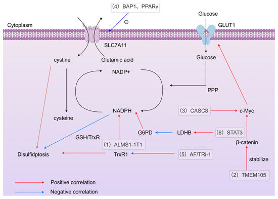
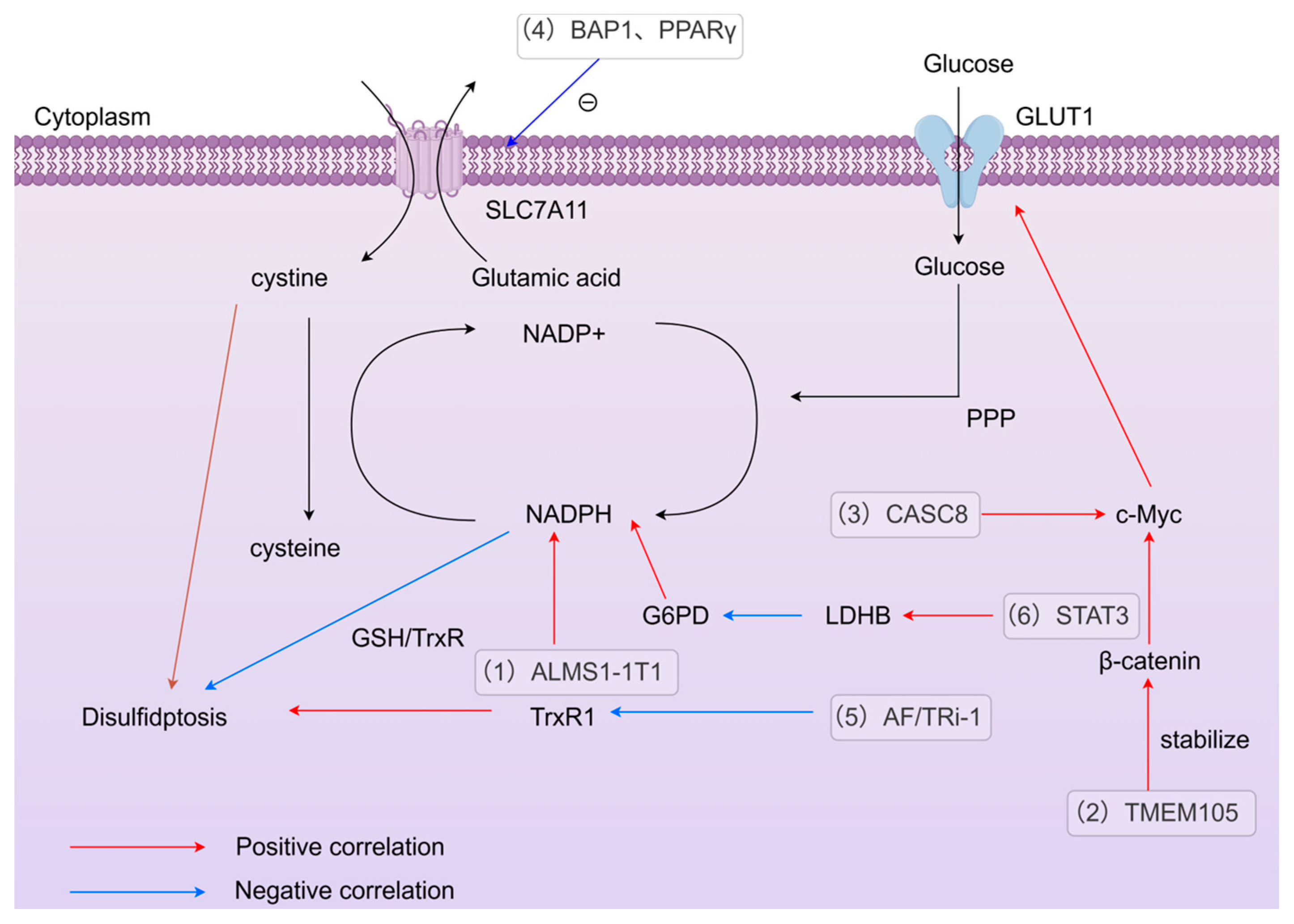
To summarize the above-mentioned regulatory mechanisms of disulfidptosis, we have created a regulatory network diagram of disulfidptosis, as shown in Figure 3.
Figure 3.
Molecules and pathways regulating disulfidptosis. (1) ALMS1-1T1 promotes NADPH production, and NADPH inhibits disulfidptosis through the reducing action of GSH/TrxR [67]. (2) TMEM105 induces GLUT expression and inhibits disulfidptosis by stabilizing the β-catenin-c-Myc axis [69]. (3) CASC8 promotes c-Myc-induced GLUT and inhibits disulfidptosis [12]. (4) BAP1 and PPARγ negatively regulate SLC7A11, reducing cystine transport and inhibiting disulfidptosis [38,86]. (5) AF/TRi-1 promotes cell death under glucose deprivation by inhibiting TrxR1 [83]. (6) STAT3 promotes LDHB expression to inhibit G6PD activity, thereby depleting NADPH and inducing disulfidptosis [90].
5. Differences and Connections Between Regulatory Cell Death
Ferroptosis and disulfidptosis are not completely separate and can occur simultaneously [86]. Although different forms of regulatory cell death (such as disulfidptosis, ferroptosis, necrotic apoptosis, pyroptosis, and apoptosis) have significant differences in molecular mechanisms, triggering conditions, and morphological characteristics, these forms of RCD are all cross-regulated by redox homeostasis, metabolic reprogramming, and key molecules (such as p53 and the Bcl-2 family) and may act synergistically or antagonistically under pathological conditions. Therefore, elucidating their specificity and interaction networks can provide precise strategies for disease treatment targeting RCD.
5.1. Ferroptosis
Ferroptosis is an iron-dependent form of cellular demise characterized by the buildup of intracellular iron, disruption of the GPX4 antioxidant defense mechanism, and a surge in reactive ROS. These factors collectively drive excessive lipid peroxidation, causing catastrophic damage to cell membranes and culminating in the cell’s destruction [94].
On the surface, SLC7A11 inhibits ferroptosis while promoting disulfidptosis, appearing contradictory. However, closer analysis reveals this as a metabolic vulnerability arising from metabolic reprogramming in cancer cells. SLC7A11 inhibits ferroptosis by supplying cysteine, the precursor for GSH, and utilizing GSH as a cofactor for GPX4 to eliminate lipid peroxides. This process, however, consumes substantial NADPH to reduce cysteine. Under conditions of ample glucose, this system functions effectively. However, under glucose deprivation or NADPH depletion, this system collapses. The continuously supplied cysteine from overexpressed SLC7A11 cannot be adequately reduced due to insufficient NADPH, leading to massive accumulation of disulfide bonds—triggering disulfidptosis.
In the previous section, inhibiting PPAγ achieved the coexistence of disulfidptosis and ferroptosis. This seemingly contradictory phenomenon primarily arises from the upregulation of HMOX1, which leads to the accumulation of abundant free iron. This triggers the Fenton reaction, generating large quantities of hydroxyl radicals that directly drive lipid peroxidation. Concurrently, the upregulation of SLC7A11 fails to inhibit ferroptosis. Instead, it allows substantial amounts of cysteine to enter the cell, competing with GPX4 for NADPH. Consequently, the cell enters a state of metabolic collapse: NADPH depletion occurs, failing both to sustain the GPX4 system for ferroptosis and to reduce excessive cysteine, leading to disulfide bond accumulation and causing disulfidptosis. However, the deeper connection between the two types of cell death requires further investigation. Excitingly, In Li’s research, the team engineered a nanoferroptosis inducer that activates itself within the tumor microenvironment. This innovative construct features a polyethylene glycol-coated Fe-Bi-SS metal–organic framework loaded with high-Z elements, designed to enhance chemotherapy outcomes by specifically targeting ferroptosis pathways. They found that SLC7A11 can be upregulated adaptively. At this point, adding GOx to MOFs induces disulfidptosis, and the hydrogen peroxide produced by glucose oxidation can also undergo a Fenton reaction with ferrous ions to form highly oxidative hydroxyl radicals, thereby enhancing ferroptosis. This shows that disulfidptosis and ferroptosis can complement each other to improve therapeutic effects [95].
5.2. Other Regulatory Deaths
In addition to ferroptosis, other regulated cell deaths may also be associated with dithiothreitol-induced cell death, and we categorize them in Table 2.
Table 2.
The association between other RCD and disulfidptosis.
Pyroptosis and disulfidptosis can work hand in hand to create a notable “synergistic” impact. In Zhu’s research, his squad harnessed GOx and SP as nano-inducers, labeling them as Pd2Sn@GOx-SP, to tweak intermetallic compounds. This clever maneuver sparked a potent anti-tumor immune reaction, thanks to the combined efforts of both pyroptosis and disulfidptosis [97].
Autophagy provides energy by breaking down cell components, which may alleviate the NADPH demand in disulfidptosis and thereby inhibit its occurrence [98]. The mitochondrial pathway of apoptosis may affect the NADPH/NADH balance. Furthermore, p53 promotes mitochondrial outer membrane permeability by regulating genes such as BAX/BAK in apoptosis, and some studies suggest that p53 may indirectly regulate disulfidptosis by regulating SLC7A11 expression, which may affect cysteine metabolism [99].
As an emerging form of regulatory cell death, disulfidptosis provides a multidimensional entry point for combined cancer therapy through its potential cross-regulation with apoptosis, necrotic apoptosis, and autophagy. In the future, it will be necessary to analyze the spatiotemporal dynamic interactions of death pathways in depth and develop combined therapies with controllable timing to ultimately achieve efficient, low-toxicity, and precise tumor elimination.
6. Disulfidptosis and Tumors
Disulfidptosis, can not only be used to establish prognostic models, but can also be induced in tumor cells as a form of tumor treatment.
6.1. Application of Disulfidptosis-Related Genes (DRGs) and Related LncRNA (DRL) in Tumors
Numerous studies have shown that discrepancies in the manifestation of genes or LncRNAs linked to disulfidptosis distinguish cancerous from healthy tissues. These disparities serve as valuable clues for identifying potential regulatory and therapeutic focal points, as well as novel biomarkers for tumor diagnosis. DPAG scores are assigned to DRGs in GC. APC11, a chosen DPAG in GC, serves as an independent prognostic marker and correlates strongly with unfavorable outcomes. APC11 is over-expressing in GC, and knocking out APC11 tremendously inhibits the proliferation and migration of GC cells [100], Similarly, Tensin4 (TNS4) in lung adenocarcinoma [101], ACTN4 and LCN2 in endometrial cancer [102], CCNA2 and RPN1 in hepatocellular carcinoma [103,104], INF2 and MEGF10 in osteosarcoma [105], CAPZB in diffuse large B-cell lymphoma [106], AC005840. 4 in bladder cancer, etc. Furthermore, the high-risk group identified using this DRL in bladder cancer showed higher sensitivity to sorafenib and oxaliplatin, demonstrating a link between disulfidptosis and existing anticancer drugs [107]. In particular, Zhang’s study found that inhibition of the disulfidptosis gene MYH9, which is highly expressed in liver cancer, is not only associated with low survival rates in liver cancer, but also with resistance to sorafenib, and that inhibition of MYH9 can reverse sorafenib resistance [108].
6.2. Inducing Disulfidptosis in Tumors to Achieve Therapeutic Effects
Combining disulfidptosis with ICIs has emerged as a novel approach [109]. In Du’s review, this approach is described as theoretically feasible, providing insights into the potential of combining the two for cancer treatment [109]. Recently, Shu’s study reveals the regulation of PD-L1 expression in glioblastoma cells by modulating the disulfidptosis-T cell exhaustion (Tex) pathway, indicating that the combination of the two has unlimited prospects for treatment [110].
Not only through the immune environment, some small molecule compounds can also induce disulfidptosis in tumors. In Shi’s research, they found that Gaudichaudione H (GH) is a natural compound, which was found in the application of hepatocellular carcinoma: GH is not only related to GSH and NADP+/NADPH metabolism, but also can upregulate SLC7A11 under glucose starvation conditions to induce disulfidptosis [13]. However, this raises some questions: How can GH be accurately localized and concentrated in in vivo experiments, especially in the microenvironment of liver cancer cells in humans? How is it degraded?
Previous research indicates that SDT (sonodynamic therapy) offers benefits like being minimally invasive and capable of deep tissue penetration, enabling effective treatment of deep-seated tumors [111]. In Wang’s latest study, nanotechnology was combined with ultrasound technology. The SPCP/CCP@Bay system consists of biodegradable photodynamic pseudoconiugate polymers (SPCP) and cysteine-containing polymers (CCP), loaded with Bay-876 (a GLUT inhibitor) [14]. This system effectively accumulates and releases cystine in bladder tumors, inhibiting GLUT and glucose metabolism and inducing disulfidptosis, while ultrasound can induce ICD and synergize with α-PD-1 to inhibit tumor.
7. The Potential Value and Application of Disulfidptosis in Chronic Diseases
Disulfidptosis is also associated with some chronic diseases and diseases related to immune dysregulation, such as osteoarthritis, intervertebral disc degeneration, pulmonary hypertension, heart failure, hypertrophic cardiomyopathy, gingivitis, and sepsis. DRGs have different degrees of differentiated expression in these diseases and are considered to be potential targets for targeted therapy. As precancerous lesions of colon adenocarcinoma, UC and NAFLD are now hot topics of research in relation to disulfidptosis.
7.1. Application of Disulfidptosis in NAFLD
In Luo’s study, it was found that DRGs were significantly elevated in NAFLD and were associated with immune cells such as regulatory T cells. MYL6 was identified as a contributor to NAFLD development and a promising future treatment target [15].
7.2. Application of Disulfidptosis in UC
Recent studies have elucidated the critical role of disulfidptosis in the pathogenesis and treatment of UC. Song et al. verified the existence of disulfidptosis in UC, observing that the upregulation of the disulfidptosis-promoting gene SLC7A11 and the downregulation of the inhibitor NUBPL significantly correlated with patient responses to TNF-α inhibitors [112]. Similarly, Yang et al. identified SLC3A2 as a central hub for disulfidptosis, finding that elevated expression of disulfidptosis-related genes (DRGs) predicted non-response to TNF-α inhibitors, whereas responders exhibited reduced DRG levels. Their work suggests disulfidptosis drives acute inflammation in UC, highlighting it as a promising therapeutic target [16]. Furthermore, Jiang et al. demonstrated that disulfidptosis-mediated regulatory cell death exacerbates inflammation and disease severity [113]. Most recently, investigations into the link between UC and colon adenocarcinoma identified CD2AP and MYH10 as key DRGs. Molecular docking analyses revealed that agents such as budesonide and sulfasalazine exhibit high binding affinity to these disulfidptosis targets, with simvastatin and phenytoin sodium also showing potential to modulate this pathway in both conditions [114].
8. Unresolved Issues in Disulfidptosis
The regulatory pathway of RCD is complex and intricate. Even though Liu’s research has revealed some of the conditions under which disulfidptosis occurs, the mechanism and regulation remain unclear and need to be further investigated.
It is well known that protein formation requires production and modification by the ER and Golgi apparatus, so disulfide bonds are also formed in the ER and Golgi apparatus, and some protein disulfide bonds even exist dynamically [115,116]. But why does the accumulation of disulfide bonds in disulfidptosis occur significantly in the cytoskeletal protein actin network? Current research has only focused on disulfide bonds between cytoskeletal proteins, and the formation of disulfide bonds in other regions has not been thoroughly investigated, which may involve other rapid signaling pathways.
Disulfidptosis correlates with oxidative stress, and ROS is significantly increased in cells that highly express SLC7A11 under glucose starvation [11]. However, different ROS scavengers show completely different effects on the rescue of disulfidptosis. Tempol and Trolox have negligible rescue effects, while catalase has a very significant rescue effect [117]. Mitochondria are essential for cellular disulfide bond formation. When the ROS produced by mitochondria decreases, the disulfide bonds of actin decrease, while the inhibition of peroxidase and catalase leads to an increase in the content of disulfide bonds [118]. Therefore, in disulfidptosis, the relationship between mitochondria, ROS (hydrogen peroxide), and disulfide bonds is worth further study.
9. The Prospect of Disulfidptosis
Liu’s research pioneered a brand new RCD, which is closely related to the basic characteristics of certain tumors or chronic diseases, providing markers, possible treatment strategies, and so on [119]. Subsequent studies have also provided many methods that can be learned to make the future of disulfidptosis traceable. This article also speculates on possible future research directions based on existing research.
9.1. Combined Use Inhibitors
In the above-mentioned induction pathways of disulfidptosis, each step has a key enzyme or molecular target, such as the expression of SLC7A11, glucose metabolism, the NADP+/NADPH ratio, TrxR, and GSH/Grxs, which can be regulated by inhibitors to regulate disulfidptosis. However, multiple inhibitors have not yet been applied simultaneously within the same cell or tissue. For example, the combination of GLUT1 inhibitors with TrXR system inhibitors may further enhance efficiency or provide additional targets for localization. Based on the previously elucidated mechanisms of disulfidptosis, NADPH emerges as a promising therapeutic target; however, achieving its precise depletion within target cells remains a challenge. Leveraging the aforementioned nanomaterial-based induction strategies, we propose targeting multiple disulfidptosis-related genes (DRGs), specifically SLC7A11. By engineering a nanoplatform that integrates NADPH-consuming agents (e.g., glucose oxidase [GOx]) with inhibitors of NADPH biosynthesis (such as those suppressing the pentose phosphate pathway or TCA cycle dehydrogenases), we aim to achieve precise NADPH exhaustion, thereby inducing disulfidptosis with enhanced efficacy.
9.2. Predicting Using Molecular Docking Between Disulfide Death-Related Genes and Potential Drugs
In Jiang’s study [16], the authors conducted enrichment analysis in ulcerative colitis to identify disulfidptosis genes that are highly expressed in UC, and used drugs such as budesonide and sulfasalazine to perform molecular docking with highly expressed genes, finding that the two have high affinity. This provides a new idea for discovering drugs that induce disulfidptosis.
Disulfidptosis is now an emerging field that deserves broader exploration. Research on disulfidptosis is also limited to important indicator targets such as SLC7A11 and NADPH. In the future, the scope of research on disulfidptosis can be expanded to include, for example, glutamate metabolism related to SLC7A11, sugar metabolism related to NADPH, the PPP, key enzymes in the tricarboxylic acid cycle, and so on. Therefore, exploring this novel form of cellular demise is truly fascinating.
Author Contributions
Funding
This study is funded by the National Natural Science Foundation of China (No. 82273321 and No. 81974383 to Wei Yan), the Natural Science Foundation of Hubei Province (No. 2023AFD044 to Wei Yan, No. 2022CFB277 to ZhenZhen Zhou).
Institutional Review Board Statement
Not applicable.
Informed Consent Statement
The Authors confirm: (1) that the work described has not been published before; (2) that it is not under consideration for publication elsewhere; (3) that its publication has been approved by all coauthors, if any; (4) that its publication has been approved (tacitly or explicitly) by the responsible authorities at the institution where the work is carried out.
Data Availability Statement
Data sharing is not applicable to this article as no new data were created or analyzed in this study.
Acknowledgments
Figures were created through figdraw.com.
Conflicts of Interest
The authors declare no conflicts of interest. The authors declare that the study was conducted in the absence of any business or financial relationship that could be interpreted as a potential conflict of interest.
Abbreviations
The following abbreviations are used in this manuscript:
| RCD | Regulatory cell death |
| ACD | Accidental cell death |
| SLC7A11 | Glutathione transporter solute carrier family 7 member 11 |
| GSH | Glutathione |
| NADPH | Nicotinamide adenine dinucleotide phosphate |
| PPP | Pentose phosphate pathway |
| GLUT1 | Glucose Transporter 1 |
| G6PD | glucose-6-phosphate dehydrogenase |
| PDI | protein disulfide isomerase |
| DRGs | Disulfidptosis-Related Gene |
| HNSCC | Head and Neck Squamous Cell Carcinoma |
| OSCC | Oral Squamous Cell Carcinoma |
| ROS | Reactive Oxygen Species |
| UC | Ulcerative Colitis |
| NAFLD | Non-Alcoholic Fatty Liver Disease |
| ER | Endoplasmic Reticulum |
| GOx | glucose oxidase |
| SP | soybean phospholipid |
| LDHB | lactate dehydrogenase B |
| BAP1 | BRCA1-Associated Protein 1 |
| CircRNA | Circular RNA |
| RBPs | RNA-binding proteins |
| LncRNA | Long non-coding RNA |
| Pca | pancreatic cancer |
| PDAC | Pancreatic Ductal Adenocarcinoma |
| WASP | Wiskott-Aldrich syndrome protein |
| Ero-1 | endoplasmic reticulum thiol oxidase 1 |
| FAD | flavin adenine dinucleotide |
| VKOR | vitamin K epoxide reductase |
| ASK1 | apoptosis signal-regulating kinase 1 |
| GSSG | glutathione disulfide |
| G6P | glucose-6-phosphate |
| SAM | S-adenosylmethionine |
| SAH | S-adenosylhomocysteine |
| Hcy | homocysteine |
| CBS | cystathionine β-synthase |
| ICIs | immune checkpoint inhibitors |
References
- Kerr, J.F.; Wyllie, A.H.; Currie, A.R. Apoptosis: A Basic Biological Phenomenon with Wide-Ranging Implications in Tissue Kinetics. Br. J. Cancer 1972, 26, 239–257. [Google Scholar] [CrossRef] [Scilit]
- Zhou, S.; Liu, J.; Wan, A.; Zhang, Y.; Qi, X. Epigenetic regulation of diverse cell death modalities in cancer: A focus on pyroptosis, ferroptosis, cuproptosis, and disulfidptosis. J. Hematol. Oncol. 2024, 17, 22. [Google Scholar] [CrossRef] [Scilit]
- Liu, X.; Nie, L.; Zhang, Y.; Yan, Y.; Wang, C.; Colic, M.; Olszewski, K.; Horbath, A.; Chen, X.; Lei, G.; et al. Actin cytoskeleton vulnerability to disulfide stress mediates disulfidptosis. Nat. Cell Biol. 2023, 25, 404–414. [Google Scholar] [CrossRef] [Scilit] [PubMed]
- Galluzzi, L.; Vitale, I.; Aaronson, S.A.; Abrams, J.M.; Adam, D.; Agostinis, P.; Alnemri, E.S.; Altucci, L.; Amelio, I.; Andrews, D.W.; et al. Molecular mechanisms of cell death: Recommendations of the nomenclature committee on cell death 2018. Cell Death Differ. 2018, 25, 486–541. [Google Scholar] [CrossRef] [Scilit] [PubMed]
- Koppula, P.; Zhuang, L.; Gan, B. Cystine transporter SLC7A11/xCT in cancer: Ferroptosis, nutrient dependency, and cancer therapy. Protein Cell 2020, 12, 599. [Google Scholar] [CrossRef] [Scilit] [PubMed]
- Stipanuk, M.H. Sulfur amino acid metabolism: Pathways for production and removal of homocysteine and cysteine. Annu. Rev. Nutr. 2004, 24, 539–577. [Google Scholar] [CrossRef] [Scilit] [PubMed]
- Yan, Y.; Teng, H.; Hang, Q.; Kondiparthi, L.; Lei, G.; Horbath, A.; Liu, X.; Mao, C.; Wu, S.; Zhuang, L.; et al. SLC7A11 expression level dictates differential responses to oxidative stress in cancer cells. Nat. Commun. 2023, 14, 3673. [Google Scholar] [CrossRef] [Scilit] [PubMed]
- Lo, M.; Wang, Y.Z.; Gout, P.W. The x(c)- cystine/glutamate antiporter: A potential target for therapy of cancer and other diseases. J. Cell. Physiol. 2008, 215, 593–602. [Google Scholar] [CrossRef] [Scilit]
- Koppula, P.; Zhang, Y.; Zhuang, L.; Gan, B. Amino acid transporter SLC7A11/xCT at the crossroads of regulating redox homeostasis and nutrient dependency of cancer. Cancer Commun. 2018, 38, 12. [Google Scholar] [CrossRef] [Scilit]
- Stipanuk, M.H.; Dominy, J.E.; Lee, J.I.; Coloso, R.M. Mammalian cysteine metabolism: New insights into regulation of cysteine metabolism. J. Nutr. 2006, 136, 1652S–1659S. [Google Scholar] [CrossRef] [Scilit]
- Liu, X.; Olszewski, K.; Zhang, Y.; Lim, E.W.; Shi, J.; Zhang, X.; Zhang, J.; Lee, H.; Koppula, P.; Lei, G.; et al. Cystine transporter regulation of pentose phosphate pathway dependency and disulfide stress exposes a targetable metabolic vulnerability in cancer. Nat. Cell Biol. 2020, 22, 476–486. [Google Scholar] [CrossRef] [Scilit]
- Yao, H.F.; Ge, J.; Chen, J.; Tang, X.; Li, C.; Hu, X.; Ahmed, A.A.; Pu, Y.; Zhou, G.; Zhang, T.; et al. CASC8 activates the pentose phosphate pathway to inhibit disulfidptosis in pancreatic ductal adenocarcinoma though the c-myc-GLUT1 axis. J. Exp. Clin. Cancer Res. CR 2025, 44, 26. [Google Scholar] [CrossRef] [Scilit] [PubMed]
- Shi, M.; Li, X.; Guo, Y.; Zhang, Y.; Xu, J.; Yan, L.; Liu, R.; Wang, H.; Tang, S.; Zhao, Y.; et al. Gaudichaudione H enhances the sensitivity of hepatocellular carcinoma cells to disulfidptosis via regulating NRF2-SLC7A11 signaling pathway. Adv. Sci. 2025, 12, E2411131. [Google Scholar] [CrossRef] [Scilit] [PubMed]
- Wang, K.; Li, L.; Liang, G.; Xiao, H.; Zhang, L.; Liu, T. Sonodynamic activated nanoparticles with Glut1 inhibitor and cystine-containing polymer stimulate disulfidptosis for improved immunotherapy in bladder cancer. Biomaterials 2025, 319, 123178. [Google Scholar] [CrossRef] [Scilit] [PubMed]
- Luo, X.; Guo, J.; Deng, H.; He, Z.; Wen, Y.; Si, Z.; Li, J. Unveiling the Role of Disulfidptosis-Related Genes in the Pathogenesis of Non-Alcoholic Fatty Liver Disease. Front. Immunol. 2024, 15, 1386905. [Google Scholar] [CrossRef] [Scilit]
- Yang, Q.-Q.; Guo, J.-A.; Zhang, K.; Li, S.-H.; Xia, W.-Y.; Wang, D.-X.; Xie, L.-S.; Wang, J.-M.; Wu, Q.-F. Disulfidptosis and its hub gene Slc3a2 involved in ulcerative colitis pathogenesis, disease progression, and patient responses to biologic therapies. Int. J. Mol. Sci. 2024, 25, 13506. [Google Scholar] [CrossRef] [Scilit]
- Fan, H.; Tan, X.; Xu, S.; Zeng, Y.; Zhang, H.; Shao, T.; Zhao, R.; Zhou, P.; Bo, X.; Fan, J.; et al. Identification and Validation of Differentially Expressed Disulfidptosis-Related Genes in Hypertrophic Cardiomyopathy. Mol. Med. 2024, 30, 249. [Google Scholar] [CrossRef] [Scilit]
- Stockwell, B.R.; Angeli, J.P.F.; Bayir, H.; Bush, A.I.; Conrad, M.; Dixon, S.J.; Fulda, S.; Gascón, S.; Hatzios, S.K.; Kagan, V.E.; et al. Ferroptosis: A regulated cell death nexus linking metabolism, redox biology, and disease. Cell 2017, 171, 273–285. [Google Scholar] [CrossRef] [Scilit] [PubMed]
- Cherqui, S.; Sevin, C.; Hamard, G.; Kalatzis, V.; Sich, M.; Pequignot, M.O.; Gogat, K.; Abitbol, M.; Broyer, M.; Gubler, M.C.; et al. Intralysosomal cystine accumulation in mice lacking cystinosin, the protein defective in cystinosis. Mol. Cell. Biol. 2002, 22, 7622–7632. [Google Scholar] [CrossRef] [Scilit] [PubMed]
- Lewerenz, J.; Hewett, S.J.; Huang, Y.; Lambros, M.; Gout, P.W.; Kalivas, P.W.; Massie, A.; Smolders, I.; Methner, A.; Pergande, M.; et al. The cystine/glutamate antiporter system xc- in health and disease: From molecular mechanisms to novel therapeutic opportunities. Antioxid. Redox Signal. 2013, 18, 522–555. [Google Scholar] [CrossRef] [Scilit]
- Bak, D.W.; Bechtel, T.J.; Falco, J.A.; Weerapana, E. Cysteine Reactivity Across the Sub-Cellular Universe. Curr. Opin. Chem. Biol. 2019, 48, 96–105. [Google Scholar] [CrossRef] [Scilit] [PubMed]
- Blachier, F.; Andriamihaja, M.; Blais, A. Sulfur-Containing Amino Acids and Lipid Metabolism. J. Nutr. 2020, 150, 2524S–2531S. [Google Scholar] [CrossRef] [Scilit]
- Combs, J.A.; DeNicola, G.M. The non-essential amino acid cysteine becomes essential for tumor proliferation and survival. Cancers 2019, 11, 678. [Google Scholar] [CrossRef] [Scilit] [PubMed]
- Kim, J.; Hong, S.J.; Park, J.H.; Park, S.Y.; Kim, S.W.; Cho, E.Y.; Do, I.G.; Joh, J.W.; Kim, D.S. Expression of cystathionine beta-synthase is downregulated in hepatocellular carcinoma and associated with poor prognosis. Oncol. Rep. 2009, 21, 1449–1454. [Google Scholar] [CrossRef] [Scilit] [PubMed]
- Zhao, H.; Li, Q.; Wang, J.; Su, X.; Ng, K.M.; Qiu, T.; Shan, L.; Ling, Y.; Wang, L.; Cai, J.; et al. Frequent epigenetic silencing of the folate-metabolising gene cystathionine-beta-synthase in gastrointestinal cancer. PLoS ONE 2012, 7, e49683. [Google Scholar] [CrossRef] [Scilit] [PubMed]
- Chen, X.; Li, J.; Kang, R.; Tang, D. Ferroptosis: Machinery and regulation. Autophagy 2021, 17, 2054–2081. [Google Scholar] [CrossRef] [Scilit]
- Lee, J.; Roh, J.L. SLC7A11 as a gateway of metabolic perturbation and ferroptosis vulnerability in cancer. Antioxidants 2022, 11, 2444. [Google Scholar] [CrossRef] [Scilit] [PubMed]
- Zhang, Y.; Shi, J.; Liu, X.; Feng, L.; Gong, Z.; Koppula, P.; Sirohi, K.; Li, X.; Wei, Y.; Lee, H.; et al. BAP1 links metabolic regulation of ferroptosis to tumour suppression. Nat. Cell Biol. 2018, 20, 1181–1192. [Google Scholar] [CrossRef] [Scilit]
- Jiang, L.; Kon, N.; Li, T.; Wang, S.J.; Su, T.; Hibshoosh, H.; Baer, R.; Gu, W. Ferroptosis as a p53-mediated activity during tumour suppression. Nature 2015, 520, 57–62. [Google Scholar] [CrossRef] [Scilit] [PubMed]
- Dixon, S.J.; Lemberg, K.M.; Lamprecht, M.R.; Skouta, R.; Zaitsev, E.M.; Gleason, C.E.; Patel, D.N.; Bauer, A.J.; Cantley, A.M.; Yang, W.S.; et al. Ferroptosis: An iron-dependent form of nonapoptotic cell death. Cell 2012, 149, 1060–1072. [Google Scholar] [CrossRef] [Scilit]
- El Mjiyad, N.; Caro-Maldonado, A.; Ramírez-Peinado, S.; Muñoz-Pinedo, C. Sugar-free approaches to cancer cell killing. Oncogene 2011, 30, 253–264. [Google Scholar] [CrossRef] [Scilit] [PubMed]
- Koppula, P.; Zhang, Y.; Shi, J.; Li, W.; Gan, B. The glutamate/cystine antiporter SLC7A11/xCT enhances cancer cell dependency on glucose by exporting glutamate. J. Biol. Chem. 2017, 292, 14240–14249. [Google Scholar] [CrossRef] [Scilit]
- Chen, L.; Zhang, Z.; Hoshino, A.; Zheng, H.D.; Morley, M.; Arany, Z.; Rabinowitz, J.D. NADPH production by the oxidative pentose-phosphate pathway supports folate metabolism. Nat. Metab. 2019, 1, 404–415. [Google Scholar] [CrossRef] [Scilit] [PubMed]
- Miller, C.G.; Holmgren, A.; Arnér, E.S.J.; Schmidt, E.E. NADPH-dependent and -independent disulfide reductase systems. Free. Radic. Biol. Med. 2018, 127, 248–261. [Google Scholar] [CrossRef] [Scilit] [PubMed]
- Lu, J.; Holmgren, A. The thioredoxin antioxidant system. Free. Radic. Biol. Med. 2014, 66, 75–87. [Google Scholar] [CrossRef] [Scilit]
- Arnér, E.S.J.; Holmgren, A. The thioredoxin system in cancer. Semin. Cancer Biol. 2006, 16, 420–426. [Google Scholar] [CrossRef] [Scilit] [PubMed]
- Lou, M.F. Redox regulation in the lens. Prog. Retin. Eye Res. 2003, 22, 657–682. [Google Scholar] [CrossRef] [Scilit]
- Tang, M.; Dirks, K.; Kim, S.Y.; Qiu, Z.; Gao, Y.; Sun, D.; Peruggia, G.; Sallavanti, J.; Li, W. Inhibition of thioredoxin reductase 1 sensitizes glucose-starved glioblastoma cells to disulfidptosis. Cell Death Differ. 2025, 32, 598–612. [Google Scholar] [CrossRef] [Scilit]
- Lillig, C.H.; Berndt, C.; Holmgren, A. Glutaredoxin systems. Biochim. Biophys. Acta BBA—Gen. Subj. 2008, 1780, 1304–1317. [Google Scholar] [CrossRef] [Scilit]
- Holmgren, A. Antioxidant function of thioredoxin and glutaredoxin systems. Antioxid. Redox Signal. 2000, 2, 811–820. [Google Scholar] [CrossRef] [Scilit]
- Xiang, H.; Shen, B.; Zhang, C.; Li, R. Bioactive Nanoliposomes for Enhanced Sonodynamic-Triggered Disulfidptosis-Like Cancer Cell Death via Lipid Peroxidation. Int. J. Nanomed. 2024, 19, 8929. [Google Scholar] [CrossRef] [Scilit] [PubMed]
- Oka, O.B.V.; Bulleid, N.J. Forming disulfides in the endoplasmic reticulum. Biochim. Biophys. Acta 2013, 1833, 2425–2429. [Google Scholar] [CrossRef] [Scilit] [PubMed]
- Reznik, N.; Fass, D. Disulfide bond formation and redox regulation in the golgi apparatus. FEBS Lett. 2022, 596, 2859–2872. [Google Scholar] [CrossRef] [Scilit] [PubMed]
- Bulleid, N.J. Disulfide bond formation in the mammalian endoplasmic reticulum. Cold Spring Harb. Perspect. Biol. 2012, 4, a013219. [Google Scholar] [CrossRef] [Scilit]
- Bulleid, N.J.; Ellgaard, L. Multiple ways to make disulfides. Trends Biochem. Sci. 2011, 36, 485–492. [Google Scholar] [CrossRef] [Scilit]
- Inaba, K. Structural basis of protein disulfide bond generation in the cell. Genes Cells Devoted Mol. Cell. Mech. 2010, 15, 935–943. [Google Scholar] [CrossRef] [Scilit]
- Cao, Z.; Tavender, T.J.; Roszak, A.W.; Cogdell, R.J.; Bulleid, N.J. Crystal structure of reduced and of oxidized peroxiredoxin IV enzyme reveals a stable oxidized decamer and a non-disulfide-bonded intermediate in the catalytic cycle. J. Biol. Chem. 2011, 286, 42257–42266. [Google Scholar] [CrossRef] [Scilit] [PubMed]
- Tavender, T.J.; Springate, J.J.; Bulleid, N.J. Recycling of peroxiredoxin IV provides a novel pathway for disulphide formation in the endoplasmic reticulum. EMBO J. 2010, 29, 4185–4197. [Google Scholar] [CrossRef] [Scilit] [PubMed]
- Wang, L.; Zhang, L.; Niu, Y.; Sitia, R.; Wang, C.C. Glutathione peroxidase 7 utilizes hydrogen peroxide generated by Ero1α to promote oxidative protein folding. Antioxid. Redox Signal. 2014, 20, 545–556. [Google Scholar] [CrossRef] [Scilit] [PubMed]
- Schulman, S.; Wang, B.; Li, W.; Rapoport, T.A. Vitamin K Vitamin K epoxide reductase prefers ER membrane-anchored thioredoxin-like redox partners. Proc. Natl. Acad. Sci. USA 2010, 107, 15027–15032. [Google Scholar] [CrossRef] [Scilit]
- Delaunay-Moisan, A.; Ponsero, A.; Toledano, M.B. Reexamining the function of glutathione in oxidative protein folding and secretion. Antioxid. Redox Signal. 2017, 27, 1178–1199. [Google Scholar] [CrossRef] [Scilit]
- Ponsero, A.J.; Igbaria, A.; Darch, M.A.; Miled, S.; Outten, C.E.; Winther, J.R.; Palais, G.; D’Autréaux, B.; Delaunay-Moisan, A.; Toledano, M.B. Endoplasmic reticulum transport of glutathione by Sec61 is regulated by Ero1 and bip. Mol. Cell 2017, 67, 962–973.e5. [Google Scholar] [CrossRef] [Scilit] [PubMed]
- Chakravarthi, S.; Bulleid, N.J. Glutathione is required to regulate the formation of native disulfide bonds within proteins entering the secretory pathway. J. Biol. Chem. 2004, 279, 39872–39879. [Google Scholar] [CrossRef] [Scilit]
- Molteni, S.N.; Fassio, A.; Ciriolo, M.R.; Filomeni, G.; Pasqualetto, E.; Fagioli, C.; Sitia, R. Glutathione limits Ero1-dependent oxidation in the endoplasmic reticulum. J. Biol. Chem. 2004, 279, 32667–32673. [Google Scholar] [CrossRef] [Scilit] [PubMed]
- Jansens, A.; van Duijn, E.; Braakman, I. Coordinated nonvectorial folding in a newly synthesized multidomain protein. Science 2002, 298, 2401–2403. [Google Scholar] [CrossRef] [Scilit]
- Kojer, K.; Riemer, J. Balancing oxidative protein folding: The influences of reducing pathways on disulfide bond formation. Biochim. Biophys. Acta 2014, 1844, 1383–1390. [Google Scholar] [CrossRef] [Scilit] [PubMed]
- Liu, X.; Zhuang, L.; Gan, B. Disulfidptosis: Disulfide stress-induced cell death. Trends Cell Biol. 2024, 34, 327–337. [Google Scholar] [CrossRef] [Scilit]
- NatureCory, G.O.C.; Ridley, A.J. Braking WAVEs. Nature 2002, 418, 732–733. [Google Scholar] [CrossRef] [Scilit]
- Eden, S.; Rohatgi, R.; Podtelejnikov, A.V.; Mann, M.; Kirschner, M.W. Mechanism of regulation of WAVE1-induced actin nucleation by Rac1 and nck. Nature 2002, 418, 790–793. [Google Scholar] [CrossRef] [Scilit] [PubMed]
- Miki, H.; Suetsugu, S.; Takenawa, T. WAVE, a novel WASP-family protein involved in actin reorganization induced by rac. EMBO J. 1998, 17, 6932–6941. [Google Scholar] [CrossRef] [Scilit] [PubMed]
- Zhong, X.P.; Kan, A.; Ling, Y.H.; Lu, L.H.; Mei, J.; Wei, W.; Li, S.H.; Guo, R.P. NCKAP1 improves patient outcome and inhibits cell growth by enhancing Rb1/p53 activation in hepatocellular carcinoma. Cell Death Dis. 2019, 10, 369. [Google Scholar] [CrossRef] [Scilit]
- Lin, C.; Yang, L. Long noncoding RNA in cancer: Wiring signaling circuitry. Trends Cell Biol. 2018, 28, 287–301. [Google Scholar] [CrossRef] [Scilit] [PubMed]
- Statello, L.; Guo, C.J.; Chen, L.L.; Huarte, M. Gene regulation by long non-coding RNAs and its biological functions. Nat. Rev. Mol. Cell Biol. 2021, 22, 96–118. [Google Scholar] [CrossRef] [Scilit]
- Kretz, M.; Siprashvili, Z.; Chu, C.; Webster, D.E.; Zehnder, A.; Qu, K.; Lee, C.S.; Flockhart, R.J.; Groff, A.F.; Chow, J.; et al. Control of somatic tissue differentiation by the long non-coding RNA TINCR. Nature 2013, 493, 231–235. [Google Scholar] [CrossRef] [Scilit] [PubMed]
- Wang, P.; Xue, Y.; Han, Y.; Lin, L.; Wu, C.; Xu, S.; Jiang, Z.; Xu, J.; Liu, Q.; Cao, X. The STAT3-binding long noncoding RNA lnc-DC controls human dendritic cell differentiation. Science 2014, 344, 310–313. [Google Scholar] [CrossRef] [Scilit]
- Li, Y.; Jiang, T.; Zhou, W.; Li, J.; Li, X.; Wang, Q.; Jin, X.; Yin, J.; Chen, L.; Zhang, Y.; et al. Pan-cancer characterization of immune-related lncRNAs identifies potential oncogenic biomarkers. Nat. Commun. 2020, 11, 1000. [Google Scholar] [CrossRef] [Scilit]
- Sun, X.Y.; Xiao, M.; Fu, M.; Gao, Q.; Li, R.F.; Wang, J.; Li, S.L.; Ge, X.Y. ALMS1-IT1: A Key Player in the Novel Disulfidptosis-Related LncRNA Prognostic Signature for Head and Neck Squamous Cell Carcinoma. Biomolecules 2024, 14, 266. [Google Scholar] [CrossRef] [Scilit] [PubMed]
- Zhang, D.; Li, J.; Wang, F.; Hu, J.; Wang, S.; Sun, Y. 2-deoxy-D-glucose targeting of glucose metabolism in cancer cells as a potential therapy. Cancer Lett. 2014, 355, 176–183. [Google Scholar] [CrossRef] [Scilit] [PubMed]
- Yin, Y.; Sun, Y.; Yao, H.; Yu, F.; Jia, Q.; Hu, C.; Zhu, Y.; Duan, Z.; Liu, D.; Sun, Y.; et al. TMEM105 modulates disulfidptosis and tumor growth in pancreatic cancer via the β-catenin-c-MYC-GLUT1 axis. Int. J. Biol. Sci. 2025, 21, 1932–1948. [Google Scholar] [CrossRef] [Scilit] [PubMed]
- Yu, F.; Yu, C.; Li, F.; Zuo, Y.; Wang, Y.; Yao, L.; Wu, C.; Wang, C.; Ye, L. Wnt/β-catenin signaling in cancers and targeted therapies. Signal Transduct. Target. Ther. 2021, 6, 307. [Google Scholar] [CrossRef] [Scilit] [PubMed]
- Liu, J.; Xiao, Q.; Xiao, J.; Niu, C.; Li, Y.; Zhang, X.; Zhou, Z.; Shu, G.; Yin, G. Wnt/β-catenin signalling: Function; biological mechanisms, and therapeutic opportunities. Signal Transduct. Target. Ther. 2022, 7, 3. [Google Scholar] [CrossRef] [Scilit] [PubMed]
- Liang, J.; Cao, R.; Zhang, Y.; Xia, Y.; Zheng, Y.; Li, X.; Wang, L.; Yang, W.; Lu, Z. PKM2 dephosphorylation by Cdc25A promotes the Warburg effect and tumorigenesis. Nat. Commun. 2016, 7, 12431. [Google Scholar] [CrossRef] [Scilit]
- Jing, Z.; Liu, Q.; He, X.; Jia, Z.; Xu, Z.; Yang, B.; Liu, P. NCAPD3 enhances warburg effect through c-myc and E2F1 and promotes the occurrence and progression of colorectal cancer. J. Exp. Clin. Cancer Res. CR 2022, 41, 198. [Google Scholar] [CrossRef] [Scilit]
- Jiang, X.; Guan, J.; Xu, Y.; Ren, H.; Jiang, J.; Wudu, M.; Wang, Q.; Su, H.; Zhang, Y.; Zhang, B.; et al. Silencing of CASC8 inhibits non-small cell lung cancer cells function and promotes sensitivity to osimertinib via FOXM1. J. Cancer 2021, 12, 387–396. [Google Scholar] [CrossRef] [Scilit] [PubMed]
- Yao, H.F.; Xu, D.P.; Zheng, J.H.; Xu, Y.; Jia, Q.Y.; Zhu, Y.H.; Yang, J.; He, R.Z.; Ma, D.; Yang, M.W.; et al. Analysis of cuproptosis-related lncRNA signature for predicting prognosis and tumor immune microenvironment in pancreatic cancer. Apoptosis Int. J. Program. Cell Death 2023, 28, 1090–1112. [Google Scholar] [CrossRef] [Scilit]
- Obi, P.; Chen, Y.G. The design and synthesis of circular RNAs. Methods 2021, 196, 85–103. [Google Scholar] [CrossRef] [Scilit]
- Chen, Z.G.; Zhao, H.J.; Lin, L.; Liu, J.B.; Bai, J.Z.; Wang, G.S. Circular RNA CirCHIPK3 promotes cell proliferation and invasion of breast cancer by sponging miR-193a/HMGB1/PI3K/AKT axis. Thorac. Cancer 2020, 11, 2660–2671. [Google Scholar] [CrossRef] [Scilit]
- Li, J.; Xu, J.; Wu, G.; Ren, Y.; Wang, X.; Zhang, Q. Circular RNA hsa_circ_0068252 functions in cisplatin resistance and immune response via miR-1304-5p/PD-L1 axis in non-small cell lung cancer. Chemotherapy 2022, 67, 223–233. [Google Scholar] [CrossRef] [Scilit]
- Huang, G.; Liang, M.; Liu, H.; Huang, J.; Li, P.; Wang, C.; Zhang, Y.; Lin, Y.; Jiang, X. CircRNA hsa_circRNA_104348 promotes hepatocellular carcinoma progression through modulating miR-187-3p/RTKN2 axis and activating Wnt/β-catenin pathway. Cell Death Dis. 2020, 11, 1065. [Google Scholar] [CrossRef] [Scilit] [PubMed]
- Zhang, N.; Wang, X.; Li, Y.; Lu, Y.; Sheng, C.; Sun, Y.; Ma, N.; Jiao, Y. Mechanisms and therapeutic implications of gene expression regulation by circRNA-protein interactions in cancer. Commun. Biol. 2025, 8, 77. [Google Scholar] [CrossRef] [Scilit] [PubMed]
- Liu, W.; Zhang, J.; Zhang, J.; Ye, Y.; Zhu, J.; Yu, Q.; Li, T.; Sun, X.; Chen, H. EIF4A3-induced circ_0022382 promotes breast cancer cell progression through the let-7a-5p/PI3K/AKT/mTOR signaling pathway and SLC7A11 axis. Front. Oncol. 2025, 14, 1476731. [Google Scholar] [CrossRef] [Scilit] [PubMed]
- Yan, X.; Zhang, X.; Wang, L.; Zhang, R.; Pu, X.; Wu, S.; Li, L.; Tong, P.; Wang, J.; Meng, Q.H.; et al. Inhibition of thioredoxin/thioredoxin reductase induces synthetic lethality in lung cancers with compromised glutathione homeostasis. Cancer Res. 2019, 79, 125–132. [Google Scholar] [CrossRef] [Scilit]
- Zhong, Z.; Zhang, C.; Ni, S.; Ma, M.; Zhang, X.; Sang, W.; Lv, T.; Qian, Z.; Yi, C.; Yu, B. NFATc1-mediated expression of SLC7A11 drives sensitivity to TXNRD1 inhibitors in osteoclast precursors. Redox Biol. 2023, 63, 102711. [Google Scholar] [CrossRef] [Scilit]
- Ahmadian, M.; Suh, J.M.; Hah, N.; Liddle, C.; Atkins, A.R.; Downes, M.; Evans, R.M. PPARγ signaling; metabolism: The good, the bad and the future. Nat. Med. 2013, 19, 557–566. [Google Scholar] [CrossRef] [Scilit]
- Bertschi, N.L.; Steck, O.; Luther, F.; Bazzini, C.; von Meyenn, L.; Schärli, S.; Vallone, A.; Felser, A.; Keller, I.; Friedli, O.; et al. PPAR-γ regulates the effector function of human T helper 9 cells by promoting glycolysis. Nat. Commun. 2023, 14, 2471. [Google Scholar] [CrossRef] [Scilit] [PubMed]
- Zhang, S.; Wang, Y.; Gu, J.; Yang, Y.; Liang, J.; Wang, Y.; Ji, N.; Liu, M.; Zhang, Y.; Sun, S.; et al. PPARγ Antagonists Exhibit Antitumor Effects by Regulating Ferroptosis and Disulfidptosis. Biomolecules 2024, 14, 596. [Google Scholar] [CrossRef] [Scilit] [PubMed]
- Jensen, D.E.; Proctor, M.; Marquis, S.T.; Gardner, H.P.; Ha, S.I.; Chodosh, L.A.; Ishov, A.M.; Tommerup, N.; Vissing, H.; Sekido, Y.; et al. BAP1: A novel ubiquitin hydrolase which binds to the BRCA1 RING finger and enhances BRCA1-mediated cell growth suppression. Oncogene 1998, 16, 1097–1112. [Google Scholar] [CrossRef] [Scilit] [PubMed]
- Wang, J.; Wang, M.; Wu, S.; Zhu, Y.; Fan, K.; Chen, Y.; Xiao, Z.; Chen, J.; Tu, K.; Huang, D.; et al. Tumor suppressor BAP1 suppresses disulfidptosis through the regulation of SLC7A11 and NADPH levels. Oncogenesis 2024, 13, 31. [Google Scholar] [CrossRef] [Scilit] [PubMed]
- Wang, G.; Dai, L.; Luo, L.; Xu, W.; Zhang, C.; Zhu, Y.; Chen, Z.; Hu, W.; Xu, X.; Pan, W. Non-essential amino acids attenuate apoptosis of gastric cancer cells induced by glucose starvation. Oncol. Rep. 2014, 32, 332–340. [Google Scholar] [CrossRef] [Scilit] [PubMed]
- Fan, S.; Wu, K.; Zhao, M.; Yuan, J.; Ma, S.; Zhu, E.; Chen, Y.; Ding, H.; Yi, L.; Chen, J. LDHB inhibition induces mitophagy and facilitates the progression of CSFV infection. Autophagy 2020, 17, 2305–2324. [Google Scholar] [CrossRef] [Scilit]
- Zou, S.; Tong, Q.; Liu, B.; Huang, W.; Tian, Y.; Fu, X. Targeting STAT3 in cancer immunotherapy. Mol. Cancer 2020, 19, 145. [Google Scholar] [CrossRef] [Scilit]
- Wan, J.; Shi, J.H.; Shi, M.; Huang, H.; Zhang, Z.; Li, W.; Guo, C.; Bao, R.; Yu, X.; Han, Q.; et al. Lactate dehydrogenase B facilitates disulfidptosis and exhaustion of tumour-infiltrating CD8+ T cells. Nat. Cell Biol. 2025, 27, 972–982. [Google Scholar] [CrossRef] [Scilit] [PubMed]
- Jiang, H.; Wei, H.; Wang, H.; Wang, Z.; Li, J.; Ou, Y.; Xiao, X.; Wang, W.; Chang, A.; Sun, W.; et al. Zeb1-induced metabolic reprogramming of glycolysis is essential for macrophage polarization in breast cancer. Cell Death Dis. 2022, 13, 206. [Google Scholar] [CrossRef] [Scilit] [PubMed]
- Jiang, X.; Stockwell, B.R.; Conrad, M. Ferroptosis: Mechanisms, biology, and role in disease. Nat. Rev. Mol. Cell Biol. 2021, 22, 266–282. [Google Scholar] [CrossRef] [Scilit]
- Li, Q.; Wu, Z.; Chen, S.; Liang, Y.; Zhu, K.; Su, N.; Liu, T.; Zhao, B. Enhancing ferroptosis-mediated radiosensitization via synergistic disulfidptosis induction. ACS Nano 2025, 19, 1757–1770. [Google Scholar] [CrossRef] [Scilit] [PubMed]
- Meng, Y.; Chen, Q.; Zhou, Z.; Li, M. Regulated cell death in cancer: Mechanisms, crosstalk, and opportunities for therapy. Cancer Lett. 2025, 635, 218077. [Google Scholar] [CrossRef] [Scilit]
- Zhu, Y.; Wang, X.; Feng, L.; Zhao, R.; Yu, C.; Liu, Y.; Xie, Y.; Liu, B.; Zhou, Y.; Yang, P. Intermetallics triggering pyroptosis and disulfidptosis in cancer cells promote anti-tumor immunity. Nat. Commun. 2024, 15, 8696. [Google Scholar] [CrossRef] [Scilit] [PubMed]
- Glick, D.; Barth, S.; Macleod, K.F. Autophagy: Cellular and molecular mechanisms. J. Pathol. 2010, 221, 3–12. [Google Scholar] [CrossRef] [Scilit]
- Yang, Y.; Ma, Y.; Li, Q.; Ling, Y.; Zhou, Y.; Chu, K.; Xue, L.; Tao, S. STAT6 inhibits ferroptosis and alleviates acute lung injury via regulating P53/SLC7A11 pathway. Cell Death Dis. 2022, 13, 530. [Google Scholar] [CrossRef] [Scilit]
- Zhang, H.; Hu, J.; Li, Y.; Liu, Y.; Shen, H.; Wang, Z.; Li, Q. Comprehensive analysis and experimental validation of disulfidptosis-associated prognostic signature and immune microenvironment characterization of gastric cancer. Cancer Immunol. Immunother. CII 2025, 74, 116. [Google Scholar] [CrossRef] [Scilit]
- Shen, T.; Lu, Z.; Yang, S.; Zhang, D.; Ke, Y.; Chen, Z.; Wu, J.; Wu, W. Development and functional validation of a disulfidoptosis-related gene prognostic model for lung adenocarcinoma based on bioinformatics and experimental validation. Front. Immunol. 2025, 16, 1540578. [Google Scholar] [CrossRef] [Scilit]
- Huang, Y.; Li, H.; Wei, Z.; He, W.; Chen, B.; Cheng, S.; Wu, J.; Wu, W. Establishment of a prognostic signature and immune infiltration characteristics for uterine corpus endometrial carcinoma based on a disulfidptosis/ferroptosis-associated signature. Front. Immunol. 2025, 16, 1492541. [Google Scholar] [CrossRef] [Scilit] [PubMed]
- Wang, T.; Li, W.; Wu, Y.; You, L.; Zheng, C.; Zhang, J.; Qu, L.; Sun, X. Construction of a prognostic model based on disulfidptosis-related genes and identification of CCNA2 as a novel biomarker for hepatocellular carcinoma. Biol. Direct 2024, 19, 128. [Google Scholar] [CrossRef] [Scilit] [PubMed]
- He, Y.; Hu, Y.; Cheng, Y.; Li, X.; Chen, C.; Zhang, S.; He, H.; Cao, F. Multi-omics insights into disulfidptosis-related genes reveal RPN1 as a therapeutic target for liver cancer. Biomolecules 2024, 14, 677. [Google Scholar] [CrossRef] [Scilit] [PubMed]
- Wang, X.; Xie, C.; Lin, L. Disulfidoptosis-related molecular subtypes and prognostic model for optimizing drug therapy in metastatic osteosarcoma patients. FASEB J. Off. Publ. Fed. Am. Soc. Exp. Biol. 2024, 38, e70258. [Google Scholar] [CrossRef] [Scilit] [PubMed]
- Wang, Y.; Tsukamoto, Y.; Hori, M.; Iha, H. Disulfidptosis: A novel prognostic criterion and potential treatment strategy for diffuse large B-cell lymphoma (DLBCL). Int. J. Mol. Sci. 2024, 25, 7156. [Google Scholar] [CrossRef] [Scilit]
- Han, L.; Yang, H.; Jiang, X.; Zhou, Z.; Ge, C.; Yu, K.; Li, G.; Wang, W.; Liu, Y. Prognostic model based on disulfidptosis-related lncRNAs for predicting survival and therapeutic response in bladder cancer. Front. Immunol. 2024, 15, 1512203. [Google Scholar] [CrossRef] [Scilit]
- Zhang, K.; Zhu, Z.; Zhou, J.; Shi, M.; Wang, N.; Yu, F.; Xu, L. Disulfidptosis-related gene expression reflects the prognosis of drug-resistant cancer patients and inhibition of MYH9 reverses sorafenib resistance. Transl. Oncol. 2024, 49, 102091. [Google Scholar] [CrossRef] [Scilit] [PubMed]
- Du, F.; Wang, G.; Dai, Q.; Huang, J.; Li, J.; Liu, C.; Du, K.; Tian, H.; Deng, Q.; Xie, L.; et al. Targeting novel regulated cell death: Disulfidptosis in cancer immunotherapy with immune checkpoint inhibitors. Biomark. Res. 2025, 13, 35. [Google Scholar] [CrossRef] [Scilit] [PubMed]
- Shu, Y.; Li, J. Disulfidptosis as a key regulator of glioblastoma progression and immune cell impairment. Front. Immunol. 2025, 16, 1526296. [Google Scholar] [CrossRef] [Scilit]
- Son, S.; Kim, J.H.; Wang, X.; Zhang, C.; Yoon, S.A.; Shin, J.; Sharma, A.; Lee, M.H.; Cheng, L.; Wu, J.; et al. Multifunctional sonosensitizers in sonodynamic cancer therapy. Chem. Soc. Rev. 2020, 49, 3244–3261. [Google Scholar] [CrossRef] [Scilit]
- Song, H.; Zhang, F.; Bai, X.; Liang, H.; Niu, J.; Miao, Y. Comprehensive Analysis of Disulfidptosis-Related Genes Reveals the Effect of Disulfidptosis in Ulcerative Colitis. Sci. Rep. 2024, 14, 15705. [Google Scholar] [CrossRef] [Scilit]
- Jiang, Y.; Meng, H.; Zhang, X.; Yang, J.; Sun, C.; Wang, X. Identification of subtypes and biomarkers associated with disulfidptosis-related ferroptosis in ulcerative colitis. Hereditas 2025, 162, 27. [Google Scholar] [CrossRef] [Scilit]
- Jiang, C.; Yue, T.; Jia, Z.; Song, L.; Zeng, X.; Bao, Z.; Liu, X.; Cheng, Z.; Wang, M.; Qin, L. Disulfidptosis links the pathophysiology of ulcerative colitis and immune infiltration in colon adenocarcinoma. Sci. Rep. 2025, 15, 5365. [Google Scholar] [CrossRef] [Scilit] [PubMed]
- Lee, C.; Lee, S.M.; Mukhopadhyay, P.; Kim, S.J.; Lee, S.C.; Ahn, W.S.; Yu, M.H.; Storz, G.; Ryu, S.E. Redox regulation of OxyR requires specific disulfide bond formation involving a rapid kinetic reaction path. Nat. Struct. Mol. Biol. 2004, 11, 1179–1185. [Google Scholar] [CrossRef] [Scilit]
- Jakob, U.; Muse, W.; Eser, M.; Bardwell, J.C. Chaperone activity with a redox switch. Cell 1999, 96, 341–352. [Google Scholar] [CrossRef] [Scilit] [PubMed]
- Joly, J.H.; Delfarah, A.; Phung, P.S.; Parrish, S.; Graham, N.A. A synthetic lethal drug combination mimics glucose deprivation-induced cancer cell death in the presence of glucose. J. Biol. Chem. 2020, 295, 1350–1365. [Google Scholar] [CrossRef] [PubMed]
- Yang, Y.; Song, Y.; Loscalzo, J. Regulation of the protein disulfide proteome by mitochondria in mammalian cells. Proc. Natl. Acad. Sci. USA 2007, 104, 10813–10817. [Google Scholar] [CrossRef] [Scilit] [PubMed]
- Zheng, P.; Zhou, C.; Ding, Y.; Duan, S. Disulfidptosis: A new target for metabolic cancer therapy. J. Exp. Clin. Cancer Res. CR 2023, 42, 103. [Google Scholar] [CrossRef] [Scilit] [PubMed]

Disclaimer/Publisher’s Note: The statements, opinions and data contained in all publications are solely those of the individual author(s) and contributor(s) and not of MDPI and/or the editor(s). MDPI and/or the editor(s) disclaim responsibility for any injury to people or property resulting from any ideas, methods, instructions or products referred to in the content. |
© 2026 by the authors. Licensee MDPI, Basel, Switzerland. This article is an open access article distributed under the terms and conditions of the Creative Commons Attribution (CC BY) license.