


Abstract
Nesfatin-1, deriving from a precursor protein, NUCB2, is a newly discovered molecule with anti-apoptotic, anti-inflammatory, antioxidant, and anorexigenic effects. It was initially identified in the central nervous system (CNS) and received increasing interest due to its energy-regulating properties. However, research showed that nesfatin-1 is also expressed in peripheral tissues, including the digestive system. The aim of this review is to give a résumé of the present state of knowledge regarding its structure, immunolocalization, and potential implications in diseases with inflammatory components. The main objective was to focus on its clinical importance as a diagnostic biomarker and potential therapeutic molecule in a variety of disorders, among which digestive disorders were of particular interest. Previous studies have shown that nesfatin-1 regulates the balance between pro- and antioxidant agents, which makes nesfatin-1 a promising therapeutic agent. Further in-depth research regarding the underlying mechanisms of action is needed for a better understanding of its effects.
1. Introduction
Identified in 2006 by a research group led by Oh-I [1] in the hypothalamic nuclei, nesfatin-1 was considered from the very beginning a potent anorexigenic neuropeptide.
Nesfatin-1 is composed of 82 amino acids and derives from nucleobindin-2, NUCB2, its precursor protein, consisting of 420 amino acids (396 amino acids and a 24 amino acid long signal peptide). To become activated, it undertakes posttranslational processing, which leads to the formation of three smaller biologically active fragments: nesfatin-1 (residues 1–82), nesfatin-2 (85–163), and nesfatin-3, respectively (166–396) [1]. The nesfatin-1 molecular structure is made of three different domains: the N-terminal part, the middle (responsible for its physiological effects), and the C-terminal part. While nesfatin-1 is directly involved in a wide variety of physiological and pathological processes, the other two peptides have thus far unknown functions [1].
Initially described in the hypothalamic nuclei, which regulate food intake (the supraoptic nucleus, paraventricular nucleus, lateral hypothalamic area, the arcuate nucleus, dorsomedial nucleus, zona incerta), nesfatin-1 immunoreactive cells were soon detected in other areas of the central nervous system (raphe pallidus, raphe obscurus, the accessorius nucleus of the oculomotor, nucleus dorsalis of the vagus nerve) [2,3]. Nesfatin-1 is directly released from the synaptic endings of the vagus nerve, and this might somewhat explain how the central nervous system (CNS) regulates both the secretory and the motor activity of the gastrointestinal (GI) tract [4,5]. Other regions of the CNS characterized by nesfatin-1 expression are the insular cortex, central amygdaloid nucleus, Purkinje cells of the cerebellum, the lumbar and sacral spinal cord, pterygopalatine parasympathetic preganglionic neurons, and some other areas of the brain stem such as locus coeruleus, dorsal vagal complex, ventrolateral medulla, and the Edinger–Westphal nucleus [5].
Since the discovery that nesfatin-1 can penetrate the blood–brain barrier in a non-saturable manner [6], new binding sites in peripheral organs have been detected. Indeed, in addition to its wide CNS distribution, nesfatin-1 has proved to be expressed in a wide variety of peripheral tissues, among which the digestive system is of particular interest [7,8]. The stomach, duodenum, and pancreas showed high nesfatin-1 expression. The main source of NUCB2/nesfatin-1 in the mammalian gut is a subset of endocrine cells located in the lower third and middle portions of the glands of the oxyntic gastric mucosa [9]. Interestingly, Stengel et al. demonstrated that in most of these endocrine cells (X/A cells), nesfatin-1 is co-expressed with the orexigenic hormone, ghrelin. However, the two peptides are stored in two different populations of intracytoplasmic vesicles [10].
The results of the studies that focused on the presence of nesfatin-1 in the small intestine are divergent. It was found that higher expression was found in the Brunner’s glands of the duodenum [9]. The glands produce a secretion containing bicarbonate and mucus, thus protecting the duodenum mucosa from acidic chyme while regulating intestinal enzyme activation. Therefore, the presence of nesfatin-1 might suggest a potential role in nutrient absorption and preservation of intestinal wall integrity. It is important to take into consideration the fact that recently nesfatin-1 has been present in both myenteric and submucosal plexuses of the duodenum in normal as well as in gastrectomized rats. This reinforces the assumption that nesfatin-1 could elicit a variety of effects as part of the brain axis. Moreover, in the rat small intestine, nesfatin-1 is mainly expressed in the Paneth cells [11].
On the other hand, nesfatin-1 expression was discovered in the enteroendocrine cells of neonatal rats [12]. As far as the localization in the large intestine is concerned, initial studies using quantitative reverse transcription polymerase chain reaction (RT-qPCR) and Western blot indicated low levels of NUCB2/nesfatin-1 in different species of dogs [13], rats, and mice [9] with distribution varying based on age and segment. However, the results of immunohistochemical studies are rather inconsistent and show species-specific characteristics. In canines, only a very small population of nesfatin-1-immunoreactive cells were identified or had no reactive cells at all [14]. Similarly, in mice and rats, no nesfatin-1 expression was identified [9].
Initially, it was revealed that in the rat pancreas, nesfatin-1 is expressed in the beta islet cells. Immunofluorescence studies conducted in humans and rats confirmed that beta cells store nesfatin-1, but the other islet cells (alfa, delta, PP) and the exocrine pancreas do not. Intracellularly, nesfatin-1 is co-stored with insulin, but they have a different subcellular distribution [15].
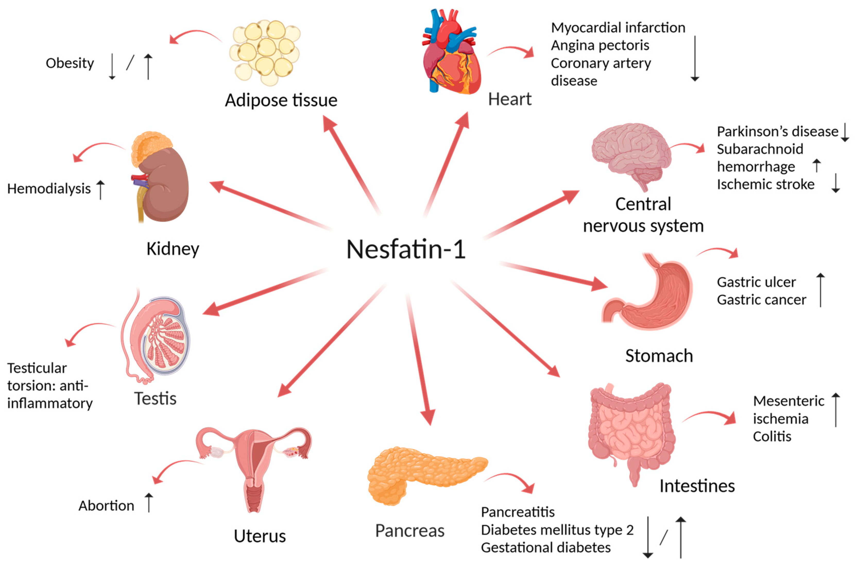
The presence of nesfatin-1 in different amounts in other peripheral tissues such as the cardiomyocytes [16], adipocytes [17], the esophagus [13], liver, kidney, ovaries [18], testis [19], uterus, and epididymis [20] is of great relevance (Figure 1). Its ubiquitous distribution explains the implication of this novel biomarker in the pathogenesis of different diseases.
Figure 1.
Nesfatin-1: localization and possible role in tissue functions regulation. Created with BioRender.com (accessed on 1 August 2024).
Despite its wide systemic distribution, the current state of knowledge regarding the mechanisms of action of nesfatin-1 is still limited. There were several attempts to detect specific intracellular or extracellular receptors, but the results were inconclusive. Nonetheless, there is strong evidence that nesfatin-1 may bind to a specific receptor that mediates its functions. By using Iodine 125-nesfatin-1 autoradiography, binding sites for nesfatin-1 were identified both in the brain as well as in peripheral organs such as the stomach, duodenum, jejunum, ileum, and the pancreas, with no signal in the colon [21]. A potential functional cooperation between the biomarker and a G protein-coupled receptor was suggested by the fact that nesfatin-1 increased the Ca2+ influx in neurons of the hypothalamus via the activation of the L, P, and Q-Ca2+ channels [22].
Currently, there is a series of proposed signaling pathways that mediate the pleiotropic effects of nesfatin-1, as summarized in Table 1.
Table 1.
Nesfatin-1: Mechanisms of action.
2. Implication of Nesfatin-1 in Multiple Disorders
After the discovery of its expression in a large number of tissues and organs, nesfatin-1 has received increased interest. Extensive research has been conducted to explore its implications in several diseases. Its anti-inflammatory, anti-apoptotic properties can explain the therapeutic potential of nesfatin-1 in a variety of diseases.
The relationship between nesfatin-1 and food intake and metabolic disorders has received special attention. Currently, there are multiple suggested mechanisms for the relationship between nesfatin-1, obesity, and metabolic dysfunction, particularly type 2 diabetes mellitus. Nesfatin-1 seems to regulate vagal neurons and the autonomic functions in the brainstem, midbrain, and hypothalamus. On the other hand, the vagus nerve also modulates nesfatin-1 expression [44]. Vagectomy increased nesfatin-1 levels and abolished the CNS negative control of the nesfatin-1 secretion in mice with induced nonalcoholic fatty liver disease (NAFLD) subjected to Roux-en-Y gastric bypass [45]. The possible implication of CNS activity on body weight regulation is also supported by the finding that the gut–brain axis may play a central role in the regulation of food intake and metabolic processes. Together with ghrelin, nesfatin-1 is now considered one of the main gastrokines involved in the gut–brain axis. Similar to ghrelin, nesfatin-1 gastric expression is regulated by nutritional status, with significantly reduced levels of nesfatin-1 being detected under fasting conditions. The mTOR signaling pathway has been proposed to modulate nesfatin-1 expression and energy balance [46]. Moreover, it has been demonstrated that the anorexigenic function can at least in part be explained by its beneficial effect on lipid metabolism. Nesfatin-1 prevents fat accumulation and accelerates lipid degradation in various tissues [47,48,49]. Even though nesfatin-1 normally inhibits gastric emptying, it has been shown that a high-fat diet stimulates gastric contractility and abolishes the effects of nesfatin-1 [50]. Another factor that influences metabolic homeostasis is food intake. Nesaftin-1 reduces food intake via the negative modulation of dopaminergic neurons, which results in a reduced rewarding feeling related to food ingestion. Taking into consideration that nesfatin-1 exerts its anorexigenic effects even when leptin resistance is present, the neuropeptide can be of therapeutic importance when it comes to the management of obesity [51,52]. Also, its therapeutic potential in eating disorders such as anorexia nervosa should not be overlooked [53]. Furthermore, nesfatin-1 exerts antihyperglycemic effects [54] as it prevents glucose formation and promotes glucose uptake in the liver [55]. Moreover, serum nesfatin-1 plays an important role in regulating β-cell insulin secretion [56] and improves the sensitivity to insulin by up-regulating the insulin receptor and GLUT8 in testis [57].
As a result, nesfatin-1 has been proposed as a potential diagnostic biomarker in metabolic disorders, mainly type 2 diabetes mellitus (DMT2). Most studies have underlined that nesfatin-1 levels are significantly lower in DMT2 patients and that there is a negative correlation between nesfatin-1 values and glucose homeostasis parameters, BMI, and atherogenic risk factors [58,59,60].
In Table 2, the potential involvement of nesfatin-1 in different pathologies with inflammatory components is summarized, with a special emphasis on its possible clinical relevance.
Table 2.
Implication of nesfatin-1 in multiple disorders.
Figure 1 illustrates the relationship between the tissular expression of nesfatin-1 and the variation in the plasmatic levels of nesfatin-1 in different disorders.
3. Implication of Nesfatin-1 in Digestive Disorders
3.1. Gastric Ulcer
Taking into consideration its anti-inflammatory properties, several studies aimed to investigate the potential effects of nesfatin-1 on gastric injury.
Kolgazi M. et al. [90] induced gastric ulcer by subcutaneous administration of indomethacin in rats. Fifteen minutes afterward, some of the rats were treated with nesfatin-1 (intraperitoneally) while the other ones were treated with saline. Serum tumor necrosis factor-α (TNF-α) and interleukin-6 (IL-6) were measured and stomach samples were collected to be microscopically examined and assessed for myeloperoxidase (MPO) activity, malondialdehyde (MDA), glutathione (GSH), and luminol- and lucigenin-enhanced chemiluminescence (CL) levels. The study demonstrated that ulcer induction led to an increase in the serum levels of TNF-α and IL-6 as well as MDA levels and MPO activity, whereas the reduced glutathione (GSH) content had decreased. Moreover, the administration of nesfatin-1 managed to reduce IL-6, CL, MDA levels, and MPO activity, and gastric GSH was restored. Nonetheless, to significantly diminish the TNF-α increase induced by indomethacin, a ten-fold dose of nesfatin-1 was needed. The decreased MPO activity suggests that nesfatin-1 exerts protective activity by inhibiting the recruitment of neutrophils. Nesfatin-1 led to a reduction in the levels of pro-inflammatory cytokines thanks to its antioxidant properties. The decreased levels of TNF-α and IL-1 might be the result of the ability of nesfatin-1 to counteract the up-regulation of mRNA expression of these molecules [91]. The restoration of GSH levels can be explained by the partial reversal of SOD mRNA down-regulation induced by nesfatin-1 [92]. Moreover, the dose administered intraperitoneally in the study managed to delay gastric emptying in comparison to previous research data that suggested that only intracerebroventricular administration had this effect [38]. However, there are no existing data regarding the intragastric administration of nesfatin-1. Furthermore, additional mechanisms have been proposed in other studies. The finding that nesfatin-1 reduces the secretion of gastric acid and increases the mucosal blood flow further supports the gastric protective effects of nesfatin-1 [91].
In rat models with gastric ulcers produced by serosal application of acetic acid, nesfatin-1 augmented mucosa regeneration by modulation of the oxidant/antioxidant balance. The inhibition of cyclooxygenase-1 (COX-1) reduced the protective effect of nesfatin-1, while the blockade of cyclooxygenase-2 (COX-2) managed to abolish all the changes induced by nesfatin-1. Hence, its gastroprotective properties involve the regulation of both COX-1 and COX-2 activity with a dominant effect of the COX-2 enzyme, which is of particular importance in ulcer healing [93].
Nesfatin-1 suppressed interleukin-1β (IL-1β) and increased the antioxidant capacity (superoxide dismutase and GSH) with direct effects on neutrophil infiltration and lipid peroxidation. Following these findings, exogenous administration managed to reduce gastric acid secretion and promote mucosal healing in rats with gastric lesions induced by water immersion and restraint stress. The NUCB2/nesfatin-1 levels in plasma as well as the blood flow in the mucosa were increased. At the same time, the study confirmed the functional cooperation between nesfatin-1 and cyclooxygenase-prostaglandin (COX-PG), and it also demonstrated that its potent gastroprotective effects rely on the nitric oxide synthase-inducible nitric oxide synthase (NOS-iNOS) system, vanilloid receptors, and the stimulation of both sensory and vagal nerve fibers. The proposed mechanism is the interaction between calcitonin gene-related peptide (CGRP) and nesfatin-1. In the rats treated with an antagonist of vanilloid receptors, capsazepine, as well as in those with denervation induced with capsaicin, the protective effects of nesfatin-1 were abolished. Therefore, nesfatin-1 probably activates the sensory afferent fibers and increases in this way the release of vasodilatory neuropeptides. CGRP has particularly been proposed to mediate the interaction between nesfatin-1 and vanilloid receptors. In addition, the combined administration of CGRP and nesfatin-1 showed superior protective effects on gastric microcirculation in comparison with the sole administration of nesfatin-1 [91].
3.2. Gastric Cancer
In the quest for a novel biomarker for gastric cancer, Wang et.al [94] tried to assess whether nesfatin-1 could potentially be used as a reliable and non-invasive diagnosis tool. Moreover, the presumed correlation between nesfatin-1 and Ki67 protein expression in normal gastric tissue and gastric cancer (GC) tissue was evaluated.
Ki67 is a well-known marker for cell proliferation. Within the cell cycle, it is solely expressed during all the active phases, but it is absent in the resting phase. As a proliferation marker, Ki67 is a close indicator of the proliferation and differentiation of cancer cells, including gastric cancer cells. Moreover, it was recently shown that it might be of great prognostic relevance and that it may consequently be useful to guide treatment strategies [95].
On the other hand, nesfatin-1 was reportedly linked to the mammalian target of rapamycin (mTOR) signaling pathway, which is involved in the dysregulation of cell proliferation. The study included 40 patients with gastric cancer and 40 controls. The results revealed that in patients with GC, the plasma nesfatin-1 levels were significantly elevated. The statistical analysis showed that nesfatin-1 has a 70% sensitivity and 95% specificity to differentiate between patients with GC and healthy individuals, while the most used cancer antigens as part of the diagnosis of GC (carcinoembryonic antigen, carbohydrate antigen 19-9 CA 19-9, carbohydrate antigen CA72-4) have low sensitivity and specificity (approximately 20–30%). Therefore, nesfatin-1 might be of clinical relevance for the screening and diagnosis of GC. Furthermore, the immunohistochemistry analyses showed that the tissue expression of ki67 is up-regulated in GC tissue, and it positively correlated with the plasma nesfatin-1 levels [94].
Gastric cancer is often associated with a higher incidence of depression. As a regulatory neuropeptide and part of the brain–gut axis, nesfatin-1 might be the key element of the relationship between gastric cancer and depressive symptoms. The levels of nesfatin-1 were evaluated both in the plasma and in the brain tissue of mice. The animals were divided into three categories: controls, the gastric cancer group that was not subjected to stress, and the gastric cancer group subjected to stress. In the last group, the levels of nesfatin-1 in tissue and plasma were the highest. The lowest expression was identified in the gastric cancer group not subjected to stress [96]. Taking into consideration the fact that nesfatin-1 is co-expressed with norepinephrine (NE) and 5-hydroxytryptamine (5-HT) in the raphe nucleus of the midbrain [5], it has been suggested that nesfatin-1 stimulates the 5-HT and NE neurons from this area, subsequently activating the corticotropin-releasing hormone (CRF) neurons and the Hypothalamic–Pituitary–Adrenal (HPA) Axis. The HPA is known to be involved in the occurrence of depression, particularly in patients suffering from cancer [97].
3.3. Necrotizing Enterocolitis
Necrotizing enterocolitis (NEC) is the most common gastrointestinal tract emergency in newborns. Because of its high mortality, new treatments are needed to improve the survival rate of the affected neonates [98]. Considering the anti-inflammatory and anti-apoptotic effects of nesfatin-1, Karadeniz et al. [99] aimed to assess its prospective therapeutic implications on NEC-induced rats.
The newborn rats were induced NEC with a hyperosmolar formula and by exposure to hypoxia. Following the induction, they were administered saline or nesfatin-1 for three consecutive days, and some of the ones treated with nesfatin-1 received capsaicin in order to ablate the afferent nerve fibers. The results indicated that the gene expression of COX-2, nuclear factor kappa-light-chain-enhancer of activated B cells (NF-kB), occludin, and claudin-3 was up-regulated in the NEC rats treated with saline, while the expression level in the nesfatin-1-treated group was not significantly changed. Its implications in the modulation of the inflammatory/anti-inflammatory balance as well as the modifications of the gut microbiome were moderately reversed by capsaicin administration. Of particular interest is the fact that NEC led to dysbiosis by stimulating the growth of Proteobacteria while inhibiting Actinobacteria and Bacteroidetes, and therefore improving the outcome of the affected newborns. The treatment with nesfatin-1 succeeded in counteracting the changes in the composition of the intestinal microbiota.
3.4. Mesenteric Ischemia
Intestinal ischemia is a condition characterized by high mortality. The majority of the lesions are caused by the restoration of blood flow resulting in an overabundance of inflammatory cytokines and free radicals [100]. Therefore, the reduction in these inflammatory molecule levels before reperfusion would reduce damage. Tatar et al. tried to investigate both the diagnostic and the therapeutic implications of nesfatin-1 in intestinal ischemia.
Twenty-one rats were divided into three different groups: the first group was subjected to intestinal ischemia for 1 h, followed by a 5-h reperfusion, the second one was exposed to 6-h ischemia, and, in the third group, a laparotomy was performed, without any other additional procedure. Intestine samples were collected for histopathological evaluation as well as intracardiac blood samples to assess a series of biomarkers including nesfatin-1.
The highest nesfatin-1 values were among the second group, while the lowest ones were in the third group. It showed a decrease in endothelial nitric oxide synthase (eNOS) activity and, subsequently, nitric oxide (NO) production. Additionally, there was a positive correlation between the pathology score and the levels of nesfatin-1, suggesting that nesfatin-1 could be an excellent biomarker in acute intestinal ischemia [101]. Similar observations were made by studies conducted on acute mesenteric ischemia-induced rat models, reinforcing the fact that nesfatin-1 reduces oxidative stress index parameters and thus may exert a protective role against ischemia/reperfusion injury [102].
3.5. Colitis
Despite its anti-apoptotic and anti-inflammatory properties, the relationship between nesfatin-1 and inflammatory bowel disease has barely been explored. Ozturk et al. were the only ones that approached this topic, having a major focus on the potential underlying mechanisms of action.
Rats were divided into two main groups: colitis and the control group. Under anesthesia, 4% acetic acid was intrarectally administered to induce colitis, whereas the controls received saline solution. Ten minutes afterward, some of the colitis mice received nesfatin-1 intracerebroventricularly. To decipher its mechanisms, three subgroups of the nesfatin-1 group were created 5 min after colitis induction, each of them receiving intracerebroventricularly growth hormone (GH) secretagogue receptor-1a (GHSR-1a) antagonist (antagonist of ghrelin receptors), atosiban (antagonist of oxytocin receptor), and trifluoroacetate salt SHU9119 (antagonist of melanocortin receptors) followed 5 min later by the administration of nesfatin-1 for 3 days. Rats were in the end decapitated and tissue samples were evaluated.
Macroscopic and microscopic damage scores were analyzed histopathologically and they were increased in mice with colitis that did not receive nesfatin-1 treatment. Since atosiban administration abolished both the macroscopic and microscopic healing changes produced by nesfatin-1 administration, nesfatin-1 might act via oxytocin receptors. Moreover, the microscopic damage-reducing properties were significantly diminished following GSHR-1a administration while the macroscopic lesions remained the same. Therefore, ghrelin might modulate the positive effects of nesfatin-1 on microscopic damage. The elevated myeloperoxidase activity, malondialdehyde levels, and luminol and lucigenin chemiluminescence characteristic for inflammation decreased thanks to the administration of nesfatin-1. As a result, nesfatin-1 seems to exert its effects by reducing lipid peroxidation and free radical damage as well as by decreasing neutrophil infiltration. In addition, its beneficial effects were counteracted by atosiban and GHSR-1a, confirming the initial assumption that nesfatin-1 is closely linked to oxytocin and ghrelin receptors. The study outlines a surprising aspect: the association between central nesfatin-1 and peripheral inflammatory status. Thus, colitis and its underlying inflammatory mechanisms might stimulate the activity of CNS nesfatin-1 neurons in order to improve the systemic inflammatory status. Even though the intracerebroventricular administration of nesfatin-1 proved to have positive effects in this regard, further research is needed to understand the link between its central and peripheral activity [103].
A single clinical study conducted by Beyaz S. and Akbal E. [104] has been carried out thus far. It included 17 patients with Crohn’s disease (CD), 18 patients with ulcerative colitis (UC), and 17 healthy patients. The serum levels of nesfatin-1 were detected both in the active and the remission stages along with other inflammation markers including C reactive protein (CRP), erythrocyte sedimentation rate (ESR), and white cell count (WCC). The results showed that nesfatin-1 was notably elevated in patients with CD as well as in patients with UC during the flare-up period. In the remission period, the values moderately decreased but they remained significantly higher in comparison to healthy individuals. Nesfatin-1 showed a high sensitivity (88,2%) and specificity (88,2%) in the active phase of the patients with Chron’s disease. Correspondingly, in patients with ulcerative colitis, the sensitivity was 83.3% and 94.1%. Thus, nesfatin-1 proved to be an excellent novel biomarker that can be used together with other markers as part of the non-invasive diagnosis of IBD. Furthermore, the study tried to assess the association with active phase reactants.
However, it is still contradictory if nesfatin-1 exerts a pro- or anti-inflammatory role. On the one hand, it has been demonstrated that the neurons expressing nesfatin-1 are stimulated by the presence of a peripheral inflammatory state [105]. Moreover, it was also shown that IL-6 and TNF-α regulate nesfatin-1 production in the human adipose tissue [17]. Thus, the increased levels of nesfatin-1 in IBD can be explained at least partially by the elevated systemic expression of these two inflammatory markers. Despite its obvious implication in several systemic inflammatory diseases, it is still unclear whether nesfatin-1 is part of a pro- or anti-inflammatory response. Taking into consideration its protective role on animal models with colitis and gastric ulcers, one cannot undermine its crucial contribution to the underlying pathophysiological mechanisms.
3.6. Pancreatitis
Similar to previous studies on colitis-induced animal models, Buzcu et al. [106] tried to investigate the potential role of nesfatin-1 as an anti-inflammatory marker in acute pancreatitis (AP) as well as the associated fundamental mechanisms. Nesfatin-1 was administered intraperitoneally five minutes before the induction of pancreatitis with two doses of caerulin. Before the pretreatment with nesfatin, atosiban (oxytocin receptor agonist), HS024 (melanocortin receptor agonist), and cortistatin (ghrelin receptor antagonist) were administered. Microscopic damage scores and several biomarkers were determined. MPO, reflecting neutrophil infiltration, malondialdehyde, an end-product of lipid peroxidation, luminol and lucigenin chemiluminescence, serum amylase, lipase, and trypsinogen 2 were increased as part of the AP inflammatory response. However, glutathione levels, showing the antioxidant protective ability, diminished. Nesfatin-1 decreased the oxidative injury, and the way the different receptor agonists/antagonists influenced the biomarkers and the histology scores revealed that nesfatin-1 modulates its activity via melanocortin receptors.
In addition, another study conducted on rat models with AP sought to evaluate the potential of nesfatin-1 not only as a diagnostic but also as a severity marker. The rats were divided into three groups: the control group, the mild pancreatitis, and the severe pancreatitis group. The serum nesfatin-1, lipase, amylase, and aspartate aminotransferase (AST) and alanine aminotransferase (ALT) levels were measured, and the severity of the induced pancreatitis was evaluated by a pathologist. There was a substantial decrease in nesfatin-1 levels in parallel with the severity of pancreatitis, but no statistically significant correlation was noted. Two main explanations can account for the reduction in nesfatin-1. Nesfatin-1 might be degraded at a higher rate to counteract the high oxidative status in acute pancreatitis. Also, the actual destruction of the pancreatic tissue can result in an evident decrease in nesfatin-1 [107].
Few clinical studies attempted to characterize the connection between nesfatin-1 and AP. In the study performed by Ulger et al. [108], 40 patients with gallstone-induced pancreatitis were enrolled in order to assess plasma nesfatin-1 along with leptin and ghrelin. Two blood samples were collected for each patient, at admission and discharge, respectively. The levels of these hormones were altered in all patients. Nesfatin-1 and leptin were significantly higher at hospitalization in comparison to discharge, whereas ghrelin values had an opposite evolution. Even if the exact mechanisms are still unclear, these findings may be linked with the clinical outcomes in patients with AP.
3.7. Cholecystitis
Tekin et al. [109] tried to determine the possible relevance of nesfatin-1 concerning the diagnosis and assessment of acute cholecystitis. The study included four groups: mild, moderate, and severe cholecystitis groups (based on the Tokyo 2018 Guidelines), and the control group. The patients were evaluated for blood nesfatin-1, leukocytes, neutrophils, lymphocytes, and neutrophil/lymphocyte ratio. Nesfatin-1 levels were found to be decreased in the cholecystitis groups compared with the controls, but no statistically significant difference was identified as far as the potential correlation between nesfatin-1 levels and the severity of the disease is concerned. At the same time, the study reinforces the remark that nesfatin-1 production and release are increased in the initial phase to balance out the inflammatory response. As inflammation progresses, nesfatin-1 levels decline. It is noteworthy that nesfatin-1 may vary depending on the severity of the inflammatory response and the type of the affected tissues.
3.8. Obstructive Jaundice
The connection between nesfatin-1 and obstructive jaundice (OJ) has barely been addressed thus far. Solmaz et al. [110] studied the effects of nesfatin-1 on OJ in rats. The animals were assigned to three different groups: sham, control, and nesfatin-1. Under general anesthesia, in the sham group, laparotomy and exploration of the common biliary duct (CBD) were performed. The other two groups undertook laparotomy, which was followed by the double ligation of the CBD above the pancreas, which was eventually cut off. Afterward, the groups were treated for 10 days with saline (control group) and nesfatin-1 (nesfatin group). Blood and liver samples were analyzed.
Biochemical parameters (AST, ALT, alkaline phosphatase, gamma-glutamyl transferase, albumin) were not significantly different within the two groups. As for the cytokine levels, MDA was the lowest and SOD was the highest in the nesfatin group, and the data were of statistical relevance. Thus, nesfatin-1 has a considerable impact on oxidative status. IL-6 levels, TNF-α, and oxidative DNA damage were lower in the nesfatin group yet without being statistically meaningful. However, the histopathological assessment indicated remarkable differences between the controls and the rats treated with nesfatin. The latter showed less neutrophil infiltration of the portal area, hepatocyte necrosis, bile duct proliferation, and edema. Even though most of the inflammation markers determined in the study did not show a critical distinction between the two groups, the histopathological results endorse the beneficial antioxidant and anti-inflammatory potential of nesfatin-1 in OJ. As suggested by the coordinators of the research, a limitation of the study that may have a great impact on the reliability of the results is the relatively short follow-up period. Thus, the relation between the role of nesfatin-1 and the evolution of the disease long-term cannot be foreseen.
4. Conclusions
This review underlines the current state of knowledge concerning nesfatin-1, a novel neuropeptide with anti-inflammatory, antioxidant, and antiapoptotic properties. It is expressed in different tissues, both centrally and peripherally, and it plays a crucial role in the pathogenesis of different diseases. As a result, nesfatin-1 can be of great clinical importance as a diagnosis biomarker, severity indicator, and therapeutic agent. However, nesfatin-1 is far from being used as a clinical application in the foreseeable future. The main limitation is the inability to identify its specific receptors. Also, while most findings are encouraging, some results are still contradictory. In particular, the relationship between nesfatin-1 and metabolic diseases is still unclearly defined, with studies leading to opposing results as previously presented in this article review. There might be several reasons behind this, for example, an inadequate selection of the subjects or animal models, faulty assessment methods, a too small sample size, additional unknown factors that can influence the course of the disease studied, inter-individual variability, and the so far not clearly known mechanisms of actions. Hence, nesfatin-1 deserves more attention and further research is clearly needed to explore its diagnostic and therapeutic potential.
Author Contributions
Conceptualization, A.-C.D.-B., R.M.P., and I.C.B.; methodology, A.-C.D.-B., D.M.M., D.-Z.D.-B. and L.C.; validation, R.M.P., I.C.B., and A.D.B.; formal analysis, A.-C.D.-B.; investigation, A.-C.D.-B., D.M.M., D.-Z.D.-B. and L.C.; resources, A.D.B. and I.C.B.; data curation, R.M.P.; writing—original draft preparation, A.-C.D.-B.; writing—review and editing, R.M.P. and I.C.B.; visualization, A.D.B.; supervision, I.C.B.; project administration, A.-C.D.-B., R.M.P., and I.C.B.; funding acquisition, I.C.B. All authors have read and agreed to the published version of the manuscript.
Funding
This research received no external funding.
Institutional Review Board Statement
Not applicable.
Informed Consent Statement
Not applicable.
Data Availability Statement
No new data were created or analyzed in this study. Data sharing is not applicable to this article.
Conflicts of Interest
The authors declare no conflicts of interest.
References
- Oh-I, S.; Shimizu, H.; Satoh, T.; Okada, S.; Adachi, S.; Inoue, K.; Eguchi, H.; Yamamoto, M.; Imaki, T.; Hashimoto, K.; et al. Identification of Nesfatin-1 as a Satiety Molecule in the Hypothalamus. Nature 2006, 443, 709–712. [Google Scholar] [CrossRef] [Scilit] [PubMed]
- Shimizu, H.; Oh-I, S.; Okada, S.; Mori, M. Nesfatin-1: An Overview and Future Clinical Application. Endocr. J. 2009, 56, 537–543. [Google Scholar] [CrossRef] [Scilit] [PubMed]
- Foo, K.S.; Brismar, H.; Broberger, C. Distribution and Neuropeptide Coexistence of Nucleobindin-2 MRNA/Nesfatin-like Immunoreactivity in the Rat CNS. Neuroscience 2008, 156, 563–579. [Google Scholar] [CrossRef] [Scilit] [PubMed]
- Stengel, A.; Goebel, M.; Taché, Y. Nesfatin-1: A Novel Inhibitory Regulator of Food Intake and Body Weight. Obes. Rev. 2011, 12, 261–271. [Google Scholar] [CrossRef] [Scilit] [PubMed]
- Goebel-Stengel, M.; Wang, L.; Stengel, A.; Taché, Y. Localization of Nesfatin-1 Neurons in the Mouse Brain and Functional Implication. Brain Res. 2011, 1396, 20–34. [Google Scholar] [CrossRef] [Scilit] [PubMed]
- Pan, W.; Hsuchou, H.; Kastin, A.J. Nesfatin-1 Crosses the Blood–Brain Barrier without Saturation. Peptides 2007, 28, 2223–2228. [Google Scholar] [CrossRef] [Scilit] [PubMed]
- Kras, K.; Muszyński, M.; Muszyński, S.; Tomaszewska, E.; Arciszewski, M.B.; Klećkowskaklećkowska-Nawrot, J.; Goździewskagoździewska-Harłajczuk, K.; Kras, K.; Muszyński, S.; Tomaszewska, E.; et al. Minireview: Peripheral Nesfatin-1 in Regulation of the Gut Activity—15 Years since the Discovery. Animals 2022, 12, 101. [Google Scholar] [CrossRef] [Scilit] [PubMed]
- Xu, Y.; Chen, F. Antioxidant, Anti-Inflammatory and Anti-Apoptotic Activities of Nesfatin-1: A Review. J. Inflamm. Res. 2020, 13, 607–617. [Google Scholar] [CrossRef] [Scilit] [PubMed]
- Zhang, A.-Q.; Li, X.-L.; Jiang, C.-Y.; Lin, L.; Shi, R.-H.; Chen, J.-D.; Oomura, Y. Expression of Nesfatin-1/NUCB2 in Rodent Digestive System. World J. Gastroenterol. 2010, 16, 1735–1741. [Google Scholar] [CrossRef] [Scilit] [PubMed]
- Stengel, A.; Goebel, M.; Yakubov, I.; Wang, L.; Witcher, D.; Coskun, T.; Taché, Y.; Sachs, G.; Lambrecht, N.W.G. Identification and Characterization of Nesfatin-1 Immunoreactivity in Endocrine Cell Types of the Rat Gastric Oxyntic Mucosa. Endocrinology 2009, 150, 232. [Google Scholar] [CrossRef] [Scilit] [PubMed]
- Puzio, I.; Muszyński, S.; Dobrowolski, P.; Kapica, M.; Pawłowska-Olszewska, M.; Donaldson, J.; Tomaszewska, E. Alterations in Small Intestine and Liver Morphology, Immunolocalization of Leptin, Ghrelin and Nesfatin-1 as Well as Immunoexpression of Tight Junction Proteins in Intestinal Mucosa after Gastrectomy in Rat Model. J. Clin. Med. 2021, 10, 272. [Google Scholar] [CrossRef] [Scilit]
- Mohan, H.; Unniappan, S. Ontogenic Pattern of Nucleobindin-2/Nesfatin-1 Expression in the Gastroenteropancreatic Tissues and Serum of Sprague Dawley Rats. Regul. Pept. 2012, 175, 61–69. [Google Scholar] [CrossRef] [Scilit]
- Jiang, S.; Zhou, W.; Zhang, X.; Wang, D.; Zhu, H.; Hong, M.; Gong, Y.; Ye, J.; Fang, F. Developmental Expression and Distribution of Nesfatin-1/NUCB2 in the Canine Digestive System. Acta Histochem. 2016, 118, 90–96. [Google Scholar] [CrossRef] [Scilit]
- Gonkowski, S.; Rychlik, A.; Nowicki, M.; Nieradka, R.; Bulc, M.; Całka, J. A Population of Nesfatin 1-like Immunoreactive (LI) Cells in the Mucosal Layer of the Canine Digestive Tract. Res. Vet. Sci. 2012, 93, 1119–1121. [Google Scholar] [CrossRef] [Scilit]
- Morton, K.A.; Hargreaves, L.; Mortazavi, S.; Weber, L.P.; Blanco, A.M.; Unniappan, S. Tissue-Specific Expression and Circulating Concentrations of Nesfatin-1 in Domestic Animals. Domest. Anim. Endocrinol. 2018, 65, 56–66. [Google Scholar] [CrossRef] [Scilit] [PubMed]
- Feijóo-Bandín, S.; Rodríguez-Penas, D.; García-Rúa, V.; Mosquera-Leal, A.; Otero, M.F.; Pereira, E.; Rubio, J.; Martínez, I.; Seoane, L.M.; Gualillo, O.; et al. Nesfatin-1 in Human and Murine Cardiomyocytes: Synthesis, Secretion, and Mobilization of GLUT-4. Endocrinology 2013, 154, 4757–4767. [Google Scholar] [CrossRef] [Scilit]
- Ramanjaneya, M.; Chen, J.; Brown, J.E.; Tripathi, G.; Hallschmid, M.; Patel, S.; Kern, W.; Hillhouse, E.W.; Lehnert, H.; Tan, B.K.; et al. Identification of Nesfatin-1 in Human and Murine Adipose Tissue: A Novel Depot-Specific Adipokine with Increased Levels in Obesity. Endocrinology 2010, 151, 3169–3180. [Google Scholar] [CrossRef] [Scilit] [PubMed]
- Gonzalez, R.; Shepperd, E.; Thiruppugazh, V.; Lohan, S.; Grey, C.L.; Chang, J.P.; Unniappan, S. Nesfatin-1 Regulates the Hypothalamo-Pituitary-Ovarian Axis of Fish. Biol. Reprod. 2012, 87, 84. [Google Scholar] [CrossRef] [Scilit]
- García-Galiano, D.; Pineda, R.; Ilhan, T.; Castellano, J.M.; Ruiz-Pino, F.; Sánchez-Garrido, M.A.; Vazquez, M.J.; Sangiao-Alvarellos, S.; Romero-Ruiz, A.; Pinilla, L.; et al. Cellular Distribution, Regulated Expression, and Functional Role of the Anorexigenic Peptide, NUCB2/Nesfatin-1, in the Testis. Endocrinology 2012, 153, 1959–1971. [Google Scholar] [CrossRef] [Scilit] [PubMed]
- Author, C.; Yang, H.; Kim, J.; Chung, Y.; Kim, H.; Im, E.; Lee, H. The Tissue Distribution of Nesfatin-1/NUCB2 in Mouse. Dev. Reprod. 2014, 18, 301–309. [Google Scholar] [CrossRef] [Scilit]
- Prinz, P.; Goebel-Stengel, M.; Teuffel, P.; Rose, M.; Klapp, B.F.; Stengel, A. Peripheral and Central Localization of the Nesfatin-1 Receptor Using Autoradiography in Rats. Biochem. Biophys. Res. Commun. 2016, 470, 521–527. [Google Scholar] [CrossRef] [Scilit]
- Brailoiu, G.C.; Dun, S.L.; Brailoiu, E.; Inan, S.; Yang, J.; Jaw, K.C.; Dun, N.J. Nesfatin-1: Distribution and Interaction with a G Protein-Coupled Receptor in the Rat Brain. Endocrinology 2007, 148, 5088–5094. [Google Scholar] [CrossRef] [Scilit] [PubMed]
- Yin, Y.; Li, Z.; Gao, L.; Li, Y.; Zhao, J.; Zhang, W. AMPK-Dependent Modulation of Hepatic Lipid Metabolism by Nesfatin-1. Mol. Cell Endocrinol. 2015, 417, 20–26. [Google Scholar] [CrossRef] [Scilit]
- Dong, J.; Xu, H.; Wang, P.F.; Cai, G.J.; Song, H.F.; Wang, C.C.; Dong, Z.T.; Ju, Y.J.; Jiang, Z.Y. Nesfatin-1 Stimulates Fatty-Acid Oxidation by Activating AMP-Activated Protein Kinase in STZ-Induced Type 2 Diabetic Mice. PLoS ONE 2013, 8, e83397. [Google Scholar] [CrossRef] [Scilit]
- Kan, J.-Y.; Yen, M.-C.; Wang, J.-Y.; Wu, D.-C.; Chiu, Y.-J.; Ho, Y.-W.; Kuo, P.-L.; Kan, J.-Y.; Yen, M.-C.; Wang, J.-Y.; et al. Nesfatin-1/Nucleobindin-2 Enhances Cell Migration, Invasion, and Epithelial-Mesenchymal Transition via LKB1/AMPK/TORC1/ZEB1 Pathways in Colon Cancer. Oncotarget 2016, 7, 31336–31349. [Google Scholar] [CrossRef] [Scilit]
- Yang, M.; Zhang, Z.; Wang, C.; Li, K.; Li, S.; Boden, G.; Li, N.; Yang, G. Nesfatin-1 Action in the Brain Increases Insulin Sensitivity through Akt/AMPK/TORC2 Pathway in Diet-Induced Insulin Resistance. Diabetes 2012, 61, 1959–1968. [Google Scholar] [CrossRef] [Scilit]
- Tan, J.; Jin, L.; Wang, Y.; Cao, B.; Wang, S.; Zhang, F.; Wei, L. Nesfatin-1 Acts as an Inhibitory Factor in Human Gastrointestinal Smooth Muscle Cells in Diabetes Mellitus-Induced Delayed Gastric Emptying. Int. J. Clin. Exp. Pathol. 2016, 9, 11214–11221. [Google Scholar]
- Ge, J.F.; Xu, Y.Y.; Qin, G.; Pan, X.Y.; Cheng, J.Q.; Chen, F.H. Nesfatin-1, a Potent Anorexic Agent, Decreases Exploration and Induces Anxiety-like Behavior in Rats without Altering Learning or Memory. Brain Res. 2015, 1629, 171–181. [Google Scholar] [CrossRef] [Scilit] [PubMed]
- Tanida, M.; Gotoh, H.; Yamamoto, N.; Wang, M.; Kuda, Y.; Kurata, Y.; Mori, M.; Shibamoto, T. Hypothalamic Nesfatin-1 Stimulates Sympathetic Nerve Activity via Hypothalamic ERK Signaling. Diabetes 2015, 64, 3725–3736. [Google Scholar] [CrossRef] [Scilit]
- Chen, Z.; Xu, Y.Y.; Ge, J.F.; Chen, F.H. CRHR1 Mediates the Up-Regulation of Synapsin I Induced by Nesfatin-1 Through ERK 1/2 Signaling in SH-SY5Y Cells. Cell Mol. Neurobiol. 2018, 38, 627–633. [Google Scholar] [CrossRef] [Scilit]
- Yuan, J.H.; Chen, X.; Dong, J.; Zhang, D.; Song, K.; Zhang, Y.; Wu, G.B.; Hu, X.H.; Jiang, Z.Y.; Chen, P. Nesfatin-1 in the Lateral Parabrachial Nucleus Inhibits Food Intake, Modulates Excitability of Glucosensing Neurons, and Enhances UCP1 Expression in Brown Adipose Tissue. Front. Physiol. 2017, 8, 239809. [Google Scholar] [CrossRef] [Scilit] [PubMed]
- Yosten, G.L.C.; Samson, W.K. Nesfatin-1 Exerts Cardiovascular Actions in Brain: Possible Interaction with the Central Melanocortin System. Am. J. Physiol. Regul. Integr. Comp. Physiol. 2009, 297, 330–336. [Google Scholar] [CrossRef] [Scilit]
- Ying, J.; Zhang, Y.; Gong, S.; Chang, Z.; Zhou, X.; Li, H.; Tao, J.; Zhang, G. Nesfatin-1 Suppresses Cardiac L-Type Ca2+ Channels Through Melanocortin Type 4 Receptor and the Novel Protein Kinase C Theta Isoform Pathway. Cell. Physiol. Biochem. 2015, 36, 555–568. [Google Scholar] [CrossRef] [Scilit] [PubMed]
- Tasatargil, A.; Kuscu, N.; Dalaklioglu, S.; Adiguzel, D.; Celik-Ozenci, C.; Ozdem, S.; Barutcigil, A.; Ozdem, S. Cardioprotective Effect of Nesfatin-1 against Isoproterenol-Induced Myocardial Infarction in Rats: Role of the Akt/GSK-3β Pathway. Peptides 2017, 95, 1–9. [Google Scholar] [CrossRef] [Scilit]
- Lu, Q.B.; Wang, H.P.; Tang, Z.H.; Cheng, H.; Du, Q.; Wang, Y.B.; Feng, W.B.; Li, K.X.; Cai, W.W.; Qiu, L.Y.; et al. Nesfatin-1 Functions as a Switch for Phenotype Transformation and Proliferation of VSMCs in Hypertensive Vascular Remodeling. Biochim. Et Biophys. Acta (BBA) Mol. Basis Dis. 2018, 1864, 2154–2168. [Google Scholar] [CrossRef] [Scilit] [PubMed]
- Li, Z.; Gao, L.; Tang, H.; Yin, Y.; Xiang, X.; Li, Y.; Zhao, J.; Mulholland, M.; Zhang, W. Peripheral Effects of Nesfatin-1 on Glucose Homeostasis. PLoS ONE 2013, 8, e71513. [Google Scholar] [CrossRef] [Scilit]
- Feng, H.; Wang, Q.; Guo, F.; Han, X.; Pang, M.; Sun, X.; Gong, Y.; Xu, L.; Xu, L. Nesfatin-1 Influences the Excitability of Gastric Distension-Responsive Neurons in the Ventromedial Hypothalamic Nucleus of Rats. Physiol. Res. 2017, 66, 335–344. [Google Scholar] [CrossRef] [Scilit]
- Stengel, A.; Goebel, M.; Wang, L.; Rivier, J.; Kobelt, P.; Mönnikes, H.; Lambrecht, N.W.G.; Taché, Y. Central Nesfatin-1 Reduces Dark-Phase Food Intake and Gastric Emptying in Rats: Differential Role of Corticotropin-Releasing Factor2 Receptor. Endocrinology 2009, 150, 4911. [Google Scholar] [CrossRef] [Scilit]
- Nakata, M.; Manaka, K.; Yamamoto, S.; Mori, M.; Yada, T. Nesfatin-1 Enhances Glucose-Induced Insulin Secretion by Promoting Ca2+ Influx through L-Type Channels in Mouse Islet β-Cells. Endocr. J. 2011, 58, 305–313. [Google Scholar] [CrossRef] [Scilit]
- Maejima, Y.; Horita, S.; Kobayashi, D.; Aoki, M.; O’hashi, R.; Imai, R.; Sakamoto, K.; Mori, M.; Takasu, K.; Ogawa, K.; et al. Nesfatin-1 Inhibits Voltage Gated K+ Channels in Pancreatic Beta Cells. Peptides 2017, 95, 10–15. [Google Scholar] [CrossRef] [Scilit]
- Ayada, C.; Turgut, G.; Turgut, S. The Effect of Nesfatin-1 on Heart L-Type Ca2+ Channel Aα1c Subunit in Rats Subjected to Chronic Restraint Stress. Bratisl. Med. J. 2015, 116, 326–329. [Google Scholar] [CrossRef] [Scilit] [PubMed]
- Yamawaki, H.; Takahashi, M.; Mukohda, M.; Morita, T.; Okada, M.; Hara, Y. A Novel Adipocytokine, Nesfatin-1 Modulates Peripheral Arterial Contractility and Blood Pressure in Rats. Biochem. Biophys. Res. Commun. 2012, 418, 676–681. [Google Scholar] [CrossRef] [Scilit] [PubMed]
- Angelone, T.; Filice, E.; Pasqua, T.; Amodio, N.; Galluccio, M.; Montesanti, G.; Quintieri, A.M.; Cerra, M.C. Nesfatin-1 as a Novel Cardiac Peptide: Identification, Functional Characterization, and Protection against Ischemia/Reperfusion Injury. Cell. Mol. Life Sci. 2013, 70, 495–509. [Google Scholar] [CrossRef] [Scilit] [PubMed]
- Rupp, S.K.; Stengel, A. Interactions between Nesfatin-1 and the Autonomic Nervous System—An Overview. Peptides 2022, 149, 170719. [Google Scholar] [CrossRef] [Scilit] [PubMed]
- Wang, G.; Wang, Q.; Bai, J.; Li, G.; Tao, K.; Wang, G.; Xia, Z. RYGB Increases Postprandial Gastric Nesfatin-1 and Rapid Relieves NAFLD via Gastric Nerve Detachment. PLoS ONE 2020, 15, e0243640. [Google Scholar] [CrossRef] [Scilit] [PubMed]
- Pena-Leon, V.; Perez-Lois, R.; Seoane, L.M. MTOR Pathway Is Involved in Energy Homeostasis Regulation as a Part of the Gut–Brain Axis. Int. J. Mol. Sci. 2020, 21, 5715. [Google Scholar] [CrossRef] [Scilit] [PubMed]
- Luo, J.; Wen, F.; Qiu, D.; Wang, S. Nesfatin-1 in Lipid Metabolism and Lipid-Related Diseases. Clin. Chim. Acta 2021, 522, 23–30. [Google Scholar] [CrossRef] [Scilit] [PubMed]
- Nasri, A.; Kowaluk, M.; Widenmaier, S.B.; Unniappan, S. Nesfatin-1 and Nesfatin-1-like Peptide Attenuate Hepatocyte Lipid Accumulation and Nucleobindin-1 Disruption Modulates Lipid Metabolic Pathways. Commun. Biol. 2024, 7, 623. [Google Scholar] [CrossRef] [Scilit] [PubMed]
- Liu, Y.; Chen, X.; Qu, Y.; Song, L.; Lin, Q.; Li, M.; Su, K.; Li, Y.; Dong, J. Central Nesfatin-1 Activates Lipid Mobilization in Adipose Tissue and Fatty Acid Oxidation in Muscle via the Sympathetic Nervous System. BioFactors 2020, 46, 454–464. [Google Scholar] [CrossRef] [Scilit] [PubMed]
- Özdemir-Kumral, Z.N.; Koyuncuoğlu, T.; Arabacı-Tamer, S.; Çilingir-Kaya, Ö.T.; Köroğlu, A.K.; Yüksel, M.; Yeğen, B.Ç. High-Fat Diet Enhances Gastric Contractility, but Abolishes Nesfatin-1-Induced Inhibition of Gastric Emptying. J. Neurogastroenterol. Motil. 2021, 27, 265–278. [Google Scholar] [CrossRef] [Scilit] [PubMed]
- Dore, R.; Krotenko, R.; Reising, J.P.; Murru, L.; Sundaram, S.M.; Di Spiezio, A.; Müller-Fielitz, H.; Schwaninger, M.; Jöhren, O.; Mittag, J.; et al. Nesfatin-1 Decreases the Motivational and Rewarding Value of Food. Neuropsychopharmacology 2020, 45, 1645–1655. [Google Scholar] [CrossRef] [Scilit] [PubMed]
- Dokumacioglu, E.; Iskender, H.; Sahin, A.; Erturk, E.Y.; Kaynar, O. Serum Levels of Nesfatin-1 and Irisin in Obese Children. Eur. Cytokine Netw. 2020, 31, 39–43. [Google Scholar] [CrossRef] [Scilit] [PubMed]
- Pałasz, A.; Rojczyk, E.; Siwiec, A.; Janas-Kozik, M. Nesfatin-1 in the Neurochemistry of Eating Disorders. Psychiatr. Pol. 2020, 54, 209–222. [Google Scholar] [CrossRef] [Scilit] [PubMed]
- Zhai, T.; Li, S.-Z.; Fan, X.-T.; Tian, Z.; Lu, X.-Q.; Dong, J. Circulating Nesfatin-1 Levels and Type 2 Diabetes: A Systematic Review and Meta-Analysis. J Diabetes Res 2017, 2017, 1–8. [Google Scholar] [CrossRef] [Scilit] [PubMed]
- Öztürk Özkan, G. Effects of Nesfatin-1 on Food Intake and Hyperglycemia. J. Am. Coll. Nutr. 2020, 39, 345–351. [Google Scholar] [CrossRef] [Scilit] [PubMed]
- Huang, K.; Liang, Y.; Wang, K.; Wu, J.; Luo, H.; Yi, B. Influence of Circulating Nesfatin-1, GSH and SOD on Insulin Secretion in the Development of T2DM. Front. Public. Health 2022, 10, 882686. [Google Scholar] [CrossRef] [Scilit] [PubMed]
- Ranjan, A.; Choubey, M.; Yada, T.; Krishna, A. Nesfatin-1 Ameliorates Type-2 Diabetes-Associated Reproductive Dysfunction in Male Mice. J. Endocrinol. Investig. 2020, 43, 515–528. [Google Scholar] [CrossRef] [Scilit] [PubMed]
- Matta, R.A.; El-Hini, S.H.; Salama, A.M.S.E.; Moaness, H.M. Serum Nesfatin-1 Is a Biomarker of Pre-Diabetes and Interplays with Cardiovascular Risk Factors. Egypt. J. Intern. Med. 2022, 34, 15. [Google Scholar] [CrossRef] [Scilit]
- Mohammad, N.; Gallaly, D. Serum Nesfatin-1 in Patients with Type 2 Diabetes Mellitus: A Cross Sectional Study. Zanco J. Med. Sci. 2020, 24, 1–7. [Google Scholar] [CrossRef] [Scilit]
- Kadim, B.M.; Hassan, E.A. Nesfatin-1—As a Diagnosis Regulatory Peptide in Type 2 Diabetes Mellitus. J. Diabetes Metab. Disord. 2022, 21, 1369–1375. [Google Scholar] [CrossRef] [Scilit] [PubMed]
- Yang, J.-Y.; Baek, S.-E.; Yoon, J.-W.; Kim, H.-S.; Kwon, Y.; Yeom, E. Nesfatin-1 Ameliorates Pathological Abnormalities in Drosophila HTau Model of Alzheimer’s Disease. Biochem. Biophys. Res. Commun. 2024, 727, 150311. [Google Scholar] [CrossRef] [Scilit] [PubMed]
- Chen, H.; Li, X.; Ma, H.; Zheng, W.; Shen, X. Reduction in Nesfatin-1 Levels in the Cerebrospinal Fluid and Increased Nigrostriatal Degeneration Following Ventricular Administration of Anti-Nesfatin-1 Antibody in Mice. Front. Neurosci. 2021, 15, 621173. [Google Scholar] [CrossRef] [Scilit]
- Acik, V.; Matyar, S.; Arslan, A.; İstemen, İ.; Olguner, S.K.; Arslan, B.; Gezercan, Y.; Ökten, A.İ. Relationshıp of Spontaneous Subarachnoid Haemorrhage and Cerebral Aneurysm to Serum Visfatin and Nesfatin-1 Levels. Clin. Neurol. Neurosurg. 2020, 194, 105837. [Google Scholar] [CrossRef] [Scilit] [PubMed]
- Kazimierczak-Kabzińska, A.; Marek, B.; Borgiel-Marek, H.; Kajdaniuk, D.; Kos-Kudła, B. Assessing the Blood Concentration of New Adipocytokines in Patients with Ischaemic Stroke. Endokrynol. Pol. 2020, 71, 504–511. [Google Scholar] [CrossRef] [Scilit] [PubMed]
- Carr, J.A.; Epelbaum, J.; Gourcerol, G.; Stengel, A.; Prinz, P.; Teuffel, P.; Lembke, V.; Kobelt, P.; Goebel-Stengel, M.; Hofmann, T.; et al. Nesfatin-1 30–59 Injected Intracerebroventricularly Differentially Affects Food Intake Microstructure in Rats Under Normal Weight and Diet-Induced Obese Conditions. Front. Neurosci. 2015, 9, 422. [Google Scholar] [CrossRef] [Scilit]
- Elthakaby, A.H.M.; Esso, H.Y.; Elsayed, H.M.; Youssef Ahmed, K.; Abd El-Ghaffar Mohamed, N.; Elthakaby, M.; Youssef, K.; Mohamed, E.-G.; Authors Ahmed M Elthakaby, A.H.; Mohamed, R.R. Effect of Nesfatin-1 on the Nutritional Status of Hemodialysis Patients. J. Med. Sci. Res. 2022, 5, 30. [Google Scholar] [CrossRef] [Scilit]
- Yin, C.; Liu, W.; Xu, E.; Zhang, M.; Lv, W.; Lu, Q.; Xiao, Y. Copeptin and Nesfatin-1 Are Interrelated Biomarkers with Roles in the Pathogenesis of Insulin Resistance in Chinese Children with Obesity. Ann. Nutr. Metab. 2020, 76, 223–232. [Google Scholar] [CrossRef] [Scilit]
- Afifi, M.A.; Khalil, F.M.; Assal, M.A.; El Matueny, R.M.; Rizk, M. Serum Nesfatin-1 in Patients with Metabolic Associated Fatty Liver Disease. Egypt. J. Hosp. Med. 2024, 94, 1056. [Google Scholar]
- Samani, S.M.; Ghasemi, H.; Bookani, K.R.; Shokouhi, B. Serum nesfatin-1 level in healthy subjects with weight-related abnormalities and newly diagnosed patients with type 2 diabetes mellitus—A case-control study. Acta Endocrinol. 2019, XV, 69–73. [Google Scholar] [CrossRef] [Scilit]
- Caroleo, M.; Carbone, E.A.; Arcidiacono, B.; Greco, M.; Primerano, A.; Mirabelli, M.; Fazia, G.; Rania, M.; Hribal, M.L.; Gallelli, L.; et al. Does NUCB2/Nesfatin-1 Influence Eating Behaviors in Obese Patients with Binge Eating Disorder? Toward a Neurobiological Pathway. Nutrients 2023, 15, 348. [Google Scholar] [CrossRef] [Scilit]
- Jiang, X.-H.; Song, H.-H.; Wang, R. Expression and Clinical Significance of Serum SHBG, Nesfatin-1 and SFRP4 in Patients with Gestational Diabetes Mellitus. J. Trop. Med. 2020, 20, 815–819. [Google Scholar]
- Çaltekin, M.D.; Caniklioğlu, A. Maternal Serum Delta-Like 1 and Nesfatin-1 Levels in Gestational Diabetes Mellitus: A Prospective Case-Control Study. Cureus 2021, 13, e17001. [Google Scholar] [CrossRef] [Scilit]
- Mahmood, N.; Al-Sammarai, R.; Al-Fanar, R. Evaluation of the Role of Nesfatin-1 and Myonectin as A Diagnostic Marker for Polycystic Ovary Syndrome and Also for Treatment Response with Metformin. Egypt. Acad. J. Biol. Sci. C Physiol. Mol. Biol. 2023, 15, 577–584. [Google Scholar] [CrossRef] [Scilit]
- Obstet, T.J.; Demir Çaltekin, M.; Caniklioğlu, A.; Yalçın, S.E.; Kırmızı, D.A.; Baser, E.; Yalvaç, E.S. Clinical Investigation/Araştırma PRECIS: DLK1 and Nesfatin-1 Protein Levels Are Lower in Women with PCOS. Polikistik over Sendromunda DLK1 ve Nesfatin-1 Düzeyleri ve Metabolik Parametrelerle Ilişkisi: Prospektif Kontrollü Çalışma. J. Turk. Soc. Obstet. Gynecol. 2021, 18, 124–130. [Google Scholar] [CrossRef] [Scilit]
- Su, R.-Y.; Geng, X.-Y.; Yang, Y.; Yin, H.-S.; Rui-Ying Su, C. Nesfatin-1 Inhibits Myocardial Ischaemia/Reperfusion Injury through Activating Akt/ERK Pathway-Dependent Attenuation of Endoplasmic Reticulum Stress. J. Cell. Mol. Med. 2021, 25, 5050–5059. [Google Scholar] [CrossRef] [Scilit] [PubMed]
- Naseroleslami, M.; Sharifi, M.; Rakhshan, K.; Mokhtari, B.; Aboutaleb, N. Nesfatin-1 Attenuates Injury in a Rat Model of Myocardial Infarction by Targeting Autophagy, Inflammation, and Apoptosis. Arch. Physiol. Biochem. 2020, 129, 122–130. [Google Scholar] [CrossRef] [Scilit]
- Nejati, A.; Doustkami, H.; Babapour, B.; Ebrahimoghlou, V.; Aslani, M.R. Serum Correlation of Nesfatin-1 with Angiographic, Echocardiographic, and Biochemical Findings in Patients with Coronary Artery Disease. Iran. Red. Crescent Med. J. 2021, 23, 245. [Google Scholar] [CrossRef] [Scilit]
- Kadoglou, N.P.E.; Korakas, E.; Lampropoulos, S.; Maratou, E.; Kassimis, G.; Patsourakos, N.; Plotas, P.; Moutsatsou, P.; Lambadiari, V. Plasma Nesfatin-1 and DDP-4 Levels in Patients with Coronary Artery Disease: Kozani Study. Cardiovasc. Diabetol. 2021, 20, 166. [Google Scholar] [CrossRef] [Scilit]
- Yilmaz, M.S.; Altinbas, B.; Guvenc, G.; Erkan, L.G.; Avsar, O.; Savci, V.; Kucuksen, D.U.; Arican, I.; Yalcin, M. The Role of Centrally Injected Nesfatin-1 on Cardiovascular Regulation in Normotensive and Hypotensive Rats. Auton. Neurosci. 2015, 193, 63–68. [Google Scholar] [CrossRef] [Scilit] [PubMed]
- Zhang, S.; Rong, G.; Xu, Y.; Jing, J. Elevated Nesfatin-1 Level in Synovium and Synovial Fluid Is Associated with pro-Inflammatory Cytokines in Patients with Rheumatoid Arthritis. Int. J. Gen. Med. 2021, 14, 5269–5278. [Google Scholar] [CrossRef] [Scilit]
- Seewordova, L.; Polyakova, J.; Papichev, E.; Akhverdyan, Y.; Zavodovsky, В. Ab0078 Nesfatin-1 Expression Associated with A Marker for Bone Matrix Formation P1np in Rheumatoid Arthritis. Ann. Rheum. Dis. 2022, 81, 1171. [Google Scholar] [CrossRef] [Scilit]
- Zhang, S.; Zhang, T.; Xu, Y.; Rong, G.; Jing, J. Inhibition of NUCB2 Suppresses the Proliferation, Migration, and Invasion of Rheumatoid Arthritis Synovial Fibroblasts from Patients with Rheumatoid Arthritis in Vitro. J. Orthop. Surg. Res. 2022, 17, 574. [Google Scholar] [CrossRef] [Scilit] [PubMed]
- Ranjan, A.; Choubey, M.; Yada, T.; Krishna, A. Direct Effects of Neuropeptide Nesfatin-1 on Testicular Spermatogenesis and Steroidogenesis of the Adult Mice. Gen. Comp. Endocrinol. 2019, 271, 49–60. [Google Scholar] [CrossRef] [Scilit] [PubMed]
- Ranjan, A.; Choubey, M.; Yada, T.; Krishna, A. Immunohistochemical Localization and Possible Functions of Nesfatin-1 in the Testis of Mice during Pubertal Development and Sexual Maturation. J. Mol. Histol. 2019, 50, 533–549. [Google Scholar] [CrossRef] [Scilit]
- Arabaci Tamer, S.; Yildirim, A.; Köroğlu, M.K.; Çevik, Ö.; Ercan, F.; Yeğen, B. Nesfatin-1 Ameliorates Testicular Injury and Supports Gonadal Function in Rats Induced with Testis Torsion. Peptides 2018, 107, 1–9. [Google Scholar] [CrossRef] [Scilit]
- Chung, Y.; Kim, H.; Seon, S.; Yang, H. Serum Cytokine Levels Are Related to Nesfatin-1/NUCB2 Expression in the Implantation Sites of Spontaneous Abortion Model of CBA/j × DBA/2. Dev. Reprod. 2017, 21, 35. [Google Scholar] [CrossRef] [Scilit]
- Wang, Z.Z.; Chen, S.C.; Zou, X.B.; Tian, L.L.; Sui, S.H.; Liu, N.Z. Nesfatin-1 Alleviates Acute Lung Injury through Reducing Inflammation and Oxidative Stress via the Regulation of HMGB1. Eur. Rev. Med. Pharmacol. Sci. 2020, 24, 5071–5081. [Google Scholar] [CrossRef]
- Jiang, L.; Xu, K.; Li, J.; Zhou, X.; Xu, L.; Wu, Z.; Ma, C.; Ran, J.; Hu, P.; Bao, J.; et al. Nesfatin-1 Suppresses Interleukin-1β-Induced Inflammation, Apoptosis, and Cartilage Matrix Destruction in Chondrocytes and Ameliorates Osteoarthritis in Rats. Aging 2020, 12, 1760–1777. [Google Scholar] [CrossRef] [Scilit]
- Sun, J.; Gao, N.; Wu, Q.; Li, Y.; Zhang, L.; Jiang, Z.; Wang, Z.; Liu, J. High Plasma Nesfatin-1 Level in Chinese Adolescents with Depression. Sci. Rep. 2023, 13, 15288. [Google Scholar] [CrossRef] [Scilit]
- Kolgazi, M.; Cantali-Ozturk, C.; Deniz, R.; Ozdemir-Kumral, Z.N.; Yuksel, M.; Sirvanci, S.; Yeğen, B.C. Nesfatin-1 Alleviates Gastric Damage via Direct Antioxidant Mechanisms. J. Surg. Res. 2015, 193, 111–118. [Google Scholar] [CrossRef] [Scilit]
- Szlachcic, A.; Sliwowski, Z.; Krzysiek-Maczka, G.; Majka, J.; Surmiak, M.; Pajdo, R.; Drozdowicz, D.; Konturek, S.J.; Brzozowski, T. New Satiety Hormone Nesfatin-1 Protects Gastric Mucosa against Stress-Induced Injury: Mechanistic Roles of Prostaglandins, Nitric Oxide, Sensory Nerves and Vanilloid Receptors. Peptides 2013, 49, 9–20. [Google Scholar] [CrossRef] [Scilit]
- Szlachcic, A.; Majka, J.; Strzalka, M.; Szmyd, J.; Pajdo, R.; Ptak-Belowska, A.; Kwiecien, S.; Brzozowski, T. Experimental Healing of Preexisting Gastric Ulcers Induced by Hormones Controlling Food Intake Ghrelin, Orexin-A and Nesfatin-1 Is Impaired under Diabetic Conditions. A Key to Understanding the Diabetic Gastropathy? J. Physiol. Pharmacol. 2013, 64, 625–637. [Google Scholar] [PubMed]
- Kolgazi, M.; Mehmet, A.; Üniversitesi, A.A.; Özdemir, Z.N.; Demirci, E.K. Anti-Inflammatory Effects of Nesfatin-1 on Acetic Acid-Induced Gastric Ulcer in Rats: Involvement of Cyclo-Oxygenase Pathway. J. Physiol. Pharmacol. 2017, 68, 765–777. [Google Scholar] [PubMed]
- Wang, X.Q.; Zheng, Y.; Fang, P.F.; Song, X.B. Nesfatin-1 Is a Potential Diagnostic Biomarker for Gastric Cancer. Oncol. Lett. 2020, 19, 1577–1583. [Google Scholar] [CrossRef] [Scilit] [PubMed]
- Ko, G.H.; Go, S.I.; Lee, W.S.; Lee, J.H.; Jeong, S.H.; Lee, Y.J.; Hong, S.C.; Ha, W.S.; Yang, F. Prognostic Impact of Ki-67 in Patients with Gastric Cancer—The Importance of Depth of Invasion and Histologic Differentiation. Medicine 2017, 96, e7181. [Google Scholar] [CrossRef] [Scilit] [PubMed]
- Zhang, N.; Li, J.; Wang, H.; Xiao, L.; Wei, Y.; He, J.; Wang, G. The Level of Nesfatin-1 in a Mouse Gastric Cancer Model and Its Role in Gastric Cancer Comorbid with Depression. Shanghai Arch. Psychiatry 2018, 30, 119–126. [Google Scholar] [PubMed]
- Yoshida, N.; Maejima, Y.; Sedbazar, U.; Ando, A.; Kurita, H.; Damdindorj, B.; Takano, E.; Gantulga, D.; Iwasaki, Y.; Kurashina, T.; et al. Stressor-Responsive Central Nesfatin-1 Activates Corticotropin-Releasing Hormone, Noradrenaline and Serotonin Neurons and Evokes Hypothalamic-Pituitary-Adrenal Axis. Aging 2010, 2, 775–784. [Google Scholar] [CrossRef] [Scilit] [PubMed]
- Weller, J.; Sampah, M.E.S.; Salazar, A.J.G.; Hackam, D.J. Necrotizing Enterocolitis. In Principles of Neonatology; Elsevier: Amsterdam, The Netherlands, 2023; pp. 707–714. [Google Scholar] [CrossRef] [Scilit]
- Karadeniz Cerit, K.; Koyuncuoğlu, T.; Yağmur, D.; Peker Eyüboğlu, İ.; Şirvancı, S.; Akkiprik, M.; Aksu, B.; Dağlı, E.T.; Yeğen, B. Nesfatin-1 Ameliorates Oxidative Bowel Injury in Rats with Necrotizing Enterocolitis: The Role of the Microbiota Composition and Claudin-3 Expression. J. Pediatr. Surg. 2020, 55, 2797–2810. [Google Scholar] [CrossRef] [Scilit] [PubMed]
- Li, G.; Wang, S.; Fan, Z. Oxidative Stress in Intestinal Ischemia-Reperfusion. Front. Med. 2022, 8, 750731. [Google Scholar] [CrossRef] [Scilit] [PubMed]
- Tatar, C.; Ahlatci, F.A.; Idiz, U.O.; Nayci, A.E.; Incir, S.; Agcaoglu, O.; Idiz, C.; Balik, E. May Nesfatin-1 Be a Biomarker in Acute Mesenteric Ischemia? J. Coll. Physicians Surg. Pak. 2019, 29, 928–931. [Google Scholar] [CrossRef] [Scilit] [PubMed]
- Gutiérrez-Sánchez, G.; García-Alonso, I.; María, J.G.S.d.S.; Alonso-Varona, A.; de la Parte, B.H. Antioxidant-Based Therapy Reduces Early-Stage Intestinal Ischemia-Reperfusion Injury in Rats. Antioxidants 2021, 10, 853. [Google Scholar] [CrossRef] [Scilit]
- Ozturk, C.; Oktay, S.; Yuksel, M.; Akakin, D.; Yarat, A.; Kasimay, C. Anti-Inflammatory Effects of Nesfatin-1 in Rats with Acetic Acid—Induced Colitis and Underlying Mechanisms. J. Physiol. Pharmacol. 2017, 66, 741–750. [Google Scholar]
- Beyaz, A.E.; Akbal, E. Increased Serum Nesfatin-1 Levels in Patients with Inflammatory Bowel Diseases. Postgrad. Med. J. 2022, 98, 446–449. [Google Scholar] [CrossRef] [Scilit] [PubMed]
- Bonnet, M.S.; Pecchi, E.; Trouslard, J.; Jean, A.; Dallaporta, M.; Troadec, J.D. Central Nesfatin-1-Expressing Neurons Are Sensitive to Peripheral Inflammatory Stimulus. J. Neuroinflam. 2009, 6, 27. [Google Scholar] [CrossRef] [Scilit] [PubMed]
- Buzcu, H.; Ozbeyli, D.; Yuksel, M.; Cilingir Kaya, O.T.; Kasimay Cakir, O. Nesfatin-1 Protects from Acute Pancreatitis: Role of Melanocortin Receptors. J. Physiol. Pharmacol. 2019, 70, 1–10. [Google Scholar] [CrossRef] [Scilit]
- Ferlengez, A.G.; Tatar, C.; Degerli, M.S.; Koyuncu, A.; Ahlatci, F.A.; Nayci, A.E.; Ari, A.; Idiz, U.O. The Role of Serum Nesfatin-1 in a Rat Model of Acute Pancreatitis. Adv. Clin. Exp. Med. 2023, 32, 545–549. [Google Scholar] [CrossRef] [Scilit]
- Ulger, B.V.; Gül, M.; Uslukaya, O.; Oguz, A.; Bozdag, Z.; Yüksel, H.; Böyük, A. New Hormones to Predict the Severity of Gallstone-Induced Acute Pancreatitis. Turk. J. Gastroenterol. 2014, 25, 714–717. [Google Scholar] [CrossRef] [Scilit] [PubMed]
- Tekin, O.; Sevinç, M.M.; Albayrak, Ö.; Batıkan, O.; İdiz, U.O. Prognostic Effect of Nesfatin-1 on the Diagnosis and Staging of Acute Cholecystitis. Ulus. Travma Acil Cerrahi Derg. 2022, 28, 1583–1589. [Google Scholar] [CrossRef] [Scilit]
- Solmaz, A.; Gülçiçek, O.B.; Erçetin, C.; Yiğitbaş, H.; Yavuz, E.; Arıcı, S.; Erzik, C.; Zengi, O.; Demirtürk, P.; Çelik, A.; et al. Nesfatin-1 Alleviates Extrahepatic Cholestatic Damage of Liver in Rats. Bosn. J. Basic. Med. Sci. 2016, 16, 247. [Google Scholar] [CrossRef] [Scilit]

Disclaimer/Publisher’s Note: The statements, opinions and data contained in all publications are solely those of the individual author(s) and contributor(s) and not of MDPI and/or the editor(s). MDPI and/or the editor(s) disclaim responsibility for any injury to people or property resulting from any ideas, methods, instructions or products referred to in the content. |
© 2024 by the authors. Licensee MDPI, Basel, Switzerland. This article is an open access article distributed under the terms and conditions of the Creative Commons Attribution (CC BY) license (https://creativecommons.org/licenses/by/4.0/).