


Abstract
A calix[4]arene-based fluorescent chemosensor containing two quinolinium units (C4-BisQ) was synthesized and evaluated for the selective detection of cyanide (CN−). C4-BisQ showed a dual-mode optical response with a green-to-blue color change under ambient light and fluorescence quenching upon CN− addition in CH3CN/H2O (9:1, v/v). UV–Vis and fluorescence titration studies indicated a 1:1 interaction between C4-BisQ and CN− with an association constant of 1.08 × 10⁵ M−1. The limits of detection were 0.080 µM by ratiometric UV–Vis analysis and 0.54 µM by fluorescence spectroscopy. C4-BisQ was highly selective for CN− in the presence of common competing anions and metal cations such as F−, Cl−, Br−, I−, ClO4−, NO3−, H2PO4−, HSO4−, Ac−, and Li+, Na+, Mg2+, Ca2+, Mn2+, Fe3+, Co2+, Zn2+, Cd2+, Al3+. Spectroscopic data suggest that cyanide recognition mainly involves deprotonation and is accompanied by fluorescence quenching under the applied conditions. Recovery experiments in tap water and seawater, together with visual detection in apricot and peach kernel extracts, confirmed the applicability of the sensor. Overall, C4-BisQ offers a simple dual-mode approach for cyanide detection in environmental water and food-related samples.
1. Introduction
The selective detection of environmentally and biologically important species remains an important topic in analytical chemistry. Among these analytes, cyanide (CN−) has attracted particular attention because of its high toxicity and widespread industrial use. Cyanide rapidly inhibits mitochondrial cytochrome c oxidase, disrupts cellular respiration, and can cause acute toxicity even at low exposure levels [1,2]. Despite its well-established toxicity, cyanide is still widely used in industrial processes such as gold extraction, electroplating, metallurgy, polymer production, and chemical synthesis, creating persistent risks of environmental release and aquatic contamination. In addition to industrial sources, cyanogenic glycosides naturally present in several edible plants can also release cyanide upon enzymatic hydrolysis. These environmental and food-related risks have sustained interest in analytical methods for rapid and sensitive cyanide detection in a wide range of sample matrices [3,4,5].
Conventional methods for cyanide determination include gas chromatography, ion chromatography, high-performance liquid chromatography, mass spectrometry, atomic absorption-based techniques, and electrochemical methods [6,7,8,9,10,11,12]. These methods provide high sensitivity and accurate quantification, but they often require expensive instrumentation, extensive sample preparation, and trained personnel, limiting their suitability for rapid on-site analysis. Optical chemosensors have emerged as an attractive alternative because they offer fast response, operational simplicity, and direct visual detection with relatively low instrumental requirements [13,14,15]. Among optical sensing approaches, dual-mode systems combining colorimetric and fluorescent responses have received considerable attention [16,17,18,19,20,21,22,23]. The colorimetric response enables rapid visual screening, whereas fluorescence provides a complementary optical signal that can support quantitative analysis. Combining both responses in a single sensor can improve analytical reliability, particularly in complex sample matrices where a single response may not always be sufficient. Consequently, dual-mode chemosensors continue to attract interest for cyanide detection in environmental and food-related samples.
Quinoline- and quinolinium-based chemosensors have been widely studied for cyanide sensing because of their electron-deficient heteroaromatic structures [24,25,26,27,28,29,30,31,32]. In many reported systems, cyanide recognition proceeds through nucleophilic addition or deprotonation, resulting in changes in conjugation, charge distribution, or emission behavior [33,34,35,36,37,38,39,40,41]. Despite these advances, there is still interest in developing new molecular designs for dual-mode cyanide sensing. Calix[n]arenes are widely used macrocyclic scaffolds in supramolecular chemistry because they are readily functionalized and can be modified with different recognition and signaling units [42,43,44,45]. As a result, they have found broad applications in the design of molecular receptors and chemical sensors. Although calixarene-based fluorescent sensors have been reported, quinolinium-functionalized calixarenes for dual-mode cyanide sensing remain relatively underexplored [46,47].
Based on these considerations, we designed and synthesized C4-BisQ, a quinolinium-functionalized calix[4]arene receptor, as a dual-mode optical chemosensor for cyanide detection under near-neutral conditions. Its sensing behaviour was investigated in CH3CN/H2O (9:1, v/v) using UV–Vis and fluorescence spectroscopy, FT-IR, and pH-dependent studies, and its applicability was further examined in environmental water samples and cyanogenic food extracts.
2. Materials and Methods
2.1. Materials and Reagents
All solvents utilized in this study were of analytical grade and sourced from either Merck (Darmstadt, Germany) or Sigma-Aldrich (St. Louis, MO, USA). Prior to use, solvents were distilled and dried according to standard literature procedures. Milli-Q grade water (18.2 MΩ·cm) was employed throughout all experiments. Metal ion sources as perchlorate salts (NaClO4, LiClO4, Ca(ClO4)2, Mg(ClO4)2, Mn(ClO4)2, Fe(ClO4)3, Co(ClO4)2, Cu(ClO4)2, AgClO4, Zn(ClO4)2, Cd(ClO4)2, Hg(ClO4)2, and Al(ClO4)3) and anion sources as tetrabutylammonium (TBA) salts (TBAF, TBACl, TBABr, TBAI, TBANO3, TBAOAc, TBAClO4, TBAH2PO4, TBAHSO4, and TBACN) were obtained from Sigma-Aldrich and used without further purification. Stock solutions of both metal ions and anions were prepared in acetonitrile (CH3CN) for subsequent experimental procedures. Nuclear magnetic resonance (1H and 13C NMR) spectra were recorded using a Bruker 400 MHz spectrometer TMS as an internal standard. Ultraviolet-visible (UV–Vis) absorption spectra were measured with a Shimadzu UV-1280 spectrophotometer, while fluorescence measurements were conducted on an Edinburgh FS5 spectrofluorometer. Infrared (IR) spectra were collected with an FT-IR spectrometer (Bruker Vertex 70). The pH measurements were performed with a digital pH meter calibrated with standard buffer solutions. All spectroscopic measurements were conducted at room temperature (25 °C).
2.2. Synthesis of C4-BisQ
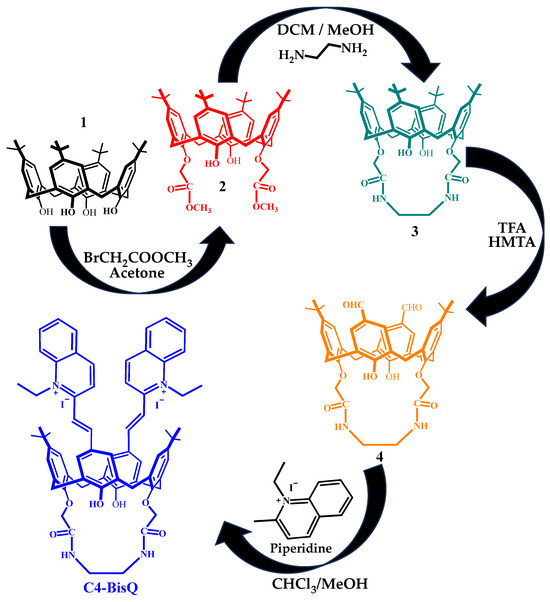
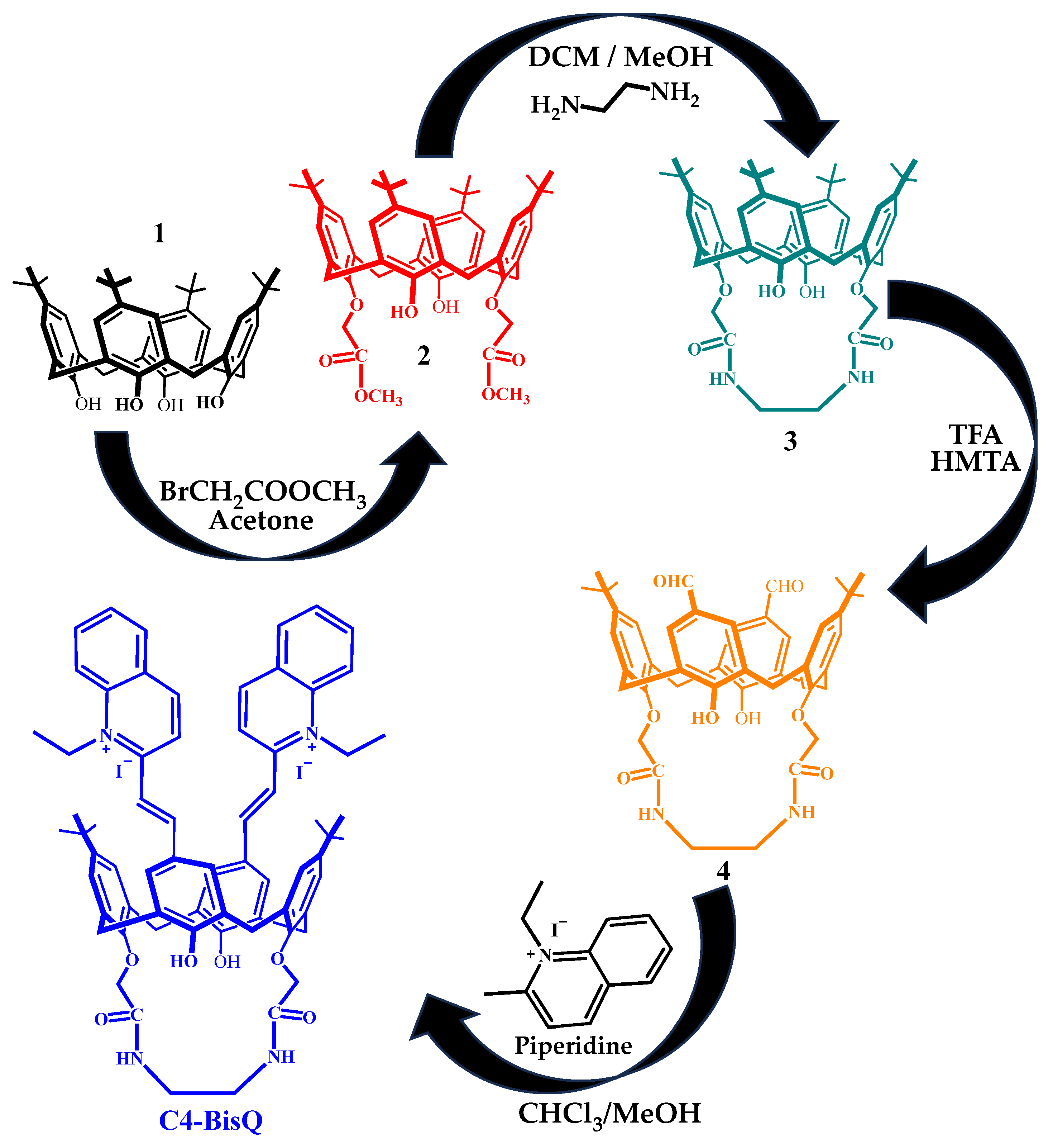
The synthetic route for the preparation of C4-BisQ is illustrated in Scheme 1. Compounds 1–4 were prepared and characterized according to literature methods [48,49,50,51], and their comprehensive synthetic pathways along with all baseline characterization spectra are made available in the Supporting Information (Figures S1–S7). To a stirred solution of the diformyl-calixarene intermediate 4 (300 mg, 0.37 mmol) and the quaternary quinolinium salt (220 mg, 0.753 mmol) in a mixture of CHCl3/MeOH (1:1, v/v, 20 mL), a few drops of piperidine (60 μL) were added as a base catalyst. The reaction mixture was heated to reflux and stirred for 48 h under a nitrogen atmosphere. The progress of the reaction was monitored by thin-layer chromatography (TLC). After completion of the reaction, the solvent was removed under reduced pressure using a rotary evaporator, and the crude product was purified by recrystallization from methanol/diethyl ether to afford C4-BisQ as a dark brown solid in 70% yield. IR (ATR): 3276 cm−1 (OH, NH), 1661 cm−1 (C=O). 1H NMR (400 MHz, DMSO-d6, 25 °C), δ (ppm): 1.20 (s, 18H, t-Bu), 1,44 (t, 6H, +NCH2CH3), 3,53 (m, 8H; 4H, -CH2CH2-; 4H, ArCH2Ar), 4.28 (d, 4H, J = 12.3 Hz, ArCH2Ar), 4.41 (s, 4H, OCH2), 4.91 (bs, 4H, +NCH2), 7.28 (s, 4H, ArH calix), 7.40–8.80 (m, 22H; 4H, ArH calix; 4H, CH=CH vinylic; 12H, ArH Quin; 2H, NH amide), 10.93 (s, 2H, OH). 13C NMR (100 MHz, DMSO-d6, 25 °C), δ (ppm): 168.64, 150.29, 148.96, 147.76, 138.75 130.37, 126.21, 121.07, 114.06, 74.97, 53.29, 52.16, 46.34, 38.41, 34.56, 31.83, 31.08, 14.43. All corresponding characterization spectra for C4-BisQ are available in the Supporting Information file (Figures S8–S10).
Scheme 1.
Synthetic pathway for the preparation of the calixarene-based fluorescent chemosensor C4-BisQ. Different colors are used only to distinguish the compounds visually.
2.3. Preparation of Solutions and Optical Measurements
UV–Vis and Fluorescence Spectroscopic Measurements. All UV–Vis absorption and fluorescence measurements were carried out at room temperature (25 °C) using a 1.0 cm quartz cuvette. A stock solution of C4-BisQ (1.0 × 10⁻² M) was prepared in CH3CN, and ultrapure Milli-Q water was used as the aqueous component for the routine spectroscopic measurements. pH-dependent experiments were performed in a CH3CN/Britton–Robinson (BR) buffer mixture (9:1, v/v). For optical measurements, the stock solution was diluted to a final probe concentration of 10 µM in CH3CN/H2O (9:1, v/v) medium, with a total volume of 3.0 mL. For fluorescence measurements, the excitation wavelength was set at 428 nm, and emission spectra were recorded over the range of 400–700 nm. For anion and cation selectivity studies, solutions of the tested analytes or metal ions (50 µM each, 5 equiv.) were added individually to separate solutions of C4-BisQ (10 µM), and the UV–Vis absorption and fluorescence spectra were recorded immediately after mixing.
For spectrophotometric titration experiments, incremental amounts of CN− stock solution (1.0 × 10⁻²–1.0 × 10⁻³ M) were added to the C4-BisQ solution (10 µM) using a micro-syringe, while the total volume change was kept negligible. Absorption and fluorescence spectra were recorded immediately after each addition. For competitive interference studies, the target CN− analyte (10 µM) was first pre-mixed with individual competing anions or metal ions (50 µM each, 5 equiv.) in the sample solution. Subsequently, the macrocyclic receptor C4-BisQ (10 µM) was added to the resulting ion mixtures. The fluorescence intensity at 526 nm and the corresponding visual color changes were then recorded immediately.
Determination of the Limit of Detection (LOD) and Limit of Quantitation (LOQ). The limit of detection and limit of quantitation for CN− ions were calculated from the fluorescence titration data according to the standard IUPAC guidelines [52], using the following equations:
where sb represents the standard deviation of the fluorescence intensity of the blank C4-BisQ solution (10 μM) measured ten times, and k is the slope obtained from the linear calibration plot of fluorescence intensity versus CN− concentration.
LOD = 3 sb/k
LOQ = 10 sb/k
LOQ = 10 sb/k
Evaluation of Binding Affinity (Benesi–Hildebrand Method). The association constant (Ka) of the C4-BisQ+CN− complex was determined from the fluorescence titration curve using the Benesi–Hildebrand expression for a 1:1 stoichiometry [53]:
where I0, I, and Imin are the fluorescence intensities of C4-BisQ at 526 nm in the absence, during the titration, and at the saturation point of CN−, respectively. The value of Ka was calculated from the ratio of the intercept to the slope of the linear plot of 1/(I0 − I) versus 1/[CN−].
Stern–Volmer Analysis. To characterize the concentration-dependent fluorescence quenching induced by CN− ions, the titration data were analyzed using the classic Stern–Volmer equation [54]:
where I0 and I are the fluorescence intensities of C4-BisQ at 526 nm in the absence and presence of CN−, respectively, Ksv is the Stern–Volmer quenching constant, and [CN–] denotes the concentration of the cyanide anion.
Stoichiometry Determination (Job’s Plot Method). The stoichiometry of the cyanide and sensor interaction was established using the continuous variation method (Job’s plot) via UV–vis absorption spectroscopy [55]. Stock solutions of both C4-BisQ and CN– were prepared at the same concentration (10 μM). A series of solutions were prepared where the total molar concentration ([C4-BisQ] + [CN−] = 10 μM) was kept constant, while the mole fraction of the receptor (X = [C4-BisQ]/([C4-BisQ] + [CN–]) was varied from 0.1 to 0.9. The ratiometric absorbance response at 605 nm and 428 nm (A605/A428) was plotted against the mole fraction of cyanide to determine the interaction ratio.
2.4. Preparation of Food Extracts and Colorimetric Assay
Fresh apricot and peach fruits were purchased from a local market, and their kernels were manually separated and used as food samples for qualitative cyanide assays. Briefly, 2.0 g of each kernel sample was crushed separately in a mortar, suspended in 50.0 mL of Milli-Q water containing 0.5% NaOH, and stirred in sealed vials at room temperature for 180 min. The resulting mixtures were then filtered through a membrane filter to obtain clear extracts [56,57]. Before analysis, the pH of the basic extracts was adjusted to approximately 7 using BR buffer to provide controlled near-neutral conditions during the sensing assay. For the colorimetric assay, 100 µL of each pH-adjusted extract was added to separate vials containing C4-BisQ (10 µM). The color change from bright green to blue was then recorded under ambient laboratory light.
2.5. Quantitative Spike-And-Recovery Assays in Environmental Water Samples
To evaluate the practical applicability of C4-BisQ, recovery experiments were carried out using tap water and seawater samples. Tap water was collected from the laboratories of Selçuk University (Konya, Türkiye) and used without further treatment because its pH was close to neutral. Seawater was collected from the Mediterranean coast of Taşucu (Mersin, Türkiye) and filtered through a 0.45 μm membrane filter to remove suspended particles. Since the slightly alkaline nature of seawater may induce the deprotonation of C4-BisQ in the absence of cyanide, the sample pH was adjusted to approximately 7.0 using BR buffer before analysis. Known amounts of CN− (2 and 5 μM) were then added to both water samples, and the fluorescence emission intensity at 526 nm was monitored to determine the recovery of CN−.
3. Results and Discussion
3.1. Synthesis and Characterization of Chemosensor C4-BisQ
The synthetic route for the preparation of C4-BisQ is illustrated in Scheme 1. Compounds 1–4 were prepared and characterized according to the reported procedures [48,49,50,51]. The target chemosensor C4-BisQ was synthesized via Knoevenagel condensation of diformyl intermediate 4 with the corresponding quinolinium salt precursor [58]. The structure of C4-BisQ was confirmed by 1H NMR and 13C NMR spectroscopy. In the 1H NMR spectrum, the disappearance of the aldehydic proton signal of intermediate 4 at 9.80 ppm, together with the appearance of new vinylic (–CH=CH–) and quinolinium aromatic proton signals, supported the formation of the desired product. The corresponding characterization spectra are provided in the Supporting Information (Figures S8–S10).
3.2. Optimization of the Sensing Conditions
Before the detailed spectroscopic studies, the experimental conditions were optimized to identify a suitable solvent system for cyanide sensing. Solvent composition and pH were selected as optimization parameters because both can influence the optical response of the probe. The fluorescence response of C4-BisQ (10 µM) was first examined in different pure solvents (Figure S11a). The free probe showed weak emission in CHCl3 and DMSO, whereas stronger fluorescence was observed in EtOH, CH3CN, and H2O. The effect of water content on the optical behavior of C4-BisQ was further examined using CH3CN/H2O mixtures with different water fractions (Figure S11b). As expected for an organic probe, the optical response showed a marked dependence on solvent composition. The highest fluorescence intensity of the free probe was observed at 50% H2O, and the corresponding UV–Vis spectra also showed the highest absorbance around 428 nm at this water fraction (Figure S12). Since fluorescence measurements were performed at an excitation wavelength of 428 nm, differences in absorbance at this wavelength may also contribute to the observed changes in fluorescence intensity. The UV–Vis response of C4-BisQ toward CN− likewise varied with water content, particularly in the relative intensities of the absorption bands around 428 and 605 nm. Under the tested conditions, CH3CN/H2O (9:1, v/v) provided a clear CN−-induced green-to-blue color change together with a distinct fluorescence response and was therefore selected as the working medium for the subsequent spectroscopic studies.
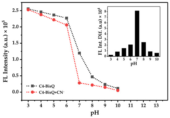
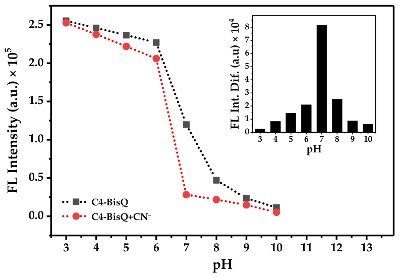
The effect of pH on the sensing behavior of C4-BisQ was then examined over the pH range of 3.0–10.0 in CH3CN/aqueous BR buffer (9:1, v/v) (Figure 1). The free probe retained relatively strong fluorescence under acidic and near-neutral conditions, whereas its emission gradually decreased at alkaline pH. To further investigate the pH-dependent behavior of C4-BisQ, UV–Vis spectra were recorded over the pH range of 3–10 in CH3CN/aqueous BR buffer (9:1, v/v), both in the absence and presence of CN− (Figure S13). The absorption spectrum of the free probe showed a clear pH dependence, particularly in the long-wavelength band around 605 nm. Fitting the absorbance at 605 nm over the pH range of 3–9 using a Henderson–Hasselbalch-type equation gave an apparent pKa value of 6.41 ± 0.21 (R2 = 0.971). Consistently, fitting the fluorescence intensity of free C4-BisQ at 526 nm as a function of pH gave an apparent pKa value of 6.92 ± 0.07 (R2 = 0.996) (Figure S13A). The small difference between the apparent pKa values obtained from absorption and fluorescence measurements can be attributed to the different optical responses monitored by the two methods. Nevertheless, both measurements place the pH-dependent optical transition of C4-BisQ in the near-neutral region. In the presence of 10 µM CN−, negligible fluorescence quenching was observed between pH 3.0 and 6.0, while a noticeable decrease in emission intensity occurred at pH 7.0. This behavior is consistent with the acid–base equilibrium of cyanide, since protonation under acidic conditions reduces the concentration of free CN− available for interaction with the receptor [59]. To identify the optimum sensing conditions, the fluorescence intensity difference (ΔF = F0 − F) between the free probe and the cyanide-treated system was plotted as a function of pH (Figure 1, inset). The maximum ΔF value was obtained at pH 7.0, indicating that near-neutral conditions provided the highest sensing contrast. Accordingly, all subsequent spectroscopic measurements, including UV–Vis and fluorescence titrations, binding studies, and selectivity experiments, were carried out in CH3CN/H2O (9:1, v/v) prepared with freshly prepared Milli-Q water (initial pH ≈ 7.0). For practical application studies, environmental water samples and food extracts were adjusted to approximately pH 7 using BR buffer, as indicated in the Experimental Section.
Figure 1.
Effect of pH on the fluorescence intensity of C4-BisQ (10 μM) at 526 nm in 9:1 CH3CN/BR buffer in the absence (solid black line) and presence of CN− (10 μM, dotted red line). Inset: The net fluorescence intensity difference (ΔF = F0 − F) plotted as a function of pH (λex = 428 nm).
3.3. UV-Vis and Fluorescence Spectroscopic Titration Studies
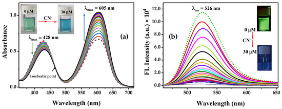
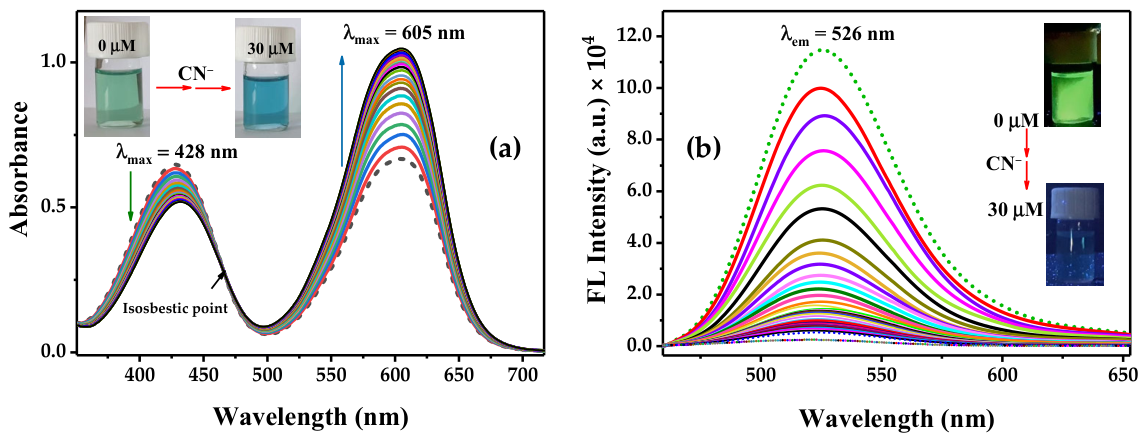
The optical response of C4-BisQ (10 µM) toward CN− was investigated by UV-Vis absorption and fluorescence emission titrations in CH3CN/H2O (9:1, v/v). As shown in Figure 2a, the absorption spectrum of the free probe exhibited a major band at 428 nm and a second band at 605 nm, giving the solution a green color under ambient light. Upon gradual addition of CN− (0–30 µM), the band at 428 nm progressively decreased, whereas the band at 605 nm increased in intensity without a significant wavelength shift. A clear isosbestic point was observed at 480 nm, suggesting a well-defined spectral conversion during the interaction process. Correspondingly, the solution color changed from green to blue, allowing visual detection of cyanide (Figure 2a, inset).
Figure 2.
UV-Vis absorption and fluorescence titration of C4-BisQ (10 µM) with CN− (0–30 µM) in CH3CN/H2O (9:1, v/v). (a) UV-Vis absorption spectra of C4-BisQ upon addition of CN−, showing a decrease at 428 nm and an increase at 605 nm, with an isosbestic point at 480 nm. Inset: Photographic images showing the color change from green to blue under ambient light. (b) Fluorescence emission spectra of C4-BisQ during titration with CN−, showing progressive quenching of the emission band at 526 nm (λex = 428 nm). Inset: Fluorescence images showing the change from green emission to a nearly non-emissive state under 365 nm UV light. The different colored curves represent increasing CN− concentrations from 0 to 30 µM.
The absorption changes were accompanied by a gradual decrease in fluorescence intensity, indicating that both optical responses followed the same concentration-dependent sensing process.
The fluorescence response of C4-BisQ was then examined under 428 nm excitation. The free probe showed a strong emission maximum at 526 nm (Figure 2b) and displayed bright green fluorescence under 365 nm UV light. With increasing CN− concentration (0–30 µM), the emission intensity decreased progressively, showing a concentration-dependent turn-off response. The gradual fluorescence quenching is consistent with the concentration-dependent optical response observed in the absorption spectra. At 30 µM CN−, the fluorescence was almost completely quenched. This change was also evident under UV illumination, where the emission color changed from bright green to a nearly non-emissive state (Figure 2b, inset).
3.4. Analytical Sensitivity and Quantification Limits
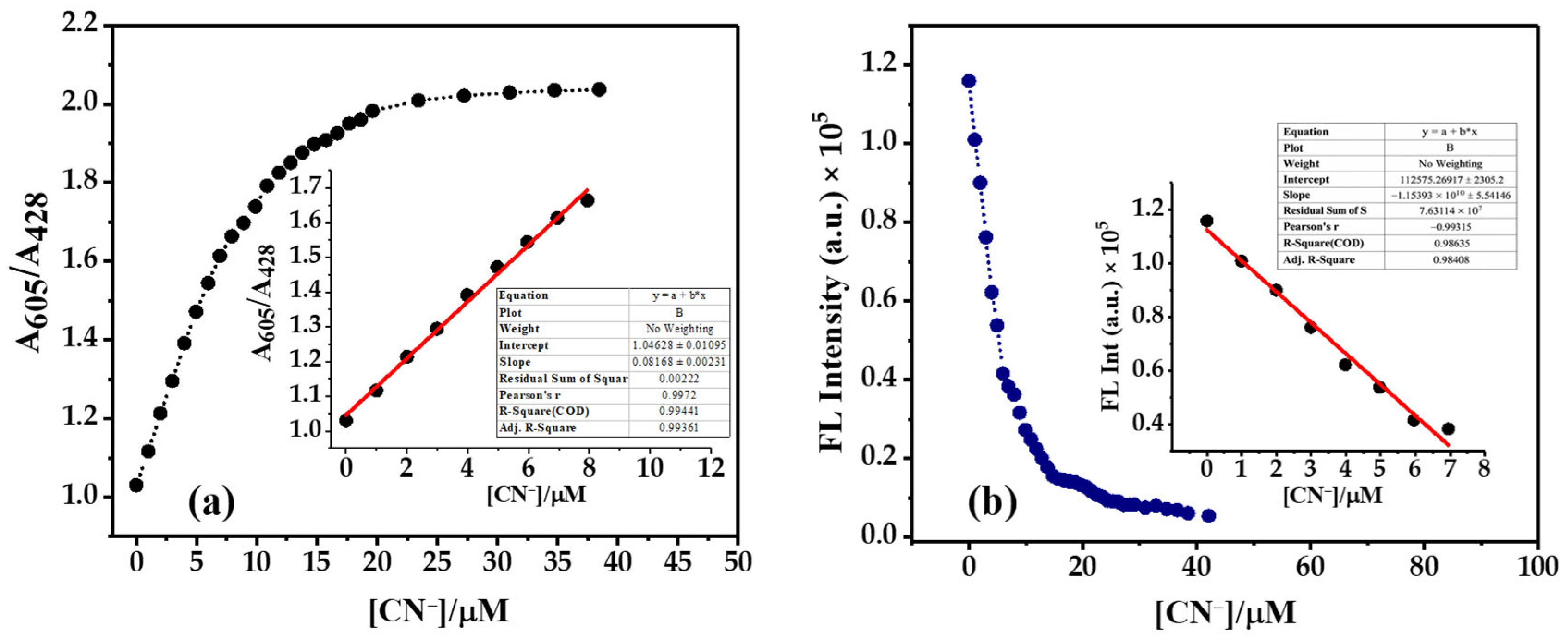
Calibration curves were constructed from the ratiometric UV–Vis response (A605/A428) and the fluorescence emission intensity at 526 nm. Both methods showed good linearity (Figure 3a,b). The UV–Vis response was linear over 0–8 μM (R2 = 0.994), whereas the fluorescence response remained linear between 0 and 7 μM (R2 = 0.986). The corresponding LOD/LOQ values were 0.080/0.265 μM for the UV–Vis method and 0.54/1.80 μM for the fluorescence method. Both detection limits are below the World Health Organization (WHO) guideline value for cyanide in drinking water (1.9 μM) [60]. The UV–Vis mode showed lower detection limits than the fluorescence mode. In addition, the ratiometric A605/A428 response provides an internal reference that reduces dependence on variations in absolute signal intensity and thereby improves measurement reliability [61,62]. In contrast, the fluorescence response is based on the intensity change in a single emission band and therefore does not provide intrinsic self-referencing, which represents a limitation of the fluorescence readout. The two responses should therefore be regarded as complementary rather than equivalent analytical modes. To place this analytical performance in context, C4-BisQ was compared with representative quinoline-, calixarene-, and other relevant optical CN− sensors reported in the literature [24,25,40,63], with a broader comparison provided in Table S1 in the Supporting Information [64,65,66,67,68,69,70,71]. As summarized in Table S1, C4-BisQ combines a ratiometric UV–Vis response (LOD = 0.080 μM) with a complementary fluorescence turn-off response (LOD = 0.54 μM) and a readily observable color change. In addition, its performance was demonstrated in environmental water samples and cyanogenic food extracts. A limitation of the present system is the relatively high proportion of CH3CN required for the sensing response (CH3CN/H2O, 9:1, v/v), which restricts its direct applicability in predominantly or fully aqueous media. Therefore, the main novelty of C4-BisQ lies in integrating complementary optical readouts and practical sample applications within a quinolinium-functionalized calix[4]arene platform rather than solely in achieving a low detection limit.
Figure 3.
Linear calibration plots and detection limit analysis for C4-BisQ (10 µM) toward CN− in CH3CN/H2O (9:1, v/v). (a) Ratiometric absorbance response (A605/A428) of C4-BisQ as a function of CN− concentration (0–40 µM). Inset: Linear calibration plot in the low-concentration range (0–8 µM). (b) Fluorescence emission intensity at 526 nm as a function of CN− concentration (0–42 µM). Inset: Linear calibration plot in the low-concentration range (0–7 µM).
3.5. Binding Stoichiometry and Association Constant
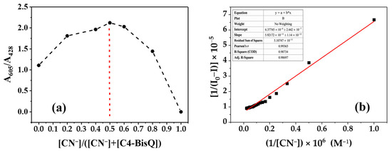
The binding stoichiometry between C4-BisQ and CN− was investigated by the method of continuous variation (Job’s plot). As shown in Figure 4a, the plot reached a maximum at a mole fraction of 0.5, indicating a 1:1 binding stoichiometry between C4-BisQ and CN−. The interaction strength was further evaluated using the Benesi–Hildebrand equation based on the fluorescence titration data. As shown in Figure 4b, the resulting plot exhibited a linear relationship (R2 = 0.987), which is consistent with a 1:1 binding model. From the ratio of the intercept to the slope of the fitted line, the association constant (Ka) was calculated to be 1.08 × 10⁵ M−1. The calculated association constant is consistent with the pronounced optical response observed during the titration studies.
Figure 4.
Binding stoichiometry and association constant analysis of C4-BisQ toward CN−. (a) Job’s plot based on the ratiometric absorbance response (A605/A428) at a constant total concentration of 10 µM, showing a maximum at a mole fraction of 0.5. (b) Benesi–Hildebrand plot constructed from the fluorescence titration data at 526 nm, consistent with a 1:1 binding model.
3.6. Stern–Volmer Analysis
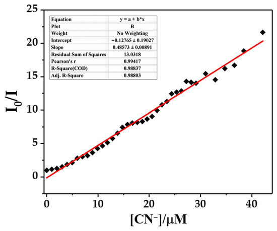
The fluorescence quenching of C4-BisQ by CN− was examined using the Stern–Volmer equation. As shown in Figure 5, the Stern–Volmer plot was linear over the 0–40 μM concentration range (R2 = 0.988), giving a quenching constant (KSV) of 4.86 × 10⁵ M−1. This linear relationship shows that the fluorescence quenching increases regularly with CN− concentration over the investigated range.
Figure 5.
Stern–Volmer plot (I0/I) as a function of CN− concentration (0–40 µM) monitored at 526 nm.
3.7. Proposed Sensing Mechanism
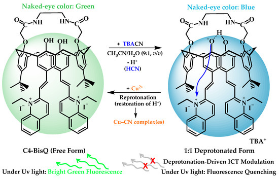
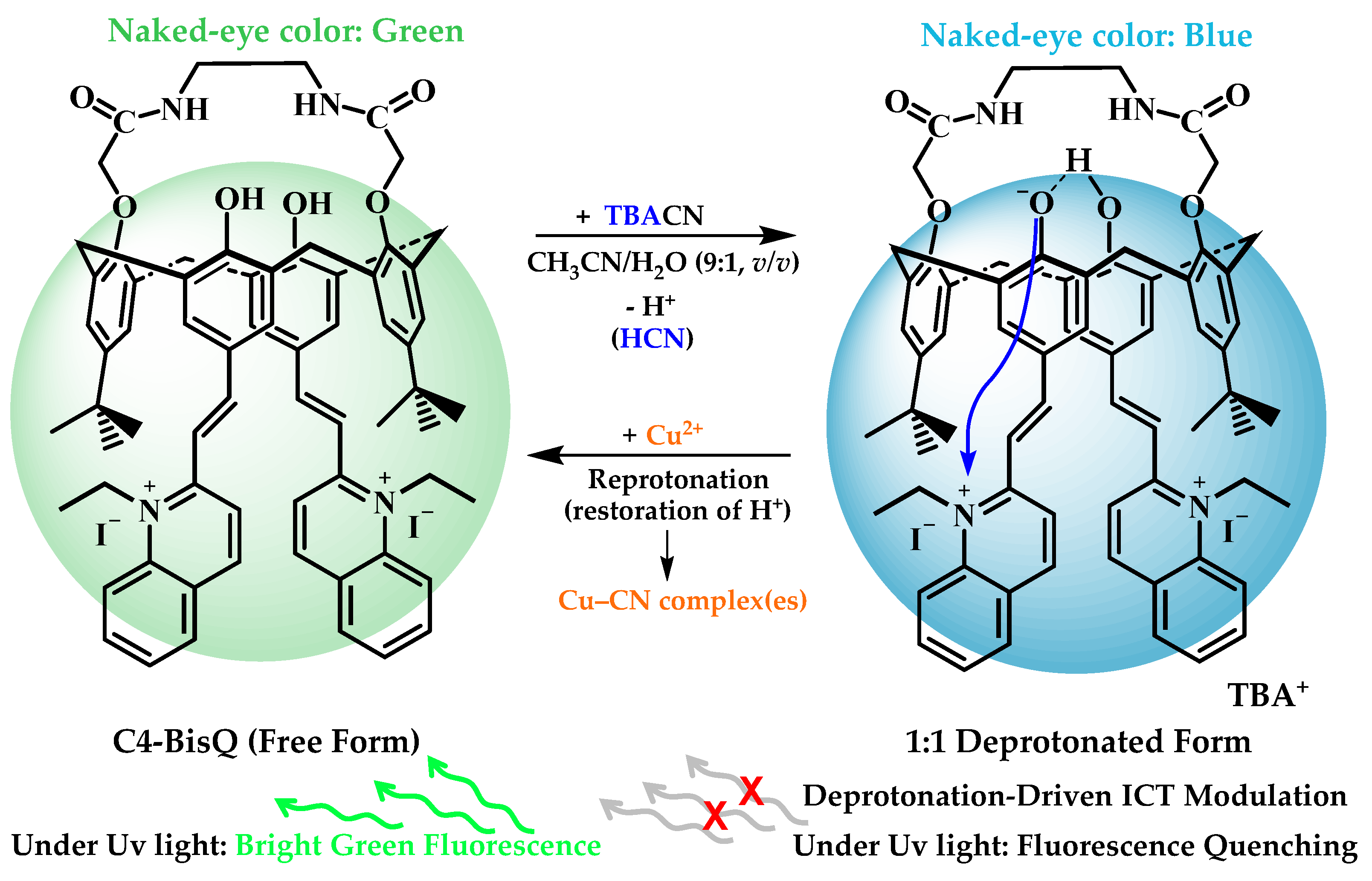
To gain insight into the sensing mechanism of C4-BisQ toward cyanide, the results obtained from pH-dependent experiments, Job’s plot analysis, UV–Vis titration, Stern–Volmer analysis, FT-IR spectroscopy, and reversibility experiments were considered collectively. Under the employed experimental conditions, the combined evidence supports a predominantly deprotonation-driven sensing pathway rather than direct assignment to an irreversible covalent transformation [20,72]. In particular, the similar optical response observed in the presence of OH− suggests that an acid–base process contributes significantly to the sensing event [73,74]. This interpretation is further supported by the Job’s plot analysis, which indicates a predominant 1:1 interaction stoichiometry between C4-BisQ and CN− (Figure 4), and by the UV–Vis titration experiments, where the presence of a well-defined isosbestic point is consistent with conversion of the free receptor into a single dominant cyanide-affected species (Figure 2) [75,76]. Taken together, these observations support the proposed deprotonation-based sensing mechanism illustrated in Scheme 2.
Scheme 2.
Proposed sensing mechanism of C4-BisQ toward CN− and its reversibility with Cu2+ via deprotonation and re-protonation pathways. CN− was introduced as tetrabutylammonium cyanide (TBACN), with TBA+ shown as the corresponding counterion.
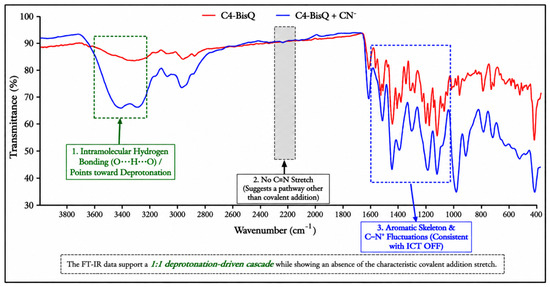
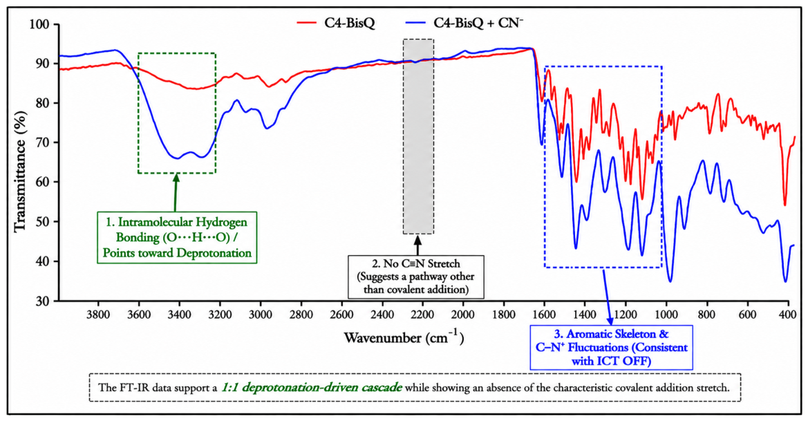
Further support for the proposed deprotonation-based pathway was obtained from the FT-IR analysis. Broadening was observed in the 3200–3500 cm−1 region, consistent with perturbation of the hydroxyl and/or hydrogen-bonding environment following CN− addition (Figure 6) [54,77]. At the same time, the absence of a distinct new absorption band attributable to a nitrile-containing covalent adduct is consistent with the view that irreversible covalent addition is not the dominant sensing pathway under the applied conditions [78,79]. The observed color change and fluorescence quenching can reasonably be attributed to deprotonation-induced perturbation of the receptor’s electronic structure. Additional solvent-dependent UV–Vis measurements revealed clear changes in the position and relative intensity of the absorption bands of C4-BisQ in CHCl3, EtOH, CH3CN, DMSO, and H2O (Figure S14), indicating that its electronic transitions are sensitive to the surrounding solvent environment. These spectral changes are consistent with charge-transfer character and support the possible involvement of an intramolecular charge transfer (ICT) process in the optical response. However, the present data do not permit an unambiguous assignment of a specific ICT mechanism.
Figure 6.
FT-IR spectra of free C4-BisQ (red line) and C4-BisQ after the addition of CN− (blue line) recorded in CH3CN/H2O (9:1, v/v) solution, along with a summary of the key spectral changes and functional group assignments. The FT-IR spectra of free C4-BisQ and C4-BisQ after CN− addition are provided in the Supporting Information (Figures S8 and S15).
Finally, restoration of the original optical response following the addition of Cu2+ demonstrates that the sensing process is reversible under the employed conditions (Figure S16). Although Cu2+ has previously been employed as an integral component of calixarene-based cyanide sensing systems through metal-displacement strategies, in the present system Cu2+ serves only to sequester CN− and restore the initial optical response, rather than participating directly in the primary sensing event [63]. The spectroscopic findings and reversibility experiments support a predominantly reversible, deprotonation-driven sensing process involving a 1:1 interaction between C4-BisQ and CN−, with the observed optical changes being consistent with a possible contribution from ICT [80].
3.8. Selectivity and Competitive Interference Studies
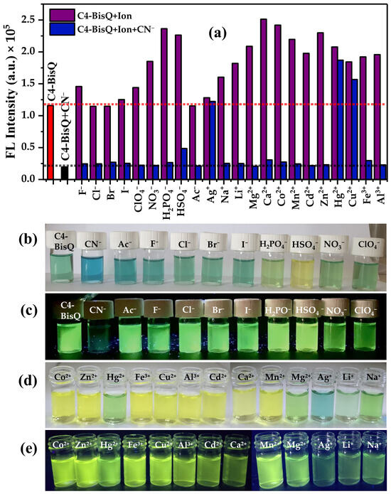
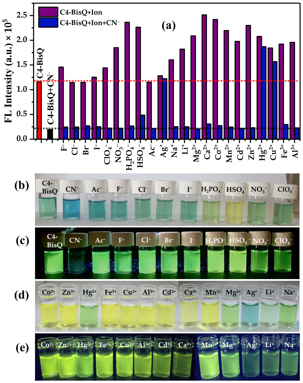
Competitive interference studies were carried out in the presence of common background anions (F−, Cl−, Br−, I−, ClO4−, NO3−, H2PO4−, HSO4−, and Ac−) and relevant metal cations (Li+, Na+, Mg2+, Ca2+, Mn2+, Fe3+, Co2+, Cu2+, Ag+, Zn2+, Cd2+, Hg2+, and Al3+). Each ion was applied at 50 μM, corresponding to a fivefold excess relative to CN− (10 μM), to evaluate the analytical selectivity of C4-BisQ under competitive conditions. As shown in Figure 7a, CN− produced a pronounced fluorescence quenching response, whereas several of the other tested ions produced changes in fluorescence intensity of different magnitudes without causing the characteristic CN−-induced turn-off response. The corresponding fluorescence emission spectra for the tested anions and metal cations are provided in the Supporting Information (Figures S17 and S18). Corresponding UV–Vis measurements were also performed to further examine these ion-dependent optical changes (Figures S19 and S20). The tested cations generally produced an increase in the absorption band around 428 nm together with a decrease in the longer-wavelength band around 605 nm, and most of the tested anions showed a similar trend. ClO4− and Ac− showed a somewhat different response, with decreases in both absorption regions. Since fluorescence excitation was performed at 428 nm, these ion-dependent changes in absorption at or near the excitation wavelength may contribute to the differences in fluorescence intensity observed for the competing ions. Small shifts in the emission maxima were also observed for some ions; however, these shifts were not assigned to a specific excited-state mechanism because the present data do not provide sufficient evidence for such an assignment. In contrast, CN− produced a decrease in the absorption around 428 nm together with a marked increase around 605 nm. Thus, the opposite direction of the UV–Vis response and the resulting increase in A605/A428 provide an additional means of distinguishing CN− from the other tested ions. The optical response was also readily observed by the naked eye. Under ambient laboratory light, only CN− induced a distinct color change from bright green to blue (Figure 7b). A corresponding fluorescence quenching was observed under 365 nm UV irradiation (Figure 7c). To evaluate competitive interference, mixed solutions containing background ions (50 μM) and CN− (10 μM) were prepared before the addition of C4-BisQ. The fluorescence intensity remained close to that of the CN−-treated sample for most of the tested ions, indicating that the cyanide response was largely preserved in the presence of coexisting species. In contrast, Ag+, Hg2+, and Cu2+ markedly suppressed the fluorescence response because of their strong affinity for CN−.
Figure 7.
Selectivity and competitive interference behavior of C4-BisQ in CH3CN/H2O (9:1, v/v). (a) Fluorescence response at λem = 526 nm (λex = 428 nm) of C4-BisQ (10 μM) in the presence of the indicated competing ions (50 μM, purple bars) and in mixtures containing the indicated competing ions (50 μM) and CN− (10 μM, blue bars). The red and black bars represent C4-BisQ alone and C4-BisQ + CN−, respectively. The red and black horizontal dashed lines indicate the reference maximum and baseline emission levels, respectively. Photographs showing the visual response of C4-BisQ toward the tested anions under (b) daylight and (c) UV illumination (365 nm), and toward the tested cations under (d) daylight and (e) UV illumination (365 nm).
3.9. Environmental Water Analysis (Tap Water and Sea Water)
The practical applicability of C4-BisQ was assessed by spike-and-recovery experiments using tap water and seawater samples (Table 1). Since natural water contains dissolved inorganic and organic species that may interfere with fluorescence measurements, recovery experiments were performed to evaluate the matrix tolerance of the sensor. Cyanide-spiked samples containing 2.0 and 5.0 μM CN− gave recoveries of 96.0–101.6%, with RSD values below 1.94% (n = 3). These results indicate that the proposed sensor system shows good reliability and matrix tolerance for cyanide determination within the investigated concentration range. Considering the fluorescence LOD of 0.54 µM, the present spike-and-recovery experiments demonstrate the applicability of C4-BisQ in complex environmental water matrices rather than its suitability for monitoring very low background levels of cyanide in natural waters. In particular, the seawater results indicate that the fluorescence response is retained despite the high ionic content and potential matrix effects of this sample. Further validation would therefore be required for applications targeting cyanide concentrations below the demonstrated analytical range.
Table 1.
Analytical determination and recovery of CN− in different environmental water samples using the C4-BisQ sensor.
3.10. Practical Assays in Food Extracts
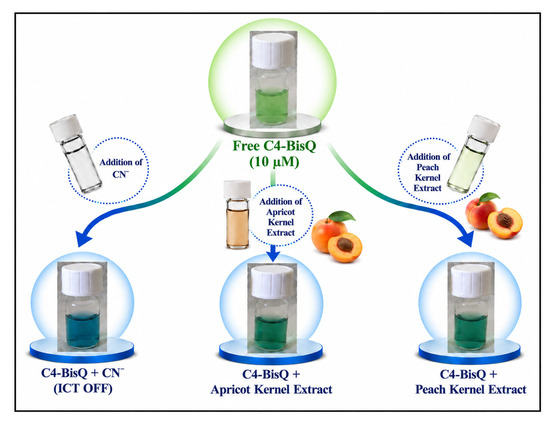
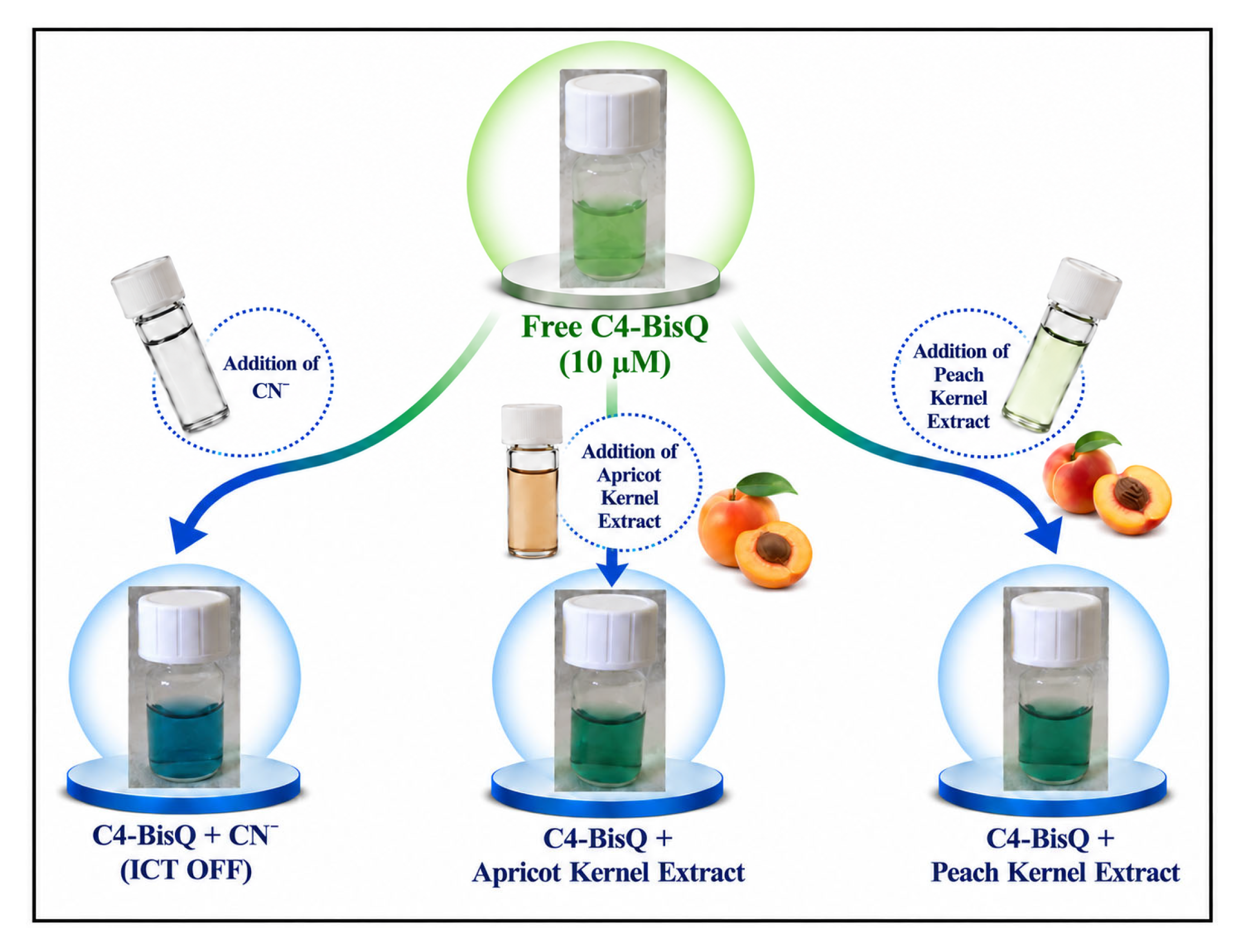
To examine the practical applicability of C4-BisQ, fresh apricot and peach kernel extracts were used for simple colorimetric assays. Mechanical disruption of Prunus kernels initiates the enzymatic hydrolysis of cyanogenic glycosides, releasing free cyanide into the aqueous phase [81,82]. As shown in Scheme 3, adding the kernel extracts to the C4-BisQ solution changed the solution color from bright green to blue under ambient light. The same color change was observed in the spectroscopic studies after the addition of CN−. The distinct visual response indicates that C4-BisQ can be used for the rapid qualitative screening of cyanide in food samples. Real-time visual responses under ambient light and 365 nm UV illumination are provided as Videos S1 and S2 in the Supporting Information. Accordingly, this application should be regarded as a proof-of-concept for visual qualitative screening in cyanogenic food matrices rather than a validated quantitative determination of total cyanide content.
Scheme 3.
Downward cascading schematic illustration of the dual-mode cyanide-sensing profile of C4-BisQ (10 µM) and its direct visual tracking in natural food matrices (apricot and peach kernel extracts). The recovery of the original optical response upon Cu2+ addition suggests that cyanide is responsible for the response of the apricot kernel extract.
4. Conclusions
In this study, we designed and synthesized a quinolinium-functionalized calix[4]arene chemosensor (C4-BisQ) for the detection of CN−. C4-BisQ exhibited a distinct green-to-blue color change accompanied by fluorescence quenching in CH3CN/H2O (9:1, v/v), enabling both visual and spectroscopic detection of cyanide. UV–Vis, fluorescence, FT-IR, and pH-dependent studies suggested that cyanide recognition mainly proceeds through a deprotonation process under the experimental conditions. The practical applicability of C4-BisQ was further evaluated in tap water, seawater, and cyanogenic food extracts. Overall, the results obtained in this study support the potential of C4-BisQ as a promising chemosensor for cyanide detection.
Supplementary Materials
The following supporting information can be downloaded at https://www.mdpi.com/article/10.3390/chemosensors14090205/s1, Figure S1: 1H-NMR spectrum of Compound 1 (25 °C) in CDCl3; Figure S2: FT-IR spectrum of Compound 2; Figure S3: 1H-NMR spectrum of Compound 2 (25 °C) in CDCl3; Figure S4: FT-IR spectrum of Compound 3; Figure S5: 1H-NMR spectrum of Compound 3 (25 °C) in CDCl3; Figure S6: FT-IR spectrum of Compound 4; Figure S7: 1H-NMR spectrum of Compound 4 (25 °C) in CDCl3; Figure S8: FT-IR spectrum of C4-BisQ; Figure S9: 1H-NMR spectrum of C4-BisQ (25 °C) in DMSO-d6; Figure S10: 13C-NMR spectrum of C4-BisQ (25 °C); Figure S11: Optimization of the solvent system for the probe C4-BisQ (10 μM). (a) Fluorescence emission spectra of C4-BisQ in different pure solvents (λex = 428 nm). (b) Fluorescence emission spectra of C4-BisQ in CH3CN/H2O mixtures containing different water volume fractions (10–90%, v/v) before and after addition of CN− (10 μM); Figure S12: Effect of water content on the optical response of C4-BisQ (a) UV–Vis absorption spectra of C4-BisQ (10 μM) in the absence (dotted lines) and presence (solid lines) of CN− (10 μM) in CH3CN/H2O mixtures containing 10, 20, 50, and 90% H2O. Corresponding photographs of C4-BisQ in the absence of CN− under (b) daylight and (c) UV illumination, and in the presence of CN− under (d) daylight and (e) UV illumination; Figure S13: pH-dependent optical behavior of C4-BisQ in CH3CN/BR buffer (9:1, v/v). (a) UV–Vis absorption spectra of C4-BisQ (10 μM) and (b) C4-BisQ (10 μM) in the presence of CN− (10 μM) over the pH range 3–10. (c) pH-dependent absorbance of C4-BisQ at 605 nm. The solid line represents fitting of the pH 3–9 data using a Henderson–Hasselbalch-type equation, yielding an apparent pKa of 6.41 ± 0.21 (R2 = 0.971). Corresponding photographs of C4-BisQ in the absence of CN− under (d) daylight and (e) UV illumination, and in the presence of CN− under (f) daylight and (g) UV illumination; Figure S13A: pH-dependent fluorescence behavior of C4-BisQ in CH3CN/BR buffer (9:1, v/v). (a) Fluorescence emission spectra of C4-BisQ (10 μM) in the absence and presence of CN− (10 μM) over the pH range of 3–10. (b) Sigmoidal fitting of the fluorescence intensity of free C4-BisQ at 526 nm as a function of pH, giving an apparent pKa value of 6.92 ± 0.07 (R2 = 0.996). λex = 428 nm; Figure S14: Solvent-dependent UV–Vis absorption spectra of C4-BisQ (10 μM) recorded in solvents of different polarity (CHCl3, EtOH, CH3CN, DMSO, and H2O), showing solvent-dependent changes in the absorption profile and band positions; Figure S15: FT-IR spectrum of C4-BisQ after CN− addition; Figure S16: Reversibility of the fluorescence response of C4-BisQ toward CN− in CH3CN/H2O (9:1, v/v). Fluorescence emission spectra of C4-BisQ (10 µM) showing the free probe, the quenched response after addition of CN−, the response of the probe after addition of Cu2+ alone, and the fluorescence recovery observed when Cu2+ was added to the CN− quenched system. Inset: Photographs of the corresponding solutions under UV light. λex = 428 nm; Figure S17: Fluorescence emission spectra of C4-BisQ (10 µM) in CH3CN/H2O (9:1, v/v) in the presence of various metal cations (50 µM each). The dashed trace corresponds to the free probe, and the solid traces represent the responses recorded after addition of Na+, Li+, Mg2+, Ca2+, Co2+, Mn2+, Cd2+, Zn2+, Hg2+, Cu2+, Fe3+, Al3+, and Ag+. λex = 428 nm.; Figure S18: Fluorescence emission spectra of C4-BisQ (10 µM) in CH3CN/H2O (9:1, v/v) in the presence of various background anions (50 µM each). The solid red trace represents the free probe, the colored solid traces represent the responses to F−, Cl−, Br−, I−, ClO4−, NO3−, H2PO4−, HSO4−, and Ac−, and the dashed black trace represents the response after addition of CN−. λex = 428 nm; Figure S19: UV–Vis absorption spectra of C4-BisQ in the presence of the tested cations (50 μM). The spectra of free C4-BisQ and C4-BisQ in the presence of CN− are included for com-parison; Figure S20: UV–Vis absorption spectra of C4-BisQ in the presence of the tested anions (50 μM). The spectra of free C4-BisQ and C4-BisQ in the presence of CN− are included for com-parison; Table S1: Comparison of C4-BisQ with representative optical chemosensors reported for CN− detection; Video S1: Real-time colorimetric response of C4-BisQ toward CN− under ambient light; Video S2: Real-time fluorescence response of C4-BisQ toward CN− under 365 nm UV illumination [23,24,25,27,40,48,49,50,51,63,64,65,66,67,68,69,70,71].
Author Contributions
Conceptualization, A.U.; methodology, I.U. and A.U.; validation, I.U. and A.U.; formal analysis, I.U.; investigation, A.U.; resources, A.U.; data curation, I.U.; writing—original draft preparation, I.U.; writing—review and editing, A.U.; visualization, I.U.; supervision, A.U.; project administration, I.U. and A.U. All authors have read and agreed to the published version of the manuscript.
Funding
This research received no external funding.
Institutional Review Board Statement
Not applicable.
Informed Consent Statement
Not applicable.
Data Availability Statement
The original contributions presented in this study are included in the article and its Supplementary Materials. Further data supporting the findings of this study are available from the corresponding author upon reasonable request.
Acknowledgments
The authors gratefully acknowledge Selçuk University for providing the necessary facilities and resources to conduct this research.
Conflicts of Interest
The authors declare no conflicts of interest.
References
- Hall, A.H.; Isom, G.E.; Rockwood, G.A. (Eds.) Toxicology of Cyanides and Cyanogens: Experimental, Applied and Clinical Aspects; John Wiley & Sons: Hoboken, NJ, USA, 2015. [Google Scholar]
- Zuhra, K.; Szabó, C. The two faces of cyanide: An environmental toxin and a potential novel mammalian gasotransmitter. FEBS J. 2021, 289, 2481–2515. [Google Scholar] [CrossRef] [Scilit] [PubMed]
- Sharma, M.; Akhter, Y.; Chatterjee, S. A review on remediation of cyanide containing industrial wastes using biological systems with special reference to enzymatic degradation. World J. Microbiol. Biotechnol. 2019, 35, 70. [Google Scholar] [CrossRef] [Scilit] [PubMed]
- Gandra, U.R.; Pandey, R.P.; Palanikumar, L.; Irfan, A.; Magzoub, M.; Belmabkhout, Y.; Hasan, S.W.; Infas, M.; Mohideen, H. Cu-TCPP metal-organic nanosheets embedded thin-film composite membranes for enhanced cyanide detection and removal: A multifunctional approach to water treatment and environmental safety. ACS Appl. Mater. Interfaces 2025, 17, 9563–9574. [Google Scholar] [CrossRef] [Scilit] [PubMed]
- Kohio, E.N.; Sossou, S.; Karoui, H.; Yacouba, H. Environmental cyanide pollution from artisanal gold mining in Burkina Faso: Human exposure risk analysis based on a conceptual site model. Int. J. Environ. Res. Public Health 2025, 22, 1125. [Google Scholar] [CrossRef] [Scilit] [PubMed]
- Cárdenas Riojas, A.A.; Wong, A.; Planes, G.A.; Sotomayor, M.D.; La Rosa-Toro, A.; Baena-Moncada, A.M. Development of a new electrochemical sensor based on silver sulfide nanoparticles and hierarchical porous carbon modified carbon paste electrode for determination of cyanide in river water samples. Sens. Actuators B Chem. 2019, 287, 544–550. [Google Scholar] [CrossRef] [Scilit]
- Bhadra, S.; Chan, A.S.; Hendry-Hofer, T.B.; Boss, G.R.; Bebarta, V.S.; Logue, B.A. Analysis of bisaminotetrazole cobinamide, a next-generation antidote for cyanide, hydrogen sulfide and methanethiol poisoning, in swine plasma by liquid chromatography-tandem mass spectrometry. J. Chromatogr. B 2022, 1208, 123392. [Google Scholar] [CrossRef] [Scilit] [PubMed]
- Pagliano, E.; Campanella, B.; D’Ulivo, A.; Mester, Z. Derivatization chemistries for the determination of inorganic anions and structurally related compounds by gas chromatography: A review. Anal. Chim. Acta 2018, 1025, 12–40. [Google Scholar] [CrossRef] [Scilit] [PubMed]
- Christison, T.T.; Rohrer, J.S. Direct determination of free cyanide in drinking water by ion chromatography with pulsed amperometric detection. J. Chromatogr. A 2007, 1155, 31–39. [Google Scholar] [CrossRef] [Scilit] [PubMed]
- Li, H.; Song, Y.; Zhou, B.; Xu, H. Nitrite: From application to detection and development. Appl. Sci. 2024, 14, 9027. [Google Scholar] [CrossRef] [Scilit]
- Ma, J.; Dasgupta, P.K. Recent developments in cyanide detection: A review. Anal. Chim. Acta 2010, 673, 117–125. [Google Scholar] [CrossRef] [Scilit] [PubMed]
- Banga, I.; Paul, A.; Poudyal, D.C.; Muthukumar, S.; Prasad, S. Recent advances in gas detection methodologies with a special focus on environmental sensing and health monitoring applications: A critical review. ACS Sens. 2023, 8, 3307–3319. [Google Scholar] [CrossRef] [Scilit] [PubMed]
- Fakayode, S.O.; Lisse, C.; Medawala, W.; Brady, P.N.; Bwambok, D.K.; Anum, D.; Alonge, T.; Taylor, M.E.; Baker, G.A.; Mehari, T.F.; et al. Fluorescent chemical sensors: Applications in analytical, environmental, forensic, pharmaceutical, biological, and biomedical sample measurement, and clinical diagnosis. Appl. Spectrosc. Rev. 2023, 59, 1–89. [Google Scholar] [CrossRef] [Scilit]
- Wu, Y.; Feng, J.; Hu, G.; Zhang, E.; Yu, H. Colorimetric sensors for chemical and biological sensing applications. Sensors 2023, 23, 2749. [Google Scholar] [CrossRef] [Scilit] [PubMed]
- Jin, Z.; Yim, W.; Retout, M.; Housel, E.; Zhong, W.; Zhou, J.; Strano, M.S.; Jokerst, J.V. Colorimetric sensing for translational applications: From colorants to mechanisms. Chem. Soc. Rev. 2024, 53, 7681–7741. [Google Scholar] [CrossRef] [Scilit] [PubMed]
- Zhou, Y.; Huang, X.; Hu, X.; Tong, W.; Leng, Y.; Xiong, Y. Recent advances in colorimetry/fluorimetry-based dual-modal sensing technologies. Biosens. Bioelectron. 2021, 190, 113386. [Google Scholar] [CrossRef] [Scilit] [PubMed]
- Li, Z.; Askim, J.R.; Suslick, K.S. The optoelectronic nose: Colorimetric and fluorometric sensor arrays. Chem. Rev. 2018, 119, 231–292. [Google Scholar] [CrossRef] [Scilit] [PubMed]
- Shen, Y.; Gao, X.; Zhang, Y.; Chen, H.; Ye, Y.; Wu, Y. Polydopamine-based nanozyme with dual-recognition strategy-driven fluorescence-colorimetric dual-mode platform for Listeria monocytogenes detection. J. Hazard. Mater. 2022, 439, 129582. [Google Scholar] [CrossRef] [Scilit] [PubMed]
- Liu, S.; Zhou, J.; Yuan, X.; Xiong, J.; Zong, M.; Wu, X.; Lou, W. A dual-mode sensing platform based on metal-organic framework for colorimetric and ratiometric fluorescent detection of organophosphorus pesticide. Food Chem. 2023, 432, 137272. [Google Scholar] [CrossRef] [Scilit] [PubMed]
- Hamzi, I.; Datoussaid, Y.; Oukebdane, N. Design strategies, mechanistic insights, and applications of N-acylhydrazone-based fluorometric and colorimetric chemosensors for anion detection. ChemistrySelect 2025, 10, 34. [Google Scholar] [CrossRef] [Scilit]
- Rashid, A.; Mondal, S.; Ghosh, P. Development and application of ruthenium(II) and iridium(III) based complexes for anion sensing. Molecules 2023, 28, 1231. [Google Scholar] [CrossRef] [Scilit] [PubMed]
- Krämer, J.; Kang, R.; Grimm, L.; De Cola, L.; Picchetti, P.; Biedermann, F. Molecular probes, chemosensors, and nanosensors for optical detection of biorelevant molecules and ions in aqueous media and biofluids. Chem. Rev. 2022, 122, 3459–3636. [Google Scholar] [CrossRef] [Scilit] [PubMed]
- Zhang, Y.; Zhou, C.; Li, J.; Kovtunets, E. A New Acridine-Based Fluorescent Sensor for the Detection of CN−. Chemosensors 2026, 14, 67. [Google Scholar] [CrossRef] [Scilit]
- Basri, R.; Ahmed, N.; Khalid, M.; Khan, M.U.; Abdullah, M.; Syed, A.; Elgorban, A.M.; Al-Rejaie, S.S.; Braga, A.A.C.; Shafiq, Z. Quinoline based thiosemicarbazones as colorimetric chemosensors for fluoride and cyanide ions and DFT studies. Sci. Rep. 2022, 12, 4927. [Google Scholar] [CrossRef] [Scilit] [PubMed]
- Sunitha, M.; Vijayakumar, V.; Lohitha, N.; Sarveswari, S. A quinoline appended isonicotino hydrazide-based colorimetric chemosensor for selective detection of cyanide ion in real samples. ChemistrySelect 2025, 10, 33. [Google Scholar] [CrossRef] [Scilit]
- Wu, C.; Wang, J.; Shen, J.; Zhang, C.; Wu, Z.P.; Zhou, H. A colorimetric quinoline-based chemosensor for sequential detection of copper ion and cyanide anions. Tetrahedron 2017, 73, 5715–5719. [Google Scholar] [CrossRef] [Scilit]
- Tang, Q.; Dan, F.; Ma, S.; Zeng, X.; Lan, H. A colorimetric and fluorescent probe based on quinoline-indolium for detection of CN− in aqueous media. ChemistrySelect 2021, 6, 6557–6563. [Google Scholar] [CrossRef] [Scilit]
- Ríos, M.C.; Bravo, N.F.; Sánchez, C.C.; Portilla, J. Chemosensors based on N-heterocyclic dyes: Advances in sensing highly toxic ions such as CN− and Hg2+. RSC Adv. 2021, 11, 34206–34234. [Google Scholar] [CrossRef] [Scilit] [PubMed]
- Udhayakumari, D. A comprehensive anthology of literature based on quinoline chemosensors from 2006 to 2022. J. Mol. Struct. 2023, 1287, 135715. [Google Scholar] [CrossRef] [Scilit]
- Swinburne, A.N.; Paterson, M.; Beeby, A.; Steed, J.W. A quinolinium-derived turn-off fluorescent anion sensor. Org. Biomol. Chem. 2010, 8, 1010–1016. [Google Scholar] [CrossRef] [Scilit] [PubMed]
- Han, Y.R.; Lee, S.B. Facile photocatalytic reactions in various conditions using quinolinium salts as photocatalysts. J. Ind. Eng. Chem. 2025, 152, 124–135. [Google Scholar] [CrossRef] [Scilit]
- Bleeker, J.; Kahn, A.P.; Baumgartner, L.M.; Grozema, F.C.; Vermaas, D.A.; Jager, W.F. Quinolinium-based fluorescent probes for dynamic pH monitoring in aqueous media at high pH using fluorescence lifetime imaging. ACS Sens. 2023, 8, 2050–2059. [Google Scholar] [CrossRef] [Scilit] [PubMed]
- Jothi, D.; Munusamy, S.; Kumar, S.M.; Enbanathan, S.; Iyer, S.K. A benzothiazole-based new fluorogenic chemosensor for the detection of CN− and its real-time application in environmental water samples and living cells. RSC Adv. 2022, 12, 8570–8577. [Google Scholar] [CrossRef] [Scilit] [PubMed]
- Barkale, H.V.; Dey, N. Selective sensing of cyanide ions: Impact of molecular design and assembly on the response of π-conjugated acylhydrazone compounds. RSC Adv. 2024, 14, 25108–25114. [Google Scholar] [CrossRef] [Scilit] [PubMed]
- Kolińska, J.; Grzelakowska, A.; Sokołowska, J. Dyes based on the 2(1H)-quinolone skeleton as potential colorimetric and fluorescent sensors for cyanide anions. Color. Technol. 2019, 135, 501–509. [Google Scholar] [CrossRef] [Scilit]
- Acar, M.; Daştan, A.; Koçak, R. Fluorometric and colorimetric sensor for selective detection of cyanide anion by dibenzosuberenone-based dihydropyridazine in aqueous solution. Talanta 2024, 277, 126241. [Google Scholar] [CrossRef] [Scilit] [PubMed]
- Padghan, S.D.; Hu, J.W.; Hsu, Y.H.; Liu, K.M.; Chen, K.Y. Swift and quantitative naked-eye detection of cyanide: A dual-mode approach using an ICT-based colorimetric and ratiometric fluorescent chemodosimeter. Dye. Pigment. 2024, 228, 112229. [Google Scholar] [CrossRef] [Scilit]
- Padghan, S.D.; Wang, L.C.; Lin, W.C.; Hu, J.W.; Liu, W.C.; Chen, K.Y. Rational design of an ICT-based chemodosimeter with aggregation-induced emission for colorimetric and ratiometric fluorescent detection of cyanide in a wide pH range. ACS Omega 2021, 6, 5287–5296. [Google Scholar] [CrossRef] [Scilit] [PubMed]
- Singh, S.; Heena; Singh, S.; Layek, S.; Goswami, T.; Kumar, S.; Dubey, R. Unveiling distinct hydrogen-bonding mechanisms: A catechol-derived probe for selective cyanide and fluoride detection. Spectrochim. Acta Part A Mol. Biomol. Spectrosc. 2025, 349, 127329. [Google Scholar] [CrossRef] [Scilit] [PubMed]
- Sunitha, M.; Sarveswari, S. A simple and coast effective quinoline based Schiff’s base SS-2 as a colorimetric chemosensor for highly selective detection of cyanide ion in food samples. Inorg. Chem. Commun. 2024, 160, 111973. [Google Scholar] [CrossRef] [Scilit]
- Poongodi, K.; Saravana Kumar, P.; Shanmugapriya, C.; Nandhini, C.; Elango, K. 2-Aminophenols based Schiff bases as fluorescent probes for selective detection of cyanide and aluminium ions: Effect of substituents. Spectrochim. Acta Part A Mol. Biomol. Spectrosc. 2020, 249, 119288. [Google Scholar] [CrossRef] [Scilit] [PubMed]
- Tian, J.; Zheng, Z.; Pan, Y.C.; Wang, Y.; Guo, D. Macrocycle-based differential sensing: Design strategies and applications. Responsive Mater. 2025, 3, e20240036. [Google Scholar] [CrossRef] [Scilit]
- Kumar, R.; Sharma, A.; Singh, H.; Suating, P.; Kim, H.S.; Sunwoo, K.; Shim, I.; Gibb, B.; Kim, J.S. Revisiting fluorescent calixarenes: From molecular sensors to smart materials. Chem. Rev. 2019, 119, 9657–9721. [Google Scholar] [CrossRef] [Scilit] [PubMed]
- Ren, H.; Wang, H.; Wen, W.; Li, S.; Li, N.; Huo, F.; Yin, C. A summary of calixarene-based fluorescent sensors developed during the past five years. Chem. Commun. 2023, 59, 13790–13799. [Google Scholar] [CrossRef] [Scilit] [PubMed]
- Marcos, P.M.; Berberan-Santos, M.N. Recent advances in calixarene-based fluorescent sensors for biological applications. Sensors 2024, 24, 7181. [Google Scholar] [CrossRef] [Scilit] [PubMed]
- Sayin, S. A highly selective fluorescence probe for Co2+ or Cu2+ detection based on a new tetraquinoline-substituted calix[4]arene derivative. Tetrahedron 2022, 122, 132960. [Google Scholar] [CrossRef] [Scilit]
- Patra, S.; Boricha, V.P.; Paul, P. Dual-mode calixarene-based chemosensor: Highly selective fluorogenic detection of Hg2+ and chromogenic detection of Cu2+ with a single ionophore. Eur. J. Inorg. Chem. 2018, 2019, 199–205. [Google Scholar] [CrossRef] [Scilit]
- Gutsche, C.D.; Rogers, J.S.; Stewart, R.D.; See, K.A. Calixarenes: Paradoxes and paradigms in molecular baskets. Pure Appl. Chem. 1990, 62, 485–491. [Google Scholar] [CrossRef] [Scilit]
- Collins, E.M.; McKervey, M.A.; Madigan, E.; Moran, M.B.; Owens, M.; Ferguson, G.; Harris, S.J. Chemically modified calix[4]arenes. Regioselective synthesis of 1,3-(distal) derivatives and related compounds. X-ray crystal structure of a diphenoldinitrile. J. Chem. Soc. Perkin Trans. 1 1991, 20, 3137–3142. [Google Scholar] [CrossRef] [Scilit]
- Uyanik, A.; Sen, N.; Yilmaz, M. Improvement of catalytic activity of lipase from Candida rugosa via sol–gel encapsulation in the presence of calix(aza)crown. Bioresour. Technol. 2011, 102, 4313–4318. [Google Scholar] [CrossRef] [Scilit] [PubMed]
- Chawla, H.M.; Pant, N.; Srivastava, B.; Upreti, S. Convenient direct synthesis of bisformylated calix[4]arenes via ipso substitution. Org. Lett. 2006, 8, 2237–2240. [Google Scholar] [CrossRef] [Scilit] [PubMed]
- Currie, L. Detection and quantification limits: Origins and historical overview. Anal. Chim. Acta 1997, 391, 127–134. [Google Scholar] [CrossRef] [Scilit]
- Benesi, H.A.; Hildebrand, J.H. A spectrophotometric investigation of the interaction of iodine with aromatic hydrocarbons. J. Am. Chem. Soc. 1949, 71, 2703–2707. [Google Scholar] [CrossRef] [Scilit]
- Gehlen, M. The centenary of the Stern-Volmer equation of fluorescence quenching: From the single line plot to the SV quenching map. J. Photochem. Photobiol. C Photochem. Rev. 2020, 42, 100338. [Google Scholar] [CrossRef] [Scilit]
- Huang, C.Y. Determination of binding stoichiometry by the continuous variation method: The job plot. In Methods in Enzymology; Purich, D.L., Ed.; Academic Press: New York, NY, USA, 1982; Volume 87, pp. 509–525. [Google Scholar]
- Erdemir, S.; Malkondu, S. On-site and low-cost detection of cyanide by simple colorimetric and fluorogenic sensors: Smartphone and test strip applications. Talanta 2020, 207, 120278. [Google Scholar] [CrossRef] [Scilit] [PubMed]
- Suna, G.; Gunduz, S.; Topal, S.; Ozturk, T.; Karakuş, E. A unique triple–channel fluorescent probe for discriminative detection of cyanide, hydrazine, and hypochlorite. Talanta 2023, 257, 124365. [Google Scholar] [CrossRef] [Scilit] [PubMed]
- Ravikumar, M.V.; Raj, A.K.; Rao, M.R.; Lakshmi, V. Structure-Property Evaluation of Knoevenagel-Derived π-Conjugated Organic Systems. Eur. J. Org. Chem. 2025, 28, e202401367. [Google Scholar] [CrossRef] [Scilit]
- Vaca-Escobar, K.; Arregui-Almeida, D.; Espinoza-Montero, P. Chemical, ecotoxicological characteristics, environmental fate, and treatment methods applied to cyanide-containing wastewater. npj Clean Water 2024, 7, 103. [Google Scholar] [CrossRef] [Scilit]
- World Health Organization. Guidelines for Drinking-Water Quality; World Health Organization: Geneva, Switzerland, 2002. [Google Scholar]
- Madhu, M.; Santhoshkumar, S.; Tseng, W.L.; Tseng, W.J. Maximizing analytical precision: Exploring the advantages of ratiometric strategy in fluorescence, Raman, electrochemical, and mass spectrometry detection. Front. Anal. Sci. 2023, 3, 1258558. [Google Scholar] [CrossRef] [Scilit]
- Zhou, Y.; Sun, Y.; Ren, G.; Huang, J. A porphyrin-chitosan Schiff base polymer for sequential ratiometric detection of Fe3+ and ascorbic acid via UV-Vis spectroscopy. Talanta 2026, 309, 129977. [Google Scholar] [CrossRef] [Scilit] [PubMed]
- Chawla, H.M.; Shukla, R.; Goel, P. Sensitive recognition of cyanide through supramolecularly complexed new calix[4]arenes. New J. Chem. 2014, 38, 5264–5267. [Google Scholar] [CrossRef] [Scilit]
- Nie, H.-M.; Gong, C.-B.; Tang, Q.; Ma, X.-B.; Chow, C.F.S. Visual and reversible detection of cyanide ions in protic solvents by a novel colorimetric receptor. Dye. Pigment. 2014, 106, 74–80. [Google Scholar] [CrossRef] [Scilit]
- Saini, N.; Prigyai, N.; Wannasiri, C.; Ervithayasuporn, V.; Kiatkamjornwong, S. Green synthesis of fluorescent N,O-chelating hydrazone Schiff base for multi-analyte sensing in Cu2+, F− and CN− ions. J. Photochem. Photobiol. A Chem. 2018, 358, 215–225. [Google Scholar] [CrossRef] [Scilit]
- Rezaeian, K.; Khanmohammadi, H.; Talebbaigy, A. Detection of CN−, Cu2+ and Zn2+ ions using a new chromone-based colorimetric chemosensor: Half-adder and integrated circuits. Anal. Methods 2020, 12, 1759–1766. [Google Scholar] [CrossRef] [Scilit]
- Sayin, S. Synthesis of new anthracene-conjugated calix[4]arene as highly selective fluorescent chemosensor for determination of CN− ion. J. Mol. Struct. 2022, 1252, 132212. [Google Scholar] [CrossRef] [Scilit]
- Panjwani, F.; Dey, S.; Kongor, A.; Kumar, A.; Panchal, M.; Modi, K.; Vora, M.; Kumar, A.; Jain, V.K. Pyrene functionalized oxacalix[4]arene architecture as dual readout sensor for expeditious recognition of cyanide anion. J. Fluoresc. 2022, 32, 1425–1433. [Google Scholar] [CrossRef] [Scilit] [PubMed]
- Gómez-Machuca, H.; Quiroga-Campano, C.; Jullian, C.; Saitz, C. Bifunctional receptor based on calix[4]arene with chromone groups as an efficient colorimetric sensor for Co2+, Cu2+, CN− and F−. ChemistrySelect 2022, 7, e202202581. [Google Scholar] [CrossRef] [Scilit]
- Oguz, A.; Oguz, M.; Kursunlu, A.N.; Yilmaz, M. A fully water-soluble Calix[4]arene probe for fluorometric and colorimetric detection of toxic hydrosulfide and cyanide ions: Practicability in living cells and food samples. Food Chem. 2023, 401, 134132. [Google Scholar] [CrossRef] [Scilit] [PubMed]
- Rani, B.; Sharma, N.; Shrivastava, R.; Agarwala, A.; Verma, V.P. Efficient detection of CN− and Cu2+ ions by styryl-BODIPY based multifunctional chemosensor in semi-aqueous medium. J. Mol. Struct. 2023, 1274, 134396. [Google Scholar] [CrossRef] [Scilit]
- Bhaskar, R.; Balakrishna, R.G. An azo dye-based colorimetric chemosensor for selective cyanide detection in food matrices. Inorg. Chem. Commun. 2026, 189, 116704. [Google Scholar] [CrossRef] [Scilit]
- Dey, S.K.; Al Kobaisi, M.; Bhosale, S.V. Functionalized quinoxaline for chromogenic and fluorogenic anion sensing. ChemistryOpen 2018, 7, 934–952. [Google Scholar] [CrossRef] [Scilit] [PubMed]
- Zali-Boeini, H.; Zareh Jonaghani, M. New fluorescent and colorimetric chemosensor for detection of cyanide with high selectivity and sensitivity in aqueous media. J. Fluoresc. 2017, 27, 1035–1040. [Google Scholar] [CrossRef] [Scilit] [PubMed]
- Wei, T.; Ding, J.D.; Chen, J.F.; Han, B.B.; Jiang, X.M.; Yao, H.; Zhang, Y.M.; Lin, Q. A Cyanide-Triggered Hydrogen-Bond-Breaking Deprotonation Mechanism: Fluorescent Detection of Cyanide Using a Thioacetohydrazone-Functionalized Bispillar[5]arene. New J. Chem. 2018, 42, 14197–14202. [Google Scholar] [CrossRef] [Scilit]
- Mahapatra, A.; Manna, S.; Pramanik, B.; Maiti, K.; Mondal, S.; Ali, S.S.; Mandal, D. Colorimetric and ratiometric fluorescent chemodosimeter for selective sensing of fluoride and cyanide ions: Tuning selectivity in proton transfer and C–Si bond cleavage. RSC Adv. 2015, 5, 10716–10722. [Google Scholar] [CrossRef] [Scilit]
- Li, G.Y.; Zhao, G.J.; Han, K.; He, G. A TD-DFT study on the cyanide-chemosensing mechanism of 8-formyl-7-hydroxycoumarin. J. Comput. Chem. 2011, 32, 668–674. [Google Scholar] [CrossRef] [Scilit] [PubMed]
- Algethami, J.S. Smart Organic Probes and Their Applications: Sequential Fluorometric and Colorimetric Sensing of Cu2+ and CN− Ions: A Review (2018–2025). Crit. Rev. Anal. Chem. 2025, 55, 1–16. [Google Scholar] [CrossRef] [Scilit] [PubMed]
- Kim, M.; Mergu, N.; Son, Y.A. Imidazole-containing ratiometric receptor for the selective and sensitive detection of cyanide and fluoride via deprotonation and a receptor-anion ensemble for Cu2+ sensing. J. Lumin. 2018, 204, 244–252. [Google Scholar] [CrossRef] [Scilit]
- Umabharathi, P.S.; Karpagam, S.; Dasgupta, T.; Tamizhselvi, R. Benzothiazole-quinoline based probe for simultaneous colorimetric detection of CN− and Cu2+ ions, with fluorescence sensing for Cu2+: Mechanistic insights and practical applications in environmental monitoring and cellular analysis. Heliyon 2024, 10, e38593. [Google Scholar] [CrossRef] [Scilit] [PubMed]
- Archana, P.; Vealan, S.M.B.; Sekar, C. Qualitative and quantitative detection of cyanide in forage crop and fibre dietary products by colorimetric and fluorescent sensor based on corn silk derived carbon dots-rutin system. Food Chem. 2025, 493, 145978. [Google Scholar] [CrossRef] [Scilit] [PubMed]
- Rodríguez Madrera, R.; Valles, S.B. Analysis of Cyanogenic Compounds Derived from Mandelonitrile by Ultrasound-Assisted Extraction and High-Performance Liquid Chromatography in Rosaceae and Sambucus Families. Molecules 2021, 26, 7563. [Google Scholar] [CrossRef] [Scilit] [PubMed]

Disclaimer/Publisher’s Note: The statements, opinions and data contained in all publications are solely those of the individual author(s) and contributor(s) and not of MDPI and/or the editor(s). MDPI and/or the editor(s) disclaim responsibility for any injury to people or property resulting from any ideas, methods, instructions or products referred to in the content. |
© 2026 by the authors. Licensee MDPI, Basel, Switzerland. This article is an open access article distributed under the terms and conditions of the Creative Commons Attribution (CC BY) license.