


Abstract
Existing colorimetric sensing strategies based on gold nanoparticles (AuNPs) are largely limited to the detection of free metal ions, leaving metal–organic complexes widely present in real water bodies undetectable. Herein, we develop a simple colorimetric method based on the anti-aggregation effect of bare AuNPs for the simultaneous detection of free Cr3+ and its organic complexes. Tetrabutylphosphonium bromide (TPB) dissociates into phosphonium cations ([P(C4H9)4]+), which neutralize the negatively charged surface of AuNPs via electrostatic attraction, thereby inducing particle aggregation and a color change from wine-red to blue. Under strongly alkaline conditions, Cr3+ converts to negatively charged [Cr(OH)6]3− species. These anionic species then compete with the AuNPs for the phosphonium cations, blocking the aggregation process and preserving the wine-red color. This competitive mechanism is further extended to the detection of Cr3+-EDTA, Cr3+-tartrate, and Cr3+-oxalate complexes, as they also transform into [Cr(OH)6]3− under alkaline conditions. Without the need for tedious surface modification, the proposed method is simple to operate and exhibits excellent sensitivity and selectivity for both free and complexed Cr3+ in real water samples. It thus provides a reliable analytical tool for monitoring chromium pollution in environmental waters.
1. Introduction
Chromium ions (Cr3+) is one of the most prevalent heavy metal contaminants in industrial wastewater, particularly originating from industries such as tanning, electroplating, textile dyeing, and metallurgy [1,2,3,4]. The extensive discharge of chromium-containing effluents has led to severe contamination of water bodies and soils on a global scale [5,6,7,8]. Although Cr3+ is an essential trace element involved in glucose and lipid metabolism at low concentrations, excessive exposure can cause skin irritation, respiratory disorders, and potential carcinogenic effects [9,10,11,12]. Therefore, the development of rapid, facile, and sensitive methods for Cr3+ detection is of great significance for both environmental protection and public health safety. In recent years, considerable efforts have been devoted to the development of point-of-care testing (POCT) methods for Cr3+ [13,14,15,16]. For instance, Zeng et al. [17] synthesized Y(V0.2P0.8O4):Eu3+ phosphovanadate nanorods, which enable ultraselective detection of Cr3+ based on the fluorescence quenching mechanism induced by Cr3+-triggered nanorod aggregation. Notably, this probe exhibits excellent photostability over a wide pH range (3~12) and high ionic strength conditions. Ruan et al. [18] constructed a novel fluorescence sensing platform based on the catechol oxidase-mimicking activity of CuSe nanoparticles (CuSeNPs). In this system, CuSeNPs catalyze the oxidation of dopamine and its subsequent reaction with α-naphthol to generate a highly fluorescent derivative. The presence of Cr3+ suppresses the catalytic activity of CuSeNPs through surface adsorption, thereby enabling fluorescence detection of Cr3+. Furthermore, Qiao et al. [19] developed a ratiometric fluorescence sensor based on an imine-linked one-dimensional covalent organic framework (Py-An COF). This sensor achieves Cr3+ detection through ratiometric signal readout, which effectively circumvents interference from external factors and significantly improves detection accuracy and sensitivity.
Gold nanoparticles (AuNPs) have firmly established themselves as premier materials in analytical chemistry, primarily due to their unique localized surface plasmon resonance (LSPR) properties [19,20]. The LSPR phenomenon imparts dispersed AuNPs with a distinct wine-red colour and a characteristic absorption peak at approximately 520 nm. Upon AuNPs aggregation, a decrease in interparticle distance is observed, resulting in a substantial red-to-blue shift in the absorption spectrum [21,22]. Concomitantly, a visual transition to blue or purple hues is evident. The basis of colorimetric sensing is formed by this distance-dependent optical modulation [23,24]. The detection capabilities offered by this method are intuitive, rapid and instrument-free, which renders them particularly advantageous for the purposes of environmental monitoring and on-site water quality screening. In comparison with conventional large-scale analytical techniques, such as inductively coupled plasma mass spectrometry, AuNP-based sensors demonstrate superior qualities in terms of portability and cost-effectiveness [25,26,27]. These attributes render them particularly well-suited for extensive deployment in settings where resources are limited.
Despite considerable advances in the detection of free metal ions (e.g., Cr3+, Hg2+, Pb2+, Cu2+) using AuNP colorimetric sensors, their application in real aquatic environments is still challenging [28,29,30,31,32]. Industrial wastewater and natural waters frequently contain ubiquitous organic ligands, including tartaric acid, oxalic acid, and ethylenediaminetetraacetic acid (EDTA), which readily form stable complexes with heavy metal ions. These metal–organic complexes have been demonstrated to exert a significant influence on the bioavailability, toxicity, and environmental mobility of heavy metals [33,34,35]. For instance, while free Cr3+ tends to adsorb onto sediment surfaces, Cr3+ bound to organic complexes exhibits enhanced solubility and migration potential due to ligand shielding [36,37,38]. It is crucial to note that this shielding effect significantly hinders the interaction between target metal ions and the specific recognition sites of traditional AuNP probes, which are predominantly designed for free ion detection [39,40,41,42,43]. Consequently, existing AuNP colorimetric strategies frequently demonstrate inadequate responsiveness or significant deviations when employed in the context of complexed metal species, thereby constituting a substantial technical impediment in the realm of environmental analysis [44,45]. Although Pan et al. [46] demonstrated that tributylhexadecylphos-phonium bromide (THPB)-modified AuNPs enable the specific recognition of Cr(III)-citrate complexes through aggregation-induced colorimetry, the synchronous quantification of free and complexed Cr(III) remains elusive. Consequently, devising a sensing strategy that can effectively circumvent the ligand shielding effect to achieve the concurrent, rapid, and facile detection of total chromium speciation has become a critical bottleneck for advancing AuNP-based colorimetric sensors toward practical environmental applications.
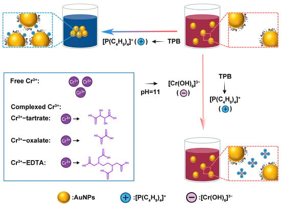
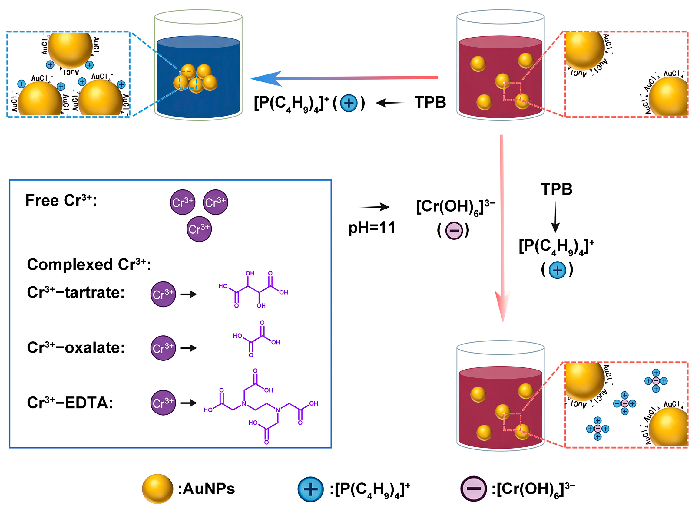
This study proposes a novel colorimetric sensing strategy based on the anti-aggregation effect of bare AuNPs for the simultaneous detection of free Cr3+ and its organic complexes. In contradistinction to conventional methodologies, which necessitate laborious surface modification of AuNPs, this approach employs tetrabutylphosphonium bromide (TPB) as an aggregation inducer (Scheme 1). In aqueous solution, TPB dissociates into phosphonium cations ([P(C4H9)4]+), which electrostatically interact with the residual AuCl4− anions on the surface of bare AuNPs, thereby neutralizing the surface negative charges and reducing the electrostatic repulsion between the particles. This process induces aggregation of the particles and a consequent colour change from wine-red to blue. In strongly alkaline conditions, free Cr3+ transforms into negatively charged hydroxy complexes ([Cr(OH)6]3−). These anionic species have been shown to competitively bind to the phosphonium cations, thereby preventing them from interacting with the AuNPs surface and inhibiting aggregation. Consequently, the AuNPs maintain their dispersion, thereby preserving the wine’s original coloration. This anti-aggregation mechanism demonstrates exceptional versatility, extending effectively to various Cr3+-organometal complexes, including Cr3+-EDTA, Cr3+-tartrate, and Cr3+-oxalate. In strongly alkaline conditions, these complexes undergo a series of reactions involving ligand exchange and hydrolysis, resulting in the formation of the same [Cr(OH)6]3− species. This species subsequently exhibits competitive inhibition of TPB-induced aggregation, operating through a mechanism analogous to that of free Cr3+. This unified detection pathway eliminates the need for specific probe modifications for each complex type, thereby significantly simplifying the analytical protocol. The method under discussion offers technical advantages, including operational simplicity, rapid response, and high sensitivity and selectivity for both free and complexed Cr3+ in real water samples.
Scheme 1.
Schematic illustration of the detection mechanism for Cr3+ and Cr3+-organic complexes based on the anti-aggregation of bare AuNPs.
2. Materials and Methods
2.1. Synthesis of Bare AuNPs
Bare AuNPs were synthesized via the sodium borohydride reduction method [47]. All glassware was pre-treated with aqua regia, followed by rinsing with ultrapure water and air-drying. Briefly, 30 mL of a 2 mmol dm−3 HAuCl4 solution (tetrachloroauric acid, Chengdu Kelong Chemical Co., Ltd., Chengdu, China) was added to 170 mL of Milli-Q water under constant stirring and heated to 55 °C. Then, 15 mL of freshly prepared ice-cold sodium borohydride (NaBH4, 20 mmol dm−3, Sinopharm Chemical Reagent Co., Ltd., Shanghai, China) solution was slowly injected into the mixture over 5 min. The immediate color change from pale yellow to wine-red indicated the successful formation of AuNPs. After stopping the heating, the colloidal solution was stirred for an additional 10 min in the dark and allowed to cool to room temperature. The resulting wine-red AuNPs colloid was stored at 4 °C in the dark for subsequent use.
2.2. Preparation of Cr3+-Organic Complex Solutions
To prepare aqueous Cr3+-organic complexes, freshly prepared Cr3+ standard solutions (Guobiao (Beijing) Inspection and Certification Co., Ltd., Beijing, China) were mixed with three organic ligands (sodium oxalate (Sinopharm Chemical Reagent Co., Ltd., Shanghai, China), tartaric acid (Sinopharm Chemical Reagent Co., Ltd., Shanghai, China), and EDTA (Sinopharm Chemical Reagent Co., Ltd., Shanghai, China)) at a 1:1 molar ratio in aqueous medium. The final concentration of each component was 10 mmol dm−3. The mixtures were allowed to react at room temperature for 48 h to ensure complete complexation.
2.3. Anti-Aggregation-Based Colorimetric Detection of Cr3+ and Cr3+-Organic Complexes Using Bare AuNPs
The detection process employed bare AuNPs as the sensing probe. Briefly, 20 μL of deionized water, 40 μL of TPB (0.4 mmol dm−3, Macklin Biochemical Technology Co., Ltd., Shanghai, China), and 300 μL of Cr3+ or Cr3+-organic complexes (e.g., Cr3+-tartrate, Cr3+-oxalate, Cr3+-EDTA) solution at varying concentrations were mixed, followed by pH adjustment to 11 with NaOH (Sinopharm Chemical Reagent Co., Ltd., Shanghai, China). Then, 300 μL of bare AuNPs colloid was added to the mixture. The reaction was completed within 1 min, and the resulting color changes and Vis–NIR spectra were recorded using a digital camera (Canon EOS 600D, Canon Inc., Tokyo, Japan) and a UV–Vis spectrophotometer (Multiskan SkyHigh, Thermo Fisher Scientific, Waltham, MA, USA), respectively.
To evaluate the specificity of the method for Cr3+ and Cr3+-organic complexes, various potential interferents including Al3+, Na+, K+, Pb2+, Cd2+, Cu2+, Fe2+, Mn2+, Ag+, Co2+, Ba2+, Ni2+, Ca2+, Mg2+, Hg2+, Sr2+, Zn2+, and Cr2O72− (Sinopharm Chemical Reagent Co., Ltd., Shanghai, China) were tested under identical procedures.
2.4. Detection of Cr3+ in Real Water Samples
To validate the practical applicability of the method, spiked recovery experiments were conducted using campus lake water, Yellow River water, and municipal tap water as real environmental samples. The water samples were pre-filtered through a 0.22 μm membrane (Millipore, Burlington, MA, USA), followed by the addition of Cr3+ standard solutions at different concentrations. The spiked samples were analyzed in parallel using both the developed AuNPs-based colorimetric method and inductively coupled plasma mass spectrometry (ICP-MS, Agilent 5110, Agilent Technologies, Santa Clara, CA, USA). The accuracy and reliability of the colorimetric method in real water matrices were assessed by comparing the results obtained from the two techniques.
To further evaluate the applicability of the proposed method for the determination of trivalent chromium (Cr3+) in actual tannery industrial wastewater, this study prepared a simulated chromium-containing wastewater referring to the recommended water quality ranges for typical tannery chromium-bearing effluent specified in the Chinese national standard HJ 2003-2010: Technical Specifications for Tannery and Fur Processing Wastewater Treatment Engineering (Ministry of Environmental Protection of China, 2010) [48]. According to this standard, the primary parameters for chromium-containing wastewater are as follows: pH 3.5~5.0, chemical oxygen demand (COD(Cr)) 3000~6500 mg dm−3, total chromium 600~2500 mg dm−3, ammonia nitrogen 150~400 mg dm−3, total nitrogen 200~500 mg dm−3, and animal/vegetable oils 400~800 mg dm−3.
To ensure experimental controllability and reproducibility, a chemical simulation approach was adopted, with the target water quality set at the median values of the above ranges. The preparation procedure for 1 L of simulated wastewater was as follows: approximately 12.8 g of chromium(III) chloride hexahydrate (CrCl3·6H2O, Sinopharm Chemical Reagent Co., Ltd., Shanghai, China) was dissolved in deionized water as the trivalent chromium source, followed by the sequential addition of 4.0 g of glucose (as the organic substrate providing COD, Sinopharm Chemical Reagent Co., Ltd., Shanghai, China), 0.6 g of ammonium chloride (providing ammonia nitrogen, Sinopharm Chemical Reagent Co., Ltd., Shanghai, China), and 0.5 mL of vegetable oil (simulating animal/vegetable oils). The mixture was continuously stirred until complete dissolution and homogenization. The pH was then adjusted to 4.0~4.5 using dilute hydrochloric acid, and the solution was made up to a final volume of 1 L. After thorough mixing, the solution was allowed to stand for 2 h to reach equilibrium. The supernatant was diluted 400-fold to fall within the linear response range of the calibration curve established for the proposed method, and then analyzed using the proposed method.
3. Results and Discussion
3.1. Characterization of Bare AuNPs
The classical NaBH4 reduction method was employed in this study to synthesize AuNPs. To validate the successful preparation of AuNPs, we utilized variety characterization techniques. First, the AuNPs solution exhibited a characteristic wine-red color, and the Visible–near-infrared (Vis–NIR) absorption spectrum revealed a sharp, symmetric LSPR peak centered at approximately 520 nm (Figure S1). It indicates the prepared AuNPs exhibit excellent dispersity. Transmission electron microscopy (TEM) observations of morphology and size revealed regular spherical AuNPs with an average diameter of approximately 20 nm and a uniform distribution (Figure S2), further confirming the excellent dispersity of the nanoparticles. Additionally, X-ray photoelectron spectroscopy (XPS) analysis was further performed. As shown in Figure S3, high-resolution Au 4f spectra clearly displayed two characteristic peaks at binding energies of 83.73 eV (Au0 4f7/2) and 87.42 eV (Au0 4f5/2), which closely matched the standard binding energies for elemental gold (Au0) [49]. This result confirms the successful reduction of HAuCl4 to elemental gold by NaBH4 and excludes the presence of other gold oxidation states. As a result, integrating macroscopic color observation, UV–Vis spectroscopy, TEM morphological analysis, and XPS chemical state characterization, we confirm the successful and efficient preparation of AuNPs with uniform particle size, and excellent dispersion via the NaBH4 reduction method. Notably, the Fourier-transform infrared (FTIR) spectra of powdered TPB and TPB-treated AuNPs were acquired and compared. After the addition of TPB to the AuNP colloid, the mixture was subjected to centrifugation, and the resulting precipitate was collected for FTIR analysis. A characteristic C−P+ stretching band of TPB at approximately 1400~1500 cm−1 was still clearly observed in the precipitate (Figure S4), confirming that TPB was effectively adsorbed onto the AuNP surface [46]. This finding provided a solid foundation for subsequent investigations.
3.2. Mechanism of the Anti-Aggregation Effect Induced by Cr3+ in the TPB/Bare AuNP System
It is well known that the LSPR effect is crucial for AuNP colorimetric sensors. When AuNPs are in a monodisperse state, their LSPR absorption peak typically appears around 520 nm, and the solution exhibits a characteristic burgundy color. Once AuNPs aggregate, the LSPR peak undergoes red shift and broadening, with a new broadened absorption peak emerging beyond 520 nm. Concurrently, the solution color changes from burgundy to blue or violet-blue. These optical changes directly reflect the dispersion and aggregation state of AuNPs, providing intuitive visual signals and spectroscopic evidence for the sensor. In this study, we found that TBP dissociates in aqueous solution to produce the phosphine cation [P(C4H9)4]+. Since bare AuNPs often carry a negative surface charge primarily due to the adsorption of incompletely reduced chloroaurate ions (AuCl4−), the [P(C4H9)4]+ cation rapidly adsorbs onto the AuNP surface via electrostatic interactions. This neutralizes the negative charge, weakening the electrostatic repulsion between nanoparticles and leading to rapid AuNP aggregation. However, when Cr3+ is present in the system, particularly under alkaline conditions, it can transform into the negatively charged hydroxyl complex [Cr(OH)4]− anion. These anionic species subsequently bind to the excess [P(C4H9)4]+ cations via electrostatic interactions. This effectively blocks the TBP-induced aggregation pathway, restoring and maintaining the monodisperse state of the AuNPs. Consequently, the solution color reverses from blue to burgundy.
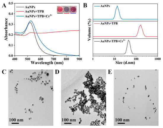
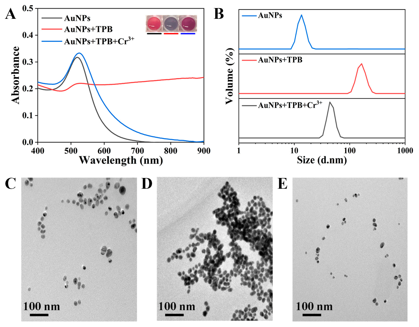
To validate the aforementioned anti-aggregation mechanism, the systematic experiments including color development behavior, Vis–NIR spectroscopy, dynamic light scattering (DLS), transmission electron microscopy (TEM), X-ray photoelectron spectroscopy (XPS), and zeta potential analysis were conducted. As shown in Figure 1A, the color development and Vis–NIR spectroscopy analysis revealed that in the initial state, bare AuNPs were uniformly dispersed and exhibited a typical burgundy color. Their Vis–NIR spectrum displayed a sharp surface plasmon resonance (SPR) absorption peak at 520 nm, a characteristic signal of monodisperse AuNPs. However, following TPB introduction, AuNPs rapidly aggregated, causing the solution color to shift to grayish-blue. Corresponding spectral changes included a significant reduction in SPR peak intensity at 520 nm and the emergence of a broadened absorption peak at 755 nm, a signature spectral feature of aggregated AuNPs. Significantly, when TPB was pre-incubated with Cr3+ before addition to the AuNP system, aggregation was significantly suppressed. Under these conditions, the solution exhibited only a slight darkening in color. The SPR absorption peak intensity at 520 nm in the Vis–NIR spectrum showed a slight increase, and no distinct aggregation-induced absorption peak appeared near 750 nm. This result indicates that Cr3+ effectively blocks TPB’s ability to induce aggregation of AuNPs.
Figure 1.
(A) Photographs and corresponding Vis–NIR spectra of bare AuNPs, bare AuNPs + TPB, and bare AuNPs + TPB + Cr3+. (B) DLS data of bare AuNPs, bare AuNPs + TPB, and bare AuNPs + TPB + Cr3+. TEM images of (C) bare AuNPs, (D) bare AuNPs + TPB, and (E) bare AuNPs + TPB + Cr3+.
Changes in hydrodynamic diameter measured by DLS also supported this observation. The hydrated diameter of pristine AuNPs was 12.56 nm, which dramatically increased to 157.76 nm after TBP treatment, indicating the formation of large aggregates. In the TBP system preincubated with Cr3+, the AuNP size only slightly increased to 48.92 nm, consistent with the conclusion that Cr3+ inhibits aggregation (Figure 1B). TEM analysis provides visual evidence for these macroscopic phenomena. As shown in Figure 1C, the initial AuNPs exhibit good monodispersity with an average particle size of approximately 20 nm. Following TBP treatment, the AuNPs formed significant aggregates. However, the individual AuNPs within these aggregates maintained their original average size of approximately 20 nm (Figure 1D). This indicates that the solution color change and SPR peak red shift stem from aggregation effects triggered by TBP-regulated interparticle spacing, rather than alterations in nanoparticle morphology or size. More importantly, when TBP was pre-incubated with Cr3+, no significant aggregation was observed in TEM images. AuNPs remained monodisperse with unchanged particle sizes (Figure 1E), further confirming that Cr3+ effectively blocks TBP’s aggregation-inducing pathway.
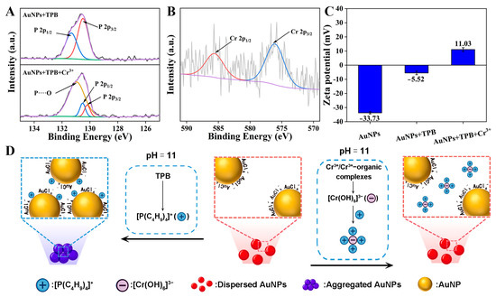
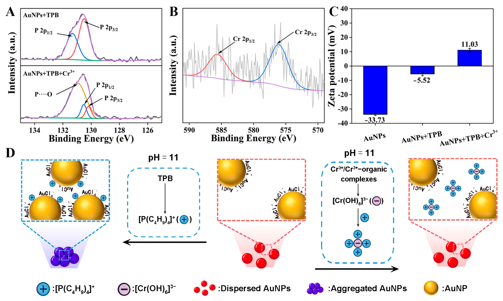
To elucidate the anti-aggregation mechanism induced by Cr3+ in the TPB/bare AuNP system, XPS analysis was further conducted. TPB solutions, before and after incubation with Cr3+, were separately mixed with bare AuNPs. The solid samples collected by centrifugation were subjected to XPS measurements. The XPS spectrum of the P element in TPB exhibited characteristic P 2p1/2 and P 2p3/2 peaks at binding energies of 131.3 eV and 130.5 eV, respectively (Figure 2A). Compared to the TPB sample without Cr3+ incorporation, the introduction of Cr3+ leads to a noticeable narrowing of the peak widths and a significant shift in the positions of the P 2p1/2 and P 2p3/2 peaks (Figure 2A), indicating that the presence of Cr3+ altered the chemical environment of the P element and suggesting a possible interaction between the two species [46]. Moreover, in the XPS spectrum of the Cr element, characteristic Cr 2p1/2 and Cr 2p3/2 peaks were observed at binding energies of 585.6 eV and 576.0 eV, respectively (Figure 2B) [46,50]. The positions of these peaks are consistent with the typical signature of Cr3+ coordinated with oxygen. Given that the experimental system was an alkaline aqueous environment, the coordinating oxygen atoms likely originated from OH−. It can thus be inferred that Cr3+ existed in the form of a hydroxo complex anion, i.e., [Cr(OH)6]−, during the reaction. This anion can electrostatically interact with the positively charged [P(C4H9)4]+ cation due to their opposite charges.
Figure 2.
(A) P 2p XPS spectra of TPB before and after incubation with Cr3+. (B) Cr 2p XPS spectrum after TPB incubation with Cr3+. (C) Zeta potential measurements of bare AuNPs, bare AuNPs + TPB, and bare AuNPs + TPB + Cr3+. (D) Schematic diagram of the surface charge change on bare AuNPs.
Zeta potential analysis further elucidates the regulation mechanism of surface charge. As shown in Figure 2C, the zeta potential of bare gold nanoparticles (AuNPs) was −33.7 mV, indicating a negatively charged surface. Upon TBP addition, the zeta potential increased to −5.5 mV, demonstrating that [P(C4H9)4]+ effectively neutralized part of the negative charge on the AuNPs surface, thereby reducing electrostatic repulsion and promoting particle aggregation (Figure 2D). When TBP was pre-incubated with Cr3+ prior to introduction into the AuNP system, the zeta potential further inverted to +11.0 mV (Figure 2C). This inversion is attributed to the conversion of Cr3+ into anionic [Cr(OH)6]− species under alkaline conditions, which subsequently interact with excess [P(C4H9)4]+ cations via electrostatic interaction. Consequently, the attenuation of interparticle electrostatic repulsion by [P(C4H9)4]+ is mitigated, thereby preserving the monodispersity of the AuNPs and retaining the characteristic wine-red coloration of the solution (Figure 2D).
3.3. Mechanism of the Anti-Aggregation Effect Induced by Cr3+-Organic Complexes in the TPB/Bare AuNP System
We also investigated the response behavior of this anti-aggregation mechanism to different complexes such as Cr3+-EDTA, Cr3+-oxalate, and Cr3+-tartrate (Figure S5). Under weakly alkaline conditions, these Cr3+-organic complexes failed to effectively inhibit TBP-induced Au NP aggregation, and the solution remained blue-gray. It may be attributed to the relatively stable structure of Cr3+-organic complexes formed with organic ligands under these conditions, where their overall net charge or spatial conformation hinders effective binding with [P(C4H9)4]+ via electrostatic interactions. However, upon converting the system to a strongly alkaline environment by adding NaOH, all three Cr3+-organic complexes successfully induced significant anti-aggregation effects, restoring the solution color to burgundy. We speculate that under strongly basic conditions, elevated OH− concentrations induce ligand exchange or hydrolysis reactions in Cr3+-organic complexes, converting them into negatively charged hydroxyl-coordinated anions such as [Cr(OH)6]3− [46,51,52]. Subsequently, these [Cr(OH)6]3− follow the aforementioned anti-aggregation mechanism to form positively charged ion clusters with excess [P(C4H9)4]+, thereby maintaining the stable dispersion of Au NPs (Figure 2D).
In summary, our findings demonstrate that both free Cr3+ and its organic complexes undergo transformation under alkaline conditions to yield the anionic hexahydroxo complex, [Cr(OH)6]3−. Crucially, these anionic species engage with dissociated [P(C4H9)4]+ cations derived from TBP via electrostatic interactions. This interaction effectively precludes the [P(C4H9)4]+-induced aggregation of AuNPs, thereby preserving their monodispersed state. The mechanistic elucidation presented herein provides a robust theoretical framework and solid experimental foundation for the rational design of AuNP-based colorimetric sensors, enabling the quantitative discrimination and detection of both free and complexed Cr3+ species.
3.4. Selectivity of Cr3+ and Cr3+-Organic Complexes Detection by the Anti-Aggregation Colorimetric Assay Based on Bare AuNPs
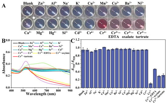
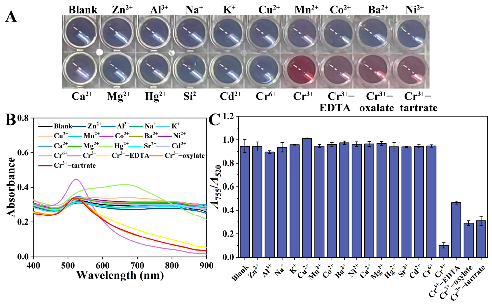
Under optimized experimental conditions, the selectivity of the anti-aggregation sensing system for detecting Cr3+ and Cr3+-organic complexes was evaluated. The TPB/bare AuNP reaction system was challenged with the target analytes, 60 μmol dm−3 Cr3+ and Cr3+-organic complexes (Cr3+-EDTA, Cr3+-oxalate, and Cr3+-tartrate), alongside 18 common potentially interfering ions at the same concentration level. These interferents included Al3+, Na+, K+, Pb2+, Cd2+, Cu2+, Fe2+, Mn2+, Ag+, Co2+, Ba2+, Ni2+, Ca2+, Mg2+, Hg2+, Sr2+, Zn2+, and Cr2O72−. As illustrated in Figure 3A, only the systems containing Cr3+ and Cr3+-organic complexes consistently exhibited the characteristic wine-red color, indicating the presence of monodispersed AuNPs. In contrast, all systems containing the interfering ions maintained the grayish-blue color characteristic of TPB-induced AuNP aggregation. This visual observation demonstrated that none of the interfering ions could effectively inhibit the TPB-mediated aggregation process. These visual findings were corroborated by the corresponding Vis–NIR spectra. The system containing Cr3+ and its complexes showed a distinct SPR absorption peak at 520 nm, which is characteristic of monodispersed AuNPs, with no discernible peak around 755 nm associated with nanoparticle aggregates (Figure 3B). Conversely, the spectra for all interfering ion systems displayed a significant attenuation of the SPR peak at 520 nm and the emergence of a prominent absorption band at 755 nm, confirming extensive nanoparticle aggregation. Quantitative analysis of the absorbance ratio A755/A520 further validated the high specificity of the system. The A755/A520 value for the system containing Cr3+ and Cr3+-organic complexes was significantly lower than that of the blank control. In contrast, the ratios for systems containing other interfering ions showed no substantial difference from the blank (Figure 3C). This may be attributed to the fact that Cr3+ and its organic complexes can be converted into strongly negatively charged hydroxo complexes under alkaline conditions, whereas other metal ions, being unable to form analogous complex structures, cannot electrostatically bind to the [P(C4H9)4]+ and thus fail to inhibit aggregation.
Figure 3.
Selectivity evaluation of the colorimetric sensing system based on bare AuNPs for Cr3+ and Cr3+-organic complexes. (A) Photographs showing solution color in the presence of different interfering substances. (B) Corresponding Vis–NIR absorption spectra. (C) Calculated absorbance ratios (A755/A520) under different conditions.
3.5. Sensitivity of Cr3+ Detection via the Anti-Aggregation Colorimetric Assay Based on Bare AuNPs
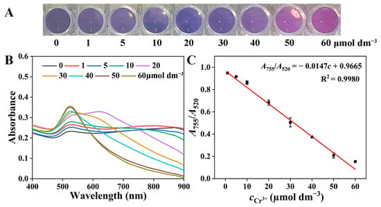
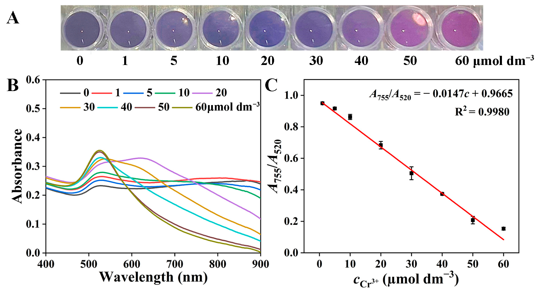
Under the optimized experimental conditions, the quantitative analytical performance of the anti-aggregation system for Cr3+ was evaluated. As the concentration of Cr3+ increased from 1 to 60 μmol dm−3, the color of the system gradually transitioned from gray-blue to wine-red (Figure 4A). Correspondingly, in the Vis–NIR spectra, the intensity of the characteristic absorption peak at 520 nm increased progressively, while the aggregation-related peak at 755 nm diminished significantly (Figure 4B). A linear regression analysis was performed between the A755/A520 ratio and the Cr3+ concentration. A good linear inverse correlation was observed within the range of 1~60 μmol dm−3, yielding the regression equation: A755/A520 = −0.0147c + 0.9664 (R2 = 0.9977) (Figure 4C). Based on a signal-to-noise ratio of 3 (S/N = 3) calculated from the standard deviation of the blank signal, the LOD for Cr3+ was determined to be as low as 0.76 μmol dm−3. This value is notably lower than the maximum contaminant level for chromium in drinking water established by the U.S. Environmental Protection Agency (US EPA, 1.9 μmol dm−3). It is worth emphasizing that the entire detection process can be completed within 1 min. As summarized in Table S1, compared with other reported POCT methods such as fluorescent and colorimetric assays, the proposed method not only obviates the need for complex surface chemical modifications but also exhibits comparable or even superior sensitivity, along with a substantially shortened assay time. On the other hand, although the sensitivity of the proposed method is somewhat lower than that of conventional instrumental techniques such as ICP-MS and atomic absorption spectrometry (AAS), it is widely recognized that these instrumental methods entail high-cost instrumentation, require professionally trained operators and intricate operational procedures, and are inherently unsuitable for on-site analysis. In contrast, our method meets the required detection threshold for Cr3+ while offering the distinct advantages of simple operation, rapid response, and cost-effectiveness. This combination of high sensitivity and rapid response makes the method a reliable technical solution for the on-site and fast detection of Cr3+ in environmental water samples.
Figure 4.
Sensitivity of the bare AuNPs-based colorimetric detection for Cr3+. (A) Photographs of solutions containing bare AuNPs with increasing concentrations of Cr3+. (B) Corresponding Vis–NIR absorption spectra. (C) Plot of the absorbance ratio A755/A520 versus Cr3+ concentration.
3.6. Sensitivity of Cr3+-Organic Complexes Detection via the Anti-Aggregation Colorimetric Assay Based on Bare AuNPs
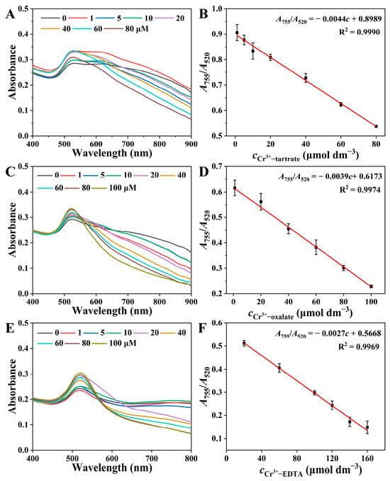
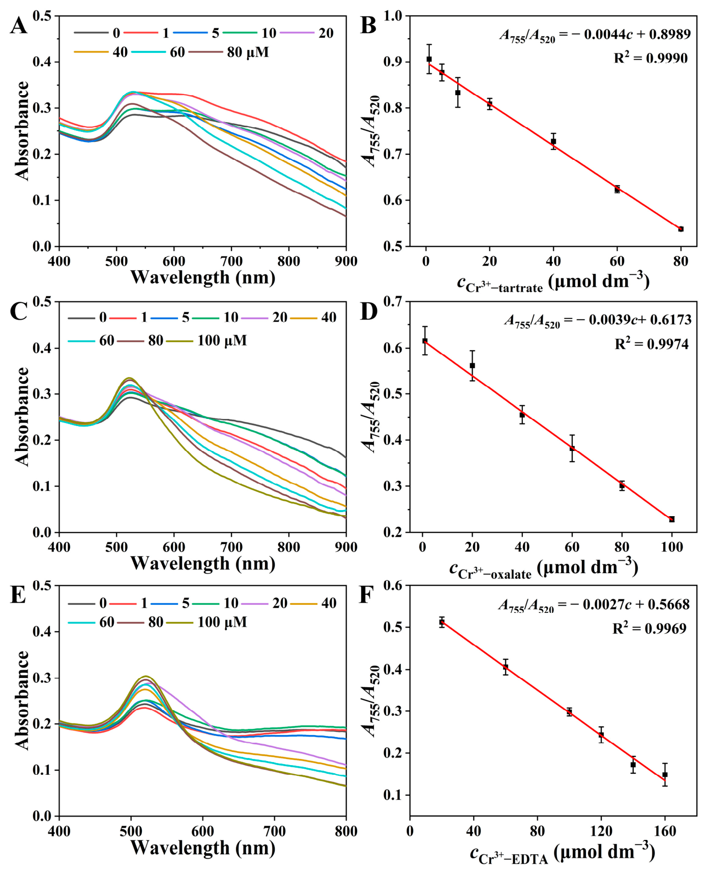
Under the optimized experimental conditions, the quantitative analytical performance and sensitivity of the sensing system were further evaluated for three typical Cr3+-organic complexes: Cr3+-tartrate, Cr3+-oxalate, and Cr3+-EDTA. As shown in Figure 5A–C, in the Vis–NIR spectra, the intensity of the dispersed-state absorption peak at 520 nm progressively increased, while the intensity of the aggregated-state absorption peak at 755 nm decreased simultaneously. This indicates a significant enhancement of the anti-aggregation effect with increasing complex concentration. Quantitative detection models for the three Cr3+-organic complexes were established via linear regression analysis: For Cr3+-tartrate, within the range of 1~80 μmol dm−3, the linear equation was A755/A520 = −0.0044c + 0.8989 (R2 = 0.9990), with an LOD of 1.49 μmol dm−3. For Cr3+-oxalate, an excellent linear relationship was observed in the concentration range of 1~100 μmol dm−3, with the regression equation A755/A520 = −0.0039c + 0.6173 (R2 = 0.9912) and an LOD of 0.93 μmol dm−3. For Cr3+-EDTA, a good linear response was maintained within 20~160 μmol dm−3, described by the equation A755/A520 = −0.0027c + 0.5668 (R2 = 0.9969), yielding an LOD of 1.82 μmol dm−3. These results demonstrate that the method possesses excellent detection sensitivity and reliable quantitative capability for different forms of Cr3+-organic complexes. It thus provides an effective technical approach for complexed Cr3+ analysis in complex environmental samples.
Figure 5.
Detection of different Cr3+-organic complexes using the bare AuNPs-based sensing system. Vis-NIR absorption spectra and corresponding calibration plots of A755/A520 for (A,B) Cr3+-tartrate, (C,D) Cr3+-oxalate, and (E,F) Cr3+-EDTA at varying concentrations.
3.7. Detection of Cr3+ in Real Water Samples
To evaluate the analytical performance of the method in real water samples, a spike recovery experiment was conducted using campus lake water, Yellow River water, and municipal tap water as representative environmental samples. Preliminary analysis confirmed that the background level of Cr3+ in the original water sample was below the method’s limit of detection (<1 μmol dm−3), meeting the requirements for a spike recovery test. To simulate practical scenarios with varying levels of Cr3+ contamination, the pretreated water samples (filtered through a 0.22 μm membrane to remove suspended particulates) were spiked with Cr3+ standard solutions at final concentrations of 20 and 40 μmol dm−3. The selection of these two spiking concentrations was based on three primary considerations: (i) these levels represent environmentally relevant contamination scenarios in industrial wastewater; (ii) they fall within the established linear detection range of our method (1~60 μmol dm−3), ensuring accurate quantification; and (iii) they induce a distinct color change that is readily discernible by the naked eye, facilitating on-site visual screening. The Cr3+ concentrations were then determined in triplicate (n = 3) using the developed anti-aggregation colorimetric sensing system. As shown in Table S2, the established colorimetric sensing system achieved spike recoveries for Cr3+ in the campus lake water ranging from 102.5% to 113.3%, with relative standard deviations (RSD) ≤ 4.90% (n = 3). These results are in good agreement with those obtained by ICP-MS, which yielded recoveries of 100.5~109.9% with RSD ≤ 4.87% (n = 3). Collectively, these findings demonstrate the favorable applicability and reliability of the proposed method for the analysis of real environmental samples, offering a practical and cost-effective technical solution for rapid and accurate on-site monitoring of Cr3+ in environmental waters. More importantly, the method was successfully applied to the direct detection of Cr3+ in simulated tannery industrial wastewater, and the results were comparable to those obtained by ICP-MS, further corroborating its potential for practical applications in tannery effluent monitoring.
4. Conclusions
We have developed an anti-aggregation colorimetric sensing strategy based on bare AuNPs for the simultaneous detection of free and complexed Cr3+ in environmental water samples. The aggregation inducer, TPB, provides cationic phosphonium species that electrostatically interact with negatively charged [Cr(OH)6]3− formed from free and complexed Cr3+ under alkaline conditions, thereby effectively inhibiting AuNPs aggregation. This mechanism overcomes the “ligand-shielding effect” that renders conventional AuNPs-based sensors incapable of recognizing metal–organic complexes. Our method requires no tedious surface modification, offers rapid response, high sensitivity, and excellent selectivity, and thus provides a simple, cost-effective, and reliable tool for speciation analysis of free and complexed Cr3+ in complex water matrices. By overcoming the ligand shielding effect that plagues traditional sensors, this strategy provides a reliable analytical tool for the comprehensive monitoring of chromium pollution in environmental waters, and therefore has a broad application prospect for speciation analysis in complex matrices.
Supplementary Materials
The following supporting information can be downloaded at https://www.mdpi.com/article/10.3390/chemosensors14090191/s1, Figure S1: Photograph and Vis–NIR spectrum of the bare AuNPs colloidal solution; Figure S2: TEM image of the synthesized bare AuNPs; Figure S3: Au 4f XPS spectrum of the bare AuNPs; Figure S4: FTIR spectrum of the TPB and bare AuNPs + TPB; Figure S5: Vis–NIR absorption spectra of (A) bare AuNPs, bare AuNPs + TPB, bare AuNPs + TPB + Cr3+-tartrate, and bare AuNPs + TPB + Cr3+-tartrate + NaOH; (B) bare AuNPs, bare AuNPs + TPB, bare AuNPs + TPB + Cr3+-oxalate, and bare AuNPs + TPB + Cr3+-oxalate + NaOH; and (C) bare AuNPs, bare AuNPs + TPB, bare AuNPs + TPB + Cr3+-EDTA, and bare AuNPs + TPB + Cr3+-EDTA + NaOH; Figure S6: Variation of the absorbance ratio A755/A520 before and after the addition of Cr3+ at different TPB concentration; Figure S7: The absorbance ratio A755/A520 of different Cr3+-organic complexes under different pH sensing systems; Table S1: Comparison of Analytical Performance of Different Methods for Cr3+ Detection [19,42,53,54,55,56,57,58,59,60,61]; Table S2: Detection of Cr3+ ions in real water samples.
Author Contributions
Conceptualization, H.R. and J.W.; methodology, H.R.; software, J.W.; validation, K.Z.; formal analysis, J.W.; investigation, H.R.; resources, H.R.; data curation, K.Z.; writing—original draft preparation, H.R. and J.W.; writing—review and editing, H.R.; visualization, Z.X.; supervision, H.R.; project administration, H.R.; funding acquisition, H.R. All authors have read and agreed to the published version of the manuscript.
Funding
This research was funded by the Industrial Support Program for Higher Education Institutions Project (Nos.2023CYZC-69, 2024CYZC-05), the Central Guided Local Science and Technology Development Fund Project (No.25ZYJA005), the Lanzhou Young Scientific and Technological Talents Innovation Project (2025-NQ-167), the Gansu Provincial Department of Education: University Teacher Innovation Fund Project (2026A-169), Lanzhou City University Ph.D. Research Fund Project (LZCU-BS2025-06).
Institutional Review Board Statement
Not applicable.
Informed Consent Statement
Not applicable.
Data Availability Statement
The original contributions presented in this study are included in the article/Supplementary Materials. Further inquiries can be directed to the corresponding authors.
Acknowledgments
The authors have reviewed and edited the output and take full responsibility for the content of this publication.
Conflicts of Interest
The authors declare no conflicts of interest.
References
- Hua, T.; Li, Y.; Hu, Y.; Yin, R.; Zhang, Y.; Hou, B.; Lu, H.; Ji, X.; Bai, X.; Lu, A.; et al. Back to chromite as a mineralogical strategy for long-term chromium pollution control. Nat. Commun. 2025, 16, 1975. [Google Scholar] [CrossRef] [Scilit] [PubMed]
- Zha, S.; Yu, A.; Wang, Z.; Shi, Q.; Cheng, X.; Liu, C.; Deng, C.; Zeng, G.; Luo, S.; Zhao, Z.; et al. Microbial strategies for effective hexavalent chromium removal: A comprehensive review. Chem. Eng. J. 2024, 489, 151457. [Google Scholar] [CrossRef] [Scilit]
- Naz, H.; Sayyed, R.Z.; Khan, R.U.; Naz, A.; Wani, O.A.; Maqsood, A.; Maqsood, S.; Fahad, A.; Ashraf, S.; Show, P.L. Mesorhizobium improves chickpea growth under chromium stress and alleviates chromium contamination of soil. J. Environ. Manag. 2023, 338, 117779. [Google Scholar] [CrossRef] [Scilit] [PubMed]
- Almeida, J.C.; Cardoso, C.E.D.; Tavares, D.S.; Freitas, R.; Trindade, T.; Vale, C.; Pereira, E. Chromium removal from contaminated waters using nanomaterials—A review. TrAC Trends Anal. Chem. 2019, 118, 277–291. [Google Scholar] [CrossRef] [Scilit]
- Banerjee, S.; Jha, S.; Chakraborty, S.; Ghosh, S.; Sarkar, D.; Datta, R.; Bhattacharya, S.S.; Bhattacharyya, P. Mycorrhiza-assisted phytoremediation of spiked chromium-contaminated soil: Assessing AMF-vetiver symbiosis for Cr accumulation and soil quality enhancement. Environ. Res. 2025, 283, 122143. [Google Scholar] [CrossRef] [Scilit] [PubMed]
- Obeidy, C.S.; Koeneke, M.W.; Duckworth, O.W.; Polizzotto, M.L. Landscape Position and Burn Intensity Influence Heat-Induced Soil Chromium Contamination. Environ. Sci. Technol. 2025, 59, 25842–25852. [Google Scholar] [CrossRef] [Scilit] [PubMed]
- Barbosa, R.F.S.; Souza, A.G.; Maltez, H.F.; Rosa, D.S. Chromium removal from contaminated wastewaters using biodegradable membranes containing cellulose nanostructures. Chem. Eng. J. 2020, 395, 125055. [Google Scholar] [CrossRef] [Scilit]
- Sujiritha, P.B.; Vikash, V.L.; Ponesakki, G.; Ayyadurai, N.; Kamini, N.R. Microbially induced carbonate precipitation with Arthrobacter creatinolyticus: An eco-friendly strategy for mitigation of chromium contamination. J. Environ. Manag. 2024, 365, 121300. [Google Scholar] [CrossRef] [Scilit] [PubMed]
- Uliana, A.A.; Pezoulas, E.R.; Zakaria, N.I.; Johnson, A.S.; Smith, A.; Lu, Y.; Shaidu, Y.; Velasquez, E.O.; Jackson, M.N.; Blum, M.; et al. Removal of Chromium and Arsenic from Water Using Polyol-Functionalized Porous Aromatic Frameworks. J. Am. Chem. Soc. 2024, 146, 23831–23841. [Google Scholar] [CrossRef] [Scilit] [PubMed]
- Liu, C.; Dong, W.; Specht, A.J.; Juhasz, A.L.; Yin, J.; Liu, C.; Ma, L.Q. Luxury at a Cost: Portable XRF-Based Bioaccessible Chromium in Sweat Predicts Dermal Exposure Risk to Chromate in Leather Products. Environ. Sci. Technol. 2026, 60, 15681–15690. [Google Scholar] [CrossRef] [Scilit] [PubMed]
- Prasad, S.; Yadav, K.K.; Kumar, S.; Gupta, N.; Cabral-Pinto, M.M.S.; Rezania, S.; Radwan, N.; Alam, J. Chromium contamination and effect on environmental health and its remediation: A sustainable approaches. J. Environ. Manag. 2021, 285, 112174. [Google Scholar] [CrossRef] [Scilit] [PubMed]
- Karunanidhi, D.; Aravinthasamy, P.; Subramani, T.; Kumar, D.; Venkatesan, G. Chromium contamination in groundwater and Sobol sensitivity model based human health risk evaluation from leather tanning industrial region of South India. Environ. Res. 2021, 199, 111238. [Google Scholar] [CrossRef] [Scilit] [PubMed]
- Liu, S.; Zhang, L.; Kim, H.; Sun, J.; Yoon, J. Recent advances and challenges in monitoring chromium ions using fluorescent probes. Coord. Chem. Rev. 2024, 501, 215575. [Google Scholar] [CrossRef] [Scilit]
- Maruthupandi, M.; Lee, N.Y. Point-of-Care Testing Kit for the Detection of Hexavalent Chromium by Carbohydrazide-Derived Graphitic Carbon Nitride. Chemosensors 2024, 12, 180. [Google Scholar] [CrossRef] [Scilit]
- B.K., V.; Bagchi, S.; T.R., S. Chromium Detection in Water Using Optical Methods: A Study of Reagent and Reagentless Approaches. Crit. Rev. Anal. Chem. 2026, 56, 494–531. [Google Scholar] [CrossRef] [Scilit] [PubMed]
- Kaur, G.; Sharma, S.; Bhardwaj, N.; Nayak, M.K.; Deep, A. Simple fluorochromic detection of chromium with ascorbic acid functionalized luminescent Bio-MOF-1. Nanoscale 2024, 16, 12523–12533. [Google Scholar] [CrossRef] [Scilit] [PubMed]
- Zeng, H.-H.; Wu, H.; Peng, D.; Liu, F.; Shi, W.-G.; Qiu, J.-D. Fast and Selective Detection of Cr(III) in Environmental Water Samples Using Phosphovanadate Y(V0.2P0.8O4):Eu3+ Fluorescence Nanorods. ACS Sens. 2018, 3, 1569–1575. [Google Scholar] [CrossRef] [Scilit] [PubMed]
- Ruan, Y.; Li, Q.; Yang, D.; Yang, Y. Fluorescence detection of valence speciation of Cr(III) based on the catechol oxidase mimic enzyme activity of CuSeNP nanozymes. Microchim. Acta 2024, 191, 496. [Google Scholar] [CrossRef] [Scilit] [PubMed]
- Qiao, S.; Wang, J.; Guo, Z. Ratiometric Fluorescent Detection of Chromium(III) Based on One-Dimensional Imine-Linked Covalent Organic Framework. Inorg. Chem. 2023, 63, 706–713. [Google Scholar] [CrossRef] [Scilit] [PubMed]
- Sun, J.; Lu, Y.; He, L.; Pang, J.; Yang, F.; Liu, Y. Colorimetric sensor array based on gold nanoparticles: Design principles and recent advances. TrAC Trends Anal. Chem. 2020, 122, 115754. [Google Scholar] [CrossRef] [Scilit]
- Yu, L.; Song, Z.; Peng, J.; Yang, M.; Zhi, H.; He, H. Progress of gold nanomaterials for colorimetric sensing based on different strategies. TrAC Trends Anal. Chem. 2020, 127, 115880. [Google Scholar] [CrossRef] [Scilit]
- Zhou, W.; Hu, K.; Kwee, S.; Tang, L.; Wang, Z.; Xia, J.; Li, X. Gold Nanoparticle Aggregation-Induced Quantitative Photothermal Biosensing Using a Thermometer: A Simple and Universal Biosensing Platform. Anal. Chem. 2020, 92, 2739–2747. [Google Scholar] [CrossRef] [Scilit] [PubMed]
- Donati, P.; Moglianetti, M.; Veronesi, M.; Prato, M.; Tatulli, G.; Bandiera, T.; Pompa, P.P. Nanocatalyst/Nanoplasmon-Enabled Detection of Organic Mercury: A One-Minute Visual Test. Angew. Chem. Int. Ed. 2019, 58, 10285–10289. [Google Scholar] [CrossRef] [Scilit] [PubMed]
- Hang, Y.; Wang, A.; Wu, N. Plasmonic silver and gold nanoparticles: Shape- and structure-modulated plasmonic functionality for point-of-caring sensing, bio-imaging and medical therapy. Chem. Soc. Rev. 2024, 53, 2932–2971. [Google Scholar] [CrossRef] [Scilit] [PubMed]
- Chang, C.-C.; Chen, C.-P.; Wu, T.-H.; Yang, C.-H.; Lin, C.-W.; Chen, C.-Y. Gold Nanoparticle-Based Colorimetric Strategies for Chemical and Biological Sensing Applications. Nanomaterials 2019, 9, 861. [Google Scholar] [CrossRef] [Scilit] [PubMed]
- Zhang, K.; Luo, M.; Rao, H.; Liu, H.; Li, J.; Chen, J.; Liu, X.; Xue, Z. Integrating plasmonic and nanozyme responses of gold nanoparticles for enhancing photothermometric sensing. Sens. Actuators B Chem. 2023, 392, 134067. [Google Scholar] [CrossRef] [Scilit]
- Revilla-Cuesta, A.; Abajo-Cuadrado, I.; Quadrini, L.; Failli, S.; Rodríguez-Rubio, A.; Cuevas, J.V.; Hernando-Muñoz, C.; García-Calvo, J.; Torroba, T. Chemical speciation of methylmercury and mercury(II) cations in fish by new fluorogenic naphthalimide alkynyl gold complexes: The ultimate test for detecting fish contamination. Sens. Actuators B Chem. 2024, 421, 136492. [Google Scholar] [CrossRef] [Scilit]
- Gao, Z.; Ye, H.; Tang, D.; Tao, J.; Habibi, S.; Minerick, A.; Tang, D.; Xia, X. Platinum-Decorated Gold Nanoparticles with Dual Functionalities for Ultrasensitive Colorimetric in Vitro Diagnostics. Nano Lett. 2017, 17, 5572–5579. [Google Scholar] [CrossRef] [Scilit] [PubMed]
- Annadhasan, M.; Muthukumarasamyvel, T.; Sankar Babu, V.R.; Rajendiran, N. Green Synthesized Silver and Gold Nanoparticles for Colorimetric Detection of Hg2+, Pb2+, and Mn2+ in Aqueous Medium. ACS Sustain. Chem. Eng. 2014, 2, 887–896. [Google Scholar] [CrossRef] [Scilit]
- Priyadarshni, N.; Nath, P.; Nagahanumaiah; Chanda, N. DMSA-Functionalized Gold Nanorod on Paper for Colorimetric Detection and Estimation of Arsenic (III and V) Contamination in Groundwater. ACS Sustain. Chem. Eng. 2018, 6, 6264–6272. [Google Scholar] [CrossRef] [Scilit]
- Lee, J.-S.; Han, M.S.; Mirkin, C.A. Colorimetric Detection of Mercuric Ion (Hg2+) in Aqueous Media using DNA-Functionalized Gold Nanoparticles. Angew. Chem. Int. Ed. 2007, 46, 4093–4096. [Google Scholar] [CrossRef] [Scilit] [PubMed]
- Zhang, K.; Luo, M.; Rao, H.; Liu, H.; Qiang, R.; Xue, X.; Li, J.; Lu, X.; Xue, Z. Plasmonic and nanozyme dual-channel-based logic judgment for enhancing gold nanoparticle based colorimetric Hg2+ ion sensing performance. Chem. Commun. 2024, 60, 10005–10008. [Google Scholar] [CrossRef] [Scilit]
- Yu, Y.; Hong, Y.; Gao, P.; Nazeeruddin, M.K. Glutathione Modified Gold Nanoparticles for Sensitive Colorimetric Detection of Pb2+ Ions in Rainwater Polluted by Leaking Perovskite Solar Cells. Anal. Chem. 2016, 88, 12316–12322. [Google Scholar] [CrossRef] [Scilit] [PubMed]
- Carnamucio, F.; Foti, C.; Saija, F.; Cassone, G.; Giuffrè, O. Speciation and Thermodynamic Study of Arsenic(III)–Pharmaceutical Complexes in Aqueous Solutions. ACS Environ. AU 2025, 5, 404–414. [Google Scholar] [CrossRef] [Scilit] [PubMed]
- Wang, S.; Song, X.; Hu, J.; Zhang, R.; Men, L.; Wei, M.; Xie, T.; Cao, J. Direct speciation analysis of organic mercury in fish and kelp by on-line complexation and stacking using capillary electrophoresis. Food Chem. 2019, 281, 41–48. [Google Scholar] [CrossRef] [Scilit] [PubMed]
- Town, R.M.; van Leeuwen, H.P. Intraparticulate Metal Speciation Analysis of Soft Complexing Nanoparticles. The Intrinsic Chemical Heterogeneity of Metal–Humic Acid Complexes. J. Phys. Chem. A 2016, 120, 8637–8644. [Google Scholar] [CrossRef] [Scilit] [PubMed]
- Van Soest, P.J.; Hall, M.B. Cobalt (III)-EDTA dissociates and chromium (III)-EDTA is slightly more stable under in vitro reducing conditions comparable to those in the rumen. J. Dairy Sci. 2020, 103, 10152–10160. [Google Scholar] [CrossRef] [Scilit] [PubMed]
- Shukla, M.; Baksi, B.; Mohanty, S.P.; Mahanty, B.; Mansi, A.; Rene, E.R.; Behera, S.K. Remediation of chromium contaminated soil by soil washing using EDTA and N-acetyl-L-cysteine as the chelating agents. Prog. Org. Coat. 2022, 165, 106704. [Google Scholar] [CrossRef] [Scilit]
- Wang, D.; Ye, Y.; Liu, H.; Ma, H.; Zhang, W. Effect of alkaline precipitation on Cr species of Cr(III)-bearing complexes typically used in the tannery industry. Chemosphere 2018, 193, 42–49. [Google Scholar] [CrossRef] [Scilit] [PubMed]
- Chen, N.; Wu, S.; Pan, B.; Yang, Z.; Pan, B. Engineering Nano-Au-Based Sensor Arrays for Identification of Multiple Ni(II) Complexes in Water Samples. Environ. Sci. Technol. 2023, 57, 9874–9883. [Google Scholar] [CrossRef] [Scilit] [PubMed]
- Chen, N.; Wu, S.; Xu, Y.; Lv, S.; Wang, X.; Zhang, Q.; Pan, B. Accurately recognizing chromium species with multi-functionalized nano Au-based sensor array. J. Hazard. Mater. 2024, 476, 134981. [Google Scholar] [CrossRef] [Scilit]
- Long, D.; Yu, H. A synergistic coordination strategy for colorimetric sensing of chromium(III) ions using gold nanoparticles. Anal. Bioanal. Chem. 2016, 408, 8551–8557. [Google Scholar] [CrossRef] [Scilit] [PubMed]
- Muthwa, S.F.; Zulu, N.S.; Kistan, M.; Onwubu, S.C.; Shumbula, N.P.; Moloto, N.; Mpelane, S.; Hlatshwayo, T.; Mlambo, M.; Mdluli, P.S. Unravelling mechanism for detecting chromium on functionalized gold nanoparticles via a smartphone and spectrophotometric-based systems supported by CIEL*a*b* colour space and molecular dynamics. J. Mol. Struct. 2023, 1274, 134394. [Google Scholar] [CrossRef] [Scilit]
- Wang, X.; Wang, X.; Liu, J.; Wang, K.; Zhao, R.; Yang, S. Ingenious aspartic acid-functionalized gold nanoparticles by one-pot protocol for the sensitive detection of chromium (III) ions. Microchem. J. 2020, 159, 105359. [Google Scholar] [CrossRef] [Scilit]
- Yang, X.; Xu, Y.; Huang, X.; Hang, J.; Guo, W.; Dai, Z. Multicolor Iridium(III) Complexes with Host–Guest Recognition Motifs for Enhanced Electrochemiluminescence and Modular Labeling. Anal. Chem. 2023, 95, 4543–4549. [Google Scholar] [CrossRef] [Scilit] [PubMed]
- Siriwardhane, T.; Sulkanen, A.; Pathirathna, P.; Tremonti, A.; McElmurry, S.P.; Hashemi, P. Voltammetric Characterization of Cu(II) Complexation in Real-Time. Anal. Chem. 2016, 88, 7603–7608. [Google Scholar] [CrossRef] [Scilit] [PubMed]
- Chen, N.; Pan, B. Tributylhexadecylphosphonium Modification Strategy to Construct Gold Nanoprobes for the Detection of Aqueous Cr(III)–Organic Complexes. Anal. Chem. 2021, 93, 1811–1817. [Google Scholar] [CrossRef] [Scilit] [PubMed]
- Shellaiah, M.; Simon, T.; Sun, K.W.; Ko, F.-H. Simple bare gold nanoparticles for rapid colorimetric detection of Cr3+ ions in aqueous medium with real sample applications. Sens. Actuators B Chem. 2016, 226, 44–51. [Google Scholar] [CrossRef] [Scilit]
- HJ 2003-2010; Technical Specifications for Tannery and Fur Processing Wastewater Treatment Engineering. Ministry of Environmental Protection: Beijing, China, 2010.
- Zhang, J.; Zheng, W.; Jiang, X. Ag+-Gated Surface Chemistry of Gold Nanoparticles and Colorimetric Detection of Acetylcholinesterase. Small 2018, 14, 1801680. [Google Scholar] [CrossRef] [Scilit] [PubMed]
- Xiu, W.; Wang, X.; Yu, S.; Li, C.; Na, Z.; Luo, Y.; Zhou, Z.; Wang, J.; Li, Y.; Ma, Y. Characterization of sweet corncob polysaccharide-chromium (III) complex and its digestion properties and hypoglycemic effect. LWT 2023, 187, 115291. [Google Scholar] [CrossRef] [Scilit]
- Wu, J.; Wang, C.; Lin, Z.; Li, N.; Fu, Y.; Li, J.; Chen, C.; Li, Y. Highly alkaline electrokinetic extraction: Characteristics of chromium mobilization, conversion and transport in high alkalinity soil. Chemosphere 2024, 361, 142531. [Google Scholar] [CrossRef] [Scilit] [PubMed]
- Qian, Q.; Liang, J.; Ren, Z.; Sima, J.; Xu, X.; Rinklebe, J.; Cao, X. Digestive fluid components affect speciation and bioaccessibility and the subsequent exposure risk of soil chromium from stomach to intestinal phase in in-vitro gastrointestinal digestion. J. Hazard. Mater. 2024, 463, 132882. [Google Scholar] [CrossRef] [Scilit] [PubMed]
- Ghanbari, B.; Zamani, P.; Janczak, J. Selective and sensitive fluorescence detection of Cr(III) or chromate by a simple derivative of a well-known dibenzodiaza-crown. Inorg. Chem. Commun. 2025, 178, 114568. [Google Scholar] [CrossRef] [Scilit]
- Chang, M.M.F.; Ginjom, I.R.; Ngu-Schwemlein, M.; Ng, S.M. Synthesis of yellow fluorescent carbon dots and their application to the determination of chromium(III) with selectivity improved by pH tuning. Microchim. Acta 2016, 183, 1899–1907. [Google Scholar] [CrossRef] [Scilit]
- Bothra, S.; Kumar, R.; Sahoo, S.K. Pyridoxal conjugated gold nanoparticles for distinct colorimetric detection of chromium(iii) and iodide ions in biological and environmental fluids. New J. Chem. 2017, 41, 7339–7346. [Google Scholar] [CrossRef] [Scilit]
- Zhan, L.; Yang, T.; Zhen, S.J.; Huang, C.Z. Cytosine triphosphate-capped silver nanoparticles as a platform for visual and colorimetric determination of mercury(II) and chromium(III). Microchim. Acta 2017, 184, 3171–3178. [Google Scholar] [CrossRef] [Scilit]
- Lin, Y.-A.; Jiang, S.-J.; Sahayam, A.C.; Huang, Y.-L. Speciation of chromium in edible animal oils after microwave extraction and liquid chromatography inductively coupled plasma mass spectrometry. Microchem. J. 2016, 128, 274–278. [Google Scholar] [CrossRef] [Scilit]
- Quarles, C.D., Jr.; Szoltysik, M.; Sullivan, P.; Reijnen, M. A fully automated total metals and chromium speciation single platform introduction system for ICP-MS. J. Anal. At. Spectrom. 2019, 34, 284–291. [Google Scholar] [CrossRef] [Scilit]
- Elahi, F.; Arain, M.B.; Khan, W.A.; Shah, N.; Kazi, T.G. Speciation and determination of chromium by ultrasound-assisted deep eutectic solvent liquid–liquid microextraction followed by flame atomic absorption spectrometry. Chem. Pap. 2021, 75, 717–724. [Google Scholar] [CrossRef] [Scilit]
- Shirani, M.; Salari, F.; Habibollahi, S.; Akbari, A. Needle hub in-syringe solid phase extraction based a novel functionalized biopolyamide for simultaneous green separation/preconcentration and determination of cobalt, nickel, and chromium (III) in food and environmental samples with micro sampling flame atomic absorption spectrometry. Microchem. J. 2020, 152, 104340. [Google Scholar] [CrossRef] [Scilit]
Disclaimer/Publisher’s Note: The statements, opinions and data contained in all publications are solely those of the individual author(s) and contributor(s) and not of MDPI and/or the editor(s). MDPI and/or the editor(s) disclaim responsibility for any injury to people or property resulting from any ideas, methods, instructions or products referred to in the content. |
© 2026 by the authors. Licensee MDPI, Basel, Switzerland. This article is an open access article distributed under the terms and conditions of the Creative Commons Attribution (CC BY) license.