


Abstract
Background/Objectives: Forest therapy has demonstrated stress-reducing and immune-enhancing effects, yet its simultaneous impact on cognitive function, stress biomarkers, and oral microbiota in older adults remains unexplored. This study aimed to evaluate the effects of an 8-week community-based village forest experience program on cognitive function, salivary cortisol, and oral pathogenic bacteria in community-dwelling older adults. Methods: A total of 125 older adults (mean age 82.2 ± 5.3 years; 87.2% female) from 17 senior centers participated in a single-arm, pre–post intervention study. Cognitive function was assessed using the Cognitive Impairment Screening Test (CIST), salivary cortisol was measured by ELISA, and seven oral bacterial species were quantified by qPCR. Results: CIST scores improved significantly (p = 0.003, d = 0.27), with the suspected cognitive impairment subgroup showing greater improvement (d = 0.66) and 48.8% transitioning to normal classification. Salivary cortisol decreased significantly (p = 0.002), and total bacterial load, Porphyromonas gingivalis, and Tannerella forsythia were significantly reduced. The 80–84-year age group showed the greatest cognitive gain, whereas participants aged 85 and older showed no significant change. Conclusions: An accessible village forest program may simultaneously benefit cognitive function, stress, and oral health in older adults with early-stage cognitive decline. Controlled studies are needed to confirm causality and elucidate the underlying mechanisms.
1. Introduction
The Republic of Korea is one of the most rapidly aging societies globally, with adults aged 65 years and older constituting 19.2% of the population in 2024 and projected to exceed 30% by 2035 [1]. The pooled prevalence of dementia among Korean older adults has been estimated at 9.2%, with mild cognitive impairment affecting an additional 24.1% [2]. Given the limited efficacy of pharmacological interventions once neurodegeneration has manifested, non-pharmacological community-based approaches targeting early-stage cognitive decline have received increasing attention [3]. Forest therapy, defined as evidence-based health promotion through structured exposure to forest environments, has demonstrated beneficial effects on cortisol reduction, mood regulation, and immune function in systematic reviews [4,5,6].
However, most forest therapy studies targeting older adults have been conducted in designated national healing forests with small sample sizes and have focused primarily on psychological outcomes such as depression and quality of life [7]. Few studies have examined the effectiveness of forest therapy delivered in village forests adjacent to senior centers, which serve as primary social and welfare hubs for community-dwelling older adults and require no dedicated transportation. Evidence on the cognitive and biological effects of forest therapy in this population remains limited.
Recent evidence has established the oral–brain axis as a biologically plausible pathway linking periodontal health to cognitive decline. Periodontal pathogens, particularly Porphyromonas gingivalis, have been identified in the brains of patients with Alzheimer’s disease, and their virulence factors have been shown to promote neuroinflammation and tau phosphorylation [8,9]. Chronic psychological stress elevates salivary cortisol, which has been demonstrated to shift the oral microbiome toward a periodontitis-associated transcriptional profile [10] and suppress mucosal immune defenses including secretory immunoglobulin A [11]. These findings indicate a bidirectional relationship among stress, oral pathogens, and cognitive function that forest-based interventions may beneficially modulate through their stress-reducing properties.
Despite this theoretical rationale, no study to date has simultaneously assessed the effects of a forest therapy intervention on cognitive function, stress biomarkers, and oral bacterial profiles within a single cohort. The present study aimed to evaluate the effects of an 8-week community-based village forest experience program on cognitive function as measured by the Cognitive Impairment Screening Test (CIST), salivary cortisol as a stress biomarker, and oral pathogenic bacteria as quantified by quantitative polymerase chain reaction in community-dwelling older adults attending senior centers in the Republic of Korea.
2. Materials and Methods
2.1. Study Design and Ethical Approval
This study employed a single-arm, pre–post intervention design to evaluate the effects of a community-based village forest experience program on cognitive function, stress biomarkers, and oral bacterial profiles in community-dwelling older adults. The study protocol was approved by the Apple Tree Dental Hospital Institutional Review Board (IRB No. ATDH-2025-0003; approved on 4 February 2025) and registered with the Clinical Research Information Service (CRIS: KCT0011498). All participants provided written informed consent prior to enrollment, including consent for the collection, storage, and analysis of biological specimens. The study was conducted in accordance with the Declaration of Helsinki. No adverse events were reported during the program.
2.2. Participants
Participants were recruited from senior centers in Goyang-si, Gyeonggi Province, Republic of Korea, between March and June 2025. The program was part of a community forest welfare initiative supported by the Korea Forest Welfare Institute for dementia prevention through the Green Fund.
Eligible participants were aged 65 years or older, regularly attended a participating senior center, and were able to walk independently or with minimal assistance to nearby village forests. Participants were excluded if they had recently used antibiotics, were concurrently enrolled in another clinical study, attended fewer than two program sessions, or consumed food or beverages immediately prior to sample collection. Only participants with complete pre- and post-intervention data (CIST scores, cortisol values, and total bacteria Ct values at both time points) were included in the final analysis.
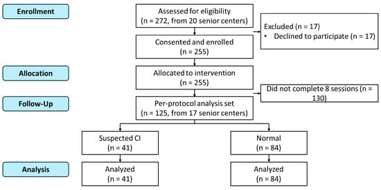
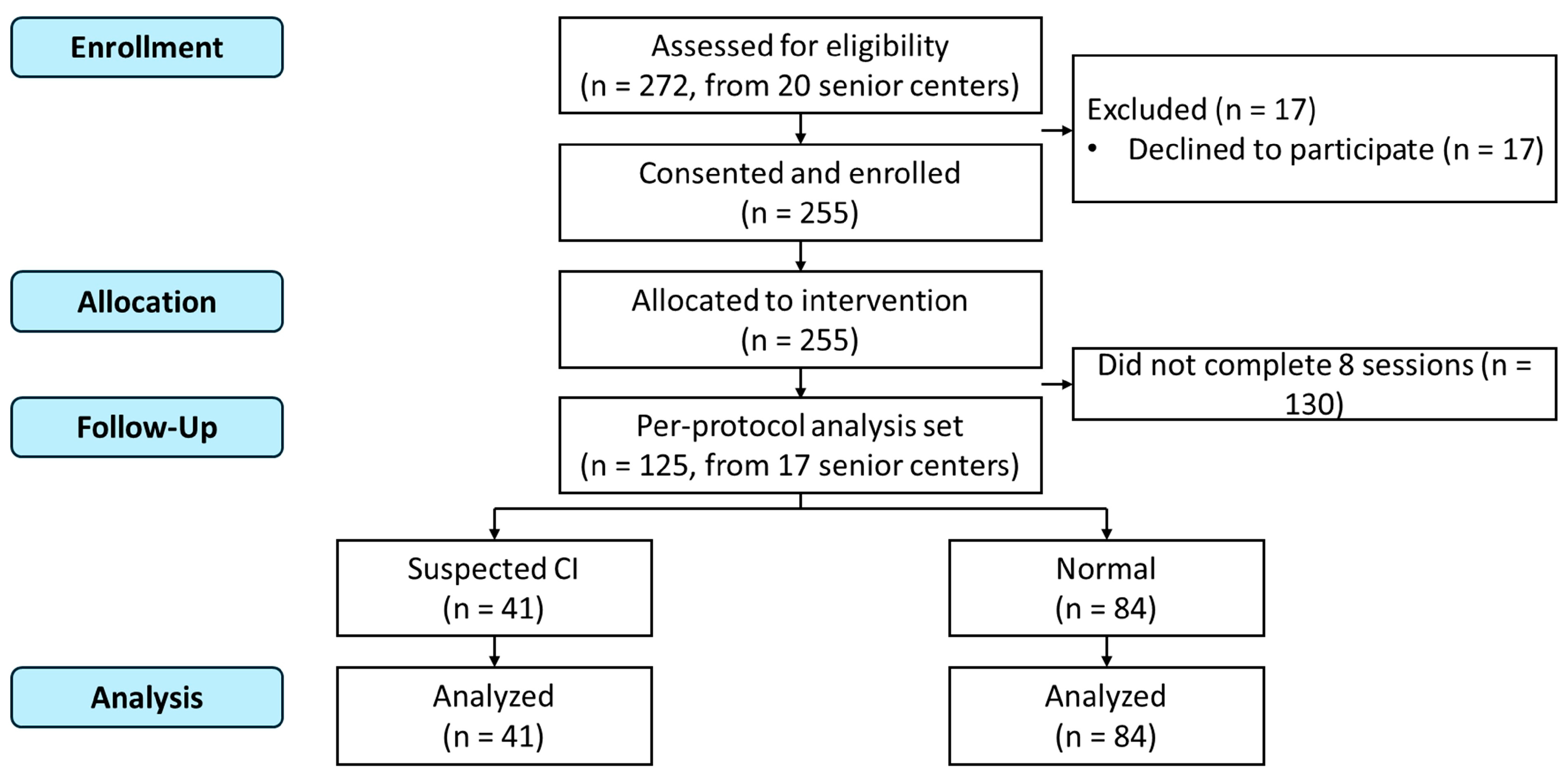
A total of 272 individuals from 20 senior centers were screened, of whom 255 provided written informed consent. After applying the above criteria, 125 participants from 17 senior centers constituted the final analytic sample (Figure 1).
Figure 1.
Participant flow diagram.
2.3. Intervention
The village forest experience program consisted of eight weekly sessions, each lasting two hours, conducted in village forests within walking distance of each participating senior center. Each session was led by a certified forest therapy instructor and comprised three phases: (1) a warm-up phase (30 min) including stretching and sensory awakening activities; (2) a forest activity phase (60 min) consisting of forest trail walking, barefoot walking, guided forest interpretation, and forest meditation; and (3) a group dialog phase (30 min) featuring facilitated peer conversations, tea, and group singing. When inclement weather prevented outdoor activities, sessions were adapted for indoor spaces. Data collection was performed at the beginning of Session 1 (baseline) and Session 8 (post-intervention), prior to the commencement of any program activity.
2.4. Outcome Measures
2.4.1. Cognitive Function
Cognitive function was assessed using the Cognitive Impairment Screening Test (CIST), a nationally standardized instrument for community-based cognitive screening developed by the National Institute of Dementia in the Republic of Korea [12]. The CIST evaluates orientation, memory, attention, visuospatial function, executive function, and language, yielding a total score of 0–30. Age- and education-adjusted cutoff scores are used to classify individuals as “Normal” or “Suspected cognitive impairment (CI).” The CIST was administered by trained evaluators at both time points.
2.4.2. Salivary Cortisol
Unstimulated whole saliva samples were collected into sterile conical tubes. Participants were instructed to refrain from eating, drinking (except water), and smoking for at least 30 min prior to collection. Samples were collected between 10:00 a.m. and 2:00 p.m., with collection times kept consistent within each center across both time points. Samples were transported on ice, centrifuged, and stored at −20 °C until analysis. Salivary cortisol concentrations (μg/dL) were determined by enzyme-linked immunosorbent assay (ELISA) at EONE Laboratories (Incheon, Republic of Korea).
2.4.3. Oral Bacteria
Oral microbial samples were collected using the Oralbiome CHECK kit (Docsmedi Co., Ltd., Goyang, Republic of Korea), which included gargle solution and dedicated collection tubes. Participants gargled vigorously for 30 s with the provided solution and then expectorated into the collection tube. Genomic DNA was extracted using the Bacteria Genomic DNA Isolation Kit (LaboPass, COSMO Genetech, Seoul, Republic of Korea) according to the manufacturer’s instructions. Quantitative real-time polymerase chain reaction (qPCR) was performed by DOCSMEDI (Seoul, Republic of Korea) to determine cycle threshold (Ct) values for total bacteria and six species of periodontal and cariogenic pathogens: Porphyromonas gingivalis (Pg), Treponema denticola (Td), Tannerella forsythia (Tf), Prevotella intermedia (Pi), Campylobacter rectus (Cr), Fusobacterium nucleatum (Fn), and Streptococcus mutans (Sm). Higher Ct values correspond to lower bacterial DNA concentrations; thus, an increase in Ct from pre- to post-intervention indicates a reduction in bacterial load. Samples with no amplification signal were excluded from species-specific analyses, resulting in variable sample sizes across species.
2.5. Statistical Analysis
Pre–post changes were analyzed using paired t-tests, and effect sizes were calculated as Cohen’s d. Changes in cognitive classification (Normal vs. Suspected CI) were evaluated using the McNemar test. Subgroup analyses were performed by baseline cognitive status, sex, and age group. All analyses were performed using R version 4.3.1 (R Foundation for Statistical Computing, Vienna, Austria), with a two-tailed p-value < 0.05 considered statistically significant. No adjustments for multiple comparisons were applied, as the analyses were exploratory. A formal sample size calculation was not performed a priori, as this was an exploratory study. The sample size was determined by the number of eligible participants from the 20 senior centers participating in the community forest welfare program.
3. Results
3.1. Participant Characteristics
The demographic and baseline characteristics of the 125 participants are presented in Table 1. The mean age was 82.2 ± 5.3 years (range: 68–97), with 109 females (87.2%) and 16 males (12.8%). The largest age group was 80–84 years (35.2%), followed by ≥85 years (32.8%). Mean years of education were 8.9 ± 4.4. At baseline, 84 participants (67.2%) were classified as “Normal” and 41 (32.8%) as “Suspected cognitive impairment” based on age- and education-adjusted CIST cutoffs.
Table 1.
Demographic and baseline characteristics of participants (N = 125).
3.2. Changes in Cognitive Function
Overall CIST scores improved significantly from 21.00 ± 6.33 to 22.22 ± 6.03 (+1.22, p = 0.003; Table 2). The improvement was driven by the suspected CI subgroup, which showed a mean increase of 3.44 points (p < 0.001, d = 0.66), whereas the normal subgroup showed no significant change (p = 0.749). By sex, significant improvement was observed among females (p = 0.002) but not males (p = 0.755). By age group, participants aged 80–84 years showed the most pronounced improvement (p = 0.009), while no significant changes were observed in the other age groups (Table 3).
Table 2.
Pre- and post-intervention comparison of outcome variables.
Table 3.
Changes in CIST scores by sex and age group.
Regarding cognitive classification changes (Table 4), 20 of 41 participants (48.8%) initially classified as suspected CI transitioned to normal after the intervention, while only three of 84 (3.6%) in the normal group declined to suspected CI. The McNemar test confirmed this asymmetry (χ2 = 11.13, p < 0.001).
Table 4.
Changes in cognitive classification before and after intervention.
3.3. Changes in Salivary Cortisol
Salivary cortisol decreased significantly from 0.12 ± 0.07 to 0.10 ± 0.06 μg/dL (p = 0.002; Table 2). When stratified by cognitive status (Table 5), the reduction was significant in the normal group (p = 0.004) but not in the suspected CI group (p = 0.173).
Table 5.
Pre- and post-intervention outcomes by baseline cognitive status.
3.4. Changes in Oral Bacteria
Significant increases in Ct values—reflecting reduced bacterial DNA concentrations, as higher Ct values correspond to lower bacterial loads—were observed for total bacteria (p = 0.026), P. gingivalis (p = 0.034), and T. forsythia (p = 0.003; Table 2). No significant changes were observed for the remaining species. When stratified by baseline cognitive status (Table 5), T. forsythia showed a notably larger reduction in the suspected CI group (+1.16, p = 0.006, d = 0.50) compared to the normal group (p = 0.083).
4. Discussion
This study simultaneously evaluated the effects of a community-based village forest experience program on cognitive function, salivary cortisol, and oral bacteria in a single cohort of older adults. Most previous forest therapy studies have assessed psychological indicators or stress biomarkers in isolation, and no study has incorporated a multidimensional assessment encompassing cognitive function and oral microbiota. Following the 8-week program, significant improvements in CIST scores, reductions in salivary cortisol, and decreases in key periodontal pathogens were observed.
The largest effect was observed in the suspected cognitive impairment (CI) group. Although CIST scores improved significantly across all participants, the suspected CI subgroup showed a mean increase more than twice that of the overall sample, with 48.8% transitioning to normal classification, whereas the normal group showed no significant change. This suggests that forest-based interventions may be associated with greater benefit in older adults at the early stages of cognitive decline. The observed associations should not be attributed solely to forest exposure per se, as the intervention was inherently multi-component in nature. Physical activity through forest trail walking and barefoot walking, cognitive stimulation through identifying and recalling the names of trees and plants, social interaction through peer dialog, and sensory engagement through guided forest meditation each represent potentially active components that may have independently or synergistically contributed to the observed outcomes. Previous studies have reported that physical activity in natural environments promotes cerebral blood flow and neuroplasticity [13,14], and evidence supports the role of social engagement and cognitive stimulation in maintaining cognitive reserve [15,16]. Park et al. (2024) administered a 20-week standardized anti-aging forest healing program to 33 older adults aged 70 years and older, recruited from dementia relief centers, and reported significant improvements in CIST total scores [17]. The present study confirmed comparable cognitive improvements with a shorter 8-week program, in everyday village forests accessible on foot from senior centers rather than in designated national healing forests, and in a sample of 125 participants. It should be noted, however, that the present study was not powered for subgroup analyses; these findings are therefore exploratory and should be interpreted with caution.
Significant improvement was observed in females but not in males; however, the male subsample comprised only 16 participants, limiting statistical power for this comparison. By age group, the greatest cognitive improvement was observed in the 80–84-year group. Korean dementia epidemiological data indicate that the prevalence of cognitive impairment and dementia increases from age 75, with age-specific prevalence doubling every 5–6 years [2,18]. In the present study, the proportion of suspected CI also increased with age; however, the 85-years-and-older group, which had the highest proportion of suspected CI, showed no significant improvement. This pattern suggests that participants aged 80–84 years may be at a stage where cognitive decline has begun but remains responsive to intervention. The absence of significant improvement in the ≥85 group may reflect a combination of factors beyond cognitive decline alone, including greater physical frailty [19] and reduced neuroplasticity associated with advanced age [20], both of which may have limited the effectiveness of an 8-week program in producing measurable cognitive change. Given the small sample sizes within individual age subgroups and the exploratory nature of these analyses, these age-related patterns warrant confirmation in adequately powered future studies.
The significant reduction in salivary cortisol is consistent with existing evidence for the stress-reducing effects of forest environments [4,5]. It should be noted that saliva samples were collected immediately before the start of each data collection session (Session 1 and Session 8), prior to any physical activity. The observed reduction in salivary cortisol therefore may reflect a change in resting-state cortisol levels following seven weeks of program participation, rather than an acute exercise-induced response. The cortisol reduction was significant only in the normal cognition group and not in the suspected CI group. One possible explanation for the absence of a significant cortisol reduction in the suspected CI subgroup is HPA axis dysregulation associated with early cognitive decline. Ouanes and Popp (2019) reviewed evidence linking elevated cortisol levels and altered HPA axis reactivity to increased risk of dementia [21]. Johar et al. (2015) further reported that older adults with cognitive impairment exhibited blunted diurnal cortisol patterns, characterized by lower morning and higher evening cortisol levels, compared to cognitively healthy individuals [22]. These findings suggest that HPA axis reactivity may be altered in the context of cognitive decline, which could attenuate the cortisol response to stress-reducing stimuli such as the present intervention.
The significant reductions in total bacterial load, P. gingivalis, and T. forsythia are, to our knowledge, the first such findings reported in a forest therapy study. These are pathogenic bacteria strongly associated with periodontal disease [23]. T. forsythia showed a larger reduction in the suspected CI group (d = 0.50) than in the normal group (d = 0.20), and this pattern occurred in parallel with the cognitive improvement observed in the same subgroup. Recent studies have proposed the oral–brain axis hypothesis, whereby periodontal pathogens reach the brain via the bloodstream and promote neuroinflammation [8,9]. The present study did not directly test this pathway; however, the concurrent reductions in cortisol and periodontal pathogens raise the possibility that stress reduction may have improved oral immune function, potentially contributing to decreased pathogenic bacterial load. These findings should be interpreted with caution, however, as data on participants’ oral hygiene practices, dietary habits, and dental care were not collected and may represent important unmeasured confounders. Future controlled studies incorporating mediation analysis are needed to directly examine the validity and directionality of this proposed pathway.
This study has several limitations. The single-arm design without a control group precludes causal inference, as the observed changes may reflect regression to the mean, practice effects on cognitive testing, or seasonal variations. Although saliva collection times were kept consistent within each center across both time points, collection times varied across centers. Given the well-established diurnal pattern of cortisol secretion, this inter-center variation may have introduced systematic differences in absolute cortisol levels between centers, representing a potential source of confounding that should be considered when interpreting the cortisol findings. The predominantly female sample (87.2%) limits the generalizability of sex-specific findings. The absence of data on participants’ concurrent activities, medications, and health behaviors represents potential unmeasured confounders. Adherence data were not collected, and the potential influence of variation in individual session attendance on the observed outcomes could not be assessed. Future studies should incorporate systematic attendance tracking to allow for dose–response analyses. Attrition from 272 to 125 participants, while typical in community-based elderly research, may have introduced selection bias. A comparison of available baseline characteristics (age, sex, and baseline CIST scores) between the final analytic sample and excluded participants revealed no significant differences (all p > 0.05), suggesting that attrition was unlikely to have introduced systematic selection bias on these measures. No a priori sample size calculation was performed, as this was an exploratory study with a sample size determined by program enrollment. The statistical power available for subgroup analyses was therefore limited, and subgroup findings should be interpreted accordingly. Additionally, CIST evaluators were not blinded to the assessment time point, which may have introduced measurement bias. Despite these limitations, this study is the first to explore the multidimensional health effects of forest therapy by simultaneously measuring cognitive function, stress biomarkers, and oral microbiota in a single cohort. The multi-site data from 125 participants across 17 senior centers, obtained in village forests accessible without dedicated transportation, provide initial evidence for the integration of community-based forest programs into dementia prevention strategies.
5. Conclusions
An 8-week village forest experience program was associated with improved cognitive function, reduced salivary cortisol, and decreased periodontal pathogenic bacteria in community-dwelling older adults. The greatest cognitive benefit was observed in the suspected cognitive impairment subgroup. As a single-arm exploratory study, these results require confirmation through randomized controlled trials with extended follow-up and mediation analyses to elucidate the mechanisms underlying these multidimensional effects.
Author Contributions
Conceptualization, M.-Y.C. and H.-S.K.; methodology, M.-Y.C. and M.-H.N.; software, M.-Y.C.; validation, M.-Y.C., J.-H.E. and J.-W.K.; formal analysis, M.-Y.C.; investigation, M.-Y.C., J.-H.E., J.-W.K. and Y.-W.K.; resources, S.-J.Y., J.H. and H.-S.K.; data curation, M.-Y.C. and J.-H.E.; writing—original draft preparation, M.-Y.C.; writing—review and editing, M.-Y.C., H.-S.K., M.-H.N. and J.H.; visualization, M.-Y.C.; supervision, M.-H.N. and H.-S.K.; project administration, H.-S.K.; funding acquisition, H.-S.K. All authors have read and agreed to the published version of the manuscript.
Funding
This research was funded by the Korea Forest Welfare Institute (2025 Green Fund Forest Experience and Education Project) and the Korea Disease Control and Prevention Agency (grant number 2024ER050701). The APC was funded by the Apple Tree Medical Foundation.
Institutional Review Board Statement
The study was conducted in accordance with the Declaration of Helsinki, and approved by the Institutional Review Board of Apple Tree Dental Hospital (protocol code ATDH-2025-0003 and date of approval 4 February 2025).
Informed Consent Statement
Informed consent was obtained from all subjects involved in the study.
Data Availability Statement
The data presented in this study are available on request from the corresponding author. The data are not publicly available due to ethical restrictions, as the dataset contains sensitive health information of elderly participants, including cognitive assessment scores, salivary cortisol levels, and oral microbial profiles, collected under IRB approval with informed consent specifying that individual-level data would remain confidential.
Acknowledgments
The authors would like to thank all elderly participants for their dedication throughout the 8-week program. We are grateful to the staff of the 20 participating senior centers for their logistical support and coordination. We also thank the Korea Forest Welfare Institute for supporting the village forest experience and education program.
Conflicts of Interest
Author Hye-Sung Kim is the CEO of DOCSmedi Co., Ltd., and authors Seung-Jo Yang and Jiyoung Hwang are employees of DOCSmedi Co., Ltd. The Oralbiome CHECK kit used for oral microbial sample collection in this study is a product of DOCSmedi Co., Ltd. Authors Mu-Yeol Cho, Je-Hyun Eom, Ji-Won Kim, Yun-Woo Kim, Mi-Hwa No and Hye-Sung Kim are affiliated with Apple Tree Medical Foundation. The funders had no role in the design of the study; in the collection, analyses, or interpretation of data; in the writing of the manuscript; or in the decision to publish the results.
Abbreviations
The following abbreviations are used in this manuscript:
| CIST | Cognitive Impairment Screening Test |
| CI | Cognitive Impairment |
| Ct | Cycle Threshold |
| ELISA | Enzyme-Linked Immunosorbent Assay |
| qPCR | Quantitative Polymerase Chain Reaction |
| IRB | Institutional Review Board |
| CRIS | Clinical Research Information Service |
References
- Eggleston, K.; Kim, D. Dementia care in a rapidly aging society. Alzheimers Dement. 2025, 21, e70268. [Google Scholar] [CrossRef] [Scilit] [PubMed]
- Kim, Y.J.; Han, J.W.; So, Y.S.; Seo, J.Y.; Kim, K.Y.; Kim, K.W. Prevalence and trends of dementia in Korea: A systematic review and meta-analysis. J. Korean Med. Sci. 2014, 29, 903–912. [Google Scholar] [CrossRef] [Scilit] [PubMed]
- Livingston, G.; Huntley, J.; Sommerlad, A.; Ames, D.; Ballard, C.; Banerjee, S.; Brayne, C.; Burns, A.; Cohen-Mansfield, J.; Cooper, C.; et al. Dementia prevention, intervention, and care: 2020 report of the Lancet Commission. Lancet 2020, 396, 413–446. [Google Scholar] [CrossRef] [Scilit]
- Antonelli, M.; Donelli, D.; Barbieri, G.; Valussi, M.; Maggini, V.; Firenzuoli, F. Forest Volatile Organic Compounds and Their Effects on Human Health: A State-of-the-Art Review. Int. J. Environ. Res. Public Health 2020, 17, 6506. [Google Scholar] [CrossRef] [Scilit]
- Li, Q. Effect of forest bathing trips on human immune function. Environ. Health Prev. Med. 2010, 15, 9–17. [Google Scholar] [CrossRef] [Scilit] [PubMed]
- Lee, I.; Choi, H.; Bang, K.S.; Kim, S.; Song, M.; Lee, B. Effects of Forest Therapy on Depressive Symptoms among Adults: A Systematic Review. Int. J. Environ. Res. Public Health 2017, 14, 321. [Google Scholar] [CrossRef] [Scilit]
- Oh, K.H.; Shin, W.S.; Khil, T.G.; Kim, D.J. Six-Step Model of Nature-Based Therapy Process. Int. J. Environ. Res. Public Health 2020, 17, 685. [Google Scholar] [CrossRef] [Scilit]
- Dominy, S.S.; Lynch, C.; Ermini, F.; Benedyk, M.; Marczyk, A.; Konradi, A.; Nguyen, M.; Haditsch, U.; Raha, D.; Griffin, C.; et al. Porphyromonas gingivalis in Alzheimer’s disease brains: Evidence for disease causation and treatment with small-molecule inhibitors. Sci. Adv. 2019, 5, eaau3333. [Google Scholar] [CrossRef] [Scilit]
- Singhrao, S.K.; Harding, A.; Poole, S.; Kesavalu, L.; Crean, S. Porphyromonas gingivalis Periodontal Infection and Its Putative Links with Alzheimer’s Disease. Mediators Inflamm. 2015, 2015, 137357. [Google Scholar] [CrossRef] [Scilit]
- Duran-Pinedo, A.E.; Solbiati, J.; Frias-Lopez, J. The effect of the stress hormone cortisol on the metatranscriptome of the oral microbiome. npj Biofilms Microbiomes 2018, 4, 25. [Google Scholar] [CrossRef] [Scilit]
- Marcotte, H.; Lavoie, M.C. Oral microbial ecology and the role of salivary immunoglobulin A. Microbiol. Mol. Biol. Rev. 1998, 62, 71–109. [Google Scholar] [CrossRef] [Scilit]
- Ministry of Health and Welfare of South Korea. Cognitive Impairment Screening Test (CIST) Manual; National Institute of Dementia: Seoul, Republic of Korea, 2021. [Google Scholar]
- Erickson, K.I.; Voss, M.W.; Prakash, R.S.; Basak, C.; Szabo, A.; Chaddock, L.; Kim, J.S.; Heo, S.; Alves, H.; White, S.M.; et al. Exercise training increases size of hippocampus and improves memory. Proc. Natl. Acad. Sci. USA 2011, 108, 3017–3022. [Google Scholar] [CrossRef] [Scilit]
- Hillman, C.H.; Erickson, K.I.; Kramer, A.F. Be smart, exercise your heart: Exercise effects on brain and cognition. Nat. Rev. Neurosci. 2008, 9, 58–65. [Google Scholar] [CrossRef] [Scilit] [PubMed]
- Stern, Y. Cognitive reserve in ageing and Alzheimer’s disease. Lancet Neurol. 2012, 11, 1006–1012. [Google Scholar] [CrossRef] [Scilit] [PubMed]
- Fratiglioni, L.; Paillard-Borg, S.; Winblad, B. An active and socially integrated lifestyle in late life might protect against dementia. Lancet Neurol. 2004, 3, 343–353. [Google Scholar] [CrossRef] [Scilit] [PubMed]
- Park, J.; Wang, S.M.; Kang, D.W.; Lee, B.; Choi, H. Effect of Anti-Aging Standard Forest Healing Program with Multiple Visits to a Forest Facility on Cognition in Older Age Patients. Dement. Neurocogn Disord. 2024, 23, 44–53. [Google Scholar] [CrossRef] [Scilit]
- Ministry of Health and Welfare of South Korea. 2023 Dementia Epidemiology Survey Results; National Institute of Dementia: Seoul, Republic of Korea, 2025. [Google Scholar]
- Ma, L.; Chan, P. Understanding the physiological links between physical frailty and cognitive decline. Aging Dis. 2020, 11, 405. [Google Scholar] [CrossRef] [Scilit]
- Park, D.C.; Bischof, G.N. The aging mind: Neuroplasticity in response to cognitive training. Dialogues Clin. Neurosci. 2013, 15, 109–119. [Google Scholar] [CrossRef] [Scilit]
- Ouanes, S.; Popp, J. High Cortisol and the Risk of Dementia and Alzheimer’s Disease: A Review of the Literature. Front. Aging Neurosci. 2019, 11, 43. [Google Scholar] [CrossRef] [Scilit]
- Johar, H.; Emeny, R.T.; Bidlingmaier, M.; Lacruz, M.E.; Reincke, M.; Peters, A.; Heier, M.; Ladwig, K.H. Lower morning to evening cortisol ratio is associated with cognitive impairment in men but not women: An analysis of 733 older subjects of the cross-sectional KORA-Age study. Psychoneuroendocrinology 2015, 51, 296–306. [Google Scholar] [CrossRef] [Scilit]
- Socransky, S.S.; Haffajee, A.D.; Cugini, M.A.; Smith, C.K.J.R.; Kent, R.L., Jr. Microbial complexes in subgingival plaque. J. Clin. Periodontol. 1998, 25, 134–144. [Google Scholar] [CrossRef] [Scilit] [PubMed]

Disclaimer/Publisher’s Note: The statements, opinions and data contained in all publications are solely those of the individual author(s) and contributor(s) and not of MDPI and/or the editor(s). MDPI and/or the editor(s) disclaim responsibility for any injury to people or property resulting from any ideas, methods, instructions or products referred to in the content. |
© 2026 by the authors. Licensee MDPI, Basel, Switzerland. This article is an open access article distributed under the terms and conditions of the Creative Commons Attribution (CC BY) license.