


Abstract
Background: Assault series focus on the mandible; midface fractures, dental injuries, teeth in the fracture line and late presentation are rarely analysed in the same cohort. We aimed to characterise and compare injury patterns and outcomes in assault-related facial fractures. Methods: Retrospective analysis of 607 patients with maxillofacial fractures caused by interpersonal violence, treated between 2001 and 2011 at one centre in north-western Romania. Results: Isolated mandibular, isolated midface and combined fractures accounted for 72.2%, 22.7% and 4.9% (one record unclassified). Midface fractures involved the zygomatic complex in 63.7% and the nasal bones in 29.2%. Patients with midface fractures were older (median 28.5 vs. 25 years, p = 0.001) and had more haematomas, lacerations and abrasions (all p < 0.001). Teeth lay in the fracture line in 78.0% of 468 mandibular fractures and were extracted in 59.5%. Osteitis was recorded after treatment in 13 patients (2.1%), all with teeth in the line (p = 0.08); in 9, an infection was already documented at treatment. No difference in osteitis was detected between extraction and retention (3.7% vs. 4.7%, p = 0.76) in an exploratory comparison subject to confounding by indication. Separately, 28 patients (4.6%) presented with established osteitis. Conclusions: Midface fractures affect older patients and carry far more soft-tissue injury than mandibular fractures. Osteitis was observed only in fractures with teeth in the line, and, in most affected patients, an infection was already documented at treatment. The small number of events and confounding by indication preclude any conclusion on extraction versus retention. Late presentation identifies a group needing structured follow-up.
1. Introduction
Interpersonal violence has become the leading cause of maxillofacial fractures in many European centres, overtaking road traffic accidents as seatbelt and helmet legislations took effect [1,2,3,4]. In the European Maxillofacial Trauma (EURMAT) prospective collaboration, assault accounted for 39% of all facial fractures, with the mandible and the zygomaticomaxillary complex as the most frequently involved sites [2,5]. Single-centre series from Turin (711 victims) and Ancona (224 victims) confirm the same profile: young men, a punch as the mechanism [6,7], a mandibular fracture in 34 to 42% of cases, and the zygoma, orbit and nasal bones sharing the remainder [8,9].
Most assault series, however, are written from the perspective of the mandible. The mandibular angle, the role of the third molar [10], and the choice between closed reduction and osteosynthesis [11] dominate the literature, while three questions that concern the whole face receive less attention. First, how do midface fractures differ from mandibular fractures in the same violent mechanism, in terms of patient profile, soft-tissue injury and treatment? Second, how often are teeth injured, and how often do they lie in the fracture line, and does their extraction protect against infection? Traumatic dental injuries have well-defined diagnostic and therapeutic pathways of their own, codified in the International Association of Dental Traumatology guidelines: uncomplicated crown fractures are restored by reattachment of the fragment or direct composite build-up; complicated crown and root fractures require pulp management before restoration; luxated teeth are repositioned and splinted; and avulsed permanent teeth are replanted as early as possible, with scheduled clinical and radiographic follow-up to detect pulp necrosis and root resorption [12,13,14]. In a maxillofacial trauma service, these injuries are easily overshadowed by the fracture itself and may neither be documented nor treated in time. Meta-analyses of angle fractures show no benefit of removing a tooth from the fracture line [15,16], yet extraction remains routine in many centres. Third, how many assault victims reach the maxillofacial surgeon late, with an already-infected fracture, and what characterises them? Late presentation is a marker of barriers to care that is seldom quantified.
These questions matter beyond the trauma service. From the standpoint of plastic and reconstructive surgery, the facial fracture is the beginning of a pathway that may end, months or years later, in secondary reconstruction: a zygoma that has healed displaced, with malar flattening, enophthalmos or diplopia; a nose with a deviated bony pyramid; a mandible with malunion and malocclusion; or a facial laceration that has become a conspicuous scar. Secondary correction of these deformities is consistently described as more difficult than primary repair, because the fragments are no longer mobile, the anatomical landmarks are lost, and osteotomies, grafts and virtual planning are needed to recover form and function [17,18]. The deformities arise most often from fractures that were severely comminuted, treated late, treated inadequately or not treated at all [18]. Assault victims combine several of these conditions: they are young, their midface fractures are frequently displaced, they are often treated by closed methods in resource-limited settings, and they are prone to delayed presentation and loss to follow-up. Describing the injury pattern, treatment and late presentations of this population is therefore a way of describing the future workload of reconstructive surgery, and of identifying where earlier referral could prevent it.
Management of each of these injuries is guided by established principles. Displaced fractures of the zygomaticomaxillary complex are reduced and, when unstable after reduction or comminuted at the buttresses, fixed by internal fixation, with exploration and reconstruction of the orbital floor when the defect or the ocular findings require it [19,20]; nasal bone fractures are reduced closed within the first two weeks; mandibular fractures are treated by closed reduction with maxillomandibular fixation or by open reduction and internal fixation, with no randomised evidence that either approach is superior for all fracture types [11]; and traumatic dental injuries follow the International Association of Dental Traumatology guidelines summarised above [12,13]. What is missing is not guidance for each injury taken separately but a description of how these injuries combine in the assault victim, who is typically young, often poorly educated, treated under resource constraints and difficult to follow up, and in whom a tooth in the fracture line, an unrecognised dental injury or a displaced zygoma treated closed each carries a specific risk of a late complication. Interpersonal violence was selected as the research focus because it is the leading cause of facial fractures in our region [21], because it allows the description of injury patterns within a shared broad aetiological category, and because its victims are the group most exposed to loss to follow-up. The specific mechanism and energy of each assault were, however, not consistently recorded, and residual heterogeneity in injury mechanisms cannot be excluded. We hypothesised that, within a single assault cohort, (i) midface and mandibular fractures would differ in patient profile and soft-tissue injury, (ii) infection of the fracture site would be concentrated in mandibular fractures with a tooth in the line, and (iii) a measurable proportion of patients would present late with an already-infected or malunited fracture.
In north-western Romania, the burden is higher still: in the 10-year series of 1007 maxillofacial fractures treated in our centre, interpersonal violence was the cause in 59%, in both the mandibular and the midface regions, and in rural as well as urban patients [21]. The mandibular fracture pattern of these assault victims has been reported previously: in 478 patients with mandibular fractures through interpersonal violence over 10 years, the angle was the most frequent site, double fractures predominated, most fractures were treated by closed reduction and maxillomandibular fixation, and the complication rate was low [22]. That report deliberately excluded patients without a mandibular fracture and did not analyse midface injuries, dental trauma or the management of teeth in the fracture line.
The present study extends the analysis to the complete cohort of 607 assault victims with any facial fracture treated in the same centre. The primary aim was to characterise and compare the pattern of maxillofacial fractures caused by interpersonal violence between patients with isolated mandibular, isolated midface and combined fractures, in terms of patient profile, soft-tissue injury, fracture sites and treatment. The secondary aims were (1) to describe the burden of dental and extrafacial injuries, (2) to describe the presence and management of teeth in the fracture line and their relation to osteitis recorded after treatment, (3) to characterise the patients who presented late with an already-infected fracture, and (4) to draw from these observations the clinical implications for follow-up and for the recognition of patients who may require secondary reconstruction.
2. Materials and Methods
2.1. Study Design, Setting and Relation to Previous Work
We performed a retrospective observational study of all patients treated for maxillofacial fractures caused by interpersonal violence by the Department of Oral and Maxillofacial Surgery of the University of Oradea, whose clinical base is the oral and maxillofacial surgery unit of Medicover Pelican Hospital, Oradea, Romania, over a 10-year period (2001 to 2011), the same period as the previous reports from this database [21,22]. The hospital serves Bihor County and neighbouring counties in north-western Romania. The overall epidemiology of the 1007 maxillofacial fractures of all aetiologies treated in the department over the same 10 years has been published [21], and the mandibular subset of the assault cohort (478 patients with at least one mandibular fracture line) has been described with respect to mandibular fracture pattern, soft-tissue lesions and treatment [22]. The present analysis uses the complete assault cohort, including 139 patients without a recorded mandibular fracture, and addresses variables not reported before: midface fracture sites, dental trauma, extrafacial injuries, the presence and management of teeth in the fracture line, osteitis at first presentation, and the comparison between fracture groups. Demographic and mandibular-pattern data overlapping with the previous report are given only in summary form to allow group comparisons. The database was constituted under the approval of the Ethics Committee of the University of Oradea (No. 35698/19.02.2018 [21]; No. 28765/11.02.2019 [22]), and the present analysis was approved by the Ethics Committee for Scientific Research of Pelican Hospital, Oradea (No. 1540, 20 October 2026). All patients, or their legal guardians for minors, signed at admission an informed consent for the use of their anonymised medical data for research and publication, as described for the previous reports [21,22]. The study was conducted in accordance with the Declaration of Helsinki.
2.2. Patients and Groups
Inclusion criteria were a mechanism of injury recorded as interpersonal violence and at least one fracture of the mandible or of the midface confirmed clinically and radiologically. Patients with facial fractures of other aetiology were excluded. Two patients in whom the assault produced a fracture on a mandible with pre-existing bone pathology were retained. The database extract contained 618 records; 11 consecutive records at the end of the file were exact duplicates, including free-text fields, of 11 earlier records and were removed, leaving 607 patients. The present analysis thus included 607 unique records after the removal of 11 exact duplicates from the available extract. Previous publications based on the same historical database reported different sample sizes (598 assault victims among 1007 fractures [21]; 478 patients with mandibular fractures [22]) and applied additional eligibility and completeness criteria. Because individual records from those analyses could not be re-identified in the anonymised extract, a patient-level reconciliation of the differences was not possible; the present cohort and its selection procedure are therefore reported independently and transparently. The 607 patients were classified into three groups: isolated mandibular fracture, isolated midface fracture (any fracture of the maxilla, zygomatic complex, nasal bones, orbital floor or maxillary alveolar process without a mandibular fracture) and combined mandibular and midface fractures.
2.3. Variables
Data were extracted from the clinical records into a structured database with a predefined coding scheme. The variables used in this analysis were age, sex, residence (urban or rural), associated systemic disease, soft-tissue injuries (haematoma, laceration, abrasion), dental trauma (crown fracture, root fracture, luxation, avulsion), other associated injuries recorded as free text (ocular and periorbital, sinus and nasal, cranial, thoracic, temporomandibular joint), mandibular fracture site and number of lines, midface fracture site (Le Fort I, II, III, zygomatic complex, nasal bones, alveolar process, orbital floor, anterior wall of the maxillary sinus), displacement, relation of the fracture to the oral cavity, presence of teeth in the fracture line, management of these teeth (extraction, apicoectomy, incision and drainage, conservative retention of vital teeth), type of fracture treatment (closed reduction with maxillomandibular fixation, plate osteosynthesis, wire osteosynthesis, combined), clinical course and complications. Free-text entries were categorised by two of the authors; ocular and periorbital injuries comprised chemosis, subconjunctival haemorrhage, eyelid ptosis, corneal erosion, diplopia and retinal detachment.
Osteitis of the fracture site was defined as clinically diagnosed infection of the bone at the fracture (persistent pain and swelling with purulent discharge or a fistula at the fracture site, exposed or sequestrating bone, or radiological bone lysis along the fracture line), as distinct from a localised odontogenic abscess (a pericoronal or periapical collection around a tooth, drained and resolving without signs of bone involvement) and from superficial wound infection. Osteitis recorded after treatment was documented in the dedicated complication field during institutional follow-up, which comprised review at removal of maxillomandibular fixation (usually 4 to 6 weeks) and at later visits when the patient attended; complications treated at other institutions or in patients who did not return were not captured, so the recorded rates are the lower bounds. Incision and drainage of an abscess around a tooth in the line at the time of treatment was recorded as management of that tooth, not as a complication. Because an abscess drained at treatment means that infection preceded the intervention, osteitis recorded as a complication in such patients, or in patients with osteitis already noted at the first consultation, was separated from osteitis without prior documented infection, referred to below as osteitis without documented pre-existing infection for brevity; both counts are reported, and the latter is presented as a descriptive proportion of the cohort rather than as an incidence estimate, because the population free of infection at baseline and the duration of follow-up could not be established precisely. Osteitis at presentation was defined as osteitis of the fracture site documented at the first consultation in our department, in patients referred late, after non-attendance for follow-up or after failed initial management elsewhere; these patients were analysed as a separate group. Displacement, relation to the oral cavity and level of education were coded as in the previous reports from this database, in which displacement of the fragments was recorded in 84% of all 1007 fractures and in 89% of assault-related mandibular fractures [22,23]; where two fracture lines in the same patient differed in displacement, the record was classified with the undisplaced lines. Level of education was grouped as no formal education, primary, middle school, high school and university.
2.4. Statistical Analysis
Continuous variables are given as mean ± standard deviation and median with interquartile range (IQR). Age was compared between the three fracture groups with the Kruskal–Wallis test and between two groups with the Mann–Whitney U test. Categorical variables were compared with chi-square test or, when any expected count was below 5, with Fisher’s exact test (2 × 2 tables) or the Fisher–Freeman–Halton exact test (larger tables; Monte Carlo estimate from 200,000 tables with fixed margins). Pairwise age comparisons after a significant Kruskal–Wallis test were Bonferroni-adjusted for three comparisons, and the gradient of dental trauma across the three fracture groups was tested with the Cochran–Armitage test for trend. Odds ratios (OR) with 95% confidence intervals (CI) are reported for 2 × 2 comparisons; where one cell contained no events, the OR is not estimable, and the absolute risk difference with its Newcombe 95% CI is given instead. Principal proportions are given with Wilson 95% CI. Because only 13 cases of osteitis were recorded after treatment, including four without documented pre-existing infection, no multivariable model was fitted; the associations are therefore presented as exploratory univariable analyses. Where expected counts in a multi-group table were below 5, pairwise Fisher’s exact tests are reported alongside the global test. The univariable associations with osteitis (Section 3.5) are ten exploratory comparisons; their p values are not adjusted for multiplicity. Variables with missing values (clinical course in 8 patients, management of the tooth in the line in 41) were analysed as recorded, with the missing category shown; no imputation was performed. All associations with osteitis were computed within the 468 patients with a mandibular fracture; the unit of analysis throughout is the patient, and fracture-line counts are stated explicitly where used. A two-sided p < 0.05 was considered significant. Analyses were performed in Python 3 (pandas 2, SciPy 1, statsmodels 0.14). The study is reported in accordance with the STROBE statement for cohort studies [24].
Several measures were taken to limit the data-quality problems inherent to a retrospective database. Eleven duplicated records were identified and removed; the coding of displacement, relation to the oral cavity and education was reconciled with the published figures from the same database before analysis; free-text entries were categorised by two authors; the unit of analysis (patient versus fracture line) was checked for every table; and the chronology of each infection was reconstructed from the management and free-text fields and is presented case by case in Supplementary Table S1, so that readers can judge the classification themselves. The original clinical records of the 13 patients with osteitis recorded after treatment were reviewed by the senior author (A.-P.T.), who confirmed the classification of each case as infection documented at treatment or osteitis without documented pre-existing infection.
3. Results
3.1. Cohort and Fracture Groups
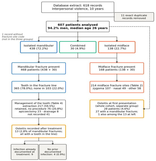
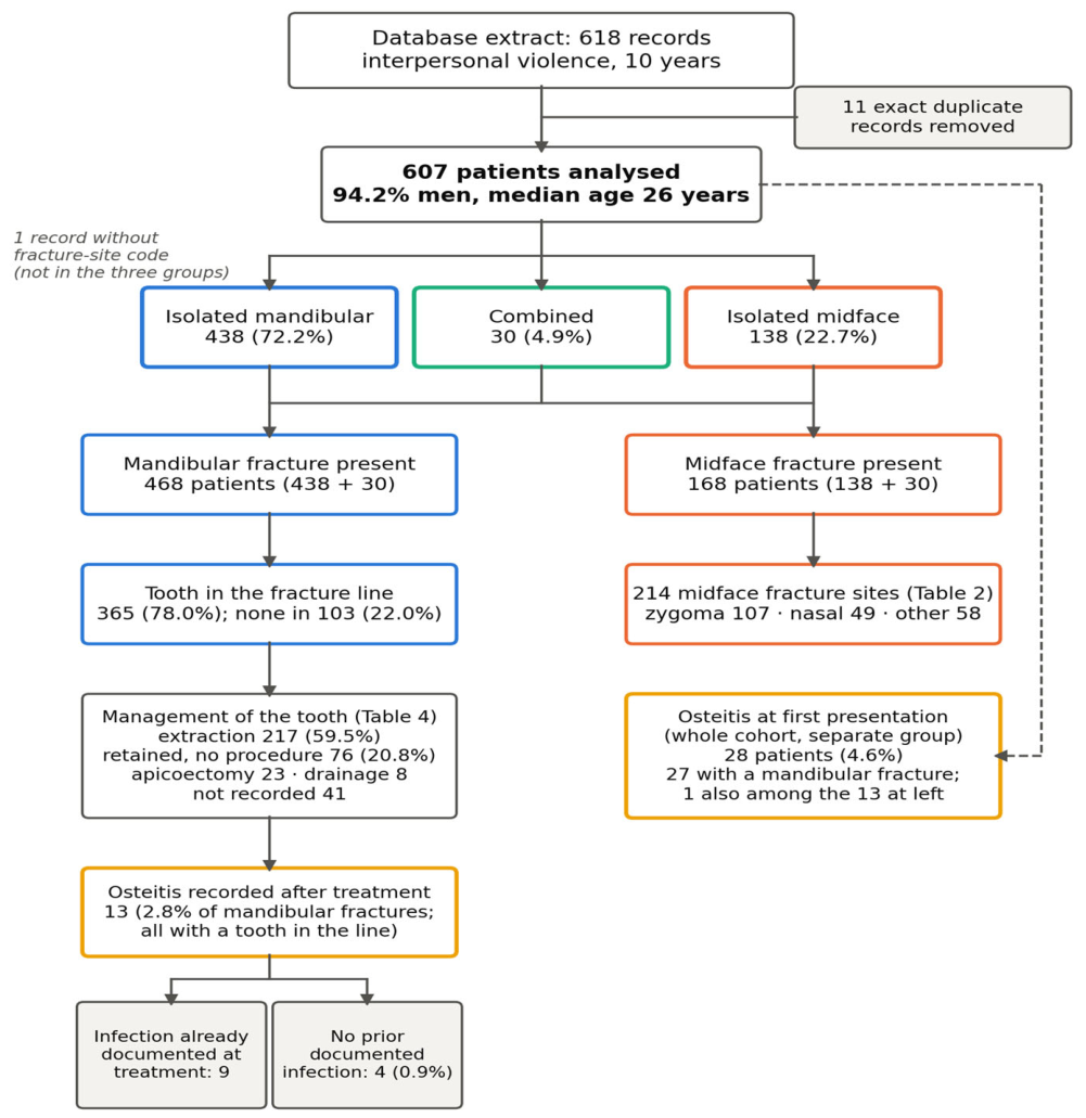
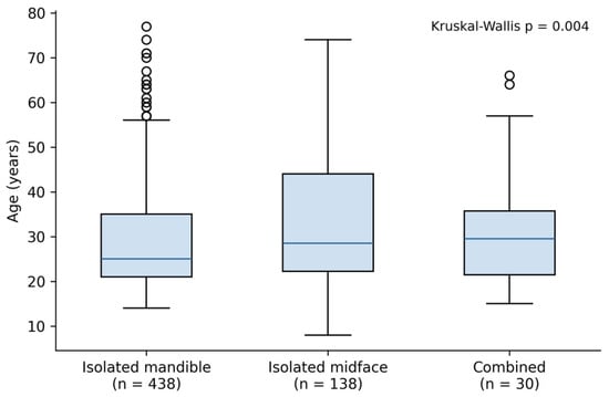
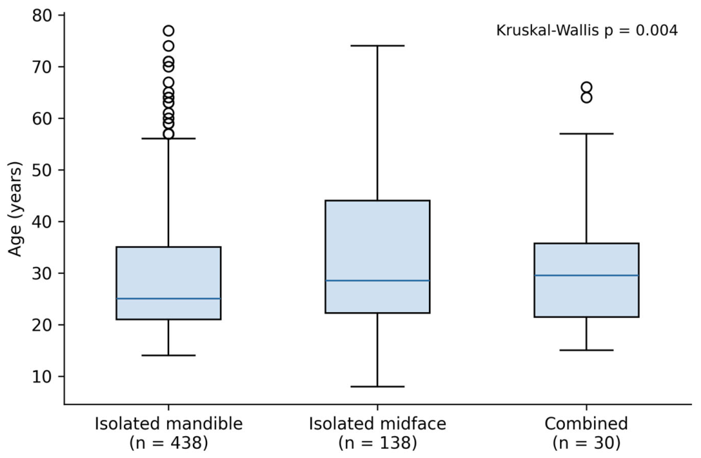
After the removal of 11 duplicated records, the cohort comprised 607 patients (Figure 1), 572 men (94.2%) and 35 women (5.8%), with a median age of 26 years (IQR 21 to 37, range 8 to 77); 55.8% lived in urban areas, 44.3% had no formal education and 9.9% had a systemic comorbidity. An isolated mandibular fracture was present in 438 patients (72.2%), an isolated midface fracture in 138 (22.7%) and combined mandibular and midface fractures in 30 (4.9%); one record carried the diagnosis of an assault-related facial fracture in the diagnosis field, on which eligibility was based, but the fracture-site code was missing in the extract and could not be recovered from the anonymised data; it is included in the cohort totals and excluded from the group comparisons, so the three groups sum to 606. Age differed between the groups (Kruskal–Wallis p = 0.004): patients with isolated midface fractures were older than those with isolated mandibular fractures (median 28.5 vs. 25 years, p = 0.001; Bonferroni-adjusted p = 0.003), and patients with combined fractures had a median age of 29.5 years. No differences in sex, residence or comorbidity were detected between groups (Table 1, Figure 2).
Figure 1.
Study flow and cohort structure. Flow of the 618 database records to the 607 analysed patients, their classification into isolated mandibular, isolated midface and combined fractures, the presence and management of teeth in the fracture line among the 468 patients with a mandibular fracture, and the two osteitis groups: osteitis recorded after treatment (13 patients, of whom 9 had an infection already documented at the time of treatment) and osteitis at first presentation (28 patients).
Table 1.
Characteristics of the three fracture groups. The three groups comprise 606 patients; one patient without a fracture-site code (Section 3.1) is included in the cohort totals (n = 607) elsewhere, but not in this table or in Figure 2 and Figure 3.
Figure 2.
Age distribution by fracture group. Boxes show the median and interquartile range, whiskers extend to the most extreme values within 1.5 interquartile ranges of the box, and individual points beyond the whiskers are outliers.
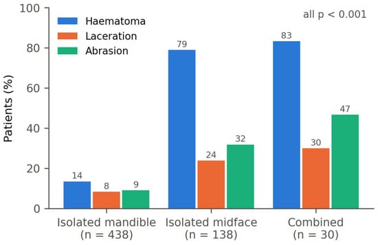
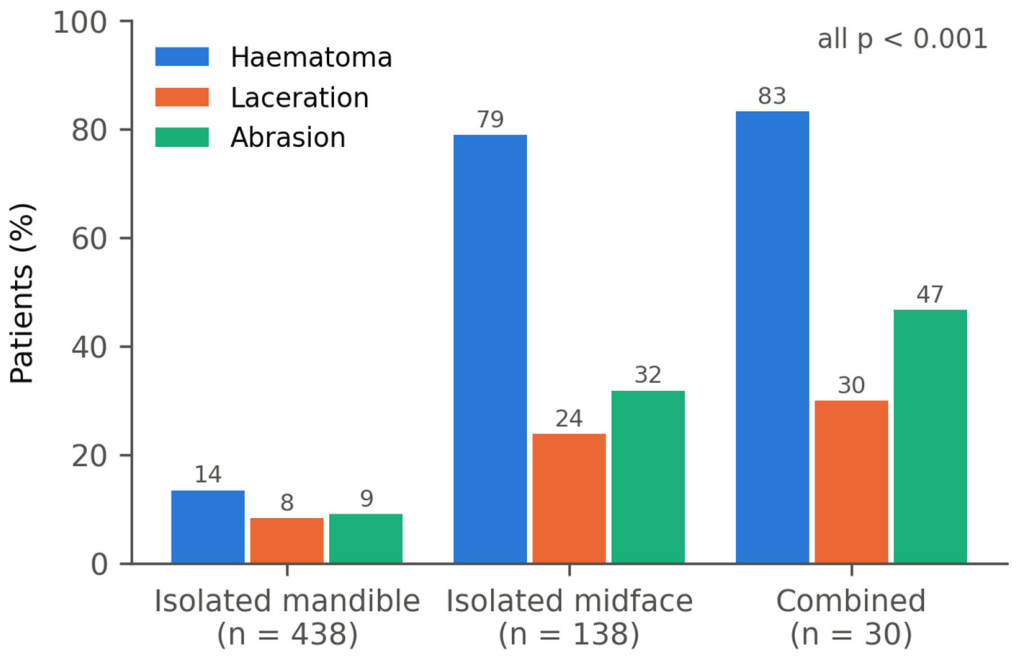
Soft-tissue injuries separated the groups sharply. Haematoma accompanied 13.5% of isolated mandibular fractures but 79.0% of isolated midface and 83.3% of combined fractures; lacerations 8.4% vs. 23.9% and 30.0%; abrasions 9.1% vs. 31.9% and 46.7% (all p < 0.001; Figure 3). Relative to an isolated mandibular fracture, an isolated midface fracture carried 24 times the odds of a haematoma (OR 24.1, 95% CI 14.8 to 39.5), 3.4 times the odds of a laceration (95% CI 2.0 to 5.7) and 4.7 times the odds of an abrasion (95% CI 2.9 to 7.6). Displacement of fragments was recorded in 91.1% of isolated mandibular, 76.8% of isolated midface and 60.0% of combined fractures (p < 0.001), with the lower figure in the combined group reflecting mixed records, with one displaced and one undisplaced line. Closed (orthopaedic) treatment predominated in all groups (82.4%, 89.1% and 73.3%; with maxillomandibular fixation for mandibular fractures, by closed reduction alone for most isolated nasal and zygomatic fractures); plate osteosynthesis alone was used in 2.3%, 4.3% and 0%, and combined techniques in 8.9%, 3.6% and 23.3%. No patient with an isolated midface fracture had an unfavourable course or any event recorded in the complication field after treatment (0 of 138, 95% CI 0 to 2.7%), against 13 of 438 (3.0%) with isolated mandibular (Fisher p = 0.045) and 2 of 30 (6.7%) with combined fractures (p = 0.031).
Figure 3.
Soft-tissue injuries by fracture group.
3.2. Midface Fractures
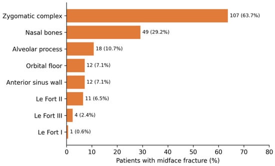
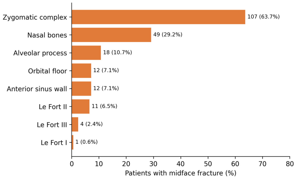
The 168 patients with a midface fracture had 214 fracture sites. The zygomatic complex was involved in 107 patients (63.7%) and the nasal bones in 49 (29.2%); fractures of the alveolar process (18, 10.7%), orbital floor (12, 7.1%), anterior wall of the maxillary sinus (12, 7.1%) and Le Fort II (11, 6.5%) were less frequent, and Le Fort III (4) and Le Fort I (1) fractures were exceptional, so that only 15 patients (one patient having fractures at two Le Fort levels) (2.5% of the cohort) had any Le Fort fracture (Table 2, Figure 4). Zygomatic fractures were displaced in 81 of 107 (75.7%) and were treated by closed reduction in 92 (86.0%), with plates in 6 and by combined techniques in 6; a concomitant mandibular fracture was present in 16 (15.0%). Nasal bone fractures were displaced in 31 of 49 (63.3%) and treated by closed reduction in 41 (83.7%). Le Fort fractures were managed by closed reduction and maxillomandibular fixation in 10 of 15, and by combined techniques in 5. Ocular and periorbital injuries were documented in 15 patients, 11 of them with a zygomatic fracture and 4 with an orbital floor fracture; haemosinus, post-traumatic sinusitis, epistaxis or oro-antral communication were documented in 10, six of them with a zygomatic fracture (8 of the 10 as the principal free-text category in Table 3).
Table 2.
Midface fracture sites (168 patients).
Figure 4.
Midface fracture sites as a proportion of the 168 patients with a midface fracture.
Table 3.
Dental trauma and free-text associated injuries or findings (607 patients).
3.3. Dental and Extrafacial Injuries
Dental trauma was recorded in 38 patients (6.3%): crown fractures in 15, luxations in 12, avulsions in 10 and root fractures in 8, with more than one type in some patients. It was most frequent in combined fractures (16.7%), intermediate in isolated midface fractures (8.7%) and least frequent in isolated mandibular fractures (4.8%; Cochran–Armitage test for trend p = 0.004). A free-text entry describing an additional injury or finding was present in 98 patients (16.1%). Each entry was assigned to one category according to its principal content (Table 3). The largest category was infection of the fracture site at presentation (osteitis, suppuration or fistula; 32 patients, described in Section 3.6), followed by ocular or periorbital injury (15), head injury (10; cranial vault or skull base fractures, extradural haematoma, cerebral contusion or oedema, mild traumatic brain injury), sinus, nasal or oro-antral findings (8), dental, alveolar or periodontal findings not captured by the dental trauma variable (7), thoracic injury (5; rib or sternal fractures, pulmonary contusion, one operated pneumothorax), haematoma, oedema or other soft-tissue findings (5), malunited or old fractures (4), temporomandibular joint contusion or dislocation (3), nerve injury or facial pain (3), and single or paired cases of carpal, scapular, renal and additional facial fractures.
3.4. Teeth in the Fracture Line
Of the 468 patients with a mandibular fracture (438 isolated and 30 combined), 365 (78.0%) had at least one tooth in a fracture line; counts in this and the following section are patients, not fracture lines, and a patient with several lines was classified by the most severe management or outcome. Teeth were present in 221 of 244 patients with an angle fracture (90.6%) but in only 83 of 145 patients with a subcondylar fracture (57.2%), in whom the tooth laid almost always in an associated fracture line of the symphysis, body or angle, rather than in the subcondylar line. Management was recorded as mutually exclusive categories after resolving combinations (Table 4): the tooth was extracted in 217 of 365 cases (59.5%), including 27 in which extraction was combined with apicoectomy, incision and drainage or observation of a second tooth; it was retained without any procedure in 76 (20.8%); it was treated by apicoectomy in 23 (6.3%), of which 2 were together with observation; incision and drainage of an abscess around the tooth, without extraction, was performed in 8 (2.2%); and, in 41 cases (11.2%), no management was recorded. Extraction was significantly more frequent in angle fractures (150 of 221, 67.9%) than in other sites (67 of 144, 46.5%; OR 2.43, 95% CI 1.58 to 3.74, p < 0.001), and tended to be more frequent in rural than in urban patients (64.5% vs. 55.1%, p = 0.07).
Table 4.
Presence and management of teeth in the fracture line (468 patients with mandibular fractures).
3.5. Osteitis
Osteitis of the fracture site was recorded in the complication field in 13 patients (2.1% of the cohort, 95% CI 1.3 to 3.6; 2.8% of mandibular fractures); the only other events recorded in the complication field were malunion in 2 patients and an infected haematoma in 1 (15 patients with any recorded event, one with two). Because nine of the 13 patients had infection documented at treatment, these 13 cases are referred to throughout as osteitis recorded after treatment and not as new postoperative complications. All 13 arose in mandibular fractures with teeth in the line (13 of 365, 3.6%, vs. 0 of 103 without; risk difference 3.6%, 95% CI −0.3 to 6.0), a difference that did not reach statistical significance (Fisher p = 0.08). Among the 13 patients with osteitis recorded during follow-up, nine had evidence of infection documented at the time of fracture treatment: eight had a localised odontogenic abscess around the tooth in the fracture line requiring drainage (4 with extraction of the tooth and 4 with the tooth retained), and one had osteitis of the fracture site documented at the initial consultation. The remaining four patients had no documented pre-existing infection. These categories describe the available clinical documentation and do not establish the onset or causation of osteitis; in particular, a drained odontogenic abscess documents infection at the tooth and not the established osteitis of the bone. Osteitis in a fracture with no infection documented at treatment occurred in 4 patients (0.9% of patients with mandibular fractures, 95% CI 0.3 to 2.2; 1.1% of those with a tooth in the line), after extraction in 3 and after apicoectomy in 1 (Supplementary Table S1). This classification was derived from the coded and free-text fields of the database and confirmed by review of the individual clinical records (Section 2.4); the proportion is a descriptive figure for the cohort rather than an incidence estimate, since neither the population free of infection at baseline nor the duration of follow-up could be established precisely.
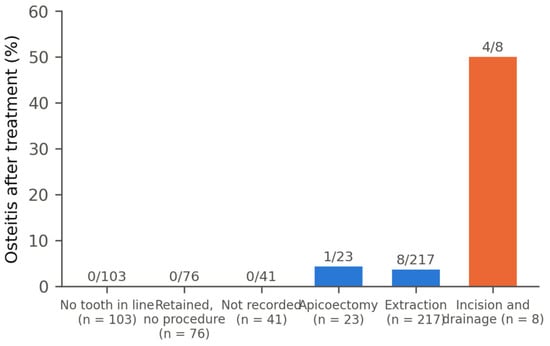
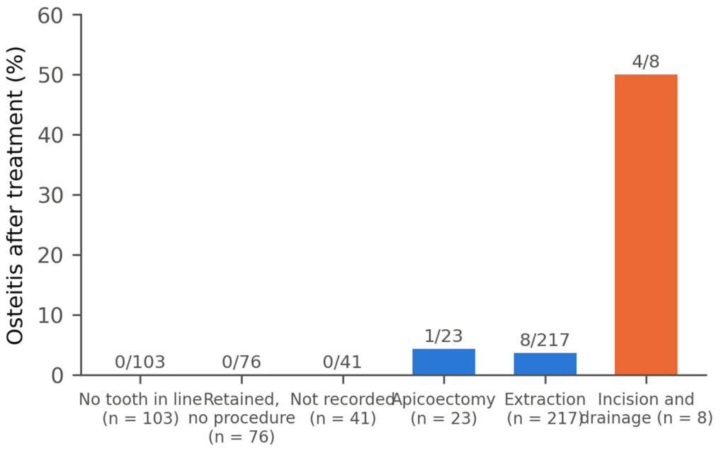
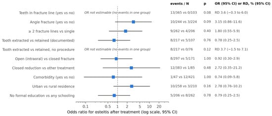
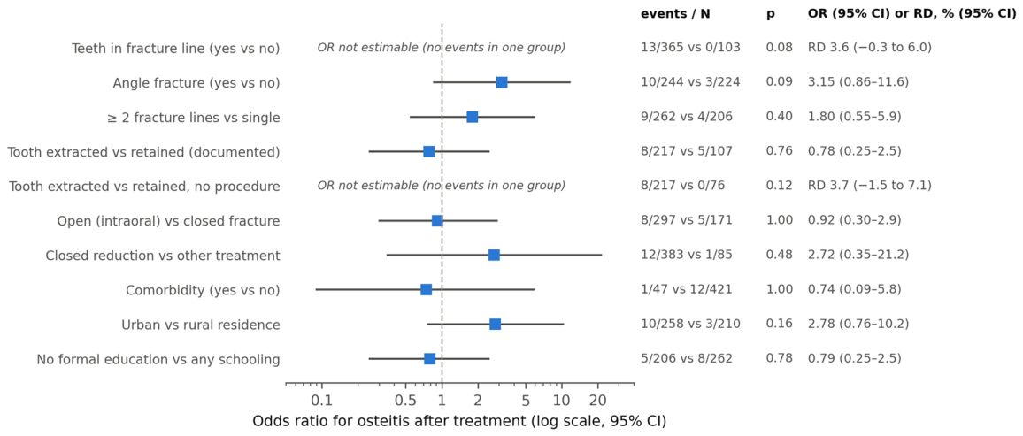
Taking the 13 recorded cases, osteitis was more frequent in angle fractures (10 of 244, 4.1%, vs. 3 of 224, 1.3%; OR 3.15, 95% CI 0.86 to 11.6, p = 0.09) and in fractures with two or more lines (3.4% vs. 1.9%; OR 1.80, 95% CI 0.55 to 5.92, p = 0.40), without significance. By management of the tooth in the line, it occurred in 8 of 217 after extraction (3.7%), in 0 of 76 after retention without a procedure, in 1 of 23 after apicoectomy, in 4 of 8 after incision and drainage with the tooth retained, and in 0 of 41 with no recorded management (Figure 5). Extraction versus documented retention in any form gave 3.7% vs. 4.7% (8 of 217 vs. 5 of 107; OR 0.78, 95% CI 0.25 to 2.45, p = 0.76), and extraction versus retention without a procedure 3.7% vs. 0% (risk difference 3.7%, 95% CI −1.5 to 7.1; p = 0.12). Osteitis was not associated with intraoral opening of the fracture (2.7% vs. 2.9%, p = 1.0), level of education (2.4% vs. 3.1%, p = 0.78), treatment modality (closed reduction 3.1% vs. other 1.2%, p = 0.48), comorbidity (2.1% vs. 2.9%, p = 1.0) or residence (urban 3.9% vs. rural 1.4%, p = 0.16), all computed within the 468 patients with mandibular fractures (Table 5, Figure 6). With only four cases without documented pre-existing infection, no association could be tested meaningfully: 4 of 365 tooth-bearing vs. 0 of 103 other fractures (p = 0.58), 2 of 244 angle vs. 2 of 224 other fractures (p = 1.0), and 3 of 217 after extraction vs. 1 of 107 after documented retention (p = 1.0).
Figure 5.
Osteitis recorded after treatment (13 patients, including 9 with infection already documented at treatment) according to the presence and management of teeth in the fracture line. Infection documented at treatment comprises a drained odontogenic abscess (8 patients) or osteitis noted at the first consultation (1 patient).
Figure 6.
Univariable associations with osteitis recorded after treatment (forest plot of Table 5). Odds ratios with 95% confidence intervals on a logarithmic scale for the 13 patients with osteitis recorded after treatment among the 468 patients with a mandibular fracture. For the two comparisons with no events in one group, the odds ratio is not estimable, and the absolute risk difference (RD, percentage points) with Newcombe 95% confidence interval is shown instead. p values from Fisher’s exact test. None of the associations reaches statistical significance. The comparisons are descriptive and exploratory; the absence of a detected difference, in particular between extraction and retention of the tooth in the fracture line, does not establish equivalence.
Table 5.
Univariable associations with osteitis recorded after treatment (13 patients, including 9 with infection already documented at treatment) in the 468 patients with mandibular fractures. RD, absolute risk difference in percentage points with Newcombe 95% CI, given where the OR is not estimable.
3.6. Osteitis at First Presentation
Twenty-eight patients (4.6% of the cohort, 95% CI 3.2 to 6.6; 27 of them with a mandibular fracture, 5.8%) had osteitis of the fracture site recorded at the first presentation to our department, which, for patients referred after treatment elsewhere, was not their first medical evaluation after the injury; one of them is also among the 13 patients with osteitis recorded after treatment, so the two groups together comprise 40 patients. In several of them, the fracture was documented as one to three months old at presentation; the circumstances recorded were delayed presentation, non-attendance for removal of fixation or failed initial management elsewhere (one patient had been immobilised incorrectly in another centre and arrived with osteomyelitis and a fistula); a further four patients had suppuration or a fistula of the fracture site without the word osteitis in the record and are counted in Table 3 but not here. These patients had a median age of 28.5 years; 57.1% lived in rural areas (vs 43.5% of the rest of the cohort, p = 0.18), 57.1% had no formal education (vs 43.7%, p = 0.18), 21 of 28 had an angle fracture (75.0%) and 25 of the 27 mandibular cases had teeth in the fracture line (92.6% vs. 77.1% of the other mandibular fractures; OR 3.71, 95% CI 0.86 to 16.0, p = 0.09); single and double fractures were equally represented. Their fractures were treated by closed reduction in 20, by wire or combined osteosynthesis in 4 and by local procedures only in 4.
4. Discussion
This analysis of 607 assault victims adds three findings to the mandibular pattern reported previously from the same centre [22]. First, approximately 28% of patients had a midface fracture, almost always of the zygomatic complex or the nasal bones, and these patients were older and far more likely to carry visible soft-tissue injury than patients with a mandibular fracture. Second, teeth laid in the fracture line in three quarters of mandibular fractures and were extracted in most of them; all recorded osteitis cases occurred in fractures with a tooth in the line, in most of them an infection (an odontogenic abscess in 8, osteitis in 1) was already documented at the time of treatment, and no association between extraction and osteitis was observed. Third, a group of patients twice the size of the group with osteitis recorded after treatment reached the service with an already-infected fracture, a burden that series reporting only complications recorded after treatment do not capture.
4.1. Midface Versus Mandibular Fractures in Assault
The proportion of midface involvement in our cohort (27.7%) is lower than in Italian assault series, where the zygomaticomaxillary complex, orbit and nasal bones together account for roughly half of the fractures [5,8,9], and the share of orbital floor fractures (2.0% of the cohort) is far below the 25% reported from Ancona [9] (Table 6). Two explanations are likely. Isolated nasal fractures and many orbital fractures are managed by otorhinolaryngology or ophthalmology in Romania, as in the Ancona series, and may not reach a maxillofacial database; and routine computed tomography, which detects orbital floor fractures that plain radiographs miss [20], was not available throughout the study period in our setting [22]. The predominance of the zygoma within the midface (63.7%) is, by contrast, in line with the EURMAT and Turin data [5,8] and with the biomechanics of a punch to the lateral face.
Table 6.
Published single-centre and multicentre series of assault-related facial fractures compared with the present cohort.
Patients with midface fractures were about three years older than those with mandibular fractures, a difference also observed in Ancona, where the median age rose from 23.5 years for mandibular to 29 years for zygomatic fractures [9]. The contrast in soft-tissue injury was striking: haematoma accompanied 79% of isolated midface fractures but only 13% of isolated mandibular fractures. In the Ancona cohort, the relation was reversed for soft-tissue wounds, which clustered with mandibular fractures [9]. The two findings are not contradictory: a blow that fractures the zygoma or the nose is absorbed by thin, vascular soft tissue over bone and produces periorbital haematoma, whereas the mandible is fractured by a blow transmitted through a thick muscular cover. In the mandibular subset of our cohort, the earlier report showed that soft-tissue lesions tracked the number and displacement of fracture lines [22]; the present data show that, across the face, the site of the fracture matters more than its severity for the soft-tissue picture. For the clinician, the message is that a periorbital haematoma after a punch should prompt imaging of the zygoma and orbit even when the mandible is intact, while a mandibular fracture can be present with little external sign.
Midface fractures in this cohort were treated almost exclusively by closed reduction, as were mandibular fractures, which differs from Western European practice where open reduction and internal fixation is used in two thirds to four fifths of assault-related fractures [5,9]. The reasons, documented previously, are the limited reimbursement of osteosynthesis material during most of the study period [22,23]; closed treatment was applied even though three quarters of midface and nine tenths of mandibular fractures were displaced, so the choice reflected resources rather than fracture pattern. The institutional indications during the study period were closed reduction, through a temporal or intraoral approach, for zygomatic fractures that were stable after reduction and for nasal bone fractures, with open reduction and plate fixation reserved for zygomatic fractures that were unstable after reduction, comminuted at the buttresses or associated with an orbital floor defect, and maxillomandibular fixation for mandibular fractures, with osteosynthesis when the fracture was unstable and material was available; the indication was not recorded as a variable for each patient, so the relation between displacement and treatment choice could not be analysed. No difference in osteitis after treatment was detected between closed reduction and the other modalities (3.1% vs. 1.2%, p = 0.48), but with 13 events and 85 patients treated otherwise than by closed reduction, this comparison has little power, and compliance with fixation and attendance at follow-up were not recorded. These results describe a regional care setting of the years 2001 to 2011, with limited CT imaging and a predominance of closed treatment, and should not be extrapolated directly to centres that rely on routine CT and open reduction. For zygoma in particular, closed reduction without fixation cannot guarantee that a displaced tripod fracture stays reduced, and inadequate primary reduction is the commonest reason for which patients later require osteotomy and repositioning of the malar complex, with or without orbital floor reconstruction [17,18]. No complication was recorded after isolated midface fractures; given the limits of the institutional follow-up described in Section 2.3, this absence of recorded complications cannot be taken as evidence that none occurred, although it is consistent with the low pooled infection rate of midface fractures (0.9%) in a recent meta-analysis of fracture-related infections [25] and with the absence of evidence, in randomised trials, that one approach to mandibular fractures is superior to another [11]. However, functional and aesthetic outcomes of zygomatic fractures (malar flattening, enophthalmos, infraorbital hypoaesthesia, which follows about a third of zygomaticomaxillary complex fractures [26]) were not systematically recorded, and the absence of complications should not be over-interpreted; long-term sequelae of facial fractures are common even after uneventful healing [27].
4.2. Dental Injuries
Dental trauma was recorded in 6.3% of patients, less than the 20 to 30% prevalence of dento-alveolar injury described in domestic violence victims [28,29] and in the Brazilian series of interpersonal violence, where dental injuries are among the most frequent lesions [30,31], and it rose from 5% in isolated mandibular to 17% in combined fractures. The low overall figure almost certainly reflects under-recording in a surgical database focused on bone, and dental injuries that were treated in the emergency department or by the patient’s dentist may have escaped documentation. The gradient across groups is nonetheless informative: dental injury accompanies the higher-energy, multi-site injuries, and a patient with both a mandibular and a midface fracture should be examined for crown and root fractures and luxations before fixation is applied. Beyond documentation, timing matters: the prognosis of a traumatised tooth depends on treatment within hours for avulsion and luxation and within days for exposed dentine or pulp, and on structured follow-ups at 2 weeks, 4 weeks, 3 months, 6 months and 1 year to detect pulp necrosis, infection-related resorption or ankylosis [12,13]. For the crown fractures that dominate dental trauma in young adults, reattachment of a preserved fragment or a direct composite restoration restores function and appearance in a single visit, whereas delayed or absent treatment converts an uncomplicated fracture into a pulpal and periapical problem [14]. In a patient immobilised in maxillomandibular fixation, these windows are easily missed, which argues for a dental assessment before fixation is applied and for a dental follow-up appointment to be built into the recall pathway proposed below.
4.3. Teeth in the Fracture Line: Extraction, Retention and Osteitis
A mandibular third molar raises the odds of an angle fracture more than threefold [10], which is why teeth lay in the line of 91% of our angle fractures. The presence of a tooth in the fracture line is the most consistently reported risk factor for infection after mandibular fracture, and our data point the same way: none of the 103 fractures without a tooth in the line had osteitis recorded during follow-up, against 3.6% of the 365 with a tooth (risk difference 3.6%, 95% CI −0.3 to 6.0), although, with 13 events, the difference did not reach significance (p = 0.08). Nine of the 13 patients had infection documented at the time of fracture treatment, although pre-existing bone infection was confirmed in only one. Consequently, the available data do not establish whether osteitis recorded during follow-up represented persistence or progression of pre-existing infection or a newly developed complication, and in these patients, the association between the tooth in the line and osteitis is descriptive: the data cannot establish whether the tooth caused the osteitis or merely accompanied it. Osteitis without documented pre-existing infection occurred in only four patients (0.9%), a descriptive proportion that should not be read as a formal incidence. Whether that tooth should be removed is a different question. Two meta-analyses of angle fractures give the answer that extraction does not reduce postoperative infection: in 13 studies pooling 1542 angle fractures, infection occurred in 10.7% after removal and 11.1% after retention of the tooth [15], and a later synthesis of 34 studies found that angle fractures with a third molar present had a higher infection rate than those with the tooth absent, but that removing the tooth at the time of treatment did not reproduce the advantage of its pre-existing absence [16]. Our series, in which extraction was performed in 60% of teeth in the line and in 68% of angle fractures, found osteitis in 3.7% after extraction and 4.7% after documented retention (p = 0.76), and in 1.4% and 0.9% respectively when only cases without documented pre-existing infection are counted. The breakdown of the retained group is informative: four of its five osteitis cases followed incision and drainage of an abscess that was already present around the tooth at the time of treatment, and none of the 76 teeth retained without any procedure had osteitis recorded during follow-up. No statistically significant association was observed between tooth extraction and osteitis recorded after treatment; however, the low event count and the likely confounding by indication—since teeth with periapical infection, mobility or root fracture are extracted preferentially—preclude any conclusion on the effectiveness of either policy. What the data do show is only that a tooth left in place without an indication for removal was not followed by recorded infection in this cohort; the absence of a detected difference between extraction and retention must not be read as equivalence of the two policies, since the study was not designed or powered to establish it; because the condition of these teeth (vital, carious, mobile or impacted) was not recorded, this observation is consistent with the pooled evidence but cannot by itself establish that retention of intact teeth is the preferable policy. The absolute osteitis rates (3 to 4% in tooth-bearing fractures as recorded, about 1% for cases without documented pre-existing infection) are low by comparison, with the 8.9% pooled infection rate of mandibular fractures [25] and with the 10 to 11% of the angle-fracture meta-analysis [15], which is consistent with a young population, closed treatment and short follow-up; infection rates after open treatment of mandibular fractures are typically higher, and depend on patient factors such as smoking, substance use and oral hygiene as much as on technique [25,32,33]. Antibiotic prophylaxis, used routinely in our department, has an uncertain effect on infection after facial fractures and was not analysed as a variable [34].
4.4. Late Presentation with an Infected Fracture
Twenty-eight patients (4.6%) arrived with osteitis of the fracture site, and, in some, the fracture was weeks or months old. This group is twice as large as the group with osteitis recorded after treatment and would be invisible in a study that counts only treatment complications. It was enriched for angle fractures and teeth in the line, as expected for a fracture that has been left untreated with an open periodontal pathway, and tended to be rural. The documented circumstances, delayed presentation, non-attendance for removal of fixation and failed initial management elsewhere, suggest that delayed access to care, interrupted follow-up and previous treatment failure may have contributed to these presentations. However, their relative contributions and potential interactions with treatment-related factors could not be determined from the available records. The same records also contained fractures that had already consolidated in a vicious position by the time the patient was seen, which at that stage are no longer a trauma problem but a reconstructive one, requiring refracture or osteotomy to restore occlusion and contour [18]. Assault victims are a population in which concealment of the mechanism, alcohol use and low health literacy are frequent [1,5,8,22,35], and the earlier report from our centre found that 44% of mandibular fracture patients had no formal education [22]. Alcohol, involved in 83% of the assaults in the mandibular subset of this cohort [22] and a recognised amplifier of fracture severity [36], adds to the difficulty of follow-up. A structured follow-up pathway, with a fixed appointment for removal of maxillomandibular fixation, nutritional counselling during fixation [37] and an active recall for patients who do not attend, is a low-cost intervention that these data justify.
4.5. Implications for Reconstructive Surgery
This section offers a clinical interpretation rather than a result of the cohort: the database did not record residual deformity, secondary reconstructive procedures or long-term aesthetic outcome, and the following considerations rest on the fracture and treatment pattern alone. Assault-related facial fractures are the point of origin of a large part of the secondary deformities that reach reconstructive surgery. Three findings of this cohort bear on that pathway. First, three quarters of zygomatic and two thirds of nasal bone fractures were displaced, and almost all were treated by closed reduction; a zygoma that heals in a displaced position produces malar flattening, enophthalmos, diplopia and infraorbital hypoaesthesia, and its secondary correction requires osteotomy, repositioning and often orbital floor reconstruction, procedures that are far more demanding than primary fixation and increasingly rely on virtual planning and patient-specific guides [17,20,26]. We hypothesise, without being able to test it in these data, that the displacement rates reported here define a population in which late referral for reconstruction is more likely; the present database, with its short institutional follow-up, cannot quantify this. Second, the free-text records contained patients who arrived with an already-malunited fracture (of the mandibular angle, the subcondylar region or the nasal bones) after treatment elsewhere or no treatment, and one patient with established facial asymmetry; these are precisely the cases that end on the reconstructive surgeon’s table. Third, lacerations accompanied one midface fracture in four and one combined fracture in three, and a laceration over a fracture is a scar that the patient will carry on the most visible part of the body; its primary closure, and the later revision of hypertrophic or misaligned scars, belong to the same reconstructive pathway. Whether these patients in fact developed residual deformity or underwent secondary reconstruction was not assessed, and the frequency of such outcomes cannot be inferred from this cohort. The observation only supports a prospective evaluation: a structured, imaging-based follow-up (clinical assessment of malar projection, ocular position and infraorbital sensation, with CT or three-dimensional photography at baseline and at 6 to 12 months) of patients treated by closed reduction for a displaced zygomatic fracture, or presenting late with a malunion, would quantify the need for secondary reconstruction that the present data can only suggest. The face is the part of the body on which appearance carries the greatest social weight, and the same sociocultural pressures that drive the demand for elective aesthetic surgery make a post-traumatic facial scar or asymmetry a burden that patients rarely accept passively [38].
4.6. Perspective: Relevance for Future Research
No artificial intelligence (AI) tool was applied in this study, and the database contains neither the imaging nor the outcome variables that the validation of such tools would require; the following remark is therefore a perspective, not a result. Deep learning detection of mandibular fractures on radiographs and CT has reached high pooled sensitivity [39], whereas midface fractures remain harder to classify [40], and the available models are trained on retrospective single-centre datasets with little external validation [41]. A population such as ours, with its predominance of angle, zygomatic and nasal fractures, its closed treatment and its late presentations, differs from most training populations, and the prevalences reported here could serve as prior information for calibrating or validating detection and triage models in a comparable setting [42]. The outcome variables that such work would need (residual displacement, enophthalmos, asymmetry, infection) are the same ones whose absence limits the present study, which is a further argument for the structured, imaging-based follow-up proposed above.
4.7. Limitations
This is a retrospective single-centre study and shares the limitations of its data source: variables were recorded for clinical rather than research purposes, their completeness depended on the treating surgeon, and selection, information and confounding biases cannot be excluded; the measures taken to limit these problems are described in Section 2.4. The mandibular subset has been published and the demographic core of the cohort is therefore not new. To state the contribution precisely: the sex and age distribution and the mandibular fracture sites and treatment summarised in Section 3.1 and in the first rows of Table 1 overlap with the earlier report [22]; everything else, the three-group comparison (Table 1 and Table 5, Figure 1, Figure 2 and Figure 3), the midface fracture sites (Table 2, Figure 4), the dental and extrafacial injuries (Table 3), the presence and management of teeth in the fracture line (Table 4), the analysis of osteitis and its chronology (Table 5, Figure 5 and Figure 6, Table S1) and the late-presentation group (Section 3.6), is reported here for the first time. Variables that would have strengthened these analyses, and that bear directly on infection risk, were not available: alcohol involvement, smoking, the mechanism of the blow, the time from injury to treatment, the condition of the tooth in the line (vital, carious, mobile, impacted) that determined extraction, the indication for closed or open treatment in the individual patient, the duration of fixation, compliance with it and the length of follow-up; systemic comorbidity was recorded but was rare in this young population. Follow-up was institutional and ended, for most patients, at removal of fixation; patients were not systematically recalled afterwards, and complications treated elsewhere were not captured, so the complication rates, including the absence of recorded complications after isolated midface fractures, are lower bounds rather than true incidences. Functional and aesthetic outcomes of midface fractures (malar asymmetry, diplopia, enophthalmos, infraorbital sensory disturbance) and secondary reconstructive procedures were not recorded. Orbital and nasal fractures are probably under-represented because of referral patterns and the imaging available. Dental injuries and extrafacial injuries were recorded as free text and are likely under-counted. The number of osteitis events (13 recorded, 4 without documented pre-existing infection) precluded multivariable analysis, so the associations reported are univariable and hypothesis-generating, and the chronology of infection relative to treatment was reconstructed from the management and free-text fields rather than from a dedicated variable, and the separation between infections present at treatment and osteitis without documented pre-existing infection, although confirmed by review of the clinical records, is limited by the documentation available in those records. The present patient count (607) differs from those of the earlier reports from the same database (598 and 478 [21,22]); as stated in Section 2.2, those analyses applied their own eligibility and completeness criteria, and, because the individual records cannot be re-identified in the anonymised extract, the difference cannot be reconciled at the patient level and no further explanation of it is offered here. The cohort is historical: the patients were treated between 2001 and 2011, before routine CT and rigid internal fixation became standard in this setting, and both fracture detection (particularly of orbital floor and nasal fractures) and the treatment distribution reflect that period. The injury pattern of assault, the behaviour of teeth in the fracture line and the problem of late presentation are unlikely to have changed, but the treatment results should be read as those of a predominantly closed-treatment era and not extrapolated to current practice with CT-based planning and open reduction. Finally, the predominance of closed treatment limits the transfer of the complication data to centres that rely on open reduction.
5. Conclusions
In 607 victims of interpersonal violence treated in north-western Romania between 2001 and 2011, we observed the following. One patient in four had a midface fracture, dominated by the zygomatic complex and the nasal bones, with Le Fort and orbital floor fractures rare. Patients with midface fractures were older and presented with haematoma, laceration or abrasion several times more often than patients with mandibular fractures, which could be present with little external sign. Dental trauma was documented in 6.3% of patients and most often when both jaws were fractured. Teeth lay in the fracture line in 78% of mandibular fractures and were extracted in 60%. All 13 cases of osteitis recorded after treatment occurred in fractures with a tooth in the line, and in 9 of them an infection was already documented at the time of treatment; no difference in osteitis was detected between extraction and retention of the tooth, but the number of events and confounding by indication preclude any conclusion on which policy is preferable. Twenty-eight patients presented with an already-infected fracture, a group larger than the one with osteitis recorded after treatment. As interpretation rather than finding, these observations suggest that a dental assessment before fixation, a structured recall after removal of fixation and a prospective, imaging-based follow-up of patients treated by closed reduction for displaced midface fractures would be the appropriate next steps; whether residual deformity and secondary reconstruction are frequent in this population was not assessed and remains to be established.
Supplementary Materials
The following supporting information can be downloaded at: https://www.mdpi.com/article/10.3390/healthcare14234014/s1, Table S1: the 13 patients with osteitis recorded after treatment, with the database fields (management of the tooth in the line, free-text notes) from which infection documented at treatment was distinguished from osteitis without documented pre-existing infection.
Author Contributions
Conceptualization, V.D., A.-P.T., A.-T.M., I.L. and M.J.; methodology, V.D., R.I.J., A.-P.T., M.I.Ș. and I.L.; software, R.F.B. and G.A.C.; validation, A.-T.M., R.I.J., F.B., M.I.Ș., I.L. and M.J.; formal analysis, V.D. and R.I.J.; investigation, V.D., A.-P.T., A.-T.M., R.F.B., F.B., G.A.C. and M.I.Ș.; resources, A.-P.T., A.-T.M., F.B., G.A.C. and M.J.; data curation, A.-P.T., R.I.J., R.F.B., G.A.C. and M.I.Ș.; writing, original draft preparation, V.D., R.I.J., A.-P.T., A.-T.M. and I.L.; writing, review and editing, A.-T.M., R.I.J., R.F.B., F.B., G.A.C., M.I.Ș., I.L. and M.J.; visualisation, V.D., R.F.B. and M.I.Ș.; supervision, I.L., A.-T.M., A.-P.T. and M.J.; project administration, V.D., A.-T.M. and F.B. All authors have read and agreed to the published version of the manuscript.
Funding
The Article Processing Charge was funded by the University of Oradea, Romania.
Institutional Review Board Statement
The study was conducted in accordance with the Declaration of Helsinki and approved by the Ethics Committee of the University of Oradea (No. 35698/19.02.2018 and No. 28765/11.02.2019, for the constitution of the database) and by the Ethics Committee for Scientific Research of Pelican Hospital, Oradea (protocol code 1540, date of approval 20 October 2026, for the present analysis).
Informed Consent Statement
Informed consent was obtained from all subjects involved in the study at the time of admission; for patients under 18 years, consent was signed by a parent or legal guardian.
Data Availability Statement
The data presented in this study are available on request from the corresponding authors because of privacy restrictions.
Acknowledgments
During the preparation of this manuscript, the authors used Claude (Claude Fable 5.1, Anthropic, San Francisco, CA, USA) for the statistical analysis scripts, language refinement and structural review of the manuscript draft, and for checking the formatting of references. The authors have reviewed and edited all output and take full responsibility for the content of this publication. The authors further acknowledge the University of Oradea, Oradea, Romania, for supporting the Article Processing Charge.
Conflicts of Interest
The authors declare no conflicts of interest.
References
- Yamamoto, K.; Matsusue, Y.; Horita, S.; Murakami, K.; Sugiura, T.; Kirita, T. Maxillofacial Fractures Associated with Interpersonal Violence. J. Craniofacial Surg. 2019, 30, e312–e315. [Google Scholar] [CrossRef] [Scilit] [PubMed]
- Boffano, P.; Roccia, F.; Zavattero, E.; Dediol, E.; Uglešić, V.; Kovačič, Ž.; Vesnaver, A.; Konstantinović, V.S.; Petrović, M.; Stephens, J.; et al. European Maxillofacial Trauma (EURMAT) project: A multicentre and prospective study. J. Cranio-Maxillofac. Surg. 2015, 43, 62–70. [Google Scholar] [CrossRef] [Scilit] [PubMed]
- Roccia, F.; Iocca, O.; Sobrero, F.; Rae, E.; Laverick, S.; Carlaw, K.; Aquilina, P.; Bojino, A.; Romeo, I.; Duran-Valles, F.; et al. World Oral and Maxillofacial Trauma (WORMAT) project: A multicenter prospective analysis of epidemiology and patterns of maxillofacial trauma around the world. J. Stomatol. Oral Maxillofac. Surg. 2022, 123, e849–e857. [Google Scholar] [CrossRef] [Scilit] [PubMed]
- Bonavolontà, P.; Dell’Aversana Orabona, G.; Abbate, V.; Vaira, L.A.; Lo Faro, C.; Petrocelli, M.; Attanasi, F.; De Riu, G.; Iaconetta, G.; Califano, L. The epidemiological analysis of maxillofacial fractures in Italy: The experience of a single tertiary center with 1720 patients. J. Cranio-Maxillofac. Surg. 2017, 45, 1319–1326. [Google Scholar] [CrossRef] [Scilit] [PubMed]
- Boffano, P.; Roccia, F.; Zavattero, E.; Dediol, E.; Uglešić, V.; Kovačič, Ž.; Vesnaver, A.; Konstantinović, V.S.; Petrović, M.; Stephens, J.; et al. Assault-related maxillofacial injuries: The results from the European Maxillofacial Trauma (EURMAT) multicenter and prospective collaboration. Oral Surg. Oral Med. Oral Pathol. Oral Radiol. 2015, 119, 385–391. [Google Scholar] [CrossRef] [Scilit] [PubMed]
- Lee, K.H. Interpersonal violence and facial fractures. J. Oral Maxillofac. Surg. 2009, 67, 1878–1883. [Google Scholar] [CrossRef] [Scilit] [PubMed]
- Goulart, D.R.; Colombo, L.A.; de Moraes, M.; Asprino, L. What is expected from a facial trauma caused by violence? J. Oral Maxillofac. Res. 2014, 5, e4. [Google Scholar] [CrossRef] [Scilit] [PubMed]
- Roccia, F.; Savoini, M.; Ramieri, G.; Zavattero, E. An analysis of 711 victims of interpersonal violence to the face, Turin, Italy. J. Cranio-Maxillofac. Surg. 2016, 44, 1025–1028. [Google Scholar] [CrossRef] [Scilit] [PubMed]
- Cirignaco, G.; Catarzi, L.; Monarchi, G.; Committeri, U.; Frosolini, A.; Togni, L.; Mascitti, M.; Balercia, P.; Santarelli, A.; Consorti, G. Interpersonal Violence-Related Facial Fractures: 12-Year Trends and Surgical Outcomes in a Southern European Level-I Trauma Centre. Medicina 2025, 61, 1443. [Google Scholar] [CrossRef] [Scilit] [PubMed]
- Armond, A.C.V.; Martins, C.C.; Glória, J.C.R.; Galvão, E.L.; dos Santos, C.R.R.; Falci, S.G.M. Influence of third molars in mandibular fractures. Part 1: Mandibular angle—A meta-analysis. Int. J. Oral Maxillofac. Surg. 2017, 46, 716–729. [Google Scholar] [CrossRef] [Scilit] [PubMed]
- Nasser, M.; Pandis, N.; Fleming, P.S.; Fedorowicz, Z.; Ellis, E.; Ali, K. Interventions for the management of mandibular fractures. Cochrane Database Syst. Rev. 2013, 2013, CD006087. [Google Scholar] [CrossRef] [Scilit] [PubMed]
- Bourguignon, C.; Cohenca, N.; Lauridsen, E.; Flores, M.T.; O’Connell, A.C.; Day, P.F.; Tsilingaridis, G.; Abbott, P.V.; Fouad, A.F.; Hicks, L.; et al. International Association of Dental Traumatology guidelines for the management of traumatic dental injuries: 1. Fractures and luxations. Dent. Traumatol. 2020, 36, 314–330. [Google Scholar] [CrossRef] [Scilit] [PubMed]
- Fouad, A.F.; Abbott, P.V.; Tsilingaridis, G.; Cohenca, N.; Lauridsen, E.; Bourguignon, C.; O’Connell, A.C.; Flores, M.T.; Day, P.F.; Hicks, L.; et al. International Association of Dental Traumatology guidelines for the management of traumatic dental injuries: 2. Avulsion of permanent teeth. Dent. Traumatol. 2020, 36, 331–342. [Google Scholar] [CrossRef] [Scilit] [PubMed]
- Zaneva-Hristova, D.; Borisova-Papancheva, T.; Yankov, Y.G.; Papanchev, G. Different Approaches to the Treatment of Anterior Tooth Fractures: Three Clinical Cases and Behavior Report. Cureus 2024, 16, e64524. [Google Scholar] [CrossRef] [Scilit] [PubMed]
- Bobrowski, A.N.; Sonego, C.L.; Chagas Junior, O.L. Postoperative infection associated with mandibular angle fracture treatment in the presence of teeth on the fracture line: A systematic review and meta-analysis. Int. J. Oral Maxillofac. Surg. 2013, 42, 1041–1048. [Google Scholar] [CrossRef] [Scilit] [PubMed]
- Fernandes, I.A.; Souza, G.M.; Silva de Rezende, V.; Al-Sharani, H.M.; Douglas-de-Oliveira, D.W.; Galvão, E.L.; Falci, S.G.M. Effect of third molars in the line of mandibular angle fractures on postoperative complications: Systematic review and meta-analysis. Int. J. Oral Maxillofac. Surg. 2020, 49, 471–482. [Google Scholar] [CrossRef] [Scilit] [PubMed]
- Krasovsky, A.; Hija, A.; Zeineh, N.; Bilder, A.; Emodi, O.; Rachmiel, A.; Shilo, D. Three-dimensional secondary reconstruction of mistreated zygomatic fractures using patient specific surgical guides and implants. Front. Oral Health 2024, 5, 1464012. [Google Scholar] [CrossRef] [Scilit] [PubMed]
- Aman, H.; Shokri, T.; Reddy, L.V.; Ducic, Y. Secondary Management of Midface Fractures. Facial Plast. Surg. 2019, 35, 640–644. [Google Scholar] [CrossRef] [Scilit] [PubMed]
- Ellis, E., III; Kittidumkerng, W. Analysis of treatment for isolated zygomaticomaxillary complex fractures. J. Oral Maxillofac. Surg. 1996, 54, 386–400. [Google Scholar] [CrossRef] [Scilit] [PubMed]
- Seifert, L.B.; Mainka, T.; Herrera-Vizcaino, C.; Verboket, R.; Sader, R. Orbital floor fractures: Epidemiology and outcomes of 1594 reconstructions. Eur. J. Trauma Emerg. Surg. 2022, 48, 1427–1436. [Google Scholar] [CrossRef] [Scilit] [PubMed]
- Juncar, M.; Tent, P.A.; Juncar, R.I.; Harangus, A.; Mircea, R. An epidemiological analysis of maxillofacial fractures: A 10-year cross-sectional cohort retrospective study of 1007 patients. BMC Oral Health 2021, 21, 128. [Google Scholar] [CrossRef] [Scilit] [PubMed]
- Rivis, M.; Juncar, R.I.; Moca, A.E.; Moca, R.T.; Juncar, M.; Țenț, P.A. Patterns of Mandibular Fractures through Human Aggression: A 10-Year Cross-Sectional Cohort Retrospective Study. J. Clin. Med. 2023, 12, 4103. [Google Scholar] [CrossRef] [Scilit] [PubMed]
- Țenț, P.A.; Juncar, R.I.; Arghir, I.A.; Arghir, O.C.; Juncar, M. A retrospective evaluation of the treatment of viscerocranial fractures in Romania. A study of 1007 patients. Ann. Ital. Chir. 2020, 91, 568–574. [Google Scholar] [PubMed]
- von Elm, E.; Altman, D.G.; Egger, M.; Pocock, S.J.; Gøtzsche, P.C.; Vandenbroucke, J.P.; STROBE Initiative. The Strengthening the Reporting of Observational Studies in Epidemiology (STROBE) statement: Guidelines for reporting observational studies. Lancet 2007, 370, 1453–1457. [Google Scholar] [CrossRef] [Scilit] [PubMed]
- Van der Cruyssen, F.; Forrest, M.; Holmes, S.; Bhatti, N. A Systematic Review and Meta-Analysis of Fracture-Related Infections in Maxillofacial Trauma: Incidence, Risk Factors, and Management Strategies. J. Clin. Med. 2025, 14, 1332. [Google Scholar] [CrossRef] [Scilit] [PubMed]
- Dubron, K.; Verbist, M.; Shaheen, E.; Dormaar, T.J.; Jacobs, R.; Politis, C. Incidence, Aetiology, and Associated Fracture Patterns of Infraorbital Nerve Injuries Following Zygomaticomaxillary Complex Fractures: A Retrospective Analysis of 272 Patients. Craniomaxillofac. Trauma Reconstr. 2022, 15, 139–146. [Google Scholar] [CrossRef] [Scilit] [PubMed]
- Petersen, L.Ø.; Ipsen, E.Ø.; Felding, U.A.; von Buchwald, C.; Steinmetz, J. Sequelae of Major Trauma Patients with Maxillofacial Fractures. Ann. Otol. Rhinol. Laryngol. 2021, 130, 475–482. [Google Scholar] [CrossRef] [Scilit] [PubMed]
- Yari, A.; Fasih, P.; Bagheri, A.; Aryanezhad, S.S.; Sani, M.K. Prevalence and pattern of maxillofacial injuries associated with domestic violence: A retrospective study at a major trauma center. Dent. Traumatol. 2024, 40, 61–68. [Google Scholar] [CrossRef] [Scilit] [PubMed]
- Boyes, H.; Fan, K. Maxillofacial injuries associated with domestic violence: Experience at a major trauma centre. Br. J. Oral Maxillofac. Surg. 2020, 58, 185–189. [Google Scholar] [CrossRef] [Scilit] [PubMed]
- Ferreira, M.C.; Batista, A.M.; de Oliveira Ferreira, F.; Ramos-Jorge, M.L.; Marques, L.S. Pattern of oral-maxillofacial trauma stemming from interpersonal physical violence and determinant factors. Dent. Traumatol. 2014, 30, 15–21. [Google Scholar] [CrossRef] [Scilit] [PubMed]
- Rodrigues, L.G.; Barbosa, K.G.N.; de Paula Silva, C.J.; Alencar, G.P.; D’avila, S.; Ferreira, E.F.E.; Ferreira, R.C. Trends of maxillofacial injuries resulting from physical violence in Brazil. Dent. Traumatol. 2020, 36, 69–75. [Google Scholar] [CrossRef] [Scilit] [PubMed]
- Boljevic, T.; Pelicic, D.; Terzic, Z.; Bojic, M. Complications in patients with facial bone fractures before and after conservative and surgical treatment, their comparison and correlation with different factors. Eur. Rev. Med. Pharmacol. Sci. 2023, 27, 11073–11081. [Google Scholar] [CrossRef] [Scilit] [PubMed]
- Munante-Cardenas, J.L.; Facchina Nunes, P.H.; Passeri, L.A. Etiology, treatment, and complications of mandibular fractures. J. Craniofac. Surg. 2015, 26, 611–615. [Google Scholar] [CrossRef] [Scilit] [PubMed]
- Goormans, F.; Coropciuc, R.; Vercruysse, M.; Spriet, I.; Willaert, R.; Politis, C. Systemic Antibiotic Prophylaxis in Maxillofacial Trauma: A Scoping Review and Critical Appraisal. Antibiotics 2022, 11, 483. [Google Scholar] [CrossRef] [Scilit] [PubMed]
- Businger, A.P.; Krebs, J.; Schaller, B.; Zimmermann, H.; Exadaktylos, A.K. Cranio-maxillofacial injuries in victims of interpersonal violence. Swiss Med. Wkly. 2012, 142, w13687. [Google Scholar] [CrossRef] [Scilit] [PubMed]
- O’Meara, C.; Witherspoon, R.; Hapangama, N.; Hyam, D.M. Mandible fracture severity may be increased by alcohol and interpersonal violence. Aust. Dent. J. 2011, 56, 166–170. [Google Scholar] [CrossRef] [Scilit] [PubMed]
- Popat, S.P.; Rattan, V.; Rai, S.; Jolly, S.S.; Malhotra, S. Nutritional intervention during maxillomandibular fixation of jaw fractures prevents weight loss and improves quality of life. Br. J. Oral Maxillofac. Surg. 2021, 59, 478–484. [Google Scholar] [CrossRef] [Scilit] [PubMed]
- Căiță, G.A.; Maghiar, T.; Bodog, F.D.; Maghiar, L.; Voiță-Mekereș, F.; Lascu, C.F. A Review of Social Factors Affecting Women’s Tendency to Cosmetic Surgery. Pharmacophore 2023, 14, 111–115. [Google Scholar] [CrossRef] [Scilit]
- Dashti, M.; Ghaedsharaf, S.; Ghasemi, S.; Zare, N.; Constantin, E.-F.; Fahimipour, A.; Tajbakhsh, N.; Ghadimi, N. Evaluation of deep learning and convolutional neural network algorithms for mandibular fracture detection using radiographic images: A systematic review and meta-analysis. Imaging Sci. Dent. 2024, 54, 232–239. [Google Scholar] [CrossRef] [Scilit] [PubMed]
- Warin, K.; Limprasert, W.; Suebnukarn, S.; Paipongna, T.; Jantana, P.; Vicharueang, S. Maxillofacial fracture detection and classification in computed tomography images using convolutional neural network-based models. Sci. Rep. 2023, 13, 3434. [Google Scholar] [CrossRef] [Scilit] [PubMed]
- Tuygunov, N.; Boymuradov, S.A.; Khurshid, Z.; Songsiripradubboon, S.; Abdulahtov, J.; Khatamov, U. Diagnostic performance of artificial intelligence for facial fracture detection: A systematic review. Oral Radiol. 2026, 42, 262–280. [Google Scholar] [CrossRef] [Scilit] [PubMed]
- Dina, V.; Tent, A.-P.; Maghiar, A.-T.; Bodog, R.F.; Grecu, T.-A.; Crețu, O.-M.; Maghiar, L.; Bodog, F.; Căiță, G.A.; Șandor, M.I.; et al. From Assistance to Autonomy: Acceptability of Progressive Artificial Intelligence Integration in Facial Reconstructive Surgery—Protocol for Within-Subjects Vignette Experiment Among Romanian Adults. Healthcare 2026, 14, 3030. [Google Scholar] [CrossRef] [Scilit] [PubMed]

Disclaimer/Publisher’s Note: The statements, opinions and data contained in all publications are solely those of the individual author(s) and contributor(s) and not of MDPI and/or the editor(s). MDPI and/or the editor(s) disclaim responsibility for any injury to people or property resulting from any ideas, methods, instructions or products referred to in the content. |
© 2026 by the authors. Licensee MDPI, Basel, Switzerland. This article is an open access article distributed under the terms and conditions of the Creative Commons Attribution (CC BY) license.