


Abstract
Background/Objectives: Hospital wound surveillance usually focuses on pressure ulcers; as a result, surgical, traumatic and moisture-related skin damage remain largely unmeasured. This study estimated the point prevalence of all wound types among inpatients of a general hospital in the Azores, Portugal, and described their characteristics and dressing workload. Methods: A single-day cross-sectional survey was conducted in May 2023 in all inpatient wards of Hospital de Santo Espírito da Ilha Terceira, using the wound characterisation instrument of the Portuguese Wound Society (ELCOS). Prevalence was estimated with Wilson 95% confidence intervals (CIs), and factors associated with having any wound and, post hoc, a non-surgical wound were explored with multivariable logistic regression. Results: Of 119 inpatients (participation rate 91.5%), 60 had at least one wound (prevalence 50.4%; 95% CI 41.6–59.2) and 35 had a non-surgical wound (29.4%; 95% CI 22.0–38.1). Of the 106 wounds recorded, 45.3% were surgical, 17.0% pressure ulcers, 17.0% traumatic and 15.1% moisture-associated skin damage. Pressure ulcer prevalence was 10.1% (95% CI 5.9–16.8). Excluding surgical wounds, which are hospital-acquired by definition, 43.1% of wounds had a hospital origin. Among adults in the medical and surgical wards, immobility was associated with having a non-surgical wound (adjusted odds ratio 7.43; 95% CI 2.35–26.93), whereas the association with surgical admission was largely explained by surgical incisions. Conclusions: Half of the inpatients had a wound, and almost one in three had a non-surgical wound. Pressure-ulcer-only audits, which assess a narrower outcome, identify far fewer patients with any wound; periodic all-wound surveillance with a standardised instrument should inform prevention and the planning of wound care resources.
1. Introduction
Wounds are a frequent, costly and often under-recognised health problem. Worldwide estimates suggest that acute and chronic wounds affect a substantial share of the population, with a growing economic and humanistic burden driven by population ageing, diabetes and multimorbidity [1,2]. In the United Kingdom, the number of patients with a wound managed by the National Health Service rose to an estimated 3.8 million in 2017/2018, at an annual cost of approximately GBP 8.3 billion, most of it incurred in the community and driven mainly by nurse visits [3,4]. Chronic wounds alone have a pooled prevalence of about 2 per 1000 in the general population, but the figure varies widely with the definitions and methods used [5].
Hospitals concentrate a large part of this burden. Inpatients combine acute illness, reduced mobility, incontinence, malnutrition and invasive or surgical procedures, all of which increase the risk of skin and tissue damage. Point-prevalence surveys in acute hospitals in Canada and England have shown that wounds are common among inpatients, that surgical wounds and pressure ulcers dominate the case mix, and that a relevant proportion of wounds are chronic or hard to heal [6,7]. However, most institutional audits focus on a single wound type, usually pressure ulcers, for which international prevalence methodology and quality indicators are well established [8,9]. Surgical wounds, skin tears and other traumatic wounds, moisture-associated skin damage and atypical ulcers are rarely measured together, so the total wound burden faced by hospital nurses remains largely invisible.
In Portugal, wound epidemiology has been studied mainly through regional surveys, including studies of leg ulceration in the community [10,11] and a survey of integrated continuing care units in the Alentejo that found a wound prevalence of 17.5% [12]. National data on hospital inpatients are scarce and heterogeneous [13], and peer-reviewed hospital data refer to pressure ulcers only [14,15]. To our knowledge, no study has described the prevalence of all wound types in hospitals of the Autonomous Region of the Azores, an insular region with its own health service and a demographic and risk-factor profile that differs from the mainland.
A study carried out in the same hospital, on the same day, applied the European Pressure Ulcer Advisory Panel (EPUAP)-based instrument to pressure ulcers only, reporting their prevalence, risk and preventive measures [16,17]. To capture the whole wound burden and to address the methodological heterogeneity of wound surveys, ELCOS also developed and validated a standardised instrument to characterise all wound types, their location, duration and origin, the patient’s clinical background and the nursing time spent on dressings [18]. The instrument showed satisfactory content validity and inter-rater reliability and was first applied in integrated continuing care units in the Alentejo [12,18], but it had not yet been used in an acute hospital. The present study applies it to provide a broader picture of the wound burden in the same institution, complementing the pressure-ulcer-only survey.
The aims of this study were (i) to estimate the point prevalence of wounds of any aetiology among inpatients of a general hospital in the Azores; (ii) to describe the typology, origin, anatomical location and duration of these wounds and the associated dressing workload; and (iii) to explore patient and care-setting factors associated with having any wound and, separately, a non-surgical wound.
2. Materials and Methods
2.1. Study Design and Setting
This was an observational, cross-sectional, single-day point-prevalence study, reported according to the Strengthening the Reporting of Observational Studies in Epidemiology (STROBE) statement for cross-sectional studies [19] (checklist in Table S1). It was conducted on Friday, 26 May 2023 at Hospital de Santo Espírito da Ilha Terceira (HSEIT), Angra do Heroísmo, one of the three public general hospitals of the Autonomous Region of the Azores, Portugal. All inpatient services were surveyed: medical and surgical wards, obstetrics/gynaecology, paediatrics, neonatology and the intensive care unit. The survey date, a weekday, was set in advance by the hospital wound care committee and submitted with the study protocol to the hospital board and Ethics Committee in March 2023; the companion pressure ulcer survey [16] was carried out on the same day.
2.2. Participants
All patients who had been hospitalised for more than 24 h on the survey day were eligible, regardless of age, diagnosis or ward; this criterion was the same as in the companion survey [16]. Patients in the emergency department, day hospital and outpatient clinics were not included. Of the 130 eligible inpatients, 11 declined to participate, and the study population comprised 119 inpatients (participation rate 91.5%). No information was recorded on the 11 non-participants, so their distribution by ward cannot be reported. Length of stay was not recorded. Because point-prevalence surveys over-represent patients with longer stays (length-biased sampling) [20], and because wounds can themselves prolong hospitalisation, the prevalence reported here describes the inpatient population present on the survey day and should not be interpreted as the risk of a wound per admission.
The companion pressure ulcer survey [16] was conducted on the same day, in the medical, surgical and obstetrics/gynaecology wards, by the same data collectors, using a separate EPUAP-based form restricted to these three wards (115 participants, one of them aged 17 years). The populations of the two surveys in these wards therefore largely overlap. Consent was sought separately for each survey, which explains the small difference in the number of participants from these wards (114 in this survey and 115 in the companion survey). The two datasets were collected on separate forms, without a common identifier, and were not linked; the all-wound data reported here have not been published elsewhere. For transparency, the pressure ulcer estimates of both surveys are presented side by side in Section 3.6.
2.3. Instrument
Data were collected with the ELCOS wound characterisation instrument, developed and validated by the Portuguese Wound Society and used with its authorisation [18]. Its development comprised four phases: (1) construction of a prototype from systematic reviews, international requirements for prevalence measurement and the opinion of a working group of ten professionals, five of them ELCOS experts; (2) content validation with ten clinical nurses coordinating wound care in different institutions, who applied successive versions to patients and discussed them in three meetings until no further suggestions remained; (3) semantic revision and production of an instruction manual defining each item; and (4) assessment of inter-rater reliability [18]. The final version is completed as an electronic form and has four parts; English versions of the form and of its instruction manual, as used in this survey, are provided as Supplementary Materials (Files S1 and S2):
- Sociodemographic data: Service where data were recorded, age, sex, marital status, family type (according to the International Classification for Nursing Practice, ICNP), educational level and usual residence.
- Clinical background: Comorbidities grouped into disease groups based on the International Classification of Diseases (ICD-10) (e.g., cardiac, endocrine, renal, neurological, neoplastic, vascular) and adverse circumstances for skin integrity (malnutrition, immobility, sedentary lifestyle, wheelchair use and other, the last recorded as free text). Immobility and malnutrition were recorded as binary items (present/absent) by applying the criteria of the corresponding Braden Scale subscales (mobility and nutrition) [21]. The subscale scores themselves were not recorded. Immobility was recorded when the patient’s mobility corresponded to a Braden mobility score of 1 (completely immobile) or 2 (very limited), that is, when the patient made no, or only occasional slight, changes in body or extremity position and could not make frequent or significant changes independently. Malnutrition was recorded when the patient’s usual nutritional intake corresponded to a Braden nutrition score of 1 (very poor) or 2 (probably inadequate), that is, when the patient never or rarely ate a complete meal, was not eating by mouth or maintained on clear liquids or intravenous fluids for more than five days, or received less than the optimum amount of liquid diet or tube feeding. “Malnutrition” therefore denotes an inadequate nutritional intake category, not a separately established clinical diagnosis; no nutritional screening tool or anthropometric or laboratory criteria were applied in the survey. Incontinence was not a listed item; the free-text entries recorded the use of walking aids and recent surgery, and none recorded incontinence.
- Wound record: Presence of wounds and, for up to six wounds per patient, wound type, anatomical region and sub-region, place of acquisition (home, hospital, nursing home, continuing care unit, primary care unit or other) and duration (years, months, weeks and days). Recording starts with the most severe wound, defined as the one posing the greatest risk or, among wounds of equal severity, the deepest.
- Care workload: For the most severe wound, the frequency of dressing changes (days per week, or more than once a day) and the time needed for each dressing change, recorded in 15 min bands in the version used in this study; the time of a dressing change was counted from the removal of the previous dressing to the completion of the new one. Frequency and time were reported by the nurse responsible for the patient, according to the usual care plan, and were not timed by the observers.
Wound types follow the ICNP and comprise 17 categories: venous, arterial and unknown-aetiology leg ulcers; neuropathic diabetic foot ulcer; pressure ulcers of categories 1 to 4, unclassifiable and suspected deep tissue injury; moisture-associated skin damage, including incontinence-associated dermatitis; malignant wound; surgical wound; traumatic wound; burn; fistula; and abscess. Pressure ulcers and moisture lesions are classified according to the international EPUAP/National Pressure Injury Advisory Panel (NPIAP)/Pan Pacific Pressure Injury Alliance (PPPIA) guideline [22,23,24], and surgical wounds, fistulas and abscesses according to the definitions of the European Centre for Disease Prevention and Control [18]. Lesions that did not fit these categories (e.g., bullous pemphigoid or a drain insertion site) were recorded as “other” and described in free text, and, as specified in the instruction manual, arterial ulcers located on the foot were recorded as arterial leg ulcers. All wounds present on the survey day were included, including closed surgical incisions and intact category 1 pressure ulcers, and each wound was recorded according to its aetiology; healed scars were not recorded. Anatomically separate wounds were recorded separately, even when they had the same aetiology (e.g., several surgical incisions), whereas contiguous lesions in the same anatomical sub-region were recorded as a single wound. Wound type was assigned clinically at the bedside by the two observers, using the definitions of the instruction manual and the distinction between pressure ulcers and moisture lesions recommended by EPUAP [25]; no additional diagnostic tests were performed for the survey.
In the validation study, two nurses independently assessed 16 patients in two integrated continuing care units: 65 of the 81 variables (80%) were recorded identically, and serious discrepancies (in wound typology, origin or duration in years) affected 3 of 37 wounds (8%). Most minor discrepancies concerned the category of pressure ulcers (category 3 vs. 4) and the omission of a comorbidity [18].
2.4. Data Collection
Data were collected by members of the HSEIT wound care committee and by the wound link nurses of each ward. Before the survey, all data collectors attended a 4 h synchronous training session on the classification of the different wound types, including pressure ulcer categories, and on how to complete the instrument according to its instruction manual. Each patient was assessed jointly by two observers, one external to the ward and one from the ward, who recorded a single consensus assessment. The observers performed a complete head-to-toe skin examination, consulted clinical records and the nurse responsible for the patient, and entered the data directly into the electronic form. All dressings were removed so that every wound could be directly observed and classified at the bedside.
2.5. Variables and Definitions
Two units of analysis were used. Prevalence was estimated at patient level (n = 119): the primary outcome was the point prevalence of patients with at least one wound, defined as the number of participants with one or more wounds of any type divided by the number of participants, and secondary outcomes were type-specific and service-specific prevalences, including the prevalence of patients with at least one non-surgical wound. Wound characteristics (type, place of acquisition, anatomical location and duration) were described at wound level, using all 106 wounds recorded. Hospital origin refers to wounds recorded as acquired in hospital; the instrument does not distinguish acquisition during the current admission from acquisition during a previous admission or at another hospital. Because surgical wounds are hospital-acquired by definition, the proportion of hospital origin is also reported for non-surgical wounds. Dressing workload, which the instrument records only for the most severe wound, was described for the 60 patients with wounds. Pressure ulcer prevalence was calculated with and without category 1 ulcers, as recommended for international comparisons [8]. Wound duration was converted to days (1 year = 365, 1 month = 30, 1 week = 7 days), and wounds lasting six weeks or more were considered chronic. Weekly dressing time was estimated only for the single most severe wound per patient, by extrapolating the care plan in place on the survey day: the number of dressing changes per week was multiplied by the midpoint of the reported time band (7.5, 22.5 or 37.5 min); “more than once a day” was counted as 14 changes per week (twice daily, the minimum compatible with that category). Assumption-based scenario bounds, which are not confidence intervals, were obtained with the lower (0, 15 and 30 min) and upper (15, 30 and 45 min) limits of each band, and an additional scenario assumed three changes per day for “more than once a day”. For illustration only, these nursing hours were also expressed as full-time equivalents of 35 h per week, the standard working week of nurses in the Portuguese public health service [26]. These full-time equivalents are illustrative and do not establish total staffing requirements, which also depend on the other wounds present, on other nursing activities and on skill mix.
2.6. Statistical Analysis
Categorical variables are presented as frequencies and percentages, and continuous variables as median and interquartile range (IQR), with mean ± standard deviation (SD) also shown. Prevalences are reported with 95% confidence intervals (CIs) calculated by the Wilson score method, which performs well for small samples and proportions close to 0 or 1 [27,28]. Prevalence was calculated among participants; to assess the possible effect of non-participation, it was recalculated with all 130 eligible inpatients as the denominator, assuming that none or all of the non-participants had a wound. Wound-level proportions, such as the proportion of wounds of hospital origin, are descriptive; because the 106 wounds were clustered in 60 patients, their 95% CIs were estimated with a cluster-robust (ratio-estimator) variance, checked against a patient-level cluster bootstrap with 20,000 resamples, instead of methods that assume independent observations. Patients with and without wounds were compared with the Mann–Whitney U test for continuous variables, Fisher’s exact test for 2 × 2 tables and the Fisher–Freeman–Halton exact test (Monte Carlo estimate, 200,000 samples) for variables with more than two categories, because several expected cell counts were below 5. These comparisons are exploratory and descriptive: p-values are reported unadjusted, and we report which differences in Table 1 remain significant after a Holm correction for the 13 comparisons [29].
Table 1.
Characteristics of the participants, overall and by wound status.
The regression analysis had an explanatory (association) aim and was not intended to develop a prediction model, so the Transparent Reporting of a multivariable prediction model for Individual Prognosis Or Diagnosis (TRIPOD) recommendations [30] were not applied and no prediction rule is proposed. Factors associated with having a wound were examined by multivariable logistic regression including age (per 10 years), sex, ward (surgical vs. medical), immobility, malnutrition and number of comorbidity groups, all specified a priori; the reference categories were female sex, medical ward and absence of immobility or of malnutrition. Age, sex and the number of comorbidity groups were treated as potential confounders of the associations of ward, immobility and malnutrition with wounds; because immobility and malnutrition may also be consequences of wounds or of the condition that led to admission, both models were additionally refitted without immobility. The model was restricted to adults admitted to the medical and surgical wards (n = 106; 54 with wounds), because the remaining services were too small for separate estimation and differ in case mix: the 12 patients in obstetrics/gynaecology (n = 7), paediatrics (n = 3), neonatology (n = 1) and intensive care (n = 1) and one patient younger than 18 years in the medical ward were excluded (119 − 13 = 106). With 54 events and six predictors (nine events per variable), the model is within the range considered acceptable for logistic regression [31,32], although the number of events per variable alone does not guarantee adequate model performance. Because admission to a surgical ward largely entails having a surgical wound, a second model was fitted with any non-surgical wound as the outcome (34 events among the same 106 adults). This model was not part of the original analysis plan: it was added during peer review, after the results of the primary model were known, and is regarded as a post hoc sensitivity analysis. To limit overfitting, it included a reduced covariate set (age, ward, immobility and malnutrition) that was fixed before this additional model was fitted, and was estimated by Firth’s penalised likelihood [33,34], with profile penalised-likelihood 95% CI and p-values; its power to detect moderate associations is limited. Results are presented as adjusted odds ratios (aORs) with 95% CI. Model fit was described with the likelihood-ratio test and Nagelkerke R2; discrimination with the area under the receiver operating characteristic curve (AUC) and its DeLong 95% CI [35], and with an optimism-corrected AUC and calibration slope obtained from 1000 bootstrap resamples [36]. The Hosmer–Lemeshow test is reported for completeness only, because a non-significant result does not demonstrate good calibration, particularly in small samples [37]. Linearity of age in the logit was assessed by adding a quadratic term and a Box–Tidwell term, collinearity with variance inflation factors, and robustness by refitting the primary model with Firth’s method and without the comorbidity count. There were no missing values in the model variables. Only the primary model (Model A), with its six covariates and reference categories, was specified before the original analysis, in which its fit was assessed with the likelihood-ratio test, Nagelkerke R2, the Hosmer–Lemeshow test and the AUC. All other regression analyses—Model B and its alternative specifications, the refits of both models without immobility, the refits of Model A with Firth’s method and without the comorbidity count, the DeLong confidence intervals, the bootstrap optimism correction, calibration slopes and significance frequencies, and the checks of linearity and collinearity—were added during peer review as sensitivity analyses and are interpreted as such.
During data cleaning, wound duration was recalculated from its recorded components (years, months, weeks and days), and every wound type in the dataset was checked against the original electronic records. This check affected three wounds: one wound-type code without a value label (the second wound of a patient) was recoded as suspected deep tissue injury, and two free-text entries that had been transposed during data export (a drain insertion site and a suspected deep tissue injury of the heel) were corrected. The number of comorbidity groups was the number of disease groups recorded for each patient, including conditions recorded as free text; fever without focus, recorded for one child, was not counted as a comorbidity. Analyses were performed with IBM SPSS Statistics, version 30 (IBM Corp., Armonk, NY, USA), and Python 3 (SciPy and NumPy; the Firth models, cluster-robust intervals and bootstrap validation used code available from the corresponding author). Statistical significance was set at p < 0.05.
2.7. Ethical Considerations
The study was approved by the Ethics Committee of HSEIT (process no. 06/2023, 5 April 2023) and was conducted in accordance with the Declaration of Helsinki. Informed consent was obtained from all participants or, for minors, from their parents; for adults lacking decision-making capacity, consent was obtained from their legal representative or closest relative, as approved by the Ethics Committee (the consent form is provided in File S3). Data were recorded anonymously, without any patient identifier.
3. Results
3.1. Characteristics of the Participants
A total of 119 inpatients were assessed (Table 1). Most were in the medical (60.5%) or surgical (29.4%) wards; the remaining 12 (10.1%) were in obstetrics/gynaecology (n = 7), paediatrics (n = 3), neonatology (n = 1) and the intensive care unit (n = 1). Mean age was 63.3 ± 21.6 years (median 67; IQR 56.0–78.5; range 0–96); 71 patients (59.7%) were aged 65 years or older and 5 were younger than 18 (3 in paediatrics, 1 in neonatology and 1 in the medical ward). Educational attainment was low: 68 patients (57.1%) had four years of schooling or less. Multimorbidity was the rule, with a median of 3 (IQR 2–4) comorbidity groups per patient; cardiac (61.3%) and endocrine (42.9%) conditions were the most frequent. Almost half of the patients (47.1%) had at least one adverse circumstance for skin integrity, mainly immobility (31.1%) and malnutrition (16.0%). In the exploratory comparisons of Table 1, only the distribution by service remained significant after Holm correction; the differences in endocrine (p = 0.026) and renal disease (p = 0.022) and in the number of comorbidity groups (p = 0.017) did not.
3.2. Prevalence of Wounds
Sixty patients had at least one wound, giving an overall point prevalence of 50.4% (95% CI 41.6–59.2); among adults it was 52.6% (60/114; 95% CI 43.5–61.6). Taking all 130 eligible inpatients as the denominator, prevalence would range from 46.2% (60/130), if none of the 11 non-participants had a wound, to 54.6% (71/130), if all of them had. Prevalence differed markedly between services (Table 2; p < 0.001): it was 37.5% in the medical ward, 77.1% in the surgical ward and 85.7% (6/7) in obstetrics/gynaecology, where five of the six patients with wounds had surgical wounds. None of the five patients in paediatrics, neonatology or intensive care had a wound; given these small numbers, their prevalences are shown for completeness only. Excluding surgical wounds, 29.4% of inpatients still had at least one wound, and 19 (16.0%; 95% CI 10.5–23.6) had at least one non-surgical wound of hospital origin. Pressure ulcer prevalence was 10.1% (12 patients), 9.2% excluding category 1, and 5.9% for ulcers of hospital origin (5.0% excluding category 1). Fifteen patients (12.6%) had at least one chronic wound (≥6 weeks).
Table 2.
Point prevalence of patients with wounds, by wound type and service (n = 119).
3.3. Characteristics of the Wounds
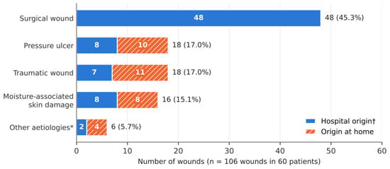
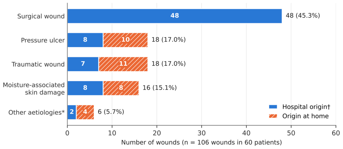
The 60 patients had 106 wounds, a mean of 1.8 (range 1–6) per patient; 28 patients (46.7%) had more than one wound, and two patients had six wounds, the maximum the form allows, so their wound count may be truncated. Surgical wounds were the most frequent type (45.3%), followed by pressure ulcers and traumatic wounds (17.0% each) and moisture-associated skin damage (15.1%) (Table 3, Figure 1). Among the 18 pressure ulcers, 1 was category 1, 3 category 2, 6 category 3, 1 category 4, 3 unclassifiable and 4 suspected deep tissue injuries: 7 (38.9%) were full-thickness (category 3 or 4) and 7 (38.9%) were of undetermined depth (unclassifiable or suspected deep tissue injury).
Table 3.
Wound types, origin and duration (n = 106 wounds in 60 patients).
Figure 1.
Distribution of the 106 wounds (in 60 patients) by type and place of origin. All surgical wounds had a hospital origin by definition; pressure ulcers, traumatic wounds and moisture-associated skin damage were split between hospital and home. * Diabetic foot ulcer (2), arterial leg ulcer (1), malignant wound (1), bullous pemphigoid lesion (1) and drain insertion site (1). † Recorded as acquired in hospital (current or previous admission).
Overall, 73 wounds (68.9%; cluster-robust 95% CI 56.7–81.0) had a hospital origin and 33 originated at home; none came from nursing homes, continuing care units or primary care. All 48 surgical wounds were hospital-acquired by definition; given their duration (1.5 to 10 years), at least three of them must have originated in previous admissions. Excluding surgical wounds, 25 of 58 wounds (43.1%; cluster-robust 95% CI 27.0–59.2) had a hospital origin, including 8 of 18 pressure ulcers, 8 of 16 moisture lesions and 7 of 18 traumatic wounds; 23 of these 25 wounds had been present for less than six weeks (median 10 days). Because the instrument does not distinguish the current admission from earlier ones, these figures describe where wounds originated, not the rate at which the hospital generates them.
More than half of the wounds (56.6%) were located in the pelvic region and lower limbs, as defined in the instrument: feet and ankles (16), legs, knees and thighs (14), buttocks, perineum and trochanters (13), sacrococcygeal region (11) and heels (6). The abdomen (18.9%) was the main site of surgical wounds. More than half of the pressure ulcers were on the feet or heels (10/18) and four on the sacrococcygeal region; moisture-associated skin damage was concentrated in the sacrococcygeal (7/16) and perianal or gluteal (7/16) areas.
Median wound duration was 9 days (IQR 2–30), but 22 wounds (20.8%) had been present for six weeks or more. Chronicity was most common among pressure ulcers (55.6%) and in the less frequent aetiologies (arterial leg ulcer, malignant wound and bullous pemphigoid). Five surgical wounds were also chronic. Three of them, present for 1.5, 2 and 10 years, were surgical wounds that had dehisced after postoperative complications and were healing by secondary intention.
3.4. Dressing Workload
For the most severe wound of each patient (n = 60), dressings were changed once a week in 14 cases (23.3%), twice a week in 7 (11.7%), three times a week in 17 (28.3%), daily in 18 (30.0%) and more than once a day in 4 (6.7%). Each dressing change took up to 15 min in 51 cases (85.0%), 15–30 min in 7 (11.7%) and 30–45 min in 2 (3.3%). Extrapolated from the care plans recorded on the survey day, the estimated weekly dressing time for the single most severe wound per patient was about 43 h (midpoint estimate 42.6 h), corresponding to about 261 dressing changes per week. This estimate rests on stated assumptions and has no confidence interval; its assumption-based scenario range was 10.0–75.3 h with the lower and upper limits of the time bands, 29.1–75.3 h if each change took at least 5 min, and 46.1 h (up to 82.3 h) if “more than once a day” meant three changes per day. For illustration only, 43 h corresponds to about 1.2 full-time equivalents of 35 h per week (scenario range 0.3–2.2); these are illustrative nursing hours and do not establish total staffing requirements. The median weekly time was 22.5 min (IQR 15.0–52.5) per patient. Daily or more frequent dressing changes were needed for 7 of 9 pressure ulcers and 6 of 7 moisture lesions recorded as the most severe wound, compared with 5 of 32 surgical wounds. These figures estimate the weekly dressing time required by the most severe wound of each patient present on the survey day; they are not a direct measurement of hospital workload.
3.5. Factors Associated with Having a Wound
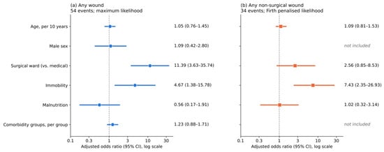
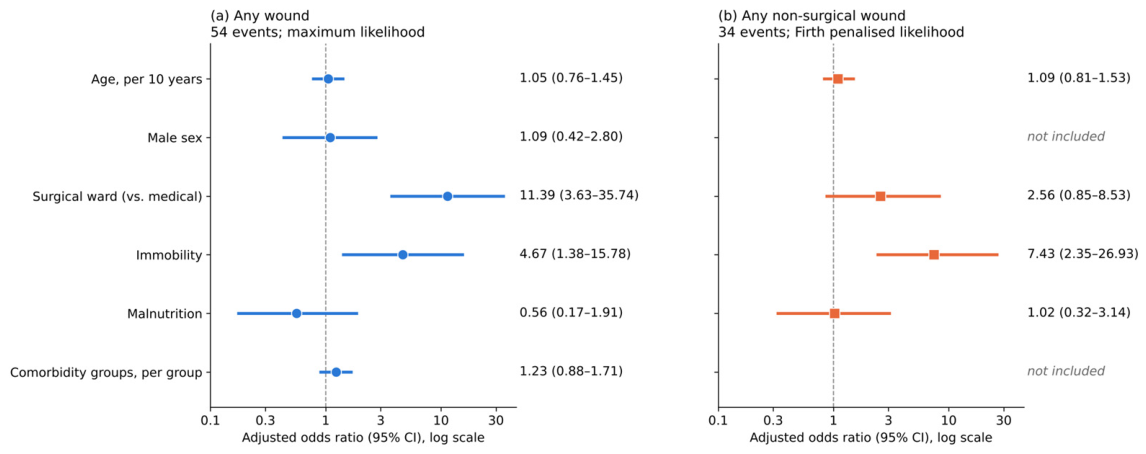
In the multivariable model of adults in the medical and surgical wards (Table 4, Model A; Figure 2), admission to the surgical ward (aOR 11.39; 95% CI 3.63–35.74) and immobility (aOR 4.67; 95% CI 1.38–15.78) were associated with having at least one wound after adjustment. Age, sex, malnutrition and number of comorbidity groups were not significantly associated. The model was significant (likelihood-ratio χ2 = 29.5; df = 6; p < 0.001), with Nagelkerke R2 = 0.32 and an apparent AUC of 0.80 (95% CI 0.70–0.88); the bootstrap optimism-corrected AUC was 0.76 and the calibration slope 0.79, indicating some overfitting. The Hosmer–Lemeshow test was non-significant (χ2 = 6.56; df = 8; p = 0.585), which does not by itself demonstrate good calibration. Age showed no departure from linearity (quadratic and Box–Tidwell terms, p > 0.8), all variance inflation factors were below 2, and the estimates were similar when the model was refitted with Firth’s method (surgical ward aOR 9.55, 95% CI 3.36–30.12; immobility aOR 4.22, 95% CI 1.36–14.27) or without the comorbidity count (surgical ward aOR 12.84, 95% CI 4.14–39.80; immobility aOR 5.18, 95% CI 1.57–17.07) (Table S2).
Table 4.
Multivariable logistic regression of factors associated with having at least one wound (Model A) and at least one non-surgical wound (Model B) in adults in the medical and surgical wards (n = 106).
Figure 2.
Adjusted odds ratios (95% CI, log scale) for having (a) at least one wound (Model A, maximum likelihood) and (b) at least one non-surgical wound (Model B, Firth penalised likelihood; post hoc analysis) among adults in the medical and surgical wards (n = 106). The association with the surgical ward weakens markedly when surgical wounds are excluded, whereas immobility remains associated; the two models have different covariate sets.
In the post hoc sensitivity analysis restricted to non-surgical wounds (Table 4, Model B; Figure 2), which was added during peer review, immobility was strongly associated with having a wound (aOR 7.43; 95% CI 2.35–26.93), whereas the association with the surgical ward was much weaker and not statistically significant (aOR 2.56; 95% CI 0.85–8.53; p = 0.095); age and malnutrition were not associated. This model had an apparent AUC of 0.76 (95% CI 0.65–0.85), an optimism-corrected AUC of 0.73 and a calibration slope of 0.89, with no evidence of non-linearity for age (p = 0.16); the Hosmer–Lemeshow test gave χ2 = 6.53 (df = 8; p = 0.588). The results were similar when using maximum likelihood estimation (immobility aOR 8.40, 95% CI 2.39–29.53; surgical ward aOR 2.74, 95% CI 0.84–8.96) and when sex and the number of comorbidity groups were added (immobility aOR 6.88, 95% CI 2.02–27.17; surgical ward aOR 2.02, 95% CI 0.61–7.12); immobility was significantly associated in 94% of 500 bootstrap resamples, compared with 34% for the surgical ward. Because immobility may also be a consequence of wounds, both models were refitted without it (Table S2): the surgical ward aOR was 5.92 (95% CI 2.20–15.89) for any wound and 1.02 (95% CI 0.41–2.47) for non-surgical wounds, and age was then associated with non-surgical wounds (aOR per 10 years 1.40; 95% CI 1.06–1.92).
3.6. Comparison with the Companion Pressure Ulcer Survey
Table 5 compares the pressure ulcer estimates of this survey with those of the companion survey carried out on the same day in the same hospital [16]. Restricted to the medical, surgical and obstetrics/gynaecology wards—the population of the companion survey—pressure ulcer prevalence was 10.5% (12/114) in this survey and 12.2% (14/115) in the companion survey, and the prevalence of ulcers of hospital origin was 6.1% and 7.0%, respectively. The two forms classify ulcers differently: the companion form has no category for unclassifiable ulcers, suspected deep tissue injury or moisture-associated skin damage, which may partly explain why it recorded more ulcers as deep ulcers or necrosis and slightly more patients with ulcers.
Table 5.
Pressure ulcer estimates in this survey and in the companion pressure ulcer survey carried out on the same day.
4. Discussion
4.1. Main Findings
This is, to our knowledge, the first study to describe the prevalence of all wound types among hospital inpatients in the Azores. On the survey day, one in two inpatients had at least one wound, almost half of them had several, and more than two-thirds of all wounds had a hospital origin—largely because all surgical wounds are hospital-acquired by definition; among non-surgical wounds, the proportion was 43.1%. Surgical wounds accounted for the largest share, as expected in a general hospital, but even after excluding them nearly one in three inpatients had a wound, mostly pressure ulcers, moisture-associated skin damage or traumatic wounds. One in five wounds was chronic, and the estimated weekly dressing time for the single most severe wound per patient, extrapolated from the survey-day care plans, was about 43 h (assumption-based scenario range 10–75 h).
4.2. Comparison with Other Studies
Direct comparison with other hospital surveys is limited by differences in case definitions, inclusion of surgical wounds and data sources. Point-prevalence surveys based on skin inspection in acute hospitals in Canada and England likewise found that wounds affect a large share of inpatients and that surgical wounds and pressure ulcers predominate [6,7]. No peer-reviewed study of all-wound prevalence in Portuguese hospitals was found, and the available peer-reviewed hospital data refer to pressure ulcers only, with prevalences of 5.8% among hospitalised patients in a multicentre regional survey [14] and 7.9% at the first skin assessment in a Portuguese hospital [15]. Our prevalence was also much higher than the 17.5% and 17.7% reported in two publications of the first application of the same instrument in Portuguese integrated continuing care units [12,18] (the same survey, analysed with slightly different denominators: 135/770 and 138/780), which is consistent with the higher proportion of surgical and acute wounds in a hospital population. Severe pressure damage appears to be a problem shared across levels of care: most pressure ulcers in the continuing care units were category 3 or 4 [12], and in this survey 7 of 18 were category 3 or 4 and a further 7 were unclassifiable or suspected deep tissue injuries.
The pressure ulcer prevalence observed (10.1%; 5.9% of hospital origin) is close to the pooled estimates of a global meta-analysis of hospitalised adults, 12.8% overall and 8.4% hospital-acquired [9], although our measure of hospital origin also includes ulcers acquired in earlier admissions. The pressure ulcers found were severe: 7 of 18 were full-thickness (category 3 or 4) and another 7 were unclassifiable or suspected deep tissue injuries, whose depth could not be established, and more than half were on the feet or heels. Heel ulcers are typically associated with inadequate offloading, whereas toe lesions in patients with diabetes or peripheral arterial disease raise the possibility of misclassification between pressure, ischaemic and diabetic foot ulcers. These findings support heel offloading and a careful aetiological assessment of foot wounds, including vascular assessment, as recommended by international guidelines [22,38].
Moisture-associated skin damage affected 11.8% of patients and was concentrated in the sacral and perianal areas, the same areas at risk of pressure ulcers. Incontinence-associated dermatitis is frequent in acute care and is associated with pressure injury [24,39], so its prevention through structured skin care and continence management is likely to benefit both conditions. However, incontinence was not recorded in the instrument, so the contribution of what is probably the main determinant of moisture-related lesions could not be assessed in this survey. Traumatic wounds affected 10.9% of patients, broadly similar to the 8.9% skin tear prevalence reported over ten years in an Australian acute hospital [40], although our traumatic category is broader than skin tears and the two figures measure different constructs. Because skin tears are not a separate category in the current version of the instrument, the inclusion of the International Skin Tear Advisory Panel (ISTAP) classification would improve the specificity of future surveys [41,42].
Five surgical wounds had been present for more than six weeks. Three of them, lasting more than a year, had dehisced after postoperative complications and were healing by secondary intention; given their duration, they must have originated in previous admissions, which illustrates why hospital origin should not be equated with acquisition during the current stay. Surgical wound dehiscence turns an acute wound into a hard-to-heal one and carries a substantial clinical and economic burden [43,44]. In the instrument, these wounds remain classified as surgical wounds, and dehiscence was identified only through the clinical information gathered at the bedside. Adding an item that distinguishes healing by primary intention from dehiscence and healing by secondary intention would allow surgical wound complications to be monitored systematically. Finally, the presence of an arterial leg ulcer, a malignant wound and bullous pemphigoid lesions among the chronic wounds is a reminder that hard-to-heal wounds often have uncommon aetiologies requiring differential diagnosis and, when indicated, biopsy [45].
4.3. Factors Associated with Wounds and Workload
Among adults in the medical and surgical wards, admission to the surgical ward and immobility were associated with having a wound after adjustment. The surgical ward association is, however, largely definitional: surgical admission usually entails a surgical incision, which is itself a wound in this survey, and uncomplicated incisions are an expected consequence of care rather than adverse events. When the outcome was restricted to non-surgical wounds, the association with the surgical ward weakened markedly and was no longer statistically significant, whereas immobility remained strongly associated. The ward estimate should therefore not be read as evidence that surgical wards independently increase the risk of avoidable wounds, and the wide confidence intervals of both models reflect the small number of events. Because the survey is cross-sectional, the associations cannot be interpreted causally: immobility may be both a risk factor for wounds and a consequence of them (reverse causation), and the models describe associations rather than estimate causal effects or predict individual risk; their apparent discrimination is optimistic for other populations. Immobility is a known determinant of pressure ulcers and moisture-related lesions [22], and the non-surgical model supports targeting prevention at immobile patients.
The estimated dressing time was considerable. Counting only the single most severe wound per patient, extrapolation of the survey-day care plans gives about 261 dressing changes and an estimated 43 h of dressing time per week. This is an assumption-based estimate, not a measurement: it rests on the midpoints of the time bands and on twice-daily changes for wounds dressed more than once a day, and the band limits give a scenario range of about 10 to 75 h, which is not a confidence interval. Expressed for illustration as full-time equivalents of 35 h per week [26], this corresponds to about 0.3 to 2.2 full-time equivalents; these are illustrative nursing hours and do not establish total staffing requirements. Because time was recorded only for the most severe wound and in 15 min bands, the true workload is uncertain, but even under the lowest assumptions it represents a relevant share of a nurse’s working week. Pressure ulcers and moisture lesions were the wounds most often dressed daily. Since nurse visits are the main cost driver of wound care [3,4], reducing preventable wounds of hospital origin would release resources as well as improve patient outcomes.
4.4. Implications for Practice and Research
The results support three practical recommendations. First, hospital quality monitoring should move from pressure-ulcer-only audits to periodic all-wound surveys using a standardised instrument; the ELCOS instrument proved feasible for a whole-hospital survey in a single day. Second, prevention priorities should include heel offloading, structured skin care for incontinent and immobile patients, and prevention of skin tears. Third, chronic and atypical wounds should be referred early to a specialised wound care team for aetiological diagnosis. Following this survey, the study group delivered wound care training to ward staff and plans to repeat the survey in three years to evaluate its impact. Future research should replicate the survey in the other Azorean hospitals and in primary care, use repeated measurements to estimate trends, and formally quantify inter-rater agreement of the instrument in acute care, adding wound photographs to the instruction manual as suggested by its developers [18]; the instrument would also benefit from items on incontinence, skin tears, the healing intention of surgical wounds and acquisition during the current admission.
4.5. Strengths and Limitations
The main strengths of this study are the inclusion of all inpatients and all wound types, the bedside examination of every patient, with dressings removed, by two trained observers (one of them external to the ward), the use of a standardised national instrument with established content validity and inter-rater reliability [18], and the availability of a companion pressure ulcer survey conducted on the same day, which allowed the pressure ulcer estimates of two instruments to be compared (Table 5).
Several limitations must be acknowledged. The study was conducted in one hospital on one day, and the sample size yields wide confidence intervals, particularly for the less frequent wound types and for the smaller services; the results apply to this hospital on the survey day and may not be generalisable to other settings. Because elective surgery and discharges vary over the week, the proportion of recent postoperative patients, and therefore the number of surgical wounds and the dressing workload, may have been different on another day. Eleven eligible patients (8.5%) declined to participate; their wound status is unknown, so the true prevalence could lie between 46.2% and 54.6%. Length of stay was not recorded; the exclusion of patients hospitalised for less than 24 h and the length-biased sampling inherent in point-prevalence surveys mean that patients with longer stays, who are more likely to have wounds, are over-represented [20]. Wound type was assigned clinically, without systematic diagnostic confirmation, so misclassification between pressure, diabetic, arterial, moisture-related and traumatic lesions is possible; pressure ulcer classification is difficult even for trained nurses [46]. Wound duration and origin were partly based on records and patient or staff reports and are subject to recall bias, and the instrument does not distinguish wounds acquired during the current admission from those acquired earlier. Immobility and malnutrition were recorded as binary items from the Braden subscale criteria, and “malnutrition” reflected inadequate intake rather than a clinical diagnosis, so misclassification of these variables may have biased their estimated associations with wounds. Incontinence was not recorded, and two patients reached the six-wound limit of the form, so their wound count may be underestimated. Workload data were collected only for the most severe wound, in time bands and from the care plans reported by the ward nurses rather than by direct timing, so total workload is uncertain and probably underestimated; the instrument also does not record wound size, infection or materials used, which limits cost estimates [18]. Its inter-rater reliability was established in continuing care rather than acute care, and the main discrepancies between observers concerned pressure ulcer category and wound typology [18]; in this survey, the two observers recorded a single consensus assessment, so agreement between them could not be quantified. Finally, the regression analysis was exploratory, restricted to adults in the medical and surgical wards and limited to the variables in the instrument; Model B and the sensitivity analyses were added after the primary results were known; the cross-sectional design precludes causal interpretation and allows reverse causation, the apparent model performance is optimistic, and residual confounding cannot be excluded.
5. Conclusions
One in two inpatients of a general hospital in the Azores had at least one wound, and almost one in three had a non-surgical wound. More than two-thirds of wounds had a hospital origin, largely because surgical wounds are hospital-acquired by definition; among non-surgical wounds, the proportion was 43.1%. Besides surgical wounds, pressure ulcers, moisture-associated skin damage and traumatic wounds were frequent; pressure ulcers were often severe or chronic, and the estimated weekly dressing time for the most severe wound of each patient alone was substantial. Immobility was the factor most consistently associated with non-surgical wounds, whereas the association with surgical admission mainly reflected surgical incisions. The ELCOS instrument made it possible to measure the prevalence of all wound types in a single day. All-wound prevalence was about five times the pressure ulcer prevalence in this survey (50.4% vs. 10.1%): because pressure-ulcer-only audits assess a narrower outcome, they capture fewer patients with any wound, but this ratio does not indicate a fivefold difference in severity, preventable harm or care requirements. Periodic all-wound surveillance should be integrated into hospital quality monitoring to guide prevention, staff training and the planning of wound care resources.
Supplementary Materials
The following supporting information can be downloaded at https://www.mdpi.com/article/10.3390/healthcare14223989/s1. Table S1: STROBE checklist. Table S2: Sensitivity analyses of the regression models. File S1: ELCOS wound characterisation form (English version). File S2: Instruction manual of the ELCOS wound characterisation form (English version). File S3: Informed consent form (English translation). References [47,48,49,50] are cited in the Supplementary Materials.
Author Contributions
Conceptualization, K.F. and A.N.V.; methodology, K.F., M.L. and P.G.; software, M.L. and P.G.; formal analysis, P.G. and K.F.; investigation, A.N.V., D.G., M.R. and M.C.; resources, A.N.V. and D.G.; writing—original draft preparation, K.F., A.N.V., D.G., M.R. and M.C.; writing—review and editing, K.F., A.N.V., M.L. and P.G.; visualization, P.G., M.R. and M.C.; project administration, K.F. and A.N.V. All authors have read and agreed to the published version of the manuscript.
Funding
This work is funded by national funds through the Foundation for Science and Technology, under the project UID/06291/2025—https://doi.org/10.54499/UID/06291/2025. The funder supported the publication costs and had no role in the design of the study; in the collection, analyses or interpretation of data; in the writing of the manuscript; or in the decision to publish the results.
Institutional Review Board Statement
The study was conducted in accordance with the Declaration of Helsinki and approved by the Ethics Committee of Hospital de Santo Espírito da Ilha Terceira (process no. 06/2023, approved on 5 April 2023).
Informed Consent Statement
Informed consent was obtained from all subjects involved in the study or, for minors, from their parents; for adults lacking decision-making capacity, consent was obtained from their legal representative or closest relative.
Data Availability Statement
The anonymised dataset supporting the conclusions of this article is available from the corresponding author upon reasonable request, subject to approval by the institution.
Acknowledgments
The authors thank the nurses of Hospital de Santo Espírito da Ilha Terceira who collected the data and ELCOS—Portuguese Wound Society for making the prevalence instrument available.
Conflicts of Interest
The authors declare no conflicts of interest.
Abbreviations
The following abbreviations are used in this manuscript:
| aOR | Adjusted odds ratio |
| AUC | Area under the receiver operating characteristic curve |
| CI | Confidence interval |
| ELCOS | Sociedade Portuguesa de Feridas (Portuguese Wound Society) |
| EPUAP | European Pressure Ulcer Advisory Panel |
| HSEIT | Hospital de Santo Espírito da Ilha Terceira |
| ICD-10 | International Classification of Diseases, 10th revision |
| ICNP | International Classification for Nursing Practice |
| IQR | Interquartile range |
| ISTAP | International Skin Tear Advisory Panel |
| NPIAP | National Pressure Injury Advisory Panel |
| PPPIA | Pan Pacific Pressure Injury Alliance |
| SD | Standard deviation |
| STROBE | Strengthening the Reporting of Observational Studies in Epidemiology |
| TRIPOD | Transparent Reporting of a multivariable prediction model for Individual Prognosis Or Diagnosis |
References
- Sen, C.K. Human Wound and Its Burden: Updated 2020 Compendium of Estimates. Adv. Wound Care 2021, 10, 281–292. [Google Scholar] [CrossRef] [Scilit] [PubMed]
- Olsson, M.; Järbrink, K.; Divakar, U.; Bajpai, R.; Upton, Z.; Schmidtchen, A.; Car, J. The Humanistic and Economic Burden of Chronic Wounds: A Systematic Review. Wound Repair Regen. 2019, 27, 114–125. [Google Scholar] [CrossRef] [Scilit] [PubMed]
- Guest, J.F.; Fuller, G.W.; Vowden, P. Cohort Study Evaluating the Burden of Wounds to the UK’s National Health Service in 2017/2018: Update from 2012/2013. BMJ Open 2020, 10, e045253. [Google Scholar] [CrossRef] [Scilit] [PubMed]
- Guest, J.F.; Ayoub, N.; McIlwraith, T.; Uchegbu, I.; Gerrish, A.; Weidlich, D.; Vowden, K.; Vowden, P. Health Economic Burden That Wounds Impose on the National Health Service in the UK. BMJ Open 2015, 5, e009283. [Google Scholar] [CrossRef] [Scilit] [PubMed]
- Martinengo, L.; Olsson, M.; Bajpai, R.; Soljak, M.; Upton, Z.; Schmidtchen, A.; Car, J.; Järbrink, K. Prevalence of Chronic Wounds in the General Population: Systematic Review and Meta-Analysis of Observational Studies. Ann. Epidemiol. 2019, 29, 8–15. [Google Scholar] [CrossRef] [Scilit] [PubMed]
- Hurd, T.; Posnett, J. Point Prevalence of Wounds in a Sample of Acute Hospitals in Canada. Int. Wound J. 2009, 6, 287–293. [Google Scholar] [CrossRef] [Scilit] [PubMed]
- Srinivasaiah, N.; Dugdall, H.; Barrett, S.; Drew, P.J. A Point Prevalence Survey of Wounds in North-East England. J. Wound Care 2007, 16, 413–419. [Google Scholar] [CrossRef] [Scilit] [PubMed]
- Baharestani, M.M.; Black, J.M.; Carville, K.; Clark, M.; Cuddigan, J.E.; Dealey, C.; Defloor, T.; Harding, K.G.; Lahmann, N.A.; Lubbers, M.J.; et al. Dilemmas in Measuring and Using Pressure Ulcer Prevalence and Incidence: An International Consensus. Int. Wound J. 2009, 6, 97–104. [Google Scholar] [CrossRef] [Scilit] [PubMed]
- Li, Z.; Lin, F.; Thalib, L.; Chaboyer, W. Global Prevalence and Incidence of Pressure Injuries in Hospitalised Adult Patients: A Systematic Review and Meta-Analysis. Int. J. Nurs. Stud. 2020, 105, 103546. [Google Scholar] [CrossRef] [Scilit] [PubMed]
- Pina, E.; Furtado, K.; Franks, P.J.; Moffatt, C.J. Leg Ulceration in Portugal: Prevalence and Clinical History. Eur. J. Vasc. Endovasc. Surg. 2005, 29, 549–553. [Google Scholar] [CrossRef] [Scilit] [PubMed]
- Furtado, K.; Pina, E.; Moffatt, C.J.; Franks, P.J. Leg Ulceration in Portugal: Quality of Life. Int. Wound J. 2008, 5, 34–39. [Google Scholar] [CrossRef] [Scilit] [PubMed]
- Furtado, K.A.X.; Infante, P.; Sobral, A.; Gaspar, P.; Eliseu, G.; Lopes, M. Prevalence of Acute and Chronic Wounds—With Emphasis on Pressure Ulcers—In Integrated Continuing Care Units in Alentejo, Portugal. Int. Wound J. 2020, 17, 1002–1010. [Google Scholar] [CrossRef] [Scilit] [PubMed]
- Barreiro, C.M.P. Feridas Crónicas em Portugal: Características, Práticas e Custos. Master’s Thesis, Universidade Católica Portuguesa, Lisboa, Portugal, 2017. Available online: http://hdl.handle.net/10400.14/39131 (accessed on 5 April 2026).
- Lopes, T.S.; Videira, L.M.M.D.S.; Saraiva, D.M.R.F.; Agostinho, E.S.; Bandarra, A.J.F. Multicentre Study of Pressure Ulcer Point Prevalence in a Portuguese Region. J. Tissue Viability 2020, 29, 12–18. [Google Scholar] [CrossRef] [Scilit] [PubMed]
- Sardo, P.M.G.; Simões, C.S.O.; Alvarelhão, J.J.M.; Costa, C.T.O.; Simões, C.J.C.; Figueira, J.M.R.; Simões, J.F.F.L.; Amado, F.M.L.; Amaro, A.J.M.; Melo, E.M.O.P. Analyses of Pressure Ulcer Point Prevalence at the First Skin Assessment in a Portuguese Hospital. J. Tissue Viability 2016, 25, 75–82. [Google Scholar] [CrossRef] [Scilit] [PubMed]
- Leal, M.; Dias, L.; Furtado, K.; Gaspar, P.; Vieira, A. Prevalência de Úlceras por Pressão: Estudo Transversal num Hospital dos Açores [Pressure Ulcer Prevalence: Cross Sectional Study in a Hospital of the Azores]. Rev. Investig. Inov. Saúde 2025, 8, e484. [Google Scholar] [CrossRef] [Scilit]
- Defloor, T.; Clark, M.; Witherow, A.; Colin, D.; Lindholm, C.; Schoonhoven, L.; Moore, Z. EPUAP Statement on Prevalence and Incidence Monitoring of Pressure Ulcer Occurrence. J. Tissue Viability 2005, 15, 20–27. [Google Scholar] [CrossRef] [Scilit] [PubMed]
- Furtado, K.A.X.; Infante, P.; Afonso, A.; Gomes, F.; Carranca, J.; Lopes, M.J. Desenvolvimento, Validação e Aplicação de um Instrumento de Caracterização das Feridas [Development, Validation and Application of a Tool to Measure Wound Characteristics]. Rev. Enferm. Atual Derme 2021, 95, e021122. [Google Scholar] [CrossRef] [Scilit]
- von Elm, E.; Altman, D.G.; Egger, M.; Pocock, S.J.; Gøtzsche, P.C.; Vandenbroucke, J.P. The Strengthening the Reporting of Observational Studies in Epidemiology (STROBE) Statement: Guidelines for Reporting Observational Studies. Lancet 2007, 370, 1453–1457. [Google Scholar] [CrossRef] [Scilit] [PubMed]
- Wolkewitz, M.; Mandel, M.; Palomar-Martinez, M.; Alvarez-Lerma, F.; Olaechea-Astigarraga, P.; Schumacher, M. Methodological Challenges in Using Point-Prevalence versus Cohort Data in Risk Factor Analyses of Nosocomial Infections. Ann. Epidemiol. 2018, 28, 475–480.e1. [Google Scholar] [CrossRef] [Scilit] [PubMed]
- Bergstrom, N.; Braden, B.J.; Laguzza, A.; Holman, V. The Braden Scale for Predicting Pressure Sore Risk. Nurs. Res. 1987, 36, 205–210. [Google Scholar] [CrossRef] [Scilit]
- European Pressure Ulcer Advisory Panel; National Pressure Injury Advisory Panel; Pan Pacific Pressure Injury Alliance. Prevention and Treatment of Pressure Ulcers/Injuries: Clinical Practice Guideline. The International Guideline, 3rd ed.; Haesler, E., Ed.; EPUAP/NPIAP/PPPIA: Prague, Czech Republic, 2019; Available online: https://www.trezzo.co.uk/documents/International-Guideline-2019.pdf (accessed on 10 June 2026).
- Kottner, J.; Cuddigan, J.; Carville, K.; Balzer, K.; Berlowitz, D.; Law, S.; Litchford, M.; Mitchell, P.; Moore, Z.; Pittman, J.; et al. Pressure Ulcer/Injury Classification Today: An International Perspective. J. Tissue Viability 2020, 29, 197–203. [Google Scholar] [CrossRef] [Scilit] [PubMed]
- Beeckman, D.; Campbell, J.; Campbell, K.; Chimentão, D.; Coyer, F.; Domansky, R.; Gray, M.; Hevia, H.; Junkin, J.; Karadag, A.; et al. Proceedings of the Global IAD Expert Panel. Incontinence-Associated Dermatitis: Moving Prevention Forward; Wounds International: London, UK, 2015. [Google Scholar]
- Defloor, T.; Schoonhoven, L.; Fletcher, J.; Furtado, K.; Heyman, H.; Lubbers, M.; Lyder, C.; Witherow, A. Statement of the European Pressure Ulcer Advisory Panel—Pressure Ulcer Classification: Differentiation between Pressure Ulcers and Moisture Lesions. J. Wound Ostomy Cont. Nurs. 2005, 32, 302–306. [Google Scholar] [CrossRef] [Scilit] [PubMed]
- Decreto-Lei n.º 71/2019, de 27 de Maio. Diário da República, 1.ª Série, n.º 101, 27 May 2019. Available online: https://diariodarepublica.pt/dr/detalhe/decreto-lei/71-2019-122403266 (accessed on 10 June 2026).
- Wilson, E.B. Probable Inference, the Law of Succession, and Statistical Inference. J. Am. Stat. Assoc. 1927, 22, 209–212. [Google Scholar] [CrossRef]
- Brown, L.D.; Cai, T.T.; DasGupta, A. Interval Estimation for a Binomial Proportion. Stat. Sci. 2001, 16, 101–133. [Google Scholar] [CrossRef] [Scilit]
- Holm, S. A Simple Sequentially Rejective Multiple Test Procedure. Scand. J. Stat. 1979, 6, 65–70. [Google Scholar]
- Collins, G.S.; Reitsma, J.B.; Altman, D.G.; Moons, K.G.M. Transparent Reporting of a Multivariable Prediction Model for Individual Prognosis or Diagnosis (TRIPOD): The TRIPOD Statement. BMJ 2015, 350, g7594. [Google Scholar] [CrossRef] [Scilit] [PubMed]
- Peduzzi, P.; Concato, J.; Kemper, E.; Holford, T.R.; Feinstein, A.R. A Simulation Study of the Number of Events per Variable in Logistic Regression Analysis. J. Clin. Epidemiol. 1996, 49, 1373–1379. [Google Scholar] [CrossRef] [Scilit] [PubMed]
- Vittinghoff, E.; McCulloch, C.E. Relaxing the Rule of Ten Events per Variable in Logistic and Cox Regression. Am. J. Epidemiol. 2007, 165, 710–718. [Google Scholar] [CrossRef] [Scilit] [PubMed]
- Firth, D. Bias Reduction of Maximum Likelihood Estimates. Biometrika 1993, 80, 27–38. [Google Scholar] [CrossRef]
- Heinze, G.; Schemper, M. A Solution to the Problem of Separation in Logistic Regression. Stat. Med. 2002, 21, 2409–2419. [Google Scholar] [CrossRef] [Scilit] [PubMed]
- DeLong, E.R.; DeLong, D.M.; Clarke-Pearson, D.L. Comparing the Areas under Two or More Correlated Receiver Operating Characteristic Curves: A Nonparametric Approach. Biometrics 1988, 44, 837–845. [Google Scholar] [CrossRef] [Scilit] [PubMed]
- Harrell, F.E.; Lee, K.L.; Mark, D.B. Multivariable Prognostic Models: Issues in Developing Models, Evaluating Assumptions and Adequacy, and Measuring and Reducing Errors. Stat. Med. 1996, 15, 361–387. [Google Scholar] [CrossRef] [Scilit]
- Van Calster, B.; McLernon, D.J.; van Smeden, M.; Wynants, L.; Steyerberg, E.W. Calibration: The Achilles Heel of Predictive Analytics. BMC Med. 2019, 17, 230. [Google Scholar] [CrossRef] [Scilit] [PubMed]
- Schaper, N.C.; van Netten, J.J.; Apelqvist, J.; Bus, S.A.; Fitridge, R.; Game, F.; Monteiro-Soares, M.; Senneville, E. Practical Guidelines on the Prevention and Management of Diabetes-Related Foot Disease (IWGDF 2023 Update). Diabetes Metab. Res. Rev. 2024, 40, e3657. [Google Scholar] [CrossRef] [Scilit] [PubMed]
- Gray, M.; Giuliano, K.K. Incontinence-Associated Dermatitis, Characteristics and Relationship to Pressure Injury: A Multisite Epidemiologic Analysis. J. Wound Ostomy Cont. Nurs. 2018, 45, 63–67. [Google Scholar] [CrossRef] [Scilit] [PubMed]
- Miles, S.J.; Fulbrook, P.; Williams, D.M. Skin Tear Prevalence in an Australian Acute Care Hospital: A 10-Year Analysis. Int. Wound J. 2022, 19, 1418–1427. [Google Scholar] [CrossRef] [Scilit] [PubMed]
- LeBlanc, K.; Baranoski, S.; Christensen, D.; Langemo, D.; Sammon, M.A.; Edwards, K.; Holloway, S.; Gloeckner, M.; Williams, A.; Sibbald, R.G.; et al. International Skin Tear Advisory Panel: A Tool Kit to Aid in the Prevention, Assessment, and Treatment of Skin Tears Using a Simplified Classification System. Adv. Skin Wound Care 2013, 26, 459–476. [Google Scholar] [CrossRef] [Scilit] [PubMed]
- LeBlanc, K.; Campbell, K.; Beeckman, D.; Dunk, A.M.; Harley, C.; Hevia, H.; Holloway, S.; Idensohn, P.; Langemo, D.; Ousey, K.; et al. Best Practice Recommendations for the Prevention and Management of Skin Tears in Aged Skin; Wounds International: London, UK, 2018. [Google Scholar]
- Sandy-Hodgetts, K.; Carville, K.; Leslie, G.D. Determining Risk Factors for Surgical Wound Dehiscence: A Literature Review. Int. Wound J. 2015, 12, 265–275. [Google Scholar] [CrossRef] [Scilit] [PubMed]
- World Union of Wound Healing Societies (WUWHS). Consensus Document. Surgical Wound Dehiscence: Improving Prevention and Outcomes; Wounds International: London, UK, 2018. [Google Scholar]
- Isoherranen, K.; O’Brien, J.J.; Barker, J.; Dissemond, J.; Hafner, J.; Jemec, G.B.E.; Kamarachev, J.; Läuchli, S.; Montero, E.C.; Nobbe, S.; et al. Atypical Wounds. Best Clinical Practice and Challenges. J. Wound Care 2019, 28, S1–S92. [Google Scholar] [CrossRef] [Scilit] [PubMed]
- Beeckman, D.; Schoonhoven, L.; Fletcher, J.; Furtado, K.; Gunningberg, L.; Heyman, H.; Lindholm, C.; Paquay, L.; Verdú, J.; Defloor, T. EPUAP Classification System for Pressure Ulcers: European Reliability Study. J. Adv. Nurs. 2007, 60, 682–691. [Google Scholar] [CrossRef] [Scilit] [PubMed]
- European Pressure Ulcer Advisory Panel; National Pressure Ulcer Advisory Panel; Pan Pacific Pressure Injury Alliance. Prevention and Treatment of Pressure Ulcers: Quick Reference Guide; Haesler, E., Ed.; Cambridge Media: Osborne Park, Australia, 2014. [Google Scholar]
- Harding, K.; Dowsett, C.; Fias, L.; Jelnes, R.; Mosti, G.; Öien, R.; Partsch, H.; Reeder, S.; Senet, P.; Verdú Soriano, J.; et al. Simplifying Venous Leg Ulcer Management. Consensus Recommendations; Wounds International: London, UK, 2015. [Google Scholar]
- Pina, E.; Furtado, K.; Albino, A.P. Boas Práticas no Tratamento e Prevenção das Úlceras de Perna de Origem Venosa; GAIF: Lisbon, Portugal, 2007. [Google Scholar]
- Vowden, K.; Kerr, A.; Mosti, G. Demystifying Mixed Arterial Venous Leg Ulcers; Wounds International: London, UK, 2020. [Google Scholar]

Disclaimer/Publisher’s Note: The statements, opinions and data contained in all publications are solely those of the individual author(s) and contributor(s) and not of MDPI and/or the editor(s). MDPI and/or the editor(s) disclaim responsibility for any injury to people or property resulting from any ideas, methods, instructions or products referred to in the content. |
© 2026 by the authors. Licensee MDPI, Basel, Switzerland. This article is an open access article distributed under the terms and conditions of the Creative Commons Attribution (CC BY) license.