


Abstract
Background and Objectives: We evaluated the incidence, spectrum, and baseline factors associated with severe infection after rituximab-based therapy for B-cell lymphoproliferative disorders in Western Romania. Materials and Methods: This retrospective cohort included 142 adults starting rituximab-containing therapy between January 2020 and December 2024. The primary outcome was infection according to the Common Terminology Criteria for Adverse Events grade ≥ 3 within 12 months. Analyses included competing-risk cumulative incidence estimation, exploratory multivariable logistic regression, restricted cubic splines, bootstrap validation, and decision-curve analysis. Results: Severe infection occurred in 38 patients (26.8%; 95% confidence interval [CI] 20.2–34.6); the 12-month cumulative incidence accounting for death as a competing event was 27.1%. Lower respiratory tract infections predominated (39.5%), and 42.1% of severe events occurred within 90 days. Hypogammaglobulinaemia at 6 months affected 39.1% of evaluable patients. In an exploratory multivariable model (36 events; nine predictors), associations were observed for baseline immunoglobulin G < 6 g/L (adjusted odds ratio [aOR] 4.96; 95% CI 1.28–19.18), serum albumin < 35 g/L (aOR 4.67; 95% CI 1.78–12.23), diabetes mellitus (aOR 2.73; 95% CI 1.03–7.24), and age per decade (aOR 1.83; 95% CI 1.11–2.99), whereas continuous immunoglobulin G showed a weaker association (aOR per 1 g/L 0.93; 95% CI 0.78–1.11). The optimism-corrected C-statistic was 0.717 (apparent, 0.789), and the corrected calibration slope was 0.671, indicating overfitting. Conclusions: Severe infections were frequent following rituximab-based therapy. Baseline immunoglobulin levels, albumin, diabetes, and age were associated with severe infection and, if confirmed, may help identify patients for closer surveillance. The exploratory prediction model showed reduced performance after internal validation and requires external validation before clinical implementation.
Keywords:
rituximab; lymphoma; non-Hodgkin; leukemia; lymphocytic; chronic; B-cell; infections; agammaglobulinemia 1. Introduction
Rituximab, a chimeric monoclonal antibody directed against CD20, transformed the treatment of B-cell lymphoproliferative disorders and remains, more than two decades after its approval, the most widely used immunotherapeutic agent in haematological oncology [1]. Its addition to cyclophosphamide, doxorubicin, vincristine, and prednisone (CHOP) improved complete response rates and overall survival in diffuse large B-cell lymphoma (DLBCL), establishing R-CHOP as the global standard of care [2]. In follicular lymphoma, rituximab-based induction followed by two years of maintenance prolongs progression-free survival [3], and in chronic lymphocytic leukaemia (CLL) chemoimmunotherapy with fludarabine, cyclophosphamide, and rituximab was the first regimen to demonstrate a survival advantage [4]. Across these indications, rituximab is administered to tens of thousands of patients each year, including an expanding population in Central and Eastern Europe.
The therapeutic benefit of rituximab is inseparable from its mechanism of action. Profound and prolonged depletion of circulating B-cells, which typically persists for six to twelve months after the last infusion, impairs humoral immunity and increases susceptibility to infection. A systematic review and meta-analysis of randomised trials did not find a significant increase in severe infections when rituximab was added to chemotherapy during induction; this result does not address the risks of prolonged maintenance exposure [5]. The spectrum of complications includes reactivation of hepatitis B virus (HBV), which has been documented in up to one in six HBsAg-positive patients and in a smaller but clinically important proportion of patients with resolved infection [6]. Secondary hypogammaglobulinaemia develops in roughly one third of patients and is associated with recurrent infections requiring immunoglobulin replacement [7], and late-onset neutropenia is a distinct, often unanticipated complication that typically emerges several weeks after the final infusion [8].
Several specific infectious syndromes merit attention in rituximab-treated patients. Pneumocystis jirovecii pneumonia occurs with sufficient frequency during rituximab-containing chemotherapy that prophylaxis is now recommended for many regimens, particularly those combining rituximab with high-dose corticosteroids or bendamustine [9]. The European Society of Clinical Microbiology and Infectious Diseases (ESCMID) consensus on the safety of targeted and biological therapies provides a structured framework for screening, prophylaxis, and vaccination in patients receiving anti-CD20 antibodies, yet it also underlines that most of the underlying evidence derives from clinical trials conducted in Western Europe and North America [10]. Such populations differ in age, comorbidity, viral epidemiology, and access to supportive care from patients treated in routine practice elsewhere.
Romania illustrates these differences. The country has one of the highest prevalences of HBV infection in the European Union, with a nationwide survey estimating that 4.4% of adults are HBsAg carriers and more than one in four have serological evidence of previous exposure [11]. Rural residence, delayed diagnosis, and a high burden of comorbidity characterise many patients reaching tertiary haematology centres, and single-centre Romanian series of DLBCL report a substantial proportion of elderly patients treated with attenuated regimens [12]. Over the past five years, the Romanian treatment landscape has also shifted markedly towards biosimilar rituximab, whose efficacy and safety were shown to be equivalent to the reference product in randomised trials of follicular lymphoma [13], but whose infectious profile in unselected populations remains incompletely described.
Despite these distinctive features, there are no published cohort data from Western Romania describing the incidence, timing, and determinants of severe infection after rituximab. Existing prognostic tools such as the International Prognostic Index were designed to predict survival rather than infectious toxicity [14], and the predictors identified in Western cohorts, including age, immunoglobulin levels, and comorbidity, have not been validated in this setting. Clinicians therefore rely on extrapolated recommendations when deciding which patients need intensified monitoring, antimicrobial prophylaxis, or immunoglobulin replacement.
The present study was designed to address this gap. Using a retrospective cohort of consecutive patients who initiated rituximab-based therapy at a tertiary centre in Timișoara between 2020 and 2024, we aimed to (i) quantify the 12-month incidence and clinical spectrum of severe infection across the three main diagnostic groups, (ii) describe the secondary outcomes of hypogammaglobulinaemia, HBV reactivation, and late-onset neutropenia, (iii) explore baseline factors associated with severe infection, and (iv) explore, with internal validation, whether a simple clinical prediction model, including flexible modelling of immunoglobulin levels, could inform future studies of risk-adapted infection prevention.
2. Materials and Methods
2.1. Study Design and Population
We conducted a single-centre, retrospective cohort study at the Department of Haematology of a tertiary university hospital in Timișoara, which serves as the referral centre for the four counties of Western Romania (Timiș, Arad, Caraș-Severin, and Hunedoara), a catchment population of approximately 1.7 million inhabitants. The study period covered rituximab initiations between 1 January 2020 and 31 December 2024, and each patient was followed from the first rituximab infusion until day 365, death, or the last documented contact, whichever came first, with the 12-month assessment scheduled at day 365 ± 30 days. Data were extracted from the electronic health record, the hospital pharmacy dispensing database, which records every rituximab vial administered, and the microbiology laboratory information system between June and September 2026, after the 12-month follow-up of all included patients had been completed. The study was reported in accordance with the Strengthening the Reporting of Observational Studies in Epidemiology (STROBE) recommendations for cohort studies.
The study was conducted in accordance with the Declaration of Helsinki and approved by the Institutional Ethics Committee of Pius Brînzeu Clinical Emergency County Hospital (decision 630/15 June 2026), before data extraction. Because of the retrospective design and the use of pseudonymised data, the Ethics Committee waived the requirement for study-specific informed consent; all patients had signed the hospital’s general consent form at admission, which permits the use of anonymised clinical data for research.
2.2. Eligibility Criteria and Group Allocation
Patients were eligible if they were aged 18 years or older, had a histologically or immunophenotypically confirmed CD20-positive B-cell lymphoproliferative disorder, and received at least one full dose of rituximab (375 mg/m2 intravenously, or 500 mg/m2 from the second cycle in CLL protocols) as part of a systemic treatment regimen started during the study period. Patients were excluded if rituximab was given for a non-malignant indication, if they had received an allogeneic stem-cell transplant, if a severe infection was ongoing at the time of the first infusion, or if follow-up data covering at least 90 days were unavailable for reasons other than death. Patients who had previously received rituximab were eligible provided that the index course began more than six months after the last prior infusion.
Participants were allocated to three mutually exclusive diagnostic groups according to the World Health Organization classification: DLBCL and other aggressive B-cell lymphomas treated with curative intent (R-CHOP, R-miniCHOP, or dose-adjusted R-EPOCH); indolent lymphomas, comprising follicular and marginal-zone lymphoma treated with R-bendamustine, R-CVP, or R-CHOP with or without rituximab maintenance; and CLL or small lymphocytic lymphoma (SLL) treated with R-bendamustine, R-FC, or R-chlorambucil. The diagnostic group was the principal comparison variable because it captures differences in disease biology, baseline immune competence, and chemotherapy backbone. Secondary comparison groups were defined by the presence or absence of the primary outcome, by baseline immunoglobulin G (IgG) status, and by the use of bendamustine, maintenance therapy, and biosimilar versus originator rituximab.
2.3. Variables, Outcomes, and Data Collection
Baseline variables, recorded at the first rituximab infusion, included demographic data (age, sex, urban or rural residence), Eastern Cooperative Oncology Group (ECOG) performance status, Ann Arbor stage for lymphoma, the Charlson Comorbidity Index (CCI), diabetes mellitus, chronic kidney disease, prior lines of therapy, chemotherapy backbone, rituximab product (originator [MabThera®, Roche, Basel, Switzerland] or biosimilar [CT-P10, Truxima®, Celltrion Healthcare, Incheon, Republic of Korea; or GP2013, Rixathon®, Sandoz, Kundl, Austria], classified by the product given at the first infusion), and laboratory values comprising serum IgG, absolute lymphocyte and neutrophil counts, haemoglobin, serum albumin, lactate dehydrogenase (LDH), β2-microglobulin, estimated glomerular filtration rate (eGFR), HBsAg, and anti-HBc antibodies. Treatment exposure was quantified as the number of rituximab infusions during induction, the cumulative rituximab dose in grams over 12 months, and the use of maintenance. Supportive measures included primary granulocyte colony-stimulating factor (G-CSF) prophylaxis, Pneumocystis prophylaxis, and antiviral prophylaxis in anti-HBc-positive patients. Institutional practice recommended, but did not mandate, Pneumocystis prophylaxis with trimethoprim–sulfamethoxazole (960 mg three times weekly, or dapsone in case of intolerance) during bendamustine- or fludarabine-containing therapy and prolonged high-dose corticosteroid treatment, continued for at least 6 months after the last dose; for other regimens, prophylaxis was at the discretion of the treating haematologist. HBsAg-positive patients were to receive antiviral prophylaxis, whereas HBsAg-negative/anti-HBc-positive patients received either prophylaxis or HBV DNA monitoring every 1–3 months with pre-emptive therapy; the antiviral agent was recorded. Immunoglobulin replacement during follow-up, COVID-19 vaccination status (number and dates of doses documented in the health record or vaccination certificate), and calendar year of treatment initiation were also recorded. Corticosteroid exposure was captured as the use of a prednisone-containing regimen; non-protocol corticosteroid use was not documented consistently and could not be quantified. Serum IgG was additionally retrieved at 3, 6, and 12 months when available.
The primary outcome was the occurrence of a first severe infection, defined as any infection of grade 3 or higher according to the Common Terminology Criteria for Adverse Events version 5.0, that is, an infection requiring intravenous antimicrobial therapy, hospitalisation, or invasive intervention, or resulting in life-threatening consequences or death, within 365 days of the first infusion. Each event was independently adjudicated by two investigators using clinical, microbiological, and imaging data, and was classified by anatomical site, causative agent, and timing (early, ≤90 days; late, 91–365 days). Disagreements were resolved by consensus or, when consensus could not be reached, by a third senior haematologist. Infection episodes were identified from inpatient, outpatient, and emergency department records, microbiology results, and antimicrobial prescriptions; infections managed at other hospitals or in primary care were identified from external discharge letters and medical documents filed in the haematology record and from the infection history obtained at each scheduled visit. Microbiological confirmation was not required: infections were classified as microbiologically documented or, when the site of infection was supported by clinical and imaging findings without an identified pathogen, as clinically documented. Febrile neutropenia without a clinically or microbiologically documented source, which is a separate term in the Common Terminology Criteria for Adverse Events, was recorded separately and was not counted as an infection. Secondary outcomes were any infection of grade ≥ 2, intensive care admission for infection, infection-related death, all-cause death, hypogammaglobulinaemia (IgG < 6 g/L) at 6 months, late-onset neutropenia (absolute neutrophil count < 1.0 × 10⁹/L occurring at least four weeks after the last infusion without an alternative cause), HBV reactivation among anti-HBc-positive patients (a ≥ 1 log10 IU/mL rise in HBV DNA or reverse seroconversion to HBsAg positivity), and symptomatic SARS-CoV-2 infection.
2.4. Statistical Analysis
Continuous variables were summarised as mean ± standard deviation (SD) or median with interquartile range (IQR) according to the Shapiro–Wilk test and visual inspection of histograms, and categorical variables as counts and percentages. Comparisons across the three diagnostic groups used one-way analysis of variance or the Kruskal–Wallis test for continuous variables and the chi-square test for categorical variables; when any expected cell count was below five, Fisher’s exact test was used for 2 × 2 tables and a Monte Carlo exact chi-square test with 5000 permutations for larger tables. Two-group comparisons between patients with and without severe infection used Welch’s t-test (with Cohen’s d) or the Mann–Whitney U test (with the rank-biserial correlation r) and the chi-square or Fisher’s exact test (with the φ coefficient). Subgroup analyses reported crude risk ratios (RR) with Wald 95% confidence intervals (CI) and absolute risk differences, and associations were explored with univariable logistic regression. Correlations between continuous variables were quantified with Spearman’s ρ. The probability of severe infection was estimated as one minus the Kaplan–Meier infection-free survival, with death and loss to follow-up treated as censoring, and curves were compared with the log-rank test. Because death without a preceding severe infection precludes the primary outcome, the cumulative incidence function was also estimated with the Aalen–Johansen method, treating such deaths as a competing event, and compared across groups with Gray’s test. Event-free patients were censored at their last documented contact within 365 days, which was usually the 12-month assessment. Longitudinal IgG trajectories were analysed with a linear mixed-effects model including a random intercept per patient and a time-by-group interaction, using all available measurements; because a linear time term averages change over 12 months, a piecewise-linear model with a knot at 6 months was also fitted.
Baseline factors associated with severe infection were explored with a pre-specified multivariable logistic regression model containing age (per 10 years), CLL/SLL diagnosis, bendamustine exposure, baseline IgG < 6 g/L, CCI ≥ 3, serum albumin < 35 g/L, diabetes, relapsed or refractory disease, and primary G-CSF prophylaxis, with a first severe infection within 365 days as the outcome; adjusted odds ratios (aOR) with 95% CI were reported, multicollinearity was assessed by variance inflation factors, and pre-specified interaction terms were tested by the likelihood-ratio test. Patients who died without severe infection or whose follow-up ended early were classified as not having the outcome. With 36 events and nine predictors (approximately four events per variable), the model was considered exploratory. The IgG threshold of 6 g/L was pre-specified rather than derived from the data, because it lies close to the lower limit of commonly used adult reference intervals and corresponds to the definition of hypogammaglobulinaemia used for the secondary outcome. Non-linear relationships between the outcome and baseline IgG, age, and cumulative rituximab dose were modelled with restricted cubic splines with three knots at the 10th, 50th, and 90th percentiles, with Wald tests for overall association and for non-linearity. Model performance was characterised by the C-statistic with bootstrap 95% CI, the Hosmer–Lemeshow test, Nagelkerke’s R2, and the Brier score; optimism was estimated by 500 bootstrap resamples, and calibration slope and intercept were derived from the linear predictor. Potential clinical usefulness was explored by decision-curve analysis across threshold probabilities of 10–40%, a range chosen to span thresholds at which clinicians might consider closer surveillance (lower thresholds) or preventive measures such as antimicrobial prophylaxis or assessment for immunoglobulin replacement (higher thresholds); net benefit was calculated from apparent predicted risks, and no specific intervention was linked to any threshold. Missing baseline albumin (n = 8; not measured before the first infusion) and 6-month IgG (n = 14; death before 6 months, 9; loss to follow-up before 6 months, 2; not measured, 3) were handled by complete-case analysis in the primary models and by multiple imputation with chained equations (20 imputed datasets, Rubin’s rules) in sensitivity analyses, which additionally excluded SARS-CoV-2 events, replaced the dichotomised IgG with its continuous form, applied ridge penalisation, and restricted the cohort to first-line patients. Consequently, analyses involving albumin, including the multivariable model, comprised 134 patients (36 events), analyses involving 6-month IgG comprised 128 patients, and all other analyses comprised 142 patients. All tests were two-sided with α = 0.05; exact p-values are reported, and no adjustment for multiplicity was applied to the exploratory subgroup analyses.
Additional analyses addressed the limited number of events and competing mortality. A reduced model was derived from the full model by backward elimination (p < 0.10), with the selection procedure repeated in each bootstrap resample so that the optimism estimate accounted for variable selection. The contribution of baseline IgG was assessed by comparing the full model with an otherwise identical model without IgG, using the likelihood-ratio test and the bootstrap difference in C-statistic. Subdistribution hazard ratios (sHR) were estimated with Fine–Gray regression including the same covariates as the multivariable model, with death without severe infection as the competing event. Further sensitivity analyses excluded patients who died without severe infection or had incomplete follow-up, and added calendar period of treatment initiation (2020–2021 versus 2022–2024) to the model. Vaccination status was not included as a covariate because it was undocumented in 21 patients and changed during follow-up, and a prednisone-containing regimen could not be added as a separate covariate because, with the regimens used, it was fully determined by diagnostic group and bendamustine exposure. Results are reported separately as unadjusted associations (crude risk ratios and univariable odds ratios), adjusted associations from the multivariable model, and internally validated, optimism-corrected estimates of model performance. Analyses were performed in Python 3.13 (Python Software Foundation, Wilmington, DE, USA) with SciPy 1.16, statsmodels 0.14, and scikit-learn 1.7; competing-risk analyses used R 4.4 (R Foundation for Statistical Computing, Vienna, Austria) with the cmprsk package.
3. Results
3.1. Patient Flow, Follow-Up, and Baseline Characteristics
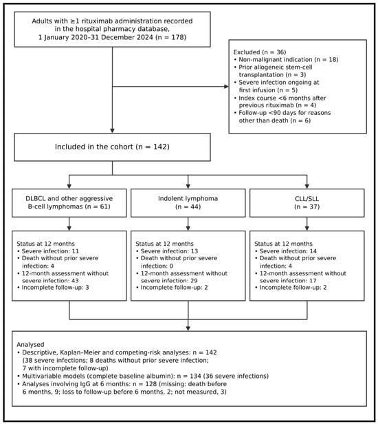
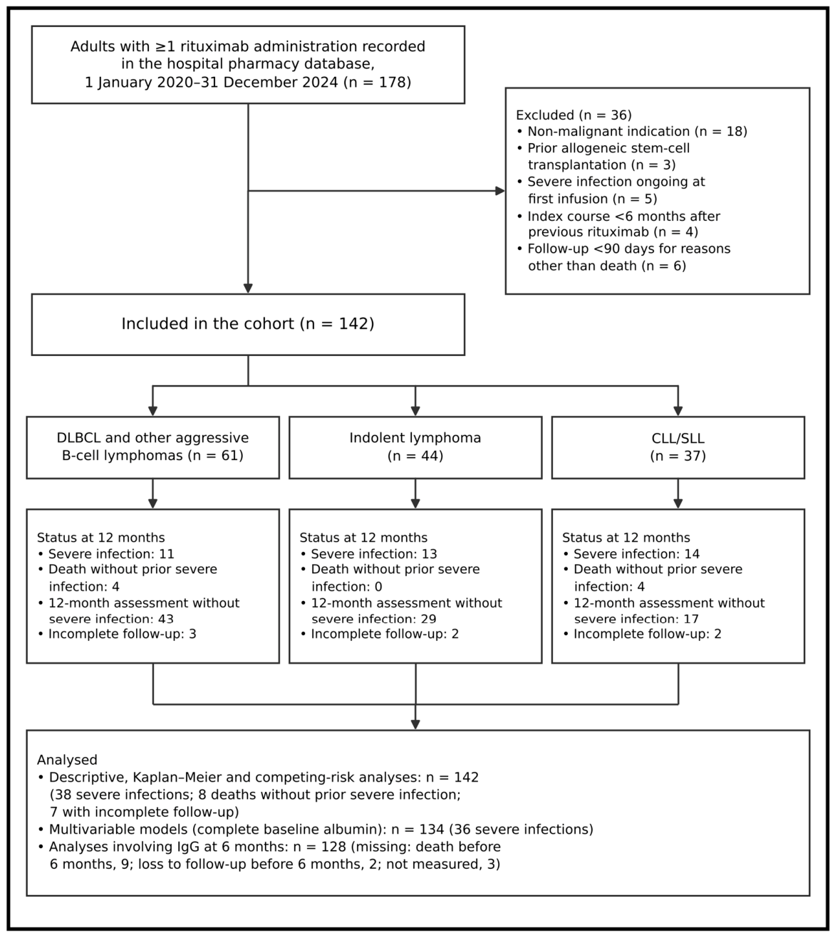
Of 178 adults with a rituximab administration recorded during the study period, 36 were excluded and 142 were included (Figure 1). Primary-outcome status at 12 months was ascertained in 135 patients (95.1%): 38 had a severe infection, eight died without a preceding severe infection, and 89 completed the 12-month assessment without severe infection. Seven patients (4.9%) had incomplete follow-up, ending after a median of 226 days (range 121–318), because of transfer to another centre (n = 4) or loss of contact (n = 3). Among event-free survivors, the 12-month assessment took place at a median of 358 days (IQR 344–371).
Figure 1.
Flow diagram of patient selection and follow-up, prepared according to the STROBE recommendations. Status at 12 months is shown for each diagnostic group; patients with a severe infection are counted in that category regardless of subsequent events. Abbreviations: CLL/SLL, chronic lymphocytic leukaemia/small lymphocytic lymphoma; DLBCL, diffuse large B-cell lymphoma; IgG, immunoglobulin G.
The cohort comprised 142 patients: 61 with DLBCL, 44 with indolent lymphoma, and 37 with CLL/SLL (Table 1). Mean age was 62.9 ± 10.9 years (range 42–88); 77 patients (54.2%) were men and 66 (46.5%) lived in rural areas. Patients with CLL/SLL had the lowest baseline IgG (7.2 ± 2.1 g/L; p < 0.001 across groups), and 24.3% had IgG < 6 g/L (p = 0.006). R-CHOP was used in 54 patients, R-bendamustine in 34, R-CVP in 17, R-miniCHOP in 13, R-chlorambucil in 12, R-FC in 10, and dose-adjusted R-EPOCH in two. Biosimilar rituximab was administered to 99 patients (69.7%), CT-P10 in 64 and GP2013 in 35, and maintenance to 22 (15.5%). Anti-HBc positivity was present in 38 patients, including eight who were HBsAg-positive; 29 anti-HBc-positive patients received antiviral prophylaxis, comprising all eight HBsAg-positive and 21 HBsAg-negative patients (entecavir, 21; tenofovir disoproxil fumarate, 5; lamivudine, 3), whereas the nine patients without prophylaxis were all HBsAg-negative and were managed with HBV DNA monitoring. Pneumocystis prophylaxis (trimethoprim–sulfamethoxazole, 44; dapsone, 2) was given to 30 of 44 patients (68.2%) receiving bendamustine- or fludarabine-containing regimens and to 16 of 98 (16.3%) receiving other regimens, and a prednisone-containing regimen was used in 86 patients (60.6%). Treatment started in 2020–2021 in 55 patients and in 2022–2024 in 87. COVID-19 vaccination status was documented in 121 patients (85.2%), of whom 52 (43.0%) had completed a primary vaccination series before the first rituximab infusion; none of the patients who started treatment in 2020 had been vaccinated before treatment.
Table 1.
Baseline demographic, clinical, treatment, and laboratory characteristics of the cohort according to diagnostic group.
3.2. Primary and Secondary Outcomes
Primary and secondary outcomes are summarised in Table 2. Severe infection occurred in 38 patients (26.8%; 95% CI 20.2–34.6), with crude proportions of 18.0% in DLBCL, 29.5% in indolent lymphoma, and 37.8% in CLL/SLL (p = 0.088). Sixteen severe infections (42.1%) occurred within 90 days; median time to the first event was 101 days (IQR 70–153). Five patients required intensive care, and seven died of infection, accounting for 41.2% of 17 deaths. Hypogammaglobulinaemia at 6 months occurred in 50 of 128 evaluable patients (39.1%), including 77.8% of those with CLL/SLL (p < 0.001 across groups). HBV reactivation occurred in three anti-HBc-positive patients: one of nine without antiviral prophylaxis and two of 29 receiving prophylaxis; none developed hepatic failure. Reactivation during prophylaxis occurred in one HBsAg-positive patient receiving lamivudine and in one HBsAg-negative patient receiving entecavir after a documented treatment interruption; the third, in an HBsAg-negative patient without prophylaxis, was detected by HBV DNA monitoring. All three were treated with entecavir or tenofovir.
Table 2.
Primary and secondary outcomes within 12 months of rituximab initiation, overall and by diagnostic group.
Agreement between the two investigators on the classification of 171 candidate infectious episodes as grade ≥ 3 was 95.9% (κ = 0.90); of the seven discordant episodes, five were resolved by consensus and two by the third investigator. Five of the 38 first severe infections (13.2%) were managed at other hospitals and were identified from external documentation. Febrile neutropenia without a documented source occurred in 12 patients (8.5%) and was not counted as infection. Immunoglobulin replacement was given to six patients (4.2%), four of whom had CLL/SLL; all had IgG < 4 g/L, no patient received replacement before rituximab, and in five patients it was started after the first severe infection.
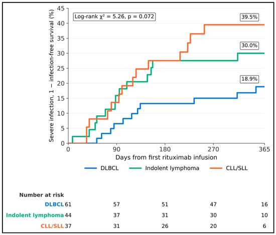
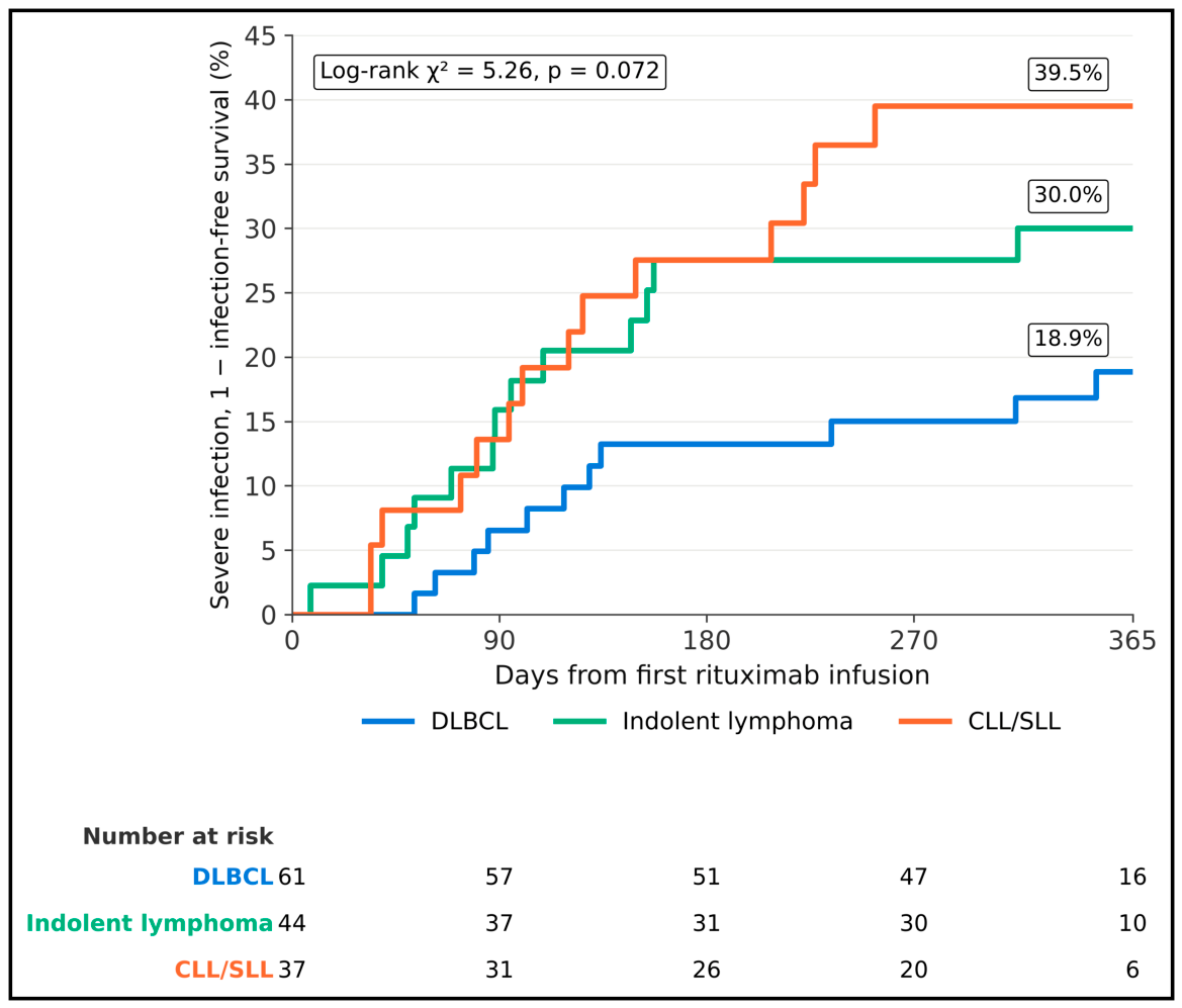
Figure 2 presents Kaplan–Meier estimates of severe infection by diagnostic group. At 90 days, the estimates were 6.6% for DLBCL, 15.9% for indolent lymphoma, and 13.6% for CLL/SLL; at 180 days, they were 13.2%, 27.5%, and 27.5%, respectively. The corresponding 12-month estimates were 18.9%, 30.0%, and 39.5% (log-rank χ2 = 5.26; p = 0.072). Numbers at risk at day 365 were 16, 10, and six, respectively; this decline reflects censoring of event-free patients at their 12-month assessment, most of which took place shortly before day 365, rather than loss to follow-up. When death without a preceding severe infection (n = 8; four with DLBCL and four with CLL/SLL) was treated as a competing event, the 12-month cumulative incidence of severe infection was 27.1% (95% CI 20.1–34.7) overall and 18.3%, 30.0%, and 38.1% in the three groups (Gray’s test p = 0.081), compared with Kaplan–Meier estimates of 27.7%, 18.9%, 30.0%, and 39.5%, respectively (Table 2). The 12-month cumulative incidence of death without severe infection was 5.8% (95% CI 2.8–10.6).
Figure 2.
Kaplan–Meier estimates of the probability of severe (grade ≥ 3) infection during the 12 months after the first rituximab infusion according to diagnostic group, calculated as one minus infection-free survival with death treated as a censoring event; these are not competing-risk cumulative incidence estimates. Labels indicate the 12-month Kaplan–Meier estimate; the log-rank test compares the three curves. Abbreviations: CLL/SLL, chronic lymphocytic leukaemia/small lymphocytic lymphoma; DLBCL, diffuse large B-cell lymphoma.
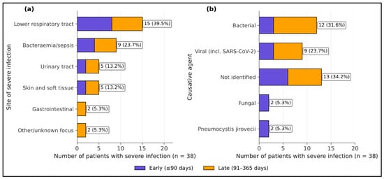
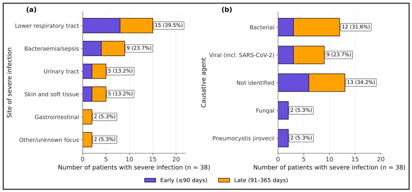
The anatomical sites and causative agents of the 38 severe infections are shown in Figure 3. Lower respiratory tract infections accounted for 15 events (39.5%), including eight early and seven late events, followed by bacteraemia or sepsis (nine events; 23.7%). An organism was identified in 25 events (65.8%): 12 bacterial, nine viral, two invasive fungal, and two Pneumocystis jirovecii infections. All four opportunistic infections occurred within 90 days in patients without Pneumocystis prophylaxis. Thirteen events (34.2%) had no identified organism.
Figure 3.
Distribution of the 38 severe infections by (a) anatomical site and (b) causative agent, stratified by timing after the first rituximab infusion (early, ≤90 days; late, 91–365 days). Labels show the number and percentage of all severe infections. Abbreviation: SARS-CoV-2, severe acute respiratory syndrome coronavirus 2.
3.3. Comparison of Patients with and Without Severe Infection
Table 3 compares patients with and without severe infection. Those with severe infection were older (66.2 ± 10.9 versus 61.7 ± 10.6 years; p = 0.031) and more often had diabetes (36.8% versus 18.3%; p = 0.020), baseline IgG < 6 g/L (23.7% versus 5.8%; p = 0.004), or albumin < 35 g/L (17/36, 47.2% versus 22/98, 22.4%; p = 0.005). Biosimilar use (63.2% versus 72.1%; p = 0.304), bendamustine exposure, maintenance, and prophylaxis did not differ significantly between groups.
Table 3.
Baseline characteristics of patients with and without severe infection during the first 12 months.
3.4. Unadjusted Subgroup Analyses
Crude subgroup risk ratios are presented in Table 4. Severe infection occurred in 60.0% of patients with baseline IgG < 6 g/L versus 22.8% with IgG ≥ 6 g/L (RR 2.63; 95% CI 1.56–4.43; p = 0.004). Higher risks were also observed with hypoalbuminaemia (RR 2.18; 95% CI 1.27–3.73), diabetes (RR 1.93; 95% CI 1.13–3.28), and age ≥ 65 years (RR 1.84; 95% CI 1.07–3.17). Confidence intervals for the remaining subgroup risk ratios included unity.
Table 4.
Incidence of severe infection within 12 months in pre-specified subgroups with crude risk ratios.
3.5. Unadjusted (Univariable) Associations with Severe Infection
Baseline IgG < 6 g/L was associated with severe infection (OR 5.07; 95% CI 1.67–15.43; p = 0.004), as were albumin < 35 g/L (OR 3.09; 95% CI 1.38–6.94; p = 0.006), diabetes (OR 2.61; 95% CI 1.14–5.96; p = 0.023), and age per decade (OR 1.48; 95% CI 1.04–2.11; p = 0.030). The OR per 1 g/L decrease in baseline IgG was 1.15 (95% CI 1.00–1.33; p = 0.043).
3.6. Correlations Between Continuous Baseline and Exposure Variables
Baseline and 6-month IgG were correlated (ρ = 0.92; p < 0.001). Absolute lymphocyte count correlated inversely with baseline IgG (ρ = −0.33; p < 0.001) and 6-month IgG (ρ = −0.42; p < 0.001). Age correlated with CCI (ρ = 0.48; p < 0.001), while cumulative rituximab dose was not correlated with the change in IgG (ρ = 0.03; p = 0.751).
3.7. Adjusted Associations in the Exploratory Multivariable Model
The multivariable model included 134 patients with complete data and 36 severe infections (Table 5), corresponding to approximately four events per candidate predictor. Adjusted associations were observed for baseline IgG < 6 g/L (aOR 4.96; 95% CI 1.28–19.18; p = 0.020), albumin < 35 g/L (aOR 4.67; 95% CI 1.78–12.23; p = 0.002), diabetes (aOR 2.73; 95% CI 1.03–7.24; p = 0.043), and age per decade (aOR 1.83; 95% CI 1.11–2.99; p = 0.017). The aOR for CLL/SLL was 1.32 (95% CI 0.46–3.80; p = 0.610). The apparent C-statistic was 0.789 (95% CI 0.711–0.865); after bootstrap optimism correction, it was 0.717 and the calibration slope was 0.671, indicating overfitting and probable overestimation of the adjusted odds ratios. The maximum variance inflation factor was 1.31. None of the tested interaction terms were statistically significant.
Table 5.
Exploratory multivariable logistic regression model for severe infection within 12 months (n = 134; 36 events).
3.8. Restricted Cubic Spline Analysis of Continuous Predictors
Relative to IgG of 9 g/L, the adjusted OR was 2.29 (95% CI 1.08–4.85) at 6 g/L and 4.55 (95% CI 1.15–18.02) at 4 g/L; the overall association had p = 0.088 and non-linearity p = 0.097. The Akaike information criterion was 167.4 for the spline model and 168.2 for the linear model. For age, the adjusted OR at 80 versus 60 years was 2.62 (95% CI 0.88–7.81; overall p = 0.157; non-linearity p = 0.582). Cumulative dose had an overall p = 0.700 and non-linearity p = 0.679.
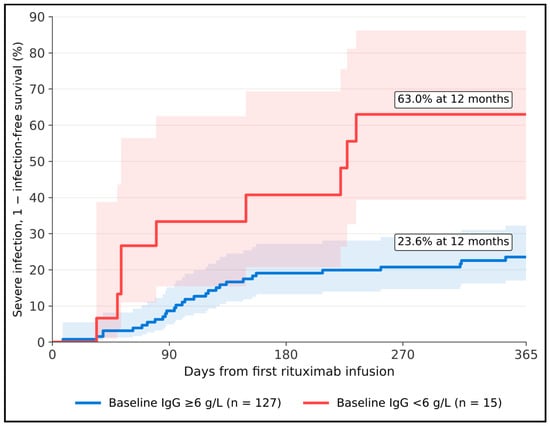
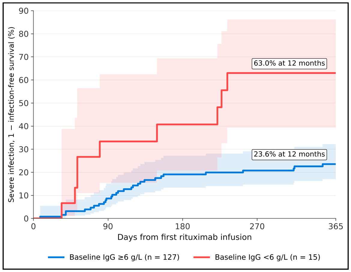
Figure 4 shows severe infection by baseline IgG status. The 12-month Kaplan–Meier estimate was 63.0% for IgG < 6 g/L (nine events among 15 patients) and 23.6% for IgG ≥ 6 g/L (29 events among 127 patients; log-rank χ2 = 13.16; p < 0.001); the corresponding cumulative incidences with death as a competing event were 60.0% and 23.0% (Gray’s test p < 0.001). Mean IgG declined from 9.3 g/L at baseline to 7.2 g/L at 6 months in 128 patients with paired values (paired t-test p < 0.001); mean values were 7.9 g/L at 3 months (n = 131) and 7.0 g/L at 12 months (n = 109). The mixed-effects model, based on all 510 available measurements from 142 patients, estimated an average linear decline of 0.18 g/L per month (p < 0.001) over 12 months. This linear estimate averages a steeper early decline and a subsequent plateau: the piecewise model estimated declines of 0.35 g/L per month (95% CI 0.29–0.41) during the first 6 months and 0.03 g/L per month (95% CI −0.05 to 0.11) thereafter, consistent with the observed decrease of 2.1 g/L at 6 months. In the linear model, the infection-group effect was approximately −1.1 g/L (p = 0.051), with time-by-group interaction p = 0.305.
Figure 4.
Kaplan–Meier estimates of the probability of severe (grade ≥ 3) infection during the 12 months after the first rituximab infusion according to baseline IgG status (<6 g/L versus ≥6 g/L), calculated as one minus infection-free survival with death treated as a censoring event; these are not competing-risk cumulative incidence estimates. Shaded areas are 95% confidence bands (log–log transformation); labels indicate the 12-month Kaplan–Meier estimate (log-rank χ2 = 13.16, p < 0.001). Abbreviation: IgG, immunoglobulin G.
3.9. Internally Validated Model Performance, Decision-Curve Analysis, and Sensitivity Analyses
Internal validation and sensitivity analyses are summarised in Table 6. After optimism correction, the C-statistic was 0.717, the calibration slope 0.671, and the Brier score 0.189. Model net benefit exceeded the treat-all and treat-none strategies at all evaluated thresholds from 10% to 40%; however, net benefit was calculated from apparent predictions and is therefore likely to be optimistic. The aOR for baseline IgG < 6 g/L was 5.88 after multiple imputation, 5.65 after exclusion of three severe SARS-CoV-2 events (only one of which was a first severe infection in the model population, the other two having followed an earlier severe infection), 3.50 with ridge penalisation, 5.74 among complete-case first-line patients, 5.32 after exclusion of patients who died without severe infection or had incomplete follow-up, and 4.89 after adjustment for calendar period. With continuous IgG, the aOR per 1 g/L was 0.93 (95% CI 0.78–1.11), and the C-statistic was 0.762.
Table 6.
Internal validation, calibration, decision-curve analysis, and sensitivity analyses of the multivariable prediction model.
In the Fine–Gray model, adjusted subdistribution hazard ratios were 3.31 (95% CI 1.42–7.72) for baseline IgG < 6 g/L, 3.05 (95% CI 1.52–6.12) for albumin < 35 g/L, 2.11 (95% CI 1.04–4.28) for diabetes, and 1.55 (95% CI 1.08–2.22) per decade of age. Backward elimination retained age, baseline IgG < 6 g/L, albumin < 35 g/L, and diabetes; this reduced model, with approximately nine events per variable, had an apparent C-statistic of 0.768, an optimism-corrected C-statistic of 0.731, and an optimism-corrected calibration slope of 0.812, and its adjusted odds ratios were 4.38 (95% CI 1.33–14.43) for baseline IgG < 6 g/L, 4.12 (95% CI 1.70–9.98) for albumin < 35 g/L, 2.49 (95% CI 1.01–6.14) for diabetes, and 1.69 (95% CI 1.10–2.60) per decade of age. Severe infection occurred in 16 of 55 patients (29.1%) who started treatment in 2020–2021 and in 22 of 87 (25.3%) who started in 2022–2024 (p = 0.618), with an aOR for the earlier period of 1.28 (95% CI 0.55–2.98); all three severe SARS-CoV-2 infections occurred in 2020–2021 in unvaccinated patients.
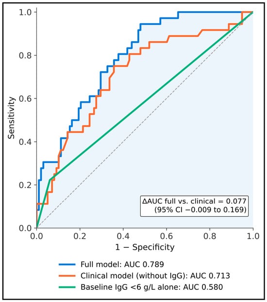
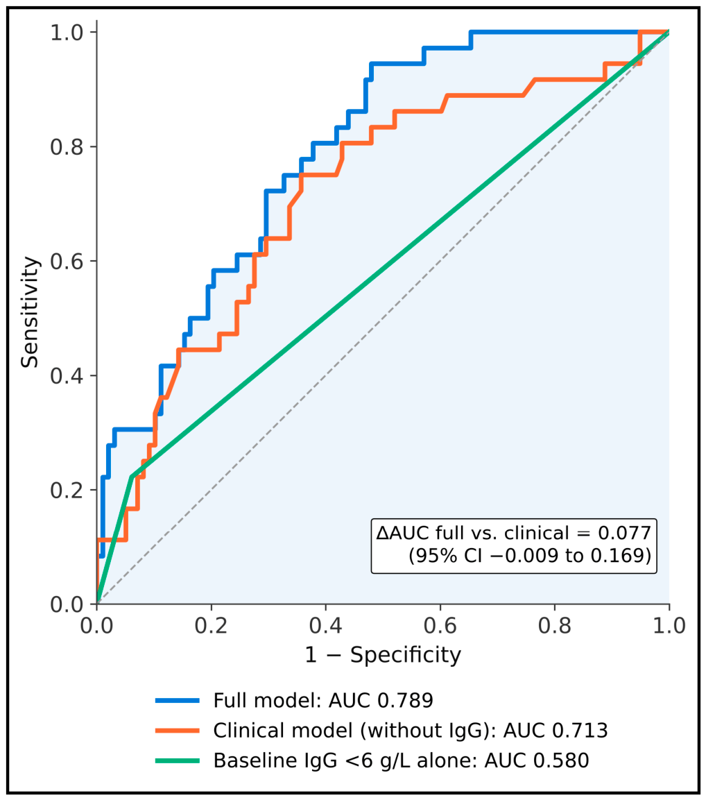
Figure 5 compares receiver operating characteristic curves. The AUC was 0.789 for the full model, 0.713 for the clinical model without IgG, which also differed from the full model in the albumin and G-CSF variables, and 0.580 for baseline IgG status alone. The reported bootstrap AUC difference between the full and clinical models was 0.077 (95% CI −0.009 to 0.169); because the two models differed in more than one variable, this difference cannot be attributed to IgG alone. At 90% sensitivity, specificity was 53.1% for the full model and 23.5% for the clinical model; at 70% sensitivity, specificity was 70.4% and 64.3%, respectively. Baseline IgG status alone had sensitivity of 22.2% and specificity of 93.9%. In an otherwise identical comparison, removing IgG from the full model reduced the apparent C-statistic from 0.789 to 0.763 (difference 0.026; 95% CI −0.006 to 0.064; likelihood-ratio test for IgG p = 0.018) and the optimism-corrected C-statistic from 0.717 to 0.698.
Figure 5.
Receiver operating characteristic curves for the prediction of severe infection within 12 months by the full multivariable model, a clinical model without IgG that also differed from the full model in the albumin and G-CSF variables, and baseline IgG < 6 g/L alone (n = 134). The dashed line represents no discrimination; the box reports the bootstrap difference in area under the curve between the full and clinical models, which therefore does not isolate the contribution of IgG; all AUC values are apparent estimates that have not been corrected for optimism. Abbreviations: AUC, area under the receiver operating characteristic curve; CI, confidence interval; IgG, immunoglobulin G.
4. Discussion
4.1. Clinical Pattern and Comparison with Previous Studies
Severe infection affected approximately one in four patients, and respiratory or bloodstream infections accounted for most events. Treating death as a competing event changed this estimate only marginally, because few patients died without a preceding severe infection. This burden should be interpreted in the context of combined malignancy-related immune dysfunction, chemotherapy, and anti-CD20 exposure. Reviews of rituximab-associated infection describe substantial variation across indications, treatment combinations, and follow-up periods [15]; a single pooled benchmark is therefore unlikely to represent this mixed cohort. Our results provide a local estimate for the diagnostic and treatment groups studied, rather than an estimate of infection attributable to rituximab alone. The persistence of events beyond the early treatment period also indicates that assessment limited to neutropenic episodes would miss part of the burden.
The association between low baseline IgG and subsequent severe infection is consistent with reports of persistent antibody deficiency after rituximab. Casulo and colleagues documented hypogammaglobulinaemia and recurrent infections in patients with B-cell lymphoma [7], while Makatsori and colleagues described clinically important deficiency that could persist after treatment completion [16]. In the health-system cohort reported by Barmettler and colleagues, severe infections increased after rituximab [17]. Differences in indications, observation windows, and immunoglobulin testing prevent direct comparison with our cohort. Together, however, these studies support measuring immunoglobulins before treatment and reassessing patients whose infection history or clinical course suggests persistent immune impairment.
Baseline IgG and IgG measured during follow-up address different questions. Baseline IgG is available when treatment is planned and was the only immunoglobulin measure included in the prediction model. In contrast, the decline in IgG during the first 6 months and the frequent hypogammaglobulinaemia at 6 months describe the evolution of humoral immunity after treatment; these measurements may be influenced by intercurrent infection, survival, and treatment modification, and most first severe infections occurred before the 6-month measurement. Follow-up IgG concentrations should therefore be interpreted as longitudinal observations rather than as predictors of a first severe infection, although they may be relevant to later infections and to decisions about immunoglobulin replacement.
4.2. Patient Characteristics and Treatment Exposure
In this exploratory analysis, older age, diabetes, and hypoalbuminaemia were associated with severe infection after adjustment, but the wide confidence intervals and the evidence of overfitting indicate that the magnitude of these associations is uncertain. Age may capture several overlapping vulnerabilities, including reduced physiological reserve and cumulative comorbidity. Experience with attenuated chemoimmunotherapy in very elderly patients illustrates why treatment tolerance and supportive care require explicit consideration [18]. Albumin should be interpreted as a nonspecific marker influenced by inflammation, nutritional intake, hepatic function, and disease activity; its association does not establish that albumin supplementation would prevent infection. Similarly, diabetes may identify patients requiring closer metabolic and infection assessment, but the present data cannot separate the effects of glycaemic control from associated vascular or renal disease.
The crude infection proportion was highest in CLL/SLL, whereas the diagnostic association was attenuated after adjustment. This is compatible with overlapping effects of age and baseline immune impairment, but the wide adjusted confidence interval does not exclude a clinically relevant diagnostic effect. The lower IgG concentrations and greater frequency of subsequent hypogammaglobulinaemia in CLL/SLL are biologically consistent with the immune abnormalities described in this disease [19]. Total lymphocyte counts should not be treated as a substitute for functional immune assessment in this setting, because an increased count can reflect the malignant clone rather than preserved protective immunity.
We did not detect an adjusted association between bendamustine exposure and severe infection. The cited meta-analysis by Gafter-Gvili and colleagues likewise found no statistically significant increase in overall or grade 3–4 infections compared with other regimens [20]. This does not establish equivalence between treatment backbones in our cohort: only a minority received bendamustine, and the confidence interval remains compatible with increased risk. Prophylaxis, treatment selection, corticosteroid exposure, and prior therapy may also modify observed associations. The nonsignificant comparison between biosimilar and originator rituximab should be interpreted with similar caution; the randomised CT-P10 study provides a stronger basis for comparative efficacy and safety than this observational subgroup [13].
4.3. Implications for Infection Prevention
The predominance of early respiratory events and the occurrence of opportunistic infections support coordinated pretreatment and on-treatment assessment. Management of neutropenic risk and prompt evaluation of febrile episodes remain central components of supportive care [21]. The observational comparison of G-CSF recipients with nonrecipients cannot measure prophylactic effectiveness, because patients at greater baseline risk may preferentially receive prophylaxis. Likewise, the occurrence of Pneumocystis infection in patients without prophylaxis is clinically relevant but too infrequent to quantify a local treatment effect, particularly as institutional prophylaxis recommendations were not consistently followed. Decisions about antimicrobial prophylaxis should therefore integrate the regimen, corticosteroid exposure, immune status, and established recommendations [9,10], rather than rely on nonsignificant subgroup comparisons.
HBV deserves particular attention in a setting with substantial prior exposure. The three reactivations observed here occurred in a small subgroup with mixed baseline serological status and cannot establish the effectiveness of a specific antiviral strategy; two occurred during prophylaxis, one with lamivudine and one after interruption of entecavir. In the randomised trial by Huang and colleagues, entecavir reduced HBV reactivation compared with lamivudine, but did not eliminate it [22]. Seto and colleagues also demonstrated that reactivation can occur during prolonged observation of patients with resolved infection receiving rituximab-containing chemotherapy [23]. HBsAg-positive and HBsAg-negative/anti-HBc-positive patients should therefore be distinguished when interpreting outcomes. These breakthrough events are consistent with that randomised comparison and with the importance of uninterrupted prophylaxis, although the numbers are too small for formal comparison.
The study period overlapped the COVID-19 pandemic, when viral circulation, access to care, and preventive measures changed substantially. The mortality burden reported in patients with haematological malignancies [24] and the impaired antibody responses to vaccination observed in CLL [25] provide context for these risks, without implying that historical estimates apply unchanged to current practice. Excluding severe SARS-CoV-2 events did not materially alter the low-IgG association, and adjustment for calendar period gave similar estimates; however, vaccination status was documented incompletely and changed over time, so residual confounding by vaccination and period-specific practices cannot be excluded. Infection surveillance should also consider late-onset neutropenia [26]. Immunoglobulin replacement is a potential option for selected patients with clinically significant antibody deficiency and recurrent or severe infections [7,16,17]; an isolated IgG threshold from this cohort is insufficient to define eligibility. In this cohort, replacement was used rarely and mostly after a severe infection, so its preventive effect could not be assessed.
Maintenance therapy requires balancing disease control against prolonged immune suppression. The PRIMA trial established a progression-free survival benefit in follicular lymphoma [3], whereas the meta-analysis by Vidal and colleagues identified increased infection-related adverse events with maintenance [27]. Our small maintenance subgroup cannot determine which patients should omit, shorten, or discontinue treatment. Its findings instead support recording infection history and immunoglobulin trends during follow-up, with individual reassessment when infectious complications threaten treatment tolerability.
4.4. Interpretation of the Prediction Model
The full model discriminated better than IgG status alone, supporting assessment of several patient characteristics together. However, the decrease in C-statistic after optimism correction and the corrected calibration slope below 0.7 indicate substantial overfitting. The optimism-corrected estimates, rather than the apparent ones, are therefore the more appropriate summary of expected performance in similar patients, and the adjusted odds ratios should be regarded as exploratory and probably overestimated. A nonsignificant Hosmer–Lemeshow test does not establish accurate individual risk prediction, particularly in a small development sample. Calibration assesses the agreement between predicted and observed risks and must be evaluated alongside discrimination [28]. The apparent full-versus-clinical AUC difference was imprecise, and the stated models also differed in albumin and G-CSF variables; the observed increment cannot therefore be attributed uniquely to IgG. In an otherwise identical comparison, the increment attributable to IgG was small and imprecise, whereas the reduced four-variable model showed less optimism, supporting the use of parsimonious models in future development studies.
The spline estimates were higher at lower IgG concentrations, but neither the overall association nor the test for non-linearity reached conventional statistical significance in that adjusted analysis. Restricted cubic splines permit examination of continuous relationships without imposing a simple linear form [29], yet this exploratory curve does not independently validate a clinical cutoff. The difference between dichotomised and continuous IgG estimates may reflect sparse observations, model specification, or sampling variability. A linear continuous term assumes a constant change in risk across the whole IgG range, whereas the spline estimates suggested that excess risk was concentrated at lower concentrations; such a pattern would be diluted by a linear term. Although the 6 g/L threshold was pre-specified, only 15 patients were below it, so the dichotomised estimate is imprecise and should not be regarded as a validated cut-off. Future analyses should retain continuous information where possible and evaluate any proposed threshold against prespecified clinical decisions in a separate cohort.
Decision-curve analysis expresses the trade-off between true-positive and false-positive classifications at an assumed intervention threshold [30]. Apparent positive net benefit across the evaluated thresholds is a reason to examine the model further, rather than evidence that a prophylaxis programme based on it improves outcomes. The intervention triggered by a risk estimate was not tested, and monitoring, antimicrobial prophylaxis, and immunoglobulin replacement have different benefits and harms. If validated, the model would be intended to support decisions at treatment initiation, such as the intensity of infection surveillance and immunoglobulin monitoring at lower risk thresholds, and consideration of antimicrobial prophylaxis or early immunological assessment at higher thresholds; each action would require its own threshold and evaluation.
4.5. Strengths and Limitations
Strengths include consecutive enrolment, a defined and independently adjudicated severe-infection endpoint, assessment across major B-cell diagnostic groups, and reporting of both discrimination and calibration. Nevertheless, the retrospective single-centre design is susceptible to selection bias and incomplete ascertainment of infections managed elsewhere; although external discharge documents were reviewed, infections managed entirely in primary care may have been missed, which would lead to underestimation of incidence. Differences in treatment backbones, prior therapy, supportive care, and disease severity limit attribution of risk to a particular drug. The small numbers of HBV reactivations, opportunistic infections, and infection-related deaths preclude reliable adjusted analyses of these outcomes. Multiple exploratory comparisons were performed without multiplicity correction, so marginal associations require confirmation.
The multivariable analysis included nine predictors and only 36 events, creating a substantial risk of unstable coefficients. Bootstrap validation, ridge penalisation, and a reduced model address some optimism but cannot substitute for a larger independent sample. Patients who died without severe infection or had incomplete follow-up were classified as event-free in the logistic models; excluding them and using Fine–Gray regression gave consistent results. Albumin and follow-up IgG were incompletely observed, and multiple imputation depends on assumptions about missingness that cannot be verified from the observed data alone.
The plotted Kaplan–Meier curves treat death as censoring and describe one minus infection-free survival; they slightly exceeded the competing-risk cumulative incidence, which is the more appropriate estimate of absolute infection probability. In addition, cumulative rituximab dose over follow-up can be affected by infection-related treatment interruption and should not be interpreted as an exposure known at baseline. Vaccination status was incompletely documented, non-protocol corticosteroid use and antimicrobial adherence were not recorded, and protocol corticosteroid exposure could not be separated from diagnosis and regimen; together with possible residual calendar-period effects, these factors limit causal interpretation. Generalisability beyond this referral centre remains uncertain.
4.6. Future Research
A prospective multicentre cohort should use harmonised infection definitions, scheduled immunoglobulin measurements, and linkage to hospital and outpatient records. It should distinguish baseline predictors from exposures that change during follow-up, account for competing mortality, and examine recurrent infections as well as the first severe event. External validation should report calibration across diagnostic groups and treatment regimens before any risk score is used to direct care. An ensuing impact study could then test a clearly defined prevention pathway, including its effects on infection-related admission, treatment interruption, patient burden, antimicrobial use, and resource requirements.
5. Conclusions
Severe infections were frequent after rituximab-based therapy in this cohort from Western Romania, with respiratory and bloodstream infections predominating. Baseline hypogammaglobulinaemia, hypoalbuminaemia, diabetes mellitus, and older age were associated with severe infection in exploratory analyses and, if confirmed in independent cohorts, could help identify patients who require closer assessment and surveillance. Coordinated evaluation of immune status, comorbidity, vaccination, and prophylaxis alongside lymphoma treatment is a potential application of these findings that requires prospective evaluation. The prediction model remains exploratory, showed reduced performance after internal validation, and requires prospective external validation and assessment of clinical benefit before it could be used to guide risk-adapted prophylaxis or monitoring.
Author Contributions
Conceptualization, B.G.; methodology, B.G.; software, B.G.; validation, B.G.; formal analysis, A.C.I.; investigation, A.C.I.; resources, A.C.I.; data curation, A.C.I.; writing—original draft preparation, B.G.; writing—review and editing, B.G.; visualization, K.C.M. and I.I.; supervision, K.C.M. and I.I.; project administration, K.C.M. and I.I.; funding acquisition, K.C.M. and I.I. All authors have read and agreed to the published version of the manuscript.
Funding
We would like to acknowledge the “Victor Babeș” University of Medicine and Pharmacy Timișoara for their support in covering the costs of publication for this research paper.
Institutional Review Board Statement
The study was conducted in accordance with the Declaration of Helsinki and approved by the Institutional Ethics Committee of Pius Brînzeu Clinical Emergency County Hospital (decision 630/15 June 2026), before data extraction.
Informed Consent Statement
Patient consent was waived by the Institutional Ethics Committee because of the retrospective design and the use of pseudonymised data; all patients had signed the hospital’s general consent form at admission, which permits the use of anonymised clinical data for research.
Data Availability Statement
The data presented in this study are available on request from the corresponding author. The data are not publicly available due to strict IRB regulations regarding data sharing with unauthorized individuals.
Conflicts of Interest
The authors declare no conflicts of interest.
References
- Salles, G.; Barrett, M.; Foà, R.; Maurer, J.; O’Brien, S.; Valente, N.; Wenger, M.; Maloney, D.G. Rituximab in B-Cell Hematologic Malignancies: A Review of 20 Years of Clinical Experience. Adv. Ther. 2017, 34, 2232–2273. [Google Scholar] [CrossRef] [Scilit] [PubMed]
- Coiffier, B.; Lepage, E.; Brière, J.; Herbrecht, R.; Tilly, H.; Bouabdallah, R.; Morel, P.; Van Den Neste, E.; Salles, G.; Gaulard, P.; et al. CHOP Chemotherapy plus Rituximab Compared with CHOP Alone in Elderly Patients with Diffuse Large-B-Cell Lymphoma. N. Engl. J. Med. 2002, 346, 235–242. [Google Scholar] [CrossRef] [Scilit] [PubMed]
- Salles, G.; Seymour, J.F.; Offner, F.; López-Guillermo, A.; Belada, D.; Xerri, L.; Feugier, P.; Bouabdallah, R.; Catalano, J.V.; Brice, P.; et al. Rituximab Maintenance for 2 Years in Patients with High Tumour Burden Follicular Lymphoma Responding to Rituximab plus Chemotherapy (PRIMA): A Phase 3, Randomised Controlled Trial. Lancet 2011, 377, 42–51. [Google Scholar] [CrossRef] [Scilit] [PubMed]
- Hallek, M.; Fischer, K.; Fingerle-Rowson, G.; Fink, A.M.; Busch, R.; Mayer, J.; Hensel, M.; Hopfinger, G.; Hess, G.; von Grünhagen, U.; et al. Addition of Rituximab to Fludarabine and Cyclophosphamide in Patients with Chronic Lymphocytic Leukaemia: A Randomised, Open-Label, Phase 3 Trial. Lancet 2010, 376, 1164–1174. [Google Scholar] [CrossRef] [Scilit] [PubMed]
- Lanini, S.; Molloy, A.C.; Fine, P.E.; Prentice, A.G.; Ippolito, G.; Kibbler, C.C. Risk of Infection in Patients with Lymphoma Receiving Rituximab: Systematic Review and Meta-Analysis. BMC Med. 2011, 9, 36. [Google Scholar] [CrossRef] [Scilit] [PubMed]
- Evens, A.M.; Jovanovic, B.D.; Su, Y.C.; Raisch, D.W.; Ganger, D.; Belknap, S.M.; Dai, M.S.; Chiu, B.C.; Fintel, B.; Cheng, Y.; et al. Rituximab-Associated Hepatitis B Virus (HBV) Reactivation in Lymphoproliferative Diseases: Meta-Analysis and Examination of FDA Safety Reports. Ann. Oncol. 2011, 22, 1170–1180. [Google Scholar] [CrossRef] [Scilit] [PubMed]
- Casulo, C.; Maragulia, J.; Zelenetz, A.D. Incidence of Hypogammaglobulinemia in Patients Receiving Rituximab and the Use of Intravenous Immunoglobulin for Recurrent Infections. Clin. Lymphoma Myeloma Leuk. 2013, 13, 106–111. [Google Scholar] [CrossRef] [Scilit] [PubMed]
- Wolach, O.; Bairey, O.; Lahav, M. Late-Onset Neutropenia after Rituximab Treatment: Case Series and Comprehensive Review of the Literature. Medicine 2010, 89, 308–318. [Google Scholar] [CrossRef] [Scilit] [PubMed]
- Jiang, X.; Mei, X.; Feng, D.; Wang, X. Prophylaxis and Treatment of Pneumocystis jiroveci Pneumonia in Lymphoma Patients Subjected to Rituximab-Contained Therapy: A Systemic Review and Meta-Analysis. PLoS ONE 2015, 10, e0122171. [Google Scholar] [CrossRef] [Scilit] [PubMed]
- Mikulska, M.; Lanini, S.; Gudiol, C.; Drgona, L.; Ippolito, G.; Fernández-Ruiz, M.; Salzberger, B. ESCMID Study Group for Infections in Compromised Hosts (ESGICH) Consensus Document on the Safety of Targeted and Biological Therapies: An Infectious Diseases Perspective (Agents Targeting Lymphoid Cells Surface Antigens [I]: CD19, CD20 and CD52). Clin. Microbiol. Infect. 2018, 24, S71–S82. [Google Scholar] [CrossRef] [Scilit] [PubMed]
- Gheorghe, L.; Csiki, I.E.; Iacob, S.; Gheorghe, C. The Prevalence and Risk Factors of Hepatitis B Virus Infection in an Adult Population in Romania: A Nationwide Survey. Eur. J. Gastroenterol. Hepatol. 2013, 25, 56–64. [Google Scholar] [CrossRef] [Scilit] [PubMed]
- Pătrașcu, A.M.; Nacea, J.G.; Pătrașcu, Ș.; Gogănău, A.M.; Delcea, F.; Mogoantă, L.; Rotaru, I. Therapeutic Options in Diffuse Large B-Cell Lymphoma—A Retrospective Study and Review of the Literature. Curr. Health Sci. J. 2017, 43, 269–274. [Google Scholar] [CrossRef] [Scilit] [PubMed]
- Kim, W.S.; Buske, C.; Ogura, M.; Jurczak, W.; Sancho, J.M.; Zhavrid, E.; Kim, J.S.; Hernández-Rivas, J.Á.; Prokharau, A.; Vasilica, M.; et al. Efficacy, Pharmacokinetics, and Safety of the Biosimilar CT-P10 Compared with Rituximab in Patients with Previously Untreated Advanced-Stage Follicular Lymphoma: A Randomised, Double-Blind, Parallel-Group, Non-Inferiority Phase 3 Trial. Lancet Haematol. 2017, 4, e362–e373. [Google Scholar] [CrossRef] [Scilit] [PubMed]
- The International Non-Hodgkin’s Lymphoma Prognostic Factors Project. A Predictive Model for Aggressive Non-Hodgkin’s Lymphoma. N. Engl. J. Med. 1993, 329, 987–994. [Google Scholar] [CrossRef] [Scilit] [PubMed]
- Kelesidis, T.; Daikos, G.; Boumpas, D.; Tsiodras, S. Does Rituximab Increase the Incidence of Infectious Complications? A Narrative Review. Int. J. Infect. Dis. 2011, 15, e2–e16. [Google Scholar] [CrossRef] [Scilit] [PubMed]
- Makatsori, M.; Kiani-Alikhan, S.; Manson, A.L.; Verma, N.; Leandro, M.; Gurugama, N.P.; Longhurst, H.J.; Grigoriadou, S.; Buckland, M.; Kanfer, E.; et al. Hypogammaglobulinaemia after Rituximab Treatment—Incidence and Outcomes. QJM 2014, 107, 821–828. [Google Scholar] [CrossRef] [Scilit] [PubMed]
- Barmettler, S.; Ong, M.S.; Farmer, J.R.; Choi, H.; Walter, J. Association of Immunoglobulin Levels, Infectious Risk, and Mortality with Rituximab and Hypogammaglobulinemia. JAMA Netw. Open 2018, 1, e184169. [Google Scholar] [CrossRef] [Scilit] [PubMed]
- Peyrade, F.; Jardin, F.; Thieblemont, C.; Thyss, A.; Emile, J.F.; Castaigne, S.; Coiffier, B.; Haioun, C.; Bologna, S.; Fitoussi, O.; et al. Attenuated Immunochemotherapy Regimen (R-miniCHOP) in Elderly Patients Older than 80 Years with Diffuse Large B-Cell Lymphoma: A Multicentre, Single-Arm, Phase 2 Trial. Lancet Oncol. 2011, 12, 460–468. [Google Scholar] [CrossRef] [Scilit] [PubMed]
- Morrison, V.A. Infectious Complications of Chronic Lymphocytic Leukaemia: Pathogenesis, Spectrum of Infection, Preventive Approaches. Best Pract. Res. Clin. Haematol. 2010, 23, 145–153. [Google Scholar] [CrossRef] [Scilit] [PubMed]
- Gafter-Gvili, A.; Gurion, R.; Raanani, P.; Shpilberg, O.; Vidal, L. Bendamustine-Associated Infections—Systematic Review and Meta-Analysis of Randomized Controlled Trials. Hematol. Oncol. 2017, 35, 424–431. [Google Scholar] [CrossRef] [Scilit] [PubMed]
- Klastersky, J.; de Naurois, J.; Rolston, K.; Rapoport, B.; Maschmeyer, G.; Aapro, M.; Herrstedt, J. Management of Febrile Neutropaenia: ESMO Clinical Practice Guidelines. Ann. Oncol. 2016, 27, v111–v118. [Google Scholar] [CrossRef] [Scilit] [PubMed]
- Huang, H.; Li, X.; Zhu, J.; Ye, S.; Zhang, H.; Wang, W.; Wu, X.; Peng, J.; Xu, B.; Lin, Y.; et al. Entecavir vs Lamivudine for Prevention of Hepatitis B Virus Reactivation among Patients with Untreated Diffuse Large B-Cell Lymphoma Receiving R-CHOP Chemotherapy: A Randomized Clinical Trial. JAMA 2014, 312, 2521–2530. [Google Scholar] [CrossRef] [Scilit] [PubMed]
- Seto, W.K.; Chan, T.S.; Hwang, Y.Y.; Wong, D.K.; Fung, J.; Liu, K.S.; Gill, H.; Lam, Y.F.; Lie, A.K.; Lai, C.L.; et al. Hepatitis B Reactivation in Patients with Previous Hepatitis B Virus Exposure Undergoing Rituximab-Containing Chemotherapy for Lymphoma: A Prospective Study. J. Clin. Oncol. 2014, 32, 3736–3743. [Google Scholar] [CrossRef] [Scilit] [PubMed]
- Vijenthira, A.; Gong, I.Y.; Fox, T.A.; Booth, S.; Cook, G.; Fattizzo, B.; Martín-Moro, F.; Razanamahery, J.; Riches, J.C.; Zwicker, J.; et al. Outcomes of Patients with Hematologic Malignancies and COVID-19: A Systematic Review and Meta-Analysis of 3377 Patients. Blood 2020, 136, 2881–2892. [Google Scholar] [CrossRef] [Scilit] [PubMed]
- Herishanu, Y.; Avivi, I.; Aharon, A.; Shefer, G.; Levi, S.; Bronstein, Y.; Morales, M.; Ziv, T.; Shorer Arbel, Y.; Scarfò, L.; et al. Efficacy of the BNT162b2 mRNA COVID-19 Vaccine in Patients with Chronic Lymphocytic Leukemia. Blood 2021, 137, 3165–3173. [Google Scholar] [CrossRef] [Scilit] [PubMed]
- Tesfa, D.; Palmblad, J. Late-Onset Neutropenia Following Rituximab Therapy: Incidence, Clinical Features and Possible Mechanisms. Expert Rev. Hematol. 2011, 4, 619–625. [Google Scholar] [CrossRef] [Scilit] [PubMed]
- Vidal, L.; Gafter-Gvili, A.; Leibovici, L.; Dreyling, M.; Ghielmini, M.; Hsu Schmitz, S.F.; Cohen, A.; Shpilberg, O. Rituximab Maintenance for the Treatment of Patients with Follicular Lymphoma: Systematic Review and Meta-Analysis of Randomized Trials. J. Natl. Cancer Inst. 2009, 101, 248–255. [Google Scholar] [CrossRef] [Scilit] [PubMed]
- Van Calster, B.; McLernon, D.J.; van Smeden, M.; Wynants, L.; Steyerberg, E.W. Topic Group ‘Evaluating Diagnostic Tests and Prediction Models’ of the STRATOS Initiative. Calibration: The Achilles Heel of Predictive Analytics. BMC Med. 2019, 17, 230. [Google Scholar] [CrossRef] [Scilit] [PubMed]
- Gauthier, J.; Wu, Q.V.; Gooley, T.A. Cubic Splines to Model Relationships between Continuous Variables and Outcomes: A Guide for Clinicians. Bone Marrow Transpl. 2020, 55, 675–680, Correction in Bone Marrow Transpl. 2023, 58, 962. https://doi.org/10.1038/s41409-023-01993-7. [Google Scholar] [CrossRef] [Scilit] [PubMed]
- Vickers, A.J.; Elkin, E.B. Decision Curve Analysis: A Novel Method for Evaluating Prediction Models. Med. Decis. Mak. 2006, 26, 565–574. [Google Scholar] [CrossRef] [Scilit] [PubMed]

Disclaimer/Publisher’s Note: The statements, opinions and data contained in all publications are solely those of the individual author(s) and contributor(s) and not of MDPI and/or the editor(s). MDPI and/or the editor(s) disclaim responsibility for any injury to people or property resulting from any ideas, methods, instructions or products referred to in the content. |
© 2026 by the authors. Licensee MDPI, Basel, Switzerland. This article is an open access article distributed under the terms and conditions of the Creative Commons Attribution (CC BY) license.